
California Associationcamlt.org/pdf_files/forms/997-form.docx · Web viewCalifornia Association....
58
California Association for Medical Laboratory Technology Distance Learning Program CAMLT Distance Learning Course DL-997 1 © California Association for Medical Laboratory Technology WHAT’S GOING ON WITH WHOOPING COUGH (PERTUSSIS)? Course # DL-997 by James I. Mangels, MA, CLS, MT(ASCP) Consultant Microbiology Services Santa Rosa, CA Approved for 3.0 CE/Contact Hours CAMLT is approved by the California Department of Public Health as a CA CLS Accrediting Agency (#0021) and is approved as a provider of continuing education programs in the clinical laboratory sciences by the ASCLS P.A.C.E. ® Program (#519) Level of Difficulty: Intermediate 1895 Mowry Ave., Ste. 112 Phone: 510- 792-4441
Transcript of California Associationcamlt.org/pdf_files/forms/997-form.docx · Web viewCalifornia Association....
California Associationfor
Medical Laboratory Technology
Distance Learning Program
CAMLT Distance Learning Course DL-9971
© California Association for Medical Laboratory Technology
WHAT’S GOING ON WITH WHOOPING COUGH (PERTUSSIS)?
Course # DL-997by
James I. Mangels, MA, CLS, MT(ASCP)Consultant
Microbiology ServicesSanta Rosa, CA
Approved for 3.0 CE/Contact HoursCAMLT is approved by the California Department of Public Health
as a CA CLS Accrediting Agency (#0021)and is approved as a provider of continuing education programs in the clinical laboratory sciences
by the ASCLS P.A.C.E.® Program (#519)
Level of Difficulty: Intermediate
1895 Mowry Ave., Ste. 112 Phone: 510-792-4441Fremont, CA 94538-1766 FAX: 510-792-3045
Notification of Distance Learning DeadlineDON’T PUT YOUR LICENSE IN JEOPARDY!
This is a reminder that all the continuing education units required to renew your license/certificate must be earned no later than the expiration date printed on your license/certificate. If some of your units are made up of Distance Learning courses, please allow yourself enough time to retake the test in the event you do not pass on the first attempt. CAMLT urges you to earn your CE units early!

OUTLINEA. IntroductionB. History of Whooping Cough InfectionC. Transmission of Bordetella pertussisD. Illness and Clinical Symptoms of Whooping CoughE. Microbiology of Bordetella pertussisF. Pathogenic Mechanisms of Bordetella pertussisG. Laboratory Diagnosis of Whooping CoughH. Treatment of Whooping CoughI. Prevention and Control of Bordetella pertussis InfectionJ. ConclusionsK. Case StudyL. References
COURSE OBJECTIVESUpon completion of this course, the participant will be able to:1. outline the history of whooping cough infection;2. discuss the incidence of B. pertussis infection in the U.S. and in California;3. list factors involved in the increased incidence of whooping cough in California;4. explain the pathogenic mechanisms of Bordetella pertussis;5. outline the clinical features of Bordetella pertussis infection;6. describe how the laboratory identifies B. pertussis;7. list methods of preventing and treating whooping cough.
A. INTRODUCTIONWhooping cough, or pertussis, is caused by the organism Bordetella
pertussis. Pertussis is an acute bacterial infectious disease, and one of the most important diseases of children throughout the world. Pertussis is a highly communicable infection of the respiratory tract spread by coughing or sneezing. The term “whooping cough” refers to the intensive cough followed by an inspiratory “whoop” sound. Bouts of coughing due to B. pertussis can last so long that patients are deprived of oxygen. At the end of a coughing bout, they forcefully draw air back into the lungs producing the characteristic “whoop.” While the whooping stage is not universal, it is the hallmark of the clinical disease and has given the disease its name. People sick with pertussis have severe coughing attacks that can occur for months. The terms “whooping cough” and “pertussis” will be used interchangeably throughout this course.
The genus Bordetella is named for Jules Bordet along with Octave Gengou, who in 1906 first described and recovered the organism on special media they developed (1). The two microbiologists from Belgium noticed epidemics of severe coughing or “whooping” associated with a high mortality among infants and young children. The organism was originally
CAMLT Distance Learning Course DL-9972
© California Association for Medical Laboratory Technology
called Bordet-Gengou bacillus, then later assigned the name Hemophilus pertussis. Bordetella spp., however, require different extraneous growth factors and have very distinct genetic differences from the genus Hemophilus spp. Thus the organism causing whooping cough was given in its own genus, Bordetella.
There are currently seven species within the genus Bordetella: B. pertussis, B. bronchiseptica, B. parapertussis, B. avium, B. hinzii, B. holmessii, and B. trematum. Although there is some genetic relatedness among B. pertussis, B. parapertussis, B. bronchiseptica, and B. holmesii (1), this course will only discuss B. pertussis because it is the pathogen responsible for the majority of pertussis cases. It also causes respiratory symptoms of greater severity than the other Bordetella species. B. pertussis produces disease only in humans, who serve as the sole reservoir for the organism.
Whooping cough occurs throughout the world in immunized and un-immunized populations causing outbreaks or epidemics in cycles of two to five years. Worldwide, B. pertussis causes an estimated 50 million cases of pertussis and 350,000 deaths annually, primarily among children in countries lacking organized vaccination programs (2). Although infection may occur at any age, the clinical disease is most frequently recognized in infants and young children and may cause serious pulmonary complications, and even death. Mortality is associated with the severity of coughing, and is especially prevalent in infants under 1 year of age.
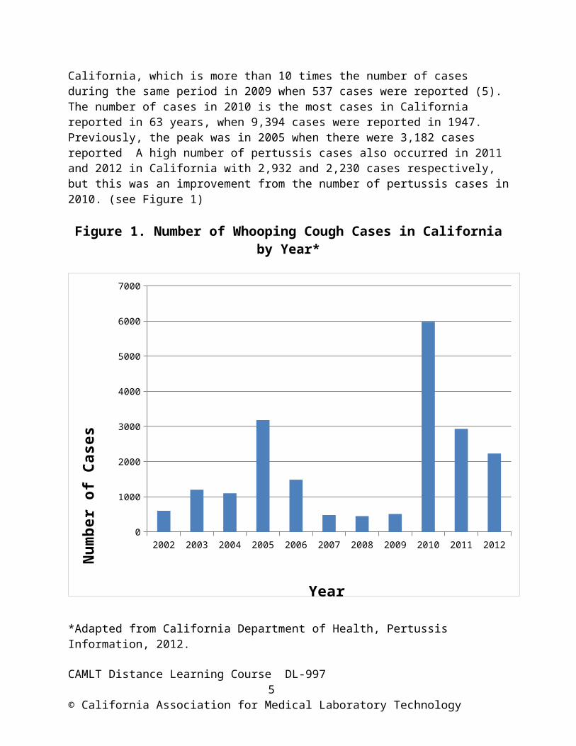
California experienced a major outbreak of pertussis in 2010. There were more than 5,900 cases of pertussis reported in California, which is more than 10 times the number of cases during the same period in 2009 when 537 cases were reported (5). The number of cases in 2010 is the most cases in California reported in 63 years, when 9,394 cases were reported in 1947. Previously, the peak was in 2005 when there were 3,182 cases reported A high number of pertussis cases also occurred in 2011 and 2012 in California with 2,932 and 2,230 cases respectively, but this was an improvement from the number of pertussis cases in 2010. (see Figure 1)
Figure 1. Number of Whooping Cough Cases in California by Year*
CAMLT Distance Learning Course DL-9973
© California Association for Medical Laboratory Technology

2002 2003 2004 2005 2006 2007 2008 2009 2010 2011 20120
1000
2000
3000
4000
5000
6000
7000
Year
Nu
mbe
r of
Cas
es
*Adapted from California Department of Health, Pertussis Information, 2012.http://www.cdph.ca.gov/Healthinfo/discond/Pages/Pertussis.ASPX
B. HISTORY OF WHOOPING COUGH INFECTION
Outbreaks of whooping cough were described as early as the 16th century in Europe. One of the first detailed reports of the disease was from Paris, France, in 1578, that described an epidemic among young infants and children that had a “whoop” sounding cough (1). Throughout the 18th and 19th centuries, whooping cough was one of the most common childhood diseases and a major cause of childhood mortality throughout the world. It was not until 1906, however, that the research of the two Belgium microbiologists, Bordet and Gengou, led to the discovery of the causative organism of whooping cough, Bordetella pertussis. Their research further led to the development of a medium (BG plate) that permitted the growth of pertussis (see Section G: Laboratory Diagnosis of Whooping Cough for composition). In 1909, Bordet and Gengou developed a method of inoculation of B. pertussis in which the patient would cough directly onto the surface of the BG plating medium. This procedure was called the
CAMLT Distance Learning Course DL-9974
© California Association for Medical Laboratory Technology
“cough plate” technique. It is no longer used because good transport systems are available to maintain the viability of the organism.
Prior to the introduction of the pertussis vaccine in the late 1940s, whooping cough was a common cause of infection and death among children. As many as 265,000 cases and 7,500 deaths were attributed yearly to B. pertussis infection in the U.S. during the 1920s and 1930s, more than the incidence of the childhood diseases of diphtheria, scarlet fever, or measles. During the six year period from 1940 through 1945, for example, more than 1 million cases of whooping cough were reported in the U.S., an average of 200,000 cases per year (1). Since the widespread use of the vaccine, however, the incidence has decreased more than 80% compared with the pre-vaccine era, gradually declining in the U.S. to 15,000 reported cases in 1960 (3).
While the incidence of pertussis is cyclical, with peaks occurring every 2-5 years in the United States, there has been a general increase in the number of reported cases of pertussis since the 1980s (3). The number of pertussis cases has fluctuated over the last few years, but has remained relatively high. For example, the number of cases reported to the Centers for Disease Control and Prevention (CDC) in 2004 was 25,827– the largest number since 1959. The number of cases in 2006 was 15,632, with 10,454 in 2007, 13,245 in 2008, 12,219 in 2009, and 14,871 cases in 2010. In 2011, the CDC reported 18,719 cases of whooping cough, while in 2012 the CDC reported over 26,000 cases with more than 10,000 cases in children ages 7 to 10. There were 18 fatalities in 2012 due to pertussis, with most deaths in children, particularly babies under 3 months of age. (3). These figures are probably lower than the true incidence because the CDC estimates that only 5-10% of all cases of pertussis are recognized and reported to public health agencies (3).
During the period of 1998-2001, the highest annual whooping cough incidence occurred in infants younger than 1 year (up to 24 percent of all cases), particularly in infants younger than 6 months of age (3). In the last few years, the incidence has also been particularly noticeable among 10 to 19 year old adolescents (3). Several factors have likely contributed to the increase in reported cases among adolescents, including increased awareness and improved recognition of pertussis among clinicians; greater access to and use of laboratory diagnostics, especially polymerase chain reaction (PCR) testing; and increased surveillance and reporting of pertussis to public health departments. However, the most likely cause for the increased number of reported cases of whooping cough among 10 to 19 year olds is believed to be a decrease in immunization among this group. Prior immunization or infection with B. pertussis does not provide lifelong immunity as earlier thought, so it is likely that adolescents or adults are actually acquiring the infection again (1,3). Public health experts believe that the increase among infants (<1 year) is probably because many infants are not receiving the correct
CAMLT Distance Learning Course DL-9975
© California Association for Medical Laboratory Technology
number of doses of the whooping cough vaccine, perhaps because of parents’ fears regarding the general safety of vaccinations, and because infants are acquiring the organism from an infected adolescent or adult (4).
Of the more than 5970 cases of pertussis reported in California in 2010, the majority were infants <6 months of age. Of hospitalized infants <6 months of age with known race and ethnicity, 178 (79%) were Hispanic. Of hospitalized infant cases in California in 2010, 236 (75%) were infants <6 month of age, and 179 (57%) were < 3 months of age. Ten deaths were reported from pertussis in California in 2010. Of these, 9 (90%) were Hispanic infants. These nine infants who died were not correctly diagnosed with the disease despite repeated visits to hospitals and clinics. In some cases, doctors only treated the infants for the common cold or nasal congestion (5). By the time test infants developed severe respiratory distress, it was too late for any intervention to prevent their deaths. These nine fatalities were infants <2 months of age at time of disease onset, and none had received any doses of pertussis-containing vaccine. The other infant who died, a premature infant born at 28 weeks, had received the first dose of DTaP at age 2 months only 15 days prior to disease onset.
As previously stated, the majority of infant cases reported in California in 2010 were infants <6 months of age. Of adolescent cases, the majority were 10-11 year-olds. While California had the majority of pertussis cases in 2010 among all the U.S states, significant whooping cough outbreaks also occurred in Texas, Idaho, Oregon, Ohio, Michigan, South Carolina, and Arizona . California, however, did not have the majority of whooping cough cases among all the U. S. states in 2011 and 2012 (3).
Worldwide, B. pertussis remains a major health problem among children, particularly in developing countries lacking organized vaccination programs. As recently as 2008, the World Health Organization estimated 294,000 pertussis deaths among children in developing countries, most prevalent in Sub-Saharan Africa and undeveloped areas of central Asia (2). It is currently estimated that pertussis causes an estimated 50 million cases and 350,000 deaths each year (1,2).
C. TRANSMISSION OF BORDETELLA PERTUSSISB. pertussis is a highly contagious infection and is transmitted from
person to person via respiratory droplets and secretions. Since B. pertussis is spread by aerosol respiratory droplets that are breathed in by the host, relatively close contact (within several feet) is required for transmission. B. pertussis in respiratory droplets may remain viable for 15 minutes (3). Transmission occurs less frequently by contact with freshly contaminated fomites (articles) from an infected person, but is possible, so precautions need to be taken. An asymptomatic carrier state is not recognized for B. pertussis, and the disease occurs only in humans. Due to their lack of or waning immunity from childhood vaccination or previous disease,
CAMLT Distance Learning Course DL-9976
© California Association for Medical Laboratory Technology

adolescents and adults with B. pertussis infection are often the source of transmission to infants.
Persons with pertussis are most infectious during the catarrhal period, the first 2-3 weeks after the initial onset of symptoms (an average of 21 days). However, since the disease may develop slowly and secretions are contagious during both the early catarrhal and the paroxysmal phases (see Section D: Illness and Clinical Symptoms of Whooping Cough), an infected person may unknowingly transmit disease for several weeks. During the first 3 weeks of infection, a child’s cough contains enough pertussis bacilli to infect many other children, but by 5 weeks most patients are noninfectious (3). B. pertussis has a very high infection rate (the rate of obtaining infection from an individual with the disease); up to 90% of family members, and up to 25 to 50% of schoolroom contacts with unvaccinated children produce clinical disease following exposure (3). This is a high rate of infection for a bacterial disease and is approximately that of viral diseases such as measles and chickenpox. Mortality from pertussis is about 1%. Sixty-four per cent of the deaths are infants under 1 year of age, while 40% of deaths occur during the first 5 months (3). Pertussis is unique in that its incidence and mortality are significantly greater among females (3). The reason for the higher incidence of pertussis in females is not known.
D. ILLNESS AND CLINICAL SYMPTOMS OF WHOOPING COUGHAlthough the infection may occur at any time of the year, most cases
of whooping cough are observed during the spring and summer months. The incubation period for whooping cough is commonly 7–10 days, with a range of 4–21 days, and rarely may be as long as 42 days (1). B. pertussis initially enters the nasopharyngeal area in aerosol droplets (exhaled droplets suspended in the air). The organism then attaches to and colonizes the ciliated respiratory epithelial cells in the bronchi and trachea. B. pertussis is the only pathogenic bacterium known to colonize this cell type exclusively. The symptoms of whooping cough can be variable in their course, but generally the illness is divided into 3 stages that often overlap: 1) the catarrhal, 2) the paroxysmal, and 3) the convalescent. (See Table 1. Stages of Whooping Cough.)
The first stage, the catarrhal stage, begins after the incubation period, about 7 to 10 days following exposure. The usual early manifestations are similar to those of any early upper respiratory viral infection and are characterized by the insidious onset of nasal congestion, irritation of the mucous membranes, sneezing, runny nose, low-grade fever, and a mild, occasional cough, similar to what one might expect for the common cold (3). Symptoms are nonspecific during the catarrhal period, and pertussis is frequently not suspected and the clinical diagnosis elusive because the typical cough with whoop appears later in the illness. The cough, however, gradually becomes more severe, and after 1–2 weeks, the second, or paroxysmal stage, begins. Fever is generally minimal throughout the course
CAMLT Distance Learning Course DL-9977
© California Association for Medical Laboratory Technology
of the illness. The first stage is probably the most communicable period of the disease. Bacterial cultures in infants and children may be useful during the first two weeks of illness for diagnosis, while the diagnosis by bacterial culture in adults is less likely to be helpful. Samples submitted for PCR testing may be useful in infants and children, and are also productive on adults during this period (See Section G. Laboratory Diagnosis of Whooping Cough).
The second stage, called the paroxysmal stage, is where the diagnosis of pertussis is usually suspected because of the development of classic symptoms. The paroxysmal stage may last 1 to 6 weeks (as long as 10 weeks in some cases). Characteristically, the patient has bursts, or paroxysms, of numerous, rapid coughs because of the difficulty of expelling thick mucus from the tracheobronchial area. The sudden crescendo of severe coughing fits (paroxysms) is usually accompanied by a characteristic high-pitched whoop when the person labors to breathe in (you can listen to the whooping cough sound at: http://health.utah.gov/epi/diseases/pertussis/pertussis_sounds.htm). During such an attack the patient may become cyanotic (turn blue) due to a lack of oxygen intake. The patient may vomit after a coughing fit and become exhausted. Infants and children generally have the most severe symptoms and appear very ill and distressed. In children under 6 months of age, the most common symptom is apnea (suspension of breathing) and cyanosis. The whoop is often absent (they may not have the strength to have a whoop), but the choking spells make it hard for infants to eat, drink, or breathe. Posttussive vomiting is often associated with the choking cough, which often leads to dehydration in the infant. Infants have the highest hospitalization and complication rates. Fifty percent or more of infants <6 months of age who contract pertussis are hospitalized. Twenty-five percent develop pneumonia, four percent develop seizures, one percent suffer permanent brain damage and another one percent die (1,2,3). Older children and adults may also lack the whoop, but have a prolonged paroxysmal cough and vomiting as symptoms. The classic signs of pertussis during this stage are: 1) a progressive cough that becomes paroxysmal (bursts of uncontrolled coughing), 2) coughing that often ends in vomiting, and 3) a forced inspiration (the whoop). Generally, bacterial cultures for B. pertussis during this later period are not useful since the organism is no longer present. The most useful laboratory diagnostic test during this late period may be PCR testing.
Ultimately, the paroxysms may be induced by any stimulus (e.g., swallowing, movement, noise, exertion), and they occur with increasing frequency, severity, and duration as the paroxysmal stage progresses. Each paroxysm may consist of several bouts separated by momentary intervals insufficient for inspiration, until hypoxia (reduction of oxygen supply to tissues) and partial asphyxia (decrease in oxygen accompanied by increase in CO2 which can lead to loss of consciousness) occur. The end of the
CAMLT Distance Learning Course DL-9978
© California Association for Medical Laboratory Technology
paroxysmal episode may be marked by a massive single inspiratory stroke causing the classic “whoop.” During severe paroxysms, patients may become very anxious, believing their life is threatened. Paroxysmal coughing may vary from mild, uncomplicated episodes to severe, protracted episodes requiring hospitalization and acute supportive care, particularly for infants. Following the early paroxysmal stage, attacks occur more frequently at night, with an average of 15 attacks per 24 hours. The attacks increase in frequency as the disease progresses, and may continue to occur over a prolonged period of time, often 4-6 weeks, but may persist for up to 10 weeks, and then gradually decrease (1,3).
In the third stage, or convalescent stage, the patient gradually recovers from the disease and may have a non-paroxysmal cough for 4 to 6 weeks (sometimes as long as 3-4 months). In the convalescent stage, recovery is gradual and slow. The cough becomes less paroxysmal and less severe. However, for many months after the initial onset of pertussis, paroxysms can often recur with subsequent respiratory infections related to other agents (e.g., colds, flu) (1,3).
Before the development of the pertussis vaccine in the late 1940s, the mortality of whooping cough was greater than 50%. Today, the mortality rate for unvaccinated infants is about 2.4%, whereas in vaccinated infant populations it is approximately 1% for infants younger than 2 months of age and less than 0.5% for infants 2 –11 months of age (4). Children who are too young to be fully vaccinated and those who have not completed the primary vaccination series are at highest risk for severe illness. Complications of pertussis are steadily declining, but can occur. The most common complication, and the cause of most pertussis-related deaths, is secondary bacterial pneumonia. Other complications can include dehydration, hypoxia seizures, encephalopathy and coma, and hemorrhagic events, particularly in the conjunctivae and soft tissues about the eyes, because of the high venous blood pressure exerted during coughing fits.
E. MICROBIOLOGY OF BORDETELLA PERTUSSISThe morphology of B. pertussis when first isolated on culture media is
a very small, oval bacillus which stains faintly Gram-negative. Its mean length and width is 0.5 micron (E. coli, for example, is commonly 2 to 3 microns in length and 0.6 microns wide). Bacterial cells from original cultures are uniform in size, but on subculture, the organism may become pleomorphic, tending toward cocco-baccillary, sometimes exhibiting a bipolar appearance. B. pertussis survives only a short time outside the human host, and is strictly aerobic. The organism has an optimal growth temperature of 35 to 37oC. Many studies show that the addition of carbon dioxide to the incubation atmosphere may slow or inhibit the growth of B. pertussis (1). B. pertussis oxidizes amino acids as an energy source, but does not utilize any carbohydrates (6). The organism is catalase-positive, urease negative, nitrate reduction negative, and nonmotile. B. pertussis is
CAMLT Distance Learning Course DL-9979
© California Association for Medical Laboratory Technology
easily killed by desiccation, heat, and chemical agents, and does not survive on environmental surfaces for any length of time.
B. pertussis is the most fastidious species of the Bordetellae, and is inhibited by constituents present in many media, including fatty acids, metal ions, sulfides, and peroxides. The growth of B. pertussis on artificial media is slow, requiring up to 7 to 10 days for its initial isolation in some cases (6). Media for the primary isolation of B. pertussis must contain protective substances, such as charcoal, blood, or starch. Bordet-Gengou, a potato infusion-based blood medium supplemented with charcoal, is one option, while another option is Regan-Lowe, a charcoal-based medium supplemented with glycerol, peptones, and horse or sheep blood. See Section G, Laboratory Diagnosis of Whooping Cough for more information on primary media. B. pertussis will not grow on common blood agar bases or chocolate agar, whereas B. parapertussis will grow on blood agar and sometimes chocolate blood agar.
F. PATHOGENIC MECHANISMS OF BORDETELLA PERTUSSISB. pertussis produces multiple virulence factors that are classified
generally as adhesins or toxins. These factors are responsible for the pathogenicity of B. pertussis and contribute to the specific clinical features of the disease. Whooping cough is primarily a toxin-mediated disease.
Once aerosols containing B. pertussis enter the nasopharynx, the organism attaches to and then multiplies on the ciliated respiratory epithelial cells, starting first in the nasopharynx and ending in the bronchi and bronchioles in the lungs. One of the major factors that permit B. pertussis to attach to the ciliated respiratory epithelial cells is an adhesin called filamentous hemagglutinin (FHA), a protein that promotes the binding of B. pertussis to specific carbohydrate structures on the ciliated respiratory epithelium, as well as to the host’s phagocytic cells. In addition to FHA, B. pertussis has fimbriae or pili (hair-like protein projections on the bacterial cell wall to allow attachment to the respiratory epithelium), that assist in infection. Recent studies indicate that fimbriae play a major role in the attachment of B. pertussis to the laryngeal mucosa, whereas FHA is important for colonization of the entire respiratory tract (1). A third class of adhesin is called pertactin, a highly immunogenic (provoking an immune response) virulence factor on the outer membrane of only B. pertussis that promotes attachment of the organism to tracheal epithelial cells and binding to phagocytes. (See Table 2: Virulence Factors Associated with the Pathogenicity of Bordetella pertussis .)
After attachment, B. pertussis multiplies on the respiratory epithelial cells and then begins to produce various toxins that aid in the pathogenesis of disease. These toxins include pertussis toxin, adenylate cyclase toxin, and tracheal cytotoxin. (See Table 2. Virulence Factors Associated with the Pathogenicity of Bordetella pertussis. ) These toxins are responsible for the further development of disease and for the specific clinical features of
CAMLT Distance Learning Course DL-99710
© California Association for Medical Laboratory Technology
whooping cough. Pertussis toxin is an exotoxin (a soluble toxin excreted by a microorganism that causes damage to the host) associated with the cell wall of B. pertussis. Pertussis toxin allows the binding of B. pertussis to host epithelial cell receptors, and permits the colonization of the organism in the respiratory tract. Pertussis toxin further allows B. pertussis to evade host defenses by altering host cell activity and disrupting host phagocytic activity. Investigations have shown that pertussis toxin and cholera and diphtheria toxins act in a similar manner. Adenylate cyclase toxin penetrates phagocytic cells, disrupting their normal function and their ability to signal to one another, thus reducing chemotaxis (the ability of phagocytic cells to be drawn to an infection site). Once adenylate cyclase toxin binds to phagocytes, it enters the phagocyte and induces apoptosis (cell death). Tracheal cytotoxin is actually a fragment of peptidoglycan (a polymer layer of the cell wall) secreted by B. pertussis. Tracheal cytotoxin destroys tracheal ciliated epithelial cells, inhibits DNA synthesis and the regeneration of the respiratory tract epithelium, and ultimately causes death of the respiratory epithelial cells. The toxins of B. pertussis not only kill respiratory epithelial cells and phagocytes, but also cause inflammation of the respiratory tract and interfere with the clearing of pulmonary secretions, leading to the classic “whoop” sound (1,3). It is common to be unable to recover the organism from the patient after several weeks, but still have toxins from B. pertussis continue to cause symptoms (1).
Many of the adhesin and toxin virulence factors of B. pertussis have been found to elicit a good antibody response in the host, and thereby are important in providing immunity after infection. Consequently, some of these virulence factors have been incorporated in the production of acellular vaccines to improve their activity (4). (See Section I. Prevention and Control of Bordetella pertussis Infection .)
G. LABORATORY DIAGNOSIS OF WHOOPING COUGHThe laboratory diagnosis of whooping cough can be achieved through
either bacterial culture, direct fluorescent antibody (DFA) tests, molecular tests such as polymerase chain reaction (PCR), or by serological tests that detect the presence of specific B. pertussis antibodies in the serum of an infected individual (see Table 3. Laboratory Tests for the Identification of B. pertussis). There are positive and negative aspects of each laboratory test, which this section will describe. The choice of which diagnostic method to use depends on the age of the patient, the duration of symptoms, the immunization status of the patient, the availability of adequate transport procedures, and the availability of required laboratory services. For any of the methods that seek to detect B. pertussis directly, such as bacterial culture, DFA, or by PCR techniques, specimen collection and transport are the most significant variables to the sensitivity (the number of positives correctly identified) of the test. Specimen Collection
CAMLT Distance Learning Course DL-99711
© California Association for Medical Laboratory Technology
The preferred specimen type for the laboratory diagnosis of pertussis is the use of a posterior nasopharyngeal (NP) swab to collect nasopharyngeal secretions. When properly collected, this specimen contains the ciliated respiratory epithelial cells for which B. pertussis exhibits tropism (selectivity to certain receptor cells). Nasopharyngeal washings or aspirates might also be a useful specimen, but generally the tendency to initiate a paroxysm makes this method unacceptable. In most cases a nasopharyngeal swab is the most practical specimen type (3,6). Throat swabs are an inferior specimen type for culture of B. pertussis and should not be accepted; they do not sample the ciliated respiratory epithelium, and they contain large numbers of the normal respiratory flora that can result in contamination and overgrowth of the requested laboratory test.
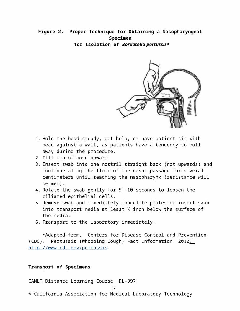
A flexible wire Dacron-tipped nasopharyngeal swab is the best collection device for NP secretions (1,3). While material collected on a calcium alginate-tipped swab is acceptable for bacterial culture, it is not acceptable for PCR assay, because residues present in the calcium alginate may inhibit the PCR test. Cotton-tipped wire swabs are not acceptable since they contain inhibitors that will decrease the isolation rate or detection of B. pertussis (1,6). The Dacron-tipped wire swab can be bent to conform to the nasal passage. The swab should be inserted deep into the nostril until it touches the pharynx and gently rotate for 5-10 seconds to loosen the ciliated epithelial cells. (See Figure 2)
CAMLT Distance Learning Course DL-99712
© California Association for Medical Laboratory Technology
Figure 2. Proper Technique for Obtaining a Nasopharyngeal Specimen for Isolation of Bordetella pertussis*
1. Hold the head steady, get help, or have patient sit with head against a wall, as patients have a tendency to pull away during the procedure.
2. Tilt tip of nose upward3. Insert swab into one nostril straight back (not upwards) and continue along the floor of
the nasal passage for several centimeters until reaching the nasopharynx (resistance will be met).
4. Rotate the swab gently for 5 -10 seconds to loosen the ciliated epithelial cells.5. Remove swab and immediately inoculate plates or insert swab into transport media at
least ½ inch below the surface of the media.6. Transport to the laboratory immediately.
*Adapted from, Centers for Disease Control and Prevention (CDC). Pertussis (Whooping Cough) Fact Information. 2010. http://www.cdc.gov/pertussis
Transport of SpecimensBecause of the fragile nature of B. pertussis outside its normal host
environment, proper handling and transport conditions are critical for optimal laboratory identification. Once an NP swab has been collected, it should be plated directly onto suitable selective culture media or immediately placed into a suitable transport medium. Direct plating of NP specimens for B. pertussis is not practical for most hospitals or medical clinics. Cough plates are not recommended, because higher rates of B. pertussis isolation are obtained from NP swabs specimens than from cough plates (1).
There are two different transport media recommended for the proper transport of B. pertussis. One is Regan-Lowe transport medium, which
CAMLT Distance Learning Course DL-99713
© California Association for Medical Laboratory Technology
contains half-strength charcoal agar base, horse blood, beef extract, starch, and tryptose. Regan-Lowe may also be purchased containing the antibiotic cephalexin to suppress growth of the normal respiratory flora. Another transport medium, Jones-Kendrick Transport Medium, contains beef heart extract, soluble starch, charcoal powder, tryptose, and yeast extract; it does not contain blood. Once the NP specimen has been collected using a Dacron tipped wire swab, it should be placed immediately into the transport medium and transported at room temperature (15–30°C) to the laboratory within four hours of collection. Isolation rates decrease when transport occurs at 4°C instead of at room temperature, or when the transport takes longer than 48 hours (1,6). Remember that B. pertussis is a fragile organism. It does not survive desiccation, temperature, or environmental changes well. Once the specimen has reached the laboratory, it should be plated as soon as possible.
For direct fluorescent-antibody (DFA) testing the specimen collection kit should contain a glass slide, on which a smear is prepared from an NP swab immediately after specimen collection. The slide is allowed to air dry and is then transported to the laboratory.
For PCR testing, NP swabs can be transported dry, in a transport medium, or in saline after specimen collection. Transport in liquid medium provides a specimen from which multiple aliquots can be tested, whereas processing a dry swab for PCR testing leaves no specimen material available for repeat analysis if required (1). Generally, the same NP specimen can be used both for culture and polymerase chain reaction (PCR) if the volume of the specimen allows. Bacterial Culture
The culture of B. pertussis is considered the “gold standard” and the preferred laboratory test for pertussis in some cases. However, the sensitivity of B. pertussis culture can vary according to the following factors: 1) collection method used, 2) transportation, 3) timing of specimen collection during illness, 4) lack of fresh media, 5) choice of media and selective agents, 6) incubation conditions and duration, and 7) various host factors. Culture is considered the most specific of the laboratory tests for pertussis, but it may take up to seven days before results are known. Culture is less sensitive than PCR, but is 100% specific (no false positives). A positive culture for B. pertussis confirms the diagnosis of pertussis; however, a negative culture result does not rule out pertussis infection. B. pertussis is most frequently recovered in the catarrhal or early paroxysmal stage of illness (i.e., first 1-2 weeks of cough); sensitivity decreases steeply (<60%) if the specimen is obtained more than 2 weeks from onset of cough. Isolation rates of B. pertussis on culture media are negatively correlated with increasing age (high rate in infants; low rate in adults), with antimicrobial pre-treatment, and with the number of pertussis vaccination doses received by the patient. Most authorities report that culture for B. pertussis is rarely positive in adults, though it is the “gold standard” test for
CAMLT Distance Learning Course DL-99714
© California Association for Medical Laboratory Technology
diagnosing pertussis in infants and children if obtained early in the infection (1). Usually adults may have been coughing for several weeks before they seek medical attention and it is often too late for culture to be useful in this age group.
The first culture medium used to isolate B. pertussis, developed in 1906 by Bordet and Gengou, is called BG medium. It was the traditional culture media for pertussis isolation and was used for many years. It consists of potatoes and glycerol, supplemented with 15% defibrinated sheep or horse blood (human blood is not an acceptable substitute). Charcoal was used in later formulations to neutralize toxic fatty acids. A more recent medium used to isolate B. pertussis is called Regan-Lowe (RL), which consists of beef heart extract, starch, charcoal, pancreatic digest of casein, niacin, and 15% defibrinated horse blood. These media are available with the addition of cephalexin or with other antibiotics to inhibit the overgrowth of normal respiratory flora. Many authorities suggest that the specimen should be inoculated onto both a non-selective pertussis medium and onto a selective medium, since inhibition of B. pertussis is sometimes observed on media containing antibiotics (1,6). Regan-Lowe medium appears to provide better isolation and faster growth of B. pertussis than does Bordet Gengou medium (1). The shelf life of media ranges from 5 days for BG medium to 4 to 8 weeks for RL medium. Bordet Gengou medium should be freshly made, if possible, for best results.Primary culture and presumptive identification
All primary plates are incubated in a humidified atmosphere under aerobic conditions without added carbon dioxide for seven days at 35–37°C and inspected every two days. The humidity must be sufficient to prevent desiccation of agar plates during the long incubation period. Growth usually appears after 4-5 days; however, cultures cannot be considered negative for B. pertussis until after 7 days of incubation. Typical B. pertussis colonies on BG or RL plates are small (1 mm in diameter after four to five days of incubation), round, domed-shaped and glistening, and resemble mercury droplets. On BG plates, B. pertussis appears slightly hemolytic.
On colonies that morphologically resemble B. pertussis and that develop after 4 days of incubation, the following steps are recommended: 1) check purity of the growth by performing a Gram stain. B. pertussis should appear microscopically as small Gram-negative coccobacilli or very short oval rods; 2) determine the organism’s biochemical characteristics by testing for oxidase, nitrate-reductase, urease, and motility, and 3) confirm the identification using specific anti-B. pertussis immunofluorescence antiserum if available. B. pertussis is catalase positive, oxidase positive, nitrate reduction test negative, urease production test negative, and is negative for motility. Since B. pertussis is catalase positive, the catalase reaction may help to distinguish the organism from catalase-negative commensal organisms. A negative urease reaction distinguishes B.
CAMLT Distance Learning Course DL-99715
© California Association for Medical Laboratory Technology
pertussis from B. parapertussis and B. bronchiseptica, which are both urease positive. For differentiation between B. pertussis and B. bronchiseptica, the oxidase reaction is helpful (B. pertussis is positive and B. parapertussis is negative). B. pertussis is non-fermentative (does not utilize carbohydrates as an energy source); it uses proteins.Direct Fluorescent Antibody Test (DFA)
The direct fluorescent antibody test (DFA) for the diagnosis of pertussis became available for use in the United States in the early 1960s. It became widely used by clinical and public health laboratories for a period of time because it provided rapid results. Although the direct DFA test for B. pertussis offers a rapid and simple diagnostic approach, the sensitivity of the test (only about 60% sensitive) is limited compared to other methods such as culture and PCR (6). Extensive cross-reaction with normal nasopharyngeal flora makes the results of DFA tests technically difficult to interpret, so DFA tests are frequently associated with false positive and negative results (3,6). The Centers for Disease Control and Prevention (CDC) believes that evaluation of B. pertussis specimens by DFA should not be considered a confirmatory test and should always be performed with concomitant culture, or with PCR testing (3). The DFA test should be regarded as a presumptive test only, not as a replacement for bacterial culture or PCR testing. Success in interpreting DFA examinations of clinical material is directly proportional to the experience of the reader. The subjectivity inherent in interpreting DFA test results presents some challenges even to good laboratories.
Commercially available reagents for DFA testing are either polyclonal antibodies (available from Becton Dickinson) or monoclonal antibodies (available from Delta Biotech, Inc., Delta, BC, Canada). The monoclonal antibody test is available as a dual-fluorochrome reagent for the detection of both B. pertussis and B. parapertussis in a single smear. This dual-fluorchrome reagent contains a fluorescein-conjugated murine monoclonal antibody that is specific for the lipooligosaccharide of B. pertussis, and a rhodamine-conjugated anti-B. parapertussis monoclonal antibody to detect B. parapertussis. With this test, B. pertussis will appear brilliant green and B. parapertussis will appear brilliant orange-red. The reported sensitivity of this dual fluorochrome reagent is 50-71%, with specificity of 92-98% (1). (See Table 3. Laboratory Tests for the Identification of B. pertussis .)
Regardless of the antibody used, the direct detection of B. pertussis by DFA lacks both sensitivity and specificity and is seldom positive in adults. DFA positive, culture-negative specimens are not unusual. In the long run, DFA testing will likely be replaced by PCR testing because the latter method combines rapidity with a much higher degree of sensitivity. Detection of Bordetella pertussis by PCR
Nucleic acid amplification methods, such as the polymerase chain reaction (PCR) technique, can be a rapid and sensitive method of diagnosing whooping cough. Since its introduction in the late 1990s, PCR
CAMLT Distance Learning Course DL-99716
© California Association for Medical Laboratory Technology
testing to identify B. pertussis has become widely used by many clinical and public health laboratories as the primary diagnostic test for whooping cough. Recommendations from the CDC are that the preferred methods for the laboratory diagnosis of pertussis are culture and polymerase chain reaction, and it is suggested that in most cases both tests be performed if possible (3). The CDC currently recommends that the PCR test should be used in addition to, and not as a replacement for, culture (3).
The CDC also recommends that PCR testing should be done on symptomatic patients only and within 3 weeks of onset of cough, but the test may provide accurate results for up to 4 weeks in infants or unvaccinated persons (3). Keep in mind that some vaccines contain PCR detectable components and may yield false positive results (3). Like bacterial culture, PCR is also affected by specimen collection technique. Nasopharyngeal swabs are the preferred method for specimen collection. An inappropriately obtained nasopharyngeal swab will likely be negative by both culture and PCR. PCR is the method of choice in adult patients with persistent cough in whom pertussis is suspected, as well as in infants and children with epidemiological and clinical features of the disease. PCR results have been shown to be unreliable after 5 days of appropriate antibiotic treatment (1,3). Since PCR detects B. pertussis DNA and not viable organisms, the sensitivity can be as low as < 1 cell. In general, the sensitivity, specificity, and rapid turnaround of PCR for B. pertussis have been shown to be very good, which is important for patient management and control of outbreaks (1).
The PCR procedure amplifies certain genes, sequences, or portions of the DNA from B. pertussis using specific primers that target certain areas of the organism’s DNA. The choice of primers (a strand of nucleic acid that serves as a starting point for DNA synthesis) and their target region are key components of the sensitivity and specificity of the PCR test. Generally, PCR for B. pertussis has a sensitivity of 95% and a specificity of 99%, and is usually more sensitive than DFA or culture (1,3). Delay in specimen collection is the main reason for a negative PCR test result in a patient with pertussis. Two major shortcomings of PCR testing, however, are: 1) the potential for false-positive results due to contamination, and 2) the interpretation of a positive result for B. pertussis DNA in the absence of a positive bacterial culture. A positive PCR result, therefore, must be interpreted in conjunction with patient symptoms, treatment status, epidemiological factors, and a similarly positive bacterial culture. PCR tests are less sensitive in previously immunized individuals, but are more sensitive than cultures in such patients. Other shortcomings of PCR testing are: 1) PCR testing requires specialized training and is technically difficult to perform; 2) PCR requires more expensive equipment and reagents; and 3) the cost of the PCR test is high (6). The charge for B. pertussis PCR testing from commercial reference laboratories is about $125.00 per test. (See Table 3 for manufacturers).
CAMLT Distance Learning Course DL-99717
© California Association for Medical Laboratory Technology
PCR testing, however, is the best option in most clinical circumstances. It provides acceptable sensitivity in both children and adults, has a relatively short turnaround time, and is available at most commercial reference laboratories.Serological Detection of B. pertussis
Serological detection of B. pertussis identifies antibodies in the patient’s serum formed in response to infection or prior immunization. Serological tests have limited value for the initial diagnosis of B. pertussis infection because of the need for paired sera before interpreting results. Valid serologic diagnosis requires data from serum collected at the beginning of cough (acute serum), and from serum collected one month later (convalescent serum) to determine a change or rise in antibody level in the serum of infected individuals. Because of the delay in collecting the convalescent sera, serological tests cannot be used as the sole method of diagnosis. The collection of a single serum specimen for diagnostic purposes is not well standardized and usually not of any use (6). Further, the use of serology as a method of diagnosis of pertussis in young infants is not sensitive and should not be used, as their immune system is immature and may not produce adequate levels of antibodies. The serodiagnosis of pertussis in vaccinated persons can also be a problem since there may be an immediate increase in various antibodies and in their titers, which may confuse serodiagnosis. In some cases, a patient who has had prior immunization to pertussis may produce an immediate high level of antibody due to an immunological phenomenon called an anamnestic response, in which the body recognizes an invading antigen and produces a high level of antibodies specific against that antigen in defense. This results in a rapid increase in antibody concentration precluding detection of a significant increase in antibody concentration between acute and convalescent phase sera. Occasionally, high IgA or IgG titers may distinguish responses to vaccination from responses in infection since IgA is rarely detected in uninfected vaccinated persons. Serologic testing could be useful for adults and adolescents who present late in the course of their illness, when both culture and PCR are likely to be negative or not available. Other conditions in which serological testing may be useful are for epidemiology purposes, or as an alternative diagnostic test when culture or PCR is not available, or when it has been over 3 weeks since the onset of cough and the individual is thus frequently PCR or culture negative (1).
Enzyme-linked immunosorbent assays (ELISA) are regarded as the best method for measuring antibodies to Bordetella pertussis. This method can be used to measure IgA, IgG, or IgM immunoglobulin levels. Some ELISA test systems can also detect specific immunoglobulins to pertussin toxin (PT) or filamentous hemagglutinin (FHA), which are considered reliable indicators of Bordetella infection (6). It is recommended that IgG and IgA titers to PT and FHA be performed in combination (3,6). In unvaccinated individuals with a primary infection, IgG and IgA antibodies to
CAMLT Distance Learning Course DL-99718
© California Association for Medical Laboratory Technology
PT and FHA are elevated. Some researchers indicate that the IgA antibody to FHA appears promising as a diagnostic test for immunized patients who have an infection (1). Commercial serological tests that use whole B. pertussis antigens, rather than using pertussis toxin or FHA, have a higher false positive rate and are not recommended (1,3).
H. TREATMENT OF WHOOPING COUGHIn general, treatment for whooping cough involves administering antibiotics and
providing supportive care. Supportive care, such as admission to a hospital, breathing support from a ventilator, and prevention of secondary infections, may provide relief of symptoms and reduce complications. Very young infants with frequent, severe paroxysms, especially if complicated by posttussive exhaustion, vomiting and malnutrition, should be hospitalized for supportive care.
Early antibiotic treatment of pertussis is very important, particularly for an infant. If antibiotics are started early in the course of illness, symptoms and duration may be lessened (1,3). If the patient is diagnosed late, antibiotics will not alter the course of the illness and do little to help the patient’s coughing. Treatment in the early stages can also reduce the further spread (communicability) of the disease. Generally, when pertussis is suspected in infants aged <1 year who are presenting with persistent cough and have the classic “whoop,” a specimen is collected and antibiotics are started immediately, especially if the patient will be in contact with other infants. Once a physician knows that pertussis is circulating within the community, any respiratory symptom has to be considered as a possible pertussis case and antibiotics initiated.
The CDC recommends limiting antibiotic treatment to those who are within three weeks of the onset of their illness unless they are: 1) infants under the age of 1 year old, 2) pregnant women, 3) persons with ongoing close contact with infants under the age of 1 year old, or 4) parents, caregivers, or daycare workers of infants (3). All household and other close contacts of pertussis cases should be given antibiotics regardless of age or immunization status. However, the initiation of antibiotics for contacts that were exposed to a pertussis case more than 3 weeks ago has limited benefit, and should not be routinely done unless the contacts are at high risk for developing severe disease (e.g., infants or persons with severe lung disease), or unless they are healthcare workers (or others) who are routinely exposed to high risk persons (3).
For those cases that require antibiotic therapy, erythromycin is usually the drug of choice and is recommended because of its higher penetration into the respiratory tract. Patients should be treated with erythromycin for a period of at least 2 weeks, as shorter courses (7-10 days) may not eliminate the organisms from the upper respiratory tract. Patients are considered non-infectious following 5 days of antibiotic therapy. Other newer macrolides such as azithromycin or clarithromycin may also be given for treatment and prophylaxis of pertussis in persons >1 month of age and
CAMLT Distance Learning Course DL-99719
© California Association for Medical Laboratory Technology
are considered as effective as erythromycin. The current CDC guidelines recommend the exclusive use of azithromycin in infants under one month of age due to fewer adverse side-reactions compared with erythromycin (3). For persons > 2 months of age, an alternative to macrolides is trimethoprim-sulfamethoxazole (TMP-SMX) for individuals intolerant of macrolides. Trimethoprim-sulfamethoxazole, however, is contraindicated in infants less than 2 months of age (1,3). Penicillin-class drugs and first and second generation cephalosporins are not effective, and should not be given. Antimicrobial Susceptibility Testing of B. pertussis
Antimicrobial susceptibility testing of B. pertussis is usually not required. Resistance to erythromycin and other macrolides is very rare. Standardized procedures from the Clinical and Laboratory Standards Institute (CLSI) to evaluate the in vitro antimicrobial susceptibility of B. pertussis have not been developed. Several methods are currently being evaluated for the in vitro antimicrobial susceptibility testing of B. pertussis, such as: 1) agar dilution susceptibility performed on Regan-Lowe agar or Bordet Gengou agar with 5% horse blood; 2) disk diffusion testing performed on Bordet Gengou agar with 5% horse blood; and 3) Etest strips (AB Biodisk, bioMerieux, Durham, NC) performed on Regan-Lowe or Bordet-Gengou agar with 20% horse blood. Susceptibility testing results using one of these methods appear to predict antimicrobial agents that may be effective against B. pertussis and provide data for a good clinical outcome, although these methods have not been approved by the CLSI (1).
I. PREVENTION AND CONTROL OF BORDETELLA PERTUSSIS INFECTION
Vaccines have dramatically reduced the health impact of pertussis throughout much of the world; however, pertussis still exists even in developed countries with good vaccination programs. Since a pregnant woman’s own protective antibodies to Bordetella pertussis do not cross the placenta, neonates are highly susceptible to whooping cough. Therefore, immunization of neonates and young infants is very important and should be implemented as soon as possible. Immunization, or a booster shot, for adults is also very important because, in many circumstances, adults contract B. pertussis and act as a reservoir for further infection.
The first pertussis vaccine, developed in the late 1940s, was a heat killed whole-cell vaccine composed of a suspension of formalin-inactived B. pertussis cells. It was used successfully for many years in clinical practice to reduce the incidence of disease. Based on efficacy trials conducted in the late 1940s, a primary series of four doses of whole-cell DTP (diphtheria, tetanus, and pertussis), also called DPT vaccine, was 70%–90% effective in preventing serious pertussis disease (1). The whole-cell vaccine was used primarily among infants and young children. It was assumed that this protocol was satisfactory because adolescents and adults seldom acquired
CAMLT Distance Learning Course DL-99720
© California Association for Medical Laboratory Technology
whooping cough. However, protection from pertussis (either from natural infection, or from prior immunization) decreases with time, resulting in little or no protection 5 to 10 years following the disease or last immunization dose. A prior episode of whooping cough does not provide life-long immunity, as previously believed, forcing changes in immunization policies (4). In recent years, concerns about the safety of whole-cell pertussis vaccine led to the development of more purified (acellular) pertussis vaccine that is associated with a lower frequency of adverse reactions and side effects. Whole-cell pertussis vaccines are no longer available in the United States but are still used in many other countries. Acellular Pertussis Vaccine
Acellular pertussis vaccines are multi-subunit vaccines that contain purified, inactivated virulence components of B. pertussis cells. (See Section F. Pathogenic Mechanisms of B. pertussis .) Currently, two different acellular B. pertussis vaccines are available in the United States. One vaccine, called DTaP, is a pediatric acellular pertussis formulation consisting of a combination of diphtheria and tetanus toxoids. The upper-case letters in DTaP denote full-strength doses, lower-case letters denote reduced doses, and the aP stands for full-strength acellular pertussis, meaning that the pertussis component contains only a part—the virulence components of Bordetella pertussis (4). The other acellular vaccine available is a formulation for adolescents and adults called Tdap. In 2005, this adolescent/adult formulation, consisting of a full-strength dose of tetanus, but a reduced dose of both diphtheria and pertussis vaccine, was approved for use in certain age groups. Efficiency trials of the acellular vaccines currently licensed in the United States show that the vaccines are 80% to 95% effective for reducing serious pertussis disease (3,4). Studies also showed that the acellular pertussis vaccines were significantly more effective than the older whole-cell DTP formulation in reducing pertussis disease, and serious side-reactions occurred less frequently among infants vaccinated with acellular pertussis vaccines than among those vaccinated with whole-cell DTP vaccine (3,4). Pediatric Formulation (DTaP)
There are a number of different manufacturers of pediatric acellular pertussis vaccine available for use in the United States. All vaccines are combined with diphtheria and tetanus toxoids as DTaP (Diphtheria, Tetanus, and acellular Pertussis), and are approved for children 6 weeks through 6 years of age (to age 7 years). Some formulations, for example, Infanrix (GlaxoSmithKline), contain three virulence factor antigens of pertussis: pertussis toxin (PT), filamentous hemagglutinin FHA, and pertactin. (See Section F. Pathogenic Mechanisms of Bordetella pertussis , for description and action of toxins.) Another formulation, Daptacel (Sanofi Pasteur) contains four components: pertussis toxin, filamentous hemagglutinin, pertactin, and fimbriae. Yet another pertussis vaccine formulation, Tripedia (Sanofi Pasteur), contains two components:
CAMLT Distance Learning Course DL-99721
© California Association for Medical Laboratory Technology
filamentous hemagglutinin and pertussis toxin. None of the available DTaP vaccines contains thimerosal (an organomercury compound used as a preservative, which some suspect may cause mercury poisoning). Since 2001, no new vaccines licensed by the FDA for use in children have contained thimerosal as a preservative (4). All DTaP vaccines are supplied in single-dose vials or syringes. Recently, manufacturers of DTaP acellular pertussis vaccine have added other infectious disease agents, such as hepatitis B, poliovirus, or Hemophilus influenza type B, in a single dose vial or syringe that can be used, under certain circumstances, to reduce the number of injections in children. The manufacturer’s product sheet should be reviewed for more information about usage recommendations (4).
CAMLT Distance Learning Course DL-99722
© California Association for Medical Laboratory Technology
DTaP Immunization ScheduleThe CDC and the California Department of Public Health recommend
that children should receive 5 doses of DTaP (4,5). Children should receive one dose at the following ages: 2 months, 4 months, 6 months, 15-18 months, and a booster at 4-6 years before kindergarten to be fully protected from the disease. (See Table 4. Summary of Pertussis Vaccine Recommendations.) The booster dose at age 4-6 increases antibody levels and may decrease the risk of school-age children transmitting the disease to younger siblings who are not fully vaccinated. While the vaccination series starts at 2 months of age, adequate protection does not occur until six months of age. An accelerated vaccine schedule to begin pertussis immunization at 6 weeks may be considered when pertussis is epidemic. The DTaP series is not licensed for adolescents, adults, or children 7 years of age and older (4). For more information about the immunization of children or their contacts who may be under-immunized, or children who have not completed the vaccine series before an exposure to pertussis, review the recommendations from the CDC in reference #4. Even with a full immunization series, some exposed infants and children may still develop disease, although usually the symptoms are much milder. Adolescent and Adult Formulation (Tdap)
The acellular pertussis vaccine called Tdap that is licensed for adolescents and adults is similar to the DTaP vaccine for children, but it contains different doses of the individual vaccines. A single dose of Tdap is recommended for people 11 through 64 years of age (4). Tdap contains a regular dose of tetanus, but a reduced dose of diphtheria and pertussis vaccine compared with the pediatric DTaP. The Tdap vaccine is currently available from two different manufactures. One vaccine, Boostrix (GlaxoSmithKline), is approved for persons 10 through 64 years of age, and contains three pertussis antigens (PT, FHA, and pertactin) in a reduced quantity compared with the GlaxoSmithKline pediatric formulation. Another, Daptacel (Sanofi Pasteur) contains four components—pertussis toxin, FHA, pertactin, and fimbriae—also in a reduced quantity. All vaccines are manufactured without preservatives and are supplied in single-dose vials or syringes.
The booster shot Tdap was introduced to combat adult-to-child whooping cough transmission due to waning immunity in adults after prior immunization or after prior natural infection as a child. But not many adults are taking advantage of the vaccine (4). All adolescents and adults should receive Tdap boosters, as indicated below.Tdap Immunization Schedule
Tdap is licensed as a onetime use vaccine for individuals 11-64, and is recommended by the CDC to be given as a replacement booster dose for the tetanus and diphtheria toxoid vaccine (Td) given every 10 years. For other recommendations for who should receive the Tdap vaccine, see Reference #4.
CAMLT Distance Learning Course DL-99723
© California Association for Medical Laboratory Technology
The CDC and the California Department of Public Health (4,5) recommend the following vaccinations to prevent pertussis in adolescents and adults, and to reduce the transmission of pertussis to infants: 1) A single dose of Tdap is recommended for people 11 through 64 years of age who have not previously received Tdap. Adults under 65 who have never received Tdap should receive a single dose and substitute it for the next tetanus and diphtheria (Td) booster dose, usually every 10 years. 2) Adults, parents, family members, and caregivers under 65 who expect to have close contact with an infant younger than 12 months of age (including women who may become pregnant) should get a dose of Tdap to reduce the risk for transmitting pertussis to infants. 3) Women who have not previously received Tdap should receive a dose of Tdap in the immediate postpartum period. 4) Healthcare workers under the age of 65 who have direct patient contact in hospitals or clinics should get a single dose of Tdap as soon as feasible if they have not previously received Tdap. Priority should be given to the vaccination of healthcare personnel who have direct contact with infants 12 months of age and younger. An interval as short as 2 years (or less) from the last dose of Td (tetanus, and diphtheria) is recommended for the Tdap dose. (See Table 4. Summary of Pertussis Vaccine Recommendations and reference #4 for more information.)
The Advisory Committee on Immunization Practices at its October 2010 meeting recommended that adults aged 65 years and older who have close contact with an infant less that 12 months of he should receive a single dose of Tdap. For other adults over 65 years a single dose of Tday may be given instead of Td vaccine. Further recommendations on the use of Tdap in adults 65 or older will be forthcoming. (4).
New 2013 CDC guidelines and those from the American Academy of Pediatrics, recommend that women receive a dose of whooping cough vaccine (Tdap) with each pregnancy (4). Ideally the booster should be given between 27 and 36 weeks. They also suggest that it is a good idea for anyone who will be around the new baby, including the father, other children and grandparents to be sure they are up to date on their vaccine.
Adolescents, adults, and children partially protected by the vaccine may become infected with B. pertussis, but may have milder disease than infants and young children. The illness, called “atypical pertussis,” may be asymptomatic, or may present as illness ranging from a mild cough to classic pertussis with persistent cough (i.e., lasting several weeks). The characteristic "whoop" may be absent in children, adolescents, and adults who were previously vaccinated. Partially immune persons and infants >6 months may not manifest all the typical symptoms, and the paroxysmal coughing may be absent. Atypical pertussis is common, endemic, and usually unrecognized in adults. Despite increasing awareness and recognition of pertussis as a disease that affects adolescents and adults, pertussis is often overlooked in the differential diagnosis of cough illness in
CAMLT Distance Learning Course DL-99724
© California Association for Medical Laboratory Technology
this population because the illness is generally less severe, and the typical “whoop” is less frequently seen in adolescents and adults.
Beginning in August 2011, a new California law requires that all 7th through 12th grade students be vaccinated against pertussis with Tdap prior to the start of the school year (5). Students will need documentation to show that they have had the Tdap booster shot before starting school. The reason the state is targeting 7th through 12 grade students is that immunization begins to wane 10 years after a previous pertussis vaccine is given, and most students were last immunized before entering kindergarten, between ages 4 and 6. The state of California is requiring the pertussis vaccinations because there is good evidence that adolescents and adults are under-immunized and are a prime source of B. pertussis infection for infants, which may have been a cause of the large epidemic California experienced in 2010 (5). New Vaccine Information
Some studies in 2012 have shown that the whooping cough vaccine loses effectiveness much faster than previously thought. A study by Sheridan (7) found that the protective effect weakens dramatically soon after a youngster gets the last of the five recommended shots around age 6. The protection rate falls from about 95 percent to 71 percent within 5 years. Healthcare officials are concerned about this finding and are considering recommending another booster shot for children or devising a new vaccine. These and other studies appear to show that there are genetic changes among some strains of B. pertussis resulting in a change in the production of specific proteins which are included in the current acellular vaccine. It is likely that these changes in proteins are in part causing the vaccine to be less effective. There is a discussion, therefore, about including some of the newer protein products in whooping cough acellular vaccine development to improve the efficiency of the vaccine. More to come about this I am sure.
CAMLT Distance Learning Course DL-99725
© California Association for Medical Laboratory Technology
Public Health LaboratoriesThe role of Public Health Laboratories is essential in prevention of the
spread of B. pertussis infection. Health care providers and laboratories need to quickly report cases of pertussis to the local public health laboratories to assist with preventing additional cases. For example, the California Department of Public Health reporting procedure is to report a case within 1 working day of identification of the case or of a suspected case, by mail, telephone, fax, or electronic transmission (4). The policy of the California Department of Public Health is not to wait until laboratory confirmation to report a possible pertussis case (4). The CDPH report form is available online at http://lapublichealth.org/acd/EpiForms/Pertussis-CDPH%208258.pdf
J. CONCLUSIONSPertussis, or whooping cough, is a highly communicable bacterial
infection caused by the organism Bordetella pertussis. It is one of the most important diseases of children and is spread by coughing or sneezing. The term “whooping cough” refers to the intensive cough, or an inspiratory whoop, experienced by many patients.
Although vaccination programs to prevent whooping cough have been employed since the 1940s, this infection still causes a high rate of infection, particularly among infants and children. Since the 1980s, the number of pertussis cases has been increasing, both in the infant and the adolescent/adult populations. In 2010, for example, California reported 5978 cases of whooping cough, the most cases reported in the last 63 years, when 9,394 cases were reported in 1947. California Department of Public Health officials believe that gaps and waning in pertussis vaccine coverage in children, as well as in adults and adolescents, is contributing to the rise in pertussis cases.
The symptoms of whooping cough can be divided into 3 stages: 1) the catarrhal, 2) the paroxysmal, and 3) the convalescent. The early manifestations of pertussis in the catarrhal stage are similar to the common cold, and are nonspecific. Pertussis is not usually suspected during this period, but this stage is probably the most communicable time. The second stage, the paroxysmal stage, is where the diagnosis of pertussis is usually suspected because of its classic symptoms. The patient characteristically has bursts, or paroxysms, of numerous, rapid coughs accompanied by a characteristic high-pitched whoop when the person labors to breathe, with bouts of coughing often causing vomiting. In the third, or convalescent stage, the patient gradually recovers from the disease and may have a non-paroxysmal cough that may occur over 4 to 6 weeks (sometimes as long as 3-4 months).
B. pertussis produces various virulence factors that promote adherence of the organism to ciliary respiratory epithelial cells, prevent the ciliary motion of the respiratory epithelial cells, and inhibit and disrupt the
CAMLT Distance Learning Course DL-99726
© California Association for Medical Laboratory Technology
function of phagocytosis. The inflammation that develops in the respiratory tract interferes with the clearing of pulmonary secretions and causes the classic “whoop” sound.
The diagnosis of pertussis can be challenging, given the frequent atypical presentation and the shortcomings of various laboratory tests. While culture is virtually 100% specific, its sensitivity is often low due to poorly collected specimens, long specimen transport time, and prior antibiotic treatment. Culture is most useful in the first two weeks of the disease, and most useful in infants and children. The sensitivity of culture in adults is poor. PCR generally offers greater sensitivity compared with culture, but its shortcomings are the potential for false-positive results due to contamination, the lack of standardization among assays, and its cost.
Immunization is the key to preventing the infection and controlling the disease. The CDC recommends five doses of pertussis vaccine for children under 7 years of age. Nearly all fatal cases of pertussis occur in infants too young to have been immunized. Even adolescents and adults need immunization against whooping cough because immunity wanes following either prior infection or prior vaccination. A single booster dose of Tdap is recommended for people 10 through 64 years old every 10 years. Evidence is overwhelming that parents and older siblings are the primary source of infection for young infants. The incidence of pertussis in preadolescents, adolescents, and adults has increased, and may be responsible for the increasing number of cases observed in infants. The CDC has concluded that many infant cases of pertussis could be prevented if immunity to pertussis in parents is maintained or boosted.
K. CASE STUDYA 62-year-old woman consulted her physician for fatigue, coryza, slight fever, and a mild
cough that had lasted over a week. She informed the doctor that her symptoms had not improved and the cough was getting worse. The physician examined the woman, obtained her history, listened to her lungs, and diagnosed bronchitis. He ordered a decongestant and an antibiotic. Because of her persistent cough, her physician prescribed a second generation cephalosporin. A sputum specimen was submitted to the laboratory for culture. A Gram stain of the sputum showed a large number of polymorphonuclear leukocytes (PMNs) and very small faintly staining Gram-negative coccobacilli. Two days later the results from the sputum culture revealed only normal respiratory flora.
The patient’s symptoms persisted despite treatment with the antibiotic. At day 14 of her illness, she developed paroxysmal coughing with post-tussive vomiting. The patient had no fever, but a severe cough that was predominantly at night. During the third week of her illness the patient returned to her physician because her symptoms persisted. The patient had no fever. During periods when she was not coughing the patient said she was feeling all right. Her physician obtained a more complete medical history and found that the woman had been traveling with her 12 year-old grandson three weeks before her symptoms started, and he was sick with a persistent cough. The grandson had developed symptoms about 5 to 7 days before the grandmother became ill, and had been with a group of his friends for a weekend before developing symptoms. Many of the grandson’s friends were also experiencing fatigue and a CAMLT Distance Learning Course DL-997
27© California Association for Medical Laboratory Technology
nonproductive paroxysmal cough with vomiting. The grandmother said her grandson was diagnosed with whooping cough but she had not mentioned that to her physician because she had had whooping cough as a child and believed she was immune; she felt that whooping cough was only a childhood disease. She said she also had different symptoms than her grandson, so she didn’t feel that she had the same illness as he did. The physician also found that the grandmother takes care of her other young grandchildren, who also recently developed a cough.
The physician obtained a nasopharyngeal (NP) secretion sample and ordered a bacterial culture for pertussis. He also sent another nasopharyngeal swab secretion sample to the County Public Health Department for Bordetella pertussis PCR testing. Without waiting for the laboratory results, the physician began treating the grandmother with erythromycin for 14 days. He also contacted the physician of the grandmother’s other grandchildren, told the physician that the young children had been exposed to whooping cough, and suggested that they be treated with erythromycin. The physician suggested to the grandmother that all her direct contacts be notified about the presumptive diagnosis of whooping cough and that they be treated with erythromycin as well. The PCR laboratory results were positive for Bordetella pertussis and were submitted to the doctor in 48 hours. The bacterial culture results were completed and sent to the physician in 7 days; however, the results were negative for B. pertussis.
The grandmother’s symptoms slowly improved, but she had a non-productive cough for several weeks; any trigger (like exertion) seemed to induce the paroxysmal coughing. The cough finally resolved after 8 weeks. Her other grandchildren, exposed to whooping cough, also developed the disease but because antibiotics were started early in the course of their disease, their symptoms were mild.Discussion
This case study describes a patient who had whooping cough as an adult. Contrary to popular belief, infants and children are not the only ones who get whooping cough. Whooping cough in adults and adolescents has been increasing over the past several years. In 2004, approximately 27% of whooping cough cases in the U.S. occurred in people 19 years of age or older. The true number of cases among adults 19 to 64 years of age is likely much higher, estimated at 600,000 annually (3).
In this case, the grandmother became infected with B. pertussis from her grandson. The grandson, who had previously been vaccinated against pertussis as a child, had a mild case of whooping cough, but was capable of infecting other people. This case is an example of an adolescent’s immunity to whooping cough waning since his last immunization as a child. The grandmother’s immunity to whooping cough probably also had waned because she had not had a Tdap booster shot. The grandmother was capable of transmitting the organism to her other grandchildren and perhaps to many other individuals. Adults are often found to have the first case of the pertussis infection in a household, and young children and infants who contract pertussis may become infected from close contact with the adult family-member.
Doctors who care for adults rarely consider the diagnosis of pertussis unless the patient has a classic whoop at the end of the cough—a symptom that is often absent in adults. B. pertussis infection among adults covers a spectrum of disease from mild cough illness to classic pertussis symptoms. Whooping cough should be suspected when an illness, thought to be a cold, is associated with the development of a non-productive paroxysmal cough that worsens at night, and when the illness is associated with post-cough vomiting. When the presentation of pertussis is not typical in adults, the cough illness can be clinically indistinguishable from other respiratory illnesses and is often misdiagnosed as bronchitis. Between coughing attacks the
CAMLT Distance Learning Course DL-99728
© California Association for Medical Laboratory Technology
patient with pertussis, however, has no symptoms; this fact is useful in differentiating pertussis from cough illness caused by respiratory viruses or allergic conditions. Prolonged cough is a common feature of pertussis in adults. In studies of adults with pertussis, the majority coughed for >3 weeks and some coughed for many months. Numerous studies worldwide have demonstrated that pertussis in adults accounts for 10% to 25% of non-improving cough illness lasting 2 weeks or longer (2, 3). Because of the prolonged coughing illness, some adults with pertussis undergo extensive medical evaluations by providers in search of a diagnosis if pertussis is not considered. Adults with pertussis often make repeated outpatient or emergency room visits for medical care, use expensive medications and antibiotics, and often will not be able to work until their symptoms improve. The morbidity and societal cost of pertussis in adults is substantial (1,3).
Adults with whooping cough may develop complications such as, difficulty sleeping, pneumonia, inguinal hernia, urinary incontinence, and subconjunctival hemorrhage. The high pressure generated during coughing attacks may cause rib fractures, pneumothorax (collapsed lung), and a herniated lumbar disc. The morbidity associated with pertussis in adults increases with age. About 3% of adults with the infection are hospitalized because of complications, usually associated with secondary pneumonia.
In this particular case study, although the grandmother did seek medical attention after one week, her symptoms were vague and resembled an upper respiratory viral infection, so pertussis was not suspected. When the grandmother returned after three weeks and pertussis was suspected, it was too late in her illness to obtain a valid bacterial culture result for B. pertussis because the organism itself was not present, just its toxin-induced damage. It is therefore not surprising that the bacterial culture for pertussis was negative. As is often the case in adults, the bacterial culture is negative for B. pertussis, whereas the PCR test for pertussis can be positive. After the start of the paroxysmal stage (generally within 2-3 weeks) the number of pertussis organisms present decreases and the isolation of B. pertussis becomes increasingly difficult. PCR testing under these circumstances makes the most sense, because it can detect B. pertussis for a longer period of time than culture. The Gram stain of the purulent sputum specimen and the patient’s symptoms suggested a preliminary diagnosis of Hemophilus influenza pneumonia, but the initial culture grew normal flora. This should have raised questions about the true nature of the infecting organism. The physician’s starting the patient on erythromycin prior to obtaining culture results was correct. Antibiotic therapy can reduce symptoms, reduce duration of the disease, and reduce transmission of the organism to other individuals. The physician was also correct in notifying the Public Health Department and the physicians of likely infected individuals.
One of the keys to controlling whooping cough is an adolescent and adult vaccination program using Tdap to reduce the transmission of the disease to infants and children. Such a program would be cost-effective, not only reducing the incidence and impact of disease in adolescents and adults, but also reducing the disease in infants as well. The CDC has analyzed the cost, health outcomes, and cost-effectiveness for such a program, and reports that an adolescent and adult vaccination program would prevent approximately 44% of infant cases over a 10-year period (3).
Recent trends in the United States indicate that pertussis has come under control in preschool and young school-age children because of the vaccination program. However, the number of pertussis cases is increasing in adolescents and adults. Most of the deaths from pertussis are in children who are too young to have completed the primary immunization series,
CAMLT Distance Learning Course DL-99729
© California Association for Medical Laboratory Technology
and most likely acquired the organism initially from an adult. Thus the grandmother in this case study would be wise to keep her Tdap vaccination current—as should we all.
L. REFERENCES1. Murray PR, Baron EJ, Jorgensen JH, Landry ML, Pfaller MA. Manual of Clinical Microbiology. 9th ed. ASM Press, American Society for Microbiology. Washington, DC. 2007.2. World Health Organization. Health Topics. Pertussis. Disease, Outbreaks, and Immunization. 2010. http://www.who.int/topics/pertussis/en/3. Centers for Disease Control and Prevention (CDC). Pertussis (Whooping Cough) Fact Information. 2012. htpp://www. cdc.gov/pertussis 4. Centers for Disease Control and Prevention (CDC). Pertussis Vaccination Recommendations. 2012. http://www.cdc.gov/pertussis/vaccines.html5. California Department of Health. Pertussis Information. 2012. http://www. cdph.ca.gov/HealthInfo/discond/Pages/Pertussis.ASPX6. World Health Organization. Laboratory Manual for the Diagnosis of Whooping Cough Caused by Bordetella pertussis. 2004. http://whqlibdoc.who.int/hq/2004/WHO_IVB_04.14_eng.pdf
CAMLT Distance Learning Course DL-99730
© California Association for Medical Laboratory Technology
7. Sheridan, DL, Ware RS, Grimwood, K, Lambert, SB. Unexpectedly Limited Durability of Immunity Following Acellular Pertussis Vaccination in Preadolescents in a North American Outbreak. Clin. Infect. Dis. 54:1730-
CAMLT Distance Learning Course DL-99731
© California Association for Medical Laboratory Technology
1735. 2012
CAMLT Distance Learning Course DL-99732
© California Association for Medical Laboratory Technology
Table 2. Virulence Factors Associated with the Pathogenicity of Bordetella pertussis*
Adhesins Mode of Action
Filamentous hemagglutinin (FHA) This filamentous protein allows B. pertussis to bind to respiratory epithelial cells as well as to phagocytic cells. It permits colonization of the entire respiratory tract.
Fimbriae Hair-like structures on the surface of B. pertussis that bind to laryngeal mucosa epithelium. Important for colonization.
Pertactin A highly immunogenic virulence factor from only B. pertussis. Pertactin promotes binding to tracheal epithelial cells, and to phagocytes.
Toxins Mode of Action
Pertussis toxin Pertussis toxin allows the binding of B. pertussis to host epithelial cells, and allows the organism to evade host defenses by altering host cell activity and disrupting host phagocytic activity. Pertussis, cholera and diphtheria toxins act in a similar manner.
Adenylate cyclase toxin Adenylate cyclase toxin penetrates phagocytic cells, disrupting their normal function and inhibiting their ability to signal to one another, reducing chemotaxis (the movement of phagocytic cells toward an infection site) in the host. Adenylate cyclase toxin eventually kills the phagocytic cells.
Tracheal cytotoxin This toxin, considered to be a component of the B. pertussis cell wall peptidoglycan (a layer outside the bacterial cell membrane), inhibits ciliary motility, inhibits DNA synthesis, and ultimately causes death of ciliated epithelial cells.
CAMLT Distance Learning Course DL-99733
© California Association for Medical Laboratory Technology
*Adapted from Murray, et al. Manual of Clinical Microbiology, 9th ed. ASM Press, American Society for Microbiology. Washington, DC. 2007, and Centers for Disease Control and Prevention (CDC). Pertussis (Whooping Cough) Fact Information. 2010. http://www.cdc.gov/pertussis
CAMLT Distance Learning Course DL-99734
© California Association for Medical Laboratory Technology
CAMLT Distance Learning Course DL-99735
© California Association for Medical Laboratory Technology
Table 4. Summary of Pertussis Vaccine Recommendations*
Age Recommendations0 through 6 years DTaP is routinely recommended at 2, 4, and 6
months, at 15 - 18 months, and at 4 through 6 years—five doses total.
11 through 18 years Tdap is routinely recommended as a single dose. Preferred administration is at 11 to 12 years. If adolescents 13 through 18 missed getting Tdap, administer at the next patient encounter, or sooner if patient will have contact with infants.
19 through 64 Any adult 19 through 64 years of age who has not received a dose of Tdap should receive one. It is not necessary to wait the typical 10 years to get an adult dose of Tdap after the last dose of Td (tetanus and diphtheria). A shorter interval may be appropriate if the patient is at high risk for contracting pertussis or is in close contact with infants.
65 years and older There is currently no pertussis vaccine licensed for this age group. Providers may choose to give Tdap in persons 65 years and older if the benefit of vaccine exceeds the risk during community outbreaks and/or for a person caring for an infant.
Pregnant women Women should ideally receive Tdap before becoming pregnant. If not vaccinated previously with Tdap, pregnant women should receive Tdap in the immediate postpartum period. The CDC guidelines in 2013 recommend that women receive a dose of whooping cough vaccine with each pregnancy. Also a good idea for anyone who will be around baby, including the father, other children and grandparents to be sure they are up to date with their vaccines.
Healthcare personnel Healthcare personnel who have direct patient contact should receive a single dose of Tdap if they have not previously received Tdap as an adult. It is not necessary to wait the typical 10 years to get the adult dose of Tdap after the last dose of Td. Tdap can help reduce B. pertussis transmission to others.
* Adapted from Centers for Disease Control and Prevention (CDC). Pertussis Vaccination Recommendations. 2012. http://www.cdc.gov/pertussis/vaccines.html
CAMLT Distance Learning Course DL-99736
© California Association for Medical Laboratory Technology
REVIEW QUESTIONSCourse #DL-997Choose the one best answer:
1. Symptoms of Bordetella pertussis infection usually include:a. paroxysmal coughing, high fever, nasal congestionb. paroxysmal coughing, no fever, sore musclesc. paroxysmal coughing, vomiting, high fever, and chillsd. paroxysmal coughing, vomiting, minimal fever
2. Infants <3 months have a tendency for severe symptoms of whooping cough due to: a. their poor nutritionb. the lack of maternal transfer of antibodies against B. pertussisc. their inadequate lung capacityd. the ineffectivenesss of pediatric antibiotics in treating B. pertussis infection
3. One reason whooping cough has become more prevalent in adults is:a. infants are infecting adultsb. infants are becoming more contagiousc. B. pertussis is becoming more virulentd. the immunity in adults needs boosting
4. The paroxysmal stage is not characterized by:a. night sweatsb. post-cough vomitingc. persistent coughd. inspiratory whoop
5. The primary plating media for isolating B. pertussis usually contain the following:a. co-enzymes, growth factors, anti-oxidantsb. vitamins, agar, human bloodc. horse blood, glycerol, starch, charcoald. sheep blood, glycerine, fatty acids
6. Patients with pertussis are most infectious:a. only during the paroxysm periodb. while coughing symptoms persistc. during the first 2-3 weeks of illnessd. during the febrile period of the first week
7. In the initiation of disease, B. pertussis first attaches to and then colonizes:a. the respiratory squamous epithelial cellsb. the ciliated respiratory epithelial cellsc. the columnar epithelial cellsd. the ciliated respiratory phagocytes
CAMLT Distance Learning Course DL-99737
© California Association for Medical Laboratory Technology
8. Which of the following best describes the catarrhal period:a. symptoms of whooping cough worsen at nightb. symptoms include a heavy cough, fever, and chillsc. symptoms include a high fever, chills at nightd. symptoms are nonspecific and pertussis is not suspected
9. Bordetella pertussis culture in adults is often negative because:a. adults have few numbers of organisms in respiratory secretionsb. adult patients wait too long to obtain medical attentionc. pertussis is a childhood diseased. adults have self-medicated to reduce number of organisms
10. The common microbiological characteristics of B. pertussis are:a. a large rod that stains Gram negative, requires CO2, and is microaerobicb. a small coccus that stains Gram positive, is anaerobic, and utilizes carbohydratesc. a coccobacillus that stains faintly Gram negative, is aerobic, and oxidizes amino acids d. a small coccobacillus that stains Gram negative, is aerobic, and utilizes carbohydrates
11. Which set of biochemical tests is characteristic for B. pertussis:a. catalase-negative, urease positive, nitrate reduction negative, motileb. catalase-positive, urease negative, nitrate reduction negative, nonmotilec. catalase-positive, urease positive, nitrate reduction positive, nonmotiled. catalase-negative, urease negative, nitrate reduction positive, nonmotile
12. B. pertussis is recognized as a toxin-mediated disease because:a. symptoms exist only when organisms are recoverableb. clinical manifestations of disease increase while organism is in log-phase growthc. virulence factors induce toxin-related shockd. toxins of B. pertussis produce inflammation and interfere with pulmonary clearing
13. The adhesins of B. pertussis include:a. FHA, fimbriae, pertactinb. adenylate cyclase, FHA, peptidoglycanc. pertussis toxin, pertactin, adenylate cyclased. FHA, adenylate cyclase, fimbriae
14. Nasopharyngeal secretions for B. pertussis are best collected using:a. an anaerobic Flexiswab kit with a polypropylene-tipped wire swabb. a flexible wire cotton-tipped swabc. a flexible wire Dacron-tipped swabd. an aerobic Flexiswab kit with a cotton tipped wire swab
CAMLT Distance Learning Course DL-99738
© California Association for Medical Laboratory Technology
15. The optimal transport conditions for B. pertussis isolation are the following:a. Cary-Blair medium, temperature of 15-30oC, transported within 24 hoursb. Stuart’s medium, temperature of 4oC, transported within 48 hoursc. Regan-Lowe medium, temperature of 4oC, transported within 24 hoursd. Regan-Lowe medium, temperature of 15-30oC, transported within 4 hours
16. For the optimal recovery of B. pertussis, the clinical specimen should be collected: a. during the first 1- 2 weeks of symptomsb. during the first 4 weeks of symptomsc. at one week intervals during the severe coughing phased. only during the paroxysmal coughing stage
17. Colonies of B. pertussis on Regan-Lowe media typically appear:a. after incubation of 4-5 days, and are 1 mm, raised, and clear-like glass beadsb. after incubation of 2-3 days, and are 1 mm, flat, round and smooth droplets c. after incubation of 4-5 days, and are 1 mm, domed, glistening-like mercury dropletsd. after incubation of 4-5 days, and are 1 mm, flat, yellowish-like droplets
18. Which statement best describes some shortcomings of the PCR test for B. pertussis:a. technically difficult to perform, expensive equipment, cost to patientb. poor sensitivity in children after 4 weeks of illness, expensive primers, cost to patientc. poor sensitivity and specificity in infants, cost to patientd. specialized training required, poor sensitivity and specificity in adults, cost to patient
19. Erythromycin is the drug of choice for treating pertussis infection because it:a. usually has a lower cost than other antibioticsb. penetrates into the respiratory tract better than other antibioticsc. causes fewer side effects in infants (<1 month) than other antibioticsd. is less likely to affect the results of laboratory tests than other antibiotics
20. Acellular pertussis vaccine contains:a. inactivated DNA of B. pertussisb. inactivated metabolic cell wall componentsc. multi-subunit, inactivated virulence components of B. pertussis cellsd. multi-subunit, inactivated whole-cell components of B. pertussis
21. Which of the following statements is not a CDC recommendation for Tdap immunization:a. a single dose for ages 11 through 64b. a single dose replacement for the typhoid and diphtheria toxoidc. adults in close contact with infantsd. healthcare workers who have direct patient contact
CAMLT Distance Learning Course DL-99739
© California Association for Medical Laboratory Technology
22. To recover B. pertussis from a patient, the clinical specimen must contain:a. flagellated respiratory cellsb. posterior nasopharyngeal cellsc. ciliated columnar cellsd. ciliated respiratory epithelial cells
23. Which of the following statements correctly describes B. pertussis immunity in adults:a. immunity can wane in adultsb. immunity prevents B. pertussis from expressing the paroxysmal stage in adultsc. previous infection with B. pertussis as a child provides life-long immunity to pertussisd. complete immunity to B. pertussis in adults requires a series of 5 immunizations
24. Which statement does not correctly describe the specific mode of action of various B. pertussis toxins:
a. binds to respiratory epithelial cellsb. binds to phagocytic cells and causes their deathc. reduces chemotaxis of phagocytesd. reduces the motility of ciliated epithelial cells
25. Which environmental conditions are necessary for the growth of B. pertussis:a. 35oC, 3 days of incubation, anaerobic conditions with added carbon dioxideb. 35oC, 7 days of incubation, aerobic atmosphere with carbon dioxidec. 35oC, aerobic atmosphere, 7 days of incubation, high humidityd. 35oC, microaerobic atmosphere, 5 days of incubation, high humidity
26. The recent increase in the number of pertussis cases in California is believed to be due to:a. the waning immunity of adolescents and adults who contact B. pertussis and infect othersb. the virulence of B. pertussis that has increased in the last 10 yearsc. infants contracting the disease and passing it along to adolescents and adultsd. the increasing antimicrobial resistance of B. pertussis
27. Which of the following does not correctly describe DTaP:a. 5 doses are administered to childrenb. an immunization used up to age 7c. a full strength diphtheria, tetanus, and activated pertussis vaccined. a booster shot is given at 4-6 years
28. Which of the following does not describe pertussis toxin (PT):a. an exotoxin associated with the cell wall of B. pertussisb. a fragment of peptidoglycanc. it interferes with the clearing of pulmonary secretionsd. it allows the binding to epithelial cell receptors and permits colonization
CAMLT Distance Learning Course DL-99740
© California Association for Medical Laboratory Technology
29. California has instituted a new whooping cough immunization law that requires:a. 12th grade students must provide proof of a Tdap booster shot before graduationb. 7th through 12th grade students must provide proof of a Tdap booster shotc. 4rd through 6th grade students must provide proof of a Tdap booster shotd. 9th through 12th grade students must provide proof of a Tdap booster shot
30. B. pertussis is inhibited by media that contains: a. fatty acids, metal ions, sulfides and peroxidesb. fatty acids, organic ions, carbohydrates and bloodc. fatty acids, peroxides, inorganic salts and bloodd. fatty acids, starch, organic ions and blood
CAMLT Distance Learning Course DL-99741
© California Association for Medical Laboratory Technology

COURSE NAME WHAT’S GOING ON WITH WHOOPING COUGH (PERTUSSIS)? COURSE # DL-997 Please circle the one best answer for each question.
NAME ______________________________ CLS LIC. # _____________ DATE _____
SIGNATURE (REQUIRED)_____________________________________________________
ADDRESS _______________________________________________________________
STREET CITY STATE/ZIP
1. a b c d 11. a b c d 21. a b c d
2. a b c d 12. a b c d 22. a b c d
3. a b c d 13. a b c d 23. a b c d
4. a b c d 14. a b c d 24. a b c d
5. a b c d 15. a b c d 25. a b c d
6. a b c d 16. a b c d 26. a b c d
7. a b c d 17. a b c d 27. a b c d
8. a b c d 18. a b c d 28. a b c d
9. a b c d 19. a b c d 29. a b c d
10. a b c d 20. a b c d 30. a b c d
DISTANCE LEARNING EVALUATION FORMAccording to state regulations, this form must be completed and returned in order to receive CE hours. Your comments help us to provide you with better continuing education materials in the distance learning format. Please circle the number that agrees with your assessment with, with 5 meaning you strongly agree and 1 meaning you strongly disagree.
1. Overall, I was satisfied with the quality of this Distance Learning course.5 4 3 2 1
2. The objectives of this Distance Learning course were met.5 4 3 2 1
3. The difficulty of this Distance Learning course was consistent with the number of CE hours.
5 4 3 2 1
CAMLT Distance Learning Course DL-99742
© California Association for Medical Laboratory Technology
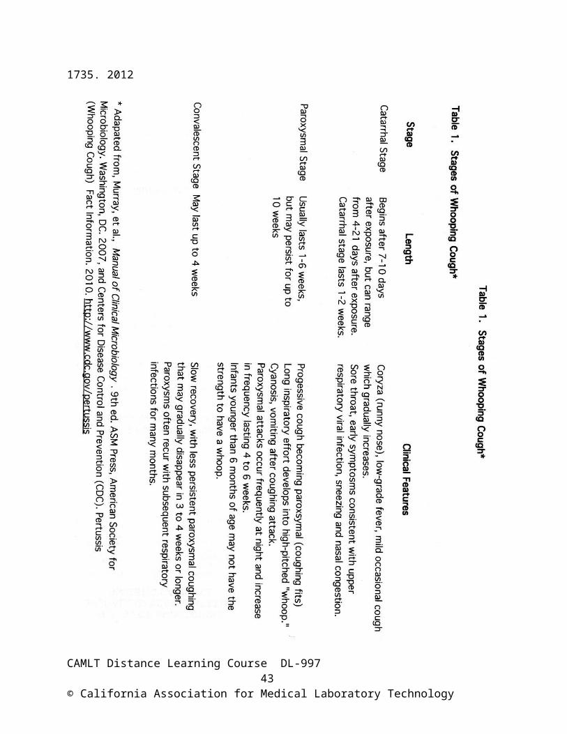
4. I will use what I learned from this Distance Learning course.5 4 3 2 1
5. The time to complete this Distance Learning course was: __________ hours. Please comment on this Distance Learning course on the back of this sheet. What did you like or dislik
CAMLT Distance Learning Course DL-99743
© California Association for Medical Laboratory Technology