California CABG Outcomes Reporting Program … California CABG Outcomes Reporting Program (CCORP) is...
76
California CABG Outcomes Reporting Program (CCORP) Data Abstractor Training Handbook Version 2.0
Transcript of California CABG Outcomes Reporting Program … California CABG Outcomes Reporting Program (CCORP) is...
California CABG Outcomes Reporting Program (CCORP)
Data Abstractor Training Handbook
Version 2.0


Presenter: Anthony Steimle, MD Kaiser - Santa Clara
CCORP Program Staff Office of Statewide Health Planning and Development
Healthcare Quality and Analysis Division 818 K Street, Suite 200 Sacramento CA, 95814
Joseph Parker, Ph.D. Holly Hoegh, Ph.D. Director, Healthcare Outcomes Center Manager, Clinical Data Programs Phone: (916) 322-9298 Phone: (916) 323-2026 Fax: (916) 445-7534 Fax: (916) 445-7534 [email protected] [email protected]
Hilva Chan Program Manager, CCORP Phone: (916) 322-9137 Fax: (916) 445-7534 [email protected] Herbert Jew Niya Fong Statistician, CCORP Program Support, CCORP Phone: (916) 322-9717 Phone: (916) 322-6375 Fax: (916) 445-7534 Fax: (916) 445-7534 [email protected] [email protected]


Table of Contents
Section One: Background CCORP ........................................................................9 Section Two: Data Elements and Definitions
By Export Order.................................................................................15 By Classification ................................................................................19 By Definition ......................................................................................21
Section Three: Quizzes Quiz 1 ................................................................................................41 Quiz 2 ................................................................................................43 Quiz 3 ................................................................................................47 Section Four: Patient Vignettes .............................................................................51 Section Five: Patient Vignettes Answers ...............................................................67


Section One:
Background on the California CABG Outcomes
Reporting Program (CCORP)


Background: CCORP _________________________________________________________________________________
What is the Office of Statewide Health Planning and Development? The Office of Statewide Health Planning and Development (OSHPD) is a department of the California Health and Human Services Agency. OSHPD is responsible for providing timely, accurate and actionable information on California’s health care system. Since 1996, OSHPD has been collaborating with the Pacific Business Group on Health (PBGH) to report risk-adjusted mortality for Californians undergoing CABG surgery. In October of 2001, Gov. Davis signed Senate Bill (SB) 680 (Figueroa) into law. SB 680 established a mandatory CABG reporting program for hospitals requiring OSHPD to report mortality rates for individual surgeons as well as hospitals. Both the California Medical Association and the American College of Cardiology were sponsors of this important legislation. The California CABG Outcomes Reporting Program (CCORP) is currently collecting and reporting CABG operative mortality data for all California hospitals and surgeons that perform the CABG procedure. Other features of CCORP include the clinical advisory panel who advise OSHPD on risk adjustment, the audit program which ensures that data submitted by hospitals are complete and accurate, and a panel review allowing surgeons to challenge their ratings. The program produces uniform hospital and surgeon-level mortality data, adjusted to account for differences across hospitals in the mix of patients undergoing isolated CABG procedures. CCORP provides comparative information to:
• Hospitals and surgeons, to stimulate and facilitate quality improvements at individual institutions;
• Purchasers of Care, to assess provider performance and make quality-based purchasing decisions;
• Consumers, to make informed treatment decisions. CCORP collects pre-operative risk factors (e.g., status of procedure, age, left ventricular ejection fraction), process of care or operative factors (e.g., IMA used as grafts), and in-hospital surgical mortality associated with a CABG surgery. We analyze the data and report on risk-adjusted mortality rates at the hospital level annually and the surgeon level biannually. To facilitate data collection, CCORP incorporates many data elements drawn from the National Society of Thoracic Surgeons (STS) cardiac reporting system. In addition, CCORP collects some data elements that STS does not collect (e.g., hepatic failure) or that are modified in important ways from STS definitions (e.g., Left Main Disease -% stenosis). Provided that data are submitted according to the format and valid values specified for each data element by OSHPD, hospitals may use approved STS vendor software, the CCORP tool, or an in-house system for data collection and submission. For hospitals that request it, OSHPD will supply, free of charge, the CCORP data collection tool (data entry software).
_________________________________________________________________________________ California CABG Outcomes Reporting Program 9

Background: CCORP _________________________________________________________________________________
Background and Development OSHPD strives to ensure that CABG data reporting is clinically and statistically sound and administratively feasible for hospitals. OSHPD’s CCORP team includes biostatisticians, programmers, and a consulting clinician with expertise in cardiology and health services research. We have reviewed the strengths and weaknesses of other CABG reporting systems and drawn on the expertise of CCORP’s Clinical Advisory Panel (CAP) to improve on them. The CAP consists of cardiac surgeons, cardiologists and researchers with expertise in quality of care measurement and risk adjustment methods. Before instituting CCORP, OSHPD staff reviewed the successes and problems experienced by other major CABG outcome reporting projects. We talked extensively with the research teams that produced the New York, Pennsylvania, and New Jersey programs, reviewed the documentation published by each project and conducted our own review of published articles. We also examined the risk models used by the National Cardiac Surgery Database maintained by the Society of Thoracic Surgeons (STS), The Veteran’s Administration, and the Northern New England Program. Our staff continues to review and examine other states practices leading us to some of the revisions in this version of the training manual (v 2.0). Importance of the STS System Why didn’t OSHPD just use the STS system? The STS system is proprietary, relatively expensive, collects more information than needed (it also collects operative and post-operative information, while CCORP focuses only on pre-operative variables) and, though widely used, it is a voluntary system at the individual surgeon (rather than hospital) level. However, CCORP recognizes that many hospitals (nearly 60% of California heart surgery hospitals) and surgeons already use the STS system. Therefore, we have maintained the STS format and data values for many of the data elements collected in CCORP. Staff has consulted closely with the STS National Database Committee and its chairman, who sits on the CCORP Clinical Advisory Panel. If your hospital already uses the STS system, you can still benefit from CCORP’s training and auditing programs, which are not part of the STS program. Since the inception of the voluntary California CABG Mortality Reporting Program (CCMRP), OSHPD has also been furnishing hospitals, free of charge, data collection software restricted to the data elements that program collects. For CCORP, we are continuing that tradition and will provide a free CCORP tool to any hospital that requests it. In addition, staff has recognized that some hospitals have invested considerable resources in developing home-grown systems that fit their hospital’s needs. We are allowing submissions using these systems, subject to a number of rules on electronic data format and export order in the CCORP Format and File Specifications (v 2.0).
_________________________________________________________________________________ California CABG Outcomes Reporting Program 10
Background: CCORP _________________________________________________________________________________
Why is Training for Data Abstraction of Risk Factors Critical? One of the central concerns is risk adjustment. Risk adjustment is a technique CCORP employs to compensate for differences among patients that may affect their hospital outcomes. It is a way to level the playing field by accounting for illness, demographics, past operations, and other factors that patients bring to the operating room. Risk adjustment begins by identifying characteristics that are associated with short-term mortality and including them in a model to predict the outcome of interest. Risk adjustment methods are a critical component of internal quality improvement initiatives and performance measurement programs, like CCORP, that involve comparisons of different providers. By accounting for key differences among patients, risk adjustment allows comparisons of “apples with apples.” Failure to adequately adjust for patient risk produces comparisons that may be flawed and misleading. Risk adjustment figures rely heavily on efforts to track quality, either internally or across facilities, by establishing a valid baseline of comparison. What Is The Purpose Of Risk Adjustment In CCORP? Most hospitals and surgeons will have case mix characteristics that are different from the average characteristics of the state. If those differences are not accounted for through risk adjustment, the hospital or surgeon will have outcomes that are not directly comparable to other providers. In effect, an unadjusted outcome report may unfairly "punish" an entity by showing that its performance is worse than average when its poor performance may be due entirely to case mix differences. The goal of risk adjustment is to help CCORP determine whether a hospital or surgeon’s outcomes are significantly better or worse than the state average, pointing to the need for quality improvement in the latter situation. The process and its results allow consumers, health plans and providers to more fairly compare the outcomes of institutions and individuals. Why the Risk Model Must Perform Adequately The risk model is a mathematical formula that is used to compute a unique "expected" value (or predicted value) for the surgical outcome of interest. The expected values for each patient are then aggregated at the hospital and/or surgeon level and average expected values are calculated. For example, a hospital’s expected mortality rate is calculated and compared to its observed mortality rate in constructing an O/E ratio, which is used to determine whether a hospital performed better, worse than, or as expected. In hospital mortality has a unique risk model and its own set of 30+ risk factors, all of which are measured at the patient level. No risk model is perfect; however, better risk models make better predictions and allow for a more level playing field among providers, enabling fairer comparisons. The more consistent the abstraction of risk factors is across hospitals, the better CCORP’s models will perform and the more valid our outcome ratings will be.
_________________________________________________________________________________ California CABG Outcomes Reporting Program 11

Background: CCORP _________________________________________________________________________________
Underreporting and Overreporting of Data Elements When data is intentionally or unintentionally abstracted from the medical chart there can be a systematic bias towards recording more severe patient risk factors than what actually exists, underreporting or overreporting of data elements results, leading to an overestimation of patient risk. In the opposite case, underreporting or overreporting leads to an underestimation of patient risk. In the first case, the hospital or surgeon’s patient’s will appear more severely ill than they truly are and the entity will benefit unfairly because it’s expected mortality rate will be higher --they will be overcompensated for the case-mix of their patient in the risk-adjustment process. In the underreporting case, the hospital will be penalized because its expected mortality rate will not reflect the actual patient case-mix. Both situations are to be avoided and proper training will help to assure that consistent abstraction of data elements exists across hospitals. CCORP uses both internal data validation methods and an independent medical records audit review to ascertain whether underreporting or overreporting exists at institutions and the degree to which it may be a problem. We are concerned by both, because we know that incomplete abstraction of risk factors can easily be as large a problem as deliberate overreporting of risk factors, especially when hospital resources are constrained. When, through the audit process or other means, CCORP finds that a hospital has not coded its data in a manner consistent with the data definitions and clarifications provided, it may ask the facility to re-abstract some or all of its data, which is a costly and time-intensive activity. We encourage data abstractors report risk factors using the guidance provided in this training manual to: make efficient use of their limited resources, ensure adequate assessment of patient risk, and to enhance the fairness of the ultimate quality comparisons.
_________________________________________________________________________________ California CABG Outcomes Reporting Program 12

Section Two:
Data Elements and Definitions Effective with 2006 Discharges


Data Elements and Definitions ____________________________________________________________________________________________________
_________________________________________________________________________________
Data Element Overview: EXPORT ORDER (Effective 2006 Discharges)
Data Element Classification Origin
1. Medical Record Number Identification STS 2. Isolated CABG Identification Non-STS 3. Date of Surgery Identification STS 4. Date of Birth Identification STS 5. Patient Age Risk Factor: Demographic STS 6. Gender Risk Factor: Demographic STS 7. Race Risk Factor: Demographic STS 8. Date of Discharge Identification STS 9. Discharge Status Identification STS 10. Date of Death Identification STS 11. Responsible Surgeon Name
Identification STS (Modified)
12. Responsible Surgeon California License Number
Identification Non-STS
13. Height (cm) Risk Factor: Demographic STS 14. Weight (kg) Risk Factor: Demographic STS 15. Diabetes Risk Factor: Comorbidity/Other STS 16. Hypertension Risk Factor: Comorbidity/Other STS 17. Peripheral Vascular Disease
Risk Factor: Comorbidity/Other STS
18. Cerebrovascular Disease
Risk Factor: Comorbidity/Other STS
19. Cerebrovascular Accident
Risk Factor: Comorbidity/Other
STS
20. Cerebrovascular Accident Timing
Risk Factor: Comorbidity/Other
STS
21. Chronic Lung Disease Risk Factor: Comorbidity/Other STS 22. Immunosuppressive Treatment
Risk Factor: Comorbidity/Other
STS
23. Hepatic Failure Risk Factor: Comorbidity/Other Non-STS 24. Dialysis Risk Factor: Comorbidity/Other STS 25. Last Creatinine Level Preop (mg/dl)
Risk Factor: Comorbidity/Other
STS
26. Left Main Disease (% Stenosis)
Risk Factor: Hemodynamic Status
STS (Modified)
27. Number of Diseased Coronary Vessels
Risk Factor: Hemodynamic Status
STS
28. Mitral Insufficiency Risk Factor: Hemodynamic Status STS (Modified) 29. Ejection Fraction Done Risk Factor: Hemodynamic Status STS 30. Ejection Fraction (%) Risk Factor: Hemodynamic Status STS 31. Ejection Fraction Method
Risk Factor: Hemodynamic Status STS
California CABG Outcomes Reporting Program 15

Data Elements and Definitions ____________________________________________________________________________________________________
_________________________________________________________________________________
Data Element Overview: EXPORT ORDER (continued)
(Effective 2006 Discharges)
Data Element Classification Origin 32. Myocardial Infarction Risk Factor: Cardiac STS (Modified) 33. Myocardial Infarction Timing
Risk Factor: Cardiac STS
34. Arrhythmia Risk Factor: Cardiac STS 35. Arrhythmia Type Risk Factor: Cardiac STS 36. Cardiogenic Shock Risk Factor: Cardiac STS 37. Angina Risk Factor: Cardiac STS 38. Angina Type Risk Factor: Cardiac STS (Modified) 39. Congestive Heart Failure
Risk Factor: Cardiac
STS
40. NYHA Classification Risk Factor: Cardiac STS 41. Resuscitation Risk Factor: Cardiac STS 42. Incidence Risk Factor: Previous Intervention
STS
43. Previous Coronary Artery Bypass Graft (CABG)
Risk Factor: Previous Intervention
STS
44. Prior Percutaneous Coronary Intervention (PCI)
Risk Factor: Previous Intervention
STS
45. Interval from Prior PCI To Surgery
Risk Factor: Previous Intervention
STS
46. Status of Procedure Risk Factor: Operative
STS
47. CPB Utilization Process of Care
STS
48. CPB Utilization – Combination
Process of Care
STS
49. Cardioplegia Process of Care
STS
50. Internal Mammary Artery(ies) Used as Grafts
Process of Care
STS
51. Radial Artery Used Process of Care
STS
52. Reoperation for Bleed/Tamponade
Reoperative Complications STS
53. Reoperation for Graft Occlusion
Reoperative Complications STS
54. Deep Sternal Wound Infection
Reoperative Complications STS
California CABG Outcomes Reporting Program 16
Data Elements and Definitions ____________________________________________________________________________________________________
_________________________________________________________________________________
Data Element Overview: EXPORT ORDER (continued) (Effective 2006 Discharges)
Data Element Classification Origin
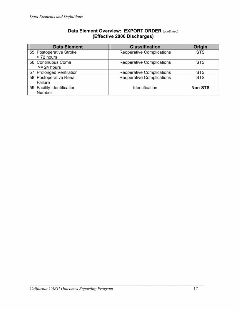
55. Postoperative Stroke > 72 hours
Reoperative Complications STS
56. Continuous Coma >= 24 hours
Reoperative Complications STS
57. Prolonged Ventilation Reoperative Complications STS 58. Postoperative Renal Failure
Reoperative Complications STS
59. Facility Identification Number
Identification Non-STS
California CABG Outcomes Reporting Program 17

Data Elements and Definitions ____________________________________________________________________________________________________
_________________________________________________________________________________
Data Element Overview: By Classification Effective with 2006 Discharges
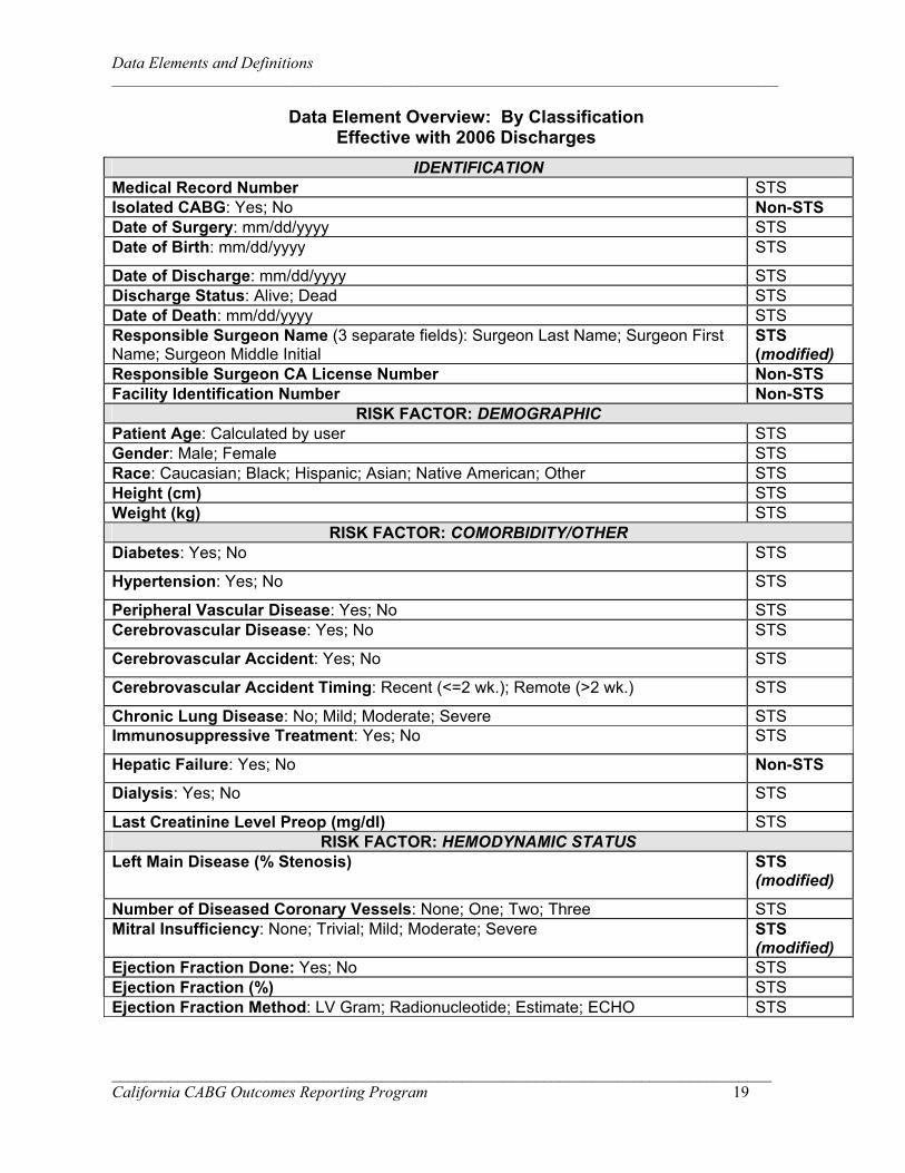
IDENTIFICATION Medical Record Number STS Isolated CABG: Yes; No Non-STS Date of Surgery: mm/dd/yyyy STS Date of Birth: mm/dd/yyyy STS
Date of Discharge: mm/dd/yyyy STS Discharge Status: Alive; Dead STS Date of Death: mm/dd/yyyy STS Responsible Surgeon Name (3 separate fields): Surgeon Last Name; Surgeon First Name; Surgeon Middle Initial
STS (modified)
Responsible Surgeon CA License Number Non-STS Facility Identification Number Non-STS
RISK FACTOR: DEMOGRAPHIC Patient Age: Calculated by user STS Gender: Male; Female STS Race: Caucasian; Black; Hispanic; Asian; Native American; Other STS Height (cm) STS Weight (kg) STS
RISK FACTOR: COMORBIDITY/OTHER Diabetes: Yes; No STS
Hypertension: Yes; No STS
Peripheral Vascular Disease: Yes; No STS Cerebrovascular Disease: Yes; No STS
Cerebrovascular Accident: Yes; No STS
Cerebrovascular Accident Timing: Recent (<=2 wk.); Remote (>2 wk.) STS
Chronic Lung Disease: No; Mild; Moderate; Severe STS Immunosuppressive Treatment: Yes; No STS
Hepatic Failure: Yes; No Non-STS
Dialysis: Yes; No STS
Last Creatinine Level Preop (mg/dl) STS RISK FACTOR: HEMODYNAMIC STATUS
Left Main Disease (% Stenosis) STS (modified)
Number of Diseased Coronary Vessels: None; One; Two; Three STS Mitral Insufficiency: None; Trivial; Mild; Moderate; Severe STS
(modified) Ejection Fraction Done: Yes; No STS Ejection Fraction (%) STS Ejection Fraction Method: LV Gram; Radionucleotide; Estimate; ECHO STS
California CABG Outcomes Reporting Program 19
Data Elements and Definitions ____________________________________________________________________________________________________
_________________________________________________________________________________ California CABG Outcomes Reporting Program 20
Data Element Overview: By Classification Effective with 2006 Discharges
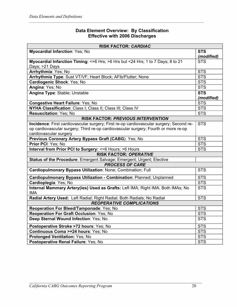
RISK FACTOR: CARDIAC
Myocardial Infarction: Yes; No STS (modified)
Myocardial Infarction Timing: <=6 Hrs; >6 Hrs but <24 Hrs; 1 to 7 Days; 8 to 21 Days; >21 Days
STS
Arrhythmia: Yes; No STS Arrhythmia Type: Sust VT/VF; Heart Block; AFib/Flutter; None STS Cardiogenic Shock: Yes; No STS Angina: Yes; No STS Angina Type: Stable; Unstable STS
(modified) Congestive Heart Failure: Yes; No STS NYHA Classification: Class I; Class II; Class III; Class IV STS Resuscitation: Yes; No STS
RISK FACTOR: PREVIOUS INTERVENTION Incidence: First cardiovascular surgery; First re-op cardiovascular surgery; Second re-op cardiovascular surgery; Third re-op cardiovascular surgery; Fourth or more re-op cardiovascular surgery
STS
Previous Coronary Artery Bypass Graft (CABG): Yes; No STS Prior PCI: Yes; No STS Interval from Prior PCI to Surgery: <=6 Hours; >6 Hours STS
RISK FACTOR: OPERATIVE Status of the Procedure: Emergent Salvage; Emergent; Urgent; Elective
PROCESS OF CARE Cardiopulmonary Bypass Utilization: None; Combination; Full STS
Cardiopulmonary Bypass Utilization - Combination: Planned; Unplanned STS Cardioplegia: Yes; No STS Internal Mammary Artery(ies) Used as Grafts: Left IMA; Right IMA; Both IMAs; No IMA
STS
Radial Artery Used: Left Radial; Right Radial; Both Radials; No Radial STS REOPERATIVE COMPLICATIONS
Reoperation For Bleed/Tamponade: Yes; No STS Reoperation For Graft Occlusion: Yes; No STS Deep Sternal Wound Infection: Yes; No STS
Postoperative Stroke >72 hours: Yes; No STS Continuous Coma >=24 hours: Yes; No STS Prolonged Ventilation: Yes; No STS Postoperative Renal Failure: Yes; No STS

Data Elements and Definitions
__________________________________________________________________________________________________________________________________ California CABG Outcomes Reporting Program 21
Data Element Overview: Definitions
Effective with 2006 Discharges
Data Element and Definition Comments and Examples Origin 1. Medical Record Number: Patient medical record number at the hospital where surgery was performed.
STS
2. Isolated CABG: Yes; No. Answer 'No' if any if any of the procedures listed were performed during coronary artery bypass graft surgery (**Refer to page 36 at the end of this section for complete definition).
Non-STS
3. Date of Surgery: mm/dd/yyyy Patient date of surgery for the CABG procedure.
STS
4. Date of Birth: mm/dd/yyyy Patient date of birth.
STS
5. Patient Age (calculated by hospital/user): Patient age in years, at time of surgery. This should be calculated from the Date of Birth and the Date of Surgery, according to convention used in the USA (the number of birth date anniversaries reached by the date of surgery).
STS
6. Gender: Male; Female. Patient gender at birth. Gender must be present for Risk Model to activate.
STS
7. Race: Caucasian; Black; Hispanic; Asian; Native American; Other. Patient race or ethnicity as determined by the patient or family.
STS
8. Date of Discharge: mm/dd/yyyy Patient date of discharge. If the patient died in the hospital, the discharge date is the date of death.
STS
9. Discharge Status: Alive; Dead. Patient status upon discharge from the hospitalization in which surgery occurred.
It is not necessary to report operative mortalities. STS
10. Date of Death: mm/dd/yyyy Patient date of death.
STS
11. Responsible Surgeon Name (3 separate fields): Surgeon Last Name (Text Length 25); Surgeon First Name (Text Length 20); Surgeon Middle Initial (Text Length 1) The responsible surgeon is the surgeon as defined in Section 97170 (**Refer to page 37 at the end of this section for detailed information).
STS (modified)

Data Elements and Definitions
__________________________________________________________________________________________________________________________________ California CABG Outcomes Reporting Program 22
Data Element Overview: Definitions
Effective with 2006 Discharges
Data Element and Definition Comments and Examples Origin 12. Responsible Surgeon CA License Number: California physician license number of responsible surgeon, assigned by the Medical Board of California of the Department of Consumer Affairs.
Non-STS
13. Height: Height of the patient in centimeters. Valid Values are between 20.0 and 251.0 cm.
RISK FACTOR NOTE: “Pre-operative” is defined as everything prior to the induction of anesthesia.
STS
14. Weight: Weight of the patient in kilograms. Valid values are between 10.0 and 250.0 kg.
STS
15. Diabetes: Yes; No. The patient has a history of diabetes, regardless of duration of disease or need for anti-diabetic agents. Includes on admission or preoperative diagnosis. Does not include gestational diabetes.
Note that this is a very liberal definition of diabetes which includes type I (juvenile/insulin dependent), type II (adult-onset); or diabetic on oral medications, insulin or diet-controlled.
STS
16. Hypertension: Yes; No. The patient has a diagnosis of hypertension, documented by one of the following: a. Documented history of hypertension diagnosed and treated with medication,
diet and/or exercise b. Blood pressure > 140 systolic or > 90 diastolic on at least 2 occasions. c. Currently on antihypertensive medication.
Note that the definition does not count isolated elevations as hypertension.
STS
17. Peripheral Vascular Disease: Yes; No. The patient has a history at any time prior to surgery of Peripheral Vascular Disease, as indicated by claudication either with exertion or rest; amputation for arterial insufficiency; aorto-iliac occlusive disease reconstruction; peripheral vascular bypass surgery, angioplasty, or stent; documented abdominal aortic aneurysm (AAA), AAA repair, or stent; positive non-invasive testing documented. Does not include procedures such as vein stripping, carotid disease, or procedures, originating above the diaphragm. Excludes Cerebrovascular Disease.
For example, abstract peripheral vascular disease if the patient has a >50% stenosis of the aortic, iliac, femoral or popliteal vessels; a history of surgery or angioplasty for such a stenosis; a history of an aortic aneurysm; classic claudication.
STS
Data Elements and Definitions
__________________________________________________________________________________________________________________________________ California CABG Outcomes Reporting Program 23
Data Element Overview: Definitions
Effective with 2006 Discharges
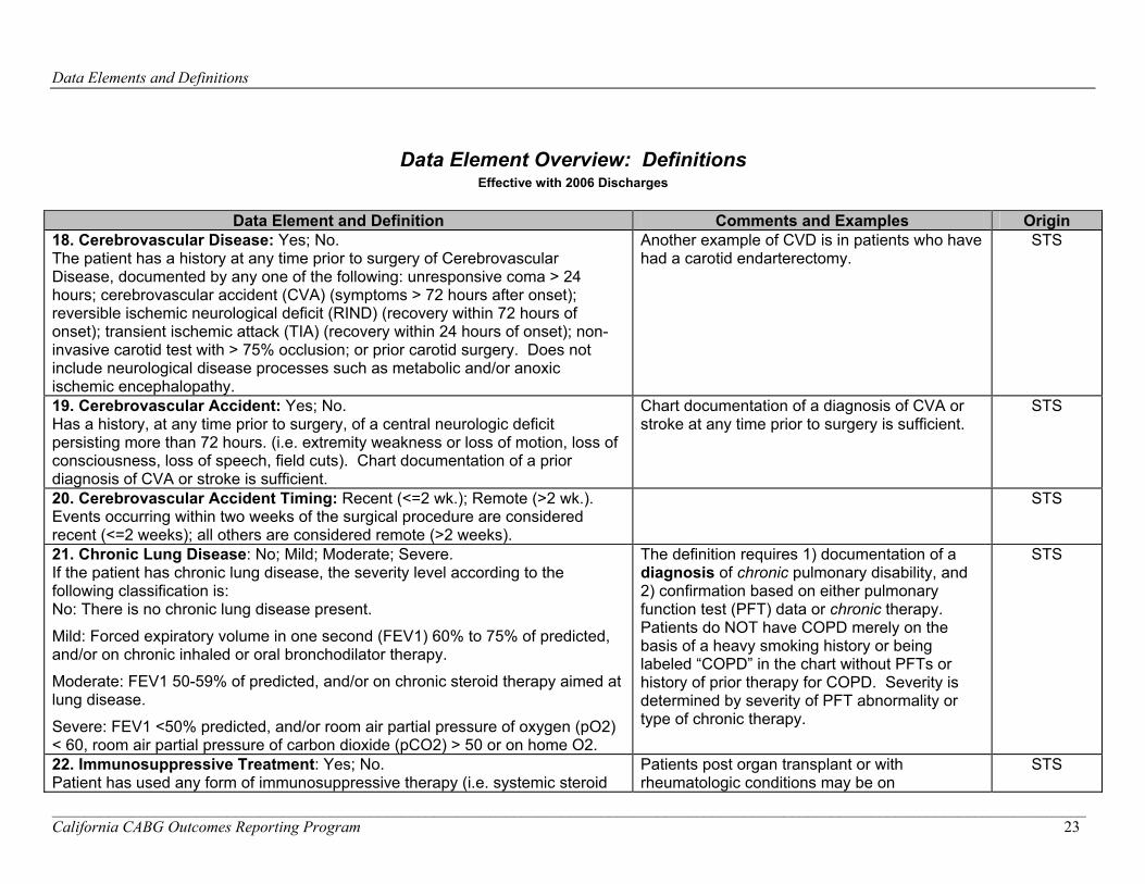
Data Element and Definition Comments and Examples Origin 18. Cerebrovascular Disease: Yes; No. The patient has a history at any time prior to surgery of Cerebrovascular Disease, documented by any one of the following: unresponsive coma > 24 hours; cerebrovascular accident (CVA) (symptoms > 72 hours after onset); reversible ischemic neurological deficit (RIND) (recovery within 72 hours of onset); transient ischemic attack (TIA) (recovery within 24 hours of onset); non-invasive carotid test with > 75% occlusion; or prior carotid surgery. Does not include neurological disease processes such as metabolic and/or anoxic ischemic encephalopathy.
Another example of CVD is in patients who have had a carotid endarterectomy.
STS
19. Cerebrovascular Accident: Yes; No. Has a history, at any time prior to surgery, of a central neurologic deficit persisting more than 72 hours. (i.e. extremity weakness or loss of motion, loss of consciousness, loss of speech, field cuts). Chart documentation of a prior diagnosis of CVA or stroke is sufficient.
Chart documentation of a diagnosis of CVA or stroke at any time prior to surgery is sufficient.
STS
20. Cerebrovascular Accident Timing: Recent (<=2 wk.); Remote (>2 wk.). Events occurring within two weeks of the surgical procedure are considered recent (<=2 weeks); all others are considered remote (>2 weeks).
STS
21. Chronic Lung Disease: No; Mild; Moderate; Severe. If the patient has chronic lung disease, the severity level according to the following classification is: No: There is no chronic lung disease present.
Mild: Forced expiratory volume in one second (FEV1) 60% to 75% of predicted, and/or on chronic inhaled or oral bronchodilator therapy.
Moderate: FEV1 50-59% of predicted, and/or on chronic steroid therapy aimed at lung disease.
Severe: FEV1 <50% predicted, and/or room air partial pressure of oxygen (pO2) < 60, room air partial pressure of carbon dioxide (pCO2) > 50 or on home O2.
The definition requires 1) documentation of a diagnosis of chronic pulmonary disability, and 2) confirmation based on either pulmonary function test (PFT) data or chronic therapy. Patients do NOT have COPD merely on the basis of a heavy smoking history or being labeled “COPD” in the chart without PFTs or history of prior therapy for COPD. Severity is determined by severity of PFT abnormality or type of chronic therapy.
STS
22. Immunosuppressive Treatment: Yes; No. Patient has used any form of immunosuppressive therapy (i.e. systemic steroid
Patients post organ transplant or with rheumatologic conditions may be on
STS
Data Elements and Definitions
__________________________________________________________________________________________________________________________________ California CABG Outcomes Reporting Program 24
Data Element Overview: Definitions
Effective with 2006 Discharges
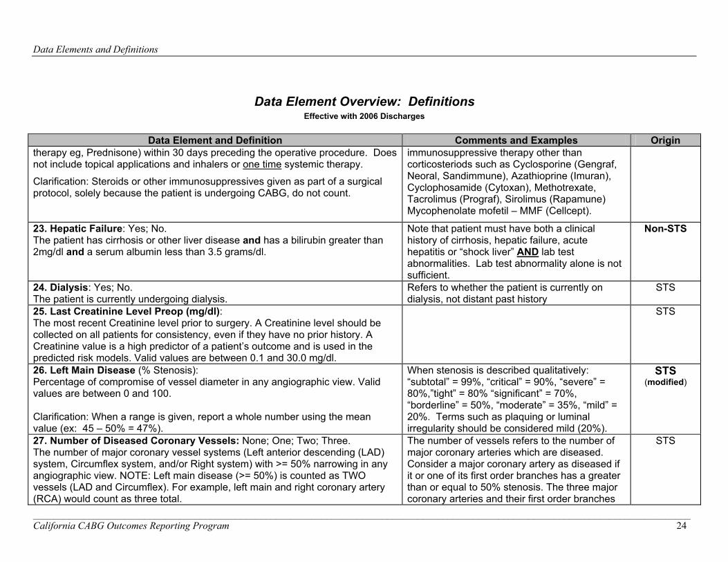
Data Element and Definition Comments and Examples Origin therapy eg, Prednisone) within 30 days preceding the operative procedure. Does not include topical applications and inhalers or one time systemic therapy.
Clarification: Steroids or other immunosuppressives given as part of a surgical protocol, solely because the patient is undergoing CABG, do not count.
immunosuppressive therapy other than corticosteriods such as Cyclosporine (Gengraf, Neoral, Sandimmune), Azathioprine (Imuran), Cyclophosamide (Cytoxan), Methotrexate, Tacrolimus (Prograf), Sirolimus (Rapamune) Mycophenolate mofetil – MMF (Cellcept).
23. Hepatic Failure: Yes; No. The patient has cirrhosis or other liver disease and has a bilirubin greater than 2mg/dl and a serum albumin less than 3.5 grams/dl.
Note that patient must have both a clinical history of cirrhosis, hepatic failure, acute hepatitis or “shock liver” AND lab test abnormalities. Lab test abnormality alone is not sufficient.
Non-STS
24. Dialysis: Yes; No. The patient is currently undergoing dialysis.
Refers to whether the patient is currently on dialysis, not distant past history
STS
25. Last Creatinine Level Preop (mg/dl): The most recent Creatinine level prior to surgery. A Creatinine level should be collected on all patients for consistency, even if they have no prior history. A Creatinine value is a high predictor of a patient’s outcome and is used in the predicted risk models. Valid values are between 0.1 and 30.0 mg/dl.
STS
26. Left Main Disease (% Stenosis): Percentage of compromise of vessel diameter in any angiographic view. Valid values are between 0 and 100. Clarification: When a range is given, report a whole number using the mean value (ex: 45 – 50% = 47%).
When stenosis is described qualitatively: “subtotal” = 99%, “critical” = 90%, “severe” = 80%,”tight” = 80% “significant” = 70%, “borderline” = 50%, “moderate” = 35%, “mild” = 20%. Terms such as plaquing or luminal irregularity should be considered mild (20%).
STS (modified)
27. Number of Diseased Coronary Vessels: None; One; Two; Three. The number of major coronary vessel systems (Left anterior descending (LAD) system, Circumflex system, and/or Right system) with >= 50% narrowing in any angiographic view. NOTE: Left main disease (>= 50%) is counted as TWO vessels (LAD and Circumflex). For example, left main and right coronary artery (RCA) would count as three total.
The number of vessels refers to the number of major coronary arteries which are diseased. Consider a major coronary artery as diseased if it or one of its first order branches has a greater than or equal to 50% stenosis. The three major coronary arteries and their first order branches
STS

Data Elements and Definitions
__________________________________________________________________________________________________________________________________ California CABG Outcomes Reporting Program 25
Data Element Overview: Definitions
Effective with 2006 Discharges
Data Element and Definition Comments and Examples Origin Clarification: 1) DO NOT USE intra-op TEE’s, 2) can use pre-op TEE’s
are 1) the left anterior descending (LAD) with its branches the diagonals; 2) the circumflex (Cx) with its branches the obtuse marginals (OM’s) or circumflex marginals; and 3) the right coronary artery (RCA) with its branch the posterior descending artery (PDA). The STS now considers Left Main Disease to count as TWO vessels–-encompassing the LAD and Circumflex (see NOTE under definition column). As such, if the chart indicates that Left Main, LAD and Circumflex are all diseased, code the number of diseased vessels as TWO, so as not to double count the Left Main. When the posterior-descending artery (PDA) is supplied by the circumflex (i.e., when the circumflex instead of the right coronary artery is dominant), standard practice is to count the PDA (but NOT the non-dominant RCA) as a major vessel. Thus, a patient with stenosis of the LAD, an obtuse marginal branch off of the circumflex, and the PDA off of the circumflex would be coded as having 3 vessel disease. NOTE: the number of major arteries which are counted as diseased may differ from the number of bypass grafts placed (e.g., a graft may be placed to a vessel with < 50% stenosis or two grafts to the LAD and diagonal even though both are part of a single major vessel).
28. Mitral Insufficiency: None; Trivial; Mild; Moderate; Severe. The evidence of mitral valve regurgitation and the severity level.
If a range of MR is given, enter the higher value (e.g. for “2 (mild) to 3 (moderate)” enter “3” or
STS (modified)
Data Elements and Definitions
__________________________________________________________________________________________________________________________________ California CABG Outcomes Reporting Program 26
Data Element Overview: Definitions
Effective with 2006 Discharges
Data Element and Definition Comments and Examples Origin moderate). Since operative conditions may artifactually alter ejection fraction and mitral regurgitation, readings from preoperative trans-thoracic echocardiograms are generally more accurate than those from trans-esophageal echocardiograms (TEE’s) done during surgery.
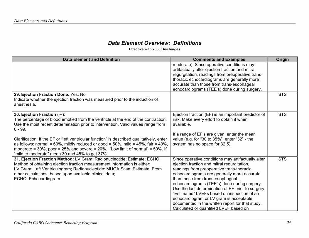
29. Ejection Fraction Done: Yes; No Indicate whether the ejection fraction was measured prior to the induction of anesthesia.
STS
30. Ejection Fraction (%): The percentage of blood emptied from the ventricle at the end of the contraction. Use the most recent determination prior to intervention. Valid values range from 0 - 99. Clarification: If the EF or “left ventricular function” is described qualitatively, enter as follows: normal = 60%, mildly reduced or good = 50%, mild = 45%, fair = 40%, moderate = 30%, poor = 25% and severe = 20%. “Low limit of normal” = 50%. If “mild to moderate” mean 30 and 45% to get 37%.
Ejection fraction (EF) is an important predictor of risk. Make every effort to obtain it when available. If a range of EF’s are given, enter the mean value (e.g. for “30 to 35%”, enter “32” - the system has no space for 32.5).
STS
31. Ejection Fraction Method: LV Gram; Radionucleotide; Estimate; ECHO. Method of obtaining ejection fraction measurement information is either: LV Gram: Left Ventriculogram; Radionucleotide: MUGA Scan; Estimate: From other calculations, based upon available clinical data; ECHO: Echocardiogram.
Since operative conditions may artifactually alter ejection fraction and mitral regurgitation, readings from preoperative trans-thoracic echocardiograms are generally more accurate than those from trans-esophageal echocardiograms (TEE’s) done during surgery. Use the last determination of EF prior to surgery. “Estimated” LVEFs based on inspection of an echocardiogram or LV gram is acceptable if documented in the written report for that study. Calculated or quantified LVEF based on
STS
Data Elements and Definitions
__________________________________________________________________________________________________________________________________ California CABG Outcomes Reporting Program 27
Data Element Overview: Definitions
Effective with 2006 Discharges
Data Element and Definition Comments and Examples Origin planimetry is not required. LVEFs which are guessed at based on clinical presentation (and not based on imaging of the ventricle) are not acceptable.
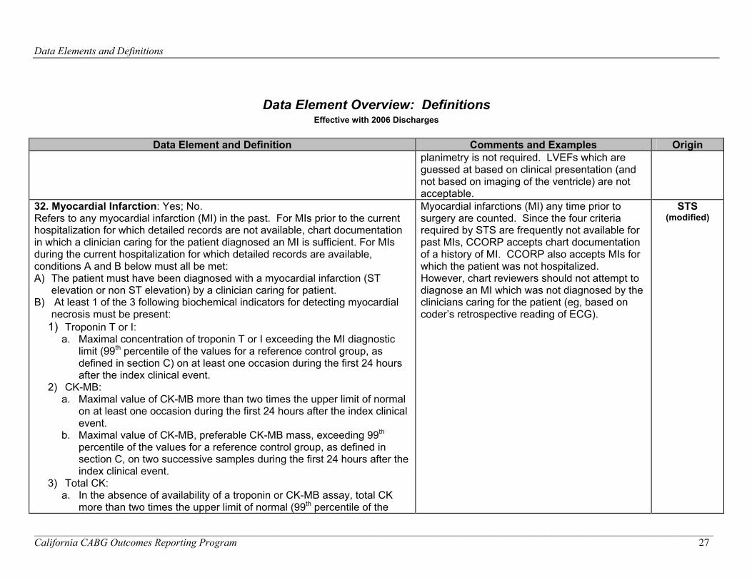
32. Myocardial Infarction: Yes; No. Refers to any myocardial infarction (MI) in the past. For MIs prior to the current hospitalization for which detailed records are not available, chart documentation in which a clinician caring for the patient diagnosed an MI is sufficient. For MIs during the current hospitalization for which detailed records are available, conditions A and B below must all be met: A) The patient must have been diagnosed with a myocardial infarction (ST
elevation or non ST elevation) by a clinician caring for patient. B) At least 1 of the 3 following biochemical indicators for detecting myocardial
necrosis must be present: 1) Troponin T or I:
a. Maximal concentration of troponin T or I exceeding the MI diagnostic limit (99th percentile of the values for a reference control group, as defined in section C) on at least one occasion during the first 24 hours after the index clinical event.
2) CK-MB: a. Maximal value of CK-MB more than two times the upper limit of normal
on at least one occasion during the first 24 hours after the index clinical event.
b. Maximal value of CK-MB, preferable CK-MB mass, exceeding 99th percentile of the values for a reference control group, as defined in section C, on two successive samples during the first 24 hours after the index clinical event.
3) Total CK: a. In the absence of availability of a troponin or CK-MB assay, total CK
more than two times the upper limit of normal (99th percentile of the
Myocardial infarctions (MI) any time prior to surgery are counted. Since the four criteria required by STS are frequently not available for past MIs, CCORP accepts chart documentation of a history of MI. CCORP also accepts MIs for which the patient was not hospitalized. However, chart reviewers should not attempt to diagnose an MI which was not diagnosed by the clinicians caring for the patient (eg, based on coder’s retrospective reading of ECG).
STS (modified)

Data Elements and Definitions
__________________________________________________________________________________________________________________________________ California CABG Outcomes Reporting Program 28
Data Element Overview: Definitions
Effective with 2006 Discharges
Data Element and Definition Comments and Examples Origin values for a reference control group, as defined in section C), or the B fraction of CK may be employed, but these last two biomarkers are considerably less satisfactory than CK-MB.
C) Reference control values (MI diagnostic limit and upper limit of normal): 1) Reference values must be determined in each laboratory by studies using
specific assays with appropriate quality control, as reported in peer-reviewed journals. Acceptable imprecision (coefficient of variation) at the 99th percentile for each assay should be defined as less than or equal to 10 percent. Each individual laboratory should confirm the range of reference values in their specific setting.
33. Myocardial Infarction Timing: <=6 Hrs; >6 Hrs but <24 Hrs; 1 to 7 Days; 8 to 21 Days; >21 Days. The time period between the last documented myocardial infarction and the CABG surgery.
STS
34. Arrhythmia: Yes; No. A preoperative arrhythmia (atrial fibrillation/flutter; third degree heart block; sustained ventricular tachycardia or ventricular fibrillation) that has been clinically documented or treated with any one of the following treatment modalities within two weeks prior to the CABG surgery: 1) Ablation therapy
2) AICD 3) Pacemaker 4) Pharmachological treatment 5) Electrocardioversion
STS
35. Arrhythmia Type: Sust VT/VF; Heart Block; Afib/Flutter; None. The type of arrhythmia present within two weeks prior to the procedure is: Sustained Ventricular Tachycardia or Ventricular Fibrillation requiring cardioversion and/or intravenous amiodarone; Third degree Heart Block; Atrial fibrillation/flutter requiring medication; None.
Sustained VT/VF is the arrhythmia of interest to CCORP. If the patient had both VT/VF and another type of arrhythmia, please choose VT/VF. Note: VT/VF must have occurred within two weeks of surgery. “Sustained” means > 30 seconds or requires electrical cardioversion.
STS

Data Elements and Definitions
__________________________________________________________________________________________________________________________________ California CABG Outcomes Reporting Program 29
Data Element Overview: Definitions
Effective with 2006 Discharges
Data Element and Definition Comments and Examples Origin Clarification: If more than one arrhythmia present, code with following priority: VT/VT, then Afib/flutter, then heart block.
Ventricular arrhythmia does NOT refer to frequent PVC’s (premature ventricular beats), bigeminy, or non-sustained ventricular tachycardia. Use of intravenous lidocaine for Sust VT/VF also qualifies.
36. Cardiogenic Shock: Yes; No
The patient, at the time,of procedure, is in a clinical state of hypoperfusion according to either of the following criteria:
1. Systolic blood pressure (BP) < 80 and/or Cardiac Index (CI) < 1.8 despite maximal treatment.
2. Intravenous inotropes and/or intra-aortic balloon pump (IABP) necessary to maintain Systolic BP > 80 and/or CI > 1.8.
Patient either 1) currently has SBP <=80 mmHg and/or CI <= 1.8, or 2) previously the SBP and/or CI met these criteria but now the patient is on inotropes or IABP.
STS
37. Angina: Yes; No, The patient has ever had angina pectoris. STS
38. Angina Type: Stable; Unstable. The type of angina present within 24 hours prior to CABG surgery is:
• Stable: Angina not meeting unstable criteria below that is controlled by oral or transcutaneous medication.
• Unstable: Requires continuous hospitalization from the episode until surgery and one of the following: 1) Angina at rest. 2) New onset angina in past 2 months** of at least Canadian Cardiovascular
Society (CCS) Class III. **Increasing angina in past 2 months - angina that has become more frequent, longer in duration, or lower in threshold; and increased by greater than or equal to one CCS class to at least CCS Class III severity.
Patients presenting with angina at rest who are subsequently diagnosed with a MI would have angina=Yes, type=unstable, CCS=class IV, MI=Yes.
STS (modified)
39. Congestive Heart Failure: Yes; No.
Whether, within 2 weeks prior to the initial surgical procedure, a physician has
The previous STS definition of CHF did not clearly specify whether a past history of heart
STS

Data Elements and Definitions
__________________________________________________________________________________________________________________________________ California CABG Outcomes Reporting Program 30
Data Element Overview: Definitions
Effective with 2006 Discharges
Data Element and Definition Comments and Examples Origin diagnosed that the patient is currently in congestive heart failure (CHF). Can be diagnosed based on careful history and physical exam, or by one of the following criteria: 1) Paroxysmal nocturnal dyspnea (PND; 2) Dyspnea on exertion (DOE) due to heart failure; 3) Chest X-Ray (CXR) showing pulmonary congestion ; 4) Pedal edema or dyspnea and receiving diuretics or digoxin.
Note: Severity is measured by NYHA Class within last two weeks
failure qualified as CHF. Note that the current STS definition clearly specifies that CHF signs or symptoms must have occurred within 2 weeks prior to surgery to code a patient as CHF=Yes. Since evidence of recent CHF symptoms is not always available in current medical record, CCORP accepts chart documentation that the patient was diagnosed with a CHF episode within the two weeks prior to surgery.
40. NYHA Classification: Class I, Class II, Class III, Class IV. The New York Heart Association Classification represents the overall functional status of the patient in relationship to both congestive heart failure and angina. Abstract the highest level leading to the episode of hospitalization and/or procedure.
1. Class I: Patients with cardiac disease but without resulting limitation of physical activity. Ordinary physical activity does not cause undue fatigue, palpitation, dyspnea or anginal pain.
2. Class II: Patients with cardiac disease resulting in slight limitation of physical activity. They are comfortable at rest. Ordinary physical activity results in fatigue, palpitations, dyspnea or anginal pain.
3. Class III: Patients with cardiac disease resulting in marked limitation of physical activity. They are comfortable at rest. Less than ordinary physical activity results in fatigue, palpitations, dyspnea, or anginal pain.
4. Patients with cardiac disease resulting in inability to carry on any physical activity without discomfort. Symptoms of cardiac insufficiency or of the anginal syndrome may be present even at rest. I f any physical activity is undertaken, discomfort is increased.
STS
41. Resuscitation: Yes; No. Whether the patient required cardiopulmonary resuscitation within one hour before the start of the operative procedure.
STS

Data Elements and Definitions
__________________________________________________________________________________________________________________________________ California CABG Outcomes Reporting Program 31
Data Element Overview: Definitions
Effective with 2006 Discharges
Data Element and Definition Comments and Examples Origin 42. Incidence: First cardiovascular surgery; First re-op cardiovascular surgery; Second re-op cardiovascular surgery; Third re-op cardiovascular surgery; Fourth or more re-op cardiovascular surgery. Whether this is the patient’s: 1) First cardiovascular surgery; 2) First re-op cardiovascular surgery; 3) Second re-op cardiovascular surgery; 4) Third re-op cardiovascular surgery; 5) Fourth or more re-op cardiovascular surgery.
CV surgeries include: CABG, valve replacement/repair, intracardiac repairs (ASD, VSD), ventricular aneurysmectormy or surgery on the aortic arch. Use of CPB is not required. CV surgeries do NOT include: PCI’s and non-cardiac vascular surgeries such as abdominal aortic aneurism repairs or fem-pop bypasses, percutaneous aortic stent grafts, percutaneous valves or pacemaker/ICD implantations.
STS
43. Previous Coronary Artery Bypass Graft (CABG): Yes; No. Whether the patient had a previous coronary artery bypass graft prior to the current admission.
STS
44. Prior PCI: Yes; No. Whether a previous Percutaneous coronary-intervention (PCI) was performed at any time prior to this surgical procedure. PCI refers to those treatment procedures that unblock narrowed coronary arteries without performing surgery. PCI may include, but is not limited to: balloon catheter angioplasty, percutaneous transluminal coronary angioplasty (PTCA), rotational atherectomy, directional atherectomy, extraction atherectomy, laser atherectomy and intracoronary stent placement.
Includes coronary stenting STS
45. Interval from prior PCI to Surgery: <=6 Hours; > 6 Hours. The interval of time between the previous PCI and the current surgical procedure is either : <=6 Hours; > 6 Hours
STS
46. Status of Procedure: Emergent Salvage; Emergent; Urgent; Elective. The status that best describes the clinical status of the patient at the time of surgery. Emergent Salvage: The patient is undergoing cardiopulmonary resuscitation en route to the operating room or prior to anesthesia induction. Clarification: If the cath was elective, the status is usually elective, even if the
Status refers to the patient’s condition immediately before surgery; it should not reflect instability which occurs after the induction of anesthesia or the operative risk but rather how expediently surgery must be performed. Thus some elective patients may be at higher risk than urgent patients; for example, an elderly
STS

Data Elements and Definitions
_________________________________________________________________________ ________________ California CABG Outcomes Reporting Program 32
Data Element Overview: Definitions
Effective with 2006 Discharges
_________________________________________
Data Element and Definition Comments and Examples Origin
patient was admitted for surgery after cath unless 1) clinical decompensation meeting definition of urgent (eg, unstable angina) or 2) left main >=80%. Emergent: The patient’s clinical status includes any of the following: (a) Ischemic dysfunction (any of the following): 1. Ongoing ischemia including rest angina despite maximal medical therapy (medical and/or intra-aortic balloon pump (IABP)); 2. Acute evolving myocardial infarction within 24 hours before surgery; or 3. Pulmonary edema requiring intubation. (b) Mechanical Dysfunction (either of the following): 1. Shock with circulatory; or 2. Shock without circulatory support. Urgent: All of the following conditions are met: (a) Not elective status (b) Not emergent status (c) Procedure required during same hospitalization in order to minimize chance of further clinical deterioration (d) Worsening, sudden chest pain; congestive heart failure (CHF); acute myocardial infarction (AMI); coronary anatomy; IABP; unstable angina (USA) with intravenous nitroglycerin; rest angina, valve dysfunction; or aortic dissection. Elective: The patient’s status has been stable in the days or weeks prior to the operation. The procedure could be deferred without increased risk of compromised cardiac outcome.
patient with an ejection fraction of 20% and COPD operated on electively compared to a young patient with a normal ejection fraction who has ongoing unstable angina. RULE OF THUMB: Elective – waits at home. Urgent – waits in hospital. Emergent – cannot wait or is not safe to wait. Emergent Salvage – no pulse. Elective surgeries are performed on patients whose cardiac function has been stable. They are usually scheduled at least one day prior to surgery, and the clinical picture allows discharge from the hospital with readmission for surgery later. Urgent surgeries are performed on patients whose medical condition requires continuous hospitalization prior to CABG. A critical feature that distinguishes urgent from elective patients is that urgent patients cannot be safely discharged prior to their CABG, but they can safely await ABG in the hospital. An intra-aortic balloon pump or IV nitroglycerin may be part of treatment. Emergent surgeries are performed on patients whose condition dictates that the surgery be performed within several hours to prevent morbidity or death. These cases should take precedence over an elective case, cause a new operating room to be opened, or be done at

Data Elements and Definitions
__________________________________________________________________________________________________________________________________ California CABG Outcomes Reporting Program 33
Data Element Overview: Definitions
Effective with 2006 Discharges
Data Element and Definition Comments and Examples Origin night or on a weekend if necessary. A critical feature which distinguishes emergent from urgent patients is that emergent patients cannot safely delay CABG even while they are in the hospital. Emergent cases are rare. Examples include CABG performed as primary revascularization during an acute MI, immediately (within minutes to a few hours) after angioplasty disaster, or while the patient is still in Cardiogenic shock. Salvage surgeries are performed on a patient undergoing CPR en route to operating room or in the operating room prior to induction of anesthesia. Patient is pulseless within hour prior to surgery.

Data Elements and Definitions
Data Element Overview: Definitions
Effective with 2006 Discharges
__________________________________________________________________________________________________________________________________ California CABG Outcomes Reporting Program 34
Data Element and Definition Comments and Examples Origin 47. CPB Utilization: None; Combination; Full. Indicate the level of CPB or coronary perfusion used during the procedure. 1) None: No CPB or coronary perfusion used during the procedure.
STS Clarification: Coronary perfusion methods are used as an alternative to complete heart and lung bypass. They are often referred to perfusion assisted devices where just the coronary artery that is being grafted is perfused (distal) to the anastomoses site (a method of supplying distal perfusion to isolated coronary arteries while new grafts are constructed). While not as invasive as cardiopulmonary bypass it is still a method of supporting the myocardium during a period of relative ischemia. These devices allow for continued myocardial perfusion to the area of myocardium that is being revascularized, therefore reducing any ischemic time to that region.
2) Combination: With or without CPB and/or with or without coronary perfusion at any time during the procedure: (a) At start of procedure: No CPB/No coronary perfusion > conversion to > CPB; (b) At start of procedure: No CPB/No coronary perfusion > conversion to > coronary perfusion; (c) At start of procedure: No CPB/No coronary perfusion > conversion to > coronary perfusion > conversion to > CPB 3) Full: CPB or coronary perfusion was used for the entire procedure.
48. CPB Utilization Combination: Planned; Unplanned. Whether the combination procedure was a planned or an unplanned conversion: 1) Planned: The surgeon intended to treat with any of the combination options described in “CPB Utilization” 2) Unplanned: The surgeon did not intend to treat with any of the combination options described in “CPB Utilization”
STS
49. Cardioplegia: Yes; No. Cardioplegia was used. STS 50. Internal Mammary Artery(ies) Used as Grafts: Left IMA; Right IMA; Both IMAs, No IMA.
Includes free graft (detached) IMAs.
STS
Indicate which internal mammary arter(ies) was/were used for grafts, if any: (a) Left IMA; (b) Right IMA; (c) Both IMAs; (d) No IMA. 51. Radial Artery Used: Left Radial; Right Radial; Both Radials, No Radial. Indicate which radial arter(ies) was/were used for grafts: (b) Left Radial artery; (b) Right Radial artery; (c) Both Radial arteries; (d) No
Radial artery.
STS

ons
__________________________________________________________________________________________________________________________________ California CABG Outcomes Reporting Program 35
Data Element Overview: Definitions
Effective with 2006 Discharges
Data Element and Definition Comments and Examples Origin 52. Reoperation for Bleed/Tamponade: Yes; No. Indicate whether an operative re-intervention was required for bleeding/tamponade.
Requires reopening the chest for bleeding.
STS
53. Reoperation for Graft Occlusion: Yes; No. Indicate whether an operative re-intervention was required for graft occlusion.
Does not include post-op PCIs. Requires reopening of the chest to revise a graft.
STS
54. Deep Sternal Wound Infection: Yes; No. Indicate whether patient had a deep sternal infection involving muscle, bone, and/or mediastinum REQUIRING OPEATIVE INTERVENTION. Must have ALL of the following conditions: 1) Wound opened with excision of tissue (I&D) or re-exploration of mediastinum; 2) Positive culture; 3) Treatment with antibiotics.
This is intended to be in-hospital infection, not a readmission for infection.
STS
STS 55. Postoperative Stroke > 72 Hours: Yes; No. Indicate whether a central neurologic deficit persisting postoperatively for more than 72 hours.
56. Continuous Coma >= 24 Hours: Yes; No. A new postoperative coma that persists for at least 24 hours secondary to anoxic/ischemic and/or metabolic encephalopathy, thromboembolic event or cerebral bleed.
Does not include medication induced coma. STS
57. Prolonged Ventilation: Yes; No. Pulmonary insufficiency requiring a ventilator. Includes (but not limited to) causes such as ARDS and pulmonary edema and/or any patient requiring mechanical ventilation for more than 24 hours postoperatively.
Postoperative period begins when patient leaves the O.R.
STS
58. Postoperative Renal Failure: Yes; No. Acute or worsening renal failure resulting in one or more of the following: 1) Increase of serum Creatinine to >2.0 and 2x most recent preoperative Creatinine level and/or 2) A new requirement of dialysis postoperatively.
STS
59. Facility Identification Number The six-digit facility identification number assigned to a hospital by the Office of Statewide Health Planning and Development, as defined in Section 97170.
Non-STS
Data Elements and Definiti
Data Elements and Definitions ____________________________________________________________________________________________________
______________________________________________________________________________
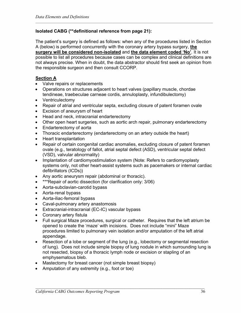
Isolated CABG (**definitional reference from page 21): The patient’s surgery is defined as follows: when any of the procedures listed in Section A (below) is performed concurrently with the coronary artery bypass surgery, the surgery will be considered non-isolated and the data element coded ‘No’. It is not possible to list all procedures because cases can be complex and clinical definitions are not always precise. When in doubt, the data abstractor should first seek an opinion from the responsible surgeon and then consult CCORP. Section A • Valve repairs or replacements • Operations on structures adjacent to heart valves (papillary muscle, chordae
tendineae, traebeculae carneae cordis, annuloplasty, infundibulectomy) • Ventriculectomy • Repair of atrial and ventricular septa, excluding closure of patent foramen ovale • Excision of aneurysm of heart • Head and neck, intracranial endarterectomy • Other open heart surgeries, such as aortic arch repair, pulmonary endarterectomy • Endarterectomy of aorta • Thoracic endarterectomy (endarterectomy on an artery outside the heart) • Heart transplantation • Repair of certain congenital cardiac anomalies, excluding closure of patent foramen
ovale (e.g., teratology of fallot, atrial septal defect (ASD), ventricular septal defect (VSD), valvular abnormality)
• Implantation of cardiomyostimulation system (Note: Refers to cardiomyoplasty systems only, not other heart-assist systems such as pacemakers or internal cardiac defibrillators (ICDs))
• Any aortic aneurysm repair (abdominal or thoracic). • ***Repair of aortic dissection (for clarification only: 3/06) • Aorta-subclavian-carotid bypass • Aorta-renal bypass • Aorta-iliac-femoral bypass • Caval-pulmonary artery anastomosis • Extracranial-intracranial (EC-IC) vascular bypass • Coronary artery fistula • Full surgical Maze procedures, surgical or catheter. Requires that the left atrium be
opened to create the ‘maze’ with incisions. Does not include “mini” Maze procedures limited to pulmonary vein isolation and/or amputation of the left atrial appendage.
• Resection of a lobe or segment of the lung (e.g., lobectomy or segmental resection of lung). Does not include simple biopsy of lung nodule in which surrounding lung is not resected, biopsy of a thoracic lymph node or excision or stapling of an emphysematous bleb.
• Mastectomy for breast cancer (not simple breast biopsy) • Amputation of any extremity (e.g., foot or toe)
California CABG Outcomes Reporting Program 36

Data Elements and Definitions ____________________________________________________________________________________________________
______________________________________________________________________________
If a procedure listed in Section B (next page) is performed concurrently with the coronary artery bypass surgery, the surgery will be considered an isolated CABG and the data element coded ‘Yes’ (unless a procedure listed in section A is performed during the same surgery). These particular procedures are listed because the Office has received frequent questions regarding their coding. Section B • Transmyocardial laser revascularization (TMR) • Pericardiectomy and excision of lesions of heart
• Repair/restoration of the heart or pericardium. ***Surgeries whose principal goal is full pericardial stripping for preoperatively identified constrictive pericarditis are non isolated (for clarification only: 3/06)
• Coronary endarterectomy • Pacemakers • Internal cardiac defibrillators (ICDs) • Fem-fem cardiopulmonary bypass (a form of cardiopulmonary bypass that should
not be confused with aortofemoral bypass surgery listed in Section A) • Thymectomy • Thyroidectomy NOTE: *** Based on the March 2006 training sessions, additional clarifications have been provided to Sections A & B to assist data abstractors. However, these will remain clarifications to the Isolated CABG definition until they are adopted officially through CCORP’s regulatory process. Responsible Surgeon Name (**definitional reference from page 21): “Responsible surgeon” means the principle surgeon who performs a coronary artery bypass procedure.
o The first and last name collected should exactly match the name assigned to the license number issued by the California Medical Board.
o The middle initial collected should match the first letter of the middle name assigned to the license number issued by the California Medical Board. Example: if a surgeon’s middle name is Harry, the middle initial should be reported as ‘H’. NOTE: do not include period (.).
o If a trainee performs this procedure, then the responsible surgeon is the physician responsible for supervising this procedure performed by the trainee. In situations in which a responsible surgeon cannot otherwise be determined, the responsible surgeon is the surgeon who bills for the coronary artery bypass procedure
California CABG Outcomes Reporting Program 37


Section Three:
Quizzes


Quizzes _________________________________________________________________________________
Quiz 1
1. A patient has a history of an asymptomatic 75% carotid stenosis of the right internal carotid artery. How would you code this patient? a) Cerebrovascular disease? Yes No b) Cerebrovascular accident? Yes No c) Peripheral vascular disease? Yes No
2. A 69 year-old male presented with unstable angina. He ruled out for a myocardial
infarction. While on telemetry, he had frequent PVC’s (> 10/hour) with rare couplets which were asymptomatic and which were treated with IV lidocaine. He had no Afib or heart block. How would you code this patient? a) Arrhythmia? Yes No b) Arrhythmia type? Sustained VT/VF Heart Block Afib/flutter
3. A 61 year-old male undergoes CABG. The coronary angiography report gives the
following result: a 60% stenosis of the left main, a 75% stenosis of the left anterior descending, an 85% stenosis of the first diagonal, a 90% stenosis of the second diagonal, plaquing of the circumflex, a 30% stenosis of the right coronary artery, and a 75% stenosis of the posterior descending artery (PDA). How would you code this patient? a) Number of diseased vessels? None One Two Three b) Left main disease _______%
4. 72 year-old male underwent CABG. The admitting note reports a history of
claudication. How would you code this patient? a) Peripheral vascular disease? Yes No
5. The left ventriculogram was read as showing an ejection fraction of 50-60% with
mild mitral regurgitation while the preoperative echocardiogram reported an ejection fraction of 62% with mild to moderate mitral regurgitation. How would you code this patient? a) Ejection fraction ________% b) Ejection fraction method? LV gram ECHO Radionucleotide Estimate c) Mitral regurgitation? None Trivial Mild Moderate Severe
______________________________________________________________________________ California CABG Outcomes Reporting Program 41

Quizzes _________________________________________________________________________________
______________________________________________________________________________
6. The preoperative left ventriculogram was read as “ejection fraction severely
reduced” with “2+” mitral regurgitation. How would you code the patient? a) Ejection fraction? ___ % b) Ejection fraction method? LV gram ECHO Radionucleotide Estimate c) Mitral regurgitation? None Trivial Mild Moderate Severe
7. A 71 year female undergoes CABG 2 days after a failed PTCA. She is not felt to
be stable enough to leave the hospital between the PTCA and CABG. She has a history of mitral valve replacement 20 years earlier for rheumatic heart disease. How would you code this patient? a) Previous CABG? Yes No b) Incidence? 1st CV surgery 1st reop CV surgery 2nd reop
3rd reop 4th reop c) Status? Elective Urgent Emergent Emergent/Salvage
8. The admitting history and physical notes a history of adult onset diabetes. The
patient is not on insulin or oral hypoglycemic agent. How would you code this patient? a) Diabetes? Yes No
9. The coronary angiography report gives the following result: 50% stenosis of the
left main, an 80% stenosis of the left anterior descending (LAD), a 70% stenosis of the first diagonal, a subtotal occlusion of the circumflex, and luminal irregularities of the right coronary artery (RCA). At surgery, the patient has four coronary bypass grafts placed to the distal LAD, the first diagonal, the second obtuse marginal, and the right coronary artery. How would you code this patient? a) Number of diseased vessels? None One Two Three b) Left main disease ________ %
10. The chart documents a history of an abdominal aortic aneurysm repair in the
past. How would you code this patient? a) Peripheral vascular disease? Yes No
California CABG Outcomes Reporting Program 42

Quizzes _________________________________________________________________________________
______________________________________________________________________________
Quiz 2 1. A healthy 71 year-old female was admitted with 30 minutes of chest pain at rest
which resolved after a single sublingual nitroglycerin in the ER. She was admitted to a telemetry bed, treated with topical nitroglycerin, aspirin, and IV heparin, and ruled out for myocardial infarction. On the second hospital day, the patient underwent a treadmill stress test which was positive. The next day, coronary angiography revealed a normal ejection fraction and a normal left main, an 80% stenosis of the mid LAD, 70% stenosis of the circumflex, and a 70% stenosis of the right coronary artery without thrombus. She remained free of recurrent chest pain. On the fourth hospital day, she underwent bypass surgery. How would you code this patient? a) Status? Elective Urgent Emergent Emergent/Salvage b) Angina? Yes No c) Angina type? Unstable Stable
2. A 63 year-old male with a history of stable angina presented to the emergency
room with 30 minutes of chest pain at rest and ECG changes consistent with ischemia. Chest pain and ECG changes initially resolved with IV nitroglycerin and heparin and the patient was admitted to the ICU. The next day, cardiac catheterization revealed a normal ejection fraction and three vessel coronary artery disease. During the catheterization, the chest pain recurred for 20 minutes and intra-aortic balloon pump was placed. Subsequently, the patient ruled out for myocardial infarction and remained hemodynamically stable without further symptoms or ECG changes. He underwent CABG two days after the catheterization. How would you code this patient? a) Status? Elective Urgent Emergent Emergent/Salvage b) Angina? Yes No c) Angina type? Unstable Stable
California CABG Outcomes Reporting Program 43

Quizzes _________________________________________________________________________________
______________________________________________________________________________
3. A 61 year-old male presented to the emergency room with three hours of
crushing chest pain at rest and ST segment elevation on electrocardiogram consistent with an anterior myocardial infarction. His blood pressure was 75/40 with a heart rate of 115 and he was in heart failure by exam. He was taken directly to cardiac catheterization, which revealed an ejection fraction of 35% and an occluded proximal LAD coronary artery with significant disease of the left main and right coronary arteries. Intravenous dopamine was begun, a balloon pump was placed, and his blood pressure improved to 100/60. He was taken directly from the cath lab to the operating room where he underwent CABG. How would you code this patient?
a) Status? Elective Urgent Emergent Emergent/Salvage b) Angina? Yes No c) Angina type? Unstable Stable d) Cardiogenic shock Yes No
4. A 55 year old female undergoing an elective angioplasty had acute closure of her
LAD resulting in a cardiac arrest. She was twice resuscitated with return of a blood pressure but went directly to the operating room for CABG while still receiving chest compressions and with an only intermittently palpable pulse. How would you code this patient? a) Status? Elective Urgent Emergent Emergent/Salvage
5a. A 73 year-old male was admitted with unstable angina and treated with IV
nitroglycerin and IV heparin. Cardiac catheterization revealed an ejection fraction of 45%, normal cardiac hemodynamics and three vessel disease. While still in the hospital the day following the catheterization, the patient had recurrent chest pain at rest that was relieved but then recurred after each of three sublingual nitroglycerins. Despite increasing doses of IV nitroglycerin, the chest pain associated with ischemic ECG changes persisted until he was taken to bypass surgery several hours later. How would you code this patient? a) Status? Elective Urgent Emergent Emergent/Salvage
5b. A 73 year-old male was admitted with unstable angina and treated with IV
nitroglycerin and IV heparin. Cardiac catheterization revealed an ejection fraction of 45%, normal cardiac hemodynamics, and three vessel disease. While still in the hospital the day following the catheterization, the patient had recurrent chest pain at rest that was not relieved despite increasing doses of IV nitroglycerin. Chest pain finally resolved with placement of an IABP. He remained stable and was taken to bypass surgery the next day. How would you code this patient? a) Status? Elective Urgent Emergent Emergent/Salvage
California CABG Outcomes Reporting Program 44

Quizzes _________________________________________________________________________________
______________________________________________________________________________
5c. A 52 year-old female presented to the ER with chest pain anterior ST elevation consistent with a myocardial infarction. She was taken immediately to cardiac catheterization for planned primary angioplasty; however, catheterization revealed anterior akinesis with ejection fraction of 35%, an 80% left main stenosis, a 100% LAD stenosis, an 80% circumflex stenosis, and a normal RCA. She was taken to CABG 2 hrs later. How would you code this patient? a) Status? Elective Urgent Emergent Emergent/Salvage b) Angina? Yes No c) Angina type? Unstable Stable d) Number of diseased vessels? None One Two Three e) Left main disease _________% f) MI? Yes No g) MI when? < 6 hr < 24 hrs 1-7days 8-21days >21days
5d. A 52 year-old female presented to the ER with chest pain anterior ST elevation
consistent with a myocardial infarction. She was taken immediately to cardiac catheterization for planned primary angioplasty; however, catheterization revealed anterior hypokinesis with ejection fraction of 40%, an 80% left main stenosis, a 95% LAD stenosis with an ulcerated plaque, an 80% circumflex stenosis, and a normal RCA. She was treated with ASA, heparin, integrelin, and beta-blockers and chest pain and ECG changes resolved. She was admitted to the ICU where she remained chest pain free and ruled in for a small myocardial infarction. She was taken to CABG 2 days later. How would you code this patient? a) Status? Elective Urgent Emergent Emergent/Salvage b) Angina? Yes No c) Angina type? Unstable Stable d) Number of diseased vessels? None One Two Three e) Left main disease __________% f) MI? Yes No g) MI when? < 6 hr < 24 hrs 1-7days 8-21days >21days
6. A 59 year male undergoes elective CABG. His medical record reports an
admission for “congestive heart failure” 2 years ago requiring IV diuretics. At the time of his surgery, however, he has not recently had symptoms of heart failure, been on any diuretics or vasodilators, or had exam or X-ray findings of heart failure (i.e., rales or chest X-ray with cardiomegaly or interstitial edema). How would you code this patient? a) Congestive heart failure? Yes No b) NYHA Class? I II III IV
California CABG Outcomes Reporting Program 45

Quizzes _________________________________________________________________________________
______________________________________________________________________________
7. A 70 year old female with a history of diabetes and hypertension undergoes
CABG. She has no angina. Prior to CABG, she has a history of congestive heart failure requiring admission to the hospital most recently 6 months ago. On her current medications, she has dyspnea with mowing the lawn but no symptoms dressing, cooing, or walking around the house. How would you code this patient?
a) Congestive heart failure? Yes No b) NYHA Class? I II III IV 8. A 77 year old female undergoes CABG. She has a history of congestive heart
failure. On her current regimen of daily lasix, digoxin, and an ACE inhibitor, she has dyspnea and occasional chest pressure with showering and dressing, trace ankle edema, and 2 pillow orthopnea. How would you code this patient? a) Congestive heart failure? Yes No b) NYHA Class? I II III IV
9. A 67 year-old female underwent elective CABG for progressive angina and three
vessel disease. The admitting history mentions a history of “chronic obstructive pulmonary disease” and a 70 pack year history tobacco use. However, the patient was not on metered dose inhalers, steroids, theophylline, or other pharmacotherapy for COPD and there is no documentation of FEV1. How would you code this patient? a) COPD? No Mild Moderate Severe
10. A 61 year-old male underwent CABG. The admitting history and physical states the patient is on atrovent and albuterol inhalers for the treatment of “chronic bronchitis.” There is no documented FEV1. How would you code this patient?
a) COPD? No Mild Moderate Severe
California CABG Outcomes Reporting Program 46
Quizzes _________________________________________________________________________________
______________________________________________________________________________
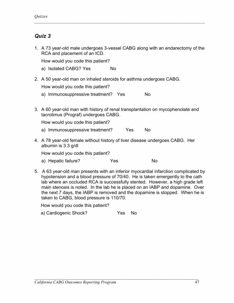
Quiz 3 1. A 73 year-old male undergoes 3-vessel CABG along with an endarectomy of the
RCA and placement of an ICD. How would you code this patient? a) Isolated CABG? Yes No
2. A 50 year-old man on inhaled steroids for asthma undergoes CABG. How would you code this patient?
a) Immunosuppressive treatment? Yes No 3. A 60 year-old man with history of renal transplantation on mycophenolate and
tacrolimus (Prograf) undergoes CABG. How would you code this patient?
a) Immunosuppressive treatment? Yes No 4. A 78 year-old female without history of liver disease undergoes CABG. Her
albumin is 3.3 g/dl How would you code this patient?
a) Hepatic failure? Yes No
5. A 63 year-old man presents with an inferior myocardial infarction complicated by hypotension and a blood pressure of 70/40. He is taken emergently to the cath lab where an occluded RCA is successfully stented. However, a high grade left main stenosis is noted. In the lab he is placed on an IABP and dopamine. Over the next 7 days, the IABP is removed and the dopamine is stopped. When he is taken to CABG, blood pressure is 110/70.
How would you code this patient? a) Cardiogenic Shock? Yes No
California CABG Outcomes Reporting Program 47

Quizzes ______________________________________________________________________________
______________________________________________________________________________
6. A 77 year old female undergoes CABG. Post-operatively she develops erythema of her sternotomy for which she receives vancomycin but does not require wound re-exploration. She is also noted to have a new left hemiparesis which does not fully resolve at the time of discharge. Preoperatively her creatinine is 1.5. During the postop period the creatinine increased to 3.4. At the time of discharge her creatinine is 2.4. How would you code this patient? a) Deep sternal wound infection? Yes No b) Postoperative stroke? Yes No c) Postoperative renal failure Yes No
California CABG Outcomes Reporting Program 48

Section Four:
Patient Vignettes


Patient Vignettes _____________________________________________________________________________
______________________________________________________________________________
Fifty-One Patient Vignettes
1. An otherwise healthy 65 year-old male presented to the emergency room with 30 minutes of chest pain at rest and ST segment depression on electrocardiogram. Chest pain and ECG changes resolved after a single sublingual nitroglycerin. He was admitted to a telemetry bed, treated with topical nitroglycerin, aspirin, IV heparin, and integrilin. He ruled out for myocardial infarction by serial enzymes. On the second hospital day, the IV heparin and integrilin were turned off and coronary angiography revealed a normal ejection fraction and a 60% stenosis of the left main, an 80% stenosis of the mid LAD, plaquing of the circumflex, and a 70% stenosis of the right coronary artery. He remained on the telemetry unit where he was free of recurrent chest pain or ECG changes on IV heparin and topical nitroglycerin. On the fourth hospital day he underwent bypass surgery. How would you code this patient? a) Status? Elective Urgent Emergent Emergent/Salvage b) Angina? Yes No c) Angina type? Unstable Stable d) NYHA class? I II III IV e) Number of diseased vessels? None One Two Three f) Left main disease? ______%
2. A 65 year-old male with a history of stable angina presented to the emergency
room with a 1 week history of severe intermittent precordial chest pressure occurring with very minimal exertion, never occurring at rest or lasting more than 5 minutes. His previous anginal pattern had been mild chest pressure with moderate to strenuous exertion about 2 times per week. He had a history of coronary artery disease, hypertension, and hypercholesterolemia. He was admitted to a telemetry bed, treated with topical nitroglycerin and IV heparin, and ruled out for myocardial infarction by serial enzymes. On the second hospital day coronary angiography revealed a normal ejection fraction, a 60% stenosis of the left main, a proximal 80% stenoses of the LAD, and plaquing of the circumflex and right coronary arteries. He remained free of recurrent chest pain while at bed rest on IV heparin and topical nitroglycerin. A day after the catheterization, he underwent bypass surgery. How would you code this patient? a) Status? Elective Urgent Emergent Emergent/Salvage b) Angina? Yes No c) Angina type? Unstable Stable d) NYHA class? I II III IV e) Number of diseased vessels? None One Two Three f) Left main disease? ________%
California CABG Outcomes Reporting Program 51

Patient Vignettes ______________________________________________________________________________
3. A 74 year-old female presented with 3 days intermittent retrosternal chest pain at rest radiating to the arm and associated with nausea. She had a history of coronary artery disease, coronary artery bypass graft surgery (CABG) 6 years earlier following a myocardial infarction, COPD on home oxygen, diabetes, and hypertension. Electrocardiogram showed old Q-waves inferiorly and ST segment depression anteriorly. Chest X-ray was consistent with heart failure. She was admitted to the ICU where she was treated with morphine and IV nitroglycerin and heparin. She ruled in for a subendocardial infarction. On the second day cardiac catheterization revealed an ejection fraction of 30-35%, severe native three vessel coronary disease, an occluded graft to the circumflex, a 95% stenosis of the vein graft to the LAD, and a patent graft to the right coronary. On the third day she had worsened shortness of breath, orthopnea, tachypnea, and renal insufficiency (creatinine increased to 3.4). She did not respond to lasix and required endotracheal intubation. Her blood pressure was 120-160/60-80. With gradual diuresis, dopamine at 3 mcg/kg/min, and vasodilators, her heart failure and renal function improved and she was extubated on the 8th hospital day. Albumin was noted to be 3.4 g/dl but she had no history of liver disease. On the 11th hospital day, she underwent bypass surgery (creatinine had improved to 1.5). Post CABG, her creatinine rose again as high as 3.1 and then declined to 2.0 by discharge. How would you code this patient? a) Status? Elective Urgent Emergent Emergent/Salvage b) Angina? Yes No c) Angina type? Unstable Stable d) CHF? Yes No f) NYHA class? I II III IV e) MI? Yes No g) MI when? < 6 hr < 24 hrs 1-7days 8-21days >21days h) Cardiogenic shock? Yes No i) Creatinine? ______ mg/dl j) COPD? No Mild Moderate Severe k) Hepatic failure? Yes No l) Post op renal failure? Yes No
______________________________________________________________________________ California CABG Outcomes Reporting Program 52

Patient Vignettes ______________________________________________________________________________
4. A 55 year-old female presented to her physician with 2 months intermittent exertional chest pressure without rest pain which was not progressive. After a positive stress test on which the patient exercised for 8 minutes, the patient was referred for catheterization which revealed a normal ejection fraction and an 70% stenosis of left main coronary artery and a 70% stenosis of the right coronary artery. She was admitted directly after the catheterization and remained symptom free at rest on aspirin and topical nitroglycerin. She underwent CABG two days later. Postoperatively she developed erythema of the sternal wound which was treated with antibiotics. How would you code this patient? a) Status? Elective Urgent Emergent Emergent/Salvage b) Angina? Yes No c) Angina type? Unstable Stable d) NYHA class? I II III IV e) Number of diseased vessels? None One Two Three f) Deep sternal wound infection? Yes No
5. A 68 year-old male with known coronary artery disease presented to the
emergency room with 30 minutes of chest pain at rest and ECG changes consistent with ischemia. Chest pain and ECG changes initially resolved with IV nitroglycerin and the patient was admitted to the ICU. The next day, cardiac catheterization revealed a normal ejection fraction and three vessel coronary artery disease. Following the catheterization, the chest pain recurred and was unrelieved despite therapy with beta-blockers, heparin, Intergrilin, and maximal dose IV nitroglycerin. An intra-aortic balloon pump was placed with immediate relief of angina. The patient ruled out for myocardial infarction and remained hemodynamically stable without further symptoms or ECG changes. The balloon pump was left in place and the patient underwent CABG the day after the catheterization. When he was extubated 4 hours after surgery, he had difficulty speaking. His aphasia resolved over the next 48 hours. How would you code this patient? a) Status? Elective Urgent Emergent Emergent/Salvage b) Angina? Yes No c) Angina type? Unstable Stable d) NYHA class? I II III IV e) Postop stroke? Yes No f) Prolonged ventilation? Yes No
6. A 73 year-old female with a history of coronary artery disease presented to the
emergency room with 45 minutes of chest pain at rest. The electrocardiogram revealed ST segment depression on consistent with ischemia. Chest pain and ECG ischemic changes resolved with sublingual nitroglycerin and IV heparin. The patient was admitted and ruled in for a small myocardial infarction. Cardiac catheterization two days later revealed an ejection fraction of 45%, normal cardiac hemodynamics and three vessel disease including a 99% diagonal lesion, which was felt to be the lesion responsible for the patient’s infarct. While
______________________________________________________________________________ California CABG Outcomes Reporting Program 53

Patient Vignettes ______________________________________________________________________________
still in the hospital the day following the catheterization, the patient had recurrent chest pain at rest that was relieved but then recurred after each of three sublingual nitroglycerins. She was started on IV nitroglycerin but the chest pain associated with ischemic ECG changes continued to come and go every few minutes despite increasing doses of nitroglycerin. Blood pressure remained stable and there were no signs of heart failure. The patient was taken to bypass surgery that afternoon. How would you code this patient? a) Status? Elective Urgent Emergent Emergent/Salvage b) Angina? Yes No c) Angina type? Unstable Stable d) NYHA class? I II III IV e) MI? Yes No f) MI when? <=6 hr >6hrs <24 hrs 1-7days 8-21days >21days
7. A 61 year-old female presented to her physician with a 2 week history of
intermittent chest pain and shortness of breath. She was felt to have mild heart failure by initial exam which was new onset. She was admitted to telemetry and treated with lasix, IV heparin, and topical nitroglycerin. She underwent cardiac catheterization later that day which revealed an elevated left ventricular end-diastolic pressure of 24, an ejection fraction of 40%, and three vessel disease. She responded well to diuresis and her heart failure resolved. However, the next day after the heparin had been stopped, she had another episode of chest pain at rest lasting 5 minutes and was again found to be in heart failure by exam. The etiology of her new onset and recurrent heart failure was felt intermittent ischemia. Heparin was restarted along with topical nitroglycerin, she remained stable for the next two days, and then underwent CABG. How would you code this patient? a) Status? Elective Urgent Emergent Emergent/Salvage b) Angina? Yes No c) Angina type? Unstable Stable d) CHF? Yes No e) NYHA class? I II III IV
______________________________________________________________________________
8. A 63 year-old male with a history of coronary artery disease and no history of heart failure presented to the emergency room with 20 minutes of chest pain at rest and ST segment depression on electrocardiogram consistent with ischemia. Chest pain and ECG ischemic changes failed to resolve with sublingual nitroglycerin but were relieved by IV nitroglycerin and Integrilin. The patient was admitted to the ICU and ruled out for a myocardial infarction. Cardiac catheterization one day later revealed an ejection fraction of 50%, normal cardiac hemodynamics, and two vessel disease of the mid left anterior descending and proximal right coronary arteries. While still on IV nitroglycerin and heparin following the catheterization, the patient had three episodes within 24 hours of recurrent chest pain that were relieved by additional nitroglycerin and beta-blocker. Blood pressure remained stable and there were no signs of heart failure. The patient was taken to bypass surgery two days after the
California CABG Outcomes Reporting Program 54

Patient Vignettes ______________________________________________________________________________
catheterization and 24 hrs after his last episode of pain. How would you code this patient? a) Status? Elective Urgent Emergent Emergent/Salvage b) Angina? Yes No c) Angina type? Unstable Stable d) CHF? Yes No e) NYHA class? I II III IV f) MI? Yes No
9. A 48 year-old male with a 2 week history of new onset, intermittent chest pain
called the paramedics during an episode of rest pain. When the paramedics arrived, he suffered a cardiac arrest, was found to be in ventricular fibrillation, and was successfully resuscitated with electrical cardioversion. Later he developed atrial fibrillation. He was treated with IV nitroglycerin and heparin and admitted to the ICU where he remained hemodynamically stable without further episodes of chest pain or arrhythmia. He ruled out for myocardial infarction. Cardiac catheterization the next day revealed an ejection fraction of 60%, lateral wall hypokinesis, and two vessel coronary disease of the LAD and circumflex. The etiology of the patient’s ventricular fibrillation and cardiac arrest was felt to be ischemia. He remained stable on IV nitroglycerin and underwent CABG the two days later. How would you code this patient? a) Status? Elective Urgent Emergent Emergent/Salvage b) Angina? Yes No c) Angina type? Unstable Stable d) CHF? Yes No e) NYHA class? I II III IV f) MI? Yes No g) Arrhythmia? Yes No h) Arrhythmia type? Sustained VT/VF Heart Block Afib/Aflutter
10. A 57 year-old female was admitted with an anterior wall myocardial infarction
which presented with 2 hours of chest pain at rest. She was treated with TPA infusion, IV nitroglycerin, and heparin and admitted to the ICU. She had no prior history of CHF or other heart disease. Four hours after the onset of her MI, she went into ventricular tachycardia with a drop in her blood pressure. She was quickly electrically cardioverted and placed on IV lidocaine. She had an episode of recurrent chest pain two days later prompting a cardiac catheterization which revealed an ejection fraction of 50%, anterior wall hypokinesis, and three vessel coronary disease. She remained stable without further chest pain or arrhythmia, IV lidocaine was stopped, and she underwent CABG on the fifth hospital day. How would you code this patient? a) Status? Elective Urgent Emergent Emergent/Salvage b) Angina? Yes No c) Angina type? Unstable Stable
______________________________________________________________________________ d) NYHA class? I II III IV
California CABG Outcomes Reporting Program 55

Patient Vignettes ______________________________________________________________________________
e) CHF? Yes No f) MI? Yes No g) Arrhythmia? Yes No h) Arrhythmia type? Sustained VT/VF Heart Block Afib/Aflutter None
11. A 51 year-old male presented to the emergency room with dyspnea at rest and
a history of 2 hrs of chest pain 3 days earlier. ECG was consistent with a recent anterior myocardial infarction. His blood pressure was 70/40, heart rate of 120 and he had heart failure on exam. Cardiac enzymes revealed a normal CK and elevated troponin. Emergency cardiac catheterization revealed an ejection fraction of 35% and an occluded proximal LAD coronary artery with significant disease of the left main and right coronary arteries. Intravenous inotropes were begun and his blood pressure improved to 95/60. He underwent CABG 2 days later. How would you code this patient? a) Status? Elective Urgent Emergent Emergent/Salvage b) Angina? Yes No c) Angina type? Unstable Stable d) NYHA class? I II III IV e) CHF? Yes No f) MI? Yes No g) MI when? <=6 hr >6hrs <24 hrs 1-7days 8-21days >21days
12. A patient has a history of an asymptomatic 90% carotid stenosis of the left
internal carotid artery but no history of stroke. How would you code this patient? a) Cerebrovascular disease? Yes No b) Cerebrovascular Accident? Yes No c) CVA Timing? Recent Remote d) Peripheral vascular disease? Yes No
13. A 56 year old female with a history of diabetes and hypertension undergoes
elective CABG for progressive class II angina and three vessel disease with a normal ejection fraction. Her medical record reports an episode of “congestive heart failure” 2 years ago requiring a brief admission for IV diuretics. At the time of her surgery, however, she has not recently had symptoms of heart failure or had exam or X-ray findings of heart failure (i.e. rales or chest X-ray with cardiomegaly or interstitial edema). On the second post operative day she becomes hypotensive and goes back to the operating room for control of bleeding and evacuation of a hemopericardium. How would you code this patient? a) Congestive heart failure? Yes No b) NYHA Class? I II III IV c) Reop Bleed / Tamponade Yes No
______________________________________________________________________________ California CABG Outcomes Reporting Program 56

Patient Vignettes ______________________________________________________________________________
14. A 56 year old female with a history of diabetes and hypertension undergoes elective CABG for progressive class III angina and three vessel disease with a normal ejection fraction. She has a history of congestive heart failure requiring admission to the hospital most recently 6 months ago. She currently is treated with daily lasix, ACE inhibitor, and digoxin but has no symptoms or signs of heart failure. Specifically, she has no dyspnea, peripheral edema, rales, or cardiomegaly or interstitial edema on chest X-ray. How would you code this patient? a) Congestive heart failure? Yes No b) NYHA Class? I II III IV
15. A 56 year old female with a history of diabetes and hypertension undergoes
elective CABG for three vessel disease with a normal ejection fraction. She has no angina but has a history of congestive heart failure requiring admission to the hospital most recently 1 year ago. She is being treated with daily lasix and ACE inhibitor. She currently has dyspnea with walking uphill or walking up stairs, trace ankle edema, and occasional PND. How would you code this patient? a) Angina Yes No b) Congestive heart failure? Yes No c) NYHA Class? I II III IV
16. A 56 year old female with a history of diabetes and hypertension undergoes
elective CABG for three vessel disease with a normal ejection fraction. Chest X-ray on admission reveals pulmonary venous congestion. She has a history of congestive heart failure requiring admission to the hospital most recently 3 months ago. She is being treated with daily lasix and an ACE inhibitor. She currently has dyspnea with walking less than 1 block on flat surfaces or walking up 1 flight of stairs, ankle edema, and 2 pillow orthopnea. How would you code this patient? a) Congestive heart failure? Yes No b) NYHA Class? I II III IV
17. A 66 year-old male with chronic atrial fibrillation presented to the emergency
room with progressive angina and chest pressure at rest consistent with unstable angina. He ruled out for a myocardial infarction. He underwent cardiac catheterization which revealed an ejection fraction of 50% and three vessel disease. While on telemetry before and after his catheterization, he had frequent PVC’s (> 10/hour) and several short runs of asymptomatic non-sustained ventricular tachycardia (up to 6 beats) for which he did not receive treatment. He underwent CABG the following day. How would you code this patient? a) Arrhythmia? Yes No b) Arrhythmia type? Sustained VT/VF Heart Block Afib/Aflutter None
______________________________________________________________________________ California CABG Outcomes Reporting Program 57

Patient Vignettes ______________________________________________________________________________
18. A 66 year-old male presented to the emergency room with progressive angina and chest pressure at rest consistent with unstable angina. He ruled out for a myocardial infarction. He underwent cardiac catheterization which revealed an ejection fraction of 50% and three vessel disease. While on telemetry before his catheterization, he had frequent PVC’s (> 10/hour) with rare couplets and a one-minute long episode of ventricular tachycardia. He underwent CABG the following day. How would you code this patient? a) Arrhythmia? Yes No b) Arrhythmia type? Sustained VT/VF Heart Block Afib/Aflutter None
19. A 66 year-old male presented to the emergency room with progressive angina
and chest pressure at rest consistent with unstable angina. He was found to be in atrial fibrillation. He ruled out for a myocardial infarction. While on telemetry, he had an episode of ventricular tachycardia lasting five minutes and associated with a fall in blood pressure. He was treated with IV lidocaine. The next day, he underwent cardiac catheterization which revealed an ejection fraction of 50% and three vessel disease. He underwent CABG the following day. How would you code this patient? a) Arrhythmia? Yes No b) Arrhythmia type? Sustained VT/VF Heart Block Afib/Aflutter None
20. A 66 year-old male undergoes elective CABG for progressive angina and three
vessel disease with a depressed ejection fraction. Six months earlier, he had an implantable cardiac defibrillator placed for an episode of ventricular tachycardia associated with syncope and requiring electrical cardioversion. Postoperatively, he remained comatose for two days before gradually becoming more responsive. How would you code this patient? a) Arrhythmia? Yes No b) Arrhythmia type? Sustained VT/VF Heart Block Afib/Aflutter None c) Cont Coma ≥24 hrs? Yes No
21. A 69 year-old male underwent elective CABG for progressive angina and three
vessel disease. The admitting history mentions a history of “chronic obstructive pulmonary disease” (COPD) and a history of heavy tobacco use. However, the patient was not on metered dose inhalers, steroids, theophylline, or other pharmacotherapy for COPD and there is no documentation of FEV1. How would you code this patient? a) COPD? No Mild Moderate Severe
22. A coronary angiogram reveals a LEFT dominant circulation with no disease of
the left main, a 70% stenoses of the LAD, an 80% stenosis of the proximal circumflex (prior to the marginals and the posterior descending artery), and a normal non-dominant right coronary. How would you code this patient? a) Number of diseased vessels? None One Two Three
______________________________________________________________________________ California CABG Outcomes Reporting Program 58

Patient Vignettes ______________________________________________________________________________
23. A 71 year-old male undergoes CABG. The coronary angiography report gives
the following result: 40% stenosis of the left main, 75% stenosis of the left anterior descending, 80% stenosis of the first diagonal, 90% stenosis of the second diagonal, plaquing of the circumflex, 60% stenosis of the first obtuse marginal, and plaquing of the right coronary artery. How would you code this patient? a) Number of diseased vessels? None One Two Three b) Left main %? _________
24. A 61 year-old male underwent CABG. The coronary angiography report gave
the following result: 60% stenosis of the left main, 50% stenosis of the left anterior descending, 70% stenosis of the second diagonal, 60% stenosis of the circumflex, and plaquing of the right coronary artery. How would you code this patient? a) Number of diseased vessels? None One Two Three b) Left main %? __________
25. A 73 year-old female was admitted to the hospital for new onset angina and
eventually underwent elective CABG. Following coronary angiography her creatinine increased from an admission level of 1.5 mg/dl to 2.5 mg/dl but then returned to a level of 1.6 mg/dl prior to CABG surgery. How would you record her creatinine? a) Creatinine? 1.5 mg/dl 1.6 mg/dl 2.5 mg/dl Other
26. A 77 year-old male underwent elective CABG for three vessel disease. The
past medical history mentions “emphysema” with an FEV1 of 50% of predicted 6 months ago. The patient is not on metered dose inhalers, steroids, theophylline, or other pharmacotherapy for COPD. How would you code this patient? a) Chronic Lung Disease ? No Mild Moderate Severe
27. A 60 year-old female underwent CABG for angina poorly controlled on medical
therapy. The admitting history and physical states the patient is on atrovent inhalers and prednisone for the treatment of “chronic bronchitis.” There is no documented FEV1. How would you code this patient? a) Chronic Lung disease? No Mild Moderate Severe b) Immunosuppressive treatment? Yes No
28. A 76 year-old male underwent CABG for unstable angina. The chart documents
a history of an abdominal aortic aneurysm repair in the past. How would you code this patient? a) Peripheral vascular disease? Yes No
______________________________________________________________________________ California CABG Outcomes Reporting Program 59

Patient Vignettes ______________________________________________________________________________
29. 76 year-old male underwent CABG for unstable angina. The admitting note reports a history of claudication in his right calf upon walking more than one block. A right femoral-popliteal bypass operation had been planned but had not yet been performed. How would you code this patient? a) Peripheral vascular disease? Yes No
30. A 79 year-old male is admitted to the hospital with chest pain at rest and is
found to have three vessel coronary disease and a depressed ejection fraction. A year earlier he had undergone right carotid endarterectomy for an asymptomatic stenosis of the right internal carotid artery. He underwent elective CABG. How would you code this patient? a) Cerebrovascular disease? Yes No b) Peripheral vascular disease? Yes No
31. A 79 year-old male is admitted to the hospital with chest pain at rest and is
found to have three vessel coronary disease and a depressed ejection fraction. Two months earlier he had temporary loss of strength in his right arm and leg which resolved after 3 hours and which was diagnosed as a transient ischemic attack (TIA). He now undergoes elective CABG. How would you code this patient? a) Cerebrovascular disease? Yes No b) Peripheral vascular disease? Yes No
32. A 79 year-old male is admitted to the hospital with chest pain at rest and is
found to have three vessel coronary disease and a depressed ejection fraction. Six months earlier he had suffered a stroke or cerebrovascular accident (CVA) with resulting left hemiparesis and partial aphasia. He now undergoes elective CABG. How would you code this patient? a) Cerebrovascular disease? Yes No b) Cerebrovascular accident? Yes No c) CVA timing Recent Remote d) Peripheral vascular disease? Yes No
33. A 56 year-old female with diabetes and hypertension undergoes elective CABG
for three vessel disease with a normal ejection fraction. She is being treated with daily lasix and enalapril for “congestive heart failure.” She currently has dyspnea with walking 1 to 2 blocks or up 1 flight of stairs, orthopnea, and a chest X-ray which is read as “cardiomegaly and mild pulmonary venous congestion”. How would you code this patient? a) Congestive heart failure? Yes No b) NYHA Class? I II III IV
______________________________________________________________________________ California CABG Outcomes Reporting Program 60

Patient Vignettes ______________________________________________________________________________
34. A 56 year-old female with diabetes and hypertension undergoes elective CABG for three vessel disease with a normal ejection fraction. She is being treated with daily lasix, hydralazine, and digoxin for “congestive heart failure.” At the time of surgery, she has dyspnea at rest, orthopnea, ankle edema, and bibasilar rales felt due to mild pulmonary venous congestion. How would you code this patient? a) Congestive heart failure? Yes No b) NYHA Class? I II III IV
35. A 67 year-old male underwent elective CABG for left main disease with a
depressed ejection fraction. In the two weeks before surgery, he had angina only with exertion, when he walked rapidly, walked up hills, or walked up two flights of stairs. Post operatively he suffered a myocardial infarction and was taken back to the operating room for acute closure of the vein graft to the RCA. How would you code this patient? a) Angina? Yes No b) Angina type? Unstable Stable c) NYHA class? I II III IV d) Reop Graft Occlusion? Yes No
36. A 67 year-old male underwent elective CABG for left main disease with a
normal ejection fraction. In the two weeks before surgery, his angina occurred only when he went on his daily 3 mile jog. How would you code this patient? a) Angina? Yes No b) Angina type? Unstable Stable c) NYHA class? I II III IV
37. A 67 year-old male underwent elective CABG for left main disease with a
depressed ejection fraction. In the week before surgery, his angina occurred with the most minimal activity such as walking to the bathroom but not at rest. How would you code this patient? a) Angina? Yes No b) Angina type? Unstable Stable c) NYHA class? I II III IV
38. A 67 year-old male underwent elective CABG for left main disease with a
depressed ejection fraction. In the two months before surgery, his angina remained exertional, occurring when he walked at a normal pace one block on level ground or up the 5 steps to his porch. How would you code this patient? a) Angina? Yes No b) Angina type? Unstable Stable c) NYHA class? I II III IV
______________________________________________________________________________ California CABG Outcomes Reporting Program 61

Patient Vignettes ______________________________________________________________________________
39. A 70 year-old male underwent elective CABG. The left ventriculogram was read as showing an ejection fraction of 40-45% while an earlier preoperative echocardiogram reported an ejection fraction of 45% with moderate mitral regurgitation. How would you code the patient? a) Ejection fraction? ____% b) Ejection fraction method? LV gram ECHO Radionucleotide Estimate c) Mitral regurgitation? None Trivial Mild Moderate Severe
40. A 70 year-old male underwent elective CABG. The preoperative left
ventriculogram was read as “ejection fraction normal” with “2+” mitral regurgitation. How would you code the patient? a) Ejection fraction? ____% b) Ejection fraction method? LV gram ECHO Radionucleotide Estimate c) Mitral regurgitation? None Trivial Mild Moderate Severe
41. A 70 year-old male underwent urgent CABG. The preoperative echocardiogram
was read as an ejection fraction of 40-45% with a calculated ejection fraction of 47% and mild to moderate mitral regurgitation. An intraoperative transesophageal echocardiogram done at the beginning of surgery revealed an ejection fraction of 55% and trivial mitral regurgitation. A postoperative echocardiogram done the day after surgery showed an ejection fraction of 55% with no mitral regurgitation. How would you code the patient? a) Ejection fraction? ____% b) Ejection fraction method? LV gram ECHO Radionucleotide Estimate c) Mitral regurgitation? None Trivial Mild Moderate Severe
42. A 79 year-old female underwent elective CABG. The preoperative left
ventriculogram was read as showing “a severely reduced ejection fraction.” How would you code the patient? a) Ejection fraction? ____%
43. A 73 year old female is admitted to the hospital for unstable angina. She has a
history of a PTCA 3 years ago and CABG surgery 2 years ago in 1994. She is found to have an occluded bypass graft to the LAD and a tight stenosis of the proximal native LAD. She is taken for angioplasty (PTCA) of the native LAD but in attempting to pass the guide wire a dissection is caused with threatened closure and the PTCA is aborted. She is taken directly from the cath lab to the operating room for CABG within 2 hrs. How would you code this patient? a) Previous CAB? Yes No b) Incidence? 1st CV surgery 1st reop CV surg 2nd reop 3rd reop 4th reop c) Prior PCI? Yes No d PCI to surgery interval? <6hrs >6hrs e) Status? Elective Urgent Emergent Emergent/Salvage
______________________________________________________________________________ California CABG Outcomes Reporting Program 62
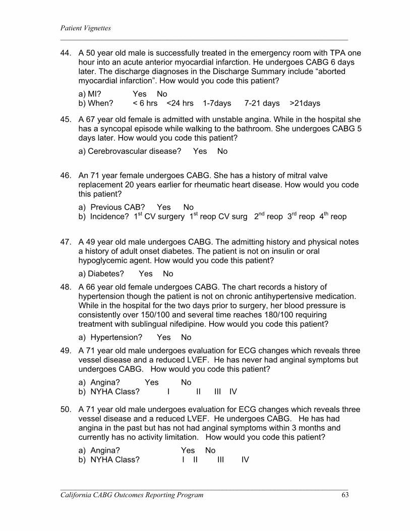
Patient Vignettes ______________________________________________________________________________
44. A 50 year old male is successfully treated in the emergency room with TPA one hour into an acute anterior myocardial infarction. He undergoes CABG 6 days later. The discharge diagnoses in the Discharge Summary include “aborted myocardial infarction”. How would you code this patient? a) MI? Yes No b) When? < 6 hrs <24 hrs 1-7days 7-21 days >21days
45. A 67 year old female is admitted with unstable angina. While in the hospital she
has a syncopal episode while walking to the bathroom. She undergoes CABG 5 days later. How would you code this patient? a) Cerebrovascular disease? Yes No
46. An 71 year female undergoes CABG. She has a history of mitral valve replacement 20 years earlier for rheumatic heart disease. How would you code this patient? a) Previous CAB? Yes No b) Incidence? 1st CV surgery 1st reop CV surg 2nd reop 3rd reop 4th reop
47. A 49 year old male undergoes CABG. The admitting history and physical notes
a history of adult onset diabetes. The patient is not on insulin or oral hypoglycemic agent. How would you code this patient? a) Diabetes? Yes No
48. A 66 year old female undergoes CABG. The chart records a history of hypertension though the patient is not on chronic antihypertensive medication. While in the hospital for the two days prior to surgery, her blood pressure is consistently over 150/100 and several time reaches 180/100 requiring treatment with sublingual nifedipine. How would you code this patient? a) Hypertension? Yes No
49. A 71 year old male undergoes evaluation for ECG changes which reveals three vessel disease and a reduced LVEF. He has never had anginal symptoms but undergoes CABG. How would you code this patient? a) Angina? Yes No b) NYHA Class? I II III IV
50. A 71 year old male undergoes evaluation for ECG changes which reveals three
vessel disease and a reduced LVEF. He undergoes CABG. He has had angina in the past but has not had anginal symptoms within 3 months and currently has no activity limitation. How would you code this patient? a) Angina? Yes No
b) NYHA Class? I II III IV
______________________________________________________________________________ California CABG Outcomes Reporting Program 63

Patient Vignettes ______________________________________________________________________________
51. A 64 year-old female undergoes CABG. She presents with a ventricular fibrillation arrest requiring resuscitation with chest compressions and electrical defibrillation. That same day, coronary angiography reveals “mild stenosis of the left main, luminal irregularities of the left anterior descending (LAD), an 70% stenosis of the first diagonal, 80% stenosis of the second diagonal, a subtotal occlusion of the circumflex after giving off the first marginal, a 30% stenosis of the first obtuse marginal, and plaquing of the right coronary artery (RCA).” At surgery two days later, the patient has four coronary bypass grafts placed to the distal LAD, the first diagonal, the second obtuse marginal, and the right coronary artery. How would you code this patient? a) Number of diseased vessels? None One Two Three b) Left main disease ________% c) Resuscitation Yes No
______________________________________________________________________________ California CABG Outcomes Reporting Program 64

Section Five:
Patient Vignette Answers


Patient Vignette Answers ______________________________________________________________________________
Patient Vignette Answers 1. a) Status = Urgent. Although this patient might conceivably have been
stabilized, discharged, and operated on electively; his surgery prior to discharge from a hospitalization for rest angina qualifies as urgent under the STS definition” “1) Procedure required during same hospitalization in order to minimize change of further clinical deterioration, and 2) worsening, sudden chest pain; congestive heart failure (CHF); acute myocardial infarction (AMI); coronary anatomy; IABP; unstable angina (USA) with intravenous nitroglycerin; rest angina; valve dysfunction; or aortic dissection.”
b) Angina = Yes. c) Angina type = Unstable. Classify anginal symptoms as unstable if they
occur at rest (as in this patient). d) NYHA class = IV. Rest symptoms are NYHA class IV, whether they are due
to angina or heart failure. e) Number of diseased vessels = Three. The left main counts as LAD and
circumflex and the RCA is >50% f) Left main % = 60% (unlike STS, where left main >50% = yes). 2. a) Status = Urgent. This patient was operated on prior to discharge from an
admission for worsened angina meeting the STS definition for urgent. As a rule of thumb, elective patients can safely await surgery at home, urgent patients can safely await surgery but only by remaining in the hospital, and emergent patients cannot safely delay surgery even in the hospital.
b) Angina = Yes. c) Angina type = Unstable. He did not have symptoms at rest but did have a
<2 week acceleration in his symptoms of at least one NYHA class to at least class III severity.
d) NYHA class = IV. Symptoms with very minimal exertion may be class IV. Symptoms occurring with walking at a normal pace one to two blocks or one flight of stairs are class III.
e) Number of diseased vessels = Two. Left main counts as LAD and circumflex but the RCA is only “plaqued” (<50%).
f) Left main % = 60% (≥50% = Yes) 3. a) Status = Urgent. She decompensated and became critically ill due to renal
failure following her angiography and was intubated for pulmonary edema. Patients operated on while intubated for pulmonary edema are emergent per the STS; however, this patient was stabilized medically and extubated by the time of CABG.
b) Angina = Yes. c) Angina type = Unstable. Chest pain at rest. d) CHF = Yes. e) NYHA class = IV. Symptoms at rest. f) MI = Yes. Ruled in for subendocardial infarction.
______________________________________________________________________________ California CABG Outcomes Reporting Program 67

Patient Vignette Answers ______________________________________________________________________________
g) MI when = 7-21d. h) Cardiogenic shock = No. Neither intubation for pulmonary edema (in the
absence of hypotension or low output) nor low-dose dopamine for diuresis meet criteria for shock. She was never hypotensive or had a low cardiac output.
i) Creatinine = 1.6 Last creatinine prior to surgery. j) COPD = Severe. Most coders consider home oxygen confirmation of the
diagnosis of COPD even though it is not mentioned by the STS; room air pO2 is assumed to be <60 mmHg in these cases.
k) Hepatic failure = No. No history of liver disease despite abnormal albumin. l) Post op renal failure = Yes. The post op creatinine rose to 2 times the
preop value of 1.5. 4. a) Status = Elective. The presence of left main disease alone is not enough to
warrant urgent status in this patient who presented with two months of stable exertional symptoms. Had she presented with unstable angina and left main disease, urgent status may have been indicated. Although in this case CABG was performed on the same hospitalization as the catheterization, the patient could also have been operated on as an outpatient.
b) Angina = Yes c) Angina type = Stable
d) NYHA class = Probably II or III (given that she went 8 minutes on her stress test), although we are not told what level of exertion precipitated symptoms.
e) Number of diseased vessels = Three. Left main counts as LAD and circumflex and RCA is diseased.
f) Deep sternal wound infection = No. The wound was not opened and there was no positive culture.
5. a) Status = Urgent. The patient’s recurrent ischemia did not respond to drug
therapy but did resolve with the placement of an intra-aortic balloon pump. The intra-aortic balloon pump by itself is not sufficient to classify this patient as emergent. If the patient’s symptoms had continued despite the IABP and the patient had been taken to surgery the same day, the operation would have been emergent (see STS urgent criteria D and emergent criteria A).
b) Angina = Yes. c) Angina type = Unstable. Angina at rest. d) NYHA class = IV. Rest symptoms are class IV. e) Postoperative Stroke > 72 hrs = No. Symptoms resolved in 48 hrs. f) Prolonged ventilation = No. Extubated less that 24 hrs post op.
______________________________________________________________________________ California CABG Outcomes Reporting Program 68

Patient Vignette Answers ______________________________________________________________________________
6. a) Status = Emergent. The myocardial infarction >24 hrs prior to CABG does not meet emergent criteria. However, this patient’s unstable anginal symptoms persisted and were ongoing up until surgery despite maximal medical therapy. The surgery was required within a few hours and warrants emergent status.
b) Angina = Yes. c) Angina type = Unstable. Rest symptoms d) NYHA class = IV. e) MI = Yes f) MI When = 1-7 days 7. a) Status = Urgent. This patient presented with new onset anginal symptoms
and CHF and underwent CABG during the same hospitalization (STS urgent criteria C & D)
b) Angina = Yes. c) Angina type = Unstable d) CHF = Yes e) NYHA class = IV. This patient had both anginal and CHF symptoms at rest
within two weeks of her surgery and thus is class IV. 8. a) Status = Urgent. This patient’s unstable angina recurred on medical therapy
and he was operated on two days later warranting urgent status. b) Angina = Yes. c) Angina type = Unstable. Rest symptoms d) CHF = No. No history or current symptoms of heart failure. e) NYHA = IV (for angina). Note: unlike CCS class for angina, there is no
“class 0” for NYHA so patients with no symptoms are class I. f) MI = No. He was “ruled out”. 9. a) Status = Urgent. This patient had unstable angina which resulted in a
malignant arrhythmia and would thus be urgent. b) Angina = Yes. c) Angina type = Unstable. Rest symptoms d) CHF = No e) NYHA = IV (for angina). He presumably has no history of CHF (none is
given). f) MI = No. He was “ruled out”. g) Arrhythmia = Yes. h) Arrhythmia type = Sustained VT/VF. He had VF requiring treatment within
two weeks of his surgery. He also had Afib, but chose VT/VF since this is a stronger risk factor for CABG.
10. a) Status = Urgent. This patient had post infarction angina following TPA
warranting urgent status b) Angina = Yes. c) Angina type = Unstable. Rest symptoms
______________________________________________________________________________ California CABG Outcomes Reporting Program 69

Patient Vignette Answers ______________________________________________________________________________
d) NYHA = IV (for angina at rest with MI). e) CHF = No. f) MI = Yes. g) Arrhythmia = Yes. h) Arrhythmia type = Sustained VT/VF. She had sustained VT requiring
cardioversion within 2 weeks of surgery. 11. a) Status = Emergent. The patient was in cardiogenic shock at the time of
surgery due to a myocardial infarction 5 days earlier. Patient in shock at the time of surgery (with or without circulatory support) are classified as emergent.
b) Angina = Yes. c) Angina type = Unstable. Rest chest pain within two weeks of the surgery d) NYHA = IV (both angina and CHF are class IV). e) CHF = Yes. Although in this exercise we do not provide a detailed enough
patient history to fully document CHF, cardiogenic shock certainly warrants this diagnosis
f) MI = Yes. g) MI when = 1-7 days. 12. a) Cerebrovascular disease = Yes. In addition to TIA, RIND, and stroke, the
revised STS definition classifies asymptomatic carotid stenoses of >75% as cerebrovascular disease.
b) Cerebrovascular accident = No. c) CVA Timing < or >2 weeks = N/A - Missing
d) Peripheral vascular disease = No. Peripheral vascular disease does not include cerebral vascular disease. Peripheral vascular disease is coded if the patient has claudication, diminished femoral pulses, a history of an aortic aneurysm, stenosis of the aortic, iliac, femoral, or popliteal vessels, or surgery for such a stenosis.
13. a) Congestive heart failure (CHF) = No. The patient has a history of CHF 2
years ago but has had no CHF symptoms within two weeks of surgery. b) NYHA = Class II (for angina). c) Reop bleed / tamponade = Yes. An operative reintervention was required
for bleeding or tamponade. 14. a) CHF = No. The patient has had no symptoms or signs of CHF within two
weeks of surgery though she is on CHF medications. b) NYHA = Class III (for angina). CHF would be NYHA class I
(asymptomatic). 15. a) Angina = No. b) CHF = Yes. She has a history of CHF and currently has STS criteria
including dyspnea on exertion and PND. c) NYHA = Class II (for heart failure). Ordinary activity results in symptoms.
______________________________________________________________________________ California CABG Outcomes Reporting Program 70
Patient Vignette Answers ______________________________________________________________________________
16. a) CHF = Yes. She has a history of CHF and currently has STS criteria -
dyspnea and pulmonary edema on chest X-ray. b) NYHA = Class III. Less than ordinary activity results in symptoms. 17. a) Arrhythmia = No. The patient had only asymptomatic and relatively brief
runs of non-sustained ventricular tachycardia. Non-sustained (<30 seconds) ventricular tachycardia is not classified as a ventricular arrhythmia.
b) Arrhythmia type = Afib/Flutter. When both a sustained ventricular and another type of arrhythmia are present, code the ventricular arrhythmia. However, in this case only atrial fibrillation is present.
18. a) Arrhythmia = No. Although this patient was treated with an intravenous
antiarrhythmic agent, he did not have sustained ventricular tachycardia or fibrillation but rather only PVC’s (premature ventricular beats) and non-sustained VT. Thus, he would not be classified as having a ventricular arrhythmia. The lidocaine was arguably unnecessary in this case.
b) Arrhythmia type = None. 19. a) Arrhythmia = Yes. b) Arrhythmia type = Sustained VT/VF. Sustained ventricular is generally
accepted as greater than 30 seconds. When both a sustained ventricular and another type of arrhythmia are present, code the ventricular arrhythmia. Most cases of ventricular fibrillation will require treatment and will qualify as a ventricular arrhythmia by STS criteria.
20. a) Arrhythmia = No. The patient’s episode of ventricular tachycardia did not
occur within 2 weeks prior to CABG, which is the time window to which this variable refers.
b) Arrhythmia type = None. c) Continuous coma >24 hrs = Yes. “A new postoperative coma that persists
for at least 24 hours secondary to anoxic/ischemic and/or metabolic encephalopathy, thromboembolic event or cerebral bleed.”
21. a) COPD = No. Merely being labeled as having “COPD” in the chart is not
sufficient. There must be documentation of therapy with bronchodilators, theophylline, steroids, or home O2, or of an FEV1 <75% of predicted. Of note, short term use of inhaled bronchodilators because of “cardiac asthma” or pulmonary infection similarly is not sufficient to classify a patient with COPD.
22. a) Number of vessels = Three. When the posterior descending coronary
artery (PDA) comes off of the circumflex instead of the right coronary artery (referred to as a left dominant circulation), the PDA is counted as a separate vessel. Therefore, the proximal stenosis in the circumflex counts as two vessels because it jeopardizes both the marginals (the usual branches of
______________________________________________________________________________ California CABG Outcomes Reporting Program 71

Patient Vignette Answers ______________________________________________________________________________
the circumflex) and the PDA. The non-dominant right coronary is not counted.
23. a) Number of vessels = Two. Although four different vessels and branches are
noted to have >50% stenoses, only two of the three major coronary artery systems (the LAD and Cx but not the RCA) are involved. If multiple branches of the same artery are diseased, they are counted as a single vessel. Plaquing refers to mild stenoses well less than 50%.
b) Left main % = 40% (≥50% = No). 24. a) Number of vessels = Two. Many clinicians consider significant coronary
stenoses to be those that are ≥70-75%. However, for the purposes of the STS and the CCORP, stenoses or narrowing ≥50% are counted. Thus, in this patient the 50% narrowing of the LAD count as LAD disease while the circumflex stenosis of 60% counts as circumflex disease. The 60% stenosis of the left main also counts as disease of the LAD and circumflex. However, do not double count the LAD and circumflex.
b) Left main disease % = 60% (≥50% = Yes). 25. a) Creatinine = 1.6 mg/dl. The last creatinine determination prior to CABG
should be entered, in this case 1.6 mg/dl. 26. a) Chronic lung disease = Moderate. An FEV1 50 to 59% of predicted, even in
the absence of pharmacological therapy, is coded as moderate COPD. FEV1 <50% predicted would be severe COPD.
27. a) Chronic lung disease = Moderate. Chronic bronchitis and emphysema are
types of COPD and thus this patient should be classified as moderate COPD because of the chronic steroid use. Had he been on bronchodilators without steroids, it would have been mild COPD.
b) Immunosuppressive treatment? = Yes (Prednisone). 28. a) Peripheral vascular disease = Yes. Peripheral vascular disease is coded if
the patient has a history of an aortic aneurysm (as in this patient), stenosis of the aortic, iliac, femoral, or popliteal vessels, or surgery for such a stenosis.
29. a) Peripheral vascular disease = Yes. Peripheral vascular disease is coded if
the patient has had claudication (as in this patient). 30. a) Cerebrovascular disease = Yes. The STS definition includes prior carotid
surgeries such as carotid endarterectomy. STS: “The patient has a history at any time prior to surgery any one of the
following:
______________________________________________________________________________ California CABG Outcomes Reporting Program 72

Patient Vignette Answers ______________________________________________________________________________
– Unresponsive coma >24 hours – Cerebro-vascular accident (CVA)
(symptoms >72 hours after onset) – Reversible ischemic neurological deficit (RIND)
(recovery within 72 hours of onset) – Transient ischemic attack (TIA)
(recovery within 24 hours of onset) – Non-invasive carotid test with >75% occlusion – Or prior carotid surgery – Does not include neurological disease processes such as metabolic
and/or anoxic ischemic encephalopathy” b) Peripheral vascular disease = No. Cerebrovascular disease is not included
in peripheral vascular disease. No other history of peripheral vascular disease is given.
31. a) Cerebrovascular disease = Yes. A history of transient ischemic attack (TIA)
is coded as cerebrovascular disease. b) Peripheral vascular disease = No. Cerebrovascular disease is not coded as
peripheral vascular disease. 32. a) Cerebrovascular disease = Yes. Strokes are coded as cerebrovascular
disease. b) Cerebrovascular accident = Yes. CVA ever is coded as yes. c) CVA timing = Remote (>2 weeks). Recent is ≤2 weeks. d) Peripheral vascular disease = No. Cerebrovascular disease is not coded as
peripheral vascular disease.
33. a) Congestive heart failure = Yes. This patient is currently symptomatic and has pulmonary congestion on chest X-ray and thus is coded as having CHF.
b) NYHA class = III. Assigning NYHA class is always a matter of some judgment but based on the limited information provided the patient has dyspnea with less than normal activities and thus is class III.
34. a) Congestive heart failure = Yes. This patient has dyspnea at rest, edema,
and rales. b) NYHA class = IV. Rest symptoms are class IV. 35. a) Angina = Yes b) Angina type = Stable c) NYHA class = II. He has slight limitation of normal activities and ordinary
activities such as walking up several flights of stairs or walking uphill can bring on his anginal symptoms.
d) Reop graft occlusion = Yes. Operative re-intervention for graft occlusion.
______________________________________________________________________________ California CABG Outcomes Reporting Program 73

Patient Vignette Answers ______________________________________________________________________________
36. a) Angina = Yes b) Angina type = Stable c) NYHA class = I. Ordinary activities do not precipitate his angina. Only
strenuous activities, in this case jogging, cause symptoms. Patients without angina (i.e., asymptomatic) are also coded class I.
37. a) Angina = Yes b) Angina type = Stable. Exertional chest pain which did not require treatment
in an ICU or IV NTG. c) NYHA class = IV. The patient cannot carry out any activities without
symptoms and should be coded with NYHA class IV angina. Patients with symptoms at rest are also coded class IV.
38. a) Angina = Yes b) Angina type = Stable. Exertional chest pain. c) NYHA class = III. The patient’s activities are markedly limited and he should
be coded class III. 39. a) Ejection fraction = 42%. For the left ventriculogram the range 40-45%
averages to 42.5%, so enter 42%. For the echocardiogram, enter 45%. b) Ejection fraction method = LV gram or echo. Most recent study is
preferred. c) Mitral regurgitation = Moderate. The MR is on the echocardiogram.
40. a) Ejection fraction = 65%. When the EF is described qualitatively it should be coded as follows: normal = 65%, mildly reduced = 50%, moderately reduced = 35%, and severely reduced = 20%.
b) Ejection fraction method = LV gram. c) Mitral regurgitation = Mild. 41. a) Ejection fraction = 47% (calculated) or 42% (visually estimated). Usually,
the visually estimated LVEF reflects what the reader believes the LVEF truly is; therefore this LVEF is usually preferred. Most patients will have ejection fractions reported from pre-operative left ventriculograms. This patient, perhaps because he was unstable (urgent status) had only an echocardiogram. The echo reported both a visually estimated EF range and a calculated value. Intraoperative and post-operative echos should not be used when other measures of EF are available.
b) Ejection fraction method = ECHO (pre-operative). The intra-operative transesophageal echocardiogram (TEE), although done at the beginning of surgery, should not be used because general anesthesia can alter EF and MR and the determination of EF and MR by TEE is less standardized and, even under identical conditions, can obtain results which differ from standard transthoracic echo. The post-operative echos are never used since they can reflect the benefits of surgery rather than the patient’s pre-operative state.
______________________________________________________________________________ California CABG Outcomes Reporting Program 74
Patient Vignette Answers ______________________________________________________________________________
c) Mitral regurgitation = Moderate. MR from the pre-operative echo should be entered as the higher of the two values (mild to moderate).
42. a) Ejection fraction = 20%. When the EF is described qualitatively it should be
coded as follows: normal = 65%, mildly reduced = 50%, moderately reduced = 35%, and severely reduced = 20%.
43. a) Previous CABG = Yes. The CABG two years ago. b) Incidence = First reop CV surgery. The previous CABG counts. If this
were the patient’s first CABG, incidence would be coded as “First CV surgery.” Incidence of cardiovascular surgeries includes CABG, valve replacement/ repair, intracardiac repairs (ASD, VSD), ventricular aneurysmectomy, or surgery on the aortic arch. Use of cardiopulmonary bypass (heart-lung machine) is not required. CV surgeries do NOT include PCIs, non-cardiac vascular surgeries such as abdominal aortic aneurysm repairs or fem-pop bypasses, or pacemaker/ICD implantations.
c) PCI = Yes. Even though the PTCA was aborted, any patient in whom the procedure is attempted should be coded as having a PCI.
d) PCI to surgery time interval = <6 hrs. e) Status = Emergent. Generally, patients with failed PTCA’s taken
immediately to CABG are coded as emergent because they have ongoing ischemia requiring immediate surgery.
44. a) MI (myocardial infarction) = Yes. Even though his MI was successfully
interrupted by thrombolysis with TPA, he should be coded as having had an MI.
b) MI When = 1-7 days. 45. a) Cerebrovascular disease = No. “Syncopal episode” refers to loss of
consciousness, also called a fainting episode. It should not be confused with a transient ischemic attack (TIA), which is a transient stroke and rarely causes fainting. Syncope is not coded as cerebrovascular disease while a TIA would be.
46. a) Previous CABG = No. The mitral valve replacement 20 yrs ago does not
count here. b) Incidence = First reop CV surgery. The previous mitral valve replacement
counts here. 47. a) Diabetes = Yes. Patients are coded with diabetes regardless duration of the
disease, the need for treatment with insulin or oral antidiabetic agents (e.g., glyburide, glypizide, micronase), or the presence of diabetic complications. Thus, any history of diabetes is coded as yes.
______________________________________________________________________________ California CABG Outcomes Reporting Program 75

Patient Vignette Answers ______________________________________________________________________________
48. a) Hypertension = Yes. Documented history of hypertension, the need for
anti-hypertensive medication, or blood pressure consistently exceeding 140/90 is coded as hypertension.
49. a) Angina = No. b) NYHA class = 1. Patients without angina or HF symptoms are class 1. 50. a) Angina = Yes. Assign yes if the patient has ever had angina.
b) NYHA class = I. Patients with a history of angina but with mild or no symptoms around the time of their surgery are class I.
51. a) Number of diseased vessels = Two. The luminal irregularities in the LAD
and the plaquing of the right coronary artery should be interpreted as a <50% stenoses and not counted. However, the LAD system is diseased because the first and second diagonals have >50% stenoses. The “subtotal occlusion” of the circumflex implies a very tight stenosis (95-99%) thus this is the second diseased vessel. The first obtuse marginal branching off of the circumflex is <50% stenosed and would not be counted but as discussed the circumflex itself is diseased. The fact that four grafts were placed, including one to the RCA which was not counted as diseased, does not influence the coding of the number of diseased vessels. In some cases, vessels with mild disease are bypassed.
b) Left main % = 20% (≥50% = No). The mild stenosis should be interpreted as less than 50.
c) Resuscitation = No. Chest compressions occurred two days prior to surgery. STS definition: “Indicate whether the patient required cardiopulmonary resuscitation within on hour before start of the operative procedure.” Cardiopulmonary resuscitation is defined as chest compressions or being placed on cardiopulmonary bypass (heart-lung machine) prior to arrival in the operating room (e.g., in the cath lab).
______________________________________________________________________________ California CABG Outcomes Reporting Program 76