
Calibre & Bullet Type Estimation - Laurentian University Henwood... · Bullet Calibre and Type...
25
1 BULLET CALIBRE AND TYPE ESTIMATION FROM GUNSHOT WOUNDS IN SUS SCROFA (LINNAEUS) LONG BONE Bailey J. Henwood Submitted in partial fulfillment of the course FORS 4095 EL 01 Department of Forensic Science Laurentian University Sudbury, Ontario P3E 2C6 ©Copyright by Bailey J. Henwood 2017
Transcript of Calibre & Bullet Type Estimation - Laurentian University Henwood... · Bullet Calibre and Type...
1
BULLET CALIBRE AND TYPE ESTIMATION FROM GUNSHOT WOUNDS IN SUS SCROFA (LINNAEUS) LONG BONE
Bailey J. Henwood
Submitted in partial fulfillment of the course FORS 4095 EL 01
Department of Forensic Science Laurentian University
Sudbury, Ontario P3E 2C6
©Copyright by Bailey J. Henwood 2017
2
Bailey J. Henwood, B. Sc. (Hons), Tracy S. Oost, B.Sc. (Hons), and Scott I. Fairgrieve, Ph. D.
Bullet Calibre and Type Estimation from Gunshot Wounds in Sus Scrofa (Linnaeus) Long Bones Abstract The calibre of bullet used in the commission of crime is not typically ascertained from a
wound in soft tissue, and cannot be reliably determined from the damage done to flat bones (1,
2). This study examined the feasibility of obtaining both bullet calibre and type from ballistic
damage to long bones. Thirty fresh pork shoulders (Sus scrofa), in three groups of ten, were
each shot with a handgun using one of three calibre. These calibres were of two bullet types,
either lead or full copper jacket. Each pork shoulder was shot through the intact humerus with
either a 0.22 round-nose lead bullet, 9mm full copper jacketed bullet, or a 0.38 round-nose lead
bullet. The long bones were subsequently defleshed and examined for evidence of bullet calibre
and type. The damaged bone was reconstructed to the fullest extent possible from the resulting
bone fragments. The minimum diameter of each entrance wound, as well as the associated
fracture pattern, were recorded. A Kruskal-Wallis nonparametric test was used to compare the
median entrance wound measurements for each calibre. Cortical bone spalling was seen only In
long bones shot with a copper jacketed bullet. This is due to the differing energy propagation of
the two bullet types through the bone tissue. Our study demonstrates that bullet type can be
ascertained from long bone damage, and the damage from a 0.22 calibre bullet can be
distinguished from that of a 0.38 or a 9mm bullet.
Keywords: forensic science, osteology, ballistics, gunshot wound, long bone, calibre, bullet
type, bone spalling
3
Introduction
The intricacies of ballistic trauma have long been researched, and many of these studies
have focused on the wound ballistics of soft tissues (3). They have, for the most part, ignored
the impact to skeletal tissues. The few studies that have been conducted on bone have
concentrated on the flat bones of the skull, and exclude long bones (1, 2). This research has
yielded little success in determining the bullet calibre, but have demonstrated the potential of
obtaining an estimation of the bullet size (1). The estimation of bullet type, be it lead or copper,
has not been explored and has the potential to add yet another dimension to the evidence of
the firearm used.
Long bones are composed of two different types of bone tissue; cortical and trabecular.
Previous studies have used Sus scrofa humeri, which is used as human analog (4). The osteon
arrangement in pork long bones is similar to human long bones, and as such, result in
comparable crack characteristics (4). Cortical bone is the dense tissue predominantly found in
the long bone diaphysis, whereas trabecular bone is found inside the articular ends (5). The
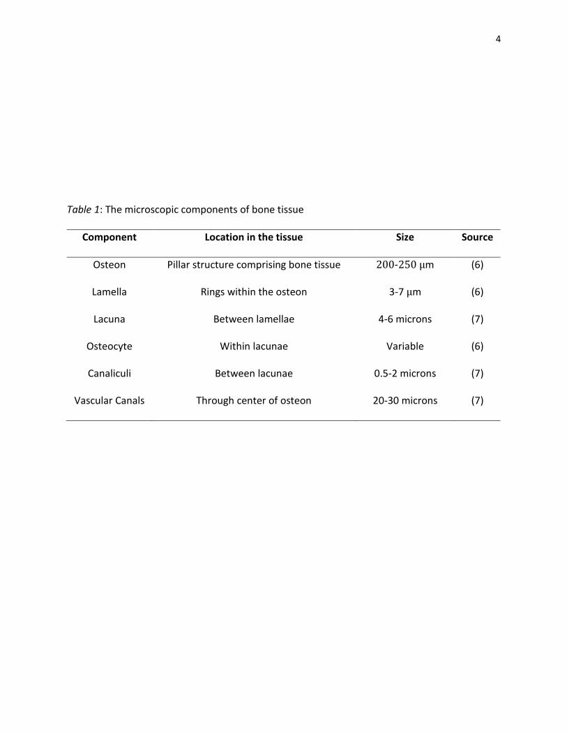
histological features of mammalian bone and their estimated sizes are summarized in Table 1
(6, 7).
4
Table 1: The microscopic components of bone tissue
Component Location in the tissue Size Source
Osteon Pillar structure comprising bone tissue 200-250 µm (6)
Lamella Rings within the osteon 3-7 µm (6)
Lacuna Between lamellae 4-6 microns (7)
Osteocyte Within lacunae Variable (6)
Canaliculi Between lacunae 0.5-2 microns (7)
Vascular Canals Through center of osteon 20-30 microns (7)
5
The microstructure summarized in Table 1 is critical to the analysis and understanding of the
nature of crack propagation through cortical bone tissue (5,8). There are five main phases that
deter the propagation of cracks, including the crystalline mineral phase, crystalline organic
phase, amorphous mineral phase, amorphous organic phase, and the liquid phase (7). Included
in the liquid phase are the canaliculi and lacunae, that help to dissipate the forces being
applied. Cracks are typically diverted along the lamellar cement lines between the osteons, but
to the decreased density at this interface. The microstructure of the bone slows the fracture
rate, as opposed to fast forming fractures, which overcome the discontinuity inherent in the
cement lines (7, 9). A fracture resulting from a gunshot wound is consequently regarded as a
fast rate fracture due to the high energy imparted to the bone and, consequently, often tend
not to follow the cement lines.
The anatomy of long and flat bones is similar in microstructure, however, differs in gross
anatomy. Although both types of bone are composed of osteon systems, their gross anatomy
differ greatly (5). Flat bones are comprised of two dense layers of cortical bone, with a layer of
spongy diploё between them. The diploic layer is composed of trabecular arches and is porous
(5). This layer disperses the impact energy throughout the area instead of concentrating it all in
one area of the bone (5). The diaphysis of a long bone is essentially a cylinder made of cortical
bone, which houses the medullary cavity. The dense cortical bone is required here to provide
stability and rigidity to the skeleton (5). In the case of the humerus, there are two epiphyseal
joints, one proximally and the other distally. The trabecular portion is made up of bony spicules,
generating a series of intersecting arches. This is a highly porous area of bone, while cortical
6
bone is very dense. The role of trabecular bone is to dampen stresses applied to the articular
surface (5).
As a result of the differences in gross anatomy, cranial bones and long bones react very
differently under stress (10). Consider a flat bone of the skull; when hit by an impact, be it blunt
force, sharp force or ballistic, the applied energy is projected straight through the outer layer of
cortical bone, and is then dissipated in an outwardly conical manner through the diploё, and
then through the inner cortical bone (11). Due to this mechanism of energy propagation, a
beveled entrance wound is typically found deep to the outer layer of flat bones that have
undergone ballistic trauma (12). Owing to the differences in the anatomy of long bones, this
process of macroscopic energy dissipation is presumptively very different, and appears to have
not been thoroughly investigated. Due to the structural differences between long and flat
bones, it is not appropriate to apply cranial bone research when examining long bone trauma.
The general trend of cranial bone research has found that the minimum diameter of the
entrance wound can lead an investigator to a general calibre size estimation (1, 2). Researchers
have successfully grouped “small” calibres, such as 0.22, 0.25, 0.28 and 0.32, separately from
“large” calibre bullets, such as 0.38 or 9mm (1, 2). Some issues with these studies include the
sheer number of calibres, how unknown intermediate targets affect the flight path of a bullet,
the range of bullet designs, materials and how they react to the forces involved (1).
Building on this previous work, it is the goal of this study to use features of the entrance
wound, such as the degree of comminution and the minimum diameter of the wound, to
elucidate possible calibre estimation from long bones exhibiting gunshot wounds. The material
of the jacket is of primary interest, as the method of force absorption differs greatly between a
7
lead bullet and a copper jacketed bullet (13). A lead bullet deforms on impact with surfaces that
are less dense, such as bone tissue. They are known as yielding bullets, and can thus absorb
some of the energy of the impact and flatten out (13). A copper jacket does not yield to any of
the forces imparted onto the bone and therefore retains its original shape (13). The differences
in effect to the bone have not been previously documented. This study seeks to remedy this
situation.
In general, this study examines how long bones differ in their reaction to ballistic trauma
compared to cranial bone with regards to the fracture patterns and dissipation of force. This
information will be useful in scene reconstruction, as well as in criminal cases where evidence,
such as the bullet itself, is not recovered or implicate a recovered bullet as the one that inflicted
the damage.
8
Materials and Methods
Sample Description
Thirty specimens of Sus scrofa L. (domestic pig) humeri were used as an analog for human
long bone. Each pork shoulder was fully fleshed, with the skin and underlying muscle intact and
attached to the bone. Each of these specimens was shot once with a handgun at close range,
approximately 0.75 meters, using a specific calibre. After shooting, each specimen was stored in
a freezer until the removal of soft tissue. Subsequently, the resulting bone fragments for each
specimen were stored at room temperature (~19°C) in a storage cabinet in the Forensic
Osteology Laboratory. Reconstructed humeri were stored in the same manner.
Firearms and Ammunition
Three different calibres of bullets were used; 0.22, 9mm and 0.38. The 0.22 calibre firearm
used lead round-nose Blazer® CCI 0.22 long rifle ammunition. The 9mm ammunition was a full
copper jacket Dominion® brand centrefire bullet. Finally, lead round-nose Dominion® brand
centrefire 0.38 special ammunition was used. The corresponding firearms used were High
Standard™ Supermatic, a Forjas Taurus™ Semi-Automatic and a Smith and Wesson® Model 639,
respectively.
9
Shooting
The pork shoulder segments were placed on their side, parallel to the ground. There were
ten specimens used for each calibre, and each section was shot once with the intended target
being in either the mid-shaft region, or head of the humerus, depending on which specimen
was being used. The range of each shot was approximately 0.75 meters. The shooting of all
specimens was completed at once. A few samples of each calibre of lead bullet were recovered
for analysis, however no copper jacketed bullets were recovered.
Sample Assessment
After shooting, the pork shoulders were frozen until cleaning. The shoulders were first
thawed and boiled in water. Most of the tissue was removed by cutting it away from the bone,
using scissors, to prevent any blades from contacting the bone. The remaining tissue was
removed using Tergazyme® and then steamed to remove the remaining cartilaginous tissues.
Once cleaned, they were stored in a dry cabinet on a tray. Each bone was photographed using a
Canon® 60D digital SLR camera. The bones were analyzed by recording degree of comminution
in affiliation with the entrance wound, compared to Rogers’ guidelines (14). The diameter of
each entrance wound was measured per specimen using a Sylvac® Model S 235 electronic
caliper. There were no measurements taken regarding the exit wound, as it does not impart any
diagnostic information regarding the size of the bullet (11). Perpendicular bullet damage to the
midshaft or humeral head, was sustained by six bones using the 0.22 firearm, eight bones by
the 9mm firearm, and six bones using the 0.38 special firearm.
10
Results
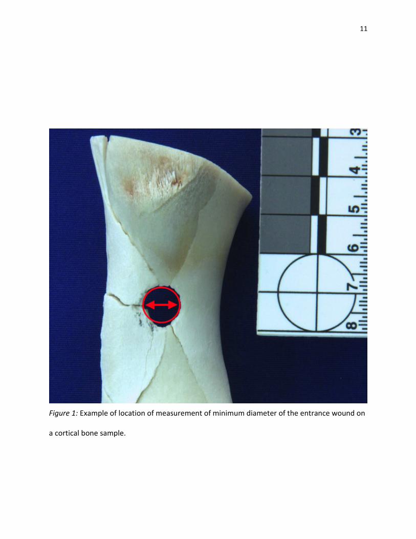
Minimum Diameter of Entry Wounds
Following the cleaning and reconstruction of the humeri, the diameter of each entrance
wound was measured. This measurement, shown in Figure 1, was taken to the nearest one
hundredth of a millimetre. The measurements were run through the non-parametric Kruskal-
Wallis test via IBM SPSS Statistics (15) to determine if the median diameter of each calibre were
significantly different. The results of this test, along with the post hoc analysis of each pairwise
comparison and their significance, are summarized in Table 2. A p-value of less than 0.05 was
considered significant.
11
Figure 1: Example of location of measurement of minimum diameter of the entrance wound on
a cortical bone sample.
12
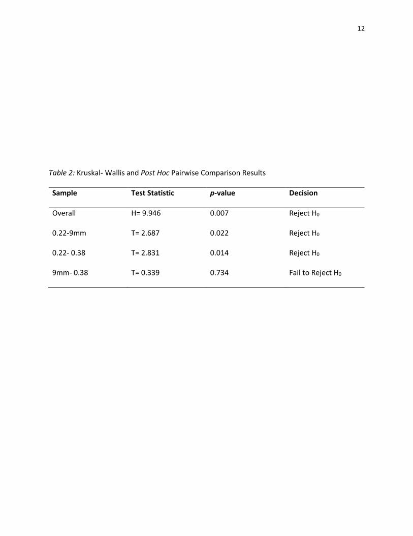
Table 2: Kruskal- Wallis and Post Hoc Pairwise Comparison Results
Sample Test Statistic p-value Decision
Overall H= 9.946 0.007 Reject H0
0.22-9mm T= 2.687 0.022 Reject H0
0.22- 0.38 T= 2.831 0.014 Reject H0
9mm- 0.38 T= 0.339 0.734 Fail to Reject H0
13
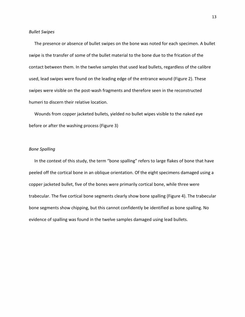
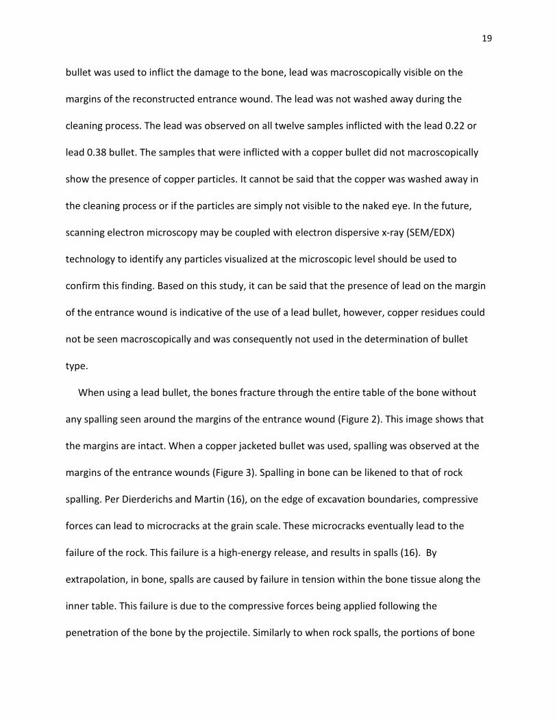
Bullet Swipes
The presence or absence of bullet swipes on the bone was noted for each specimen. A bullet
swipe is the transfer of some of the bullet material to the bone due to the frication of the
contact between them. In the twelve samples that used lead bullets, regardless of the calibre
used, lead swipes were found on the leading edge of the entrance wound (Figure 2). These
swipes were visible on the post-wash fragments and therefore seen in the reconstructed
humeri to discern their relative location.
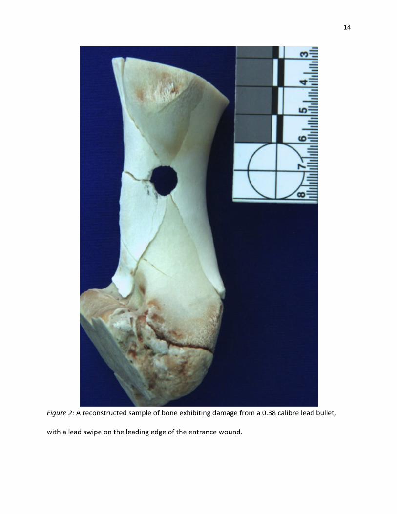
Wounds from copper jacketed bullets, yielded no bullet wipes visible to the naked eye
before or after the washing process (Figure 3)
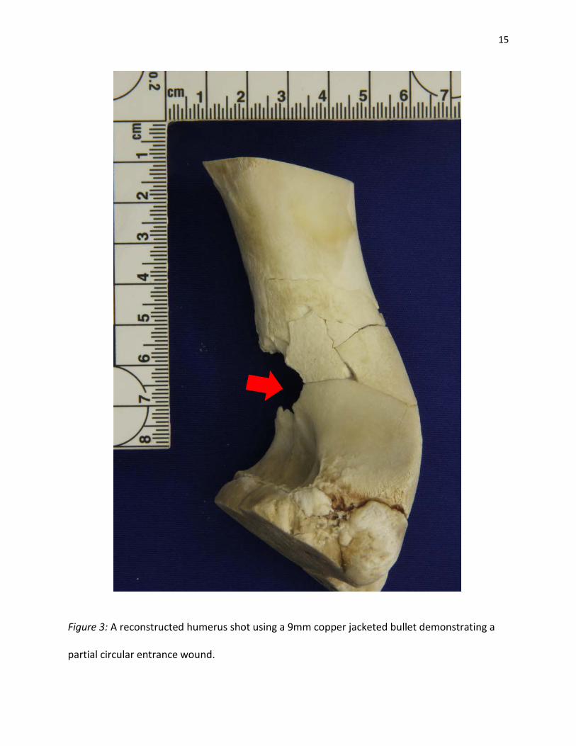
Bone Spalling
In the context of this study, the term “bone spalling” refers to large flakes of bone that have
peeled off the cortical bone in an oblique orientation. Of the eight specimens damaged using a
copper jacketed bullet, five of the bones were primarily cortical bone, while three were
trabecular. The five cortical bone segments clearly show bone spalling (Figure 4). The trabecular
bone segments show chipping, but this cannot confidently be identified as bone spalling. No
evidence of spalling was found in the twelve samples damaged using lead bullets.
14
Figure 2: A reconstructed sample of bone exhibiting damage from a 0.38 calibre lead bullet,
with a lead swipe on the leading edge of the entrance wound.
15
Figure 3: A reconstructed humerus shot using a 9mm copper jacketed bullet demonstrating a
partial circular entrance wound.
16
Figure 4: Bone spalling observed in a bone shot using a 9mm copper jacketed bullet. Note the
absence of bullet swipe evidence.

17
Discussion
Calibre Estimation
A bullet’s calibre is reported in either imperial measurements or in metric measurements. If
the reported calibre is given in the metric system, the calibre reflects the diameter in
millimetres, such as a 9mm, while the imperial system names the bullet reflective of the
diameter in inches, such as a 0.38. In this study, three different calibres were used. Of these
three calibres, bullets with two different diameters were used. A 0.22 calibre bullet measures
5.65mm in diameter, while a 9mm and a 0.38 calibre each measure 9.00mm. Using the 0.22
lead bullets, six humeri sustained enough damage to necessitate reconstruction prior to
analysis. The median measurement of the entry wound in this sample set was 8.00mm, with no
statistical outliers. Of the ten specimens damaged used a 9mm copper jacketed bullet, eight
humeri were analyzed. The median entrance wound size resulting from the 9mm bullet was
found to be 9.69mm, again with no statistical outliers. Finally, six humeri were inflicted with
adequate damage to require reconstruction and were analyzed after reconstruction. The
median entrance wound size was found to be 10.02mm, without any outliers in the data.
Because the data set is small, non-parametric statistical analysis was conducted. The null
hypothesis was not rejected if there were no differences between the median entrance wound
size for each calibre. The Kruskal-Wallis test was used to determine if there were differences
between any of the three calibres overall. It was found that there were differences between a
0.22 calibre entrance wound and a 0.38 calibre entrance wound, as well as a 0.22 calibre
entrance wound and a 9mm entrance wound (p= 0.007), and the null hypothesis was rejected.
Following the Kruskal-Wallis test, a pairwise-comparison post hoc test was completed. It was
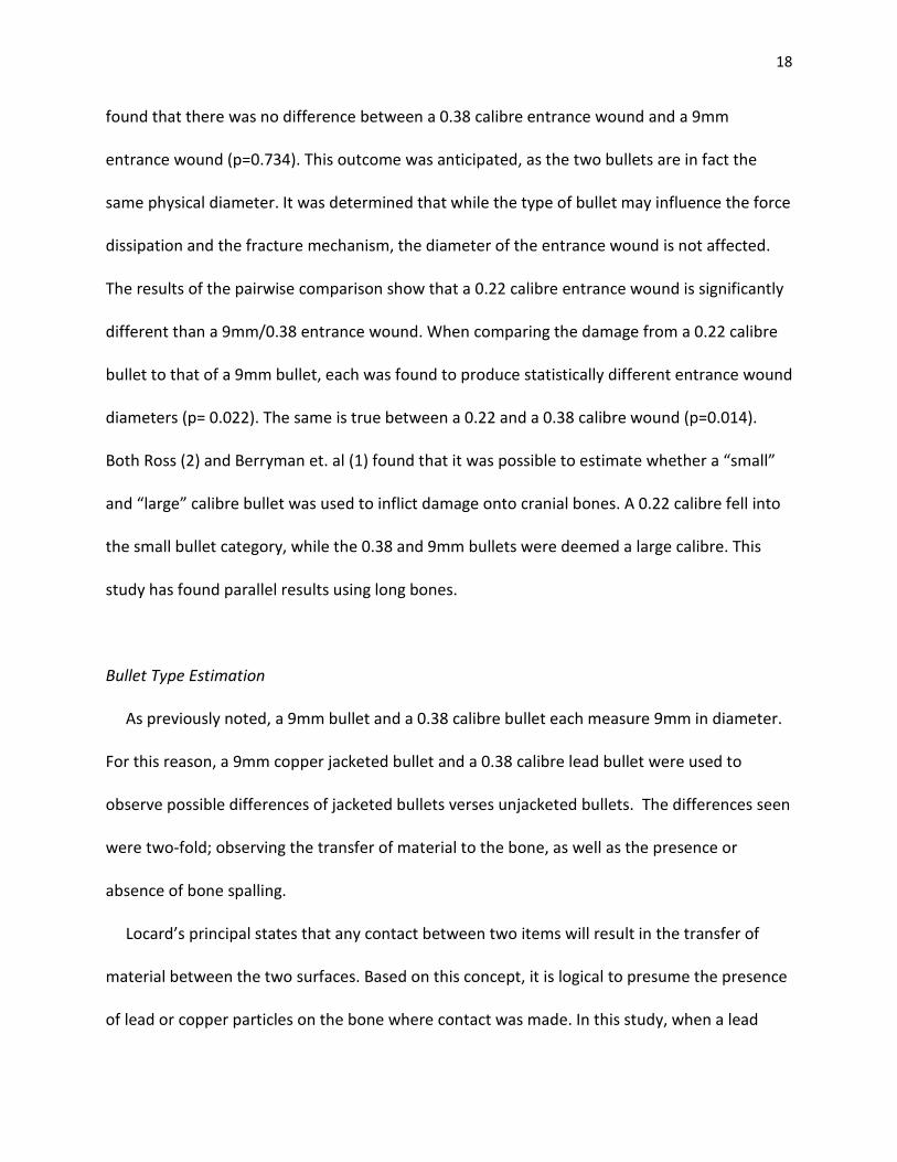
18
found that there was no difference between a 0.38 calibre entrance wound and a 9mm
entrance wound (p=0.734). This outcome was anticipated, as the two bullets are in fact the
same physical diameter. It was determined that while the type of bullet may influence the force
dissipation and the fracture mechanism, the diameter of the entrance wound is not affected.
The results of the pairwise comparison show that a 0.22 calibre entrance wound is significantly
different than a 9mm/0.38 entrance wound. When comparing the damage from a 0.22 calibre
bullet to that of a 9mm bullet, each was found to produce statistically different entrance wound
diameters (p= 0.022). The same is true between a 0.22 and a 0.38 calibre wound (p=0.014).
Both Ross (2) and Berryman et. al (1) found that it was possible to estimate whether a “small”
and “large” calibre bullet was used to inflict damage onto cranial bones. A 0.22 calibre fell into
the small bullet category, while the 0.38 and 9mm bullets were deemed a large calibre. This
study has found parallel results using long bones.
Bullet Type Estimation
As previously noted, a 9mm bullet and a 0.38 calibre bullet each measure 9mm in diameter.
For this reason, a 9mm copper jacketed bullet and a 0.38 calibre lead bullet were used to
observe possible differences of jacketed bullets verses unjacketed bullets. The differences seen
were two-fold; observing the transfer of material to the bone, as well as the presence or
absence of bone spalling.
Locard’s principal states that any contact between two items will result in the transfer of
material between the two surfaces. Based on this concept, it is logical to presume the presence
of lead or copper particles on the bone where contact was made. In this study, when a lead
19
bullet was used to inflict the damage to the bone, lead was macroscopically visible on the
margins of the reconstructed entrance wound. The lead was not washed away during the
cleaning process. The lead was observed on all twelve samples inflicted with the lead 0.22 or
lead 0.38 bullet. The samples that were inflicted with a copper bullet did not macroscopically
show the presence of copper particles. It cannot be said that the copper was washed away in
the cleaning process or if the particles are simply not visible to the naked eye. In the future,
scanning electron microscopy may be coupled with electron dispersive x-ray (SEM/EDX)
technology to identify any particles visualized at the microscopic level should be used to
confirm this finding. Based on this study, it can be said that the presence of lead on the margin
of the entrance wound is indicative of the use of a lead bullet, however, copper residues could
not be seen macroscopically and was consequently not used in the determination of bullet
type.
When using a lead bullet, the bones fracture through the entire table of the bone without
any spalling seen around the margins of the entrance wound (Figure 2). This image shows that
the margins are intact. When a copper jacketed bullet was used, spalling was observed at the
margins of the entrance wounds (Figure 3). Spalling in bone can be likened to that of rock
spalling. Per Dierderichs and Martin (16), on the edge of excavation boundaries, compressive
forces can lead to microcracks at the grain scale. These microcracks eventually lead to the
failure of the rock. This failure is a high-energy release, and results in spalls (16). By
extrapolation, in bone, spalls are caused by failure in tension within the bone tissue along the
inner table. This failure is due to the compressive forces being applied following the
penetration of the bone by the projectile. Similarly to when rock spalls, the portions of bone

20
that are not spalled away show a fan of energy propagation, which extend away from the
location of the initial impact. This fan of energy dispersion can be seen in Figure 5. This study
proposes that this difference in fracture pattern, spalling versus none, is due to the yielding
nature of the type of bullet used. Lead bullets are softer than bone, and when they contact the
bone they assume some of the impact energy and consequently flatten out (13). Copper
jacketed bullets are denser than the bone tissue and are not deformed on impact (13). They are
not able to assume any energy from the impact, which leaves all the energy to disperse within
the bone. This creates an frenzied reaction from the bone, sending energy between the osteon
layers, resulting in spalls. Based on the results of this study, the presence of bone spalling can
be used to identify when a copper bullet has been used to inflict trauma to bone tissue, while
the absence of spalls is indicative of a bullet made of lead.
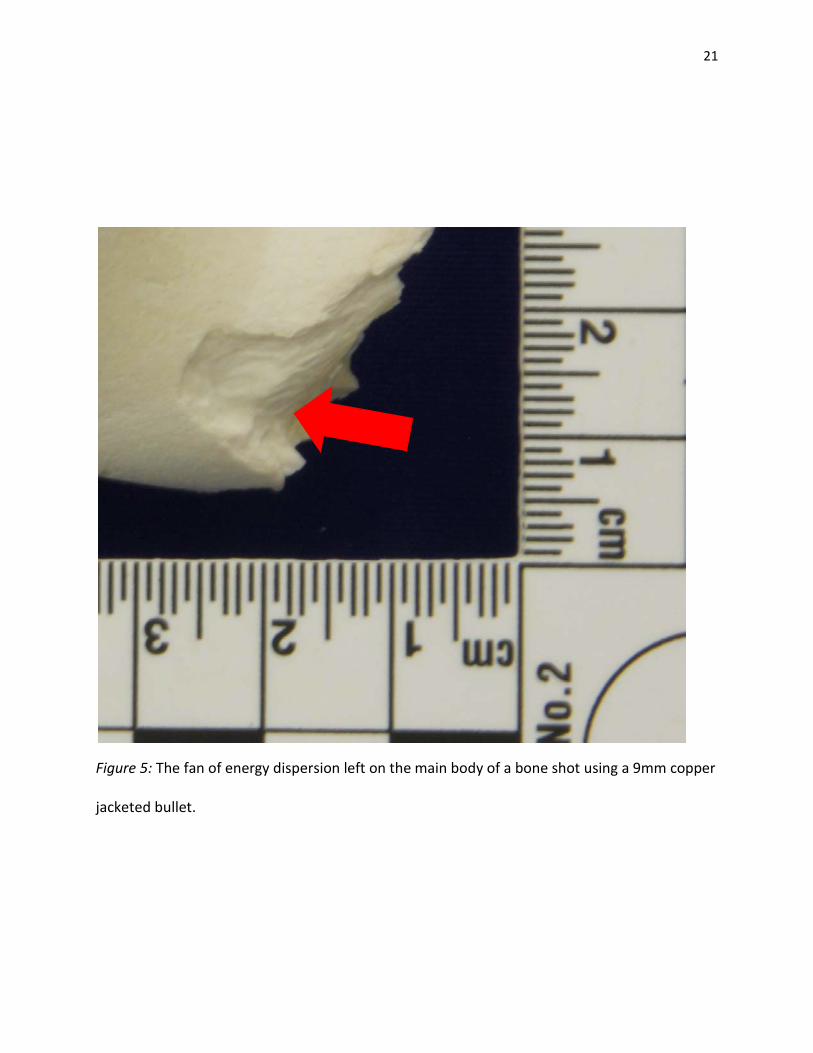
21
Figure 5: The fan of energy dispersion left on the main body of a bone shot using a 9mm copper
jacketed bullet.

22
Considerations
The main limitation of this study is that the ammunition used only included two different
sizes and two different types. While using three different calibres, both the 9mm and 0.38
bullets measured 9mm in diameter. This limited the calibre estimation portion of this study
because only two groups of entry wound sizes were compared. Both the research previously
done and this study confirm that calibres can be grouped into small versus large based on
damage to bone (1, 2). As this study should be considered a beginning, a more diverse
assortment of calibres should be used to obtain a more definitive conclusion on whether
specific calibre estimation is possible. The types of ammunition are far greater than what is
reflected in this study. Future research should focus on the effects of other types of bullets,
such as hollow point or soft-point jacketed bullets. It would be of value to see how a soft-point
jacketed bullet affects the bone tissue and if spalling occurs as it is composed of both yielding
and non-yielding materials. The statistical analysis would benefit from increasing the sample
size, as normality may be obtained in the measurements of entrance wounds. Tripling the
sample size to thirty specimens per calibre would remedy sampling constraints.
This study has forensic relevance in cases where no bullet is recovered from the scene of
victim of a gunshot wound. In this case, only the physical evidence left on a body can be used to
piece together the perimortem events surrounding the death of an individual. A forensic
anthropologist or pathologist would be able to look at the size of the entrance wound, the
presence of lead on the margins of the wound, as well as the presence or absence of bone
spalling to determine what size and type of bullet was used to inflict the damage to the victim’s
bones. This research lends itself to the now more focused description of ammunition used
23
during the search and seizure of evidence. While this study does not identify the actual bullet
that was used to shoot the victim, it does provide additional information that is germane to an
investigation.
Conclusion
The findings of this study were found to be consistent with the results generated regarding
calibre estimation in cranial bones (1, 2). It was found that a 0.22 calibre entrance wound is a
significantly different size than those generated using a 9mm bullet and a 0.38 calibre bullet,
regardless of the type of bullet used. Because a 9mm and a 0.38 are the same physical size,
they produce entrance wounds that are not different.
This study found that bone spalling is indicative of the use of a jacketed bullet, as spalling is
not present when a lead bullet is used. While further research is recommended, this study finds
that bullet type can be estimated using the presence or absence of bone spalling.
24
References
(1) Berryman H, Smith O, Symes S. Diameter of cranial gunshot wounds as a function of bullet
caliber. J Forensic Sci. 1995; 40: 751-54.
(2) Ross A. Caliber estimation from cranial entrance defect measurements. J Forensic Sci. 1996;
41: 629-33.
(3) Di Maio V. Gunshot Wounds: Practical Aspects of Firearms, Ballistics and Forensic
Techniques. First Edition. 1998, CRC Press.
(4) Hillier M. and Bell L. Differentiating Human Bone from Animal Bone: A Review of
Histological Methods. J Forensic Science. 2007; 52: 249- 63.
(5) Gray H. Gray’s anatomy. Edited by Williams P, Warwick R, Dyson M & Bannister L. 37th
edition. 1989; Churchill Livingstone.
(6) Piekarski K. Fracture of bone. J App Physics. 1970; 41: 215-25
(7) Rho J, Kuhn-Spearing L, Zioupos P. Mechanical properties and the hierarchical structure of
bone. Mechanical engineering & physics. 1998; 20.2: 92-102.
(8) Vashishth D, Tanner K, Bonfield W. Contribution, development and morphology of
microcracking in cortical bone during crack propagation. J Biomech. 2000; 33: 1169-74.
(9) Cohen H, Kugel C, May H, Medlej B, Stein S, Slon V, Hershkovitz I, Brosh T. The impact
velocity and bone fracture pattern: forensic perspective. J Forensic Sci. 2016; 266: 54-62.
(10) Langley N. An anthropological analysis of gunshot wounds to the chest. J Forensic Sci.
2007; 52: 532-7.
25
(11) Galloway A. Chapter 3: Fracture patterns and skeletal morphology: introduction and the
skull. Broken bones: anthropological analysis of blunt force trauma. Edited by Galloway A.
1999; Charles C Thomas Publisher.
(12) Coe J. External beveling of entrance wounds by handguns. Am J Fors Med Path. 1982; 3:
215-19.
(13) Carter H. Method of Making a Bullet. United States Patent US4793037. December 27,
1988.
(14) Rogers L. Radiology of Skeletal Trauma. Second Edition, New York: Churchill Livingstone;
1992. As cited by Galloway A, Broken Bones; 1999.
(15) IBM SPSS Statistics for Windows, Version 23.0.0.2. Armonk, NY: IBM Corp (2015)
(16) Diederichs M and Marin C. Measurement of Spalling Parameters from Lab Testing. Int
Soc for Rock Mech. 2010; 2: 323-326.