Calcifying extracellular mucus substances (EMS) of Madrepora ...
14
Freiwald A, Roberts JM (eds), 2005, Cold-water Corals and Ecosystems. Springer-Verlag Berlin Heidelberg, pp 731-744 Calcifying extracellular mucus substances (EMS) of Madrepora oculata – a first geobiological approach Joachim Reitner Geobiology-GZG, Göttingen University, Goldschmidtstr. 3, D-37077 Göttingen, Germany ([email protected]) Abstract. Colonial non-zooxanthellate corals from deep-water coral reefs, Lophelia pertusa and Madrepora oculata, produce large amounts of extracellular mucus (EMS). This mucus has various functions, e.g., an antifouling capability protecting the coral skeleton from attacks of endolithic and boring organisms. Both corals show thick epithecal and exothecal skeletal parts with a clear lamellar growth pattern. The formation of the epitheca is unclear. It is supposed that the EMS play a central role during the calcification process of the epithecal skeletal parts. Staining with the fluorochrome tetracycline has shown an enrichment of Ca 2+ ions in the mucus. In order to investigate this hypothesis, the protein content of the mucus and the intracrystalline organic matter from newly formed epithecal aragonite of Madrepora oculata was determined via sodium dodecyl sulfate (SDS) gel electrophoresis. Identical band patterns within both substances could be detected, one around 45 kDa molecular weight and a cluster around 30-35 kDa molecular weight. The occurrence of identical protein patterns within the mucus and in the newly formed aragonite confirms the idea that the mucus plays an important role during the organomineralization of the coral epitheca. Keywords. Biomineralisation, extracellular mucus, martix proteins, Madrepora, SDS gel electrophoresis Introduction Many aspects of the biomineralization of scleractinian skeletons are still unclear. Various authors have described in detail the microstructure of the aragonitic skeleton (Bryan and Hill 1941; Johnston 1980; Gladfelter 1984; Constantz 1986; Constantz and Meike 1989; Cuif et al. 1996; Cuif and Dauphin 1998; Stolarski 2003; and many others). Only some of them have studied the interaction of organic matter and skeletal formation. The hypothesis that acidic organic macromolecules control the nucleation of calcium carbonate minerals developed by Wilbur and Simkiss (1979),
Transcript of Calcifying extracellular mucus substances (EMS) of Madrepora ...

Freiwald A, Roberts JM (eds), 2005, Cold-water Corals and Ecosystems. Springer-Verlag Berlin Heidelberg, pp 731-744
Calcifying extracellular mucus substances (EMS) of Madrepora oculata – a first geobiological approach
Joachim Reitner
Geobiology-GZG, Göttingen University, Goldschmidtstr. 3, D-37077 Göttingen, Germany
Abstract. Colonial non-zooxanthellate corals from deep-water coral reefs, Lophelia pertusa and Madrepora oculata, produce large amounts of extracellular mucus (EMS). This mucus has various functions, e.g., an antifouling capability protecting the coral skeleton from attacks of endolithic and boring organisms. Both corals show thick epithecal and exothecal skeletal parts with a clear lamellar growth pattern. The formation of the epitheca is unclear. It is supposed that the EMS play a central role during the calcification process of the epithecal skeletal parts. Staining with the fluorochrome tetracycline has shown an enrichment of Ca2+ ions in the mucus. In order to investigate this hypothesis, the protein content of the mucus and the intracrystalline organic matter from newly formed epithecal aragonite of Madrepora oculata was determined via sodium dodecyl sulfate (SDS) gel electrophoresis. Identical band patterns within both substances could be detected, one around 45 kDa molecular weight and a cluster around 30-35 kDa molecular weight. The occurrence of identical protein patterns within the mucus and in the newly formed aragonite confirms the idea that the mucus plays an important role during the organomineralization of the coral epitheca.
Keywords. Biomineralisation, extracellular mucus, martix proteins, Madrepora, SDS gel electrophoresis
Introduction
Many aspects of the biomineralization of scleractinian skeletons are still unclear. Various authors have described in detail the microstructure of the aragonitic skeleton (Bryan and Hill 1941; Johnston 1980; Gladfelter 1984; Constantz 1986; Constantz and Meike 1989; Cuif et al. 1996; Cuif and Dauphin 1998; Stolarski 2003; and many others). Only some of them have studied the interaction of organic matter and skeletal formation. The hypothesis that acidic organic macromolecules control the nucleation of calcium carbonate minerals developed by Wilbur and Simkiss (1979),

732 Reitner
Weiner et al. (1983) and Addadi and Weiner (1985, 1992) provides a fundamentally new view of the calcium carbonate formation and entire architecture of the coral skeleton. The basic idea is that COO--rich (acidic) proteins form peptides with ß- sheet structures. These ß-sheets or flat-monolayer proteins have highly organized COO- groups with distinct distances. Divalent cations bond to this COO- and form an interface which is characterized by a crystal base plane (001 plane). Few authors have used this idea and have started to extract intracrystalline organic matter and to analyse it (Young 1971; Mitterer 1978; Constantz and Weiner 1988; Cuif and Gautret 1995; Cuif et al. 1996; Dauphin and Cuif 1997; Gautret et al. 1997; Allemand et al. 2001).
The focus of the present paper is to show that the proteins of extracellular mucus substances (EMS) mediate the growth of the epitheca of the skeleton of Madrepora oculata. The EMS is a complex mixture of weakly acidic proteins and carbohydrates and has some similarities with microbial exopolymeric substances (EPS). Formation of calcium carbonate minerals via EPS is not an enzymatically controlled biomineralization. This process is mediated by macromolecules forming the EPS and is called EPS-controlled organomineralization (Reitner 1993; Defarge and Trichet 1995; Reitner et al. 1995, 2001; Trichet and Defarge 1995; Arp et al. 2003).
Freiwald and Wilson (1998) discussed the role and importance of the mucus of Lophelia in the formation of deep cold-water coral reefs. They observed that the mucus has a strong antifouling capability including development of biofilms, stops boring attacks, and cleans the tissue from extraneous particles. The most intriguing observation was the reaction of the coral by the settlement of Foraminifera (e.g., Hyrrokkin sarcophaga) and the tube-forming polychaete Eunice norvegica on the coral branches. Due to the settlement stress, the coral produces large amounts of mucus entrapping the upward growing organisms and via this process thickens the epitheca. The organic tubes of Eunice norvegica are completely calcified in this way which additionally stabilizes the coral colony. Comparable mucus-controlled process was observed in cementing freshly broken coral branches. This organomineralizing process via stress mucus stabilizes the entire reef of azooxanthellate colonial corals. This is a crucial process in the growth of deep-water coral reefs.
Material and methods
The corals were collected during an expedition with the RV “Poseidon” in 1999 (POS 254) near the Sula Ridge (64°N/8°E) 80 km east of Trondheim (Norway). The cold-water Lophelia reefs occur in water depth around 250-350 m (Freiwald et al. 1997, 2002; Reitner and Hoffmann 2003). The specimens were collected using the manned German submersible “Jago”.
The specimens were immediately fixed with 2 % buffered formol and later stored in 70 % ethanol. Some specimens were fixed with 2 % glutardialdehyde and post fixed with 2 % osmium tetroxide. For biochemical analyses the corals were immediately frozen at -20°C. The coral mucus was extracted from living

Calcifying extracellular mucus substances (EMS) of Madrepora oculata 733
specimens by stressing them with freshwater (Fig. 1). This shock-stress induces the organism to produce an enormous amount of mucus. More than 100 ml mucus was produced by a 20 cm-sized Madrepora dendroid colony. 50 ml of the mucus were sterilized using HgCl2 and immediately frozen. The non-sterilized mucus was also immediately frozen. The Madrepora colony was treated three times with fresh water after a recovery period of 15 minutes. The first mucus was slightly pink in colour, the others were clear.
The formalin-fixed specimens were embedded in resin and then cut with a hard part microtome and stained with various histochemical dyes. The Ca2+ load was
Fig. 1 Living Madrepora specimen stressed with freshwater. This shock-stress induces the organism to produce an enormous amount of mucus. Picture was taken during the RV “Poseidon” expedition POS 254 in 1999

734 Reitner
qualitatively determined using the fluorochrome tetracycline in living colonies of Madrepora and Lophelia. The mucus was enriched in Ca2+ exhibiting a strong yellow-green fluorescence when a narrow band UV-filter linked with an epifluorescence microscope Axioplan/Zeiss (Fig. 2) was applied. For description of the detailed procedure see Reitner (1993).
Fig. 2 Living Madrepora specimen stained with the Ca2+ detecting fluorochrome tetracycline. Ca2+-rich mucus substances fluoresce bright yellow using a narrow band UV-filter. The EMS exhibits a strong yellow fluorescence
The samples for biochemical analyses using High Pressure Liquid Chromatography-HPLC and electrophoresis were frozen. For decalcification, 100 mg of the sample were dissolved in pH 4-controlled acetic acid for 10 hours on a shake table. The insoluble fraction was removed by centrifugation. The insoluble pellet was removed and frozen again. The supernatant was concentrated using Omega-Microsept 3-10 kDa concentrators. The concentrate was carefully desalted using 3-4x HPLC-H2O (ultrafiltered MilliQ water) with a minimum of 90 minutes centrifugation. After this procedure the concentrate was hydrolyzed with 6N HCl. The hydrolyzed extract was derivatised with the Waters AccQ Flour Kit and analysed using a Waters HPLC. Further detailed descriptions of the analytical procedure see Gautret et al. (1997) and Reitner et al. (1995). For electrophoresis the samples were decalcified with EDTA. Protein concentrations were determined

Calcifying extracellular mucus substances (EMS) of Madrepora oculata 735
by the protein quantification assay Bradford (1976) (bovine serum albumin (BSA) BIO-RAD). Samples were electrophoresed on a 14 % sodium dodecyl sulfate (SDS) gel and silver stained. Isoelectrical focussing of proteins was done with precast gels (pH-range 3-10, SERVA). Molecular weights were determined using SDS-page electrophoresis (SDS-polyacrylamid) (further detailed descriptions of the analytical procedure see Lange et al. (2001)).
In vitro mineralization-“inhibition” assay
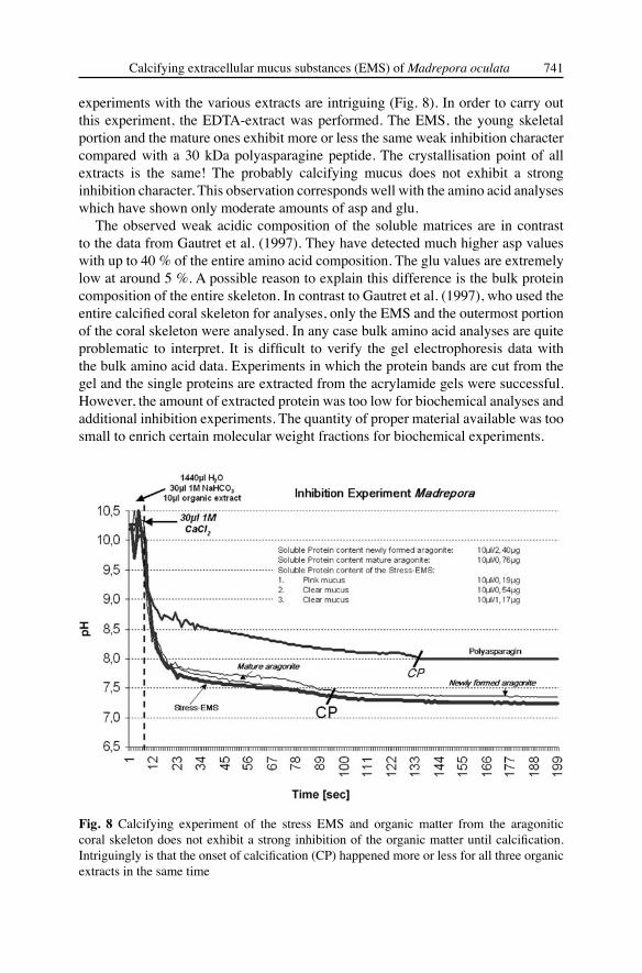
This experiment is crucial to the understanding of the role of organic molecules during the calcium carbonate crystal formation. In vitro mineralization inhibition experiments were carried out based on the procedures described by Wheeler et al. (1981), Gunthorpe et al. (1990) and Lange et al. (2001) using bulk intracrystalline native organic matter that has not been treated with SDS. It is possible to control the molecular weights using SDS-page electrophoresis cutting the stained bands and extraction of the organic matter from the gel. Inhibition experiments give indications of the Ca2+-binding capability of the organic matter and consequently the importance for skeletal and organomineral formation. Mineralization was studied in a reaction tube containing 400 μl NaHCO3 (1 M), 19 ml aqua bidest., and 200 μl sample or 200 μl aqua bidest. in the control assay, respectiveley. The solution contains total proteins or Ca2+-binding proteins. Protein concentration was 0.5 μg ml- 1 if not indicated otherwise. In order to start the reaction, 400 μl CaCl2 (1 M) were added. Inhibition of mineralization was determined by measuring the pH over a certain period. The extremely acidic artificial peptide polyasparagine 30 kDa and bidest H2O were used as standard reaction molecules. The mineralization event could be observed through the occurrence of the turbidity of the solution and the drop of the pH to 7 (CP-calcification point). During this process protons were released from NaHCO3 according to the following reaction:
NaHCO3 + CaCl2 CaCO3 + H+ + 2 Cl- + Na+ (Fig. 8).
Results
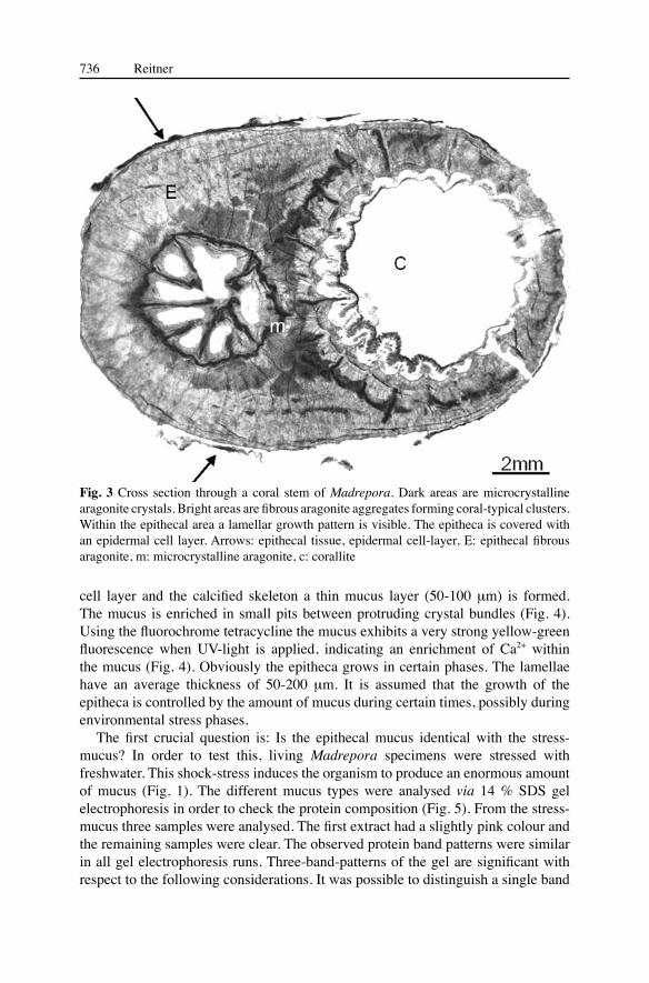
Sections through the dendroid stems of a Madrepora colony exhibit two types of mineralization pattern of the aragonite (Fig. 3). The inner corallite area including septa is formed by a microcrystalline fabric, whereas the outer and superficial areas are formed of fibre bundles of elongated aragonite crystals (sclerodermites) which are modified spherulites starting in the so-called “centre of calcification”. These centres (“seeds”) are small spherulites and clearly distinct from the sclerodermites (Constantz and Meike 1989; Cuif et al. 1999 ). These seed areas show an enrichment of organic remains and are areas of initial calcification. The microcrystalline fabric is also part of this initial growth process. However, this process is not the goal of this investigation. The outer epitheca demonstrates a clear lamellar skeletal growth pattern. The epitheca is covered by an epidermal cell layer. Between the epidermal
736 Reitner
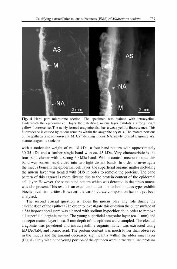
cell layer and the calcified skeleton a thin mucus layer (50-100 μm) is formed. The mucus is enriched in small pits between protruding crystal bundles (Fig. 4). Using the fluorochrome tetracycline the mucus exhibits a very strong yellow-green fluorescence when UV-light is applied, indicating an enrichment of Ca2+ within the mucus (Fig. 4). Obviously the epitheca grows in certain phases. The lamellae have an average thickness of 50-200 μm. It is assumed that the growth of the epitheca is controlled by the amount of mucus during certain times, possibly during environmental stress phases.
The first crucial question is: Is the epithecal mucus identical with the stress-mucus? In order to test this, living Madrepora specimens were stressed with freshwater. This shock-stress induces the organism to produce an enormous amount of mucus (Fig. 1). The different mucus types were analysed via 14 % SDS gel electrophoresis in order to check the protein composition (Fig. 5). From the stress-mucus three samples were analysed. The first extract had a slightly pink colour and the remaining samples were clear. The observed protein band patterns were similar in all gel electrophoresis runs. Three-band-patterns of the gel are significant with respect to the following considerations. It was possible to distinguish a single band
Fig. 3 Cross section through a coral stem of Madrepora. Dark areas are microcrystalline aragonite crystals. Bright areas are fibrous aragonite aggregates forming coral-typical clusters. Within the epithecal area a lamellar growth pattern is visible. The epitheca is covered with an epidermal cell layer. Arrows: epithecal tissue, epidermal cell-layer, E: epithecal fibrous aragonite, m: microcrystalline aragonite, c: corallite
Calcifying extracellular mucus substances (EMS) of Madrepora oculata 737
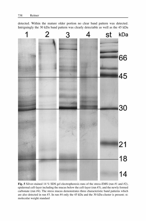
with a molecular weight of ca. 18 kDa, a four-band-pattern with approximately 30-35 kDa and a further single band with ca. 45 kDa. Very characteristic is the four-band-cluster with a strong 30 kDa band. Within control measurements, this band was sometimes divided into two tight-distant bands. In order to investigate the mucus beneath the epidermal cell layer, the superficial organic matter including the mucus layer was treated with SDS in order to remove the proteins. The band pattern of this extract is more diverse due to the protein content of the epidermal cell layer. However, the same band pattern which was detected in the stress mucus was also present. This result is an excellent indication that both mucus types exhibit biochemical similarities. However, the carbohydrate composition has not yet been analysed.
The second crucial question is: Does the mucus play any role during the calcification of the epitheca? In order to investigate this question the outer surface of a Madrepora coral stem was cleaned with sodium hypochloride in order to remove all superficial organic matter. The young superficial aragonite layer (ca. 1 mm) and a deeper mature layer in ca. 3 mm depth of the epitheca were sampled. The cleaned aragonite was powdered and intracrystalline organic matter was extracted using EDTA/NaN3 and formic acid. The protein content was much lower than observed in the mucus and the amount decreased significantly within the older inner layer (Fig. 8). Only within the young portion of the epitheca were intracrystalline proteins
Fig. 4 Hard part microtome section. The specimen was stained with tetracycline. Underneath the epidermal cell layer the calcifying mucus layer exhibits a strong bright yellow fluorescence. The newly formed aragonite also has a weak yellow fluorescence. This fluorescence is caused by mucus remains within the aragonite crystals. The mature portions of the epitheca is non-fluorescent. M: Ca2+-binding mucus, NA: newly formed aragonite, AS: mature aragonitic skeleton
738 Reitner
Fig. 5 Silver-stained 14 % SDS gel electrophoresis runs of the stress-EMS (run #1 and #2), epidermal cell-layer including the mucus below the cell-layer (run #3), and the newly formed carbonate (run #4). The stress mucus demonstrates three characteristic band patterns which are also detected in run #3. In run #4 only the 45 kDa and the 30 kDa-cluster is present; st: molecular weight standard
detected. Within the mature older portion no clear band pattern was detected. Intriguingly the 30 kDa band pattern was clearly detectable as well as the 45 kDa

Calcifying extracellular mucus substances (EMS) of Madrepora oculata 739
band within the newly formed aragonite. The 30 kDa band cluster consists of three bands, of which the 30 kDa band is the most prominent one and in some electrophoresis runs it is divided into two tight-distant bands as seen in the two mucus types. The 30 kDa band pattern and the 45 kDa band within the skeletal carbonate are identical with the pattern seen in the stress mucus and within the epithecal mucus. This is a clear indication that the EMS is part of the mineralizing system of the epitheca (Fig. 5).
From all three EMS extracts, young, and mature portion of the Madrepora epitheca, bulk amino acid analyses (fraction <10 kDa) were carried out (Figs. 6, 7). Amino acid analyses from native proteins separated via electrophoresis unfortunately did not give good data sets which was due to the extremely small amount of sampled proteins. With ca. 13 mol% asparagine (asp) and glutamine (glu) the overall EMS is only moderately acidic (Fig. 6). Isoelectric focusing of the mucus proteins demonstrates weakly acidic protein band pattern which corresponds with the measured amounts of asp and glu. More important is the observation that the ratio of asp and glu within the EMS is roughly identical.
In contrast to the extracts from the carbonate, the amount of the amino-sugar D-glucosamine is relativley high. It is assumed that the mucus contains high amounts of glycoproteins, however, the glucidic compounds have not yet been analysed. Histological staining with Alcian blue supports this assumption, because the mucus is strongly stained blue. Goreau (1956) has described a mucopolysaccharid layer located outside the basal ectodermal cell-layer and it was supposed that this layer may play a role as a template during the skeleton growth (Milliman 1974).
Fig. 6 Bulk amino acid analyses of the stress-EMS. The three extracts are more or less identical, except the excursion of glycine in the 3rd extraction. The acidic amino acids are weakly represented. The ratio of asp/glu (0.79) is more or less similar in all three extracts. All extracts have moderate amounts of amino sugars
740 Reitner
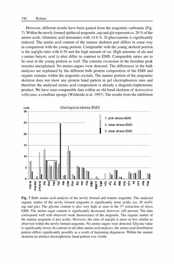
However, different results have been gained from the aragonitic carbonate (Fig. 7). Within the newly formed epithecal aragonite, asp and glu represent ca. 20 % of the amino acids. Glutamic acid dominates with 14.6 %. D-glucosamine is significantly reduced. The amino acid content of the mature skeleton part differs in some way in comparison with the young portion. Comparable with the young skeletal portion is the asp/glu ratio with 0.39 and the high amount of ser. High amounts of ala and γ-amino butyric acid (γ-aba) differ in contrast to EMS. Comparable ratios are to be seen in the young portion as well. The extreme excursion of the histidine peak remains unexplained. No amino-sugars were detected. The differences in the bulk analyses are explained by the different bulk protein composition of the EMS and organic remains within the aragonite crystals. The mature portion of the aragonitic skeleton does not show any protein band pattern in gel electrophoresis runs and therefore the analysed amino acid composition is already a diagentic/taphonomic product. We have seen comparable data within an old basal skeleton of Astrosclera willeyana, a coralline sponge (Wörheide et al. 1997). The results from the inhibition
Fig. 7 Bulk amino acid analysis of the newly formed and mature aragonite. The analysed organic matter of the newly formed aragonite is significantly more acidic (ca. 20 mol% asp and glu). The glycine content is also very high as seen in the 3rd extraction of stress EMS. The amino sugar content is significantly decreased, however, still present. The data correspond well with observed weak fluorescence of the aragonite. The organic matter of the mature aragonite is less acidic. However, the ratio of asp/glu is more or less similar as observed within the newly formed aragonite. No amino sugars were detected. Glycine value is significantly lower. In contrast to all other amino acid analyses, the amino acid distribution pattern differs significantly possibly as a result of beginning diagenesis. Within the mature skeleton no distinct electrophoretic band pattern was visible
Calcifying extracellular mucus substances (EMS) of Madrepora oculata 741
experiments with the various extracts are intriguing (Fig. 8). In order to carry out this experiment, the EDTA-extract was performed. The EMS, the young skeletal portion and the mature ones exhibit more or less the same weak inhibition character compared with a 30 kDa polyasparagine peptide. The crystallisation point of all extracts is the same! The probably calcifying mucus does not exhibit a strong inhibition character. This observation corresponds well with the amino acid analyses which have shown only moderate amounts of asp and glu.
The observed weak acidic composition of the soluble matrices are in contrast to the data from Gautret et al. (1997). They have detected much higher asp values with up to 40 % of the entire amino acid composition. The glu values are extremely low at around 5 %. A possible reason to explain this difference is the bulk protein composition of the entire skeleton. In contrast to Gautret et al. (1997), who used the entire calcified coral skeleton for analyses, only the EMS and the outermost portion of the coral skeleton were analysed. In any case bulk amino acid analyses are quite problematic to interpret. It is difficult to verify the gel electrophoresis data with the bulk amino acid data. Experiments in which the protein bands are cut from the gel and the single proteins are extracted from the acrylamide gels were successful. However, the amount of extracted protein was too low for biochemical analyses and additional inhibition experiments. The quantity of proper material available was too small to enrich certain molecular weight fractions for biochemical experiments.
Fig. 8 Calcifying experiment of the stress EMS and organic matter from the aragonitic coral skeleton does not exhibit a strong inhibition of the organic matter until calcification. Intriguingly is that the onset of calcification (CP) happened more or less for all three organic extracts in the same time

742 Reitner
Conclusions
1. It was possible to distinguish two anatomical sites with protein-rich mucus substances. Madrepora produces an extracellular stress mucus which covers the outer epidermal cell layer of the epitheca. The second mucus is located between the epidermal cell layer and the aragonitic skeleton. Both mucus types bear a comparable protein composition analysed via SDS gel electrophoresis. It is possible to distinguish a single protein with a molecular weight of ca. 18 kDa, a four-protein-cluster with approximately 30-35 kDa and a further single protein with ca. 45 kDa.
2. It was possible to extract a soluble protein composition from the newly formed aragonite crystals. Two band patterns are identical with the results obtained from both mucus types. A single protein band with a molecular weight fraction of ca. 45 kDa and the 30-35 kDa protein cluster was detectable. In some electrophoresis runs the very prominent 30 kDa band is divided into two. The IEF-gel analyses show that the mucus proteins and matrix proteins from the epitheca are weakly acidic (pH 5).
3. These data correspond with the bulk amino acids from the mucus and the skeleton. The inhibition experiment shows an intriguing result. The crystallisation point, which stops the inhibition potential of the macromolecules, was the same as in all extracts. This means that only a certain group of molecules inhibit a rapid calcification. However, in contrast to polyasparagine and other very acidic macromolecules, the measured inhibition is only weak.
4. Based on the electrophoresis data, the EMS obviously play an important role in the formation of the thick epithecal skeletal parts of Madrepora oculata and obviously plays a crucial role in the formation of extended deep-water azooxanthellate coral reefs.
Acknowledgements
The “Deutsche Forschungsgemeinschaft” is greatfully acknowledged for financing the investigations on biomineralization (Re 665/18, Re665/12 Leibniz award – Evolution of Multicelular Systems and Organomineralisation EMSO). Financial support was also provided by the German Bundesministerium für Bildung und Forschung (BMBF). This paper represents publication no. 40 of the research program, BOSMAN (BMBF 03F0358 C). The material was collected during the RV “Poseidon” expedition POS 254 in 1999. The author thanks the ship-board and scientific crew, specially the crew of the submersible “Jago”, Karin Hissmann and Jürgen Schauer for excellent support. I thank also Birgit Röring and Wolfgang Dröse (Geobiology-Göttingen) for assistance with laboratory work. My special thanks are provided to André Freiwald (Erlangen) for many intriguing discussions dealing with the mucus problem and helpful comments when reviewing the manuscript.

Calcifying extracellular mucus substances (EMS) of Madrepora oculata 743
ReferencesAddadi L, Weiner S (1985) Interactions between acidic proteins and crystals: stereochemical
requirements in biomineralisation. Proc Natl Acad Sci USA 82: 4110-4114Addadi L, Weiner S (1992) Kontroll- und Designprinzipien bei der Biomineralisation. Angew
Chem 104: 159-176Allemand D, Tambutté È, Girard J-P, Jaubert J (2001) Organic matrix synthesis in the
scleractinian coral Stylophora pistillata: role in biomineralization and potential target of the organotin tributyltin. J Exp Biol: 201: 2001-2009
Arp G, Reimer A, Reitner J (2003) Microbialite formation in seawater of increased alkalinity, Satonda Crater Lake, Indonesia. J Sediment Res 73: 105-127
Bradford M (1976) A rapid and sensitive method for the quantitation of microgram quantities of protein utilizing the principle of protein-dye binding. Anal Biochem 72: 248-254
Bryan W, Hill D (1941) Spherulitic crystallization as a mechanism of skeletal growth in the Hexacorals. Proc R Soc Queensland 52: 78-91
Constantz BR (1986) Coral skeleton construction: a physiochemically dominated process. Palaios 1: 152-157
Constantz BR, Meike A (1989) Calcite centres of calcification in Mussa angulosa (Scleractinia). In: Crick RE (ed) Origin, Evolution and modern Aspects of Biomineralization in Plants and Animals. Plenum Press, New York, pp 201-207
Constantz BR, Weiner S (1988) Acidic macromolecules associated with the mineral phase of scleractinian coral skeletons. J Exp Zool 248: 253-258
Cuif J-P, Dauphin Y (1998) Microstructural and physico-chemical characterization of “centers of calcification” in septa of some Recent scleractinian corals. Paläont Z 72: 257-270
Cuif J-P, Dauphin Y, Denis A, Gautret P (1996) The organomineral structure of coral skeletons: a potential source of new criteria for scleractinian taxonomy. Bull Inst Océanogr Monaco Spec Issue 14: 359-367
Cuif J-P, Dauphin Y, Gautret P (1999) Compositional diversity of soluble mineralizing matrices in some recent coral skeletons compared to fine-scale growth structures of fibres: discussion of consequences for biomineralization and diagenesis. Int J Earth Sci 88: 582-592
Cuif J-P, Gautret P (1995) Gluides et proteins de la matrice soluble des biocristaux de scleractiniaires acroporides. CR Acad Sci Paris 320 Ser IIa: 273-278
Dauphin Y, Cuif J-P (1997) Isoelectric properties of the soluble matrices in relation to the chemical composition of some scleractinian skeletons. Electrophoresis 18: 1180-1183
Defarge C, Trichet J (1995) From biominerals to “organominerals”: the example of the modern lacustrine calcareous stromatolites from Polynesian atolls. Bull Inst Océanogr Monaco Spec Issue 14: 265-271
Freiwald A, Wilson JB (1998) Taphonomy of modern deep, cold-temperate water coral reefs. Hist Biol 13: 37-52
Freiwald A, Henrich R, Pätzold J (1997) Anatomy of a deep-water coral reef mound from Stjernsund. SEPM Spec Publ 56: 141-161
Freiwald A, Hühnerbach V, Lindberg B, Wilson JB, Campbell J (2002) The Sula Reef Complex, Norwegian Shelf. Facies 47: 179-200
Gautret P, Cuif, J-P, Freiwald A (1997) Composition of soluble mineralizing matrices in zooxanthellate and non-zooxanthellate scleractinian corals: biochemical assessment of photosynthetic metabolism through the study of a skeletal feature. Facies 36: 189-194
Gladfelter EH (1984) Skeletal development in Acropora cervicornis. A comparison of monthly rates of linear extension and calcium carbonate accretion measured over a year. Coral Reefs 3: 51-57
744 Reitner
Goreau T (1956) Histochemistry of mucopolysaccharide-like substances and alkaline phosphatase in Madreporaria. Nature 177: 1029-1030
Gunthorpe ME, Sikes CS, Wheeler AP (1990) Promotion and inhibition of calcium carbonate crystallization in vitro by matrix protein from blue crab exoskeleton. Biol Bull 179: 191-200
Johnston I (1980) The ultrastructure of skeletogenesis in hermatypic corals. Int Rev Cyt 67: 171-214
Lange R, Bergbauer M, Szewzyk U, Reitner J (2001) Soluble proteins control growth of skeleton crystals in three coralline demosponges. Facies 45: 195-202
Milliman JD (1974) Marine carbonates. In: Milliman JD, Müller G, Förstner U (eds) Recent sedimentary Carbonates. Springer, Berlin
Mitterer RM (1978) Amino acid composition and metal binding capability of the skeletal protein of corals. Bull Mar Sci 28: 173-180
Reitner J (1993) Modern cryptic microbialite/metazoan facies from Lizard Island (Great Barrier Reef, Australia). Formation and concepts. Facies 29: 3-39
Reitner J, Hoffmann F (2003) Schwämme in Kaltwasser-Korallenriffen. Kleine Senckenberg-Reihe 45: 75-87
Reitner J, Gautret P, Marin F, Neuweiler F (1995) Automicrites in a modern marine microbialite. Formation model via organic matrices (Lizard Island, Great Barrier Reef, Australia). Bull Inst Océanogr Monaco Spec Issue 14: 237-263
Reitner J, Wörheide G, Lange R, Schumann-Kindel G (2001) Coralline demosponges – a geobiological portait. Bull Tohoku Univ Mus 1: 219-235
Stolarski J (2003) Three-dimensional micro- and nanostructural characteristics of the scleractinian coral skeleton: a biocalcification proxy. Acta Palaeontol Pol 48: 497-530
Trichet J, Defarge C (1995) Non-biologically supported organomineralisation. Bull Inst Océanogr Monaco Spec Issue 14: 203-236
Weiner S, Traub W, Lowenstam HA (1983) Organic matrix in calcified exoskeletons. In: Westbroek P, de Jong EW (eds) Biomineralization and Biological Metal Accumulation. Reidel, Amsterdam, pp 205-224
Wheeler AP, George JW, Evans CA (1981) Control of calcium carbonate nucleation and crystal growth by soluble matrix of oyster shell. Science 212: 1397-1398
Wilbur KM, Simkiss K (1979) Carbonate turnover and depostion by Metazoa. In: Trudinger PA, Swaine DJ (eds) Studies in Environmental Sciences. Biochemical Cycling of Mineral-forming Elements. Elsevier, Amsterdam, pp 69-106
Wörheide G, Gautret P, Reitner J, Böhm F, Joachimski MM, Thiel V, Michaelis W, Massault M (1997) Basal skeletal formation, role and preservation of intracrystalline organic matrices, and isotopic record in the coralline sponge Astrosclera willeyana Lister, 1900. Bol R Soc Esp Hist Nat (Sec Geol) 91: 355-374
Young SD (1971) Organic material from scleractinian coral skeleton. 1. Variation in composition between several species. Cop Biochem Physiol 40B: 113-120