Calcific Metamorphosis with Pathological Root Resorption ... · Calcific Metamorphosis with...
7
712 Int. J. Morphol., 33(2):712-718, 2015. Calcific Metamorphosis with Pathological Root Resorption in Permanent Teeth: Morphohistometric Evaluation of Two Cases Metamorfosis Calcificante Asociada a Reabsorción Patológica Radicular en Dientes Permanentes: Evaluación Morfohistométrica de Dos Casos Gabriel M. Fonseca *,** & Miguel M. Fonseca ** FONSECA, G. M. & FONSECA, M. M. Calcific metamorphosis with pathological root resorption in permanent teeth: morphohistometric evaluation of two cases. Int. J. Morphol., 33(2):712-718, 2015. SUMMARY: Calcific Metamorphosis (CM) is a pulpal response to dental trauma characterized by a deposition of hard tissue within the canal space. A tooth with CM usually presents a discoloration and a partial or total obliteration of the pulp canal space, and its vital pulp tests and symptoms are difficult. Since pulp necrosis cannot be supposed even the negative responses, the periradicular status is the only reliable criterion. Two permanent teeth diagnosed as CM and pathological root resorption, extracted from two males (22 and 53 years of age) due to severe mobility were prepared and sectioned for histological and histometrical evaluation. Images were captured, processed and measured in a total mapping of each specimen with software Pinnacle Studio 9.4® (Pinnacle Systems Inc.), Adobe® Photoshop® (CS 8.0.1, Adobe Systems Inc.) and Image J® (National Institute of Health, Bethesda, MD, USA). The images showed the simultaneous occurrence of CM and root resorption. The means of the total measurements showed that the area of radicular affection (R) was higher than the coronal affection (C), both in quantity (R: 13.75 mm 2 -28.75%-/C: 4.7 mm 2 -5.47%-) and quality (R: absence of CM / C: presence of CM). CM showed a fibrotic pattern, a cortical bone-like pattern and a cancellous bone-like pattern, representing a kind of reparative reaction probably initiated by the resorption process. Concurrent CM and pathological root resorption are uncommon in the same tooth, and the possibility to obtain specimens of permanent teeth to make histological and histometric evaluations of them represents a situation even more infrequent. This morphohistometric evaluation can expand the spectrum of useful variables to make clinical and therapeutic odontological decisions. KEY WORDS: Dental pulp; Calcific metamorphosis; Pathological root resorption; Diagnoses; Endodontics. INTRODUCTION Traumatic dental injuries can affect all the hard and soft tissues in and surrounding the teeth. Concussion, subluxation and luxation are moderate injuries in a majority of cases; they can be associated with minor symptoms and, therefore, go unnoticed both by the patient and/or by the clinician (Malhotra & Mala, 2013; Velásquez et al., 2014). These events and their clinical management can present important challenges to the odontologist; since the sequel can arise several years after the injury, proper medical and dental history, as well as a detailed history of the dental trau- ma, a thorough clinical examination will assist the dental provider in formulating a proper diagnosis and subsequently, adequate treatment (Ajmera & Mulay, 2013). Pulpal reactions to traumatic injuries can vary from immediate pulpal necrosis to long-term response depending on the degree and type of trauma, age of the patient and the condition of the tooth in the post-traumatic period. In a pulp with moderate or very good prognosis for healing, the tooth can show an obliteration of the pulp chamber and canal in periods of 18 months to five years or more. This is a common sequel to dental trauma and it was referred as Calcific Metamorphosis (CM) also called Pulp Canal Obliteration (Malhotra & Mala; Oginni et al., 2009). This condition is defined by the American Association of Endodontists as “…a pulpal response to trauma characterized by rapid deposition of hard tissue within the canal space” (Siddiqui, 2014). The frequency of CM depends on the extent and severity of the injury and the stage of the root development at the time of the event, and up to 24% of traumatized teeth develops varying degrees of this condition (Malhotra * Professor, Department of Integral Adult Dentistry, CIMA Research group, Faculty of Dentistry, Universidad de La Frontera, Temuco, Chile ** Professor, Department of Oral Pathology, Faculty of Dentistry, National University of Cordoba, Cordoba, Argentina.
Transcript of Calcific Metamorphosis with Pathological Root Resorption ... · Calcific Metamorphosis with...
712
Int. J. Morphol.,33(2):712-718, 2015.
Calcific Metamorphosis with Pathological Root Resorption inPermanent Teeth: Morphohistometric Evaluation of Two Cases
Metamorfosis Calcificante Asociada a Reabsorción Patológica Radicularen Dientes Permanentes: Evaluación Morfohistométrica de Dos Casos
Gabriel M. Fonseca*,** & Miguel M. Fonseca**
FONSECA, G. M. & FONSECA, M. M. Calcific metamorphosis with pathological root resorption in permanent teeth: morphohistometricevaluation of two cases. Int. J. Morphol., 33(2):712-718, 2015.
SUMMARY: Calcific Metamorphosis (CM) is a pulpal response to dental trauma characterized by a deposition of hard tissuewithin the canal space. A tooth with CM usually presents a discoloration and a partial or total obliteration of the pulp canal space, and itsvital pulp tests and symptoms are difficult. Since pulp necrosis cannot be supposed even the negative responses, the periradicular statusis the only reliable criterion. Two permanent teeth diagnosed as CM and pathological root resorption, extracted from two males (22 and53 years of age) due to severe mobility were prepared and sectioned for histological and histometrical evaluation. Images were captured,processed and measured in a total mapping of each specimen with software Pinnacle Studio 9.4® (Pinnacle Systems Inc.), Adobe®Photoshop® (CS 8.0.1, Adobe Systems Inc.) and Image J® (National Institute of Health, Bethesda, MD, USA). The images showed thesimultaneous occurrence of CM and root resorption. The means of the total measurements showed that the area of radicular affection (R)was higher than the coronal affection (C), both in quantity (R: 13.75 mm2 -28.75%-/C: 4.7 mm2 -5.47%-) and quality (R: absence of CM/ C: presence of CM). CM showed a fibrotic pattern, a cortical bone-like pattern and a cancellous bone-like pattern, representing a kindof reparative reaction probably initiated by the resorption process. Concurrent CM and pathological root resorption are uncommon in thesame tooth, and the possibility to obtain specimens of permanent teeth to make histological and histometric evaluations of them representsa situation even more infrequent. This morphohistometric evaluation can expand the spectrum of useful variables to make clinical andtherapeutic odontological decisions.
KEY WORDS: Dental pulp; Calcific metamorphosis; Pathological root resorption; Diagnoses; Endodontics.
INTRODUCTION
Traumatic dental injuries can affect all the hard andsoft tissues in and surrounding the teeth. Concussion,subluxation and luxation are moderate injuries in a majorityof cases; they can be associated with minor symptoms and,therefore, go unnoticed both by the patient and/or by theclinician (Malhotra & Mala, 2013; Velásquez et al., 2014).These events and their clinical management can presentimportant challenges to the odontologist; since the sequelcan arise several years after the injury, proper medical anddental history, as well as a detailed history of the dental trau-ma, a thorough clinical examination will assist the dentalprovider in formulating a proper diagnosis and subsequently,adequate treatment (Ajmera & Mulay, 2013).
Pulpal reactions to traumatic injuries can vary fromimmediate pulpal necrosis to long-term response depending
on the degree and type of trauma, age of the patient and thecondition of the tooth in the post-traumatic period. In a pulpwith moderate or very good prognosis for healing, the toothcan show an obliteration of the pulp chamber and canal inperiods of 18 months to five years or more. This is a commonsequel to dental trauma and it was referred as CalcificMetamorphosis (CM) also called Pulp Canal Obliteration(Malhotra & Mala; Oginni et al., 2009). This condition isdefined by the American Association of Endodontists as “…apulpal response to trauma characterized by rapid depositionof hard tissue within the canal space” (Siddiqui, 2014).
The frequency of CM depends on the extent andseverity of the injury and the stage of the root developmentat the time of the event, and up to 24% of traumatizedteeth develops varying degrees of this condition (Malhotra
* Professor, Department of Integral Adult Dentistry, CIMA Research group, Faculty of Dentistry, Universidad de La Frontera, Temuco, Chile** Professor, Department of Oral Pathology, Faculty of Dentistry, National University of Cordoba, Cordoba, Argentina.
713
& Mala). There appears to be a significant correlationbetween CM and a history of tooth mobility as a result ofthe trauma (Heling et al., 2000; Holcomb & Gregory, 1967;Siddiqui). Although it can be recognized clinically as earlyas 3 months after injury, in most cases it goes undetectedfor about one year or more (Oginni et al.).
The clinical picture of CM has been described as atooth with a yellowish or yellowish brown discolorationthan the normal adjacent teeth because of a decrease intranslucency due to a greater thickness of dentine (Ajmera& Mulay; Manuel et al., 2010). However, not all such teethbecome discolored, and there is no correlation betweenthe amount of discoloration and the degree of CM(Holcomb & Gregory). The radiographic appearance ofCM is a partial (limited to the coronal part of the tooth) ortotal (extended to the coronal and radicular pulp canalspaces) obliteration of the pulp canal space with normalperiodontal membrane space and intact lamina dura (Holan,1998; Malhotra & Mala). When CM is associated withapical periodontitis, a thickening of the periodontalligament space or peri-radicular radiolucency may beobserved, with or without subjective symptoms (Malhotra& Mala).
Vital pulp tests and symptoms of CM are difficult;responses to heat and cold decrease with time and theresponse to electric pulp tests may be normal in the earlystages but absent in the later stages. Generally there is nosensitivity to percussion. Teeth with CM may or may nothave symptoms, depending on the status of pulp tissuewithin the mineralized canals (Holcomb & Gregory;Malhotra & Mala; Oginni et al.). There also seems to beno correlation between the amount of discoloration andthe response to the vitalometer (Holcomb & Gregory).Jacobsen & Kerekes (1977) concluded that a negativerespond to sensitivity tests not necessarily indicate pulpnecrosis, and the periradicular status seems to be the onlyreliable criterion. The therapeutic significance of all thesesigns has been a recurrent topic in the literature. Eventhough some classical authors recommended theendodontic therapy, apical surgery and retrogradeendodontic treatment or even the extraction due CM couldrepresents a potential focus of infection (Patterson &Mitchell, 1965), other authors underlined that this conditiondoes not justify these procedures since endodonticcomplications (pulp necrosis and apical rarefaction)occurred in less than one-third of teeth with CM and routineendodontic therapy is virtually impossible (Holcomb &Gregory; Jacobsen & Kerekes; Munley & Goodell, 2005).
Histology of CM is characterized by an abnormaldeposition with characteristics of bone-like, dentine-like
or fibrotic tissue with entrapment of some pulpal cells(Malhotra & Mala; Piattelli & Trisi, 1993; Robertson etal., 1997). Holan mentioned that the histologic appearanceof this calcified tissue was different from typical irregularsecondary dentine adjacent to the primary dentine in thesame section, excluding the possibility that this abnormaltissue was some kind of dentine. CM is a response of avital pulp to trauma; it was proposed that the temporarydisruption of blood supply in trauma occurs followed bydestruction of odontoblasts and activation ofundifferentiated mesenchimal cells that form the reparativetissue (Ajmera & Mulay). Heling et al., mentioned thatCM might be seen as a metaplastic transformation of thepulp into bone-like tissue.
Even though the degree of obliteration, the type of tissueand their distribution in the pulp chamber, seem to be fun-damental considerations to be able to make the appropriatedecisions, it is not possible to determine the extent of theobliteration only from a clinical or radiographicexamination (Malhotra & Mala). It was accepted that theobliteration of the pulp canal spaces advances in a corono-apical direction, but only the histologic evaluation of thenarrow pulp canals confirmed the existence of pulp tissuewhen the obliteration was total (Malhotra & Mala).Robertson et al., suggested that even though different typesof obliterating tissues did not correspond to clinicaldiagnoses, it could correspond to certain histologicalcombinations of these types.
The pathological root resorption has numerousreports in the literature and it has been associated with along-standing chronic inflammation in the pulp. Betweenthe different contributory factors, trauma has beensuggested as one of the primary triggers. Regardless, thiscondition has been classified as internal and external (withsubdivisions), the devastation rate of resorption may berapid or slow and an opportune treatment is recommendedin all diagnosed cases. When root destruction makes in-compatible the permanence of the teeth in the arch,extractions must be planned (Caliskan & Türkün, 1997;Armas et al., 2008).
Concurrent CM and pathological root resorption areuncommon in the same tooth (Armas et al.; Kuster, 1981;Peterson et al., 1985) and the possibility to obtainspecimens of permanent teeth to make histological andhistometric evaluations of them represents a situation evenmore infrequent (Kuyk & Walton, 1990; Piattelli & Trisi).We present a morphohistometric evaluation of twopermanent teeth where the characterization and distributionof these conditions can expand the spectrum of useful va-riables to make clinical and therapeutic decisions.
FONSECA, G. M. & FONSECA, M. M. Calcific metamorphosis with pathological root resorption in permanent teeth: morphohistometric evaluation of two cases.Int. J. Morphol., 33(2):712-718, 2015.
714
MATERIAL AND METHOD
The material consisted of two permanent teethobtained from two males (aged 22 and 53 years) referred tothe Oral Pathology at the Faculty of Dentistry of theUniversity of Cordoba (Argentina). Teeth had been exposedto trauma and extracted at different time intervals after injury.CM associated with pathological root resorption wasdiagnosed in both cases according to the almost completeobliteration of pulp chamber and the extensive root resorptionradiographically (Table I). After having obtained informedconsent from the patients, extractions were made due to theextensive loss of marginal bone and severe mobility.
The teeth were washed with physiological solution,fixed and stored immediately after extraction in 10% neu-tral-buffered formalin until sectioning. All teeth weredecalcified by immersing in 7.5% nitric acid and dehydratedin ascending concentrations of ethyl alcohol andconventionally prepared for paraffin embedding, then seriallysectioned to obtain 5- to 10-µm slides. Sections were stainedwith hematoxylin and eosin for histological evaluation. Allprocedures were made in according with previous reports toavoid artifacts of space and staining, and to obtain a verygood microscopic resolution of all of the structures (Patterson& Mitchell; Piattelli & Trisi).
Histological and histometric evaluations were madeat the Oral Pathology Department, Laboratory of Experi-mental Pathology and Tissue Engineering, Dental School,University of Tucuman (Tucuman, Argentina). The morerepresentative sections were selected evaluated with a Sonydigital camera (Sony Corp., Tokyo, Japan) mounted on anOlympus CH30 light microscope (Olympus, Center Valley,PA, USA). Images were captured in horizontal and verticalsequences by using software Pinnacle Studio 9.4® (PinnacleSystems Inc.) at magnification x 56.7. Adobe® Photoshop®software (CS 8.0.1, Adobe Systems Inc.) was used to fuse
the saved images in a total mapping of each specimens andimages were measured off-line using Image J® (NationalInstitute of Health, Bethesda, MD, USA).
RESULTS
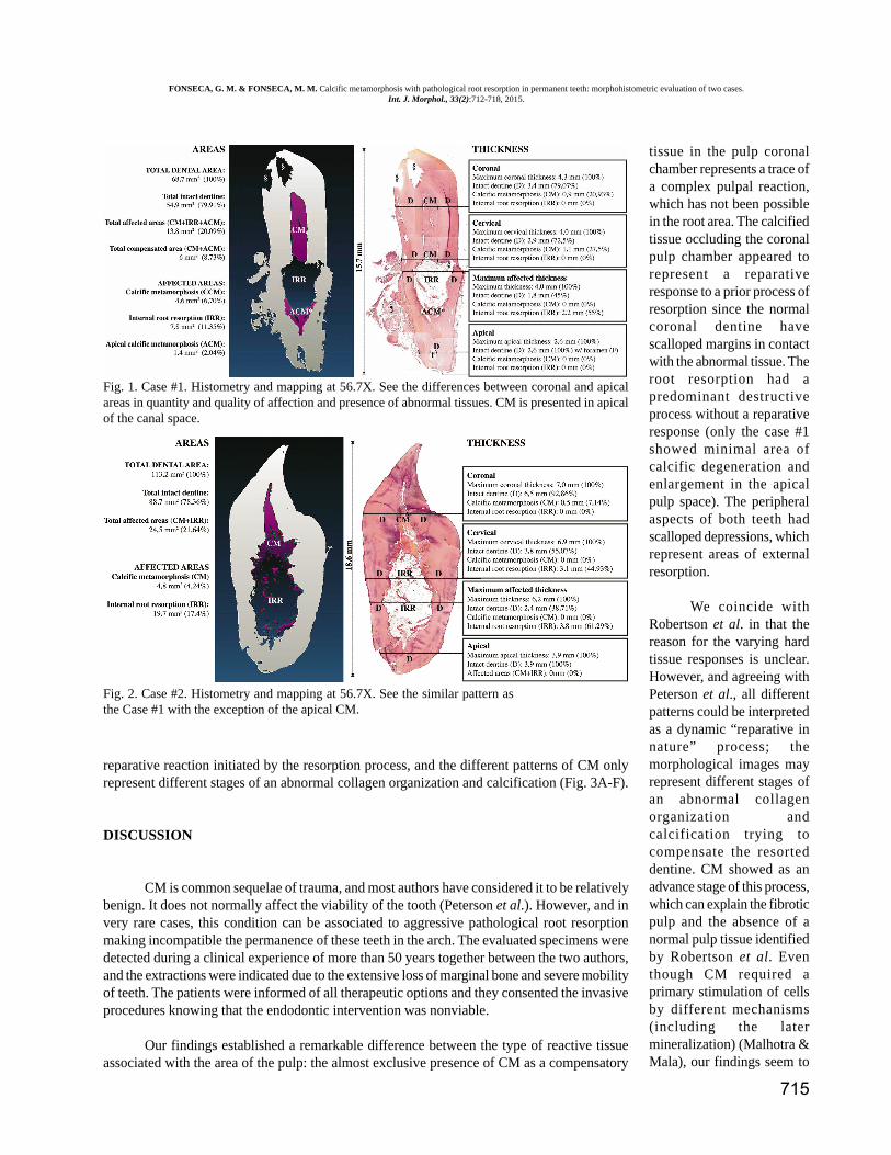
The histologic findings showed the simultaneousoccurrence of CM and root resorption. Although the vitalitypulp tests were negative, pulps had degenerative images butno necrosis images. Whereas there was an almost completeabsence of the pulp chamber in both cases with a calcifiedtissue occluded the pulpal lumen, an internal resorption wasrepresented by resorption lacunae along the root canal wallswithout this hard tissue apposition. The histometric evaluationis represented in Figures 1 and 2. The means of the totalmeasurements showed that the area of radicular affection (R)was higher than the coronal affection (C), both in quantity (R:13.75 mm2 -28.75%- /C: 4.7 mm2 -5.47%-) and quality (R:absence of CM / C: presence of CM).
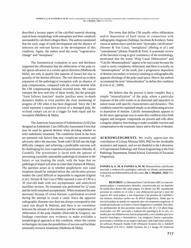
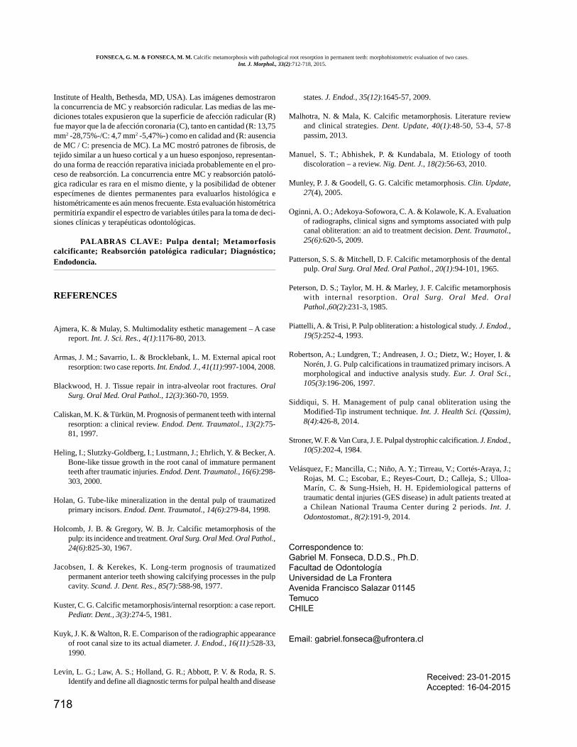
An abnormal tissue occluding the coronal pulpdemonstrated the CM. This tissue showed three differentpatterns, all of a collagen structure: a) a fibrotic appearanceresembling collagenous connective tissue, with courses ofcollagen-like fibers and cells. In parts this pattern filled theresorption cavities; b) a calcified mass resembling a “corticalbone” with a concentric layer structure and numerous butempty cell lacunae. This pattern was located in the peripheralof the CM; c) a trabecular pattern resembling a “cancellousbone”, located in the most internal part of the CM andsurrounding the pulp tissue; this pattern has the sameconcentric layer structure and lacunae and in parts resembleddenticles-like or barriers-like forms. The remains of pulpshowed signs of aging, atrophy and fibrosis with loss ofodontoblasts. In contact with the active resorption cavities,inflammatory cells were found. The main conclusion of thehistological evaluation was that the CM could represent a
Case # Age Tooth Age oftrauma
Type oftrauma
Diagnosisat trauma Clinic image Radiograph Pulp vitality
cold/heat tests
1 22 12 9 Direct(Fall)
Lateralluxation
No discoloration.Asymptomatic. Severe
mobility.
Almost completeobli teration of pulp
chamber,pathological root
resorption.Extensive loss of
marginal bone
Negative
2 53 11 23 Direct(Blow)
Lateralluxation
Yellowish discolored.Asymptomatic. Severe
mobility
Almost completeobli teration of pulp
chamber,pathological root
resorption
Negative
Table I. Clinical characteristics and history of the samples included in the calcific metamorphosis study.
FONSECA, G. M. & FONSECA, M. M. Calcific metamorphosis with pathological root resorption in permanent teeth: morphohistometric evaluation of two cases.Int. J. Morphol., 33(2):712-718, 2015.
715
Fig. 1. Case #1. Histometry and mapping at 56.7X. See the differences between coronal and apicalareas in quantity and quality of affection and presence of abnormal tissues. CM is presented in apicalof the canal space.
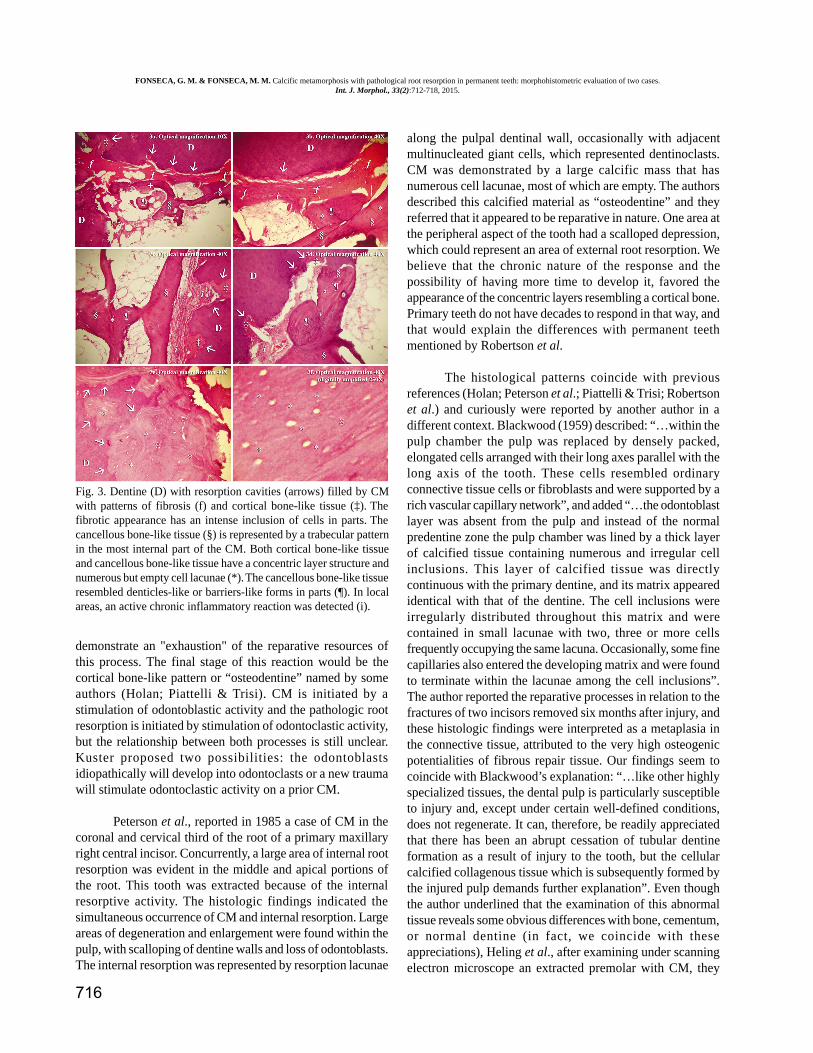
Fig. 2. Case #2. Histometry and mapping at 56.7X. See the similar pattern asthe Case #1 with the exception of the apical CM.
reparative reaction initiated by the resorption process, and the different patterns of CM onlyrepresent different stages of an abnormal collagen organization and calcification (Fig. 3A-F).
DISCUSSION
CM is common sequelae of trauma, and most authors have considered it to be relativelybenign. It does not normally affect the viability of the tooth (Peterson et al.). However, and invery rare cases, this condition can be associated to aggressive pathological root resorptionmaking incompatible the permanence of these teeth in the arch. The evaluated specimens weredetected during a clinical experience of more than 50 years together between the two authors,and the extractions were indicated due to the extensive loss of marginal bone and severe mobilityof teeth. The patients were informed of all therapeutic options and they consented the invasiveprocedures knowing that the endodontic intervention was nonviable.
Our findings established a remarkable difference between the type of reactive tissueassociated with the area of the pulp: the almost exclusive presence of CM as a compensatory
tissue in the pulp coronalchamber represents a trace ofa complex pulpal reaction,which has not been possiblein the root area. The calcifiedtissue occluding the coronalpulp chamber appeared torepresent a reparativeresponse to a prior process ofresorption since the normalcoronal dentine havescalloped margins in contactwith the abnormal tissue. Theroot resorption had apredominant destructiveprocess without a reparativeresponse (only the case #1showed minimal area ofcalcific degeneration andenlargement in the apicalpulp space). The peripheralaspects of both teeth hadscalloped depressions, whichrepresent areas of externalresorption.
We coincide withRobertson et al. in that thereason for the varying hardtissue responses is unclear.However, and agreeing withPeterson et al., all differentpatterns could be interpretedas a dynamic “reparative innature” process; themorphological images mayrepresent different stages ofan abnormal collagenorganization andcalcification trying tocompensate the resorteddentine. CM showed as anadvance stage of this process,which can explain the fibroticpulp and the absence of anormal pulp tissue identifiedby Robertson et al. Eventhough CM required aprimary stimulation of cellsby different mechanisms(including the latermineralization) (Malhotra &Mala), our findings seem to
FONSECA, G. M. & FONSECA, M. M. Calcific metamorphosis with pathological root resorption in permanent teeth: morphohistometric evaluation of two cases.Int. J. Morphol., 33(2):712-718, 2015.
716
demonstrate an "exhaustion" of the reparative resources ofthis process. The final stage of this reaction would be thecortical bone-like pattern or “osteodentine” named by someauthors (Holan; Piattelli & Trisi). CM is initiated by astimulation of odontoblastic activity and the pathologic rootresorption is initiated by stimulation of odontoclastic activity,but the relationship between both processes is still unclear.Kuster proposed two possibilities: the odontoblastsidiopathically will develop into odontoclasts or a new traumawill stimulate odontoclastic activity on a prior CM.
Peterson et al., reported in 1985 a case of CM in thecoronal and cervical third of the root of a primary maxillaryright central incisor. Concurrently, a large area of internal rootresorption was evident in the middle and apical portions ofthe root. This tooth was extracted because of the internalresorptive activity. The histologic findings indicated thesimultaneous occurrence of CM and internal resorption. Largeareas of degeneration and enlargement were found within thepulp, with scalloping of dentine walls and loss of odontoblasts.The internal resorption was represented by resorption lacunae
along the pulpal dentinal wall, occasionally with adjacentmultinucleated giant cells, which represented dentinoclasts.CM was demonstrated by a large calcific mass that hasnumerous cell lacunae, most of which are empty. The authorsdescribed this calcified material as “osteodentine” and theyreferred that it appeared to be reparative in nature. One area atthe peripheral aspect of the tooth had a scalloped depression,which could represent an area of external root resorption. Webelieve that the chronic nature of the response and thepossibility of having more time to develop it, favored theappearance of the concentric layers resembling a cortical bone.Primary teeth do not have decades to respond in that way, andthat would explain the differences with permanent teethmentioned by Robertson et al.
The histological patterns coincide with previousreferences (Holan; Peterson et al.; Piattelli & Trisi; Robertsonet al.) and curiously were reported by another author in adifferent context. Blackwood (1959) described: “…within thepulp chamber the pulp was replaced by densely packed,elongated cells arranged with their long axes parallel with thelong axis of the tooth. These cells resembled ordinaryconnective tissue cells or fibroblasts and were supported by arich vascular capillary network”, and added “…the odontoblastlayer was absent from the pulp and instead of the normalpredentine zone the pulp chamber was lined by a thick layerof calcified tissue containing numerous and irregular cellinclusions. This layer of calcified tissue was directlycontinuous with the primary dentine, and its matrix appearedidentical with that of the dentine. The cell inclusions wereirregularly distributed throughout this matrix and werecontained in small lacunae with two, three or more cellsfrequently occupying the same lacuna. Occasionally, some finecapillaries also entered the developing matrix and were foundto terminate within the lacunae among the cell inclusions”.The author reported the reparative processes in relation to thefractures of two incisors removed six months after injury, andthese histologic findings were interpreted as a metaplasia inthe connective tissue, attributed to the very high osteogenicpotentialities of fibrous repair tissue. Our findings seem tocoincide with Blackwood’s explanation: “…like other highlyspecialized tissues, the dental pulp is particularly susceptibleto injury and, except under certain well-defined conditions,does not regenerate. It can, therefore, be readily appreciatedthat there has been an abrupt cessation of tubular dentineformation as a result of injury to the tooth, but the cellularcalcified collagenous tissue which is subsequently formed bythe injured pulp demands further explanation”. Even thoughthe author underlined that the examination of this abnormaltissue reveals some obvious differences with bone, cementum,or normal dentine (in fact, we coincide with theseappreciations), Heling et al., after examining under scanningelectron microscope an extracted premolar with CM, they
Fig. 3. Dentine (D) with resorption cavities (arrows) filled by CMwith patterns of fibrosis (f) and cortical bone-like tissue (‡). Thefibrotic appearance has an intense inclusion of cells in parts. Thecancellous bone-like tissue (§) is represented by a trabecular patternin the most internal part of the CM. Both cortical bone-like tissueand cancellous bone-like tissue have a concentric layer structure andnumerous but empty cell lacunae (*). The cancellous bone-like tissueresembled denticles-like or barriers-like forms in parts (¶). In localareas, an active chronic inflammatory reaction was detected (i).
FONSECA, G. M. & FONSECA, M. M. Calcific metamorphosis with pathological root resorption in permanent teeth: morphohistometric evaluation of two cases.Int. J. Morphol., 33(2):712-718, 2015.
717
described a lacunar aspect of the calcified material showingtypical bone morphology with osteoplasts and their canaliculisurrounded by calcified collagen fibers. The authors mentionedthat the early stage of tooth development and the absence ofinfection are relevant factors in the development of thiscondition. Again, the author used the terms “regenerativechange” and “metaplasia”.
The histometrical evaluation in area and thicknesssupported the affirmation that the obliteration of the pulp ca-nal spaces advances in a corono-apical direction (Malhotra &Mala), not only in quality (the patterns of tissue) but also inquantity of the dentine affection. The root showed an evidentexpansion of the pathological resorption with an absence ofpulp compensation, compared with the coronal dentine withthe CM compensating minimal resorted areas. We cannotinterpret the how-and-why of these results, but the principle“form follows function” maybe justifies more in-depthdynamics studies or even justifies the close monitoring ofprogress of CM when it has been diagnosed. Since the CMcould represent a reparative process of a damaged pulp, theocclusal contact can act as a trigger for both repair and forresorption (Malhotra & Mala).
The American Association of Endodontists (AAE) hasdesigned an Endodontic Case Difficulty Assessment Form thatmay be used by general dentists when deciding whether torefer endodontic treatment. The conditions listed in the formare potential risk factors that may complicate treatment andadversely affect the outcome. Teeth with CM fall into the highdifficulty category and achieving a predictable outcome willbe challenging for even experienced practitioners (Munley &Goodell). The practitioner is faced with the options ofpreventing a possibly untreatable pathological situation in thefuture or not treating the tooth, with the hope that nopathological sequel will arise at a later date (Ajmera & Mulay).The question arises as to whether prophylactic root canaltreatment should be initiated before the calcification processrenders the canal difficult or impossible to negotiate (Oginniet al.). Stroner & Van Cura (1984) reported a case of CM in a36 year-old male with two asymptomatic and uninvolvedmaxillary incisors. No treatment was performed for 12 yearsand the teeth remained asymptomatic. When treatment becamenecessary because of crown fracture, a perforation occurredduring the attempt to create a canal. Since the apparentradiographic diameter size does not always correspond to truecanal size (Kuyk & Walton), and there is no correlationbetween the amount of discoloration and the completeness ofobliteration of the pulp chamber (Holcomb & Gregory), ourfindings contribute new evidence to make available amorphological approach to this condition, where the currenttechnologies increase the possibilities of success and facilitateminimally invasive treatments (Malhotra & Mala).
The terms that define CM usually refers obliterationand/or deposition of hard tissue in connection withradiographic diagnosis (Siddiqui; Jacobsen & Kerekes). Someclassical authors have used the term “dystrophic calcification”(Stroner & Van Cura), “metaplasia” (Heling et al.) and“osteodentine” (Holan; Piattelli & Trisi). A systematic reviewof the literature trying to give consistency of the terminology,mentioned that the terms “Pulp Canal Obliteration” and“Calcific Metamorphosis” appear to be inaccurate because thecanal is rarely completely obliterated, and there is actually no“metamorphosis” of the tooth, just a progressive depositionof dentine (secondary or tertiary) resulting in radiographicallyapparent shrinkage of the pulp canal space. Hence, the authorsrecommend the term “mineralization” to define this condition(Levin et al., 2009).
We believe that the process is more complex than asimple “mineralization” of the pulp, where a particularresponse of the cells results in an abnormal and reparative innature tissue with specific characteristics and dynamics. Thiscondition cannot be explained simply as an obliterating processor deposition of dentine. Calcific metamorphosis appears tobe the more appropriate way to name this condition since bothorganic and inorganic compounds are present and cells showa singular adaptive functioning to make available reaction andcompensation to the traumatic injury and to the loss of dentine.
ACKNOWLEDGMENTS . We really appreciate thecontribution of Liliana Missana DDS, PhD for her invaluableassistance and support, and we are thankful to the Laboratoryof Experimental Pathology and Tissue Engineering at the OralPathology Department, Dental School, University of Tucuman(Argentina).
FONSECA, G. M. & FONSECA, M. M. Metamorfosis calcificanteasociada a reabsorción patológica radicular en dientes permanentes:evaluación morfohistométrica de dos casos. Int. J. Morphol., 33(2):712-718, 2015.
RESUMEN: La Metamorfosis Calcificante (MC) es una res-puesta pulpar a traumatismos dentales, caracterizada por un depósitode tejido duro dentro del canal pulpar. Un diente con MC usualmentepresenta un cambio en el color y una obliteración parcial o total delespacio del canal pulpar, y sus respuestas a las pruebas de vitalidad ylos mismos síntomas suelen ser de difícil diagnóstico. Dado que lanecrosis pulpar no puede ser supuesta aún con respuestas negativas, elestado periradicular es el único criterio diagnóstico confiable. Dos dien-tes permanentes de dos pacientes masculinos (de 22 y 53 años) diag-nosticados con MC asociada a reabsorción patológica de la raíz, ex-traídos por movilidad severa fueron preparados y seccionados para eva-luación histológica e histométrica. Las imágenes fueron capturadas,procesadas y medidas en un mapeo total para cada espécimen utilizan-do software Pinnacle Studio 9.4® (Pinnacle Systems Inc.), Adobe®Photoshop® (CS 8.0.1, Adobe Systems Inc.) e Image J® (National
FONSECA, G. M. & FONSECA, M. M. Calcific metamorphosis with pathological root resorption in permanent teeth: morphohistometric evaluation of two cases.Int. J. Morphol., 33(2):712-718, 2015.
718
Institute of Health, Bethesda, MD, USA). Las imágenes demostraronla concurrencia de MC y reabsorción radicular. Las medias de las me-diciones totales expusieron que la superficie de afección radicular (R)fue mayor que la de afección coronaria (C), tanto en cantidad (R: 13,75mm2 -28,75%-/C: 4,7 mm2 -5,47%-) como en calidad and (R: ausenciade MC / C: presencia de MC). La MC mostró patrones de fibrosis, detejido similar a un hueso cortical y a un hueso esponjoso, representan-do una forma de reacción reparativa iniciada probablemente en el pro-ceso de reabsorción. La concurrencia entre MC y reabsorción patoló-gica radicular es rara en el mismo diente, y la posibilidad de obtenerespecímenes de dientes permanentes para evaluarlos histológica ehistométricamente es aún menos frecuente. Esta evaluación histométricapermitiría expandir el espectro de variables útiles para la toma de deci-siones clínicas y terapéuticas odontológicas.
PALABRAS CLAVE: Pulpa dental; Metamorfosiscalcificante; Reabsorción patológica radicular; Diagnóstico;Endodoncia.
REFERENCES
Ajmera, K. & Mulay, S. Multimodality esthetic management – A casereport. Int. J. Sci. Res., 4(1):1176-80, 2013.
Armas, J. M.; Savarrio, L. & Brocklebank, L. M. External apical rootresorption: two case reports. Int. Endod. J., 41(11):997-1004, 2008.
Blackwood, H. J. Tissue repair in intra-alveolar root fractures. OralSurg. Oral Med. Oral Pathol., 12(3):360-70, 1959.
Caliskan, M. K. & Türkün, M. Prognosis of permanent teeth with internalresorption: a clinical review. Endod. Dent. Traumatol., 13(2):75-81, 1997.
Heling, I.; Slutzky-Goldberg, I.; Lustmann, J.; Ehrlich, Y. & Becker, A.Bone-like tissue growth in the root canal of immature permanentteeth after traumatic injuries. Endod. Dent. Traumatol., 16(6):298-303, 2000.
Holan, G. Tube-like mineralization in the dental pulp of traumatizedprimary incisors. Endod. Dent. Traumatol., 14(6):279-84, 1998.
Holcomb, J. B. & Gregory, W. B. Jr. Calcific metamorphosis of thepulp: its incidence and treatment. Oral Surg. Oral Med. Oral Pathol.,24(6):825-30, 1967.
Jacobsen, I. & Kerekes, K. Long-term prognosis of traumatizedpermanent anterior teeth showing calcifying processes in the pulpcavity. Scand. J. Dent. Res., 85(7):588-98, 1977.
Kuster, C. G. Calcific metamorphosis/internal resorption: a case report.Pediatr. Dent., 3(3):274-5, 1981.
Kuyk, J. K. & Walton, R. E. Comparison of the radiographic appearanceof root canal size to its actual diameter. J. Endod., 16(11):528-33,1990.
Levin, L. G.; Law, A. S.; Holland, G. R.; Abbott, P. V. & Roda, R. S.Identify and define all diagnostic terms for pulpal health and disease
states. J. Endod., 35(12):1645-57, 2009.
Malhotra, N. & Mala, K. Calcific metamorphosis. Literature reviewand clinical strategies. Dent. Update, 40(1):48-50, 53-4, 57-8passim, 2013.
Manuel, S. T.; Abhishek, P. & Kundabala, M. Etiology of toothdiscoloration – a review. Nig. Dent. J., 18(2):56-63, 2010.
Munley, P. J. & Goodell, G. G. Calcific metamorphosis. Clin. Update,27(4), 2005.
Oginni, A. O.; Adekoya-Sofowora, C. A. & Kolawole, K. A. Evaluationof radiographs, clinical signs and symptoms associated with pulpcanal obliteration: an aid to treatment decision. Dent. Traumatol.,25(6):620-5, 2009.
Patterson, S. S. & Mitchell, D. F. Calcific metamorphosis of the dentalpulp. Oral Surg. Oral Med. Oral Pathol., 20(1):94-101, 1965.
Peterson, D. S.; Taylor, M. H. & Marley, J. F. Calcific metamorphosiswith internal resorption. Oral Surg. Oral Med. OralPathol.,60(2):231-3, 1985.
Piattelli, A. & Trisi, P. Pulp obliteration: a histological study. J. Endod.,19(5):252-4, 1993.
Robertson, A.; Lundgren, T.; Andreasen, J. O.; Dietz, W.; Hoyer, I. &Norén, J. G. Pulp calcifications in traumatized primary incisors. Amorphological and inductive analysis study. Eur. J. Oral Sci.,105(3):196-206, 1997.
Siddiqui, S. H. Management of pulp canal obliteration using theModified-Tip instrument technique. Int. J. Health Sci. (Qassim),8(4):426-8, 2014.
Stroner, W. F. & Van Cura, J. E. Pulpal dystrophic calcification. J. Endod.,10(5):202-4, 1984.
Velásquez, F.; Mancilla, C.; Niño, A. Y.; Tirreau, V.; Cortés-Araya, J.;Rojas, M. C.; Escobar, E.; Reyes-Court, D.; Calleja, S.; Ulloa-Marín, C. & Sung-Hsieh, H. H. Epidemiological patterns oftraumatic dental injuries (GES disease) in adult patients treated ata Chilean National Trauma Center during 2 periods. Int. J.Odontostomat., 8(2):191-9, 2014.
Correspondence to:Gabriel M. Fonseca, D.D.S., Ph.D.Facultad de OdontologíaUniversidad de La FronteraAvenida Francisco Salazar 01145TemucoCHILE
Email: [email protected]
Received: 23-01-2015Accepted: 16-04-2015
FONSECA, G. M. & FONSECA, M. M. Calcific metamorphosis with pathological root resorption in permanent teeth: morphohistometric evaluation of two cases.Int. J. Morphol., 33(2):712-718, 2015.