Caffey's Pediatric Diagnostic Imaging: J.P. Kuhn, T.L. Slovis, J.O. Haller (Eds.); 2 volume set,...
2
reporting sessions. However, this text may be useful as a resource and reference for those who, like myself, are engaged in developing and deliver- ing practitioner reporting courses across the radi- ology spectrum. It is helpful to have information about the key elements of the radiology curricu- lum, although I am not entirely convinced that there has been input from UK radiologists to en- sure that it matches the Royal College of Radiolog- ists’ radiology curriculum. At this reasonable price, a copy of this book would be a useful central reference resource for academic institutions and radiology departments. I would not, however, recommend it as a key text for postgraduate reporting radiographers or students, although elements of it would be useful if they could be accessed in a postgraduate library. Julie Nightingale is Director of Postgraduate Studies for the Faculty of Health and Social Care. She is also Course Leader for an Advanced Practice award in Gastrointestinal Imaging. Julie Nightingale University of Salford Allerton Building Frederick Road Salford M6 6PU, UK Tel.: C44-161-295-2154; fax: C44-161-736-9989. E-mail address: [email protected] doi:10.1016/j.radi.2004.01.010 Caffey’s Pediatric Diagnostic Imaging J.P. Kuhn, T.L. Slovis, J.O. Haller (Eds.); 2 volume set, 10th edn, Mosby, Pennsylvania, 2003, 2502 pages, hardbound £230, ISBN 0323011098 This is a comprehensively rewritten and newly titled edition of Caffey’s ‘‘Pediatric X-ray Diag- nosis’’. Unlike previous editions, three Professors of Radiology edit this latest version with over 60 specialist contributors from within the paediatric radiology field across the United States. The two volume book has 2502 pages packed with good quality images from all modalities. These are, in places, accompanied by line diagrams to aid inter- pretation of the images. There are also colour plates at the beginning of each book which relate to some of the black and white images within the text. The two volumes are divided into nine sections. Each section is further broken down into parts and then into chapters, thus making it easier to locate relevant information. Some of the parts are not broken down into chapters, where perhaps they could be and some parts do not have content lists which, when used, are useful. However, the overall format lends itself to a well organised ref- erence book rather than a text to be read from cover to cover. This latest edition has maintained some of the old style. The organ system approach has been used with a brief anatomical description of each system. The chapters covering Skull and Facial Bones have changed very little and, as might be expected, normal and normal variants are almost unchanged. However, the other sec- tions have had major rewrites in order to take into account the changes which have taken place in medical imaging since the last publication some 10 years ago. There are seven sections covering the organ sys- tems. Each section contains an overview of the sys- tem, followed by numerous chapters which cover such topics as anatomy and imaging appearances, congenital anomalies, tumours and tumour-like conditions, trauma, infections and system spe- cific subjects. The sections also contain imaging findings covering all modalities and many lists of syndromes related to that system. Within the section covering Skull, Spine and Central Nervous System there are useful imaging findings for vascular malformations. Protocols and myelination timetables are also included. There is an excellent part relating to the brain which will be of interest to any department carrying out MR scans on children, as there are many cross-sectional images demonstrating both normal and abnormal appearances. PET and its applica- tions are also discussed. The Respiratory System comprises indications and applications for specific modalities along with lists of differential diagnosis related to image appearances. The Heart and Great Vessels introduces evaluation of the heart using echocardiography. It looks at the uses of CT, MR and Nuclear Cardiology in the imaging of the paediatric heart. Cardiac operations and inter- ventional catheterisation are also discussed. The plain film has, by no means been forgotten. It is still very much in use within the Abdomen and Gas- trointestinal Tract section, where it is used as a guide to select the preferred imaging modality for specific problems. The Urinary Tract and Retro- peritoneum section dedicates a complete chapter to Nuclear Imaging, whilst all the other imaging modalities are dealt with in a separate chapter. The main modality used within the Reproductive 88 Book reviews
-
Upload
amanda-martin -
Category
Documents
-
view
216 -
download
2
Transcript of Caffey's Pediatric Diagnostic Imaging: J.P. Kuhn, T.L. Slovis, J.O. Haller (Eds.); 2 volume set,...
reporting sessions. However, this text may beuseful as a resource and reference for those who,like myself, are engaged in developing and deliver-ing practitioner reporting courses across the radi-ology spectrum. It is helpful to have informationabout the key elements of the radiology curricu-lum, although I am not entirely convinced thatthere has been input from UK radiologists to en-sure that it matches the Royal College of Radiolog-ists’ radiology curriculum.
At this reasonable price, a copy of this bookwould be a useful central reference resource foracademic institutions and radiology departments.I would not, however, recommend it as a keytext for postgraduate reporting radiographersor students, although elements of it would beuseful if they could be accessed in a postgraduatelibrary.
Julie Nightingale is Director of Postgraduate Studies for theFaculty of Health and Social Care. She is also Course Leaderfor an Advanced Practice award in Gastrointestinal Imaging.
Julie NightingaleUniversity of Salford
Allerton BuildingFrederick Road
Salford M6 6PU, UKTel.: C44-161-295-2154; fax: C44-161-736-9989.
E-mail address: [email protected]
doi:10.1016/j.radi.2004.01.010
88 Book reviews
Caffey’s Pediatric Diagnostic ImagingJ.P. Kuhn, T.L. Slovis, J.O. Haller (Eds.);2 volume set, 10th edn, Mosby, Pennsylvania,2003, 2502 pages, hardbound £230, ISBN0323011098
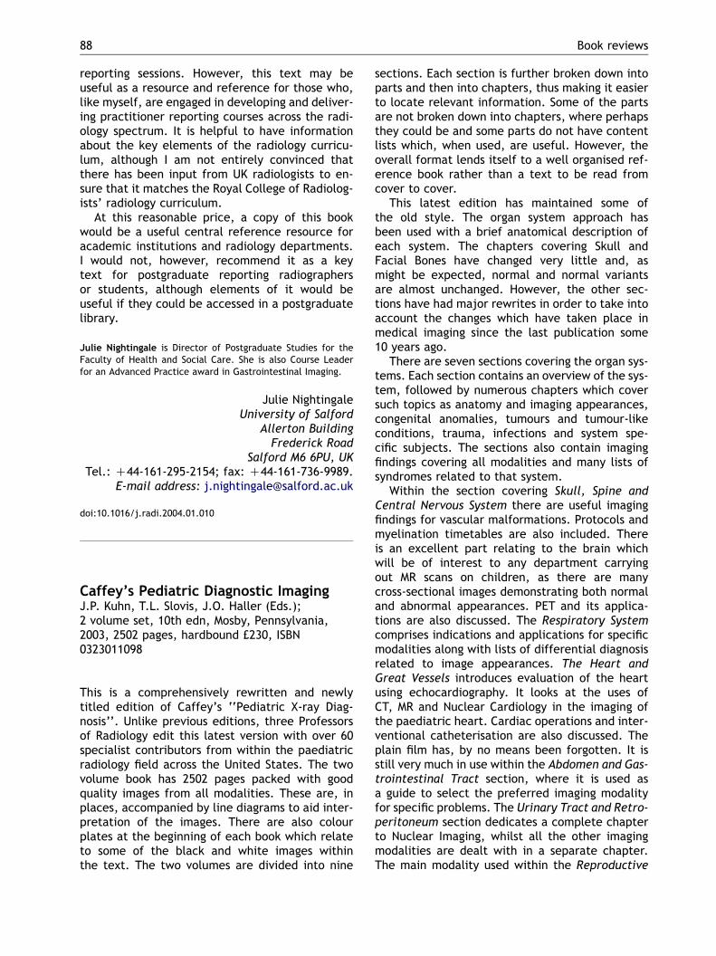
This is a comprehensively rewritten and newlytitled edition of Caffey’s ‘‘Pediatric X-ray Diag-nosis’’. Unlike previous editions, three Professorsof Radiology edit this latest version with over 60specialist contributors from within the paediatricradiology field across the United States. The twovolume book has 2502 pages packed with goodquality images from all modalities. These are, inplaces, accompanied by line diagrams to aid inter-pretation of the images. There are also colourplates at the beginning of each book which relateto some of the black and white images withinthe text. The two volumes are divided into nine
sections. Each section is further broken down intoparts and then into chapters, thus making it easierto locate relevant information. Some of the partsare not broken down into chapters, where perhapsthey could be and some parts do not have contentlists which, when used, are useful. However, theoverall format lends itself to a well organised ref-erence book rather than a text to be read fromcover to cover.
This latest edition has maintained some ofthe old style. The organ system approach hasbeen used with a brief anatomical description ofeach system. The chapters covering Skull andFacial Bones have changed very little and, asmight be expected, normal and normal variantsare almost unchanged. However, the other sec-tions have had major rewrites in order to take intoaccount the changes which have taken place inmedical imaging since the last publication some10 years ago.
There are seven sections covering the organ sys-tems. Each section contains an overview of the sys-tem, followed by numerous chapters which coversuch topics as anatomy and imaging appearances,congenital anomalies, tumours and tumour-likeconditions, trauma, infections and system spe-cific subjects. The sections also contain imagingfindings covering all modalities and many lists ofsyndromes related to that system.
Within the section covering Skull, Spine andCentral Nervous System there are useful imagingfindings for vascular malformations. Protocols andmyelination timetables are also included. Thereis an excellent part relating to the brain whichwill be of interest to any department carryingout MR scans on children, as there are manycross-sectional images demonstrating both normaland abnormal appearances. PET and its applica-tions are also discussed. The Respiratory Systemcomprises indications and applications for specificmodalities along with lists of differential diagnosisrelated to image appearances. The Heart andGreat Vessels introduces evaluation of the heartusing echocardiography. It looks at the uses ofCT, MR and Nuclear Cardiology in the imaging ofthe paediatric heart. Cardiac operations and inter-ventional catheterisation are also discussed. Theplain film has, by no means been forgotten. It isstill very much in use within the Abdomen and Gas-trointestinal Tract section, where it is used asa guide to select the preferred imaging modalityfor specific problems. The Urinary Tract and Retro-peritoneum section dedicates a complete chapterto Nuclear Imaging, whilst all the other imagingmodalities are dealt with in a separate chapter.The main modality used within the Reproductive
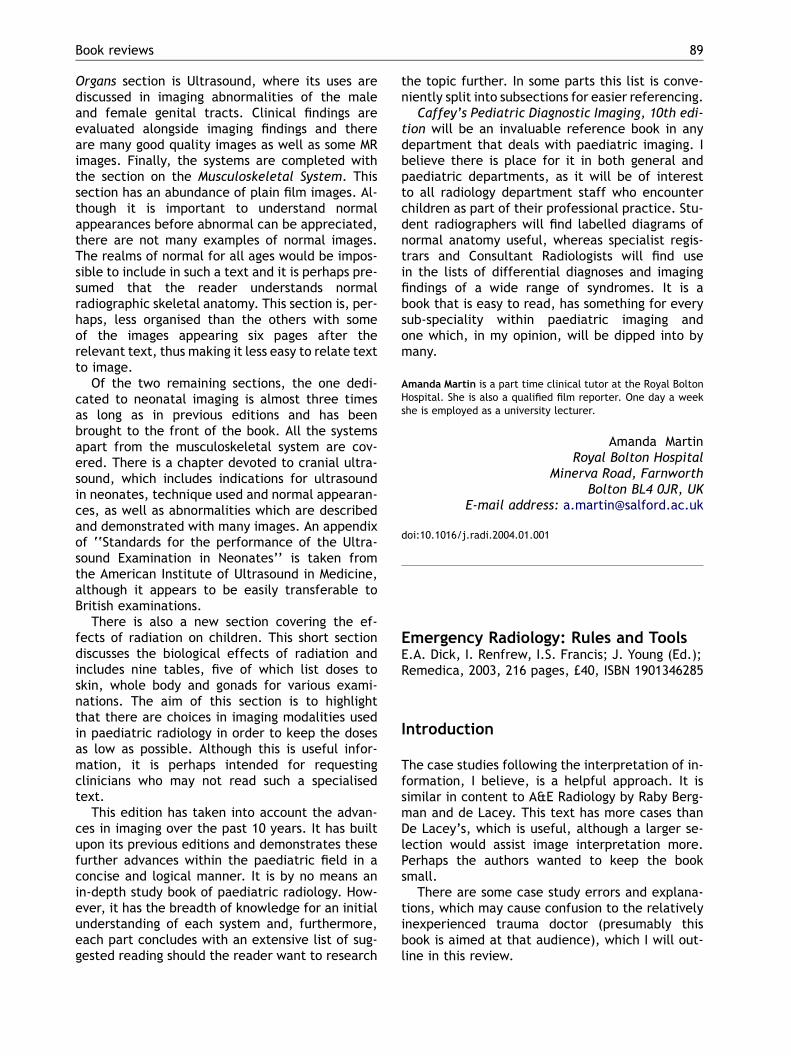
Emergency Radiology: Rules and ToolsE.A. Dick, I. Renfrew, I.S. Francis; J. Young (Ed.);Remedica, 2003, 216 pages, £40, ISBN 1901346285
Introduction
The case studies following the interpretation of in-formation, I believe, is a helpful approach. It issimilar in content to A&E Radiology by Raby Berg-man and de Lacey. This text has more cases thanDe Lacey’s, which is useful, although a larger se-lection would assist image interpretation more.Perhaps the authors wanted to keep the booksmall.
There are some case study errors and explana-tions, which may cause confusion to the relativelyinexperienced trauma doctor (presumably thisbook is aimed at that audience), which I will out-line in this review.
Book reviews 89
Organs section is Ultrasound, where its uses arediscussed in imaging abnormalities of the maleand female genital tracts. Clinical findings areevaluated alongside imaging findings and thereare many good quality images as well as some MRimages. Finally, the systems are completed withthe section on the Musculoskeletal System. Thissection has an abundance of plain film images. Al-though it is important to understand normalappearances before abnormal can be appreciated,there are not many examples of normal images.The realms of normal for all ages would be impos-sible to include in such a text and it is perhaps pre-sumed that the reader understands normalradiographic skeletal anatomy. This section is, per-haps, less organised than the others with someof the images appearing six pages after therelevant text, thus making it less easy to relate textto image.
Of the two remaining sections, the one dedi-cated to neonatal imaging is almost three timesas long as in previous editions and has beenbrought to the front of the book. All the systemsapart from the musculoskeletal system are cov-ered. There is a chapter devoted to cranial ultra-sound, which includes indications for ultrasoundin neonates, technique used and normal appearan-ces, as well as abnormalities which are describedand demonstrated with many images. An appendixof ‘‘Standards for the performance of the Ultra-sound Examination in Neonates’’ is taken fromthe American Institute of Ultrasound in Medicine,although it appears to be easily transferable toBritish examinations.
There is also a new section covering the ef-fects of radiation on children. This short sectiondiscusses the biological effects of radiation andincludes nine tables, five of which list doses toskin, whole body and gonads for various exami-nations. The aim of this section is to highlightthat there are choices in imaging modalities usedin paediatric radiology in order to keep the dosesas low as possible. Although this is useful infor-mation, it is perhaps intended for requestingclinicians who may not read such a specialisedtext.
This edition has taken into account the advan-ces in imaging over the past 10 years. It has builtupon its previous editions and demonstrates thesefurther advances within the paediatric field in aconcise and logical manner. It is by no means anin-depth study book of paediatric radiology. How-ever, it has the breadth of knowledge for an initialunderstanding of each system and, furthermore,each part concludes with an extensive list of sug-gested reading should the reader want to research
the topic further. In some parts this list is conve-niently split into subsections for easier referencing.
Caffey’s Pediatric Diagnostic Imaging, 10th edi-tion will be an invaluable reference book in anydepartment that deals with paediatric imaging. Ibelieve there is place for it in both general andpaediatric departments, as it will be of interestto all radiology department staff who encounterchildren as part of their professional practice. Stu-dent radiographers will find labelled diagrams ofnormal anatomy useful, whereas specialist regis-trars and Consultant Radiologists will find usein the lists of differential diagnoses and imagingfindings of a wide range of syndromes. It is abook that is easy to read, has something for everysub-speciality within paediatric imaging andone which, in my opinion, will be dipped into bymany.
Amanda Martin is a part time clinical tutor at the Royal BoltonHospital. She is also a qualified film reporter. One day a weekshe is employed as a university lecturer.
Amanda MartinRoyal Bolton Hospital
Minerva Road, FarnworthBolton BL4 0JR, UK
E-mail address: [email protected]
doi:10.1016/j.radi.2004.01.001