Caecilian jaw-closing mechanics: integrating two muscle
14
Caecilian jaw-closing mechanics: integrating two muscle systems Thomas Kleinteich 1, * , Alexander Haas 1 and Adam P. Summers 2 1 Universita¨t Hamburg, Biozentrum Grindel und Zoologisches Museum, Martin-Luther-King-Platz 3, 20146 Hamburg, Germany 2 University of California Irvine, 321 Steinhaus, Irvine, CA 92697, USA Caecilians (Lissamphibia: Gymnophiona) are unique among vertebrates in having two sets of jaw-closing muscles, one on either side of the jaw joint. Using data from high-resolution X-ray radiation computed tomography scans, we modelled the effect of these two muscle groups (mm. levatores mandibulae and m. interhyoideus posterior) on bite force over a range of gape angles, employing a simplified lever arm mechanism that takes into account muscle cross- sectional area and fibre angle. Measurements of lever arm lengths, muscle fibre orientations and physiological cross-sectional area of cranial muscles were available from three caecilian species: Ichthyophis cf. kohtaoensis; Siphonops annulatus; and Typhlonectes natans. The maximal gape of caecilians is restricted by a critical gape angle above which the mm. levatores mandibulae will open the jaw and destabilize the mandibular joint. The presence of destabilizing forces in the caecilian jaw mechanism may be compensated for by a mandibular joint in that the fossa is wrapped around the condyle to resist dislocation. The caecilian skull is streptostylic; the quadrate–squamosal complex moves with respect to the rest of the skull. This increases the leverage of the jaw-closing muscles. We also demonstrate that the unusual jaw joint requires streptostyly because there is a dorsolateral movement of the quadrate–squamosal complex when the jaw closes. The combination of the two jaw- closing systems results in high bite forces over a wide range of gape angles, an important advantage for generalist feeders such as caecilians. The relative sizes and leverage mechanics of the two closing systems allow one to exert more force when the other has a poor mechanical advantage. This effect is seen in all three species we examined. In the aquatic T. natans, with its less well-roofed skull, there is a larger contribution of the mm. levatores mandibulae to total bite force than in the terrestrial I. cf. kohtaoensis and S. annulatus. Keywords: Gymnophiona; amphibian cranial morphology; bite force modelling; high resolution mCT; automated measurement of muscle fibre angles 1. INTRODUCTION Caecilians (Gymnophiona) are fossorial limbless amphi- bians, comprising 171 species with circumtropical distri- bution ( Frost 2007). Their burrowing lifestyle places special demands on their cranial anatomy, and the caecilian skull is compact and wedge shaped, with many bones fused into compound elements (for a review see Wake 2003). Caecilians are also unique among vertebrates in possessing two sets of jaw-closing mechanisms that act on either side of the jaw joint. In addition to the usual complement of jaw adductors anterior to the jaw joint (mm. levatores mandibulae), a hyobranchial muscle posterior to the joint (m. interhyoideus posterior (IHP)) acts as an accessory jaw-closing muscle. The jaw-closing function of the IHP was proposed by Bemis et al. (1983) and Nussbaum (1983) who realized that its insertions on the ventral side of the retro- articular process of the lower jaw allowed it to adduct the mandible (figure 1). Using electromyography, Bemis et al. (1983) showed that the IHP acts syner- gistically with the primary jaw-closing muscles. The jaw-closing function of the IHP has been proposed as an adaptation to a fossorial lifestyle ( Nussbaum 1983). The reasoning is that in compact caecilian skulls, space for the mm. levatores mandibulae is restricted by the squamosal, a bone that covers large parts of the lateral face of the skull. The restriction on the size of the mm. levatores mandibulae demands the use of the IHP as an accessory jaw-closing muscle to generate appropriate bite forces. Although the cranium in caecilians is compact and solid, the morphology of the quadrate and the squamosal (quadrate–squamosal complex) led to the hypothesis that caecilian skulls are kinetic (strep- tostylic; Luther 1914; Marcus et al. 1933; De Villiers 1936; Iordanski 1990, 2000; Wilkinson & Nussbaum 1997). Indeed, manipulating the skull of a freshly killed J. R. Soc. Interface (2008) 5, 1491–1504 doi:10.1098/rsif.2008.0155 Published online 15 May 2008 Electronic supplementary material is available at http://dx.doi.org/ 10.1098/rsif.2008.0155 or via http://journals.royalsociety.org. *Author for correspondence ([email protected]). Received 21 April 2008 Accepted 23 April 2008 1491 This journal is q 2008 The Royal Society on September 19, 2010 rsif.royalsocietypublishing.org Downloaded from
Transcript of Caecilian jaw-closing mechanics: integrating two muscle
on September 19, 2010rsif.royalsocietypublishing.orgDownloaded from
Electronic su10.1098/rsif.2
*Author for c
Received 21 AAccepted 23 A
Caecilian jaw-closing mechanics:integrating two muscle systems
Thomas Kleinteich1,*, Alexander Haas1 and Adam P. Summers2
1Universitat Hamburg, Biozentrum Grindel und Zoologisches Museum,Martin-Luther-King-Platz 3, 20146 Hamburg, Germany
2University of California Irvine, 321 Steinhaus, Irvine, CA 92697, USA
Caecilians (Lissamphibia: Gymnophiona) are unique among vertebrates in having two sets ofjaw-closing muscles, one on either side of the jaw joint. Using data from high-resolution X-rayradiation computed tomography scans, we modelled the effect of these two muscle groups(mm. levatores mandibulae and m. interhyoideus posterior) on bite force over a range of gapeangles, employing a simplified lever arm mechanism that takes into account muscle cross-sectional area and fibre angle. Measurements of lever arm lengths, muscle fibre orientationsand physiological cross-sectional area of cranial muscles were available from three caecilianspecies: Ichthyophis cf. kohtaoensis; Siphonops annulatus; and Typhlonectes natans. Themaximal gape of caecilians is restricted by a critical gape angle above whichthe mm. levatores mandibulae will open the jaw and destabilize the mandibular joint. Thepresence of destabilizing forces in the caecilian jaw mechanism may be compensated for by amandibular joint in that the fossa is wrapped around the condyle to resist dislocation. Thecaecilian skull is streptostylic; the quadrate–squamosal complex moves with respect to therest of the skull. This increases the leverage of the jaw-closing muscles. We also demonstratethat the unusual jaw joint requires streptostyly because there is a dorsolateral movement ofthe quadrate–squamosal complex when the jaw closes. The combination of the two jaw-closing systems results in high bite forces over a wide range of gape angles, an importantadvantage for generalist feeders such as caecilians. The relative sizes and leverage mechanicsof the two closing systems allow one to exert more force when the other has a poor mechanicaladvantage. This effect is seen in all three species we examined. In the aquatic T. natans, withits less well-roofed skull, there is a larger contribution of the mm. levatores mandibulae tototal bite force than in the terrestrial I. cf. kohtaoensis and S. annulatus.
Keywords: Gymnophiona; amphibian cranial morphology; bite force modelling;high resolution mCT; automated measurement of muscle fibre angles
1. INTRODUCTION
Caecilians (Gymnophiona) are fossorial limbless amphi-bians, comprising 171 species with circumtropical distri-bution (Frost 2007). Their burrowing lifestyle placesspecial demands on their cranial anatomy, and thecaecilian skull is compact and wedge shaped, with manybones fused into compound elements (for a review seeWake 2003).Caecilians are also unique among vertebratesin possessing two sets of jaw-closing mechanisms thatact on either side of the jaw joint. In addition to the usualcomplement of jaw adductors anterior to the jaw joint(mm. levatores mandibulae), a hyobranchial muscleposterior to the joint (m. interhyoideus posterior (IHP))acts as an accessory jaw-closing muscle.
The jaw-closing function of the IHP was proposed byBemis et al. (1983) and Nussbaum (1983) who realized
pplementary material is available at http://dx.doi.org/008.0155 or via http://journals.royalsociety.org.
orrespondence ([email protected]).
pril 2008pril 2008 1491
that its insertions on the ventral side of the retro-articular process of the lower jaw allowed it to adductthe mandible (figure 1). Using electromyography,Bemis et al. (1983) showed that the IHP acts syner-gistically with the primary jaw-closing muscles. Thejaw-closing function of the IHP has been proposed asan adaptation to a fossorial lifestyle (Nussbaum 1983).The reasoning is that in compact caecilian skulls, spacefor the mm. levatores mandibulae is restricted by thesquamosal, a bone that covers large parts of the lateralface of the skull. The restriction on the size ofthe mm. levatores mandibulae demands the use of theIHP as an accessory jaw-closing muscle to generateappropriate bite forces.
Although the cranium in caecilians is compact andsolid, the morphology of the quadrate and thesquamosal (quadrate–squamosal complex) led to thehypothesis that caecilian skulls are kinetic (strep-tostylic; Luther 1914; Marcus et al. 1933; De Villiers1936; Iordanski 1990, 2000; Wilkinson & Nussbaum1997). Indeed, manipulating the skull of a freshly killed
J. R. Soc. Interface (2008) 5, 1491–1504
doi:10.1098/rsif.2008.0155
Published online 15 May 2008
This journal is q 2008 The Royal Society

Figure 1. Representation of the lever arm model used to predict bite forces in the caecilian jaw-closing mechanism drawn overa reconstruction of the skull in lateral view of I. cf. kohtaoensis. Bite forces were calculated for a range of gape angles (a) from08 to 908. Two jaw-closing mechanisms, one actuated by the mm. levatores mandibulae and the other actuated by the IHP, areshown. The bite force at the tip of the lower jaw Fbite is a function of the forces generated by the two closing systems. Themm. levatores mandibulae closure system is described by the force generated by the muscles (FLEV), acting on an input lever armlLEV, with a muscle fibre orientation of b. The IHP closure system acts on a lever arm lRP, with a force of FIHP and a fibreorientation of 3. Two aspects of skull anatomy are captured with angular measurements: the angle of the quadrate–squamosalcomplex (d) and the angle of the retroarticular process of the lower jaw with a line drawn from the lower jaw joint to the tip ofthe lower jaw (g).
1492 Caecilian jaw-closing mechanics T. Kleinteich et al.
on September 19, 2010rsif.royalsocietypublishing.orgDownloaded from
Dermophis mexicanus specimen showed that the quad-rate and the squamosal could rotate slightly (Wake &Hanken 1982). A model of bite force generated by theIHP demonstrated the unusual and counter-intuitiveresult that force steadily decreases during jaw closure.However, two factors can ameliorate this loss in force: aretroarticular process that curves upward, giving abetter mechanical advantage (O’Reilly 2000) and thestreptostylic joints in the skull, which act as a secondleverage system (Summers & Wake 2005).
Previous mathematical descriptions of the function ofthe IHP were based on the assumption that muscle fibreorientation in the IHP is aligned exactly along the longaxis of the animal and the force acts in caudal direction(Summers & Wake 2005). However, the studies of thecranial musculature in caecilians (Wiedersheim 1879;Luther 1914; Lawson 1965; Bemis et al. 1983; Nussbaum1983; Iordanski 1996; Wilkinson & Nussbaum 1997;Kleinteich & Haas 2007) show that the fibre orientationof the IHP inmost caecilian species is oblique rather thanpurely anteroposterior; the muscle fibres run in thecaudal and ventral directions. A second simplification inthe previous model was to ignore the jaw-closing functionof themm. levatores mandibulae. These are the only jaw-closing muscles in most other vertebrates and are quitesubstantial in size, so it is important that any modelof caecilian jaw function includes the contribution ofthese muscles.
The goals of this study are fivefold: (i) we propose anew model of jaw function that includes the contri-bution of fibre angles that are not aligned with the longaxis of the body, (ii) we extend the model to include theancestral jaw-closing muscles, the mm. levatoresmandibulae, (iii) we estimate the effective mechanicaladvantages (EMAs) of these two jaw-closing systemsover different gape angles to test the hypothesis that the
J. R. Soc. Interface (2008)
two systems contribute best to different parts of jawclosure, (iv) with values derived from high-resolution,synchrotron-based X-ray radiation computed tomo-graphy (CT) scan data, we calculate a theoretical biteforce across a range of gapes and the contributions ofthe two closing systems to the maximal bite force, and(v) we use physical and computer models to describethe complex movements at the jaw joint during thedoubly actuated closure of the gape.
2. MATERIAL AND METHODS
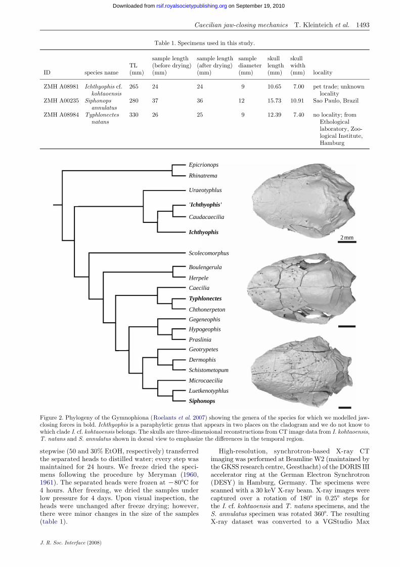
We studied the jaw lever mechanics of three specimensfrom different caecilian species (table 1; figure 2):Ichthyophis kohtaoensis Taylor, 1960; Typhlonectesnatans (Fischer in Peters, 1880); and Siphonopsannulatus (Mikan, 1820). All the specimens are storedin the herpetological collection of the zoologicalmuseum, Hamburg (ZMH). Our sampling comprisesbasal (Ichthyophis cf. kohtaoensis) and derived cladeswithin the Gymnophiona (figure 2) and two differentskull architectures: I. cf. kohtaoensis and S. annulatushave stegokrotaphic skulls (closed temporal region;figure 2), and T. natans has a zygokrotaphic skull(temporal region with wide gap between squamosaland parietal; figure 2). Owing to unsolved problemsin the taxonomy of and within the genus Ichthyophis(see Gower et al. 2002), we use I. cf. kohtaoensis hereinfor specimen ZMH A08981. The current taxonomicstatus of I. kohtaoensis is highly debated and underrevision. Further, the genus Ichthyophis has beenshown to be paraphyletic (Gower et al. 2002; Frostet al. 2006; Roelants et al. 2007; figure 2).
The specimens were decapitated between the fifthand sixth annuli (i.e. in the anterior trunk region). Allspecimens had been stored in 70% EtOH. We
Epicrionops
Rhinatrema
Uraeotyphlus
'Ichthyophis'
Caudacaecilia
Ichthyophis2mm
Scolecomorphus
Boulengerula
Herpele
Caecilia
Typhlonectes
Chthonerpeton
Gegeneophis
Hypogeophis
Praslinia
Geotrypetes
Dermophis
Schistometopum
Microcaecilia
Luetkenotyphlus
Siphonops
Figure 2. Phylogeny of the Gymnophiona (Roelants et al. 2007) showing the genera of the species for which we modelled jaw-closing forces in bold. Ichthyophis is a paraphyletic genus that appears in two places on the cladogram and we do not know towhich clade I. cf. kohtaoensis belongs. The skulls are three-dimensional reconstructions from CT image data from I. kohtaoensis,T. natans and S. annulatus shown in dorsal view to emphasize the differences in the temporal region.
Table 1. Specimens used in this study.
ID species nameTL(mm)
sample length(before drying)(mm)
sample length(after drying)(mm)
samplediameter(mm)
skulllength(mm)
skullwidth(mm) locality
ZMH A08981 Ichthyophis cf.kohtaoensis
265 24 24 9 10.65 7.00 pet trade; unknownlocality
ZMH A00235 Siphonopsannulatus
280 37 36 12 15.73 10.91 Sao Paulo, Brazil
ZMH A08984 Typhlonectesnatans
330 26 25 9 12.39 7.40 no locality; fromEthologicallaboratory, Zoo-logical Institute,Hamburg
Caecilian jaw-closing mechanics T. Kleinteich et al. 1493
on September 19, 2010rsif.royalsocietypublishing.orgDownloaded from
stepwise (50 and 30% EtOH, respectively) transferredthe separated heads to distilled water; every step wasmaintained for 24 hours. We freeze dried the speci-mens following the procedure by Meryman (1960,1961). The separated heads were frozen at K808C for4 hours. After freezing, we dried the samples underlow pressure for 4 days. Upon visual inspection, theheads were unchanged after freeze drying; however,there were minor changes in the size of the samples(table 1).
J. R. Soc. Interface (2008)
High-resolution, synchrotron-based X-ray CTimaging was performed at Beamline W2 (maintained bythe GKSS research centre, Geesthacht) of the DORIS IIIaccelerator ring at the German Electron Synchrotron(DESY) in Hamburg, Germany. The specimens werescanned with a 30 keV X-ray beam. X-ray images werecaptured over a rotation of 1808 in 0.258 steps forthe I. cf. kohtaoensis and T. natans specimens, and theS. annulatus specimen was rotated 3608. The resultingX-ray dataset was converted to a VGStudio Max

1494 Caecilian jaw-closing mechanics T. Kleinteich et al.
on September 19, 2010rsif.royalsocietypublishing.orgDownloaded from
(Volume Graphics GmbH, Heidelberg, Germany)volume rendering dataset. The datasets have a resolutionof 6.83 mm (I. cf. kohtaoensis and T. natans) and 9.2 mm(S. annulatus) in x, y and z orientations. Neighbouringvoxels of the volume dataset have been merged to reducethe size of the dataset (binding). Different graduationsof these resampled datasets (twofold, threefold andfourfold bindings) were available for analysis. We usedthe software packagesVGSTUDIOMAXv. 1.2 andAMIRA v.4.1 (Mercury Computer Systems) for processing, analys-ing and segmentation of the resulting volumetric three-dimensional datasets. Surface rendering and animationwere performed with Alias Wavefront MAYA v. 6.0.
The model used to calculate force transmission of theIHP is based on the model presented by Summers &Wake (2005). To account for fibre orientations of theIHP, we added fibre angle 3 to the model (figure 1). Wecalculated the EMAIHP (bite force per unit muscleforce; based on Biewener 1989) for the IHP by (figure 1)
EMAIHP ZFbite=FIHP Z sinðaCgC3Þ!ðlRP=lLJÞCsinðdK3Þ!cosðaCdÞ; ð2:1Þ
where Fbite is the output bite force; FIHP is the forcegenerated by the IHP; lRP is the length of the retro-articular process of the lower jaw; lLJ is the distance fromthe rostral tip of the lower jaw to the jaw articulation; ais the gape angle; g is the retroarticular angle withrespect to the anteroposterior axis; d is the quadrate–squamosal angle with respect to the anteroposterioraxis; and 3 is themuscle fibre orientation of the IHPwithrespect to the anteroposterior axis. In this equation andthe next, the first term represents the force generatedthrough the conventional simple lever systemof the jawsand the second term accounts for the streptostylicsuspension of the jaws.
We developed a second lever arm model for themm. levatores mandibulae group (figure 1). This groupcomprises three muscles in adult caecilians: m. levatormandibulae articularis;m. levator mandibulae internus;and m. levator mandibulae longus (Wiedersheim 1879;Luther 1914; Edgeworth 1935; Lawson 1965; Bemiset al. 1983; Iordanski 1996; Wilkinson & Nussbaum1997; a table of synonyms was presented by Kleinteich &Haas 2007). The EMA for single muscles in this groupEMALEV is calculated by
EMALEV ZFbite=FLEV
Z sinðbKaÞ!ðlLEV=lLJÞCsinðbCdÞ!cosðaCdÞ; ð2:2Þ
where Fbite is the output bite force; FLEV is the forcegenerated by muscles of the mm. levatores mandibulaegroup; lLEV is the distance from the insertion of themuscles to the jaw articulation; lLJ is the distance fromthe rostral tip of the lower jaw to the jaw articulation;a isthe gape angle; b is the muscle fibre orientation of themuscles with respect to the anteroposterior axis; and d isthe quadrate–squamosal angle with respect to theanteroposterior axis.
Besides the mm. levatores mandibulae group, thenervus trigeminus innervated jaw musculature of caeci-lians comprises three additional muscles that are notconsidered in this paper, i.e. the m. intermandibularis,
J. R. Soc. Interface (2008)
the m. levator quadrati and the m. pterygoideus(Iordanski 1996; Haas 2001; Kleinteich & Haas 2007).The m. interhyoideus lowers the buccal floor. Thefunction of the m. levator quadrati and m. pterygoideusis poorly known; however, both insert on the quadrateand thus are likely to be involved in movements of thequadrate (streptostyly) rather than jaw closure.
Measurements of anatomical characters are basedon lateral views of rendered CT datasets. We usedthe freely available image analysis software IMAGEJ v.1.36b (NIH; http://rsb.info.nih.gov/ij/download.html) for all measurements. We did all measurementsfor both sides of the body of the animals; the averagevalue of both measurements was used for furthercalculations.
For measurements of muscle fibre orientations(b and 3), we generated 8 bit grey-scale image stacksof sagittal sections parallel to the muscle in the CTdatasets. These image stacks were generated with theoblique slice function of AMIRA. For every investigatedmuscle, we adjusted the plane of section so that sectionswere parallel to the muscle fibres in lateral view. Weconverted the grey-scale image stacks into binary datawith the adjustOthreshold function of IMAGEJ. Theresulting dataset (figure 3c) contained only muscula-ture (black) and background (white). Muscle fibreorientations were measured automatically with IMAGEJ,and to reduce noise we excluded particles less than 25pixels in area and with a circularity greater than 0.3(figure 3d ) from the analysis. Measured muscle fibreangles are distributed (figure 3e) around a mean value.Standard deviations of muscle fibre orientations aregiven in table 2. The standard deviation of muscle fibreangles can be interpreted as an estimate for thediversity of fibre orientations within a muscle. Wecalculated EMAs for the mean and the minimal andmaximal values that were derived from the standarddeviation of muscle fibre orientations. However,muscles are treated as idealized parallel-fibredstructures herein and the mean fibre orientation wasused for interpretation of the jaw-closing mechanism.
Theoretical forces that can be generated by a singlemuscle were calculated by
Fmuscle ZV
l
� �!pMIS; ð2:3Þ
where Fmuscle is the force generated by a muscle; V is thevolume of the muscle; l is the length of the muscle inthe direction of the fibre orientation; and pMIS is themaximal isometric stress (pMISZ250 kPa; Herzog 1994).The ratio of muscle volume tomuscle length is a measurefor physiological cross-sectional area (PCSA).
Total bite force for the entire jaw-closing system(i.e. both jaw-closing systems and both sides of theskull) is calculated as the doubled sum of bite forcesfrom single muscles, assuming bilateral symmetry.
We measured volumes and lengths of the muscles byseparating single muscles out of the CT datasets(segmentation). Surfaces were generated out of thesegmented datasets and measured with the lineprobeand areavolume tools in AMIRA. Volume and lengthmeasurements are averages of values from both sides ofthe body. To interactively explore possible movements

mm. levatores mandibulaeproc. retroarticularis
m. interhyoideus posterior
(c)
(e)
(d )
(b)
(a)
1mm
1mm
800700600500400300200100
900
00 180
muscle fibre angle (deg.)
no. o
f m
uscl
e fi
bres
Figure 3. A method for measuring muscle fibre angle from high resolution, synchrotron-based X-ray CT scan data. (a) A volumerendering of the CT image data for I. cf. kohtaoensis shown in lateral view with the skin removed to reveal the underlyingskeleton andmuscles. The squamosal bone has also been removed to show the levator mandibulae muscle complex. (b) A contrastenhanced, grey-scale image of a parasagittal section of the IHP from the CT image data. (c) The section in (b) thresholded toemphasize the muscle fibres. (d ) The section shown in (c) with all connected areas smaller than 25 pixels in area removed, and allareas with a circularity of more than 0.3 removed. The remaining areas are muscle fibres with measurable angles. (e) Histogramshowing the distribution of angular measurements for the 6797 fibres measured from the image stack for the IHP from the left sideof I. cf. kohtaoensis that included image (b). Count 6797, mean 44.61 (56.24), s.d. 32.08, min. K41.988 (08), max. 137.93 (1808).
Caecilian jaw-closing mechanics T. Kleinteich et al. 1495
on September 19, 2010rsif.royalsocietypublishing.orgDownloaded from
of the cranial bones, we produced physical models of thecaecilian skulls including the lower jaws on a ZPrinter310 rapid prototyping machine (ZCorp, Burlington,MA). The models were based on the CT datasets. Wesegmented the quadrate–squamosal complex, the pter-ygoid and the stapes on the right-hand side of the skullwith AMIRA v. 4.1 in order to produce separated modelsfor those parts. The resulting physical models are scaledup in size (I. cf. kohtaoensis and T. natans scale factor:14.5; S. annulatus scale factor: 10.7) compared to theskulls in the specimens. The physical models weremanipulated by hand to estimate the degrees of lowerjaw and quadrate–squamosal movements. We thenused MAYA v. 6.0 to animate the VRML dataset of theI. cf. kohtaoensis specimen. The computer animation isbased on the results of the interaction with the physical
J. R. Soc. Interface (2008)
model for this species. The movie files of the animationare available as information in the electronic supple-mentary material.
3. RESULTS
All measurements used for the calculations of EMAsand bite forces over gape angles are listed in table 2.The ratios of the lengths of the in-lever to out-leverfor the mm. levatores mandibulae range from 0.055(m. levator mandibulae articularis in S. annulatus) to0.264 (m. levator mandibulae longus in T. natans). Thelever arm ratios are higher for the IHP in all the threeinvestigated species: 0.404 in I. cf. kohtaoensis; 0.58 inS. annulatus; and 0.538 in T. natans.

Table 2. Measurements of functionally important anatomical characters and calculation of maximal force of single muscles forthe three investigated species.
Ichthyophis cf.kohtaoensis
Siphonopsannulatus
Typhlonectesnatans
m. levator mandibulae articularis length in-lever lLEV (mm) 0.775 0.575 0.635lLEV/lLJ 0.100 0.055 0.093fibre angle bLMA (8) 88.3G24.7 97.2G30.6 88.0G34.7volume (mm3) 0.695 0.424 0.304length (mm) 1.699 1.999 1.181volume/length (mm2) 0.409 0.212 0.257force (N) 0.102 0.053 0.064
m. levator mandibulaeinternus
length in-lever lLEV (mm) 1.655 2.105 1.805lLEV/lLJ 0.214 0.200 0.264fibre angle bLMA (8) 116.1G29.7 102.5G32.6 125.5G32.0volume (mm3) 1.385 6.860 0.819length (mm) 2.296 4.214 2.772volume/length (mm2) 0.603 1.628 0.295force (N) 0.151 0.407 0.074
m. levator mandibulae longus length in-lever lLEV (mm) 1.655 2.105 1.805lLEV/lLJ 0.214 0.200 0.264fibre angle bLMA (8) 91.5G27.8 96.6G31.3 79.2G35.4volume (mm3) 1.195 4.025 2.685length (mm) 2.433 3.981 2.941volume/length (mm2) 0.491 1.011 0.913force (N) 0.123 0.252 0.228
m. interhyoideus posterior length in-lever lRP (mm) 3.120 6.100 3.680lRP/lLJ 0.404 0.580 0.538fibre angle 3 (8) 41.2G30.9 36.2G31.4 57.3G28.2volume (mm3) 22.400 30.600 12.100length (mm) 5.135 7.294 5.687volume/length (mm2) 4.363 4.195 2.128force (N) 1.09 1.05 0.53
length out-lever lowerjaw lLJ (mm)
7.718 10.525 6.840
angle processusretroarticularis g (8)
32.7 16.8 22.3
angle quadrate–squamosal complexd (8)
26.0 27.5 26.2
1496 Caecilian jaw-closing mechanics T. Kleinteich et al.
on September 19, 2010rsif.royalsocietypublishing.orgDownloaded from
Average muscle fibre angle orientations of thelevator mandibulae complex relative to the anteropos-terior axis range from 79.28 (m. levator mandibulaelongus in T. natans) to 125.58 (m. levator mandibulaeinternus in T. natans). The fibres of the IHP areoriented on average 41.28 in I. cf. kohtaoensis, 36.28 inS. annulatus and 57.38 in T. natans.
The m. levator mandibulae articularis has thesmallest ratio of muscle volume to muscle length(PCSA) in all the investigated specimens. The PCSAvalues of the m. levator mandibulae articularis arebetween 0.212 (T. natans) and 0.409 mm2 (I. cf.kohtaoensis). The PCSA value ranges from 0.295(T. natans) to 1.628 mm2 (S. annulatus) for the m.levator mandibulae internus and from 0.491 (I. cf.kohtaoensis) to 1.011 mm2 (S. annulatus) for the m.levator mandibulae longus. In the three investigatedcaecilian species, the IHP has the largest PCSA valueof all muscles: 4.363 mm2 in I. cf. kohtaoensis; 4.195 mm2
in S. annulatus; and 2.128 mm2 in T. natans.Calculated maximal bite forces range from 0.053
(m. levator mandibulae articularis in T. natans) to0.407 N (m. levator mandibulae internus in S. annulatus)for the levator mandibulae group and from 0.53(T. natans) to 1.09 N (I. cf. kohtaoensis) for the IHP.
J. R. Soc. Interface (2008)
The retroarticular process of the lower jaw is angled32.78 dorsally in I. cf. kohtaoensis, 16.88 in S. annulatusand 22.38 inT. natanswith respect to the anteroposterioraxis. The quadrate–squamosal complex is oriented at 268to the anteroposterior axis in I. cf. kohtaoensis, 27.58 inS. annulatus and 26.28 in T. natans.
3.1. Effective mechanical advantageand gape angle
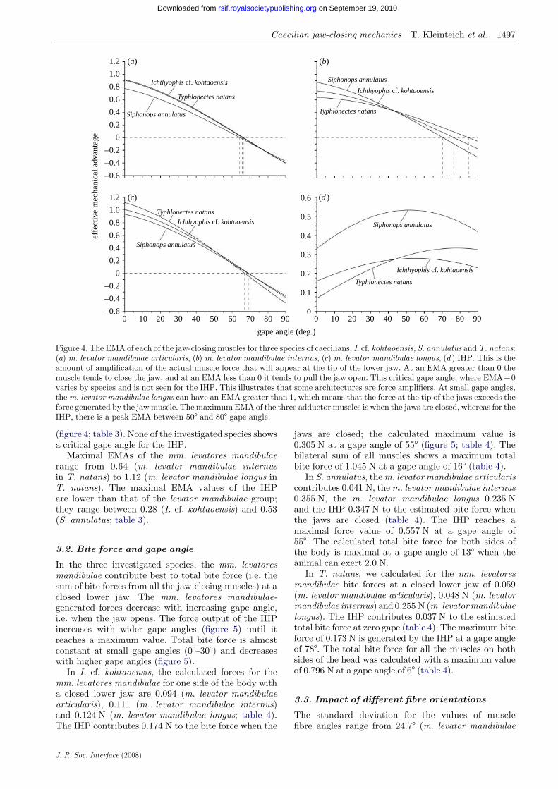
The EMAs of all mm. levatores mandibulae aremaximal at a closed lower jaw in all the investigatedspecies (figure 4; table 3). With increasing gape angle,the EMA of the mm. levatores mandibulae decreases.The muscles of the levator mandibulae group in theinvestigated species have critical values for gape anglesabove which the EMAs become negative (figure 4).Critical gape angles range from 64.48 (m. levatormandibulae articularis in S. annulatus) to 84.88(m. levator mandibulae internus in T. natans; table 3).
The EMA of the IHP in the three investigated speciesincreases with increasing gape angle, reaches a maxi-mum value at gape angles of 55.28 (I. cf. kohtaoensis),51.88 (S. annulatus) and 78.38 (T. natans), and decreaseswith increasing gape angles above the optimal gape
effe
ctiv
e m
echa
nica
l adv
anta
ge
0.2
1.2
1.0
0.8
0.6
0.4
0.2
0
–0.2
–0.4
–0.6
1.2
1.0
0.8
0.6
0.4
0 0.2
0.1
0.6
0.3
0.4
0.5
0
–0.2
–0.4
–0.60 10 20 30 40 50 60 70 80 90
gape angle (deg.)
Typhlonectes natans
Ichthyophis cf. kohtaoensis
Siphonops annulatus
Siphonops annulatus
Siphonops annulatus
Siphonops annulatus
Ichthyophis cf. kohtaoensisIchthyophis cf. kohtaoensis
Ichthyophis cf. kohtaoensis
Typhlonectes natans
Typhlonectes natans
Typhlonectes natans
0 10 20 30 40 50 60 70 80 90
(a) (b)
(c) (d )
Figure 4. The EMA of each of the jaw-closing muscles for three species of caecilians, I. cf. kohtaoensis, S. annulatus andT. natans:(a) m. levator mandibulae articularis, (b) m. levator mandibulae internus, (c) m. levator mandibulae longus, (d ) IHP. This is theamount of amplification of the actual muscle force that will appear at the tip of the lower jaw. At an EMA greater than 0 themuscle tends to close the jaw, and at an EMA less than 0 it tends to pull the jaw open. This critical gape angle, where EMAZ0varies by species and is not seen for the IHP. This illustrates that some architectures are force amplifiers. At small gape angles,the m. levator mandibulae longus can have an EMA greater than 1, which means that the force at the tip of the jaws exceeds theforce generated by the jawmuscle. The maximumEMA of the three adductor muscles is when the jaws are closed, whereas for theIHP, there is a peak EMA between 508 and 808 gape angle.
Caecilian jaw-closing mechanics T. Kleinteich et al. 1497
on September 19, 2010rsif.royalsocietypublishing.orgDownloaded from
(figure 4; table 3). None of the investigated species showsa critical gape angle for the IHP.
Maximal EMAs of the mm. levatores mandibulaerange from 0.64 (m. levator mandibulae internusin T. natans) to 1.12 (m. levator mandibulae longus inT. natans). The maximal EMA values of the IHPare lower than that of the levator mandibulae group;they range between 0.28 (I. cf. kohtaoensis) and 0.53(S. annulatus; table 3).
3.2. Bite force and gape angle
In the three investigated species, the mm. levatoresmandibulae contribute best to total bite force (i.e. thesum of bite forces from all the jaw-closing muscles) at aclosed lower jaw. The mm. levatores mandibulae-generated forces decrease with increasing gape angle,i.e. when the jaw opens. The force output of the IHPincreases with wider gape angles (figure 5) until itreaches a maximum value. Total bite force is almostconstant at small gape angles (08–308) and decreaseswith higher gape angles (figure 5).
In I. cf. kohtaoensis, the calculated forces for themm. levatores mandibulae for one side of the body witha closed lower jaw are 0.094 (m. levator mandibulaearticularis), 0.111 (m. levator mandibulae internus)and 0.124 N (m. levator mandibulae longus; table 4).The IHP contributes 0.174 N to the bite force when the
J. R. Soc. Interface (2008)
jaws are closed; the calculated maximum value is0.305 N at a gape angle of 558 (figure 5; table 4). Thebilateral sum of all muscles shows a maximum totalbite force of 1.045 N at a gape angle of 168 (table 4).
In S. annulatus, them. levator mandibulae articulariscontributes 0.041 N, them. levator mandibulae internus0.355 N, the m. levator mandibulae longus 0.235 Nand the IHP 0.347 N to the estimated bite force whenthe jaws are closed (table 4). The IHP reaches amaximal force value of 0.557 N at a gape angle of558. The calculated total bite force for both sides ofthe body is maximal at a gape angle of 138 when theanimal can exert 2.0 N.
In T. natans, we calculated for the mm. levatoresmandibulae bite forces at a closed lower jaw of 0.059(m. levator mandibulae articularis), 0.048 N (m. levatormandibulae internus) and 0.255 N (m. levatormandibulaelongus). The IHP contributes 0.037 N to the estimatedtotal bite force at zero gape (table 4). The maximum biteforce of 0.173 N is generated by the IHP at a gape angleof 788. The total bite force for all the muscles on bothsides of the head was calculated with a maximum valueof 0.796 N at a gape angle of 68 (table 4).
3.3. Impact of different fibre orientations
The standard deviation for the values of musclefibre angles range from 24.78 (m. levator mandibulae

Table
3.Calculationsofeffectivemechanicaladvantages(E
MA)andfunctionallyim
portantgapeangles(a)forsingle
muscles.
(Resultsshownforcalculationswithmeanmuscle
fibre
angles(b
mean/3 m
ean),
minim
alfibre
angles(b
min/3 m
in)andmaxim
alfibre
angles
(bmax/3 m
ax).)
Ichthyophis
cf.kohtaoen
sis
Siphonopsannulatus
Typhlonectesnatans
EMA
max
amax[8]
criticalaZ08
EMA
max
amax[8]
criticalaZ08
EMA
max
amax[8]
criticalaZ08
bmean(b
min;bmax)
bmean(b
min;bmax)
bmean(b
min;bmax)
bmean(b
min;bmax)
bmean(b
min;bmax)
bmean(b
min;bmax)
bmean(b
min;bmax)
bmean(b
min;bmax)
bmean(b
min;bmax)
m.levatormandibulae
articularis
0.92(0.99;0.68)
0(0;0)
66(64;70)
0.78(0.93;0.41)
0(0;0)
64(63;69)
0.91(0.95;0.54)
0(0;0)
66(63;72)
m.levatormandibulae
internus
0.74(1.04;0.27)
0(0;24)
76(68;—
)0.87(1.07;0.41)
0(0;1)
70(64;—
)0.64(1.04;0.27)
0(0;81)
85(70;—
)
m.levatormandibulae
longus
1.01(1.09;0.69)
0(0;0)
69(64;78)
0.93(1.07;0.53)
0(0;0)
69(63;83)
1.12(1.02;0.80)
0(0;0)
67(59;78)
EMA
max
amax[8]
EMA
aZ08
EMA
max
amax[8]
EMA
aZ08
EMA
max
amax[8]
EMA
aZ08
3 mean(3
min;3 m
ax)
3 mean(3
min;3 m
ax)
3 mean(3
min;3 m
ax)
3 mean(3
min;3 m
ax)
3 mean(3
min;3 m
ax)
3 mean(3
min;3 m
ax)
3 mean(3
min;3 m
ax)
3 mean(3
min;3 m
ax)
3 mean(3
min;3 m
ax)
m.interhyoideus
posterior
0.28(0.55;0.21)
55(19;90)
0.16(0.52;K0.25)
0.53(0.66;0.35)
52(33;89)
0.33(0.56;0.01)
0.33(0.52;0.21)
78(44;90)
0.07(0.38;K0.26)
1498 Caecilian jaw-closing mechanics T. Kleinteich et al.
J. R. Soc. Interface (2008)
on September 19, 2010rsif.royalsocietypublishing.orgDownloaded from
articularis in I. cf. kohtaoensis) to 35.48 (m. levatormandibulae longus in T. natans; table 2). EMAs andthus bite forces increase with smaller values formuscle fibre angles in all the investigated species andmuscles; larger fibre angles decrease EMA and biteforce (tables 3 and 4).
The gape angle at which the muscles have their bestEMA increases with increasing fibre angle (table 3) forthe m. levator mandibulae internus and the IHP, andthus variation in fibre orientation has an effect on totalbite force over gape angle (table 4).
The critical gape angles increase with increasingfibre angle for all the muscles. For the m. levatormandibulae internus, the increase in critical gape angleresults in a critical gape that is beyond the maximalgape angle considered herein (908; table 3).
3.4. Rapid prototyping and animation
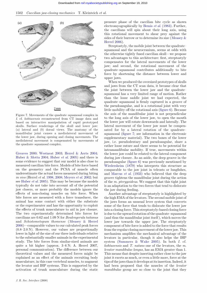
The scaled-up, rapid prototyped models of the lowerjaw clearly show a partially captured mandibular joint(figure 6a,d,g) in that the fossa of the jaw joint in thepseudoangular is a deep mediocaudally oriented ridge(figure 6c,f,i ) that wraps around the condyle of thequadrate. Interactive manipulation of the rapid proto-typed models and the resulting three-dimensionalcomputer animation (see information in the electronicsupplementary material) show substantial dorsolateralmovement of the quadrate–squamosal complex as thelower jaw closes. This movement is driven by themediocaudally oriented fossa of the lower jaw, whichnecessitates some degree of lateral movement of thelower jaw as the jaw opens (figure 7; see information inthe electronic supplementary material).
4. DISCUSSION
Our analysis of the caecilian jaw function has broughtto light several interesting features of these unusualamphibians. When the gape angle exceeds some criticalangle, the force generated by one set of jaw musclestransitions from closing the jaws to opening them(figure 4). The previous model of jaw function did notpredict this because it did not account for the fibreorientation of the muscles, realistic orientations of thequadrate–squamosal complex or the levator musclecomplex (Summers & Wake 2005). The critical gapeangle has two important consequences: first, as gapeincreases the contribution of the levator complex willdecrease (figures 4 and 5); and second, the forcesoriented at oblique angles relative to the long axis of thejaw are one set that will tend to destabilize the jawjoint. Our model suggests that at gape angles higherthan approximately 708, the mm. levatores mandibulaewill open the lower jaw and thus counteract to the IHP.Although the IHP has its peak mechanical advantageat rather high gape angles (table 3; figure 4) and thushas the potential to compensate for the mm. levatoresmandibulae, this theoretical scenario seems to be oflittle biological relevance. The gape angle data areavailable from I. kohtaoensis, T. natans and Hypogeo-phis rostratus, which show peak gape angles of not morethan 608 (O’Reilly 2000). This coincides with our model

total bite force
total bite force
total bite force
sum mm. levatoresmandibulae
sum mm.levatores mandibulae
sum mm.levatores mandibulae
m. interhyoideusposterior
m. interhyoideusposterior
m. interhyoideusposterior
m. interhyoideusposterior
m. interhyoideusposterior
m. interhyoideus posterior
m. lev. mand.internus m. lev. mand. internus
m. lev. mandinternus
m. lev. mand. internus
m. lev. mand. internus
m. lev. mand. longus
m. lev. mand. articularis
m. lev. mand.longus
m. lev. mand.longus
m. lev. mand.longus
m. lev. mand. articularis
m. lev. mand. longus
m. lev. mand. longus
m. lev. mand. articularis m. lev. mand. articularis
m. lev. mand. articularism. lev. mand. articularis
m. lev. mand. internus
–0.2
–0.5
0
0.5
1.0
1.5
2.0
2.5
0
0.2
0.4
0.6
0.8
1.0
1.2
–0.2
0
0.2
0.4
0.6
0.8
1.0
1.2
10
20
30
40
50
60
70
80
90
100
0
10
20
30
40
50
60
70
80
90
100
0
10
20
30
40
50
60
70
80
90
100
cont
ribu
tion
to b
ite f
orce
(%
)
bite
for
ce (
N)
gape angle (deg.)
0 10 20 30 40 50 60 70 80 90 0 10 20 30 40 50 60 70 80 90
(a) (b)
(c) (d )
(e) ( f )
Figure 5. Modelled bite forces at varying gape angles for three species of caecilians: (a,b) I. cf. kohtaoensis, (c,d ) S. annulatusand (e, f ) T. natans. (a,c,e) Absolute force generated by the four jaw-closing muscles and the sum of the forces of the threemm. levatores mandibulae as well as the total bite force as sum of all four muscles for both sides of the skull. (b,d, f ) The relativecontributions of single muscles (for both skull sides) to the total bite force.
Caecilian jaw-closing mechanics T. Kleinteich et al. 1499
on September 19, 2010rsif.royalsocietypublishing.orgDownloaded from
predictions that critical gape angles for single musclesof the mm. levatores mandibulae are at approximately658 for the three investigated species (table 3; figure 4).
The synchrotron-based X-ray radiation CT imagedata made it possible to measure fibre angles for a largenumber of fibres relative to the methods that rely on adissecting scope and goniometer (thousands versusdozens) in a reasonable investment of time. However,the technique may also introduce errors. These fall intotwo categories, those which affect the measurement ofcross-sectional area and those which affect the anglesmeasured. Two sources of error are apparent, the first isthat the specimen must be freeze dried in order tovisualize the soft tissues clearly and the second is thatthe method we developed is two dimensional, whereas
J. R. Soc. Interface (2008)
the muscles are three-dimensional structures. We donot suppose that the estimation of volume from thefreeze-dried specimens is very different from that whichwould be obtained from fresh material. Shrinkage thatoccurs during the fixation of specimens in formalin andstorage in ethanol usually decreases the volume byapproximately 20–25% (Bock 1989). However, most ofthis shrinkage (approx. 20%; Bock 1989) is caused bydehydration, which was at least partially compensatedby rehydration of the specimens prior to freeze drying.Freeze drying itself is known to reduce the volume byapproximately 15% (Boyde 1978). Thus, we estimatethe volume shrinkage due to fixation and drying to notmore than approximately 25%. Bite force would then beunderestimated by approximately 17%. We virtually
Table
4.Calculatedmaxim
um
biteforcevalues
andbiteforces
ataclosedlower
jawforcranialmusclesin
thethreeinvestigatedspecies.(R
esultsshownforcalculationswithmeanmuscle
fibre
angles
(bmean),
minim
alfibre
angles(b
min)andmaxim
alfibre
angles(b
max).)
Ichthyophis
cf.kohtaoen
sis
Siphonopsannulatus
Typhlonectesnatans
forceaZ0[N
]forcemax[N
]amax[8]
forceaZ0[N
]forcemax[N
]amax[8]
forceaZ0[N
]forcemax[N
]amax[8]
bmean(b
min;bmax)
bmean(b
min;bmax)
bmean(b
min;bmax)
bmean(b
min;bmax)
bmean(b
min;bmax)
bmean(b
min;bmax)
bmean(b
min;bmax)
bmean(b
min;bmax)
bmean(b
min;bmax)
m.levatormandibulae
articularis
0.09(0.10;0.07)
0.09(0.10;0.07)
0(0;0)
0.04(0.05;0.02)
0.04(0.05;0.02)
0(0;0)
0.06(0.06;0.04)
0.06(0.06;0.04)
0(0;0)
m.levatormandibulae
internus
0.11(0.16;0.04)
0.11(0.16;0.04)
0(0;24)
0.35(0.43;0.17)
0.35(0.43;0.17)
0(0;1)
0.05(0.08;0.00)
0.05(0.08;0.02)
0(0;81)
m.levatormandibulae
longus
0.12(0.13;0.09)
0.12(0.13;0.09)
0(0;0)
0.23(0.27;0.13)
0.23(0.27;0.13)
0(0;0)
0.25(0.23;0.18)
0.25(0.23;0.18)
0(0;0)
m.interhyoideus
posterior
0.17(0.57;K0.27)
0.31(0.60;0.23)
55(19;90)
0.35(0.59;0.01)
0.56(0.70;0.37)
52(33;89)
0.04(0.20;K0.14)
0.17(0.28;0.11)
78(44;90)
sum
mm.levatores
mandibulae
0.33(0.39;0.19)
0.33(0.39;0.19)
0(0;0)
0.63(0.75;0.32)
0.63(0.75;0.32)
0(0;0)
0.36(0.37;0.22)
0.36(0.37;0.22)
0(0;0)
totalbiteforce
1.00(1.92;K0.16)
1.05(1.92;0.41)
16(1;90)
1.96(2.67;0.65)
2.00(2.67;0.95)
13(0;47)
0.79(1.14;0.17)
0.80(1.14;0.24)
6(0;45)
1500 Caecilian jaw-closing mechanics T. Kleinteich et al.
J. R. Soc. Interface (2008)
on September 19, 2010rsif.royalsocietypublishing.orgDownloaded from
sectioned the three-dimensional muscle volume in aplane that was as nearly parallel to the fibre directionas we could estimate from a lateral projection. Thedistribution of fibre angles (figure 3e) suggests that wehave successfully estimated the orientations frommuscle fibres and the mean fibre angle. Furthermore,figure 3c shows all the potential fibres and whencompared with figure 3d, which shows only themeasured fibres, it is clear that we have managed tosection in a plane parallel to the majority of the fibres.The most extreme fibre angles that were derived fromstandard deviation values within the sample can resultin a different behaviour of the model (tables 3 and 4).In vivo it seems also possible that muscle fibres changetheir orientation during shortening, an effect that is notconsidered herein. However, based on the distributionof fibre angles (figure 3e), we suggest that the averagevalue of fibre angle is a decent estimation to interpretmuscle function. Most of the fibres have an average fibreorientation and higher and lower values for muscle fibreangles seem likely to outweigh each other in the animal.Literature estimates offibre directions are alsomade fromtwo-dimensional projections of the muscles and owing tothe laborious nature ofmaking angularmeasurements byhand are always on a very small subset of muscle fibres.We suggest that the synchrotron method yields resultsthat are more representative of the actual musclearchitecture and it has the advantage of being able to beextended into true three dimensionality.
The unusual, partially captive rotating joint of thelower jaw of caecilians (figure 6) could be explained bytwo factors. Consider the force generated by the jawmuscles. There is a component of this force perpen-dicular to the jaw or retroarticular process that closesthe gape. Another component, parallel to the jaw, tendsto dislocate the jaw joint. The existence of a criticalangle implies that as the gape increases the componentperpendicular to the jaws decreases, therefore theparallel component of the muscle force will increase.So, with increasing gape, there is a greater force tendingto dislocate the jaws. The IHP itself, because it acts onthe opposite side of the jaw than the levator mandibulaecomplex, will exert tension to the mandibular joint.This force tends to loose the connection between lowerjaw and jaw joint. The way in which the mandibularfossa of the lower jaw is wrapped around the condyle ofthe articular bone stabilizes the joint against theseforces. An analogue for this joint is seen in mammalcarnivores (particularly the mustelids), in which thecondylar process of the mandible is captured by thesquamosal bone (Scapino 1976, 1981; Radinsky 1982;Riley 1985). Besides the skeletal features, them. pterygoideus (innervated by cranial nerve V)seems likely to stabilize the mandibular joint as well.
Every jaw-closing muscle has its own critical angle,and the two sets of jaw closers have quite differentaverage critical angles—this means that for anybiologically relevant gape, there is some muscle in oneof the two systems that can exert a closing force. Animplication of the different critical angles is that thecontribution of single muscles to closing force will varywith gape angle. For example, in I. cf. kohtaoensis at agape of 108 the mm. levatores mandibulae contribute

(a)
(b)
(d ) ( f )
(e)
(g)
(h)
(c)
2 mm
(i)
Figure 6. Anatomy of the mandibular joint in three species of caecilians: (a–c) I. cf. kohtaoensis, (d– f ) S. annulatus and (g–i )T. natans reconstructed from CT image data. Surface renderings of the skull and lower jaw in lateral view (a,d,g) showing thearticulation between the pseudoangular and the quadrate (arrow). The disarticulated lower jaw showing the deep groove of themandibular fossa (b,e,h). Dorsal views of the lower jaws (c,f,i ) showing the fossa on the left side (arrow) and tracing its obliqueorientation on the right side (solid line).
Caecilian jaw-closing mechanics T. Kleinteich et al. 1501
on September 19, 2010rsif.royalsocietypublishing.orgDownloaded from
approximately 60% of the total closing force, whereaswhen the mouth is open to 508 they contribute only35%. The net effect of this variation is that as the forcefrom the mm. levatores mandibulae is decreasing, theforce of the second jaw-closing system, the IHP, isincreasing and the total force exerted at the tips of thejaws is similar over a wide range of gape. If we considerthe jaw-closing force of all the three caecilians between08 and 358 gape angles, the closing force varies by lessthan 10% from its peak value (figure 5), and overthe most extreme gape angles (approx. 608) seen in the
J. R. Soc. Interface (2008)
literature the force drops just 29 (I. cf. kohtaoensis), 33(S. annulatus) or 43% (T. natans) from the peak.Maintaining high closing forces over a wide range ofgapes seems important for dietary generalists, such asthese three species (Moodie 1978; Gudynas et al. 1988;Verdade et al. 2000; Presswell et al. 2002; Kupfer &Maraun 2003; Gaborieau & Measey 2004; Measey et al.2004; Kupfer et al. 2005).
These theoretical models of bite force have provenquite robust relative to measured bite force in awide variety of organisms (Herrel et al. 1998, 2002;
2 mm
(a) (b)
Figure 7. Movements of the quadrate–squamosal complex inI. cf. kohtaoensis reconstructed from CT image data andbased on interactive manipulation of rapid prototypedskulls. Surface renderings of the skull and lower jaw:(a) lateral and (b) dorsal views. The anatomy of themandibular joint causes a mediolateral movement ofthe lower jaw, during opening and closing movements. Themediolateral movement is compensated by movements ofthe quadrate–squamosal complex.
1502 Caecilian jaw-closing mechanics T. Kleinteich et al.
on September 19, 2010rsif.royalsocietypublishing.orgDownloaded from
Greaves 2000; Westneat 2003; Herrel & Aerts 2004;Huber & Motta 2004; Huber et al. 2005) and there issome evidence to suggest that our model is also close tomeasured caecilian bite force. Models of bite force basedon the geometry and the PCSA of muscle oftenunderestimate the actual forces measured during bitingin vivo (Herrel et al. 1998, 2008; Meyers et al. 2002; butsee Huber et al. 2005). This may be because the modelstypically do not take into account all of the potentialjaw closers, or more probably the models ignore theeffects of non-closing muscles on bite force. Whenbite forces are measured with a force transducer, theanimal has some contact with either the substrateor the experimenter and has the opportunity to exploitthe effects of trunk musculature to aid in jaw closure.The two experimentally determined bite forces forcaecilians are 0.62 and 1.09 N for Boulengerula taitanusand Schistometopum thomense (Measey & Herrel2006)—comparable values to our calculated peak forces(0.8–2.0 N). However, our values are proportionallylower in light of the size of our three individuals relativeto the substantially smaller animals in the experimentalstudy. The bite forces from similar-sized animals arequite a bit higher (approx. 3–6 N; A. Herrel 2007,personal communication). The difference between ourtheoretical values and the measured forces might beexplained as an effect of the animals recruiting bodymusculature, in this case vertebral muscles, to augmentthe levator and IHP systems. This is supported by theactivation of trunk musculature during the static
J. R. Soc. Interface (2008)
pressure phase of the caecilian bite cycle as shownelectromyographically by Bemis et al. (1983). Further,the caecilians will spin about their long axis, usingthis rotational movement to shear prey against thesides of their burrow or to determine its size (Measey &Herrel 2006).
Streptostyly, the mobile joint between the quadrate–squamosal and the neurocranium, seems at odds withthe otherwise tightly fused caecilian skull—we proposetwo advantages to this architecture: first, streptostylycompensates for the lateral movements of the lowerjaw; and second, the rotational movement of thequadrate–squamosal contributes additionally to biteforce by shortening the distance between lower andupper jaws.
When we produced the oversized prototypes of skullsand jaws from the CT scan data, it became clear thatthe joint between the lower jaw and the quadrate–squamosal has a very limited range of motion. Ratherthan the loose saddle joint we had expected, thequadrate–squamosal is firmly captured in a groove ofthe pseudoangular, and is a rotational joint with verylittle mobility off the rotational axis (figure 6). Becausethe axis of the mandibular joint is not perpendicularto the long axis of the lower jaw, to open the mouththe lower jaw will rotate downwards and laterally. Thelateral movement of the lower jaw must be compen-sated for by a lateral rotation of the quadrate–squamosal (figure 7; see information in the electronicsupplementary material). The two bones of the lowerjaw, i.e. pseudodentary and pseudoangular, share arather loose suture and there seems to be potential forintramandibular mobility. If true, movements withinthe lower jaw could be related to the lateral componentduring jaw closure. As an aside, the deep groove in thepseudoangular (figure 6) was previously mentioned byWiedersheim (1879) who interprets this structure ascomparable to the jaw joint in teleosts or lungfishes,and Marcus et al. (1933) who believed that the deepgroove tightens the mandibular joint during the actionof the m. pterygoideus. We suggest that this tight jointis an adaptation to the two forces that tend to dislocatethe jaw during feeding.
Another advantage of streptostyly is highlighted bythe high EMAof the levators. The secondmobile joint ofthe jaws forms an unusual lever system that convertssome of the force that tends to dislocate the lower jawinto a closing force. This streptostyly-based closing forceis due to the upward rotation of the quadrate–squamosal(and thus the mandibular joint itself ), which moves thelower jaw towards the upper jaw. The streptostylycomponent of bite force is added to the force that resultsfrom the regular closingmovement of the lower jaw.Thismechanism amplifies the mechanical advantage of thelevators in particular, though it also helps the IHPsystem (Summers & Wake 2005). In both I. cf.kohtaoensis and T. natans one of the levators, the m.levator mandibulae longus, has an EMA greater than 1.This means that despite inserting rather close to the jawjoint it exerts as much, or even a little more, force at thetips of the jaws than it develops at its insertion. Indeed, ithad been proposed that the muscles of the levatormandibulae group are so close to the joint that they

Caecilian jaw-closing mechanics T. Kleinteich et al. 1503
on September 19, 2010rsif.royalsocietypublishing.orgDownloaded from
might be adapted for high-speed closure of the jaws(Summers & Wake 2005); we find instead that like theIHP they are well suited to exerting large forces on prey.
An interesting connection to skull architecture comesfrom understanding the mm. levatores mandibulae asimportant contributors to bite force. Many caecilianskulls, including two of the species we looked at here,are stegokrotaphic; that is, the temporal region of theskull is roofed with bone. By contrast, T. natans iszygokrotaphic—there is a significant opening betweenthe squamosal and the parietal bones, which leavessubstantially more room for jaw levator muscles. Thisarchitectural difference is reflected in the relativecontributions to force of the two jaw adductor systemsto closing force. In the two stegokrotaphic species, thelevators contribute approximately 65–42% of the totalforce at gapes from 08 to 358, but in the zygokrotaphicspecies these same muscles contribute approximately91–64% at the same gape angles. We attribute thisdifference to the zygokrotaphic species being able topack more muscle, with better lever arms, into the skullthan can the two species with the roofed temporalregion. It is tempting to extrapolate further from thissmall dataset, perhaps into the realm of ecology, whereT. natans is an aquatic animal, while both I. cf.kohtaoensis and S. annulatus are terrestrial andfossorial. However, with the confounding effects ofphylogeny and ecology and the very limited sample sizeused for our study, there is nothing that can be said atthe moment. If a large contribution from themm. levatores mandibulae is characteristic of aquaticfeeding, we might expect to see similar force contri-bution patterns in those species of caecilians withaquatic larvae (i.e. Rhinatrematidae, Ichthyophiids,Uraeotyphlidae and some Caeciliidae). There is also anopportunity to further our understanding by examin-ing an independent radiation of zygokrotaphic butterrestrial caecilians, i.e. the Scolecomorphidae.
We thankAnthonyHerrel (Harvard) for helpful comments andfor sharing information on caecilian in vivo bite forces. We arealso grateful to Marvalee Wake (Berkeley), Mark Wilkinsonand David Gower (both London) for their fruitful discussionson caecilian cranial anatomy. The members of the UC IrvineBiomechanicsLabhavebeenhelpful byproviding comments toimprove earlier drafts of the manuscript. Georg Petschenka(Hamburg) gave a helpful introduction to freeze drying. TheCT imagingwas performed at theDORIS III accelerator ring ofthe DESY Hamburg with the tremendous support of FelixBeckmann and Julia Herzen from the GKSS research centre,Geesthacht (project number I-20060152).T.K. is supported bythe Studienstiftung des deutschen Volkes. T.K. and A.H. arefunded by the German Research Foundation (DFG) grantHA2323/10-1; A.P.S. is supported by the National ScienceFoundation (NSF) grant IOB-0616322.
REFERENCES
Bemis, W. E., Schwenk, K. & Wake, M. H. 1983 Morphologyand function of the feeding apparatus in Dermophismexicanus (Amphibia: Gymnophiona). Zool. J. Linn.Soc. 77, 75–96.
Biewener, A. A. 1989 Scaling body support in mammals: limbposture and muscle mechanics. Science 245, 45–48.(doi:10.1126/science.2740914)
J. R. Soc. Interface (2008)
Bock, P. 1989 Romeis mikroskopische Technik, p. 697. Balti-more, MD: Urban und Schwarzenberg.
Boyde, A. 1978 Pros and cons of critical point drying andfreeze drying for SEM. Scan. Electron. Microsc. 2,303–314.
De Villiers, C. G. S. 1936 Some aspects of the Amphibiansuspensorium, with special reference to the paraquadrateand quadratomaxillary. Anat. Anz. 81, 225–247.
Edgeworth, F. H. 1935 The cranial muscles of vertebrates,p. 493. London, UK: Cambridge University Press.
Frost, D. R. 2007 Amphibian species of the world: an onlinereference, version 5.1 (10 October 2007). New York, NY:American Museum of Natural History. Electronic data-base accessible at http://research.amnh.org/herpetology/amphibia/index.php.
Frost, D. R. et al. 2006 The amphibian tree of life. Bull. Am.Mus. Nat. Hist. 297, 1–370. (doi:10.1206/0003-0090(2006)297[0001:TATOL]2.0.CO;2)
Gaborieau, O. & Measey, G. J. 2004 Termitivore ordetritivore? A quantitative investigation into the diet ofthe East Arican caecilian Boulengerula taitanus (Amphibia:Gymnophiona: Caeciliidae). Anim. Biol. Leiden Neth. 54,45–56. (doi:10.1163/157075604323010042)
Gower, D. J. et al. 2002 A molecular phylogeny ofichthyophiid caecilians (Amphibia: Gymnophiona:Ichthyophiidae): out of India or out of South East Asia?Proc. R. Soc. B 269, 1563–1569. (doi:10.1098/rspb.2002.2050)
Greaves, W. S. 2000 Location of the vector of jaw muscleforce in mammals. J. Morphol. 243, 293–299. (doi:10.1002/(SICI)1097-4687(200003)243:3!293::AID-JMOR6O3.0.CO;2-5)
Gudynas, E., Williams, J. D. & De Las Mercedes Azpelicueta,M. 1988 Morphology, ecology, and biogeography of theSouth American caecilian Chthonerpeton indistinctum(Amphibia: Gymnophiona: Typhlonectidae). ZoologischeMededelingen 62, 5–28.
Haas, A. 2001 Mandibular arch musculature of anurantadpoles, with comments on homologies of amphibianjaw muscles. J. Morphol. 247, 1–33. (doi:10.1002/1097-4687(200101)247:1!1::AID-JMOR1000O3.0.CO;2-3)
Herrel, A. & Aerts, P. 2004 Biomechanical studies of foodand diet selection. In Encyclopedia of life sciences.Chichester, UK: Wiley. See http://www.els.net/. (doi:10.1038/npg.els.0003213)
Herrel, A., Aerts, P. & De Vree, F. 1998 Ecomorphology ofthe lizard feeding apparatus: a modelling approach. Neth.J. Zool. 48, 1–25. (doi:10.1163/156854298X00183)
Herrel, A., Adriaens, D., Verraes, W. & Aerts, P. 2002 Biteperformance in clariid fishes with hypertrophied jawadductors as deduced by bite modeling. J. Morphol. 253,196–205. (doi:10.1002/jmor.1121)
Herrel, A., De Smet, A., Aguirre, L. F. & Aerts, P. 2008Morphological and mechanical determinants of bite forcein bats: do muscles matter? J. Exp. Biol. 211, 86–91.(doi:10.1242/jeb.012211)
Herzog, W. 1994 Muscle. In Biomechanics of the musculos-keltal system (eds B. M. Nigg & W. Herzog), pp. 154–187.Chichester, UK: Wiley.
Huber, D. R. & Motta, P. J. 2004 Comparative analysis ofmethods for determining bite force in the spiny dogfishSqualus acanthias. J. Exp. Zool. 301A, 26–37. (doi:10.1002/jez.a.20003)
Huber, D. R., Eason, T. G., Hueter, R. E. & Motta, P. J. 2005Analysis of the bite force and mechanical design of thefeeding mechanism of the durophagous horn sharkHeterodontus francisci. J. Exp. Biol. 208, 3553–3571.(doi:10.1242/jeb.01816)

1504 Caecilian jaw-closing mechanics T. Kleinteich et al.
on September 19, 2010rsif.royalsocietypublishing.orgDownloaded from
Iordanski, N. N. 1990 Evolution of cranial kinesis in lowertetrapods. Neth. J. Zool. 40, 32–54. (doi:10.1163/156854289X00174)
Iordanski, N. N. 1996 Evolution of the musculature of the jawapparatus in the Amphibia. Adv. Amph. Res. Former Sov.Union 1, 3–26.
Iordanski, N. N. 2000 Cranial kinesis in the amphibia: areview. Zh. Obshch. Biol. 61, 102–118.
Kleinteich, T. & Haas, A. 2007 Cranial musculature in thelarva of the caecilian, Ichthyophis kohtaoensis (Lissam-phibia: Gymnophiona). J. Morphol. 268, 74–88. (doi:10.1002/jmor.10503)
Kupfer, A. &Maraun, M. 2003 Ichthyophis kohtaoensis (Koh-Tao caecilian) diet. Herpetol. Rev. 34, 226.
Kupfer, A., Nabhitabhata, J. & Himstedt, W. 2005 Fromwater into soil: trophic ecology of a caecilian amphibian(genus Ichthyophis). Acta. Oecol. 28, 95–105. (doi:10.1016/j.actao.2005.03.002)
Lawson, R. 1965 The anatomy of Hypogeophis rostratusCuvier (Amphibia: Apoda or Gymnophiona). Part II. Themusculature. Proc. Univ. Newcastle Phil. Soc. 1, 52–63.
Luther, A. 1914 N. trigeminus versorgte Muskulatur derAmphibien. Acta Soc. Sci. Fenn. 7, 3–151.
Marcus, H., Winsauer, O. & Hueber, A. 1933 Der kinetischeSchadel von Hypogeophis und die Gehorknochelchen.Beitrag zur Kenntnis der Gymnophionen XVIII. Z.Anat. Entwicklungsgesch. 100, 149–193. (doi:10.1007/BF02118905)
Measey, G. J. & Herrel, A. 2006 Rotational feeding incaecilians: putting a spin on the evolution of cranial design.Biol. Lett. 2, 485–487. (doi:10.1098/rsbl.2006.0516)
Measey, G. J., Gower, D. J., Oommen, O. V. &Wilkinson, M.2004 A subterranean generalist predator: diet of the soildwelling caecilian Gegeneophis ramaswamii (Amphibia;Gymnophiona; Caeciliidae) in southern India. C. R. Biol.327, 65–76. (doi:10.1016/j.crvi.2003.11.001)
Meryman, H. T. 1960 The preparation of biological museumspecimens by freeze-drying. Curator 3, 5–19.
Meryman, H. T. 1961 The preparation of biological museumspecimens by freeze-drying: II. Instrumentation. Curator4, 153–174.
Meyers, J. J., Herrel, A. & Birch, J. 2002 Scaling ofmorphology, bite force and feeding kinematics in aniguanian and a scleroglossan lizard. In Topics in functionaland ecological vertebrate morphology (eds P. Aerts, K.D’Aout, A. Herrel & R. Van Damme), pp. 47–62.Maastricht, The Netherlands: Shaker Publishing.
Moodie, G. E. E. 1978 Observations on the life history ofthe caecilian Typhlonectes compressicaudus (Dumeriland Bibron) in the Amazon basin. Can. J. Zool. 56,1005–1008.
J. R. Soc. Interface (2008)
Nussbaum, R. A. 1983 The evolution of an unique dual jaw-closing mechanism in caecilians (Amphibia: Gymno-phiona) and its bearing on caecilian ancestry. J. Zool.Lond. 199, 545–554.
O’Reilly, J. C. 2000 Feeding in caecilians. In Feeding: form,function and evolution in tetrapod vertebrates (ed. K.Schwenk), pp. 149–166. San Diego, CA: Academic Press.
Presswell, B., Gower, D. J., Oommen, O. V., Measey, G. J. &Wilkinson, M. 2002 Scolecophidian snakes in the dietsof south Asian caecilian amphibians. Herpetol. J. 12,123–126.
Radinsky, L. B. 1982 Evolution of skull shape in carnivores. 3.The origin and early radiation of the modern carnivorefamilies. Paleobiology 8, 177–195.
Riley, M. A. 1985 An analysis of masticatory form andfunction in three mustelids (Martes americana, Lutracanadensis, Enhydra lutris). J. Mammal. 66, 519–528.(doi:10.2307/1380927)
Roelants, K., Gower, D. J.,Wilkinson, M., Loader, S. P., Biju,S. D., Guillaume, K., Moriau, L. & Bossuyt, F. 2007 Globalpatterns of diversification in the history of modernamphibians. Proc. Natl Acad. Sci. USA 104, 887–892.(doi:10.1073/pnas.0608378104)
Scapino, R. P. 1976 Function of the digastric muscle incarnivores. J. Morphol. 150, 843–860. (doi:10.1002/jmor.1051500405)
Scapino, R. P. 1981 Morphological investigation into func-tions of the jaw symphysis in carnivorans. J. Morphol. 167,339–375. (doi:10.1002/jmor.1051670308)
Summers, A. P. & Wake, M. H. 2005 The retroarticularprocess, streptostyly and the caecilian jaw closing system.Zoology 108, 307–315. (doi:10.1016/j.zool.2005.09.007)
Verdade, V. K., Schiesari, L. C. & Bertoluci, J. A. 2000 Diet ofjuvenile aquatic caecilians, Typhlonectes compressicauda.J. Herpetol. 34, 291–293. (doi:10.2307/1565428)
Wake, M. H. 2003 The osteology of caecilians. In Amphibianbiology 5: osteology (eds H. Heatwole & M. Davies),pp. 1810–1876. Chipping Norton, UK: Surrey Beattyand Sons.
Wake, M. H. & Hanken, J. 1982 Development of the skull ofDermophis mexicanus (Amphibia: Gymnophiona), withcomments on skull kinesis and amphibian relationships.J. Morphol. 173, 203–223. (doi:10.1002/jmor.1051730208)
Westneat, M. W. 2003 A biomechanical model for analysis ofmuscle force, power output and lower jaw motion in fishes.J. Theor. Biol. 223, 269–281. (doi:10.1016/S0022-5193(03)00058-4)
Wiedersheim, R. 1879 Die Anatomie der Gymnophionen,p. 101. Jena, Germany: Fischer.
Wilkinson, M. & Nussbaum, R. A. 1997 Comparativemorphology and evolution of the lungless caecilianAtretochoana eiselti (Taylor)(Amphibia: Gymnophiona:Typhlonectidae). Biol. J. Linn. Soc. Lond. 62, 39–109.


![A-Occlusion - Principles [Read-Only] - Brian Palmer, DDS ... · PDF fileOcclusion The KEY to dentistry. ... development. A59. The jaw is like a nutcracker. • Strongest muscle forces](https://static.fdocuments.us/doc/165x107/5aad63397f8b9aa9488e2f65/a-occlusion-principles-read-only-brian-palmer-dds-the-key-to-dentistry.jpg)