CAARI Book of Abstracts · Web viewNotebook, ultrabook, or desktop users: the Microsoft Word...
358
Book of Abstracts 25 th International Conference on the Application of Accelerators in Research and Industry (CAARI 2018) Gaylord Texan Resort in Grapevine, Texas, USA August 12 – 17, 2018
Transcript of CAARI Book of Abstracts · Web viewNotebook, ultrabook, or desktop users: the Microsoft Word...
Book of Abstracts
25th InternationalConference on the Application of
Accelerators in Research andIndustry
(CAARI 2018)
Gaylord Texan Resort in Grapevine, Texas, USAAugust 12 – 17, 2018
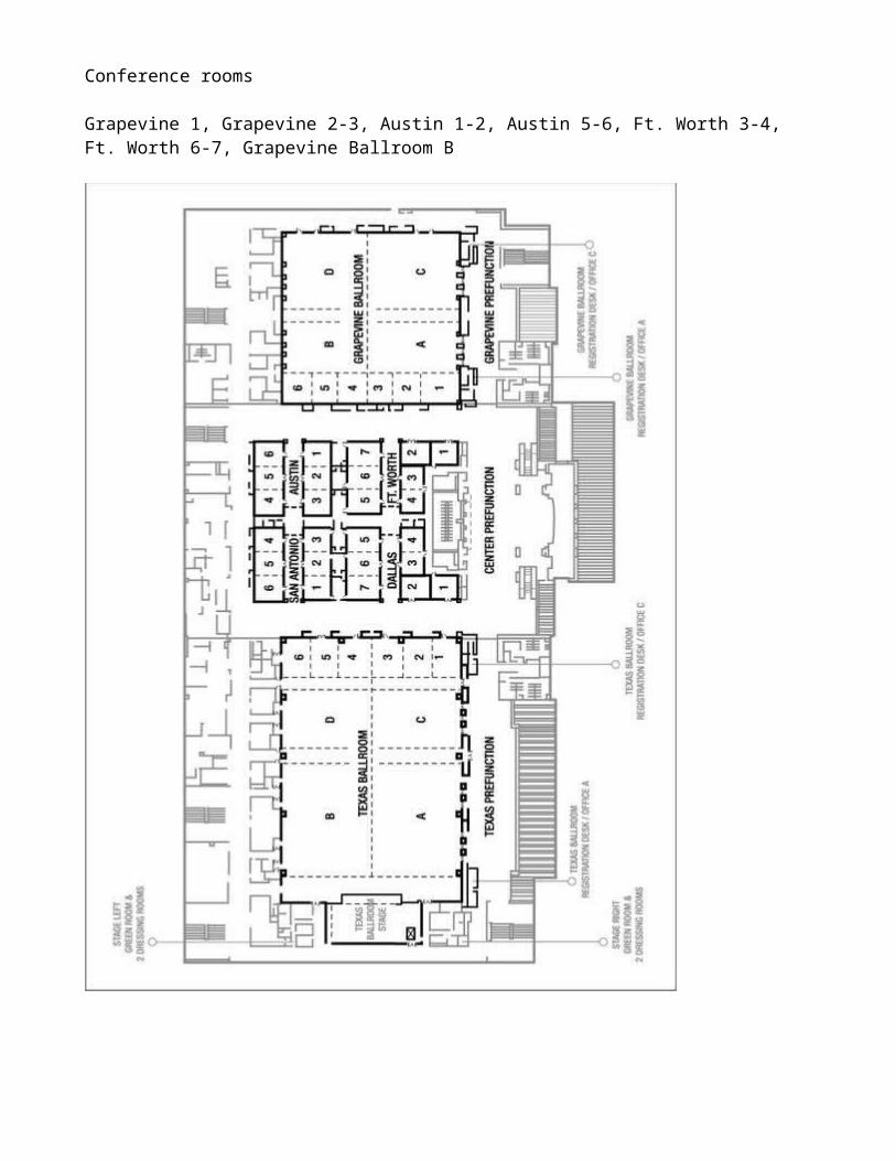
Conference rooms
Grapevine 1, Grapevine 2-3, Austin 1-2, Austin 5-6, Ft. Worth 3-4, Ft. Worth 6-7, Grapevine Ballroom B

Using this Abstract Book
This Abstract Book contains:1. the session summary, 2. full text of accepted abstracts, and 3. the index of authors and co-authors.
The Abstract Book can be downloaded from the conference website at http://www.caari.com and is available in several formats. All formats feature mutual hypertext links between the schedule, abstracts, and the author index.
Notebook, ultrabook, or desktop users: the Microsoft Word version offers popup hints - to see them, please, hover the mouse over the hyperlink. Alternatively, a PDF version is also available.
Users of small mobile devices (tablets or smartphones): While you can certainly use the desktop Word and PDF versions if your device supports them, we have re-flown the Abstract Book to about 4“ width so that if you are viewing the book you do not need to shift the screen horizontally. The mobile version of the Adobe Reader can be downloaded from the corresponding app stores, for example, from Google Play for Android.
Based on the type of program you chose to read this book, you will be able to search for keywords in the abstracts that relate to your areas of interest or add your own notes or bookmars.
This listing contains information about each presentation including all of the authors and their affiliations. If you wish to find out, for example, when Barney Doyle’s talk is to be given, just look up his name in the author index at the end of this book:
Then click on the abstract number to read the full abstract or the session code to view other presentations in the same session.
When you are viewing the abstract click on the author’s name to skip to the author index again or click on the session time and location to see all other presentations in this session.
The abstract details include the abstract number: 318The type of presentation: Invited TalkThe day and time Session RE03 starts: Tuesday 1:30 PMThe location: Presidio C
Hypertext content of this Abstract Book was facilitated by the Meeting247 tools

Session Summary
Please note: the presentations marked (Poster session) in the listing below will be presented during the two poster sessions only. They are listed at the end of each talk session only to provide the session context.
PS-01-MON: Monday Plenary SessionMonday at 9:15 AM in Grapevine Ballroom B
# 381 Applications for a Laser-Driven Accelerator on a Chip by R. Joel England
AP-IA-02: Energy and Environmental ApplicationsMonday at 10:30 AM in Austin 1-2
# 56 Accelerator Technology for Large-Scale Energy Production by Robert W. Garnett# 184 Development Status of the Myrrha Injector by Holger Hoeltermann# 230 Applications of Heavy Ion Linear Accelerator for Studies of Radiation Effects in Nuclear Fuel and Structural materials by Abdellatif Yacout# 3 Reduction of the uncertainty due to fissile clusters in radioactive waste characterization with the Differential Die-away Technique by Rodolphe Antoni# 249 PIXE Analysis of Dust in Rainwater Collected on Polysulfone Filters* by Todd A. Byers# 359 Pulsed hydrogen cold-cathode Penning ion source with high monatomic fraction and high current in stable operation by Kyumin Choe# 21 (Poster session) The application of accelerator technology in treating wastewater and haze in BEPC by Dong Dong# 158 (Poster session) 3-D Simulation and Efficiency Optimization of Thermionic Energy Converters by Jonathan P Edelen# 177 (Poster session) ISDE SEE test facilities based on JINR heavy ion accelerators by VASILY ANASHIN# 284 (Poster session) Controllable defects production and property modification in single-layer MoS2 by using ion irradiation by Kedi Yin
AP-SD-02: Detectors for Accelerator-Based Security and Defense - IMonday at 10:30 AM in Ft. Worth 6-7
# 341 Advanced Detector Materials for Accelerator-Based Security and Defense by Alan Janos# 14 New Detectors for High Energy Radiography by Kanai S Shah# 272 Advances in Solid Organic Scintillators for Wide Energy Range Neutron Detection by Andrew M. Glenn# 267 Mitigation of Photon Active Interrogation Background for Fast Neutron Detection by Cameron A. Miller# 39 Development of a Portable Active Interrogation System for Characterizing Special Nuclear Material by Calvin E Moss# 95 (Poster session) The Use of Fast-Neutron Imaging Detectors for Security Applications by Tobias Achtzehn
AP-TA-03: Undergraduate Education in the Accelerator LaboratoryMonday at 10:30 AM in Ft. Worth 3-4
# 31 Implementing PIXE and PIGE at the Texas A&M University Cyclotron Institute by A. Rodriguez Manso# 67 The Naval Academy Accelerator Facility by Akaa Daniel Ayangeakaa# 242 Undergraduate Education at the University of Kentucky Accelerator Laboratory by Anthony Paul D. Ramirez
# 84 An Inexpensive XRF Lab for Undergraduates and Other Educational Activities at Tarleton's Nuclear Laboratory by Daniel Keith Marble# 259 Cyclotrons and Their Design - an Undergraduate Education by Amber Johnson
AR-ISM-02: Helium Ion Interactions with Functional MaterialsMonday at 10:30 AM in Austin 5-6
# 34 Strain Doping in Functional Oxides by T. Zac Ward# 50 Polarization Control via He-ion Beam Induced Nanofabrication in Layered Ferroelectric Semiconductors by Alex Belianinov# 195 High Resolution Inert-Gas Bombardment Microscopy, Nanofabrication and Secondary Ion Mass Spectrometry: Recent Results from ZEISS ORION NanoFab. by Sybren Sijbrandij# 215 Helium and Emerging Focused Ion Beams by Gregor Hlawacek# 86 (Poster session) Temperature range of helium retention in austenitic stainless steel implanted helium at different temperatures: 100, 300 and 620 K by Oleksandr Morozov
AR-NST-04: Focused Ion Implantation for Novel Device Fabrication - IIMonday at 10:30 AM in Grapevine 2-3
# 32 Fabrication of Single Atom Devices by Direct Write Nanofabrication by Edward Bielejec# 309 Statistics of Deterministic Single Ion Implantation by Roger Paul Webb# 198 Two-axis control of a coupled quantum dot - donor qubit in Si-MOS by Peter Sharma# 213 Simulation and Experimental Analysis of Fe and Co implanted Si Nano wires by Satyabrata Singh# 132 Ion-beam fabricated optically active color centers in diamond for quantum optics and quantum-enhanced sensing. by Sviatoslav Ditalia Tchernij# 373 Advanced Applications in Nanoscale Device Fabrication Enabled by Novel Focused Ion Beam Instrumentation by Joseph Klingfus
PR-AMP-02: Physics of MoleculesMonday at 10:30 AM in Grapevine 1
# 211 Fully-differential and initial-state selective studies of single ionization in ion-lithium collisions by Daniel Fischer# 301 Experimental and computational study of gold nanoparticles as a radiosensitizer for ion radiation by Jefferson L Shinpaugh# 46 The role of multiple electron processes for fast ion H2O collisions by Sebastian Otranto# 164 Measured Absolute Cross Section of Charge Transfer in D2
+ + H between 2 keV/u - 10 keV/u by V M Andrianarijaona# 151 Fragmentation pathways following ionization of Water molecule by electron impact by Lucas Sigaud# 18 (Poster session) Low energy electron scattering by methane molecules in a spherical model by HARSH MOHAN# 57 (Poster session) Study of scattering cross sections for collision of low energy electrons with polar molecule: Hydrogen Chloride by Gurpreet Kaur# 153 (Poster session) Production pathways for symmetric molecular dications: N2
++, O2++ and C2H4
++ by Lucas Sigaud
AA-IBTM-02: Cultural Heritage/Forensic ScienceMonday at 2:00 PM in Grapevine 2-3
# 181 Application of MeV SIMS for forensic document examination by Iva Bogdanovic Radovic# 11 A match made in heaven: Forensic hair screening with ion beam analysis by Karen Jacqueline Cloete# 49 Archaeometry with ion beams - application on the objects from the 1st millennium BC by Ziga Smit# 58 Analysis of Forensic Traces using Direct Analyte-Probed Nanoextraction Mass Spectrometry (DAPNe-MS) and Ion Beam Analysis (IBA) by Holly-May Lewis# 174 (Poster session) The New Microbeam Setup for Cultural Heritage and Bio-medical Applications at the Lebanese Accelerator by Mohamad Roumie

# 379 (Poster session) PIXE Analysis of Ceramics from the Clement Archaeological Site (ca. A.D. 1000-1200), a Caddo Mound Complex in the Middle Red River Valley by Stewart Bragg Younger-Mertz# 380 (Poster session) Identification of a Metal Alloy of Unknown Composition from Oklahoma: Explorations in Twentieth-Century Industrial Archaeology by Stewart Bragg Younger-Mertz
AC-TD-04: Ultra-Compact and Mini AcceleratorsMonday at 2:00 PM in Austin 1-2
# 33 A Portable X-ray Source based on Dielectric Accelerators by Roman Kostin# 35 Inexpensive Brazeless Accelerator Prototype by Roman Kostin# 36 High Shunt Impedance Accelerating Structure with Distributed Microwave Coupling by Roman Kostin# 206 A compact RF-based ion accelerator by Thomas Schenkel# 210 (Poster session) Study of electron cyclotron resonance acceleration by cylindrical TE011 mode by Oswaldo Otero
AR-ISM-01: In situ and 3D Analysis of Irradiation Induced Changes in Microstructure of MaterialsMonday at 2:00 PM in Ft. Worth 6-7
# 123 In-situ irradiation tolerance investigation of nanocrystalline W-Ti-Cr-V high entropy and T-TiC alloys by Osman El Atwani# 138 Exploring the Interplay Between Grain Boundaries and Radiation Damage by Khalid Hattar# 293 Irradiation response of twin boundaries in face-centered cubic metals with low stacking fault energy by Jin Li# 65 Radiation-Induced Effects on Contact Angle and Field Emission of Vertically Aligned Silicon Nanowires by Pathak A P
AR-RE-05: Radiation Effects in materials for Fission ApplicationsMonday at 2:00 PM in Grapevine 1
# 226 Radiation Effects in Concentrated Solid Solution Alloys by Yanwen zhang# 162 Atom probe tomography study on irradiation induced Nb redistribution in ZrNb alloys by Zefeng Yu# 207 Radiation effects in ceramic composite by Maulik Patel# 149 Radiation Enhanced Xenon Diffusion in Yttria Stabilized Zirconia at High Temperatures by Joseph Graham# 79 (Poster session) SMoRE-II Round Robin of Irradiated T91 Steel by Ian Peter Swainson
PR-SP-02: Neutrino PhysicsMonday at 2:00 PM in Austin 5-6
# 322 PROSPECT, A Precision Reactor Oscillation and SPECTrum Short-Baseline Antineutrino Experiment by Chris Bass# 350 Characterization of Stray Magnetic Fields Near the PROSPECT Detector by Corey Gilbert# 394 Barium Tagging with Single Molecule Fluorescence Imaging for Neutrinoless Double Beta Decay by Austin McDonald# 398 (Poster session) Background characterization at the High Flux Isotope Reactor (HFIR) for PROSPECT by Alan Salcedo Gomez
AA-IBTM-03: IBA of Samples Exposed to Extreme EnvironmentsMonday at 4:00 PM in Grapevine 1
# 148 Correlation between Cr3+ Luminescence and Oxygen Vacancy Disorder in SrTiO3 under MeV Ion Irradiation by Joseph Graham# 337 Progress in Coupling Electron Microscopy and Ion Beam Induced Luminescence by Khalid Hattar# 241 Materials modification with ion pulses from laser-plasma acceleration by Thomas Schenkel# 345 Development of ERD Technique for Quantifying Light Isotope Concentrations in Irradiated TPBAR Materials by Caitlin Anne Taylor

AP-SD-05: Software and Simulation for Security and DefenseMonday at 4:00 PM in Ft. Worth 6-7
# 247 Automated container inspection for 100% screening by Denis Dujmic# 399 Applications of MRED for Predicting Single Event Effects by R.A. Reed# 294 Performance of a sparse-data tomography algorithm for active cargo interrogation by Luke Maloney# 90 Integration of CZT and CLYC Radiation Detectors into Robotic Platforms using ROS by Monia Kazemeini# 333 Cloud-Based Radionuclide Source Injection for Real-Time Training Dataset Generation by Michael J King
AP-TA-02: Undergraduate Education and Experiments with Accelerators - IIMonday at 4:00 PM in Ft. Worth 3-4
# 199 Diffusion in sulfide and sulfate minerals with planetary applications by Heather C Watson# 121 Ion beam-based projects for undergraduate students by Daryush ILA# 111 PIXE Analysis of Synthetic Turf by Michael F. Vineyard# 80 Undergraduate research with a 400 KeV Accelerator by Andrew David Roberts
AR-NST-03: Focused Ion Implantation for Novel Device Fabrication - IMonday at 4:00 PM in Grapevine 2-3
# 62 Creation of silicon vacancy in silicon carbide device by proton beam writing toward quantum applications by Takeshi Ohshima# 72 Engineering and coherent control of defects in silicon carbide by G. V. Astakhov# 200 Point defect creation using strain-sensitive x-ray imaging for quantum technologies by F. Joseph Heremans# 171 Single Photon Sources in SiC with Applications in Quantum Technologies by Jeffrey C McCallum# 396 Scalable Nanoscale Patterning of Quantum Emitters in Diamond via Focused Ion Beam by Matthew E Trusheim
PR-SP-08: Nuclear SpectroscopyMonday at 4:00 PM in Austin 5-6
# 193 Doppler-Shift Attenuation Lifetime Measurement of 36Ar with the TIGRESS Integrated Plunger by P. Voss# 76 High eff. β-decay study of 75Ga and structure of 75Ge nuclei. by Umesh Silwal# 169 The next generation neutron detector for the studies of exotic nuclei by Mustafa M Rajabali# 160 Detection of Selenium in Soil Samples Using Photon Activation Analysis by Leyton Brenner# 89 (Poster session) High resolution β-decay study of neutron rich 74Zn into odd-odd 74Ga using LeRIBSS. by Durga P. Siwakoti
PS-02-TUE: Tuesday Plenary SessionTuesday at 8:00 AM in Grapevine Ballroom B
# 368 New Horizons in Particle Therapy Systems by Jonathan B. Farr# 374 Illuminating the Nucleus with Neutrinos by Kate Scholberg
AA-NBAT-01: Nuclear Based AnalysisTuesday at 10:00 AM in Ft. Worth 3-4
# 320 Photon Activation Analysis and Fundamental Problems in Nuclear Physics by Douglas P Wells# 156 Elemental Analysis of Jade Stones using Photon Activation Analysis by Mayir Mamtimin# 13 A Novel Equation for Activity Calculation in Pulse Irradiation by Zaijing Sun# 16 Detection of the Counterfeited Sildenafil Tablets Using Neutron Activation Analysis by Faisal Alrumayan# 303 Determining Trace Elements in Cotton Seeds with Instrumental Neutron Activation Analysis (INAA) by N. Isa
AC-AS-01: Accessing the National Labs and Technology TransferTuesday at 10:00 AM in Austin 5-6
# 370 Update on DOE's Accelerator Stewardship Program by Cherri J Schmidt# 290 Spinning out and in: Technology Commercialization at Berkeley Lab by Elsie Quaite-Randall# 270 DOE's Technologist in Residence (TIR) Program by Eli Levine# 281 Accessing Los Alamos National Neutron Science Center (LANSCE) and Technology Transfer at Los Alamos by Antonio Redondo# 268 5 things to know about working with Argonne National Laboratory by Gregory Halder# 300 Exploring your ROI with A2D2 at Fermilab by Thomas K Kroc
AC-TD-03: Emerging Accelerator Technologies - IITuesday at 10:00 AM in Austin 1-2
# 188 EuPRAXIA - a Compact, Cost-Efficient Particle and Radiation Source by Maria Katharina Weikum# 157 Exploration of Electrochemical Processes for Creating Nb3Sn Thin Films via Bronze Route by Choong-Un Kim# 127 Superconducting Cable-in-Conduit: Enabling Technology for Industrial Applications by Peter McIntyre# 141 Bronze routes that facilitate Nb3Sn superconducting cavity technology by Shreyas Balachandran# 352 Intense, pulsed ion beams for materials research by Thomas Schenkel# 147 (Poster session) Axial Emission Rate of Charged Particles from a Dual-Solenoid Magnetized Plasma by Marisol Hermosillo# 339 (Poster session) Development of Novel Seamless Cavity Forming Methods by John S. Buttles
AP-MA-03: Clinical Progress with Hadrons and Start-up Logistics for New Treatment FacilitiesTuesday at 10:00 AM in Grapevine 2-3
# 364 Prospects for an Advanced Heavy Ion Therapy Center in the Chicago Area by Brahim Mustapha# 321 Preparation for facility startup - experience at the New York Proton Center by Haibo Lin# 371 Intensity Modulated Proton Therapy for lung cancer patients by Heng Li# 307 Toshiba ESS's contribution and future vision for the cancer therapy technology. by Shinya Matsuda
PR-AMP-01: Physics of AtomsTuesday at 10:00 AM in Grapevine 1
# 30 High Resolution X-ray Measurements Following Charge Exchange with Atomic H: Data for a New Observational Window on Diffuse Astrophysical Sources by Ruitian Zhang# 304 Modeling ion-neutral collisions using 'universal' scattering for drifting plasmas or ion beams in PIC-DSMC codes by Jose L. Pacheco# 61 Ultrafast dissociation of the CD2Cl2 and CH2Cl2. by Débora Nunes Barros de Vasconcelos# 391 Ion recoil laser driven by surface plasmons by Pooja Singh# 19 M X-ray production cross sections of 78Pt, 79Au, 82Pb and 83Bi using C3+ and Si3+ ions by Shivcharan Verma# 75 (Poster session) Concentric Cone Antihydrogen Gravity Experiment by Steven R Sun# 106 (Poster session) Variational Calculations for the Ps-Ps System by Gabriel Medrano# 154 (Poster session) L-shell x-ray production cross sections induced by heavy ion impact: searching for a universal curve. by Javier Miranda# 219 (Poster session) Absolute measurement of total cross sections of N7+ - H charge exchange towards thermal energies by R. T. Zhang
PR-SP-06: Nuclear Reactions ITuesday at 10:00 AM in Ft. Worth 6-7
# 159 The fissionTPC by mike heffner# 55 The Dependence of Fission Mass Yields on the Nuclear Structure of the Compound Nucleus by Jenifer Shafer# 4 Photonuclear cross-section and yields of 100Mo(g,x)99Mo, 100Mo(g,np)98mNb, and 59Co(g,xn; x=1-4) 58-55Co reactions with intermediate bremsstrahlung energies from electron linac by Md. Shakilur Rahman
# 139 Energy Dependence of Fission Product Yields for 235U, 238U, and 239Pu with Monoenergetic Neutrons Between Thermal and 14.8 MeV by Matthew Gooden# 287 The Oklo natural fission reactors and improved limits on the variation in fine structure constant by Edward David Davis# 5 Some Calculated (p,α) Cross-sections using the Alpha Particle Knock-on and Triton Pick-up Pre-equilibrium Reaction Mechanisms by Felix S. OLISE# 125 (Poster session) Binding energy and Einstein's mass energy equation by AJAY SHARMA
AC-AS-02: Future Workforce and Capability DevelopmentTuesday at 1:30 PM in Austin 5-6
# 239 Accelerator Science & Engineering Traineeship Program at Michigan State University by Alireza NASSIRI# 349 Program at the Center for Bright Beams to recruit and train the next generation of scientists in accelerator and related fields by Young-Kee Kim# 140 US Particle Accelerator School activities in workforce training for accelerator science and engineering by Steven M Lund
AC-TD-02: Emerging Accelerator Technologies - ITuesday at 1:30 PM in Austin 1-2
# 276 Muon Colliders, Neutrino Factories, and Results from the MICE Experiment by Daniel M. Kaplan# 319 The Integrated Laser-driven Accelerator System by Paul R. Bolton# 42 C-14 Isotopic Targets by Richard L Fink# 51 An Emerging Solid-State UHF Technology based on 100 VDC GaN for Powering Particle Accelerators by Gabriele Formicone# 98 (Poster session) A Multiphysics Simulation Tool for Vacuum System Design and Optimization for Next Generation Light Sources by Nicholas B Goldring# 209 (Poster session) Neural network based virtual diagnostics at FAST by Jonathan P Edelen# 405 (Poster session) Preparation of bronze for niobium coatings to make superconducting Nb3Sn radio-frequency cavities for accelerator applications by Chris Reis
AP-IA-01: Accelerators for Geo-Physical ApplicationsTuesday at 1:30 PM in Ft. Worth 6-7
# 197 Compensated Neutron Logging Tool Using DD Neutron Generator For AmBe Replacement by Brian Jurczyk# 286 Am-Be Versus Neutron Generator-based Alternatives for Well Logging Measurements by Ahmed Badruzzaman# 54 Porosity Measurement in Oil-Well Logging Using a Pulsed-Neutron Tool by Weijun Guo# 366 Advances in Accelerator Technology for the Oil and Gas Industry: High-Output Small Form Factor Neutron Generator for Advanced Neutron Measurements by Frederic Gicquel# 53 Development of a Test Facility for Studies Relevant to Replacing Dangerous Radiological Sources by Long Vo# 376 Ion-Beam Analysis for Non-Destructive Direct Measurement of Hydrocarbon Content and Mineralogical Elements within Shale by Khalid Hossain
AP-MA-01: Technological Developments and Future Aspirations - ITuesday at 1:30 PM in Grapevine 2-3
# 362 Planning and Supporting a New Generation of Centers for Particle Beam Radiation Therapy Research by John Robb# 336 Ion microbeam SNAKE: Technology and future developments for radiobiological and medical research by Judith Reindl# 365 A New Model for Future Ion Beam Therapy Research and Treatment Centers - From the Laboratory to the Clinic by James Welsh

# 403 Use of Thermoacoustics to Image Therapeutic Charged Particle Beams by Keith Stantz# 47 (Poster session) Development of a very wideband RF-knockout system for a spot scanning irradiation by Tetsuya Nakanishi# 137 (Poster session) Studies of Fricke-PVA-GTA xylenol orange hydrogels for 3D measurements in radiotherapy dosimetry by Grazia Gambarini
AP-TA-04: Graduate Programs ITuesday at 1:30 PM in Ft. Worth 3-4
# 190 Resource sharing in the Nuclear Physics laboratory classes: a distribuited data acquistion system for experiments with shared resources and data managament by Cristiano Lino Fontana# 258 Graduate Research Programs in Nuclear and Radiochemistry at Hunter College by Jennifer Shusterman# 253 Development of the St. Andre Ion Beam Analysis Facility at Notre Dame by S.R. McGuinness
AR-RE-02: Multiscale studies of irradiated materials for fusion applications - IITuesday at 1:30 PM in Grapevine 1
# 323 On the Radiation Tolerance of Nanocrystalline Tungsten Materials by Stuart Maloy# 388 Utilizing the Helium ion beam microscope for implantation studies to understand the effect of He bubble structures in solids by Peter Hosemann# 28 Enhanced radiation tolerance of nanochannel materials by Feng Ren# 402 Atmospheric Neutron Testing by Laura Dominik
AA-IBTM-05: Ion and Micro Beam AnalysisTuesday at 3:30 PM in Grapevine 1
# 173 Use of Proton Beam and its Rutherford Backscattering on CR39 for Radiation Dose Assessment by Mohamad Roumie# 262 ToF-ERD analysis software Potku 2.0 with integrated MCERD by Mikko Ilkka Laitinen# 314 Ion Channeling In Nanotubes by J Napagoda# 133 Preliminary formation of a negative ion microbeam in a compact ion microbeam system by Takeru Ohkubo# 351 Design of an electrostatic focusing system for low MeV multi-ion micro-beam by V A Chirayath# 23 (Poster session) Measurement of α-7Li scattering cross-section for time-of-flight and transmission ERDA by Kohtaku Suzuki# 99 (Poster session) New Installation of AMS at Dongguk University by Sang-Hun Lee# 170 (Poster session) Prototype of a penning ionization gauge type ion source with a permanent magnet for a MeV compact ion microbeam system by Takeru Ohkubo# 332 (Poster session) High Resolution Ion Beam Analysis of GaAs(100) Oxides combined with Electron Spectroscopy for Chemical Analysis (ESCA/XPS) and Surface Energy Analysis: Comparison with Si(100) by Sukesh Ram# 397 (Poster session) Measurement of Helium Diffusion through Nuclear Reaction Analysis by Matthew Chancey
AP-MA-02: Technological Developments and Future Aspirations - II Tuesday at 3:30 PM in Grapevine 2-3
# 264 Impact of imaging and beam scanning technologies on the particle therapy penetration to the mainstream clinical practice. by Zelig Tochner# 348 Robust Iterative Methods: Convergence and Applications to Proton Computed Tomography by Keith E Schubert# 308 Pluridirectional High-energy Agile Scanning Electronic Radiotherapy (PHASER) by Vinod Bharadwaj# 280 PET-aided hadron therapy based on 11C by Johanna Pitters
AR-ISM-03: Ion Beam Modification and Radiation Effects in SolidsTuesday at 3:30 PM in Austin 1-2
# 92 Pulsed plasma generated in coaxial accelerator for the synthesis of thermodynamic unstable materials by KATARZYNA NOWAKOWSKA-LANGIER# 306 Analyzing ion beam irradiation effects in materials by multimodal microstructural characterization by ARUN DEVARAJ# 97 Radiation response in single-phase concentrated solid solution alloys by Chenyang Lu# 191 Modification of crystalline phase in space silicate analogues with light ion irradiation by Joshua Michael Young# 87 (Poster session) Deuterium сoncentrations in austenitic stainless steel by deuterium irradiation. Effects dose and temperature irradiation by Volodumyr Zhurba# 88 (Poster session) Impact of Ionizing Radiation and Additive on Chitosan-based Biodegradable Matrices by Gnansagar B Patel# 96 (Poster session) Modification of Indium Tin Oxide (ITO) thin films on glass substrate by Vanadium keV ion implantation: Structural, electrical and optical properties by Olakunle Oluwaleye# 100 (Poster session) Evidence of a Simple Damage Energy Scaling Rule for Proton Irradiation of Luminescent Materials by William A. Hollerman# 109 (Poster session) Synthesis of Nickel nanoclusters embedded within Indium Phosphide lattice via low energy ion implantation by Daniel C Jones# 122 (Poster session) Implementation of high energy ions implantation for the adjustments of properties of complex semiconductor structures by Vitalij Kovalevskij# 255 (Poster session) Development of Ion Beam Induced Luminescence (IBIL) and Proton Beam Induced UV Spectroscopy (PUV) at the Ion Beam Modification and Analysis Laboratory by Gerard Munyazikwiye
AR-RE-07: Accelerator-based Irradiation capabilities for Nuclear Energy ResearchTuesday at 3:30 PM in Ft. Worth 6-7
# 375 IAEA activities in support of Materials Research using Ion Beams by Ian Peter Swainson# 212 High Intensity D-T Fusion Neutron Generator and Its Applications by Yongfeng Wang# 196 Mitigation of carbon contamination in ion irradiation experiments through environmental conditioning by Fabian U. Naab# 257 Roadmap for the Application of Ion Beam Technologies to Challenges for the Advancement and Implication of Nuclear Energy Technologies by Simon M. Pimblott# 110 Coupling a 6 MV Tandem and an Ion Gun to a Scanning Electron Microscope by Samuel A Briggs
PR-SP-05: Physics with Lasers and TrapsTuesday at 3:30 PM in Austin 5-6
# 105 Laser ion source as a research tool at the ISOLDE/CERN radioactive ion beam facility by Valentin Fedosseev# 221 TITAN-TRIUMF: Ion traps for precision experiments with radioactive ion beams by A. A. Kwiatkowski# 180 Laser Isotope Separation revisited - Production and characterization of highest purity radioisotope samples for neutrino mass determination and more by Klaus D.A. Wendt# 166 Development of Laser Ion Sources at IMP by Huanyu Zhao# 194 High Efficiency Resonance Laser Ionization of Pu by Elisa Romer-Romero
PS-03-WED: Wednesday Plenary SessionWednesday at 8:00 AM in Grapevine Ballroom B
# 114 Ten qubits in five years: Building a near-term quantum computer device by deterministic ion implantation based on donors in silicon by David Norman Jamieson# 81 Harvey Recovery: Drones, Cores, and Photon Activation Analysis by Philip L Cole
AA-IBTM-01: Advances in MeV SIMSWednesday at 10:00 AM in Grapevine 1
# 179 Chemical Analysis under Ambient Conditions using MeV-Energy Heavy Ion by Toshio Seki
# 175 Exploring the compatibility of MeV SIMS and Desorption Electrospray Ionisation (DESI) for multimodal imaging of biomedical samples by Melanie J Bailey# 94 Secondary ion yield and fragmentation in MeV-SIMS performed on organic samples by Klaus-Ulrich Miltenberger# 296 Detection of Cocaine Parent and Fragment Molecules using Ambient Pressure MeV-SIMS by Roger Paul Webb# 214 Recent developments and applications of MeV SIMS at the Ruđer Bošković Institute in Zagreb by Iva Bogdanovic Radovic# 224 (Poster session) Hydrogen Mobility in Materials by Kwyntero Van Kelso
AC-AF-01: Accelerator Facilities for Industry and MedicineWednesday at 10:00 AM in Austin 1-2
# 354 Isotope Research Applied to Life Sciences in the Planned Institute for Advanced Medical Isotopes (IAMI) by Monika Kinga Stachura# 265 IsoDAR: A Cyclotron based Neutrino Source with Applications to Medical Isotope Production by Loyd Hoyt Waites# 144 The HVEE Single Ended Particle Accelerators - Performance and Applications by Nicolae C. Podaru# 7 Improvements in the Design of Dynamitron Accelerators by Richard A Galloway# 244 Spreading the Wealth of Accelerator Knowledge: The Illinois Accelerator Research Center by Thomas K Kroc# 360 Towards the Next Generation of Ion Therapy and Imaging Accelerators by Carol Johnstone# 59 (Poster session) Influence of the Frequency Detuning to Electrodynamics Parameters of an Electron Linac by Alexey Igorevich Pronikov# 183 (Poster session) The new heavy ion irradiation facility at KVI-CART by Brian Nathaniel Jones# 390 (Poster session) Capabilities of the Fermilab Test Beam Facility by Carol Johnstone
AP-MA-06: Accelerator-Based Boron Neutron Capture Therapy (BNCT)Wednesday at 10:00 AM in Grapevine 2-3
# 260 Boron Neutron Capture Therapy in Finland: the Past, the Present and the Future by Hanna Koivunoro# 295 Status of the accelerator based BNCT projects Worldwide by YOSHIAKI KIYANAGI# 311 Overview of Cyclotron-based Epithermal Neutron Source(C-BENS) for BNCT by Hiroki Tanaka# 334 Beam performance of the iBNCT as a compact linac-based BNCT neutron source developed by University of Tsukuba by Hiroaki Kumada# 356 Accelerator Technologies at TAE: from Fusion to BNCT by Alexander Dunaevsky# 136 Problems in dose measurements in Adrotherapy and BNCT due to dosimeter sensitivity quenching by Grazia Gambarini# 63 (Poster session) Studies of Boron-10 Doped Nanodiamonds Made by Ion Implantation for Boron Neutron Capture Therapy by Yueh-Chung Yu
AP-SD-06: Neutron Generators for Security and DefenseWednesday at 10:00 AM in Ft. Worth 6-7
# 202 Using Particle-in-Cell (PIC) Models to Optimize Short-Pulse Neutron Sources for National Security and Industry Applications by Andrea Schmidt# 327 Explosive Detection Using a Field Deployable Neutron Generator by Paul McBride# 167 R&D on a Portable Active Neutron Interrogation System for Special Nuclear Materials by Kai Masuda# 217 Short-pulse Photoneutron Production on Beryllium Using the Mercury Pulsed Power X-ray Source* by Joseph W Schumer# 185 Characterization of a Portable Neutron Generator for Neutron Imaging by Matthew D Coventry# 358 Advanced Neutron Generators for Activation Analysis and Imaging by Charles K Gary
AR-ISM-07: Applications of Ion Beams in Frontiers of Material ResearchWednesday at 10:00 AM in Austin 5-6
# 228 Effects on Electronic Energy Loss on Irradiation Response of SiC by William J Weber# 229 Ionization Effects in Oxides under Ion Irradiation by Yanwen Zhang# 161 Additively Manufactured grade 91 steel for reactor applications by Benjamin Paul Eftink# 383 Universal"Ion-cut" for Nanopatterning, modification and heterointegration of semiconductors by Xin Ou# 305 Helium Ion Microscope: Past, Present and Future by Vaithiyalingam Shutthanandan
AR-NST-01: Nanoscale Surface Patterns Produced by Broad Beams of Particles - IWednesday at 10:00 AM in Ft. Worth 3-4
# 285 Spontaneous pattern formation on crystalline surfaces induced by low energy ion irradiation by Stefan Facsko# 93 Nonlinear theory of ion-induced solid flow by Rodolfo Cuerno# 115 Prediction of ion-induced pattern formation using Monte Carlo Simulations and comparison with experiments by Hans Hofsaess# 343 Sputtering of silicon: a comparison between simulations and experiments by Naresh T. Deoli# 150 (Poster session) Tailoring Si Nanocone Arrays via Simultaneous Low Energy Helium Ion Sputtering on Metal and Si Surfaces by Nicholas Carl Termini# 172 (Poster session) Concurrent segregation and erosion effects in medium-energy iron beam patterning of silicon surfaces by Rodolfo Cuerno
AP-MA-04: Particle Beam Radiobiology: Modeling and Simulation for Accelerator-Based Medical Applications
Wednesday at 1:30 PM in Grapevine 2-3# 335 Radiobiological and preclinical, medical physics research at the ion microprobe SNAKE by Judith Reindl# 400 A comparison of biophysical models predicting the relative biological effectiveness (RBE) for treatment planning in ion beam therapy by M. Scholz# 235 Simulating Boron Enhancement in Proton Therapy by Jacob Daniel Baxley
AP-SD-01: X-Ray Sources for NII SystemsWednesday at 1:30 PM in Ft. Worth 6-7
# 236 From Science to Industry: A Truly High-Power Electron Accelerator for Multiple Industrial Applications by Thomas K Kroc# 41 X-ray Sources for Adaptive Radiography and Computed Tomography by Sergey V Kutsaev# 250 Novel Linear Accelerators for X-ray Sources by VINOD BHARADWAJ
AR-ISM-04: New Frontiers of SIMSWednesday at 1:30 PM in Austin 1-2
# 201 Towards Elucidation of Plant and Bacteria Interactions Using In Situ Liquid SIMS by Xiao-Ying Yu# 178 SIMS Analysis of Liquid Materials in Low Vacuum with Large Cluster Ion Beam by Toshio Seki# 45 Molecular Examination of Ion Solvation using in situ Liquid SIMS by Wen Liu# 103 Using ToF-SIMS to advance the quantification of stopping powers for heavy ions in ceramics by Ke Jin# 44 Molecular tracking of mass transfer in electric double layer at electrode-electrolyte interface using in situ liquid SIMS by Zihua Zhu
AR-RE-04: Radiation Effects in Nanostructured MaterialsWednesday at 1:30 PM in Grapevine 1
# 74 Imperfect WSe2 monolayer is better than perfect ones as a platform for SERS by Yang Tan# 392 Development and Testing of Advanced Alloys for High Dose Applications in Next Generation of Nuclear Reactors by Osman Anderoglu# 9 In-situ TEM Observation of the Response of Tungsten Nanoparticles under He+ Irradiation by E Aradi# 218 Ion introduced defects in graphene and their applications on ion seperation and transport by Huijun Yao

# 289 (Poster session) Synthesis and modification of Ti2SnC nanolaminates with high-fluence ions by Jiri Vacik# 291 (Poster session) Enhanced radiation tolerance of YSZ/Al2O3 multilayered nanofilms with pre-existing nanovoids by Hui Wang# 378 (Poster session) Electron Emission from Fast Ion Interactions with Metallic and Biological Materials by Wilson L Hawkins
PR-SP-07: Nuclear Reactions IIWednesday at 1:30 PM in Austin 5-6
# 223 Radioactive Targets at Los Alamos National Laboratory: A quasi-philosophical approach by Christiaan Vermeulen# 187 Measurements of (p,n) reactions relevant to the neutrino-p process in the ReA3 facility by Panagiotis Gastis# 240 Experiments with Radioactive Beams at the Texas A&M University Cyclotron Institute by Gregory Christian# 269 Elastic and Inelastic Scattering with TwinSol Radioactive Beams: Study of Cluster Structure in Light Nuclei by Tan Ahn
AC-TD-01: Accelerator Technology for Security and Defense ApplicationsWednesday at 3:30 PM in Austin 1-2
# 342 MeV LINAC Systems for Security and Cargo Scanning with Pulse-to-Pulse Control of Stability, Energy and Dose by William Leo Nighan# 344 Monoenergetic Photon Radiography in Active Interrogation of Special Nuclear Material by Anna Erickson# 298 Practical optimisation of radiation sources for border security applications by Ceri David Clemett
AP-IA-03: Neutron Applications for IndustryWednesday at 3:30 PM in Ft. Worth 6-7
# 328 Neutron Radiography using a High-Flux Compact Thermal Neutron Generator by Katie Rittenhouse# 367 Advances in High Flux Compact D-D Neutron Generator using an Off-Resonance Electron Cyclotron Resonance Driven Microwave Ion Source by Allan Xi Chen# 326 Commercial Applications of High-Yield Accelerator-Based Neutron Generators by Katie Rittenhouse# 315 Characterization of a Compact 4/2 MeV D+/p RFQ Accelerator System for Fast-, Epithermal- & Thermal-Neutron Radiography by Thomas J. Houlahan, Jr.# 329 Neutron Generator Driven Active Fuel Scanner by Katie Rittenhouse
AP-MA-05: Accelerator Production of Medically Relevant IsotopesWednesday at 3:30 PM in Grapevine 2-3
# 129 Strong-Focusing Cyclotron - High-Current Proton Driver for Isotope Production by Peter McIntyre# 130 Improving Properties of Carbon Stripper (Extractor) Foils by Use of Multiple Layers by Constance G. Stoner# 203 Prospects of the use of laser resonance ionization in production of medical radioisotopes by Vadim Gadelshin# 330 Accelerator-Based Production of Mo-99: Photonuclear Approach by Sergey Chemerisov# 331 Production of Medical Isotopes at Argonne Low Energy Accelerator Facility (LEAF) by Sergey Chemerisov# 43 (Poster session) Isotopic Targets with Graphene Backing by Richard L Fink
AP-TA-05: Graduate Programs IIWednesday at 3:30 PM in Ft. Worth 3-4
# 283 Production of 89Zr and development of nHap molecular imaging agents by Stacy L. Queern# 263 Ion Beam Experiments for Nuclear Nonproliferation and Security Applications by Jason Nattress
# 317 Isotope Harvesting and Ion Beam Analysis as Graduate Research Projects by Graham F Peaslee
AR-RE-06: Radiation Effects in Non-Metallic MaterialsWednesday at 3:30 PM in Grapevine 1
# 237 Defect Engineering of Molybdenum Disulfide (MoS2) to Tunable Hydrogen Evolution Behavior through Ion Irradiation by Engang Fu# 15 Effects of Swift Heavy Ion irradiation on the structural and electrical properties of HfO2 based Resistive Random Access Memory Devices by Arun Nimmala# 101 (Poster session) New Half Brightness Fluence Measurements for Large-Grained ZnS:Mn, EuD4TEA, and MnD4TEA Samples by William A. Hollerman# 163 (Poster session) Analysis of Leg Bones of Rats exposed to Simulated Microgravity and Space Radiation† by Sidney G. Freyaldenhoven
PR-SP-09: Nuclear Data ApplicationsWednesday at 3:30 PM in Austin 5-6
# 238 Nuclear Data Research Activities at Argonne National Laboratory by Filip G. Kondev# 246 Neutron-induced reaction rates away from stability for astrophysics applications: Uncertainties in statistical model calculations and implications for neutron-induced nucleosynthesis. by Georgios Perdikakis# 252 Nuclear Data in Defense Program Applications by Michelle A. Mosby# 393 Revealing the signature of individual fission products from nuclear reactors' antineutrino spectra by Libby McCutchan
PS-04-THU: Thursday Plenary SessionThursday at 8:45 AM in Grapevine Ballroom B
# 355 Ion Implantation for Electronic Devices: From Dennard Scaling to Qubits by Michael Ira Current
AA-NBAT-02: Neutron Based Techniques & Nuclear DataThursday at 10:00 AM in Ft. Worth 3-4
# 299 14 MeV Neutron Tests at the Sandia Ion Beam Laboratory by William Wampler# 387 Neutron diagnostic characterization at the Ion Beam Laboratory for inertial confinement fusion experiments conducted at the Z-accelerator facility by Jedediah Styron# 312 Measurement of flux-weighted average cross-section of 100Mo(g,x)99Mo, 100Mo(g,np)98mNb, and 59Co(g,xn; x=1-4) 58-55Co reactions with bremsstrahlung end-point energies of 55-65 MeV by Md. Shakilur Rahman# 302 Study of Li diffusion in Li-ion batteries by Thermal Neutron Depth Profiling by Jiri Vacik# 112 Development of a Portable Neutron Generator Based on Inertial Electrostatic Confinement D-D Fusion Reaction by Mahmoud A Bakr# 277 (Poster session) High energy proton interactions in organic tissue by Oksana Vladlenivna Kozak
AC-AF-02: Accelerator Facility Updates - IThursday at 10:00 AM in Austin 1-2
# 52 An AC dipole for the AGS Booster to overcome spin resonances* by Nicholaos Tsoupas# 70 CAS - The new Centre for Accelerator Science in Australia. Its facilities and capabilities for Australia and the Asian region. by Armand J Atanacio# 232 An Overview of the Facilities, Activities, and Developments at the University of North Texas Ion Beam Modification and Analysis Laboratory (IBMAL) by Jack E Manuel# 266 Early Experience Using a Variable-Energy, High-Intensity, Pulsed-Mode Ion Source for Low-Energy Nuclear Astrophysics Studies at LENA by Andrew Leland Cooper# 20 (Poster session) Measurement of the background radiation at the KFSHRC CS30 cyclotron by Faisal Alrumayan# 69 (Poster session) Buncher Control System for the 88-Inch Cyclotron by Alex Kireeff# 278 (Poster session) Scientific results of the Institute of Nuclear Research of Ukraine for the needs of nuclear medicine by Oksana Vladlenivna Kozak
AP-SD-03: Detectors for Accelerator-Based Security and Defense - IIThursday at 10:00 AM in Ft. Worth 6-7
# 325 Detectors and algorithms for active interrogation by Sara A. Pozzi# 347 New scintillator development, large crystal growth, and the challenges of maintaining high performance by Stacy E Swider# 134 Signal processing optimization for a neutron scintillator read-out with SiPMs by Enrico Gazzola
AR-RE-01: Multiscale studies of irradiated materials for fusion applications - IThursday at 10:00 AM in Grapevine 2-3
# 275 Characterization of helium plasma-induced damage of tungsten surfaces using helium ion microscopy, ion channeling, and in-situ spectroscopic ellipsometry by Robert D. Kolasinski# 234 Long-timescale atomistic simulations of Helium in Tungsten for fusion applications by Danny Perez# 155 Multi-scale electron microscopy study on the damage mechanisms of materials under fusion irradiation environments by Kun Wang# 346 Direct Comparison of Helium Aging in Ion Implanted and Tritium Loaded Metals by Caitlin Anne Taylor
PR-AMP-03: Radiation Effects in Biological and Chemical SystemsThursday at 10:00 AM in Grapevine 1
# 256 Radiation track structure and the charge cycling of ions in nuclear materials by Simon M. Pimblott# 292 The application of ion beam accelerators to the study of radiation damage in biological systems : some perspectives from a small lab. by Andy Smith# 126 Thionated Uracils under UV Irradiation: Intramolecular Micro-Environmental Effects on the Intersystem Crossing Dynamics by Susanne Ullrich# 401 A Generalized Sturmian Functions approach to proton-impact double ionization of He by Marcelo Ambrosio# 182 Photon Data in Radiation Dosimetry: Analysis of ICRU Report 90 Recommendations and Beyond by Paul Bergstrom# 220 Bimolecular Damage Induced by Ionizing Radiation: The Direct and Indirect Effects of Low-Energy Electrons on DNA by Elahe Alizadeh# 377 (Poster session) Sensitization of malignant cells by nanoparticles to proton radiation by Nichole Cheri Libby
PR-SP-03: AstrophysicsThursday at 10:00 AM in Austin 5-6
# 85 Spectroscopic strengths of low-lying levels in 18Ne by Patrick D O'Malley# 38 Use of (3He,n) Indirect Measurements to Study H and He burning reactions in Type-I X-Ray Bursts by Doug Soltesz# 68 New S-Wave Resonances Found in 19Ne for the 18F(p,α)15O Reaction by Matthew R Hall# 205 Development of a Long Counter for (alpha,n) measurements at the Ohio University Edwards Accelerator by Kristyn Holley Brandenburg# 40 (Poster session) Use of (3He,n) Indirect Measurements to Study H and He burning reactions in Type-1 X-Ray Bursts by Doug Soltesz# 222 (Poster session) Transfer of the Oak Ridge Enge Split-Pole Spectrograph to Notre Dame by D.W. Bardayan# 318 (Poster session) Piercing the Veil of Modern Physics. Part 3 & Superconductivity by Jian DING
AP-TA-01: Undergraduate Education and Experiments with Accelerators - IThursday at 1:30 PM in Ft. Worth 6-7
# 152 The Value of Undergraduate Research, From the Perspective of an Early-Career Nuclear Scientist by Christopher John Prokop

# 117 Analysis of the 11B(p,a)2a nuclear reaction using an ion implanter by Hans Hofsaess# 142 Distribution of Heavy Metals Along New York's East River by Scott LaBrake# 145 (Poster session) Proton Induced X-ray Emission (PIXE) Analysis to Measure Trace Metals in Soil along the East River in Queens, NY by Sajju Chalise
AR-ISM-05: High Energy ions in Advanced Functional MaterialsThursday at 1:30 PM in Austin 1-2
# 64 The degradation study of organic perovskite single-crystals and solar cell devices by in-situ ion beam analysis by MALLIKARJUNA RAO MOTAPOTHULA# 24 Sequential MeV implantation effect on the refractive index and nanoparticle nucleation in silica by John Derek Demaree# 279 Ion Beams and Lasers for Synthesis , modifications and characterization of NanoMaterials by Anand P Pathak# 25 Ion Induced Structural Changes in Graphite by Lenore S. Miller# 6 (Poster session) Selection of positive and negative photoconductance in doped black phosphorus with ultra-broad response from 632.8 nm to 10 μm by Yang Tan
AR-NST-02: Nanoscale Surface Patterns Produced by Broad Beams of Particles - IIThursday at 1:30 PM in Grapevine 2-3
# 128 Nanoscale ripple patterns on a surface of growing multilayer films by Dmitriy L. Voronov# 60 Terraced Topographies and Blazed Diffraction Gratings Produced by Ion Sputtering by R. Mark Bradley# 135 Surface nanopatterning induced by low-energy ion irradiation: Experimental investigations of non-equilibrium pattern formation by Denise Erb# 273 Co-GISAXS Analysis for Investigating Surface Growth Dynamics of Ar+ Bombardment of SiO2 by Mahsa Mokhtarzadeh
PR-SP-04: Fundamental SymmetriesThursday at 1:30 PM in Austin 5-6
# 231 Fundamental weak interaction studies using ion traps by Praveen D Shidling# 363 Fundamental Physics at the LANSCE Ultracold Neutron Source by Chen-Yu Liu# 386 Fundamental Physics at the Oak Ridge Spallation Neutron Source by Vince Cianciolo
AA-CR-01: Highlights of Accelerator-Based Conferences held in 2018Thursday at 3:30 PM in Austin 5-6
# 297 The 9th International Particle Accelerator Conference, IPAC-18 by Oliver Kester# 310 Report on the ICNMTA and COSIRES conferences by Roger Paul Webb
AA-IBTM-04: IBA of Liquids and Biological SamplesThursday at 3:30 PM in Grapevine 2-3
# 274 Accurate Electrolyte Measurements by Ion Beam Analysis using Microliter-Size Blood Drops Congealed Into Homogeneous Thin Solid Films via Hemadrop™ Coatings and DropFilmStrip™ Substrate by Nicole Herbots# 118 Development of external beam coincidence ERDA : Hydrogen analysis of thin films and moist samples by Hans Hofsaess# 225 Rapid Human Blood Diagnostics via Ion Beam Analysis of 10 μl Droplets Congealed into Homogeneous Thin Solid Films (HTSF) via Hemadrop™ Coatings by Harshini L. Thinakaran# 176 (Poster session) Exploring the possibility of IBA and Direct Analyte Probed Nano Extraction (DAPNe) for protein and elemental speciation by Mason Malloy# 282 (Poster session) Determination of Minimum Detectable Levels and Backing Film for Micro-PIXE Analysis of Rat Brain Tissue by Shawn C. Hampton# 385 (Poster session) Optimizing the Surface Energy and Solidification Rate of Hyper-Hydrophilic Coatings (InnovaDropTM) That Allow Fluids To Be Analyzed In Vacuo by Jack M Day
AP-IA-04: Associated Particle Neutron Generator ApplicationsThursday at 3:30 PM in Ft. Worth 6-7
# 77 Application of associated particle neutron techniques for soil carbon analysis by Galina Yakubova# 189 Fast Neutron Induced Gamma-ray Imaging Study by Bonnie Elise Canion# 108 Associated Particle Imaging of Carbon in Soil by Mauricio Ayllon Unzueta# 372 Advances in Associated Particle Imaging Neutron Generators by Charles k Gary# 357 (Poster session) Comparison of YAP and ZnO scintillators for Associated Particle Imaging by Charles K Gary
AP-TA-11: Cyclotrons: Beam Production and ApplicationsThursday at 3:30 PM in Ft. Worth 3-4
# 382 Cyclotrons: Beam Production and Applications by Brian T. Roeder
AR-ISM-06: Defect Engineering in ion Modified MaterialThursday at 3:30 PM in Austin 1-2
# 102 Investigating compositional effects on irradiation response in single-crystalline concentrated solid-solution alloys using ion beam techniques by Ke Jin# 113 Nanomechanical properties of ion implanted ferritic/martensitic steels. by Lukasz Kurpaska# 251 Ion Beam Analysis Correlated with Surface Energy Measurements on Silicon Oxides as a Function of Dopant Species and Concentration by Saaketh R. Narayan# 208 Defect Engineering in Transition Metal Oxides and Dichalcogenide Using Heavy Ion Irradiation by Yan Chen# 10 (Poster session) A review on the role of ion implantation of h-BN in the nucleation of c-BN nanocrystals by E. Aradi
PR-AMP-04: PIXE Basics and ApplicationThursday at 3:30 PM in Grapevine 1
# 124 Universal empirical and theoretical fits to L-shell x-ray production cross sections by protons by Gregory Lapicki# 104 Micro-PIXE analysis in plant biology at Jozef Stefan Institute by Paula Pongrac# 313 Towards Aerosol Analysis at the PIXE-RBS Beamline in the University of Jordan Van de Graaff Accelerator (JUVAC) by Hanan Sa'adeh# 204 (Poster session) Elemental quantification of homogeneous thick samples using particle-induced x-ray emission (PIXE), including hydrogen profiling. by Juan Manuel Restrepo Arteta
PS-05-FRI: Friday Plenary SessionFriday at 8:45 AM in Grapevine Ballroom B
# 233 Applications of Ion Accelerators in Nuclear Materials Science: Issues and Perspectives by Lin Shao
AC-AF-03: Accelerator Facility Updates - IIFriday at 10:00 AM in Austin 1-2
# 78 Current status of plasma spectroscopic experiments with Hyper-ECR ion source at CNS, University of Tokyo by Hideshi Muto# 82 First Beam at FRIB: Commissioning the Facility for Rare Isotope Beams by Steve Lidia# 71 RHIC Status and Plans by Christoph Montag# 192 The 88-Inch Cyclotron and its Applications by Michel Kireeff Covo# 324 Status of the Louisiana Accelerator Center by Naresh T. Deoli# 143 (Poster session) Solid-State Thyratron Replacement by John Kinross-Wright
AP-SD-04: Border, Airport, Rail Car, and Maritime SecurityFriday at 10:00 AM in Ft. Worth 6-7

# 243 Cargo Screening Using High Duty Cycle Sources by Cody M Wilson# 271 Z-SCAN, Z-SPEC and Radiography Detectors for X-Ray Cargo Inspection by Willem G. J. Langeveld# 146 Transmission Cargo Inspection with Ramping-Energy X-ray Pulses by Anatoli Arodzeroo# 186 First results of the integration tests of the Rapidly Relocatable Tagged Neutron Inspection System (RRTNIS) of the C-BORD project by Cristiano Lino Fontana# 361 (Poster session) Design of a high-repetition-rate gamma-ray source based on Inverse Compton Scattering by Aaron Thomas Fetterman
AR-RE-03: Radiation Effects in ElectronicsFriday at 10:00 AM in Grapevine 1
# 254 Overview of Radiation Effects in Semiconductor Devices by Jeffrey S George# 338 Space Environment Effects on Space Systems by Suzanne F Nowicki# 389 Radiation Effects on High Performance Computers by Sean Blanchard# 288 Effects of Electron Beam Induced Current on Breakdown Voltage of GaN P-N Junction Diodes and AlGaN/GaN Schottky Diodes by Albert Colon# 131 Thin Carbon Foils for Time-of-Flight (TOF) Measurements of Low-Energy Ions' Velocities by Robert B. Stoner# 22 (Poster session) Heat transport in silicon carbide bombarded by proton beams by Maher Soueidan# 66 (Poster session) Sensitivity of HfO2 based RRAM devices to gamma irradiation by N Arun# 91 (Poster session) Study of Radiation Effects in Electronics of a Hexapod Robotic Platform by Monia Kazemeini# 404 (Poster session) Electret formation by substrate charging method using MeV protons by Kyle L. Coutee
PR-SP-01: New FacilitiesFriday at 10:00 AM in Austin 5-6
# 216 High Energy Circular Electron Positron Collider (CEPC) As A Higgs Factory by Xinchou Lou# 395 Status of Helium-Jet Ion-Source development at NSCL/FRIB. by Jiban Jyoti Das# 107 Physics design of the next-generation spallation neutron target-moderator-reflector-shield assembly at LANSCE by Lukas Zavorka# 165 (Poster session) Assessing the potentiality of the 250KV-SSAMS for stopping power measurements at low energies by Lucas Sigaud
Regular PostersWill be presented on poster boards Monday 6:00 PM to 7:30 PM and Tuesday 5:30 PM to 7:30 PM
AA-IBTM-01 # 224 Hydrogen Mobility in Materials by Kwyntero Van KelsoAA-IBTM-02 # 174 The New Microbeam Setup for Cultural Heritage and Bio-medical Applications at the Lebanese Accelerator by Mohamad RoumieAA-IBTM-02 # 379 PIXE Analysis of Ceramics from the Clement Archaeological Site (ca. A.D. 1000-1200), a Caddo Mound Complex in the Middle Red River Valley by Stewart Bragg Younger-MertzAA-IBTM-02 # 380 Identification of a Metal Alloy of Unknown Composition from Oklahoma: Explorations in Twentieth-Century Industrial Archaeology by Stewart Bragg Younger-MertzAA-IBTM-04 # 176 Exploring the possibility of IBA and Direct Analyte Probed Nano Extraction (DAPNe) for protein and elemental speciation by Mason MalloyAA-IBTM-04 # 282 Determination of Minimum Detectable Levels and Backing Film for Micro-PIXE Analysis of Rat Brain Tissue by Shawn C. HamptonAA-IBTM-04 # 385 Optimizing the Surface Energy and Solidification Rate of Hyper-Hydrophilic Coatings (InnovaDropTM) That Allow Fluids To Be Analyzed In Vacuo by Jack M DayAA-IBTM-05 # 23 Measurement of α-7Li scattering cross-section for time-of-flight and transmission ERDA by Kohtaku SuzukiAA-IBTM-05 # 99 New Installation of AMS at Dongguk University by Sang-Hun LeeAA-IBTM-05 # 170 Prototype of a penning ionization gauge type ion source with a permanent magnet for a MeV compact ion microbeam system by Takeru OhkuboAA-IBTM-05 # 332 High Resolution Ion Beam Analysis of GaAs(100) Oxides combined with Electron Spectroscopy for Chemical Analysis (ESCA/XPS) and Surface Energy Analysis: Comparison with Si(100) by Sukesh RamAA-IBTM-05 # 397 Measurement of Helium Diffusion through Nuclear Reaction Analysis by Matthew ChanceyAA-NBAT-02 # 277 High energy proton interactions in organic tissue by Oksana Vladlenivna KozakAC-AF-01 # 59 Influence of the Frequency Detuning to Electrodynamics Parameters of an Electron Linac by Alexey Igorevich PronikovAC-AF-01 # 183 The new heavy ion irradiation facility at KVI-CART by Brian Nathaniel JonesAC-AF-01 # 390 Capabilities of the Fermilab Test Beam Facility by Carol JohnstoneAC-AF-02 # 20 Measurement of the background radiation at the KFSHRC CS30 cyclotron by Faisal AlrumayanAC-AF-02 # 69 Buncher Control System for the 88-Inch Cyclotron by Alex KireeffAC-AF-02 # 278 Scientific results of the Institute of Nuclear Research of Ukraine for the needs of nuclear medicine by Oksana Vladlenivna KozakAC-AF-03 # 143 Solid-State Thyratron Replacement by John Kinross-WrightAC-TD-02 # 98 A Multiphysics Simulation Tool for Vacuum System Design and Optimization for Next Generation Light Sources by Nicholas B GoldringAC-TD-02 # 209 Neural network based virtual diagnostics at FAST by Jonathan P EdelenAC-TD-02 # 405 Preparation of bronze for niobium coatings to make superconducting Nb3Sn radio-frequency cavities for accelerator applications by Chris ReisAC-TD-03 # 147 Axial Emission Rate of Charged Particles from a Dual-Solenoid Magnetized Plasma by Marisol HermosilloAC-TD-03 # 339 Development of Novel Seamless Cavity Forming Methods by John S. ButtlesAC-TD-04 # 210 Study of electron cyclotron resonance acceleration by cylindrical TE011 mode by Oswaldo OteroAP-IA-02 # 21 The application of accelerator technology in treating wastewater and haze in BEPC by Dong DongAP-IA-02 # 158 3-D Simulation and Efficiency Optimization of Thermionic Energy Converters by Jonathan P Edelen
AP-IA-02 # 177 ISDE SEE test facilities based on JINR heavy ion accelerators by VASILY ANASHINAP-IA-02 # 284 Controllable defects production and property modification in single-layer MoS2 by using ion irradiation by Kedi YinAP-IA-04 # 357 Comparison of YAP and ZnO scintillators for Associated Particle Imaging by Charles K GaryAP-MA-01 # 47 Development of a very wideband RF-knockout system for a spot scanning irradiation by Tetsuya NakanishiAP-MA-01 # 137 Studies of Fricke-PVA-GTA xylenol orange hydrogels for 3D measurements in radiotherapy dosimetry by Grazia GambariniAP-MA-05 # 43 Isotopic Targets with Graphene Backing by Richard L FinkAP-MA-06 # 63 Studies of Boron-10 Doped Nanodiamonds Made by Ion Implantation for Boron Neutron Capture Therapy by Yueh-Chung YuAP-SD-02 # 95 The Use of Fast-Neutron Imaging Detectors for Security Applications by Tobias AchtzehnAP-SD-04 # 361 Design of a high-repetition-rate gamma-ray source based on Inverse Compton Scattering by Aaron Thomas FettermanAP-TA-01 # 145 Proton Induced X-ray Emission (PIXE) Analysis to Measure Trace Metals in Soil along the East River in Queens, NY by Sajju ChaliseAR-ISM-02 # 86 Temperature range of helium retention in austenitic stainless steel implanted helium at different temperatures: 100, 300 and 620 K by Oleksandr MorozovAR-ISM-03 # 87 Deuterium сoncentrations in austenitic stainless steel by deuterium irradiation. Effects dose and temperature irradiation by Volodumyr ZhurbaAR-ISM-03 # 88 Impact of Ionizing Radiation and Additive on Chitosan-based Biodegradable Matrices by Gnansagar B PatelAR-ISM-03 # 96 Modification of Indium Tin Oxide (ITO) thin films on glass substrate by Vanadium keV ion implantation: Structural, electrical and optical properties by Olakunle OluwaleyeAR-ISM-03 # 100 Evidence of a Simple Damage Energy Scaling Rule for Proton Irradiation of Luminescent Materials by William A. HollermanAR-ISM-03 # 109 Synthesis of Nickel nanoclusters embedded within Indium Phosphide lattice via low energy ion implantation by Daniel C JonesAR-ISM-03 # 122 Implementation of high energy ions implantation for the adjustments of properties of complex semiconductor structures by Vitalij KovalevskijAR-ISM-03 # 255 Development of Ion Beam Induced Luminescence (IBIL) and Proton Beam Induced UV Spectroscopy (PUV) at the Ion Beam Modification and Analysis Laboratory by Gerard MunyazikwiyeAR-ISM-05 # 6 Selection of positive and negative photoconductance in doped black phosphorus with ultra-broad response from 632.8 nm to 10 μm by Yang TanAR-ISM-06 # 10 A review on the role of ion implantation of h-BN in the nucleation of c-BN nanocrystals by E. AradiAR-NST-01 # 150 Tailoring Si Nanocone Arrays via Simultaneous Low Energy Helium Ion Sputtering on Metal and Si Surfaces by Nicholas Carl TerminiAR-NST-01 # 172 Concurrent segregation and erosion effects in medium-energy iron beam patterning of silicon surfaces by Rodolfo CuernoAR-RE-03 # 22 Heat transport in silicon carbide bombarded by proton beams by Maher SoueidanAR-RE-03 # 66 Sensitivity of HfO2 based RRAM devices to gamma irradiation by N ArunAR-RE-03 # 91 Study of Radiation Effects in Electronics of a Hexapod Robotic Platform by Monia KazemeiniAR-RE-03 # 404 Electret formation by substrate charging method using MeV protons by Kyle L. CouteeAR-RE-04 # 289 Synthesis and modification of Ti2SnC nanolaminates with high-fluence ions by Jiri VacikAR-RE-04 # 291 Enhanced radiation tolerance of YSZ/Al2O3 multilayered nanofilms with pre-existing nanovoids by Hui WangAR-RE-04 # 378 Electron Emission from Fast Ion Interactions with Metallic and Biological Materials by Wilson L HawkinsAR-RE-05 # 79 SMoRE-II Round Robin of Irradiated T91 Steel by Ian Peter SwainsonAR-RE-06 # 101 New Half Brightness Fluence Measurements for Large-Grained ZnS:Mn, EuD4TEA, and MnD4TEA Samples by William A. Hollerman
AR-RE-06 # 163 Analysis of Leg Bones of Rats exposed to Simulated Microgravity and Space Radiation† by Sidney G. FreyaldenhovenPR-AMP-01 # 75 Concentric Cone Antihydrogen Gravity Experiment by Steven R SunPR-AMP-01 # 106 Variational Calculations for the Ps-Ps System by Gabriel MedranoPR-AMP-01 # 154 L-shell x-ray production cross sections induced by heavy ion impact: searching for a universal curve. by Javier MirandaPR-AMP-01 # 219 Absolute measurement of total cross sections of N7+ - H charge exchange towards thermal energies by R. T. ZhangPR-AMP-02 # 18 Low energy electron scattering by methane molecules in a spherical model by HARSH MOHANPR-AMP-02 # 57 Study of scattering cross sections for collision of low energy electrons with polar molecule: Hydrogen Chloride by Gurpreet KaurPR-AMP-02 # 153 Production pathways for symmetric molecular dications: N2
++, O2++ and C2H4
++ by Lucas SigaudPR-AMP-03 # 377 Sensitization of malignant cells by nanoparticles to proton radiation by Nichole Cheri LibbyPR-AMP-04 # 204 Elemental quantification of homogeneous thick samples using particle-induced x-ray emission (PIXE), including hydrogen profiling. by Juan Manuel Restrepo ArtetaPR-SP-01 # 165 Assessing the potentiality of the 250KV-SSAMS for stopping power measurements at low energies by Lucas SigaudPR-SP-02 # 398 Background characterization at the High Flux Isotope Reactor (HFIR) for PROSPECT by Alan Salcedo GomezPR-SP-03 # 40 Use of (3He,n) Indirect Measurements to Study H and He burning reactions in Type-1 X-Ray Bursts by Doug SolteszPR-SP-03 # 222 Transfer of the Oak Ridge Enge Split-Pole Spectrograph to Notre Dame by D.W. BardayanPR-SP-03 # 318 Piercing the Veil of Modern Physics. Part 3 & Superconductivity by Jian DINGPR-SP-06 # 125 Binding energy and Einstein's mass energy equation by AJAY SHARMAPR-SP-08 # 89 High resolution β-decay study of neutron rich 74Zn into odd-odd 74Ga using LeRIBSS. by Durga P. Siwakoti
CAARI AbstractsAbstract 381 MON-PS-01-MON-1 Plenary Talk - Monday 9:15 AM - Grapevine Ballroom B
Applications for a Laser-Driven Accelerator on a Chip
R. Joel England
SLAC National Accelerator Laboratory, 2575 Sand Hill Rd., Menlo Park CA 94043, United States
Acceleration of particles in laser-driven dielectric structures fabricated using semiconductor manufacturing techniques is a new and promising approach to developing future generations of ultra-compact particle accelerators. There has been substantial progress in this area in recent years, fueled by a growing international collaboration of universities, national laboratories, and companies. We present a conceptual layout for a wafer-scale device based on this approach and examine some potential near-term and longer-term uses for such accelerators in medical, industrial, and scientific fields.
Abstract 56 MON-AP-IA-02-1 Invited Talk - Monday 10:30 AM - Austin 1-2
Accelerator Technology for Large-Scale Energy Production
Robert W. Garnett
Accelerator Operations and Technology Division, Los Alamos National Laboratory, MS H809, Los Alamos NM 87545, United States
Although not currently being aggressively pursued in the US, accelerator technology is being applied worldwide in the hopes of one-day using accelerators to generate electrical power while simultaneously burning legacy waste produced by the current generation of operating nuclear reactors. Demonstration facilities are in various states of progress worldwide. The common design features, and common reliability and availability requirements for these accelerator-driven systems (ADS) will be discussed. Additionally, a review of the landscape of applicable accelerator technology for ADS, including some recent technology areas where advances have been made, and the status of planned demonstration facilities and developing commercial approaches will also be discussed. This work is supported by the United States Department of Energy, National Nuclear Security Agency, under contract DE-AC52-06NA25396.
Abstract 184 MON-AP-IA-02-2 Contributed Talk - Monday 10:30 AM - Austin 1-2
Development Status of the Myrrha Injector
Holger Hoeltermann1, Klaus Kuempel1, Ulrich Ratzinger1, Malte Schwarz1, Waldemar Schweizer, Carmen Angulo3, Dirk Vandeplassche3, Jorik Belmans3, Francois Davin3, Holger Podlech2, Marco
Busch2, Hendrik Hähnel2, Wouter DeCock3, Angélique Gatera3, Franck Pompon3, Frederic Doucet3, Philippe della Faille3
(1)BEVATECH GmbH, Altenhoeferallee 3, Frankfurt Hessen 64546, Germany(2)IAP, Johann Wolfgang Goethe-University, Max-von-Laue Strasse 1, Frankfurt Hessen 60438, Germany
(3)Studiecentrum voor Kernenergie - Centre d'Étude de l'énergie Nucléaire SCK-CEN, Boretang 200, Mol Belgium 2400, Belgium
The MYRRHA project aims at coupling a cw 600 MeV, 4 mA proton linac with a sub-critical reactor as the very first prototype nuclear reactor to be driven by a particle accelerator (ADS). Among several applications, MYRRHA main objective is to demonstrate the principle of partitioning and transmutation (P&T) as a viable solution to drastically reduce the radiotoxicity of long-life nuclear waste. For this purpose, the linac needs an unprecedented level of reliability in terms of allowable beam trips. The normal conducting injector delivers 16.6 MeV protons to the superconducting main linac. The first section of the injector (up to 5.9 MeV) consists of an ECR source, a 4-Rod-RFQ and a rebunching line followed by 7 individual CH- type cavities; this entire section will be set up and operated by SCK∙CEN in Louvain-la-Neuve, Belgium,
for ample performance and reliability testing. The most recent status of the testing of the first 2 CH cavities and the development status of the quadrupole magnets and beam diagnostics of the MYRRHA injector is presented in this paper.
Abstract 230 MON-AP-IA-02-3 Invited Talk - Monday 10:30 AM - Austin 1-2
Applications of Heavy Ion Linear Accelerator for Studies of Radiation Effects in Nuclear Fuel and Structural materials
Abdellatif Yacout
Chemical and Fuel Cycle Technologies , Argonne National Laboratory, 9700 S. Cass Ave, Argonne IL 60565, United States
Several existing ion irradiation facilities in the US are being used for accelerated testing of materials to support the DOE-NE mission related to investigation of nuclear materials behavior under irradiation. However, each existing facility has limitations that affect interpretation of the data and comparisons to radiation damage effects induced by neutron irradiations. For example, existing facilities can provide ion species with several MeV energy for irradiation of structural materials that can cause damage away from the surface; however, the width of the damage zone can limit the interpretation of the damage, and it also does not work well with fuel material. Other facilities can perform irradiations with proton beams at a few MeV energy range and produce a uniform damage in a sample similar to neutron damage, but the dose achieved can be insufficient, especially for fuel materials studies. Meanwhile, there is an existing, unique large-scale user facility, the Argonne Tandem Linac Accelerator System (ATLAS) which is capable of producing high-energy heavy ions (up to 100 MeV level, or > 1 MeV/u) that are suitable for irradiation effects studies in materials. Compared with conventional ion accelerators, which can only accelerate heavy ions to several MeV, this ATLAS high-energy heavy ion beam has outstanding advantages in simulating radiation damage in two aspects: (1) the ion species and energies match those of fission products so that the ATLAS heavy ion beams can replicate the damage of in-pile-irradiated nuclear fuel materials that can reach 1000's of displacements per atoms (dpa); (2) the high-energy heavy ion beam creates a deep damage region, minimizing the surface effects that conventional ion irradiation experiments can suffer. In addition, ion beams can be used to simulate damage in structural materials, where it will also have the advantage of producing wide damage zones free from added interstitials. An overview of recent use of high energy heavy ion irradiation at ATLAS to study irradiation effects in nuclear fuel and structural materials is presented here.
Abstract 3 MON-AP-IA-02-4 Contributed Talk - Monday 10:30 AM - Austin 1-2
Reduction of the uncertainty due to fissile clusters in radioactive waste characterization with the Differential Die-away Technique
Rodolphe Antoni, Christian Passard, Bertrand Perot, François Guillaumin, Clément Mazy, Marc Batifol, Gabriele Grassi
DTN/SMTA/LMN, CEA, centre CEA de cadarache , saint paul lez durance 13115, France
AREVA NC is preparing to process, characterize and compact old used fuel metallic waste stored at La Hague reprocessing plant in view of their future storage ("Haute Activité Oxyde" project). For a large part of these historical wastes, the packaging is planned in CSD-C canisters ("Colis Standard de Déchets Compactés") in the ACC hulls and nozzles compaction facility. This paper presents a new method to take into account the possible presence of fissile material clusters, which may have a significant impact in the active neutron interrogation (Differential Die-away Technique) measurement of the CSD-C canisters, in the industrial neutron measurement station "P2-2". A matrix effect correction has already been investigated to predict the prompt fission neutron calibration coefficient (which provides the fissile mass) from an internal "drum flux monitor" signal provided during the active measurement by a boron-coated proportional counter located in the measurement cavity, and from a "drum transmission signal" recorded in passive mode by the detection blocks, in presence of an AmBe point source in the measurement cell. Up to now, the relationship between the calibration coefficient and these signals was obtained from a factorial design that did not consider the potential for occurrence of fissile material clusters. The interrogative neutron self-shielding in these clusters was treated separately and resulted in a penalty coefficient larger
than 20 % to prevent an underestimation of the fissile mass within the drum. In this work, we have shown that the incorporation of a new parameter in the factorial design, representing the fissile mass fraction in these clusters, provides an alternative to the penalty coefficient. This new approach finally does not degrade the uncertainty of the original prediction, which was calculated without taking into consideration the possible presence of clusters. Consequently, the accuracy of the fissile mass assessment is improved by this new method, and this last should be extended to similar DDT measurement stations of larger drums, also using an internal monitor for matrix effect correction.
Abstract 249 MON-AP-IA-02-5 Contributed Talk - Monday 10:30 AM - Austin 1-2
PIXE Analysis of Dust in Rainwater Collected on Polysulfone Filters*
Todd A. Byers1, Jack E. Manuel1, Alexandra G. Ponette-Gonzalez2, Thomas E. Gill3, Christopher M.B. Lehmann4, Joe D. Collins5, Kathleen C. Weathers6, Gary A. Glass1
(1)Department of Physics, Ion Beam Modification and Analysis Laboratory, University of North Texas, Denton TX 76203, United States(2)Department of Geography and the Environment, University of North Texas, Denton TX 76203, United States
(3)Department of Geological Sciences, University of Texas, El Paso, El Paso TX 79968, United States(4)University of Illinois at Urbana-Champaign, Champaign IL 61820, United States
(5)Department of Science and Mathematics, Texas A&M University-San Antonio, San Antonio TX 78224, United States(6)Department of Ecology, Cary Institute of Ecosystem Studies, Millbrook NY 12545, United States
Bulk elemental composition of rainwater particles collected on National Atmospheric Deposition Program polysulfone filters was analyzed using Particle Induced X-ray Emission spectroscopy (PIXE) at the Ion Beam Modification and Analysis Laboratory (IBMAL), University of North Texas (UNT). X-ray Fluorescence (XRF) was initially used to determine the uniformity of particle deposition on the filters by evaluating changes in Fe Kα x-ray emissions. X-rays induced by 2 MeV protons produced by the IBMAL National Electrostatics Corporation (NEC) 9SH 3 MV Pelletron accelerator were collected by a 4-segment Bruker QUANTAX FlatQuad Silicon drift x-ray detector with an energy resolution of 127 eV @ MnKα Fe55. Typical proton beam current was 1-5 nA. High sulfur content native to the filters and deposited particulates required the use of a 100 μm aluminum filter in addition to the already present 80 μm carbon impregnated polyethylene filter to eliminate pile-up and also protect the detector from back scattered protons. Quantitative analysis of the spectra was performed using GeoPIXE v6.6 software. The minimum detectable levels (MDL) at a 99% confidence of elements in the filter substrate for 21 > Z > 36 was between 0.1 ng/cm2 and 10 ng/cm2 while MDLs for 36 < Z < 83 ranged from >10 ng/ cm2 to <30 ng/cm2. Precision of elemental quantification was typically better than 10%. The areal density of 18 elements (Ti, V, Cr, Mn, Fe, Ni, Cu, Zn, Ga, As, Se, Br, Rb, Sr, Y, Zr, Hg, Pb) was investigated but Cr, As, Se, Y, Zr, Hg were found to be below detection limits in all samples and therefore only 12 elements are reported.
*Support by NSF grants 1600902 and 1600947
Abstract 359 MON-AP-IA-02-6 Contributed Talk - Monday 10:30 AM - Austin 1-2
Pulsed hydrogen cold-cathode Penning ion source with high monatomic fraction and high current in stable operation
Kyumin Choe, Jaeyoung Choi, Kyoung-Jae Chung, Y. S. Hwang
Department of Nuclear Engineering, Seoul National University, 1 Gwanak-ro, Gwanak-gu, Seoul 08826, Korea
Hydrogen (or deuterium) cold-cathode Penning ion source in pulsed operation is popular for portable neutron generators due to its simple structure, the simple power system and the easiness of operation. It is based on DC plasma discharge with external magnetic field. DC plasma has discharge operation regimes such as glow or arc depending on the current-voltage characteristics. In this study, we focus on finding the optimum operating condition of the Penning plasma source by controlling discharge current, operating pressure, magnetic field, and so on. To characterize the plasma, we measured the plasma parameters such as plasma density and electron temperature with the triple probe diagnostics. To characterize the ion beam, we measured the ion species fraction of the extracted hydrogen beam which includes monatomic ions (proton) and molecular ions (H2
+ and H3+) using a home-made time-of-flight mass spectrometry system. Finally, we found an
optimum Penning plasma operation regime satisfying both high monatomic fraction of nearly 70% and high plasma density to produce high beam current, while keeping the plasma state stable.
Abstract 341 MON-AP-SD-02-1 Invited Talk - Monday 10:30 AM - Ft. Worth 6-7
Advanced Detector Materials for Accelerator-Based Security and Defense
Alan Janos
Countering Weapons of Mass Destruction (CWMD) Office, Systems Support Directorate (SSD), Research & Development Division (RDD), U.S. Department of Homeland Security (DHS), Washington DC, United States
Abstract under governmental review for approval.
Abstract 14 MON-AP-SD-02-2 Invited Talk - Monday 10:30 AM - Ft. Worth 6-7
New Detectors for High Energy Radiography
Kanai S Shah
Radiation Monitoring Devices, Inc., 44 Hunt Street, Watertown MA 02472, United States
In this presentation, an overview of novel scintillation materials that can be used in high energy radiography and related applications will be given. The focus will be placed on materials that provide high sensitivity and light output, fast response and low afterglow. New oxide materials including some candidates from the garnet family will be covered. Due to very high melting point for some of these oxides, fabrication of large specimens using ceramic processing will be discussed. In addition, examples of halide scintillation materials that have been tuned to provide low afterglow will be provided. Recent advances in dual-mode elpasolite materials will be discussed. Finally, recent developments in plastic scintillators that are capable of gamma-neutron separation (PSD) and improved photopeak efficiency for gamma-events will also be covered. Applications of these scintillators in accelerator based security systems will be discussed.This work has been supported by the US Department of Homeland Security, Domestic Nuclear Detection Office, under competitively awarded contract(s) HSHQDC-15-C-B0032, HSHQDC-15-C-B0042 and HSHQDC-15-C-B0041; and the US Defense Threat Reduction Agency, under competitively awarded contract HDTRA1-14-C-0020. This support does not constitute an express or implied endorsement on the part of the Government. DISTRIBUTION A: Approved for public release: distribution unlimited
Abstract 272 MON-AP-SD-02-3 Invited Talk - Monday 10:30 AM - Ft. Worth 6-7
Advances in Solid Organic Scintillators for Wide Energy Range Neutron Detection
Andrew M. Glenn1, Natalia P. Zaitseva2, Andrew N. Mabe2, M. Leslie Carman2, Stephen A. Payne2
(1)Nuclear and Chemical Sciences Division, Lawrence Livermore National Laboratory, 7000 East Ave, Livermore CA 94550, United States
(2)Materials Science Division, Lawrence Livermore National Laboratory, Lawrence Livermore National Laboratory, 7000 East Ave, Livermore CA 94550, United States
Solid organic scintillator materials developed and advanced at Lawrence Livermore National Laboratory over the recent past are being adopted for use in a broad range of applications including fusion diagnostics and SNM detection and characterization in both passive and active environments. More recently, applications for neutrino physics, nuclear reaction measurements at low-energy accelerators, and positronium monitoring for antihydrogen studies are under active study. The primary focus of our work has been on the development of single crystal and plastic scintillators with excellent neutron/gamma pulse shape discrimination (PSD) for fast neutron detection in the presence of gamma radiation. This need has been historically addressed with liquid scintillators, but the associated oxygen susceptibility, hazards, and handling issues have limited adoption for some critical applications. Our research has thus far led to commercial implementations in

the form of EJ-276 (and the EJ-299-33 predecessor) plastic scintillator produced by Eljen Technology and solution-grown single-crystal stilbene produced by Inrad Optics.
Additional advantages have been introduced by plastics doped with neutron capture agents, such as B-10 and Li-6, that can be used without external moderation for combined detection of both thermal and fast neutrons, offering PSD for signal separation between fast neutrons, thermal neutrons, and gamma-rays. Work with mixed crystals led to an enhanced understanding of organic scintillator decays and allowed for the development of crystal scintillators with fast decay and extremely low afterglow. We are currently investigating the application of similar techniques to plastic scintillator compositions in order to produce high light output scintillators with very fast rise and decay characteristics. A discussion of material developments, trade-offs, and performance, with a focus on recent advances such as the growth of a large deuterated stilbene crystal, will be presented.
Prepared by LLNL under Contract DE-AC52-07NA27344.
Abstract 267 MON-AP-SD-02-4 Contributed Talk - Monday 10:30 AM - Ft. Worth 6-7
Mitigation of Photon Active Interrogation Background for Fast Neutron Detection
Cameron A. Miller, Christopher A. Meert, Shaun D. Clarke, Sara A. Pozzi
Nuclear Engineering and Radiological Sciences, University of Michigan, 2355 Bonisteel Blvd., Ann Arbor MI 48109, United States
Preventing the proliferation of nuclear materials is a difficult problem due to the ease of concealment and the small quantities of materials that could be damaging. Preventing the proliferation of highly enriched uranium is especially challenging because it emits signatures that can be easily attenuated to avoid detection. A more significant signal can be induced in the target through active interrogation. A variety of interrogating particles can be used, but one readily available option is high-energy photons. To study this method for material detection a Varian 9-MV, 1 kW linear accelerator has been installed at the University of Michigan. The accelerator contains a permanent conversion target that produces bremsstrahlung radiation from the incident electrons. This conversion yields an X-ray energy spectrum with a portion of the photons greater than the photonuclear threshold for special nuclear material (SNM) of interest. By inducing photonuclear reactions, a detectable signal of fast neutrons will be produced yielding a positive confirmation of nuclear material presence.
The Varian accelerator represents a readily available option for nuclear material detection in cargo screening or safeguards applications and enables development of a suitable, mass-producible detection system. The challenges of this detection environment include isolating the nuclear material- confirming fast neutron signal from interrogating photons and detecting a large flux of particles on a short time scale. These photons are either scattered into the detector by the interrogation target or surrounding material, or originate from the accelerator itself. To separate the neutron signal from these photons a pulse shape discrimination (PSD) capable trans-stilbene organic scintillator was chosen as the detector because it has a high neutron detection efficiency and excellent PSD. Stilbene also produces very short pulses, limiting the effects of dead-time caused by the time scale of an accelerator pulse.
Based on previous simulation work, shielding of both the source and detector will be necessary to reduce the photon signal to levels that will allow for detection of neutrons produced in the interrogation target. Along with the photon active background, nuisance photoneutrons are produced by active background photons reacting with shielding and laboratory materials. Methods are discussed for mitigating the effects of these background signals. Through a parametric study of the radiation environment spatial and energy distribution this work demonstrates optimal experimental configuration for conducting photon induced active interrogation measurements. Performing experiments with this configuration will allow for the implementation of neural network pulse processing algorithms currently under development for more efficient neutron detection. These algorithms will enable scaling from a laboratory setting to field applications. With a robust method for detecting neutrons in a challenging photon environment we show that an organic scintillator detection system can be paired with a readily available accelerator for nuclear material screening.
Abstract 39 MON-AP-SD-02-5 Contributed Talk - Monday 10:30 AM - Ft. Worth 6-7
Development of a Portable Active Interrogation System for Characterizing Special Nuclear Material

Calvin E Moss1, Matthew L Baruzzini1, David L Chichester2, John C Determan1, Jesson D Hutchinson1, William L Myers1, Eric B Sorensen1
(1)NEN-2: Advanced Nuclear Technology, Los Alamos National Laboratory, PO Box 1663, Los Alamos New Mexico 87545, United States(2)Global Security and International Safeguards, Idaho National Laboratory, PO Box 1625, Idaho Falls Idaho 83415, United States
Detection, identification, and characterization of shielded Special Nuclear Material (SNM), and especially of Highly Enriched Uranium (HEU), often require active interrogation. Active interrogation with neutrons or high energy photons induces fission and thus causes the emission of characteristic gamma and neutron radiation. The detection of the emitted radiation with simple detector systems often only indicates the presence of SNM. Analysis of the correlations of the neutron emissions can provide characteristics of a material, such as multiplication and mass. Such analyses are used in well counters for characterizing SNM. Our system consists of a portable pulsed neutron generator and a portable list-mode neutron detection system. The neutron correlations between the intense interrogating pulses from the neutron generator are analyzed to determine characteristics of the SNM. After the last interrogating pulse, the die away is analyzed to identify the type of SNM. We report data from a variety of SNM objects and present preliminary analysis results.
Abstract 31 MON-AP-TA-03-1 Invited Talk - Monday 10:30 AM - Ft. Worth 3-4
Implementing PIXE and PIGE at the Texas A&M University Cyclotron Institute
A. Rodriguez Manso1, A. B. McIntosh1, G. F. Peaslee3, J. Gauthier1, K. Hagel1, L. Heilborn1,2, A. Jedele1,2, M. McCarthy2, Y. Pajouhafsar2, A. Zarrella1,2, A. Wakhle1, S. Yennello1,2
(1)Cyclotron Institute, Texas A&M University, 120 Spence Street, College Station TEXAS 77843, United States(2)Chemistry Department, Texas A&M University, 120 Spence Street, College Station TEXAS 77843, United States
(3)Physics Department, University of Notre Dame, 225 Nieuwland Hall, Notre Dame IN 46556, United States
Accelerator-based Ion Beam Analysis (IBA) has been actively used at the Cyclotron Institute at Texas A&M University (TAMU) to support the undergraduate research program. PIXE (Particle Induced X-ray Emission) and PIGE (Particle Induced Gamma-ray Emission) are examples of traditional ion beam techniques for elemental analysis. In the past year, our laboratory has assembled PIXE and PIGE experiments with the intention of systematically collaborating with other departments and universities in the study of contaminants' pathways to the environment and characterization of materials and, in addition, contribute didactically to undergraduates involved in the project. The implementation of routine PIXE and PIGE analysis at the cyclotron allows students to perform elemental composition analyses nondestructively, for a large number of samples in a variety of matrices (environmental or biological) with minimal time and sample preparation typically. PIGE in particular has very few spectral interferences as well. The experiments were performed at the TAMU Cyclotron Institute, using the K150 cyclotron. Each matrix was bombarded with a proton beam that ranged from 3.6 - 6.3 MeV, with an intensity between 5 - 9 nA and a beam spot size of 5 - 10 mm. The resulting x- and gamma-rays were measured with SiPIN, SDD and CdTe high performance x- and gamma-ray detectors, located at 450 and 1350 with respect to beam direction. We will discuss our recent projects in detail and the impact they have on the research program and on undergraduate education.
Abstract 67 MON-AP-TA-03-2 Invited Talk - Monday 10:30 AM - Ft. Worth 3-4
The Naval Academy Accelerator Facility
Akaa Daniel Ayangeakaa, Jeffrey Vanhoy, Daryl Hartley
Department of Physics, United States Naval Academy, 572c Holloway Road, Annapolis MD 21402, United States
The Naval Academy Tandem Accelerator Laboratory is somewhat unique among undergraduate institutions in that it houses a research-grade particle accelerator explicitly dedicated to undergraduate education. It is typically used for materials analysis investigations where trace-element compositions are determined using the non-destructive PIXE (proton-induced x-ray emission), PIGE (proton-induced gamma ray emission) and RBS (Rutherford backscattering) techniques. The laboratory provides independent-study opportunities for several midshipmen per year (from the Oceanography and

Physics departments) and is utilized in the Atomic and Nuclear Physics courses. Public-service analyses are also performed. We begin with a tour of the equipment and a discussion of the analysis techniques. We will take a brief look at Native American trade patterns and space rock evolution.
Abstract 242 MON-AP-TA-03-3 Invited Talk - Monday 10:30 AM - Ft. Worth 3-4
Undergraduate Education at the University of Kentucky Accelerator Laboratory
Anthony Paul D. Ramirez1,2, Beemnet Alemayehu3, Sally F. Hicks3, John Lowrie3, Marcus T. McEllistrem2, Erin E. Peters1, Sharmistha Mukhopadhyay1,2, Jeffrey R. Vanhoy4, Steven W. Yates1,2
(1)Department of Chemistry, University of Kentucky, Lexington KY 40506-0055, United States(2)Department of Physics and Astronomy, University of Kentucky, Lexington KY 40506-0055, United States
(3)Department of Physics, University of Dallas, Irving TX 7506-9991, United States(4)Department of Physics, United States Naval Academy, Annapolis MD 21402-5026, United States
The University of Kentucky Accelerator Laboratory (UKAL) provides undergraduate students an opportunity to experience scientific research in nuclear physics. They are introduced to our research program, which focuses on the examination of basic nuclear properties using the 7 MV CN Van de Graaff accelerator and the time-of-flight facility to perform inelastic neutron scattering measurements with γ-ray and neutron detection. Students are given a series of projects to support measurements including experimental equipment setup, kinematics calculations, data collection, experiment diagnostics, MCNP simulations, analysis code development, and preliminary analysis of the data.
Recently, undergraduate physics majors from the University of Dallas took part in (n,n'γ) measurements for LiF. Neutron scattering cross sections for these nuclei, which are of significant interest in coolant designs for molten salt reactors, were determined for incident neutron energies between 0.8 and 4.5 MeV. By measuring the γ-ray production cross sections and feeding contributions from higher lying levels, students extracted the neutron level cross sections for Li and F. Additional (n,n') measurements for Li metal will be performed in the near future to supplement the γ-ray data. An overview of the research program at UKAL and the preliminary results obtained by the students will be presented.
This research was funded in part by the U.S. DOE NNSA-SSAA under Grant No. DE-NA0002931, U.S. NSF under Grant No. PHY-1606890, and the Donald A. Cowan Physics Institute at UD.
Abstract 84 MON-AP-TA-03-4 Invited Talk - Monday 10:30 AM - Ft. Worth 3-4
An Inexpensive XRF Lab for Undergraduates and Other Educational Activities at Tarleton's Nuclear Laboratory
Daniel Keith Marble
Chemistry, Geoscience, and Physics, Tarleton State University, Box T-540, Stephenville Texas 76402, United States
Since 2001, Tarleton State University's nuclear laboratory has been providing educational labs and research activities to support both physics and non-physics majors. Using Internet based multichannel analyzers, the lab supports not only Tarleton students, but undergraduates at other Texas institutions in the Texas Physics Consortium (TPC) of which Tarleton is a founding member. A discussion of the facility, various undergraduate activities, and the laboratory's K-12 outreach activities will be presented. Among those activities to be discussed is the development of an XRF lab to measure the charge on an electron that can be performed by high schools or undergraduate institutions without x-ray machines or non-exempt radioactive sources and its use in our high school summer physics camp.
*Funding provided by the Nuclear Power Institute and the Texas Work Force Commission
Abstract 259 MON-AP-TA-03-5 Invited Talk - Monday 10:30 AM - Ft. Worth 3-4

Cyclotrons and Their Design - an Undergraduate Education
Timothy Koeth, Brian Beaudoin, Amber Johnson, Kiersten Ruisard, Heidi Komkov, Jason Osheroff, Zach Gude
Materials Science and Engineering, University of Maryland, 4418 Stadium Dr., College Park MD 20742, United States
Initiated as a DOE United States Particle Accelerator School (USPAS) class in 2013, the authors developed a cyclotron design course, with an emphasis on hands-on demonstrations and measurement-based labs. The course is structured to provide students with fundamental understanding of common accelerator systems including vacuum technology, radiofrequency systems, ion sources, magnet design, and beam transport. All of the components are brought together in an operational 12-cyclotron that the students directly use. This course has evolved into a 3-credit senior-level undergraduate capstone design class at the University of Maryland. The class is co-listed in Physics, Electrical Engineering, and Material Science and Engineering. This class satisfies the physics student's senior lab requirement and the engineering student's capstone design requirements. In addition to accelerator theory and cyclotron based lab class, the students focus on designing and constructing a new 19-inch, 5 MeV proton cyclotron. In the end, our students are finding placement in industry, healthcare, government and academic positions as a direct result of their unique educational cyclotron experience.
Abstract 34 MON-AR-ISM-02-1 Invited Talk - Monday 10:30 AM - Austin 5-6
Strain Doping in Functional Oxides
T. Zac Ward, Andreas Herklotz
Materials Science and Technology Division, Oak Ridge National Laboratory, 1 , Knoxville TN 37831-6056, United States
The way that functional materials behave-are they magnetic, are they superconductors, are they structurally strong-can be simply thought of as a response to what the electrons in the material are doing. Controlling the atoms' arrangement to one another in a crystal lattice changes where the electrons reside and how they interact with one another. If we can control the atomic structure and makeup, we can then manipulate what the electrons are doing. Understanding this fact is particularly important in materials where strong electronic correlations are present. In these systems, the nearly equivalent energies of the spin, charge, and orbital order parameters mean that even slight variations to the crystal lattice can have a dramatic impact on what functional phenomena emerges.
This talk will focus on some of our recent work which relies on low doses of low energy helium ion irradiation to manipulate the structure and symmetry of several functional oxides. We find that lattice expansion induced by the irradiation process allows us to effectively strain dope single crystal films. In epitaxial films coherently strained to an underlying substrate, this provides the unprecedented capability of iteratively increasing only the out-of-plane lattice expansion. Tuning uniaxial lattice expansion is shown to be tremendously useful in functional materials whose properties are dominated by symmetry or orbital polarizations. I will demonstrate how optical bandgaps, magnetic behavior, and ferroelectric responses can be manipulated using several different classes of examples oxide materials.
Supported by the US DOE Office of Basic Energy Sciences, Materials Sciences and Engineering Division
Abstract 50 MON-AR-ISM-02-2 Invited Talk - Monday 10:30 AM - Austin 5-6
Polarization Control via He-ion Beam Induced Nanofabrication in Layered Ferroelectric Semiconductors
Alex Belianinov, Matthew Burch, Holland E Hysmith, Anton V Ievlev, Vighter Iberi, Michael A Susner, Michael A McGuire, Peter Maksymovych, Marius Chyasnavichyus, Stephen Jesse, Olga S
Ovchinnikova
Center for Nanophase Materials Sciences, Oak Ridge National Laboratory, 1 Bethel Valley Rd., PO Box 2008-6493, Oak Ridge TN 37831-6493, United States

Rapid advances in nanoscience rely on improvements in material manipulation at atomic and near-atomic scales. Currently, the workhorse of nanofabrication is resist-based lithography and its various derivatives. However, electron, ion, and physical probes are expanding into the fabrication arena, driven largely by the need for quick turnaround, smaller scale fabrication without the contamination from resists and solvents. Furthermore, probe-based methods extend beyond nanofabrication to nanomanipulation, and to imaging which are all vital for a rapid transition to the prototyping and testing of devices. In this work we study helium ion interactions with the surface of bulk copper indium thiophosphate CuMIIIP2X6 (M = Cr, In; X= S, Se), a novel layered 2D material, with a Helium Ion Microscope (HIM). Using this technique, we can control ferrielectric domains and grow conical nanostructures with enhanced conductivity. Compared to the copper indium thiophosphate (CITP) from which they grow, the nanostructures are oxygen rich, sulfur poor, and with virtually unchanged copper concentration as confirmed by Energy Dispersive X-ray spectroscopy (EDX). Scanning Electron Microscopy (SEM) imaging contrast as well as Scanning Microwave Microscopy (SMM) measurements suggest enhanced conductivity in the formed particles, whereas Atomic Force Microscopy (AFM) measurements indicate that the produced structures have lower dissipation and a are softer as compared to the CITP.
Acknowledgements
Research was supported (V. I., A.T., D. J., S. V. K., S. J., A. J. R. O. S. O) and partially conducted (AFM, Data Analysis) at the Center for Nanophase Materials Sciences, which is sponsored at Oak Ridge National Laboratory by the Scientific User Facilities Division, Office of Basic Energy Sciences, US Department of Energy. Research was partially sponsored by the Laboratory Directed Research and Development Program of Oak Ridge National Laboratory, managed by UT-Battelle, LLC, for the U. S. Department of Energy (Helium Ion Microscopy A.B.; crystal growth, M.A.S. and M.A.M.).
Abstract 195 MON-AR-ISM-02-3 Invited Talk - Monday 10:30 AM - Austin 5-6
High Resolution Inert-Gas Bombardment Microscopy, Nanofabrication and Secondary Ion Mass Spectrometry: Recent Results from ZEISS ORION NanoFab.
Sybren Sijbrandij, Alexander Lombardi, Brett Lewis, Chuong Huynh, David Ferranti, Deying Xia, Doug Wei, Fouzia Khanom, Jennifer Braggin, John Notte
Ion Microscopy Innovation Center, Carl Zeiss Microscopy, LLC, 1 Corporation Way, Peabody MA 01960, United States
Owing to the ultra-high brightness of its Gas Field Ion Source (GFIS), which can be run with either helium or neon, ZEISS ORION NanoFab has numerous applications in high-resolution sample characterization and nanofabrication. Helium Ion Microscopy (HIM) was commercially introduced twelve years ago [1], and offers sub-nm resolution, excellent depth of focus, and unique image contrast mechanisms and beam-sample interaction characteristics. The capability of using neon gas was added five years ago [2]. The neon ion beam enhances its performance for sub-10 nm sample modification processes, such as sputtering, lithography, and beam-induced chemical etching or deposition [3,4]. The increased sputter rate of neon also enables rapid secondary ion imaging and analysis techniques, which has led to the recent development of a GFIS-based Secondary Ion Mass Spectrometry (SIMS) implementation with a lateral resolution better than 20 nm, limited mainly by beam-sample interactions [5,6].
In this contribution, details of the technologies, architecture and performance of the ORION NanoFab will be briefly reviewed, and selected examples of the most recent application results in imaging, nanofabrication and materials analysis using SIMS will be presented.
1. B.W. Ward, J.A. Notte, and N.P. Economou, J Vac. Sci. & Tech. B 24, 2871-2874 (2006).
2. F.H.M. Rahman, J. A. Notte, R.H. Livengood and S. Tan, Ultramicroscopy 34, 10-18 (2013).
3. S. Tan, R. Livengood, P. Hack, R. Hallstein, D. Shima, J. Notte, and S. McVey, J Vac. Sci. & Tech. B 29, 06F604 (2011).
4. D. Winston, V.R. Manfrinato, S.M. Nicaise, L.L. Cheong, H. Duan, D. Ferranti, J. Marshman, S. McVey, L. Stern, J. Notte, and K. Berggren, Nano Letters 11, 4343-4347 (2011).
5. T. Wirtz, D. Dowsett, J.-N. Audinot, and S. Eswara, Microscopy and Microanalysis, 22(S3), 160-161 (2016).
6. T. Wirtz, P. Philipp, J.-N. Audinot, D. Dowsett and S. Eswara, Nanotechnology, 26 434001 (2015).
Abstract 215 MON-AR-ISM-02-4 Invited Talk - Monday 10:30 AM - Austin 5-6
Helium and Emerging Focused Ion Beams
Gregor Hlawacek
Institute for Ion Beam Physics and Materials Research, Helmholtz Zentrum Dresden Rossendorf, Bautzner Landstr. 400, Dresden 01328, Germany
Gas field ion sources (GFIS) using helium and neon as ion species are new and rapidly growing ion beam techniques. However, GFIS based focused ion beams (FIB) are not the only new ion beam techniques offering new capabilities that go beyond what classic Ga based FIB can do. Based on the contributions to the recently held meeting on Helium and Emerging Focused Ion Beams (HeFIB) I will report on the newest developments in this field.
I will try to highlight new technological developments in the field of GFIS based FIBs, but also present new and emerging alternative FIB source techniques such as Laser cooled sources, liquid metal alloy source, or Xe plasma FIBs. However, such new techniques also open up many new application fields. I will present selected examples of in which focused ion beams have been used for imaging, localized materials modification as well as classical FIB based fabrication of nano-structures.
Abstract 32 MON-AR-NST-04-1 Invited Talk - Monday 10:30 AM - Grapevine 2-3
Fabrication of Single Atom Devices by Direct Write Nanofabrication
Edward Bielejec
Radiation-Solid Interactions, Sandia National Laboratories, 1515 Eubank Blvd, Albuquerque NM 87185-1056, United States
We present on-going work to fabricate single atom devices via direct write nanofabrication using the Sandia National Laboratories nanoImplanter (A&D FIB100NI). This is a multi-species 10-100 kV focused ion beam system with a minimum spot size of 10 nm setup for both mass resolution using an ExB filter and single ion implantation using fast blanking and chopping (see Figure 1 for system overview and typical mass spectrum). We use a Raith lithography pattern generator for nanofabrication. The combination of high spatial resolution, variable energy and the ability to implant a range of elements from the periodic table makes this a versatile machine for a range of topics such as deterministic seeding of TaOx memristor devices[1], high resolution ion beam induced charge collection (IBIC) for probing the structure of defect cascades[2], deterministic single donor devices for quantum computing research[3], as well as, the formation of individual SiV centers in diamond[4],[5] using in-situ detectors[6].
The idea for donor based quantum computing goes back to Kane[7]. We implement a fabrication pathway that combines focused ion implantation (FIB) with in-situ counted ion detection. We integrate avalanche photodiodes with quantum transport nanostructures and demonstrate low temperature transport in counted samples3. This FIB approach allows for a positioning accuracy of <35 nm, limited by the beam spot size. Figure 2 shows (a) the combined ion detector and nanostructure, (b) quantized ion detection and (c) transport data showing a charge offset from a counted donor at low temperature.
Color centers in diamond have been used for a range of applications from metrology to single photon sources for secure quantum communication[8]. We demonstrate the ability to deterministically implant ions into photonic nanostructures with high spatial resolution4,5. Separately, we demonstrate the ability to detect single ion implants using an in-situ diamond
detector with a SNR approaching 10 for detection of single 200 keV Si ions6. Figure 3 shows (a) an SEM image of a 2D photonic crystal in diamond, (b) IBIC map for in-situ diamond detectors fabricated at SNL.
A versatile multi-species FIB capability demonstrates utility for a range of applications including the direct write nanofabrication of single atom devices in both silicon and diamond substrates.
Sandia National Laboratories is a multimission laboratory managed and operated by National Technology & Engineering Solutions of Sandia, LLC, a wholly owned subsidiary of Honeywell International Inc., for the U.S. Department of Energy's National Nuclear Security Administration under contract DE-NA0003525.
[1] D. R. Hughart et al., TNS 61, 2965-2971 (2014)
[2] E. Auden et al., Physics Procedia 66, 561-567 (2015)
[3] M. Singh et al., APL 108, 062101 (2016)
[4] A. Sipahigil et al., Science 354, 847-850 (2016)
[5] T. Schroder et al., arVix:1610.09492
[6] J. B. S. Abraham et al., APL 109, 063502 (2016)
[7] B. E. Kane, Nature 393, 133-137 (1998)
[8] I. Aharonovich et al., Rep. Prog. Phys. 74, 076501 (2011)
Abstract 309 MON-AR-NST-04-2 Contributed Talk - Monday 10:30 AM - Grapevine 2-3
Statistics of Deterministic Single Ion Implantation
Roger Paul Webb, Ben Murdin1, Dave Cox1, Nathan Cassidy2, Paul Blenkinsopp, Richard Curry3
(1)Surrey Ion Beam Centre, University of Surrey, University of Surrey Ion Beam Centre, Guildford Surrey GU2 7XH, United Kingdom(2)Ionoptika, Southampton Hampshire GU2 7XH, United Kingdom
(3)The Photon Science Institute, University of Manchester, Manchester M13 9PL, United Kingdom
Quantum technology devices based on solid-state quantum objects are not currently as advanced as those involving atoms trapped in vacuum because of the much stronger interaction with the environment. However the advantages of solid state devices in that they provide strong microelectronic integration (e.g. shallow donors in semiconductors like Si:P allowing electrical readout etc). A number of techniques have been demonstrated to construct solid state quantum devices including the use of AFM/SPM for single atom manipulation. These have demonstrated the potential of such devices but are very limited in their flexibility and ability to scale-up. Single ion implantation has less precision and creates damage that has to be annealed, but offers both flexibility and scale-up. A new single ion implanter designed by Ionoptika aims to implant single ion species to within 20nm of each other. With high quality focussed ion beams this part is relatively straight forward, the tricky part is placing precisely two atoms side-by-side. Three is too many and one is not enough. In an array of 10 x 10 (or more), gaining a high yield relies on a high success rate of detection of a single ion. Here we look at the underlying statistics for single ion doping and explore the requirements for the detection system such that implantation is achieved improved over just a reliance upon Poisson statistics. We also investigate the behaviour of the implanted ions. Even if two ions arrive at exactly the same place they will, of course, end up at different positions statistically. How good does the targeting need to be to achieve the optimum and what is the maximum energy that we can use for the primary beam to allow the two ions to still be with 20nm of each other? Does the impact of one ion effect the position of the second ion?
Abstract 198 MON-AR-NST-04-3 Invited Talk - Monday 10:30 AM - Grapevine 2-3
Two-axis control of a coupled quantum dot - donor qubit in Si-MOS
Martin Rudolph1, Patrick Harvery-Collard1,3, Tobias Jacobson1, Joel Wendt1, Tammy Pluym1, Jason Dominguez1, Greg Ten-Eyck1, Mike P Lilly1,2, Malcom S Carroll1, Peter Sharma
(1)Sandia National Labs, 1515 Eubank Blvd SE, Albuquerque NM 87123, United States(2)Center for Integrated Nanotechnologies, Sandia National Labs, 1515 Eubank Blvd SE, Albuquerque NM 87123, United States
(3)Universite Sherbrooke, Sherbrooke, Canada
Si-MOS based QD qubits are attractive due to their similarity to the current semiconductor industry. We introduce a highly tunable MOS foundry compatible qubit design that couples an electrostatic quantum dot (QD) with an implanted donor. We show for the first time coherent two-axis control of a two-electron spin logical qubit that evolves under the QD-donor exchange interaction and the hyperfine interaction with the donor nucleus. The two interactions are tuned electrically with surface gate voltages to provide control of both qubit axes. Qubit decoherence is influenced by charge noise, which is of similar strength as epitaxial systems like GaAs and Si/SiGe. This work was performed, in part, at the Center for Integrated Nanotechnologies, an Office of Science User Facility operated for the U.S. Department of Energy (DOE) Office of Science. Sandia National Laboratories is a multi-mission laboratory managed and operated by National Technology and Engineering Solutions of Sandia, LLC., a wholly owned subsidiary of Honeywell International, Inc., for the U.S. Department of Energy's National Nuclear Security Administration under contract DE-NA-0003525.
Abstract 213 MON-AR-NST-04-4 Contributed Talk - Monday 10:30 AM - Grapevine 2-3
Simulation and Experimental Analysis of Fe and Co implanted Si Nano wires
Satyabrata Singh, Joshua M. Young, Daniel C. Jones, Wickramaarachchi J. Lakshantha, Bibhudutta Rout
Department of Physics, University of North Texas, 155 Union Circle #311427, Denton Texas 76203, United States
In last several years Monte-Carlo based codes have been developed to simulate the distributions of implanted ions and defects in the target matrix. The range and redistribution of the ions have been simulated considering the dynamic changes in the target matrix when low energy heavy ions have been implanted. In these simulation codes, the target is generally defined as a stack of sharp interfaced layers which has infinite lateral extension. However, it is difficult to make these simulation with target layers involving three-dimensional nanostructures such as nanowires. Recently codes like Iradina, IM3D, Corteo, etc. have been developed to incorporate target layers of three dimensional structures. In this presentation, we will be reporting the simulated results of ion and energy distribution in silicon nanowires obtained from these ion beam simulation codes. We have chosen silicon nanowire of diameter 300 nm as target and the implanted ions were Fe and Co separately. The simulation were initially performed with just a few ions and then the number of ions were increased, till the noise of the results was sufficiently small. The energy of the ions were varied from 50 keV to 80 keV to obtain the distribution of implanted ions in the silicon nanowire. The simulation were also carried out using different straggling models. The presence of Fe and Co ions are expected to facilitate the formation of ternary alloy with subsequent thermal annealing. X-ray diffraction spectrometry and scanning electron microscopy measurements on Co implanted Si nanowire indicated formation of CoSi2 alloy with decreased diameters than the original nanowire due to sputtering of silicon by implanted ions.
Abstract 132 MON-AR-NST-04-5 Invited Talk - Monday 10:30 AM - Grapevine 2-3
Ion-beam fabricated optically active color centers in diamond for quantum optics and quantum-enhanced sensing.
Sviatoslav Ditalia Tchernij1,2
(1)Physics Department, University of Turin, via Pietro Giuria 1, Torino Piemonte 10125, Italy
(2)National Institute for Nuclear Physics, sez. Torino, INFN, via Pietro Giuria 1, Torino Piemonte 10125, Italy
"Color centers" in diamond are attracting an ever-growing interest in quantum optics, quantum information and quantum sensing, due to their appealing photo-physical properties combined with ease of access and manipulation in a solid state system characterized by high transparency and structural stability. Literally hundreds of optically active color centers can be created and controlled in the diamond matrix, to be employed either as bright and stable single-photon sources or individual spin systems with optical readout, with record performances even at room temperature.
In concurrence with the remarkable results obtained at the state of the art on the exploitation of the unique properties of the negatively-charged nitrogen-vacancy complex (NV-), new and appealing color centers are continuously being discovered and characterized.
In the present contribution, the most recent results obtained at the University of Torino in close collaboration with the Italian National Institutes of Metrologic Research (INRiM) and Nuclear Physics (INFN), as well with other partner institutions, will be overviewed and critically assessed in their future perspectives:
the implementation of innovative schemes for the electrical control and stimulation of nitrogen-vacancy centers in bulk single-crystal diamond by means of integrated graphitic electrodes [1, 2];
the employment of ensembles of NV centers for the quantum-sensing and high-resolution mapping of local electrical fields in diamond-based devices [3];
the cretion and photo-physical characterization of novel color center based on He-, Sn- and Pb-related impurities in the diamond lattice [4, 5];
the implementation of novel criteria to assess non-classical behaviors in the fluorescence emission from ensembles of color centers in diamond [6].
References
[1] J. Forneris et al., "Electrical stimulation of non-classical photon emission from diamond color centers by means of sub-superficial graphitic electrodes", Scientific Reports 5, 15901 (2015)
[2] J. Forneris et al., "Electrical control of deep NV centers in diamond by means of sub-superficial graphitic micro-electrodes", Carbon 113, p 76-86 (2017)
[3] J. Forneris et al., "Mapping the local spatial charge in defective diamond by means of NV sensors - A "self-diagnostic" concept", arXiv: 1706.07935 (2017)
[4] G. Prestopino et al., "Photo-physical properties of He-related color centers in diamond", Applied Physics Letters 111, 111105 (2017)
[5] S. Ditalia Tchernij et al., "Single-photon-emitting optical centers in diamond fabricated upon Sn implantation", ACS Photonics 4, 10, 2580-2586 (2017)
[6] E. Moreva et al., "Direct experimental observation of nonclassicality in ensembles of single photon emitters", Physical Review B 96, 19, 195209 (2017)
Abstract 373 MON-AR-NST-04-6 Contributed Talk - Monday 10:30 AM - Grapevine 2-3
Advanced Applications in Nanoscale Device Fabrication Enabled by Novel Focused Ion Beam Instrumentation
Joseph Klingfus1, Jason E. Sanabia1, Sven Bauerdick2, Ralf Jede2

(1)Raith America, Inc., 1377 Long Island Motor Parkway, Islandia NY 11749, United States(2)Raith GmbH, Konrad-Adenauer-Allee 8, PHOENIX West, Dortmund 44263, Germany
Nanofabrication techniques are enabling many advances in nanoscale science and engineering. Therefore, the expansion of the available nanofabrication techniques will have an immediate impact on our exploration of the nanoscale. We report on a myriad of different applications achieved through the use of state-of-the-art focused ion beam (FIB) nanofabrication instrumentation.
FIB-based nanofabrication is a viable and useful alternative to Electron Beam Lithography (EBL) for impressing patterns into, and otherwise directly physically and chemically modifying, material systems at the nanoscale. A key motivation for using FIB nanofabrication instead of EBL is the relative simplification of the overall nanofabrication process, especially for the direct processing at the nanometer scale of novel materials for which EBL processes have yet to be developed, or any material systems for which EBL processes do not exist or are otherwise difficult to access. Furthermore, FIB nanofabrication has direct, resistless, and three-dimensional patterning as advantages over EBL-centric nanofabrication.
The increasing availability of FIB-SEM instrumentation is also motivating FIB nanofabrication as a complement to EBL. However, traditional FIB-SEM instruments are lacking patterning resolution, stability, large, corrected fields-of-view, and laser interferometer stages, which are essential components in EBL instrumentation and are mandatory in some applications, such a plasmonics and nanophotonics, which often require high resolution nanolithography with tight dimensional control over areas much larger than a single field-of-view.
Here, we briefly introduce the VELION, a new FIB-SEM nanofabrication instrument concept containing these essential components of EBL instrumentation, but moreover review a wide range of FIB nanofabrication applications in nanoscale science and engineering. Over the last fifteen years, Raith has been pursuing its vision that nanofabrication has special requirements that should drive the development of FIB technology. This effort has culminated in the nanoFIB Three, a state-of-the-art FIB column that produces record performance in stability and patterning resolution for Gallium-FIB. Furthermore, this technology opens the door to the stable delivery of non-Gallium nanobeams, such as Silicon-FIB, Gold-FIB, Germanium-FIB, and Cluster-FIB.
With the appreciation that an ion's properties can have dramatic consequences on the physical and chemical nature of the resulting nanostructures, we discuss the motivations behind applications employing either Gallium or non-Gallium species. We will survey the unique FIB nanofabrication applications that are enabled by this sub-10nm instrument, including maskless ion implantation for color center creation and nanomaterial synthesis, metasurfaces and other plasmonic and nanophotonic devices, quantum optics, nanopores for DNA sequencing, and compound semiconductor materials.
Abstract 211 MON-PR-AMP-02-1 Invited Talk - Monday 10:30 AM - Grapevine 1
Fully-differential and initial-state selective studies of single ionization in ion-lithium collisions
Daniel Fischer
Physics, Missouri University of Science and Technology, 1315 N Pine St, Rolla Missouri 65409-6511, United States
Understanding the dynamics of correlated few-body systems is one of the most fundamental and challenging tasks in physics. The theoretical obstacle is solving the equations of motion, which is analytically not possible for more than two-bodies. Therefore, the advancement of our knowledge on few- and many- body phenomena relies on the comparison of theoretical predictions with detailed experimental observations. In this presentation we report on the investigation ion-collision induced single-ionization of lithium atoms. The target atoms are cooled and trapped in a near-resonant optical trap and can be ionized from the 2s ground-state or from the optically excited and polarized 2p state. After the ionization the target fragments are detected in a "reaction microscope" enabling to extract fully differential cross sections. The experimental results are compared with 3-body continuum-distorted wave calculations and reasonable good agreement is found between theory and experiment. The electron angular distributions reveal a very strong dependence on the target initial state and polarization and interference effects due to the electronic structure of the target will be discussed.
Abstract 301 MON-PR-AMP-02-2 Invited Talk - Monday 10:30 AM - Grapevine 1

Experimental and computational study of gold nanoparticles as a radiosensitizer for ion radiation
Jefferson L Shinpaugh1, Eric C Maertz1, Wilson L Hawkins1, Nichole Libby1, Myer Milbrath1, Nathan Carlson1, Christopher Boyd1, Jacek Teller1, Larry H Toburen1, Cindy Putnam-Evans2, Robert A
McLawhorn3, Michael Dingfelder1
(1)Department of Physics, East Carolina University, Greenville NC 27858, United States(2)Department of Biology, East Carolina University, Greenville NC 27858, United States
(3)21st Century Oncology, Greenville NC 27858, United States
Radiation therapy is used in over half of the cases in the treatment of cancer. There has been extensive interest in radiosensitizers to enhance the efficacy of the treatment, and the use of metallic nanoparticles is widely being studied for enhancing tumor cell killing. Most of these studies have been for photon radiation, the predominant method of treatment. However, as hadron therapy continues to expand, we have initiated at East Carolina University an experimental and computational study of gold nanoparticles as a radiosensitizer for particle radiation. Results are presented for cell survival and radiation damage for in-vitro irradiation by protons of malignant prostate and breast epithelial cells treated with gold nanoparticles in an energy range approaching the Bragg peak (maximum stopping power near the end of the ion track). These results are intended to serve as benchmark data as interest expands for using a wide range of materials as radiosensitizers in hadron therapy.
In addition, we are expanding current Monte Carlo track structure simulation models to include secondary electron production from gold. To test the models, we have measured absolute doubly differential electron emission yields from gold surfaces, hydrated gold surfaces, and amorphous solid water induced by fast proton and carbon ion impact. The experimental results are compared to electron transport simulations using the PARTRAC track structure code.
Abstract 46 MON-PR-AMP-02-3 Contributed Talk - Monday 10:30 AM - Grapevine 1
The role of multiple electron processes for fast ion H2O collisions
Nicolas Bachi1, Guadalupe Sol Otero1, Sebastian Otranto1, Ronald E Olson2
(1)Instituto de FÃsica del Sur, CONICET-Universidad Nacional del Sur, Av. Alem 1253, BahÃa Blanca Buenos Aires 8000, Argentina(2)Physics Department, Missouri University of Science and Techonolgy, Rolla MO 65409, United States
A new semiclassical model has been developed to investigate multiple ionization processes for collisions involving H+, C6+, O 8+and Si13+ ions with H2O molecules at intermediate impact energies. This model is based on quantum mechanical molecular structure calculations for water and its ionized components. The H2O molecule has been described as consisting of the 8 electrons of the valence molecular orbitals within a 3-center water target framework. Collisions with ions are propagated using the classical trajectory Monte Carlo method and a dynamic change of the electrons' binding energies is included during ionizing collisions. The present model combines two desirable properties of the well-established independent electron method (IE) and the sequential model (SE). These are the correct electron density associated to the valence electrons of the former and the correct energy deposition in processes involving multiple electron removal of the latter.
The obtained cross sections for net and multiple ionization and single differential cross sections in energy and angle are benchmarked against the experimental data reported in the literature and compared to the predictions of the IE and SE methods.
The advantages and limitations of the new procedure are discussed together with its potential extension to more complex targets.
Abstract 164 MON-PR-AMP-02-4 Contributed Talk - Monday 10:30 AM - Grapevine 1
Measured Absolute Cross Section of Charge Transfer in D2+ + H between 2 keV/u - 10 keV/u
V M Andrianarijaona1, A Z Watson1, L Wegley2, K Kim1, J J Taylor1, E J Nuss1, D G Seely2, R T Zhang3, C C Havener3
(1)Department of Mathematics, Physics, and Engineering, Pacific Union College, Angwin CA 94508, United States(2)Department of Physics, Albion College, Albion MI 49224, United States
(3)Physics Division, Oak Ridge National Laboratory, Oak Ridge TN 37831, United States
The ion-atom merged-beams apparatus at Oak Ridge National Laboratory (ORNL) in Oak Ridge, Tennessee, has been successful for many years in measuring charge transfer (CT) for atomic ions [1]. Moreover, the first molecular ion measurements with the upgraded version of the apparatus, performed for D2
+ + H, are used to benchmark theory between 0.5 - 2 keV/u, and vibrationally specific adiabatic theory for the (H2-H)+ complex, the most fundamental ion-molecule two-electron system [2]. The absolute cross section of direct charge transfer (CT) in D2
+ + H is measured at the merged-beam apparatus between 2.0 keV/u - 10 keV/u, and are presented here. Our measurements agree with McGrath et al. measurements at their lowest energy range 10 keV/u [3]. The ab initio calculation of Errea et al. on H2
+ + H that uses the sudden approximation for rotation and vibration [4] predicts a larger cross section than McGrath's measurements. No structure due to interference effects, such as Rosenthal oscillations, was observed.
[1] J. Woodall, M. Agundez, A. J. Markwick-Kemper and T. J. Miller. "The UMIST Database for Astrochemistry 2006." A&A 466, 1197-1204 (2007).
[2] V. M. Andrianarijaona, J. J. Rada, R. Rejoub and C. C. Havener, J. Phys. :Conf. Ser. 194, 012043 (2009).
[3] C. McGrath, M. B. Shah, P. C. E. McCartney, and J. W. McConkey Phys. Rev. A 64 062712 (2001).
[4] L. F. Errea, A. MacÍas, L. Méndez, I. RabadÁn, and A. Riera, Nucl. Instrum. Methods Phys. Res. B 235 362 (2005)
Abstract 151 MON-PR-AMP-02-5 Invited Talk - Monday 10:30 AM - Grapevine 1
Fragmentation pathways following ionization of Water molecule by electron impact
Lucas Sigaud1, Natalia Ferreira3, Eduardo C Montenegro2
(1)Institute of Physics, Universidade Federal Fluminense (UFF), Av. Gal. Milton Tavares de Souza, s/nº - Campus da Praia Vermelha, Niterói Rio de Janeiro 24210-346, Brazil
(2)Institute Of Physics, Universidade Federal do Rio de Janeiro, Centro Tecnológico, Bloco A - Cidade Universitária, Rio de Janeiro Rio de Janeiro 21941-972, Brazil
(3)Physics, CEFET/RJ, Av. Maracanã, 229, Rio de Janeiro Rio de Janeiro 20271-110, Brazil
Fragmentation of water molecules following electron impact has been studied via the DETOF technique, which combines a standard time of flight mass spectrometer with a delay controlled retarded extraction field for the recoil ions that allows the determination of different kinetic energy distributions for each fragment and their respective absolute cross sections. The structures and substructures in the kinetic energy distribution of OH+ associated with both single and double ionization are identified qualitatively and quantitatively. A comparison with the kinetic energy distribution of the complementary channel OH + H+ shows marked differences between the two, mainly regarding the relative role between the fragmentation involving the H2O+ ground state or via transitions to repulsive states. On the other hand, the ionization of water with energy transfers close to the 2a1 inner valence orbital is accompanied by a fast electronic rearrangement driven by electron relaxation and electron-electron correlations. Satellite excited states are here assigned to single ionization. We focus on the H + O+ + H fragmentation, where it was found that the H ejections occur after a large angular rearrangement, in an approximate linear geometry of the molecule, and leaving the O+ near at rest. Approximately ∼50% of the O+ produced in water fragmentation by swift electrons was found to be present this feature.
Abstract 181 MON-AA-IBTM-02-1
Invited Talk - Monday 2:00 PM - Grapevine 2-3
Application of MeV SIMS for forensic document examination
Iva Bogdanovic Radovic1, Zdravko Siketic1, Marko Barac1, Mason C. Malloy2
(1)Department of experimental physics, Rudjer Boskovic Institute, P.O.Box 180, Zagreb 10000, Croatia(2)Department of Chemistry, University of Surrey, Guildford Surrey, United Kingdom
In the present work we explore potential of Secondary Ion Mass Spectrometry using MeV ions (MeV SIMS) for the analysis of questioned documents. For the forensic investigation of questioned documents, it is important to know if all parts of the document are written at the same time and with the same writing tools or is document manipulated in any way. Another important information is deposition sequence of intersecting lines of two different inks or ink and toner. Very often analysis should be performed without destroying the document by keeping the full document integrity.
MeV SIMS setup attached to the heavy ion microprobe in Zagreb [1] is characterized by imaging capability, surface sensitivity, and high detection efficiency for secondary molecular ions. Mass spectra of different inks were collected in order to determine ink molecular composition.After that imaging of intercepting lines of two inks was performed using focused MeV Siions with a typical lateral beam resolution of 5x5 μm2. To test the MeV-SIMS sensitivity, the smallest possible amount of sample was taken from a piece of handwritten text keeping visually document integrity. It was shown that this small amount of sample was sufficient to identify used ink from a MeV SIMS spectrum [2].
In case of MeV SIMS analysis of paper or toner situation is more complicated due to the fact that mass spectra of paper as well as of toner do not have significant peaks in the higher mass region (m/z > 100) as is case with colorants in the ink mass spectra. Therefore, MeV SIMS was combined with two other Ion Beam Analysis (IBA) techniques, Proton Induced X-Ray Emission (PIXE) and Elastic Backscattering (EBS) to analyze and compare paper and toner at several different pages in the questioned document. Obtained results will be presented and discussed.
[1] T. Tadić, I. Bogdanović Radović, Z. Siketić, D. D. Cosic, N. Skukan, M. Jakšić, J. Matsuo, Development of a TOF SIMS setup at the Zagreb heavy ion microbeam facility, Nucl. Instr. and Meth. B 332 (2014) 234-237
[2]M. Malloy, I. Bogdanović Radović, Z. Siketić, M. Jakšić, Determination of deposition order of blue ballpoint pen lines by MeV SIMS, Forensic Chemistry 7 (2018) 75
Abstract 11 MON-AA-IBTM-02-2 Invited Talk - Monday 2:00 PM - Grapevine 2-3
A match made in heaven: Forensic hair screening with ion beam analysis
Karen Jacqueline Cloete1, Bostjan Jencic2, Janella de Jesus3, Ziga Smit2,4, Mitja Kelemen2, Melanie Bailey3, Primoz Pelicon2
(1)Materials Research Department, iThemba Laboratory for Accelerator Based Sciences, National Research Foundation, PO Box 722, Somerset West 7129, South Africa
(2)Low and Intermediate Energy Physics, Jo?ef Stefan Institute, Jamova cesta 39, Ljubljana SI-1001, Slovenia(3)Department of Chemistry, University of Surrey, Guildford GU2 7XH, United Kingdom
(4)Faculty of Mathematics and Physics, University of Ljubljana, Jadranska ulica 19, Ljubljana SI-1001, Slovenia
Hair analysis has grown by leaps and bounds to cater for translational applications in multidisciplinary fields such as forensic science. Advantages of hair as a testing medium in forensic science include non-invasive sampling and collection under supervision that limits cross-contamination or adulteration of samples. Hair further has the unique ability to provide a retrospective profile of exposure to drugs or pharmaceutical compounds depending on its length. The forensic chemical data obtained from hair analysis however need to be carefully scrutinized as choice of analytical technique and sample preparation methodology may significantly affect outcome. For example, some analytical instruments may have poor detection limits for specific compounds, whereas sample preparation methodologies utilizing bulk processing methods destroy historical and spatial information contained within single hair fibres. To overcome these analytical pitfalls, we present here a multidimensional analytical approach for chemical screening of individual scalp hair fibres. Such analytical

approach exploiting established and relatively novel ion beam methodologies allows quantitative screening of chemical elements in individual, intact hair fibres at ppm level sensitivity as well as mapping of organic and inorganic chemical compounds in the hair shaft across a longitudinal profile. In addition, a brief overview of sample preparation and analytical methodology will be provided. This unique multidimensional approach for chemical hair screening may become significant for diverse downstream translatory applications in forensic science.
Abstract 49 MON-AA-IBTM-02-3 Contributed Talk - Monday 2:00 PM - Grapevine 2-3
Archaeometry with ion beams - application on the objects from the 1st millennium BC
Ziga Smit(1)Faculty of Mathematics and Physics, University of Ljubljana, Jadranska 19, Ljubljana SI-1000, Slovenia
(2)Jo?ef Stefan Institute, Jamova 30, POB 3000, Ljubljana SI-1001, Slovenia
The methods of PIXE and PIGE are routinely used in archaeometrical research at the Tandetron accelerator of the Jo?ef Stefan Institute, covering the objects from almost all historic periods. From the first millennium BC we concentrated on glass and metallic objects. A systematic study on prehistoric glass beads was performed on a series of breads covering uniformly the period between 11-10th c. BC until 3rd c. BC. The most enigmatic period involves changing of the glass technology that took place until 8th c. BC. Since the Bronze Age, alkalis for making glass were mainly harvested from plant ash, which was later replaced by mineral sources from Egypt. The oldest glass beads excavated in Slovenia were made of mixed alkalis, that can be either of ash or mineral origin, and the connection with Egypt are manifested in blue pigments that may originate from Egyptian alum. Glass of the later periods show high or low level of impurities, that can be connected to the glassmaking workshops in Egypt or in the Near East.
The measurements of metals involve raw bronze and finished bronze objects of the Early Iron Age. Raw bronze often appears in the form of the so called shaft hole axes that contain a great fraction of lead. As the lead makes the alloy non-functional, they are seen as an early premonetary form. We also tried to divide the raw copper into groups according to the trace elements. This attempt were so far unsuccessful, it was only evident that certain sites show a greater variety of different metal sources. Silver was used in Celtic coinage since the 2nd c. BC. As silver in these coins is usually very inhomogeneously distributed due to metal segregation, we look for alternative bulk-sensitive methods.
Abstract 58 MON-AA-IBTM-02-4 Contributed Talk - Monday 2:00 PM - Grapevine 2-3
Analysis of Forensic Traces using Direct Analyte-Probed Nanoextraction Mass Spectrometry (DAPNe-MS) and Ion Beam Analysis (IBA)
Holly-May Lewis1, Roger Webb2, Guido Verbeck4, Josephine Bunch5, Janella de Jesus3, Catia Costa2, Vladimir Palitsin2, Primo? Pelicon6, Melanie Bailey3
(1)Advanced Technology Institute, University of Surrey, Guildford, United Kingdom(2)Ion Beam Centre, University of Surrey, Guildford, United Kingdom
(3)Department of Chemistry, University of Surrey, Guildford, United Kingdom(4)University of North Texas, Denton Texas, United States
(5)National Physical Laboratory, Teddington, United Kingdom(6)Jo?ef Stefan Institute, Ljubljana, Slovenia
Direct Analyte-Probed nanoextraction mass spectrometry (DAPNe-MS) is a spatially resolved technique which has the potential to analyse discrete areas of a sample to determine the molecular composition. DAPNe, which is coupled with nanospray ionization mass spectrometry (NSI-MS), uses a capillary tip filled with solvent which is placed in a nanomanipulator. An area of interest is found using a microscope and the capillary tip is positioned above an area of interest on a surface. The solvent is injected onto the sample, the analytes are dissolved and then the solvent is re-aspirated into the tip. The tip is then placed on a nanospray ionisation source and a mass spectrum is acquired. Due to the discrete extraction area, DAPNe causes minimal destruction to the sample. Although DAPNe-MS is a very sensitive technique, no elemental information is given. Like DAPNe, ion beam analysis is spatially resolved, and is non-destructive to the sample. The technique can successfully map the elemental composition of a sample however, gives no molecular information. The combination of these two techniques gives potential to simultaneously map the elemental and molecular composition of discrete areas of sample whilst causing minimal destruction.
In this project, DAPNe-MS and IBA were combined and applied to forensic trace evidence requiring spatially resolved analysis; in particular automotive paint and hair. IBA was used to determine the elemental composition of a sample's surface to find areas of interest. Once these areas have been found, DAPNe was used to provide molecular speciation of localised areas on the surface of the sample. With both techniques being sensitive to trace elements and molecules respectively, there is potential when analysing many other trace forensic samples. The project successfully distinguished between car paints from different manufacturers through both elemental and chemical differences in the paint layers. DAPNe-MS was also used to extract cocaine from the cortex of a drug user's hair that had previously been mapped using MeV SIMS.
Abstract 33 MON-AC-TD-04-1 Invited Talk - Monday 2:00 PM - Austin 1-2
A Portable X-ray Source based on Dielectric Accelerators
Roman Kostin, Chinguang Jing, Sergey Antipov, Alexei Kanareykin
Radiation sources, Euclid Beamlabs, 365 Remington blvd, Bolingbrook IL 60440, United States
The portable low energy accelerator based X-ray sources have attractive applications in the non-destructive examination as a replacement of radiological gamma isotope sources. We are developing an inexpensive ultra-compact dielectric accelerator technology for low energy electron beams. The portability in the realm of this proposal is unprecedented ~ 1 ft3 volume with ~ 50 lbs of weight. The use of ceramics makes the transverse size of the accelerating waveguide comparable to that of a pencil. Because of this size reduction, additional weight reduction of shielding becomes possible. The article will report on the progress of this project. The work has been supported by the U.S. Department of Homeland Security (DHS), Domestic Nuclear Detection Office (DNDO), under a competitively awarded contract No. HSHQDC-17-C-00007. This support does not constitute an endorsement on the part of the Government.
Abstract 35 MON-AC-TD-04-2 Contributed Talk - Monday 2:00 PM - Austin 1-2
Inexpensive Brazeless Accelerator Prototype
Sergey Antipov1, Roman Kostin1, Sergey Kuzikov1, Alexander Vikharev2
(1)Radiation sources, Euclid Techlabs, 365 Remington blvd, Bolingbrook Illinois 60440, United States(2)Institute of Applied Physics, Nizhny Novgorod, Russia
A simple, inexpensive way to manufacture a standard radio frequency (RF) driven particle accelerator is presented. The simplification comes from two innovations: utilization of LCLS gun type RF design to avoid an expensive brazing process and copper plating of stainless steel that further reduces manufacturing cost. This is realized by a special structure design where accelerating structure cells are made out of copper plated stainless steel with knife edges and structure irises - copper disks acts also as gaskets for vacuum and RF seal. Besides the reduced cost, brazeless assembly allows integration of effective cooling and magnet optics elements into accelerator cells. Here we report on manufacturing and testing of brazeless accelerator prototype.
Abstract 36 MON-AC-TD-04-3 Contributed Talk - Monday 2:00 PM - Austin 1-2
High Shunt Impedance Accelerating Structure with Distributed Microwave Coupling
Sergey Antipov1, Roman Kostin1, Sergey Kuzikov1, Valeri Dolgashev2
(1)Radiation sources, Euclid Techlabs, 365 Remington blvd, Bolingbrook Illinois 60440, United States(2)SLAC National Accelerator Laboratory, Menlo Park CA 94025, United States
Conventional traveling wave or pi-phase advance standing wave structures use coupling of the microwave power through the beam pipe. This feature constrains the cavity shunt impedance (efficiency) to relatively small values. As microwave power flows through the accelerating cells in such structures, the probability of breakdown in high gradient operation is greatly increased. In this paper we present results from an accelerating structure prototype with distributed microwave coupling, an approach invented at SLAC [1, 2]. These structures include one or more parallel waveguides which are loaded by accelerating cavities. In this configuration accelerating cavities are fed independently and completely isolated at the beam pipe. Thus there is no microwave power flow through the accelerating cavity, making this geometry favorable for high gradient operation and maximizing the shunt impedance.
Abstract 206 MON-AC-TD-04-4 Contributed Talk - Monday 2:00 PM - Austin 1-2
A compact RF-based ion accelerator
Qing Ji1, Arun Persaud1, Peter A. Seidl1, William L. Waldron1, Kadayra B. Vinayakumar2, Di Ni2, Amit Lal2, Thomas Schenkel1
(1)ATAP, Lawrence Berkeley National Laboratory, 1 Cyclotron Road, Berkeley CA 94720, United States(2)School of Electrical and Computer Engineering, Cornell University, 120 Phillips Hall, Ithaca NY 14853, United States
Reducing the size, power requirements, and cost for accelerators provides an exciting opportunity to advance current technologies, for example, accelerator mass spectrometry and ion implantation. We recently showed the feasibility of a compact accelerators structure [1, 2] based on the concept of a Multiple Electrostatic Quadrupole Array Linear Accelerator (MEQALAC). Our technology is based on wafer-based components (silicon or circuit boards) that implement focusing and accelerating components where the beam transport is in the direction of the surface normal to the wafer. This allows stacking of these components to build an ion accelerator while limiting the peak voltage to several kilovolts in the accelerator. Furthermore, another advantage is that the planar, wafer-based implementation allows us to operate multiple ions beams on a single wafer in parallel. This allows for higher overall current densities per wafer in a multi-beamlet arrangement compared to a single beam with a large aperture. We report on current progress in our project, showing the integration of all three main components (matching section, focusing elements and acceleration stages). We will discuss a possible path to an even more integrated solution by generating the high voltage radio-frequency signal on or near the wafer. This work was supported by the US Department of Energy through the ARPA-E ALPHA program under contract DE-AC02-05CH11231 (LBNL).
[1] Persaud, A. et al. A compact linear accelerator based on a scalable microelectromechanical-system RF-structure. Rev. Sci. Instrum. 88, 063304 (2017).
[2] Seidl, P. A. et al. Source-to-accelerator quadrupole matching section for a compact linear accelerator. Rev. Sci. Instrum. 89, 053302 (2018).
Abstract 123 MON-AR-ISM-01-1 Invited Talk - Monday 2:00 PM - Ft. Worth 6-7
In-situ irradiation tolerance investigation of nanocrystalline W-Ti-Cr-V high entropy and T-TiC alloys
Osman El Atwani1, Erika Esquivel1, Williams Cunningham2, Meimei Li3, Enrique Martinez1, Arun Devaraj4, Jason Trelewicz2, Blas Uberuaga1, Stuart Maloy1
(1)Materials Science and Technology Division (MST-8), Los Alamos National laboratory, Los Alamos, United States(2)Department of Materials Science and Chemical Engineering, Stony Brook University, Stony Brook NY, United States
(3)Division of Nuclear Engineering, Argonne National Laboratory, Chicago IL, United States(4)Physical and Computational Sciences Directorate, Pacific Northwest National Laboratory, Richland WA, United States
Refining grain size and addition of alloying elements are two alternative approaches of existing nuclear materials for enhanced irradiation tolerance. Here, we present detailed in-situ irradiation research on defect evolution behavior and irradiation tolerance of ultrafine W-TiC alloys and W-Ti-Cr-V high entropy alloy (HEA) irradiated with 1 MeV Kr+2 at RT and 1073 K, and compare their overall performance to pure coarse grained tungsten. In the case of W-TiC alloys, dislocation loop type, loop density, average loop area, and overall damage are reported as a function of irradiation dose revealing distinct defect evolution behavior from pure materials. The overall damage followed the average loop size trend which decreased with time for both temperatures. The overall irradiation damage resistance in the alloys is discussed. In the case of the HEA, no loop formation occurred up to 10 dpa although segregation of Cr occurred as confirmed by detailed transmission electron microscopy analysis and atomic probe tomography. The results are to promote further experimental and modeling research that aim to understand the many different phenomena occurring at different time scales.
Abstract 138 MON-AR-ISM-01-2 Invited Talk - Monday 2:00 PM - Ft. Worth 6-7
Exploring the Interplay Between Grain Boundaries and Radiation Damage
Christopher M. Barr, Brittany R. Muntifering, Caitlin A. Taylor, Daniel C. Bufford, David Adams, Khalid Hattar
Sandia National Laboratories, PO Box 5800, Albuquerque NM 87185, United States
To first order, grain boundaries are thought of as static two-dimensional features in a microstructure that serve as excellent sinks for any point defect present in the material. This assumption probably holds most true for simple high-purity or binary Face Centered Cubic (FCC) metal systems, but complexities arise when grain boundary structure and chemistry are examined in greater detail. In this presentation, we will demonstrate recent in-situ ion irradiation transmission electron microscopy (I3TEM) experiments in a range of FCC metals that contradict this generalization of grain boundary and radiation damage interaction. In order to watch this interaction with nanometer resolution in real time, a JEOL 2100 TEM have been coupled to 6 MV Tandem and 10 kV Colutron accelerators. The following examples will be presented:
1) Grain growth and texture evolution in nanocrystalline Au exposed to 10 MeV Si irradiation
2) Stacking fault tetrahedra directly vicinal to grain boundaries in self-ion irradiated Cu
3) Complex interactions between evolving He cavities and mobile grain boundaries in annealed Pd
4) A potential break-down in the radiation defect density to grain size scaling relationship in nanocrystalline Pt
5) Preference for bubble nucleation at grain boundaries in He implanted Ni at ambient conditions; however, no preference in He implanted Ni annealed after irradiation
6) No correlation between thermal and radiation stability in a "stable" binary alloy (Pt-Au)
The direct observations of these interactions are made possible by the coupling a TEM and accelerators, which subsequently demonstrates the complex interplay between grain boundaries and radiation damage even in simple FCC systems. A detailed understanding of the radiation environment utilized and the character of the grain boundary network are needed to develop predict which diverse mechanism might be active.
This research was partially funded by the U.S. Department of Energy, Office of Science, Office of Basic Energy Sciences, Division of Materials Sciences and Engineering. This work was performed, in part, at the Center for Integrated Nanotechnologies, an Office of Science User Facility operated for the U.S. Department of Energy (DOE) Office of Science. Sandia National Laboratories is a multi-mission laboratory managed and operated by National Technology and Engineering Solutions of Sandia, LLC., a wholly owned subsidiary of Honeywell International, Inc., for the U.S. DOE's National Nuclear Security Administration under contract DE-NA-0003525. The views expressed in the article do not necessarily represent the views of the U.S. DOE or the United States Government.
Abstract 293 MON-AR-ISM-01-3 Invited Talk - Monday 2:00 PM - Ft. Worth 6-7
Irradiation response of twin boundaries in face-centered cubic metals with low stacking fault energy
Jin Li1, Youxing Chen2, Dongyue Xie5, Cuncai Fan1, Haiyan Wang1,3, Jian Wang4,5, Xinghang Zhang1
(1)School of Materials Engineering, Purdue University, 701 West Stadium Ave., West Lafayette IN 47907-2045, United States(2)Department of chemical engineering and materials science, University of Minnesota, Minneapolis MN 55455-0132, United States
(3)School of Electrical and Computer Engineering, Purdue University, 701 West Stadium Ave., West Lafayette IN 47907-2045, United States
(4)Nebraska Center for Materials and Nanoscience, University of Nebraska-Lincoln, Lincoln NE 68583-0857, United States(5)Mechanical and Materials Engineering, University of Nebraska-Lincoln, Lincoln NE 68583-0857, United States
Face-centered cubic metals with low stacking fault energy, such as Ag and Cu, are in general vulnerable to high-energy ion irradiation. Recent studies have shown that twin boundaries (TBs) are effective defect sinks in heavy ion irradiated nanotwinned (NT) metals. Here we report on the study on TB affected zone in irradiated NT Ag wherein time accumulative defect density and defect diffusivity are substantially different from those in twin interior. In situ studies also reveal surprising resilience and self-healing of TBs under irradiation. Moreover, we show that the irradiation tolerance of NT Ag can be significantly improved by adding merely 1 at.% of Fe solute atoms into Ag matrix. Density function theory calculations suggest that Fe solutes stabilize nanotwins by pinning TBs. The mechanisms of enhanced radiation tolerance enabled by solute-twin boundary networks are discussed.
Abstract 65 MON-AR-ISM-01-4 Contributed Talk - Monday 2:00 PM - Ft. Worth 6-7
Radiation-Induced Effects on Contact Angle and Field Emission of Vertically Aligned Silicon Nanowires
VENDAMANI V S1, Kanjilal D1, Kabiraj D1, Pathak A P2,3
(1)Material Science, Inter University Accelerator Centre, Arun Asaf Ali Marg, Vasanth Kunj, , New Delhi 110067, India(2)Department of Physics, Sikkim University, sikkim 737102, India
(3)School of Physics, University of Hyderabad, Gachibowli, Hyderabad Telangana 500046, India
Kink silicon nanowire has been a great challenge due to its versatility in device fabrication. The kink silicon nanowires (KSiNWs) were grown by a simple silver assisted electroless etching (SAE) of proton implanted silicon. The length and the aspect ratio of the KSiNWs are found to decrease with an increase in implanted fluence. The as-prepared samples were then modified by hydrofluoric acid treatment. All the pure and modified samples were characterized by field emission scanning electron microscope (FESEM), Fourier transformed infrared (FTIR) and UV-vis spectroscopy. The surface wettability of the samples, both pure and modified with HF, was studied by measuring the contact angle of water. The field emission characteristics of the modified samples have been studied using a high vacuum electron field emission set up. It has been shown that the emission characteristic gets significantly enhanced with increasing aspect ratio of the wires. These results will be discussed in detail during the conference.
Abstract 226 MON-AR-RE-05-1 Invited Talk - Monday 2:00 PM - Grapevine 1
Radiation Effects in Concentrated Solid Solution Alloys
Yanwen zhang1, Gihan Velisa1, Neila Sellami1, Ke Jin1, Shijun Zhao1, Mohammad Ullah, Hongbin Bei1, Yury N. Osetsky, German D. Samolyuk1, G. Malcolm Stocks1, Chenyang Lu2, Lumin Wang2,
William J. Weber1,3
(1)Oak Ridge National Laboratory, 1 Bethel Valley Road, Oak Ridge Tennessee 37831-6138, United States(2)University of Michigan, Ann Arbor Michigan 48109-2104, United States
(3)University of Tennessee, Knoxville Tennessee 37996, United States
Radiation effects in metals and alloys have been studied intensively for over 70 years, yet there are still important issues to be addressed both in terms of practical applications and in the fundamental understanding of defect dynamics far from equilibrium. Most research and applications have been focused on traditional alloys, to which the addition of alloying elements in low concentrations or nano-scale features with complex microstructures to mitigate damage and to improve radiation resistance. In sharp contrast to traditional alloys, recent success in the synthesis of single-phase concentrated solid solution alloys (SP-CSAs) has enabled a novel approach to investigate radiation effects. In these alloys, a random arrangement of multiple transition metal elements on a simple lattice (fcc or bcc) results in unique site-to-site disordered chemical environments. Within an Energy Frontier Research Center (EFRC) for Energy Dissipation to Defect Evolution (EDDE), we aim to develop a fundamental understanding of energy dissipation mechanisms to control defect evolution of structural alloys in a radiation environment.
Intense radiation in nuclear energy power systems and nuclear waste forms transfers energy to the electrons and atoms that make up the material, and thereby produces defects that ultimately compromise material's strength and lifetime. A grand challenge in materials research is to understand complex electronic correlations and non-equilibrium atomic interactions, and how such intrinsic properties and dynamic processes affect energy transfer and defect evolution in irradiated materials. Since SP-CSAs possess unique links between intrinsic material properties, energy dissipation and various defect dynamic processes; they are ideal systems to fill knowledge gaps between electronic-/atomic-level interactions and radiation resistance mechanisms. In our work, we show that chemical disorder and compositional complexity in SP-CSAs have an enormous impact on defect dynamics through substantial modification of energy dissipation pathways. Based on a closely integrated computational and experimental study using a novel set of Ni-based SP-CSAs, we have explicitly demonstrated that increasing chemical disorder can lead to a substantial reduction in the electron mean free path and electrical and thermal conductivity. These reductions have a significant impact on energy dissipation and consequentially on defect evolution during ion irradiation. Considerable enhancement in radiation resistance with increasing chemical complexity from pure nickel to binary, and to more complex high entropy alloys, is observed under ion irradiation. Tailoring chemical complexity to control energy dissipation, defect evolution, and materials degradation under irradiation conditions may provide science-based principles for the accelerated design of radiation-tolerant alloys.
Work supported by the Energy Dissipation to Defect Evolution Center (EDDE), an Energy Frontier Research Center funded by the U.S. Department of Energy, Office of Science.
Abstract 162 MON-AR-RE-05-2 Contributed Talk - Monday 2:00 PM - Grapevine 1
Atom probe tomography study on irradiation induced Nb redistribution in ZrNb alloys
Zefeng Yu, Adrien Couet, Mukesh Bachhav
Engineering Physics, University of Wisconsin, Madison, 1500 Engineering Drive, Madison Wisconsin (WI) 53726, United States
ZrNb alloys such as ZIRLO, M5, and E110 are currently used in the nuclear industry as fuel cladding in Pressurized Water Reactor due to the significant suppression of in-reactor corrosion acceleration compared to other traditional zirconium alloys based on ZrSnFeCrNi system (i.e. Zircaloys). Therefore, ZrNb enhanced in-reactor corrosion resistance properties have generated significant attention in the nuclear materials community to understand the irradiation effects on ZrNb alloys responsible for this improved corrosion kinetics. Due to the relative complex handling and characterization of in-reactor neutron irradiated samples, charged particle irradiation has become an alternative method to emulate neutron irradiation induced damage and alteration of the microstructure and microchemistry. ZrNb alloys are usually in metastable state after processing, and in-reactor irradiation can drive them towards thermally equilibrium states, which are accompanied by redistribution of Nb in the solid solution through precipitation of new phases or dissolution of existing ones. It is well known that in-reactor irradiation induces needle shaped Nb rich precipitates which are few nanometers long in the α-Zr matrix. Similar irradiation induced microstructure have also been generated by proton and electron irradiation. Even though it is clear that irradiation helps generate Nb-rich precipitates, the mechanism of the formation of those precipitates and their impact on corrosion is not fully understood. There are some indirect experimental evidences that the Nb diffusivity in solid solution is enhanced by irradiation to precipitate via inverse Kinderdall effect. Thus, it is critical to directly measure the Nb concentration in the solid solution after irradiation to fully understand the irradiation effect on ZrNb alloy.
Thus, this study focuses on using atom probe tomography (APT) to precisely characterize irradiation induced microchemistry changes of ZrNb alloys, with the objective to precisely measure Nb concentration in solid solution before and after irradiation, as this variable has been shown to have major impact on corrosion. The advantages of using APT is
not only that it can provide 3D distribution of elements at nanoscale, but it can also precisely yield more reliable quantification of very low element concentration (below 0.1 at%) compared to TEM. In order to simulate in-reactor irradiation, Zr-xNb (x=0.2,0.5,1.0%) have been irradiated at the University of Wisconsin Ion Beam Laboratory using 2-MeV protons up to 0.55 and 1 dpa at 350 °C with precise temperature control. (S)TEM and EDS have confirmed the formation of irradiation induced Nb-rich precipitates (>10nm). APT experiments in laser mode have been performed on both unirradiated and irradiated Zr-xNb alloys. As expected, 3D reconstruction of unirradiated needles show homogeneous Nb distribution. However, irradiated samples shows nano-scale Nb rich clusters (5-10 nm). Careful cluster analysis on irradiated sample demonstrates a significant reduction of Nb% in solid solution upon irradiation to the benefit of Nb rich cluster formation, which is suspected to be the reason for the enhanced corrosion resistance. Thus, this study represents the first attempt to measure Nb redistribution upon irradiation at the atomic scale using APT and results are critical to fully understand the coupling effect of irradiation and corrosion on ZrNb alloys.
Abstract 207 MON-AR-RE-05-3 Invited Talk - Monday 2:00 PM - Grapevine 1
Radiation effects in ceramic composite
Kenta Ohtaki1, Maulik Patel2, Miguel Crespillo3, Keyur Karandikar5, Yanwen Zhang3,4, Olivia Graeve5, Martha Mecartney1
(1)Chemical Engineering & Materials Science, University of California-Irvine, Irvine, Irvine CALIFORNIA 92697-2575, United States(2)Mechanical Materials Aerospace, University of Liverpool, Harrison Hughes Building, Brownlow Hill, Liverpool L25 4SB, United
Kingdom(3)Materials Science and Engineering, University of Tennessee, Knoxville, Knoxville Tennessee 37919, United States
(4)Ion Beam Materials Laboratory, University of Tennessee, Knoxville, Knoxville Tennessee 37919, United States(5)Mechanical and Aerospace Engineering, University of California-San Diego, San Diego, San Diego CALIFORNIA 92093-0411, United
States
Advanced nuclear fuels and waste matrices with enhanced thermo-physical properties are expected to perform under extreme conditions of high temperature and radiation with minimal microstructure evolution. This may be achieved either by engineering specific interfaces with specific structure and chemistry or by producing ceramics with nano grained microstructure. The former approach, although has discovered fascinating physics and chemistry at interfaces, large scale manufacturing seems far from realisation. While the single phase nano-grained ceramics undergo grain growth at high temperature hence, reducing the grain boundary concentration that act as sinks for defects. In the present talk, I will present a ceramic-ceramic composite approach where three or more ceramic phases are combined to produce a material with minimal grain growth at high temperature and under radiation. These materials can be produced by conventional synthesis approaches that allow some control over grain size and hence may be more appealing in nuclear technology.
Abstract 149 MON-AR-RE-05-4 Contributed Talk - Monday 2:00 PM - Grapevine 1
Radiation Enhanced Xenon Diffusion in Yttria Stabilized Zirconia at High Temperatures
Joseph Graham1, Miguel Crespillo2
(1)Mining and Nuclear Engineering, Missouri University of Science and Technology, 222 Fulton Hall 301 W. 14th St., Rolla MO 65409, United States
(2)Materials Science and Engineering, The University of Tennesse, Knoxville, 414 Ferris Hall, Knoxville TN 37996, United States
Cubic Stabilized Zirconia (CSZ), being a cubic fluorite oxide, is isostructural to the actinide fluorites, UO2, PuO2 and ThO2 and therefore useful as a surrogate for studying radiation effects in oxide nuclear fuel. The solubility, mobility and ultimate release of certain volatile fission products in nuclear fuel is of importance during both normal operating conditions and accident conditions. Swelling, cracking and cladding pressurization from Xenon gas is particularly important as it affects cladding creep and the pellet clad interaction. To further investigate the detailed kinetics of Xenon diffusion, single crystal Yttria Stabilized Zirconia (YSZ) and nano-Stabilized Zirconia (nSZ) were implanted with Xenon ions to produce an approximately Gaussian Xenon concentration depth profile. Subsequent high-energy, high-temperature ion irradiations and Rutherford Backscattering Spectrometry (RBS) measurements were performed to quantify the Xenon diffusion coefficients in different regimes of temperature and displacement damage. Three regimes were identified: a low temperature vacancy
assisted regime, a high temperature intrinsic regime and an intermediate temperature regime where defect recombination at sinks is rate limiting.
Abstract 322 MON-PR-SP-02-1 Invited Talk - Monday 2:00 PM - Austin 5-6
PROSPECT, A Precision Reactor Oscillation and SPECTrum Short-Baseline Antineutrino Experiment
Bryce R Littlejohn, Chris Bass
Physics, Illinois Institute of Technology, 3105 S Dearborn St, Chicago Illinois 60647, United States
PROSPECT (Precision Reactor Oscillation and Spectrum) is a short-baseline reactor antineutrino experiment. PROSPECT consists of a segmented 4-ton 6Li liquid scintillator antineutrino detector that will precisely measure the U-235 fission antineutrino spectrum from the High-Flux Isotope Reactor (HFIR) at Oak Ridge National Laboratory (ORNL). PROSPECT's high statistics and high resolution measurements of the antineutrino energy spectrum and flux from HFIR's 235U core will be vital to understanding the discrepancies between predicted and measured antineutrino spectra and fluxes observed in previous commercial power reactor neutrino experiments. PROSPECT's assembly was completed in late 2017 and physics data taking at HFIR began in 2018. This talk will explain PROSPECT's physics objectives, experimental design, and describe its installation and first data taken at ORNL.
Abstract 350 MON-PR-SP-02-2 Contributed Talk - Monday 2:00 PM - Austin 5-6
Characterization of Stray Magnetic Fields Near the PROSPECT Detector
Corey Gilbert1,2, for the PROSPECT collaboration(1)Department of Physics and Astronomy, University of Tennessee, 1408 Circle Dr, Knoxville TN 37916, United States
(2)Physics Division, Oak Ridge National Laboratory, 1 Bethel Valley Rd., Oak Ridge TN 37831, United States
PROSPECT is a reactor antineutrino experiment located in the High Flux Isotope Reactor (HFIR) at Oak Ridge National Laboratory. PROSPECT's primary goals are to probe short-baseline oscillations and perform a precise measurement of the U-235 reactor antineutrino spectrum. The detector consists of 154 segmented cells filled with 6Li-doped liquid scintillator. Each segment holds two PMTs, one on each end of the cell. The PMTs can be exposed to stray magnetic fields from superconducting magnets operating on the experiment hall one floor below the detector. The monitoring of the magnetic fields is an important part of our background characterization as the magnetic fields can directly influence the energy measurement. This study presents the characterization and monitoring of stray magnetic fields using triple axis magnetometers near the detector.
Abstract 394 MON-PR-SP-02-3 Invited Talk - Monday 2:00 PM - Austin 5-6
Barium Tagging with Single Molecule Fluorescence Imaging for Neutrinoless Double Beta Decay
Austin McDonald
Physics, UT Arlington, 108 Science Hall, 502 Yates St, Arlington TX 76019, United States
It has long been recognized that detection of the single barium ion produced as a result of the double beta decay of xenon 136 would enable a new class of ultra-low background neutrinoless double beta decay experiments. However, despite more than 20 years of R&D, a credible method to collect and identify individual barium ions in bulk xenon has remained elusive. We will present a recent milestone in barium tagging R&D: single barium dication resolution using the technique of single molecule fluorescence imaging (SMFI). This R&D adapts techniques from biochemistry and microscopy to yield a novel technology with potential to extend the sensitivity of searches for neutrinoless double beta decay. Individual ions are resolved with high statistical significance and with super-resolution on the nanometer scale. We will present on recent developments and current status.
Abstract 148 MON-AA-IBTM-03-1
Invited Talk - Monday 4:00 PM - Grapevine 1
Correlation between Cr3+ Luminescence and Oxygen Vacancy Disorder in SrTiO3 under MeV Ion Irradiation
Joseph Graham1, Miguel Crespillo2
(1)Mining and Nuclear Engineering, Missouri University of Science and Technology, 222 Fulton Hall 301 W. 14th St., Rolla MO 65409, United States
(2)Materials Science and Engineering, 414 Ferris Hall, 1508 Middle Dr., Knoxville TN 37996, United States
Strontium titanate (SrTiO3) - a textbook example of a strongly correlated electronic system - has recently attracted much attention because it exhibits a rich variety of outstanding physicochemical properties and considerable potential for technological applications. Novel insights into the near infrared luminescence emission of Cr3+ centers in stoichiometric SrTiO3 are presented under 3 MeV proton irradiation at temperatures of 100 K, 170 K and room temperature. Our main purpose is to investigate the role of the oxygen vacancies introduced by irradiation on the shape and yield of induced luminescence spectra, offering a route to explore the interplay between the Cr3+ luminescence at 1.55 eV and oxygen disorder. A correlation is found suggesting irradiation-induced oxidation of Cr3+ (Cr3+ → Crn+, n>3) by trapping of holes, while electrons are self-trapped as Ti3+. This work was supported in part by the U.S. Department of Energy, Office of Basic Energy Sciences, Materials Sciences and Engineering
Abstract 337 MON-AA-IBTM-03-2
Invited Talk - Monday 4:00 PM - Grapevine 1
Progress in Coupling Electron Microscopy and Ion Beam Induced Luminescence
Khalid Hattar
CINT, Sandia National Laboratories, PO Box 5800, Albuquerque NM 87185, United States
Ion Beam Induced Luminescence (IBIL) is a powerful technique to elucidate the atomistic point defects that are produced during displacement damage. At subsequently larger length scales, in-situ ion irradiation transmission electron microscopy (I3TEM) and scanning electron microscopy (I3SEM) are strong techniques to characterize the external and internal structural defects. The Ion Beam Lab (IBL) at Sandia has a rich history of utilizing IBIL to explore the response of various phosphors ranging from metal organic frameworks (MOF) to high-Z tungstates. The fundamental principles behind IBIL has also been utilized in the development of complex radiation effects techniques, such as ion photon emission microscopy (IPEM). Simultaneously, the IBIL has been pioneering advancements in I3TEM and I3SEM techniques. This presentation will focus on the recent efforts to couple IBIL, as well as photoluminescence (PL) and cathodoluminescence (CL), with the in-situ electron microscopy capabilities already developed. By coupling these techniques, we hope to provide additional insight into how structural defect evolution in radiation environments alter the emitted light spectrum.
Abstract 241 MON-AA-IBTM-03-3
Contributed Talk - Monday 4:00 PM - Grapevine 1
Materials modification with ion pulses from laser-plasma acceleration
Thomas Schenkel1, Leonard Feldman2, Norman Tolk3, Ryan Thorpe2, Andrey Baydin3, Halina Krzyzanowska3, Sven Steinke1, Qing Ji1, Jianhui Bin1, Wim P Leemans1, Jaehong Park1, Stepan S
Bulanov1
(1)Accelerator Technology and Applied Physics Division, Lawrence Berkeley National Laboratory, 1 Cyclotron Road, Berkeley CA 94720, United States
(2)Physics & Astronomy, Rutgers University, 136 Frelinghuysen Rd., Piscataway NJ 08854, United States(3)Physics & Astronomy, Vanderbilt University, 6301 Stevenson Center Ln., Nashville TN 37235, United States
We report on studies of materials modification effects with semiconductors (Si, SiC, GaAs) which had been exposed to pulse of ions. Ion pulses were generated by laser-plasma acceleration at the BELLA petawatt laser. Ion pulses had a broad energy distribution up to 7 MeV, contained protons and a series of heavy ions with an intensity of about 1 J/cm^2 at the sample location. We conducted channeling RBS, SIMS, photo-luminescence and coherent acoustic phonon spectroscopy to characterize residual damage levels, the presence of color centers, and other modification effects.
Abstract 345 MON-AA-IBTM-03-4
Contributed Talk - Monday 4:00 PM - Grapevine 1
Development of ERD Technique for Quantifying Light Isotope Concentrations in Irradiated TPBAR Materials
Caitlin Anne Taylor, Brittany Muntifering, Khalid Hattar, Barney Doyle
Sandia National Laboratories, Albuquerque NM, United States
Lithium aluminate contained in Tritium Producing Burnable Absorber Rods (TPBARs), which are utilized to produce the U.S. supply of tritium, will contain hydrogen, tritium, helium-3, and helium-4, in addition to the already present lithium-6 and lithium-7, after being irradiated in the Watts Bar 1 reactor. Recent progress will be reported in refurbishing the Heavy Ion (HI) ERD system at Sandia National Laboratories for use in measuring the relative concentrations of these light isotopes, most importantly the Li-6 and Li-7 ratio, He-4 and T-3 in radioactive lithium aluminate pellets after tritium extraction has occurred. The experimental configuration and progress toward measurement of radioactive materials will be discussed. The system is currently using 42 MeV Si to impinge the lithium aluminate ceramic, which is tilted 10 degrees from grazing incidence. Forward scattered silicon is stopped in 15 micron thick Mylar. Recoiled light elements first lose a little energy in the Mylar, and then some more in a fully depleted 44 micron thick Si detector (dE detector), and then deposit the remainder of their energy into a much thicker E detector. The detectors are located at an angle of 20 degrees with respect to the ceramic surface. Because the dE and E detectors are in coincidence, the energy lost by an ion can be correlated with its final energy deposited in the E detector, providing the information necessary to distinguish between all the light isotopes from H-1 to Li-7 in the n-exposed and T-extracted lithium aluminate ceramics that would otherwise have overlapping energies. This work is supported by the NNSA Tritium Sustainment Program. Sandia National Laboratories is a multimission laboratory managed and operated by the National Technology and Engineering Solutions of Sandia, LLC, a wholly owned subsidiary of Honeywell International, Inc., for the U.S. Department of Energy's National Nuclear Security Administration under contract DE-NA0003525.
Abstract 247 MON-AP-SD-05-1 Invited Talk - Monday 4:00 PM - Ft. Worth 6-7
Automated container inspection for 100% screening
Denis Dujmic
R&D, L3 Security and Detection Systems, 10E Commerce Way, Woburn MA 01801, United States
Inspection of all containers passing through a port or border crossing is hardly achievable even in countries where it is mandated by law. Implementation of such policy would cause interruption in the flow of commerce and significant financial burden. There is a good indication that the main obstacle is the manpower required to review scan data. In this talk we propose automated cargo inspection to reduce the labor cost and increase the throughput.
Abstract 399 MON-AP-SD-05-2 Invited Talk - Monday 4:00 PM - Ft. Worth 6-7
Applications of MRED for Predicting Single Event Effects
R.A. Reed, R.A. Weller, K.M. Warren, B. Sierawski, R.S. Schrimpf, L.W. Massengill, J. Trippe
Vanderbilt University, Nashville TN 37235, United States
MRED (Monte Carlo Radiative Energy Deposition) is Vanderbilt University's Geant4 application for simulating radiation events in semiconductors. Geant4 is comprised of the best available computational physics models for the transport of radiation through matter. Geant4 is a library of c++ routines for describing radiation interaction with matter assembled by a large and diverse international collaboration. MRED includes a model developed by researchers at Vanderbilt University for screened Coulomb scattering of ions (currently available in the latest Geant4 release), tetrahedral geometric objects, a cross section biasing and track weighting technique for variance reduction, and a number of additional features relevant to semiconductor device applications. The Geant4 libraries contain alternative models for many physical processes, which differ in levels of detail and accuracy. Generally, MRED is structured so that all physics relevant for radiation effects applications are available and selectable at run time.
The underlying physical mechanisms for Single Event Effect (SEE) response are: 1) ionizing radiation-induced energy deposition within the device, 2) initial electron-hole pair generation 3) the transport of the charge carriers through the semiconductor device and 4) the response of the device and circuit to the electron-hole pair distribution and subsequent transport. Each of these occur on a different time scale and they are often assumed to be sequential, i.e., energy deposition determines the initial electron-hole pair generation, which in-turn impacts device and circuit response. We will provide a review of MRED applications that address issues as they relate to the mechanisms listed above.
Abstract 294 MON-AP-SD-05-3 Contributed Talk - Monday 4:00 PM - Ft. Worth 6-7
Performance of a sparse-data tomography algorithm for active cargo interrogation
Luke Maloney, Joseph Harms, Anna Erickson
Nuclear and Radiological Engineering and Medical Physics, Georgia Institute of Technology, 770 State St NW, Atlanta GA 30332, United States
Cargo radiography is an important aspect of nuclear security and nonproliferation. Traditional projection radiography is a useful tool for assays of closed cargo containers but is unable to distinguish superposed structures. Special nuclear materials (SNM) which are shielded from an active accelerator beam may be difficult or impossible to detect without recourse to more complex computational methods like tomography.
Iterative reconstruction techniques such as algebraic reconstruction technique (ART) perform well for ill-conditioned, underdetermined inverse problems, and when gradient images are relatively sparse. A tomographic-like approach to reconstruction of attenuation coefficients within a container may be used when multiple detector views are available. Acquired data is assumed to be very sparsely sampled for short scan times and low system cost.
The algorithm presented is a modification of solution by total variation regularization with projection onto convex sets (TV-POCS). Successive iterates from each projection view are weighted by the 2-norm of the residual, improving robustness to detector response nonuniformity and low SNR. Loopwise fractional drift in residual 2-norm is one stop criterion. Forward-projected test images are evaluated on quality metrics including structural similarity and error norm. Total variation regularization is incorporated in the solution model, as cargo is assumed to consist of piecewise-constant distributions of attenuation coefficient.
Abstract 90 MON-AP-SD-05-4 Contributed Talk - Monday 4:00 PM - Ft. Worth 6-7
Integration of CZT and CLYC Radiation Detectors into Robotic Platforms using ROS
Monia Kazemeini, Joon Lee, Alexander Barzilov, Woosoon Yim
Mechanical Engineering, University of Nevada, Las Vegas, 4505 S Maryland Pkwy, Las Vegas NV 89154, United States

Remote radiation sensing of radiological and nuclear materials is important for safety and security. Gamma-ray and neutron detectors can be deployed in the field using robotic platforms. Unmanned aerial systems (UAS) and ground vehicles (UGV) allow remote radiation sensing operations in hard to reach hazardous areas where the contamination is unknown keeping users away from the risk of exposure. In order to achieve this, radiation sensors must be integrated with the robotic platforms. The Cadmium Zinc Telluride (CZT) detector and the Cs2LiYCl6:Ce3+ (CLYC) scintillation detector were integrated with a multicopter aerial platform and a ground vehicle. Robot Operating System (ROS) was used for the detector's data fusion. Functions were developed within ROS to perform automated gamma spectrum analysis and neutron/gamma discrimination. Each measurement was supplied with the GPS data and the timestamp enabling analysis of the radiation fields in temporal and spatial coordinates. Moreover, the plug-and-play interface was designed for both detectors to enable their field use. Results of detectors testing installed on the robotic platforms are discussed.
Abstract 333 MON-AP-SD-05-5 Contributed Talk - Monday 4:00 PM - Ft. Worth 6-7
Cloud-Based Radionuclide Source Injection for Real-Time Training Dataset Generation
Michael J King1, John Kwong2, Daniel Chivers1, Austin Kuhn1
(1)Berkeley Applied Analytics, 6425 Christie Ave, Suite 617, Emeryville CA 94608, United States(2)Insight Data Science, 260 Sheridan Ave, Palo Alto CA 94306, United States
Isotope identification algorithms are integral to the successful detection of clandestine nuclear materials used in nuclear terrorist acts. Passive detection of these materials to prevent such acts is a widely used technique but is susceptible to various NORM and medical false alarms. Currently, medium resolution detectors such as NaI and CsI are used because large volume and area detectors are required to extend the detection coverage to tens of meters in an urban environment. Robust isotope identification algorithms are required because of the highly variable background encountered in urban environment deployments where count rates can vary by an order of magnitude. Neural network based spectral algorithms can provide the required high probability of detection and low false alarm rates but require thousands of spectral data sets for training purposes. A cloud-based source injection process has been developed to create the required training sets. The development, operation and capabilities of the source injection will be discussed.
Abstract 199 MON-AP-TA-02-1 Contributed Talk - Monday 4:00 PM - Ft. Worth 3-4
Diffusion in sulfide and sulfate minerals with planetary applications
Heather C Watson1, Zhihai Zhang1, Daniele Cherniak2, Sherrie Wang2, Kelsie Sausville2, John Linarelli1, Thomas Clement1, Scott Labrake1, Michael Vineyard1
(1)Physics and Astronomy, Union College, Dept. Physics and Astronomy Union College 807 Union St., Schenectady NY 12308, United States
(2)Earth and Environmental Sciences, Rensselaer Polytechnic Institute, 110 Eighth St, Troy NY 12308, United States
Retention of isotopic signatures is a key parameter in using isotopes as a recorder of geologic or planetary processes. We have performed two separate series of experiments on diffusion of specific elements (S and Pb) in sulfur-bearing minerals with two distinct planetary-themed applications. Both sets of experiments have been analyzed using accelerator techniques including Rutherford backscattering spectroscopy and nuclear reaction analysis.
The first set of experiments is aimed at understanding self-diffusion of sulfur in minerals. Isotope ratios of sulfur are used to gain insight into geochemical cycling within Earth's crust, as well as conditions on the Martian surface and early solar system processes. In order to evaluate the reliability of S isotope signatures as they persist through geologic time, accurate knowledge of S diffusion within pertinent minerals at relevant conditions is required. Self-diffusion of sulfur in sphalerite (ZnS) and sulfate minerals barite (BaSO4) and celestine (SrSO4) has been characterized at ~1 bar pressure over the temperature range 575-850°C,. The resulting 34S diffusive-exchange profiles were measured using Rutherford backscattering spectroscopy (RBS), or nuclear reaction analysis (NRA) with the reaction 34S(p,γ)35Cl. we obtain the following Arrhenius relation for S diffusion in sphalerite: DS-sphal = 4.0x10⁻⁸ exp (-244 ± 20 kJ mol-1/RT) m2sec-
1indicating that sphalerite will have greater retentivity of S isotopic signatures than pyrite at lower temperature (< 500°C) conditions.
(2) Many iron meteorites have enough radiogenic lead in their sulfide phases to allow for absolute age dating using the Pb-Pb system and have been measured by 207Pb/206Pb dating of troilite nodules in the IVA iron meteorites. Limited data on Pb diffusion in sulfides precludes using an accurate closure temperature for this system, which limits its utility in constraining thermal histories. We have conducted experiments to measure Pb diffusion in FeS with direct applicability to the closure temperature of Pb in this system. Diffusion experiments were performed in evacuated silica capsules at 1 atm and 500°C to 850°C for a period of 24 hours to two weeks. The resulting concentration profiles were measured by Rutherford backscattering spectroscopy (RBS). Preliminary results suggest that the closure temperature for Pb is significantly higher than previously estimated, and wide scale melting (of sulfides and metals) would likely be required to reset this isotopic clock.
Abstract 121 MON-AP-TA-02-2 Invited Talk - Monday 4:00 PM - Ft. Worth 3-4
Ion beam-based projects for undergraduate students
Daryush ILA, Zhiping Luo, John Derek Demaree
Research and Technology Transfer/Physics-Materials, Fayetteville State University, P. O. Box 1602, Fayetteville NC 28302, United States
We have selected several ion beam materials modifications and ion beam analysis of materials which are appropriate for undergraduate capstone projects at the Department of Chemistry and Physics. Projects highlighted in this presentation are; A) Ion Beam Modification of Silica; B) Ion Beam Analysis of Graphite; C) Ion Beam Analysis of Paint on Toys; D) Ion Beam Modification of hardness of Silica. The scope of both project A and B are, now, expanded and you will see the results in separate presentations in partnership with colleagues at US Army Research Laboratory (ARL). The projects were, carefully, selected based on their scientific merit, relevance scope to the existing research, and plausibility conduct of the project by an undergraduate student in a short period of time. Majority of physical properties (UV-Vis spectrometry, and 3D Optical Microscopy), mechanical properties, hardness testing, Atomic Force Microscopy (AFM) study, Scanning Electron Microscopy, and Microprobe analysis as well as annealing were conducted at Fayetteville State University, where ion implantation, ion bombardment, Rutherford Scattering Spectrometry (RBS), X-ray photoelectron spectroscopy (XPS) were conducted at ARL. Two of the students working on these projects are now moved to a PhD program in Materials Science and Engineering, one to Materials Engineering and one is graduating with BS Fall 2018.
Abstract 111 MON-AP-TA-02-3 Invited Talk - Monday 4:00 PM - Ft. Worth 3-4
PIXE Analysis of Synthetic Turf
Michael F. Vineyard, Scott M. LaBrake, Sajju Chalise, Morgan L. Clark, Skye T. Conlan, Zachary H. Porat
Physics and Astronomy, Union College, 807 Union Street, Schenectady New York 12308-3107, United States
There have been considerable concerns in recent years about potential health effects associated with chemicals that may be released from synthetic turf, used in many athletic fields and playgrounds throughout the world. Of particular concern is the crumb rubber infill from recycled tires that is used to cushion the surfaces. While the presence of heavy metals and carcinogenic chemicals have been reported in a number of studies, more data and research are needed to determine if there is a real link between synthetic turf and adverse health effects. We performed a proton-induced X-ray emission (PIXE) analysis of synthetic turf blade and crumb rubber infill samples to search for heavy metals and other possibly toxic substances. Samples were collected from eight athletic fields installed in the Capital District of New York between 2009 and 2016. The samples were bombarded with 2.2-MeV proton beams from the 1.1-MV tandem Pelletron accelerator in the Union College Ion-Beam Analysis Laboratory and the emitted X-rays were measured using a silicon drift detector with an energy resolution of about 130 eV. All of the infill samples contained Zn at levels above soil standards. Approximately 17% of the infill samples contained measurable concentrations of Pb and one had a level (110 ± 10 ppm) exceeding soil standards. Bromine was detected in approximately 42% of the infill samples with a maximum concentration of 1500 ± 200
ppm and may be due to the presence of brominated flame retardants. The distributions and relative concentrations of elements measured in synthetic turf blade samples of different colors are indicative of the metal-oxide pigments used to color the blades. For example, V and Bi observed in yellow blade samples are from the environmentally friendly, yellow pigment bismuth vanadate.
Abstract 80 MON-AP-TA-02-4 Invited Talk - Monday 4:00 PM - Ft. Worth 3-4
Undergraduate research with a 400 KeV Accelerator
Andrew David Roberts
Department of Physics and Astronomy, Minnesota State University Mankato, Trafton N141, Mankato MN 56001, United States
Applied Nuclear Science has been an active field at Minnesota State University, Mankato, for over a decade. Most recent work has been built around the HVE AN400, 400KV Van de Graaff accelerator. While the limited energy and available beam currents put severe restrictions on the types of projects undertaken, there are still opportunities for performing relevant and timely research as part of the overall training in accelerator operation and techniques. We report here on the recent efforts to characterize and optimize the accelerator for intermediate radiation dose studies in biological systems. Projects include dose characterization, voltage optimization, and preliminary studies on irradiating fruit fly larvae at doses comparable to those experienced in near system space travel.
Abstract 62 MON-AR-NST-03-1 Invited Talk - Monday 4:00 PM - Grapevine 2-3
Creation of silicon vacancy in silicon carbide device by proton beam writing toward quantum applications
Takeshi Ohshima1, Yuichi Yamazaki1, Yuji Chiba1,2, Naoto Yamada1, Shin-ichiro Sato1, Takahiro Satoh1, Yasuto Hijikata2, Sang-Yun Lee3, Kazutoshi Kojima4
(1)National Institutes for Quantum and Radiological Science and Technology, 1233 Watanuki, Takasaki Gunma 370-1292, Japan(2)Saitama University, Urawa, Saitama Saitama 338-0825, Japan
(3)Center for Quantum Information, Korea Institute of Science and Technology, 5, Hwarang-ro 14-gil, Seongbuk-gu, Seoul 02792, Korea(4)National Institute of Advanced Industrial Science and Technology, 1-1-1 Umezono, Tsukuba Ibaraki 305-8568, Japan
Silicon vacancy (VSi) in silicon carbide (SiC) is a half integer spin (S=3/2) center and shows the photoluminescence around 900 nm at room temperature. Since its spin can be manipulated by applying radio wave (70 MHz) and its spin state can be optically detected (Optically Detected Magnetic Resonance, ODMR) at room temperature. Therefore, VSi in SiC is regarded as a promising candidate for quantum technologies such as quantum sensor working at room temperature. For this purpose, it is important to develop fabrication methods of VSi in certain locations. We demonstrated the creation of VSi in certain locations of SiC using Proton Beam Writing (PBW) without any post-irradiation process. In this study, we introduce VSi in near the pn junction in in-plane SiC pn diodes. Photoluminescence and electroluminescence from VSi are induced by excitation laser of 671 nm and current injection due to forward biases to the diodes, respectively, and their properties are observed using a home-built confocal microscopy. We discuss luminescence properties of VSi with different irradiation fluence (number of protons in an irradiation spot) and suggest reasonable irradiation condition to create VSi in SiC diodes.
Abstract 72 MON-AR-NST-03-2 Invited Talk - Monday 4:00 PM - Grapevine 2-3
Engineering and coherent control of defects in silicon carbide
G. V. Astakhov1, C. Kasper2, H. Kraus3, W. Kada4, T. Ohshima5, V. Dyakonov2
(1)Institute of Ion Beam Physics and Materials Research, Helmholtz-Zenrum Dresden-Rossendorf, Dresden 01328, Germany(2)Experimental Physics VI, Julius Maximilian University of Wuerzburg, Wuerzburg 97074, Germany
(3)NASA Jet Propulsion Laboratory, California Institute of Technology, Pasadena CA 91109, United States
(4)Faculty of Science and Technology, Gunma University, Kiryu Gunma 376-8515, Japan(5)National Institutes for Quantum and Radiological Science and Technology (QST), Takasaki Gunma 370-1292, Japan
Building quantum devices based on silicon carbide (SiC) is highly desirable, facilitated by established SiC CMOS technology. Optoelectronic SiC devices have already been demonstrated, however, the signal-mediating quantum defects are usually introduced in a semi-random manner, by bulk electron or neutron irradiation. We present the controlled generation of quantum centers in silicon carbide (SiC) by focused proton beam in a noncomplex manner without need for pre- or postirradiation treatment [1]. The generation depth and resolution can be predicted by matching the proton energy to the material's stopping power, and the amount of quantum centers at one specific sample volume is tunable from ensembles of millions to discernible single photon emitters [2]. We identify the generated centers as silicon vacancies through their characteristic magnetic resonance signatures and demonstrate that they possess highly coherent spin properties even at room temperature [3].
[1] H. Kraus et al., Nano Lett. 17, 2865 (2017).
[2] F. Fuchs et al., Nat. Commun. 6, 7578 (2015).
[3] D. Simin et al., Phys. Rev. B 95, 161201(R) (2017).
Abstract 200 MON-AR-NST-03-3 Invited Talk - Monday 4:00 PM - Grapevine 2-3
Point defect creation using strain-sensitive x-ray imaging for quantum technologies
F. Joseph Heremans1,2,3, Stephan O. Hruszkewycz2, Samuel J. Whiteley3, Martin V. Holt4, David D. Awschalom1,2,3
(1)Institute for Molecular Engineering, Argonne National Lab, 9700 Cass Ave., Argonne IL 60439, United States(2)Materials Science Division, Argonne National Lab, 9700 Cass Ave., Argonne IL 60439, United States
(3)Institute for Molecular Engineering, University of Chicago, 5640 S. Ellis Ave., Chicago Illinois 60637, United States(4)Center for Nanoscale Materials, Argonne National Lab, 9700 Cass Ave., Argonne IL 60439, United States
The local crystalline strain surrounding point defects, like the NV center in diamond and the divacancy complexes in SiC, create perturbations to the local environment. These local strain perturbations affect the optical transition frequency and the charge stability of the quantum defect, which in turn often limits the use of quantum defects as nanoscale sensors and qubits for quantum information processing. While this local strain can be both mitigated using electric fields or external static strain, and harnessed by dynamically driving nanomechanical devices, a direct observation of nanoscale strain fields surrounding quantum defects remains extremely challenging. Furthermore, defect creation through ion implantation inherently causes damage to the local crystal structure, further motivating the need to understand lattice strain at the nanoscale. Here we present work on developing synchrotron-based, strain-sensitive x-ray imaging to measure the local lattice strain within diamond and SiC. These tools could help understand the point defect creation process and minimize implantation-induced crystalline strain, as well as image dynamically driven strain fields. We present two techniques: strain-sensitive Bragg coherent diffraction imaging (BCDI) that measures three-dimensional lattice strain in individual diamond and SiC nanoparticles as a function of annealing temperature [1,2], and a stroboscopic scanning x-ray diffraction microscopy (s-SXDM) imaging approach to spatially map surface acoustic waves in SiC [3]. Combining these techniques with 3D localized nanoimplanted quantum defects provides a direct means to understand the local crystalline damage surrounding the defects and offers a pathway for improving the defect creation process.
[1] S.O. Hruszkewycz, et al., 'In situ study of annealing-induced strain relaxation in diamond nanoparticles using Bragg coherent diffraction imaging.' APL Materials 5, 026105 (2017).
[2] S.O. Hruszkewycz, et al., 'Improving strain homogeneity of defective SiC nanocrystals for quantum technologies.' Submitted (2018).
[3] S. J. Whiteley, et al., 'Nanoscale strain imaging of near-defect surface acoustic waves with stroboscopic Scanning X-ray Diffraction Microscopy (s-SXDM).' in preparation.
Abstract 171 MON-AR-NST-03-4 Invited Talk - Monday 4:00 PM - Grapevine 2-3
Single Photon Sources in SiC with Applications in Quantum Technologies
Jeffrey C McCallum, Brett C Johnson
School of Physics, The University of Melbourne, Melbourne Victoria 3010, Australia
Single photon sources (SPSs) that are bright and produce high-purity single photons on-demand are essential components needed for advancements in quantum technologies. Quantum technologies seek to utilise the inherent properties of quantum states to push performance capabilities in the fields of sensing, metrology, communications, computing and simulation beyond the classical limit of current technologies. SPSs have applications in most areas of quantum technology but are particularly important in sensing, metrology, quantum communications and optical quantum computing. SPSs need to emit single photons with well-defined polarisation and tightly controlled spatial and temporal characteristics. Indistinguishability of emitted photons is an important requirement for many applications. Solid-state SPSs can offer key engineering advantages for developments in this field including potential for scalability, compatibility with existing electronics industry fabrication processes and potential for systems integration. These sources include quantum-dot (QD) based systems and defect centres in solid-state materials exhibiting atom-like optical transitions. The QD-based SPS systems represent the highest level of achievement thus far in terms of performance and sophistication of engineering. Recently, QD-based single-photon sources have been demonstrated with near-unity indistinguishability between successively-generated photons, high single-photon purity and brightnesses. SPSs based on defect centres in wide band gap materials such as diamond, SiC, BN and ZnO where the energy levels of the defect centres lie deep within the band-gap far from the band edges are attractive because they often exhibit bright, comparatively sharp single photon characteristics at room-temperature even without the use of an optical cavity structure. In many cases the defect centres can be produced using straightforward defect engineering techniques and there have been many research reports on the room-temperature operation of these SPSs and important demonstrations of their potential in a number of quantum technologies but until recently there has been far less emphasis on the conditions required to produce indistinguishability. The current challenge is to engineer the defect centres into optical cavity structures to increase the spontaneous emission rate and control the far field emission profile of the single photons for efficient coupling into optical systems. To maximise performance defect centres need to be optimally located and oriented within cavities and offsets between cavity mode and emission wavelength of the centre compensated by application of some form of tuning. We have made considerable progress in the development of SiC single photon sources. In this presentation we will present our work on controlled formation of ultra-bright, fully polarized SPSs in SiC using device-engineering compatible oxidation and etching techniques and our work on targeted fabrication of optical micro-resonators at the locations of pre-characterized single photon emitters. We will also present results on SPS formation in patterned structures in SiC and in coupling of SPSs to plasmonic nano-structures. These are important steps toward developing optimal coupling between optical cavity structures and SPSs and provide a foundation for our work toward development of a practical single photon source for applications in quantum technologies.
Acknowledgements:The Australian Research Council Centre of Excellence for Quantum Computation and Communication Technologyis acknowledged for support of this project.
Abstract 396 MON-AR-NST-03-5 Invited Talk - Monday 4:00 PM - Grapevine 2-3
Scalable Nanoscale Patterning of Quantum Emitters in Diamond via Focused Ion Beam
Matthew E Trusheim2, Edward Bielejec1, Dirk Englund2
(1)Sandia National Labs, Sandia National Labs, 1515 Eubank SE, Albuquerque NM 87123, United States(2)Massachusetts Institute of Technology, 77 Massachusetts Avenue, Cambridge MA 02139, United States
Quantum emitters in diamond, such as the nitrogen-vacancy (NV) and silicon-vacancy (SiV) centers, are leading candidates for a variety of applications from sensing to long-distance quantum communication. Precise control over emitter locations at the nanoscale, a critical requirement for the development of complex devices e.g. locally-entangled qubit clusters and spin-photon interfaces, can be achieved through selective introduction of constituent dopants (nitrogen, silicon) via ion beam implantation. In particular, we show that implantation via focused ion beam can produce high spatial precision and accuracy while retaining excellent quantum properties of the resulting emitters, with alignment into pre-existing photonic nanostructures. As the field of integrated quantum photonics continues to expand in both depth of complexity and breadth
of discovered emitters, focused ion beam implantation is likely to become a key element of the quantum nanophotonic toolbox.
Abstract 193 MON-PR-SP-08-1 Contributed Talk - Monday 4:00 PM - Austin 5-6
Doppler-Shift Attenuation Lifetime Measurement of 36Ar with the TIGRESS Integrated Plunger
P. Voss1,2, T. E. Drake3, K. Starosta2, C. Andreoiu2, R. Ashley2, G. C. Ball6, P. C. Bender6, A. Chester2, R. Churchman6, D. S. Cross2, A. B. Garnsworthy6, G. Hackman6, B. Hadinia4, R.
Henderson6, B. Jigmeddorj4, S. Ketelhut6, R. Kruecken5,6, A. T. Laffoley4, K. G. Leach4, D. Miller6, R. Orlandi7, C. J. Pearson6, J. Pore2, M. M. Rajabali6, E. T. Rand4, C. E. Svensson4, E. Tardiff6, C.
Unsworth6, Z.-M. Wang2,6, A. Signoracci8,9
(1)Department of Physics, Albion College, Albion MI 49224, United States(2)Department of Chemistry, Simon Fraser University, Burnaby BC V5A 1S6, Canada
(3)Department of Physics, University of Toronto, Toronto ON M5S 1A7, Canada(4)Department of Physics, University of Guelph , Guelph ON N1G 2W1, Canada
(5)Department of Physics, University of British Columbia, Vancouver BC V6T 1Z4, Canada(6)TRIUMF, 4004 Wesbrook Mall, Vancouver BC V6T 2A3, Canada
(7)Institutos del CSIC, Instituto de Estructura de la Materia, Madrid 28006, Spain(8)Physics Division, Oak Ridge National Laboratory, Oak Ridge TN 37831, United States
(9)Department of Physics and Astronomy, University of Tennesee, Knoxville TN 37996, United States
Doppler-shift lifetime measurements play an important role in quantifying the evolution of nuclear structure with increasing proton-neutron asymmetry. The TIGRESS Integrated Plunger (TIP) was developed for such studies of short-live nuclear states produced at the ISAC-II facility of TRIUMF, via in-beam gamma-ray spectroscopy with TIGRESS. To maximize the lifetime sensitivity of weak gamma-ray line shapes, a suite of TIP ancillary particle detector systems- among them a 44-element silicon PIN diode array- are utilized to provide exit-channel selectivity via kinematic gating and light-ion identification.
For an in-beam test of the TIP silicon PIN diode array, the lifetime of the first 2+ excited state in self-conjugate 36Ar was measured using the Doppler-shift attenuation method following low-energy Coulomb excitation on a gold-backed carbon target. Recoiling charged particles were detected in the PIN diode array in coincidence with gamma rays in TIGRESS. Offline rise-time discrimination of digitized silicon waveforms was employed to separate Coulomb excitation reaction signatures from competing alpha-particle transfers. This improved the lifetime sensitivity of the gamma-ray line shapes; fitting these to simulated line shapes generated within the Geant4 framework yielded a model-independent lifetime result of 490 ± 50 fs.
More than a successful first lifetime measurement with the TIP silicon PIN diode array, this result resolved transition-rate discrepancies in the literature for 36Ar between previous Doppler-shift lifetime and intermediate energy Coulomb excitation measurements. A discussion of the 36Ar experiment, analysis, and results, with an emphasis on the implementation of digital rise-time discrimination of silicon waveforms for precision lifetime studies, will be presented.
Abstract 76 MON-PR-SP-08-2 Contributed Talk - Monday 4:00 PM - Austin 5-6
High eff. β-decay study of 75Ga and structure of 75Ge nuclei.
Umesh Silwal1, S.V. Ilyushkin2, J.A. Winger1, K.P. Rykaczewski3, C.J. Gross3, J.C. Batchelder4, L. Cartegni5, I.G. Darby5, R. Grzywacz5, J.H. Hamilton6, A. Korgul5, W. Krolas7, S.N. Liddick8, C.
Mazzocchi9, T. Mandez3, S. Padgett5, M.M. Rajabali10, D. Shapira3, D.W. Stracener3, E.F. Zganjar11
(1)Department of Physics and Astronomy, Mississippi State University, Mississippi state Mississippi 39762, United States(2)Department of Physics and Astronomy, Colorado School of Mines, Golden Colorado 80401, United States
(3)Physics Division, Oak Ridge National Laboratory, Oak Ridge Tennessee 37831, United States(4)UNIRIB, Oak Ridge Associated Universities, Oak Ridge Tennessee 37832, United States
(5)Department of Physics and Astronomy, University of Tennessee, Knoxville Tennessee 37996, United States

(6)Department of Physics and Astronomy, Vanderbilt University, Nashville Tennessee 37235, United States(7)Joint institute , For Heavy Ion- Reactions, Oak Ridge Tennessee 37831, United States
(8)National Superconducting Cyclotron Laboratory, Michigan State University, East Lansing Michigan 48824, United States(9)Faculty of Physics, University of Warsaw, Warsaw PL 00 -681, Poland
(10)Department of Physics and Astronomy, Michigan Tech. University, Houghton Michigan 49931, United States(11)Department of Physics and Astronomy, Louisiana State University, Baton Rouge Louisiana 70803, United States
Pure 75Cu beam was developed at the Holifield Radioactive Ion Beam facility (HRIBF) of Oak Ridge National Lab (ORNL) and β-decay of the A=75 decay chain was studied using four High Purity Germanium (HPGe) clover detectors in the Low-energy Radioactive Ion Beam Spectroscopy Station (LeRIBSS) set-up. In this measurement, data on γ ray emission following β-decay, including βγ and γγ coincidences were obtained. The gated γγ spectra were analyzed to identify the statistically significant coincidences, and decay schemes have been developed for all daughter nuclei within the decay chain. Presented here is the case for 75Ga decay. The proposed decay scheme contains a total of 31 energy levels occupying up to 2.75 MeV with the placement of 77 γ rays. This work has been able to significantly increase the number of both energy levels and transitions, and has compared the established energy levels with the shell model calculations.
Abstract 169 MON-PR-SP-08-3 Contributed Talk - Monday 4:00 PM - Austin 5-6
The next generation neutron detector for the studies of exotic nuclei
Robert Grzywacz1, David Perez-Loureiro1, Joe Heideman1, Leonard Mostella2, Joseph Owens2, Cole howell2, Kyle Smith1, Mustafa M Rajabali2
(1)Department of Physics & Astronomy, University of Tennessee - Knoxville, 401 Nielsen Physics Building 1408 Circle Drive, Knoxville TN 37996-1200, United States
(2)Physics, Tennessee Technologocal University, Box 5051, Cookeville TN 38505, United States
With the advent of new generation radioactive ion beam facilities comes the possibility of exploring very-neutron rich nuclei. These exotic nuclei will exhibit lower neutron separation energies far from the valley of beta stability and as they get closer to the neutron drip line, single and multi-neutron emission will become even more prominent. Beta-delayed neutron spectroscopy will be an indispensable tool for studying the nuclear structure of these exotic nuclei. To meet the challenges of neutron spectroscopy, we are developing the new Neutron dEtector with Tracking (NEXT). The NEXT detector will be a high resolution neutron detector array based on time of flight measurements. It will be composed of small form factor modules of neutron-discriminating plastic scintillators coupled to silicon photomultipliers (SiPMs) for the readout, making a compact array. The development of the prototype modules involves the testing of different materials and geometries involved in the design as well as investigating the timing capabilities of these detectors using digital electronics data acquisition. In parallel, detailed Montecarlo simulation codes are being developed to optimize light collection efficiency and define the final geometry of the array. The status of the project and most recent results will be presented in this contribution.
This research was sponsored in part by the National Nuclear Security Administration under the Stewardship Science Academic Alliances program through DOE Award No. DOE DE-NA000293.
Abstract 160 MON-PR-SP-08-4 Contributed Talk - Monday 4:00 PM - Austin 5-6
Detection of Selenium in Soil Samples Using Photon Activation Analysis
Leyton Brenner
Physics, Idaho State University, 921 South 8th Avenue, Pocatello Idaho 83209, United States
Photon Activation Analysis (PAA) is often used to measure nuclear isotopic concentrations by observing the characteristic gamma decay of radioisotopes produced during irradiation. The photons used to irradiate a sample were generated by accelerating electrons to an energy of 32 MeV at the Idaho Accelerator Center and impinging those electrons onto a Tungsten/Aluminum converter to create Bremsstrahlung radiation. Four soil samples, with varying concentrations of
Selenium by mass (50%, 10%, 1%, 0.1%), were irradiated. The purpose of the experiment was to determine the minimum concentration in which Se-81m and Se-75 can be accurately identified when embedded in a soil matrix. This minimum observable concentration, referred to as the minimum detectable limit, was measured in term of the signal to noise ratios for both the 103 keV gamma decay from Se-81m and the 265 keV gamma decay from Se-75. The detection limit of Selenium in soil using PAA will be reported as well as the potential to determine its provenance.
Abstract 368 TUE-PS-02-TUE-1 Plenary Talk - Tuesday 8:00 AM - Grapevine Ballroom B
New Horizons in Particle Therapy Systems
Jonathan B. Farr1,2
(1)Medical Physics, Appications of Detectors and Accelerators to Medicine (ADAM SA), Rue du Veyrot 11, Meyrin 1206, Switzerland(2)Advanced Oncotherapy plc, 4 Tenterden Street, London W1S 1TE, United Kingdom
Particle therapy is rapidly expanding and claiming its position as the treatment modality of choice in teletherapy. However, the rate of expansion continues to be restricted by the size and cost of the associated particle therapy systems and their operation. Additional technical limitations such as dose delivery rate, treatment process efficiency, and achievement of superior dose conformity potentially hinder the complete fulfilment of the promise of particle therapy. These topics are explored in this plenary talk considering the current state of particle therapy systems and what improvements are required to overcome the current challenges. Beam production systems (accelerators), and beam delivery systems are specifically addressed in these regards.
Abstract 374 TUE-PS-02-TUE-2 Plenary Talk - Tuesday 8:00 AM - Grapevine Ballroom B
Illuminating the Nucleus with Neutrinos
Kate Scholberg
Physics, Duke University, Box 90305, Durham NC 27708, United States
Neutrinos, the "ghostly particles", interact only very rarely with matter, and their detection is challenging. But when we detect neutrino interactions, they bring unique information about particle physics, nuclear physics and astrophysics. Neutrinos come from numerous natural sources, including the Sun, cosmic ray collisions in the atmosphere, and supernovae; neutrinos are also generated by artificial sources such as reactors and accelerators, either by design or as by-products. This talk will describe the basics of neutrino physics, accelerator sources for study of neutrino interactions with nuclei, and recent results from scattering experiments.
Abstract 320 TUE-AA-NBAT-01-1
Invited Talk - Tuesday 10:00 AM - Ft. Worth 3-4
Photon Activation Analysis and Fundamental Problems in Nuclear Physics
Douglas P Wells
Physics, New Mexico Tech, 801 Leroy Place, Socorro New Mexico 87801, United States
Photon activation analysis (PAA) has numerous applications beyond nuclear physics in areas such as archaeometry, forensics, and environmental applications; however, there are also important questions in basic nuclear science in which PAA is well-positioned to contribute. For example, fundamental nuclear astrophysics questions about the origins and abundance of proton-rich nuclei (p-nuclei) remain unanswered and PAA methods could contribute significantly to resolving those questions. Similarly, while photonuclear reactions below the pion production threshold, with photons in either the entrance channel or the exit channel (or both), have been intensively studied since the late 1950s to yield reasonably well-established integrated photo-absorption cross sections up to pion photo-production threshold, and the distribution of E1, E2 and M1 strength; nonetheless numerous nuclear data questions about particular reaction channels remain unanswered.
Because of this dearth of measurements, there are large uncertainties in the cross sections and systematics of individual reaction channels and the models that predict them. Moreover, many stable isotope photo-nuclear cross sections are unmeasured or only measured over a small energy range. And, importantly, there are many proposed applications of photo-nuclear physics that require or would greatly benefit from well-measured cross sections of individual reaction channels.
Abstract 156 TUE-AA-NBAT-01-2
Contributed Talk - Tuesday 10:00 AM - Ft. Worth 3-4
Elemental Analysis of Jade Stones using Photon Activation Analysis
Mayir Mamtimin1, Valeriia Starovoitova1, Tony Forest2, Zaijing Sun3
(1)Applied Physics, Niowave, Inc., 1012 N Walnut St, Lansing MI 48906, United States(2)Department of Physics, Idaho State University, Pocatello Idaho 83209, United States
(3)South Carolina State University, Orangeburg SC 29115, United States
Jade, which symbolizes wealth, health, and superiority, has a long and prestigious history in Chinese culture. The beauty and rarity of jade stones has made them one of the most premium gems in China. In fact, jade stones were used (with gold) to make gold medals for the 2008 Summer Olympics games held in Beijing. Due to its high price, manmade surrogates of jade have become widespread. A detailed elemental composition analysis is required to authenticate jade and provide information on its origin. Nuclear activation analysis is a family of highly sensitive, non-destructive techniques which can be used to examine the elemental composition of precious samples which cannot be destroyed or degraded. One of these techniques, photon activation analysis (PAA), can reveal information about the elemental composition of samples of significant volume due to photons' highly penetrating nature. PAA, therefore, is applicable to a wide range of samples ranging from milligrams of dust samples to kilograms of rocks and ore. We will present the results of the PAA of several samples from the north-western province of China. Samples include authentic Khotan jade (HeTianYu), counterfeit manmade jade samples, and regular stones from the same location. We will discuss the results of the analysis showing similarities and differences in the elemental composition of the samples as well as the potential to determine their provenance.
Abstract 13 TUE-AA-NBAT-01-3 Invited Talk - Tuesday 10:00 AM - Ft. Worth 3-4
A Novel Equation for Activity Calculation in Pulse Irradiation
Zaijing Sun
Nuclear Engineering Program, South Carolina State University, 300 College Street NE, Orangeburg SC 29036, United States
To calculate the radioactivity of product nuclides generated in pulse irradiation, it is generally assumed that the irradiation is approximately continued in the entire irradiation period (t_i) and the flux of incoming irradiation particle can be obtained by averaging their intensity in each pulse period (T). However, this approximation fails to acknowledge the fact that the product nuclides are not created in each pulse period (T) evenly: they are only produced in a very short pulse width (t_p) and then decay in a relatively long rest time (t_r=T-t_p). Given by the enormous number of pulses, the sum of these decays may not be negligible. To make the activity calculation in accordance with the real situation in pulse irradiation, we scrutinize the details of irradiation and decay processes in each pulse, applies the geometric series to obtain the activity superimposition of millions of pulses and derives a novel activity equation particularly suitable for pulse irradiation. The experimental results, numerical simulations, and activity measurements from photon activation driven by a pulsed electron LINAC have confirmed the validity of this new equation. The comparison between the new and traditional equations indicates that their discrepancy could be significant under certain conditions. The limitations of the new activity equation for pulse irradiation are discussed as well.
Abstract 16 TUE-AA-NBAT-01-4 Contributed Talk - Tuesday 10:00 AM - Ft. Worth 3-4
Detection of the Counterfeited Sildenafil Tablets Using Neutron Activation Analysis
Amal Al-najjar2,4, Hamoud Kassim3, Abdul-Wali Ajlouni2, Mohamed Al Saad2, Ibrahim Aljammaz, Faisal Alrumayan1
(1)Cyclotron and Radiopharmacuticles Department, King Faisal Specialist Hospital and Research Centre, mbc 03 p.o box, Riyadh 11211, Saudi Arabia
(2)Forensic Chemistry, Naif Arab University for Security Sciences, Riyadh 11211, Saudi Arabia(3)Physics Department, King Saud University, Riyadh, Saudi Arabia
(4)Pharmacy Department, Security Forces Hospital, Riyadh, Saudi Arabia
Neutron Activation Analysis was proposed as accurate, fast and safe method in detecting counterfeited drugs. Sildenafil tablets were used as example. By proofing the accuracy and reproducibility of this technique, it can be used on large scale in detecting and controlling counterfeited drugs, as well as assessing the regulatory bodies in limiting the spread of this counterfeited drugs as well as the expected consequences if not detected.
Experimental design included three types of sildenafil tablets, one of which was the authentic sample of Sildenafil (Viagra), and the other two counterfeit samples.There was a discrepancy in the type of trace elements in the original sample and other samples. This could be the result of adulteration in drug manufacturing or due to the poor quality, which in both ways will be harmful to health when taken for long term.Results showed that NAA plays an important role in the detection of trace elements in drugs.
Abstract 303 TUE-AA-NBAT-01-5
Contributed Talk - Tuesday 10:00 AM - Ft. Worth 3-4
Determining Trace Elements in Cotton Seeds with Instrumental Neutron Activation Analysis (INAA)
Z.J. Sun1, N. Isa1, E. Nwosu1, Q.S. Cai2, S. Lassel2
(1)Nuclear Engineering Program, South Carolina State University, 300 College Street NE, Orangeburg SC 29117, United States(2)Department of Nuclear Engineering, North Carolina State University, 2500 Stinson Dr, Raleigh NC 27607, United States
Cotton has been a major cash crop in South Carolina since revolutionary times to current day. Throughout the growing season, cotton assimilates numerous trace elements from the soil, including the toxic ones. Some of these trace elements are accumulated and enriched in cotton seeds. Therefore, cotton seeds can serve as a biological indicator of heavy metal contamination in local soil. Like other states, South Carolina is subject to the environmental impact of human behaviors. Dozens of heavily-polluted superfund sites are scattered in the state, and some of them are close to cotton plantations. It is conceivable that cotton may be under contamination impact of these sites through groundwater movement or other migration paths.
In this study, several cotton seeds and corresponding local soil samples from the Midlands Region of South Carolina were studied by instrumental neutron activation analysis (INAA). After irradiation of the samples with thermal and epithermal neutrons from the PULSTAR Reactor, all of short-lived, medium-lived and long-lived isotopes spectra were collected with HPGe spectrometers. The pilot study indicated that: (1) INAA is a competent tool for conducting multi-element analysis in agricultural products, especially cash crops. It is capable to determine the level of elements in soil and cotton seeds with high accuracy and extreme sensitivity. (2) cotton, as a life form, works like a refining factory which has the ability to not only increase the concentration of essential elements in seeds but also block some elements which it does not need.
Abstract 370 TUE-AC-AS-01-1 Invited Talk - Tuesday 10:00 AM - Austin 5-6
Update on DOE's Accelerator Stewardship Program
Eric Colby1, Cherri J Schmidt2
(1)U.S. Department of Energy, Washington DC, United States(2)Fermi National Accelerator Laboratory, Batavia IL, United States
The session will lead off with a brief update on DOE's Accelerator Stewardship Program, including information about upcoming workshops.
Abstract 290 TUE-AC-AS-01-2 Invited Talk - Tuesday 10:00 AM - Austin 5-6
Spinning out and in: Technology Commercialization at Berkeley Lab
Elsie Quaite-Randall
Intellectual Property Office, Lawrence Berkeley National Laboratory, One Cyclotron Road, Berkeley CA 94720, United States
LBNL is the oldest DOE National Lab and is situated in Berkeley, CA. It is operated by the University of California and is physically located close the University of California's Berkley campus. We actively seek partners who can benefit from our unique expertise and facilities and use the usual set of DOE partnering agreements - as well as helping the private sector become more innovative through technology licensing opportunities.
All DOE Labs perform cutting edge research in many different areas that can provide solutions for many technology challenges. Across the 17 lab system, no technology area is left untouched - the labs look at everything from life sciences through energy efficiency to the origins of the universe. This work, mostly funded by DOE, generates significant discoveries that often translate into valuable patents and software, providing an valuable library of intellectual assets.
These intellectual assets are promoted widely by the labs and are available for licensing on an exclusive or non-exclusive basis to US companies and small businesses as well as global entities. The labs also have significant successes in spinning out start-up companies that are based solely on one or more of these intellectual assets. Often, such licensees also partner with the labs through CRADs, SPPs etc. in order to provide continued access to the unique expertise and facilities that helped make the technology possible in the first case.
In addition to spinning out companies, the DOE has a program to 'spin in' young cleantech innovators who have a technology that would benefit from the Labs unique expertise and facilities. These innovators spend time working at the bench, perfecting their technology, while learning how to be successful at developing a real cleantech start-up company from leading entrepreneurs and incubator programs.
Examples of how DOE funded basic science has led to product innovation will be discussed.
Abstract 270 TUE-AC-AS-01-3 Invited Talk - Tuesday 10:00 AM - Austin 5-6
DOE's Technologist in Residence (TIR) Program
Eli Levine
Advanced Manufacturing Office, U.S. Dept of Energy, 1000 Independence Ave SW, Washington DC 20585, United States
To boost jobs and grow their businesses, American manufacturers are looking for any edge they can find to strengthen their competitiveness. The Department of Energy's (DOE) national laboratories are the crown jewels of America's research and development (R&D) system, possessing unparalleled facilities and expertise. Unfortunately, many potential industry partners aren't aware of the resources available at the national laboratories, or aren't sure how to work with them.
The Technologist in Residence (TIR) Program pairs senior technical staff from national laboratories and manufacturing companies to work together towards long-term strategic collaborative partnerships and impactful manufacturing solutions. The vision of the TIR Program is to catalyze strong national laboratory-industry relationships that result in significant growth in high-impact research and development. More than a one company-one lab partnership, industry will gain insight and build relationships across the national laboratory system, developing streamlined methods for establishing long-term relationships that result in collaborative research and development.
This talk will present the TIR program, and how companies can leverage the National Labs' resources and expertise.
Abstract 281 TUE-AC-AS-01-4 Invited Talk - Tuesday 10:00 AM - Austin 5-6
Accessing Los Alamos National Neutron Science Center (LANSCE) and Technology Transfer at Los Alamos
Antonio Redondo
Richard P Feynman Center for Innovation, Los Alamos National Laboratory, PO Box 1663, Mail Stop C334, Los Alamos New Mexico 87545, United States
We will present a brief description of mechanisms to access the Los Alamos Neutron Science Center (LANSCE). This will include a discussion of the facilities available for collaborations and as a user facility as well as the procedures to visit and access LANSCE facilities. In addition, we will give a short overview of technology transfer activities conducted by the Richard P Feynman Center for Innovation.
Abstract 268 TUE-AC-AS-01-5 Invited Talk - Tuesday 10:00 AM - Austin 5-6
5 things to know about working with Argonne National Laboratory
Gregory Halder
Technology Commercialization & Partnerships Division, Argonne National Laboratory, 9700 S Cass Ave, Argonne IL 60439, United States
Argonne National Laboratory is an Office of Science laboratory of the Department of Energy (DOE), and conducts cutting-edge research across a wide range of scientific areas for a variety of federal and industry sponsors. A significant portion of the work is conducted through the five user facilities we operate for the DOE, and which host upwards of 8000 users every year, from academia and industry. Primary among these are the two accelerator-related facilities, the Advanced Photon Source (APS) and the Argonne Tandem Linac Accelerator System (ATLAS). The science and technology performed at these facilities finds application in areas as diverse as radio-isotopes for pharmaceuticals and batteries for electrical vehicles, and we seek to enhance the impact of our DOE-funded work by engaging in technology transfer activities with a diverse range of partners. Argonne scientists work with their counterparts in companies in many industry verticals by utilizing DOE's sponsored research mechanisms, namely Cooperative Research & Development Agreements (CRADAs) and Strategic Partnership Projects (SPPs). This presentation will delve into how you can use these mechanisms to engage with us.
Abstract 300 TUE-AC-AS-01-6 Invited Talk - Tuesday 10:00 AM - Austin 5-6
Exploring your ROI with A2D2 at Fermilab
Thomas K Kroc
IARC, Fermilab, PO Box 500, MS 312, Batavia IL 60510, United States
The Accelerator Application Development and Demonstration (A2D2) machine provides opportunities for the investigation of new applications for electron beams. A2D2 allows you to try new applications and processes using electron beams to explore new business opportunities and determine the feasibility of investing in new areas. Accelerator manufacturers can explore new opportunities for their products without having to dedicate their own resources. They can engage with existing or potential clients to develop new business opportunities. Product manufacturers can explore electron beams as new sources of chemical or radiological processes. These efforts can be conducted on the behalf of clients through Strategic Partnership Projects (SPP) or in collaboration with Fermilab through Cooperative Research and Development Agreements (CRADAs).

IARC, located at the Fermi National Accelerator Laboratory (Fermilab), was created with a vision to facilitate partnerships with industry, universities, and other national labs to promote the development of accelerator technology and applications, leading to new products, capabilities, and businesses. Through a collaborative research, development, and demonstration model, IARC helps bridge the gap between Fermilab's primary mission space of high energy physics and the space where industry is willing and able to invest their own resources to commercialize new technologies. IARC's buildings and equipment provide a unique setting where accelerator-based technologies for industry can be researched, developed, and demonstrated.
Abstract 188 TUE-AC-TD-03-1 Contributed Talk - Tuesday 10:00 AM - Austin 1-2
EuPRAXIA - a Compact, Cost-Efficient Particle and Radiation Source
Maria Katharina Weikum, Paul Andreas Walker, Ralph Wolfgang Assmann
Deutsches Elektronensynchrotron DESY, Notkestr. 85, Hamburg 22607, Germany
A promising route to advancing existing and novel applications based on particle accelerators in the future will be through the development of more compact accelerator technologies. Plasma accelerators present one of the most suitable candidates in this context - having demonstrated extremely high accelerating gradients and the potential to miniaturize acceleration distances by multiple orders of magnitude compared to radiofrequency technology -, yet what this method still lacks today is the beam quality, control and stability necessary for many more complex utilizations.
With the Horizon 2020 project EuPRAXIA ("European Plasma Research Accelerator with eXcellence In Applications") we want to present a possible approach to overcoming this hurdle towards "table-top" accelerators and the many opportunities that come with this concept. Currently in its conceptual design phase, the future EuPRAXIA facility will center around a compact plasma electron accelerator with high beam quality and energies up to 5 GeV. Additionally, several radiation sources will be developed and made accessible, including a Free-Electron Laser in the nanometer to sub-nanometer wavelength range and more compact radiation generation schemes, together with access to sub-petawatt, femtosecond laser pulses.
Thanks to the small spatial and temporal scales of such plasma-accelerated beams as well as the focus of EuPRAXIA on R&D activities in the planned user access, a wide variety of applications will be possible at this machine ranging from crystallography and imaging to the development and testing of diagnostics and novel concepts in accelerator science, high energy physics, material analysis, medical physics and others. In this paper, we present some examples of these foreseen user activities at EuPRAXIA and will provide an overview over the current project status as well as plans for the implementation of the facility.
*Submitted on behalf of the EuPRAXIA Collaboration
Abstract 157 TUE-AC-TD-03-2 Contributed Talk - Tuesday 10:00 AM - Austin 1-2
Exploration of Electrochemical Processes for Creating Nb3Sn Thin Films via Bronze Route
Choong-Un Kim, Geng Li1, Lance D. Cooley2, Peter Lee2, Shreyas Balachandran2, John Buttles3
(1)Materials Science and Engineering, The University of Texas at Arlington, Arlington Texas 76019, United States(2)Mechanical Engineering, Florida State University and National High Magnetic Field Laboratory, Tallahassee Florida 32310, United
States(3)Bailey Tools Manufacturing, Lancaster Texas 75146, United States
We investigate whether the precursors for Nb3Sn coating on a seamless Cu superconducting radio-frequency cavities can be formed by electrochemical methods. Our particular interest is to utilize electropolishing to prepare the surface of a Cu or bronze cavity for deposition of Nb and bronze layers. We are also interested in using electroplating to create Nb and/or bronze layers in sequence on polished surfaces. Thereby, the coatings enable growth of superconducting Nb3Sn via solid state diffusion during a post-deposition reaction. Electrochemical processes offer various unique advantages over physical deposition, especially when line-of-site methods are not available as for cavities. However, when coupled with bronze, Nb
creates incompatibilities with electrochemical processes in an aqueous media, which necessitates exploration of non-conventional electrochemistry. There are plausible routes, but so far attempts to plate Nb, or plate other metals on Nb, using non-aqueous electrolyte system have been unsuccessful. We were motivated to continue the development and overcome the specific factors that frustrated previous efforts. Systematic study of the electrochemistry of Nb in two types of non-aqueous media, a deep eutectic ionic liquid and an organic solvent, were coupled with weighted emphasis on understanding the mechanism leading to plating/etching failure, thus defining directions for success. Our investigation, although not fully optimized, finds that both plating and etching of Nb are possible in methanol-based electrolyte systems. Also, Cu-Sn alloys (bronze) can be plated over a Nb surface under certain conditions. In this paper, we will discuss the electrochemistry of a cell consisting of a Nb electrode and a non-aqueous solvent as the electrolyte, and we will present evidence supporting Nb coating on various substrates that could lead to formation of a Nb3Sn layer.
This work was supported by the US Department of Energy under contract DE-SC0018379
Abstract 127 TUE-AC-TD-03-3 Contributed Talk - Tuesday 10:00 AM - Austin 1-2
Superconducting Cable-in-Conduit: Enabling Technology for Industrial Applications
Peter McIntyre, Jeffrey Britschopf, Daniel Chavez, Joshua Kellams, Akhdiyor Sattarov
Physics & Astronomy, Texas A&M University, https://physics.tamu.edu/people/p-mcintyre/, College Station TX 77843, United States
Superconducting cable-in-conduit (CIC) has been used to benefit in a variety of applications: particle accelerator magnets, fusion tokomak magnets, power transmission lines, and magnetic energy storage. A new generation of CIC technology has been developed that provides cost-effective performance for those and other applications. One or two layers of superconducting wires are wound with twist pitch onto a perforated SS center tube. An over-wrap of SS foil tape is applied, and the cable is pulled with loose fit through a sheath tube. The sheath tube is then drawn down onto the cable to compress all wires against the center tube and immobilize them.
We report the cable technology with which to retain full short-sample performance of the wires in the finished cable, even after forming in the small-radius U-bends necessary for windings for motors and dipoles. We report the coil technology with which to form the U-bends and flared saddles for compact windings while retaining full cable performance. We have report a demountable splice technology with which to join windings with full cable performance. We report a method for quench protection that can quench an entire winding so that stored energy is dissipated through the entire coil mass with modest temperature rise.
We describe applications for the windings of wind turbine generators, multiplex transmission lines, and a next generation of magnets for colliding beam storage rings.
Abstract 141 TUE-AC-TD-03-4 Contributed Talk - Tuesday 10:00 AM - Austin 1-2
Bronze routes that facilitate Nb3Sn superconducting cavity technology
Shreyas Balachandran1, Shengzhi Zhang1,2, Christopher Reis1, Peter J Lee1, Wan Kyu Park2, Lance D Cooley
(1)Applied Superconductivity Center, National High Magnetic Field Laboratory, Florida State University, 3123 E. Paul Dirac Dr, Tallahassee Florida 32305, United States
(2)National High Magnetic Field Laboratory, Florida State University, 3123 E. Paul Dirac Dr, Tallahassee Florida 32305, United States
Nb3Sn superconducting cavity technology could enable future electron accelerating machines based on compact cryogenic systems at 5-8 K, which is well below the critical temperature ~18 K. One of the approaches to form Nb3Sn is commonly referred to as the bronze route, where a copper-tin alloy in contact with Nb forms Nb3Sn at temperatures as low as 600°C in argon or vacuum. In this study we evaluate the potential for using commercially available high-tin bronzes (CDA90300 and CDA90700) as substrate materials to form Nb3Sn from coatings of Nb. DC magnetron sputtering was used to deposit micrometer-thick Nb films on bronze substrates held at a temperature of 450°C. The Nb-sputtered bronze was then heat treated between 650°C-750°C in argon to undergo reactions. We report on the microstructure, and electromagnetic
characterization of the Nb-Sn phases formed after heat treatments, including measurements of superconducting properties. Variations in the structure of the film, and influences of the bronze substrate will be discussed to evaluate the potential of this route to a low-cost SRF Nb3Sn cavity system.
Abstract 352 TUE-AC-TD-03-5 Contributed Talk - Tuesday 10:00 AM - Austin 1-2
Intense, pulsed ion beams for materials research
Thomas Schenkel1, Qing Ji1, Sven Steinke1, Stepan Bulanov1, Jaehong Park1, Wim Leemans1, Peter A Seidl1, Arun Persaud1, Jian H Bin2
(1)Accelerator Technology and Applied Physics, Lawrence Berkeley National Laboratory, 1 Cyclotron Road, Berkeley CA 94720, United States
(2)Accelerator Technology and Applied Physics, Lawrence Berkeley National Laboratory, 1 Cyclotron Road, Berkeley CA 94720, United States
Intense, pulsed ion beams enable access to novel regimes of ion-solid interactions where high peak ion intensities in excess of 10 A/cm2 can be delivered to targets in (sub)-ns pulses. Ion pulses can be formed with induction linacs, such as NDCX-II, or by laser-plasma acceleration. We report on results from NDCX-II with 1 MeV proton and helium pulses [1], and from experiments BELLA-i, where we conduct ion acceleration experiments with the BELLA petawatt laser [2], generating up to 8 MeV ion pulses with >1012 ions/pulse. Short ion pulse durations provide access to the time domain and the dynamics of radiation effects in materials and we can study dose rate effects e. g. on the damage of materials and electronic devices with high ion pulse intensities during short pulses.
[1] P. A. Seidl, et al., Laser and Particle Beams 35, 373 (2017)
[2] K. Nakamura et al., IEEE J. Quant. Electr. 53, 1200121 (2017)
This work was supported by LDRD funds at LBNL and by the US DOE under contract DE-AC0205CH11231. J. H. Bin acknowledges support from the Alexander von Humboldt Foundation.
Abstract 364 TUE-AP-MA-03-1 Invited Talk - Tuesday 10:00 AM - Grapevine 2-3
Prospects for an Advanced Heavy Ion Therapy Center in the Chicago Area
Brahim Mustapha1, Ali Nassiri1, Jerry Nolen1, John Noonan1, Bulent Aydogan2, Mark Pankuch3, James Welsh4, Reinhard Schulte5, John Robb6
(1)Argonne National Laboratory, 9700 S Cass Ave, Lemont IL 60439, United States(2)Radiation and Cellular Oncology, The University of Chicago, Chicago IL, United States
(3)Chicago Proton Center, Northwestern Medicine, Warrenville IL, United States(4)Edward Hines Jr. VA Medical Center, Hines IL, United States
(5)Division of Biomedical Engineering Sciences, Loma Linda University , Loma Linda CA, United States(6)National Center for Particle Beam Radiation Therapy Research, Las Vegas NV, United States
Argonne National Laboratory and the Department of Radiation and Cellular Oncology at the University of Chicago (UChicago) are investigating the feasibility of developing an innovative heavy ion therapy center by combining an advanced compact heavy ion linac (ACCIL) designed at Argonne with real-time image-guided therapy. The proposed technology will not only be the first linac-based heavy-ion therapy technology in the world, with the advantage of much desired fast energy and ion switching capability, but also the first real-time image-guided heavy ion therapy modality. ANL has a prime location to house such a development within existing infrastructure and radiation enclosure at the old Intense Pulsed Neutron Source (IPNS) site. The cost of developing the technology and building a linac-based ion therapy facility on the existing ANL site will be significantly lower than the "green field" carbon ion therapy centers proposed elsewhere. Such a facility would prove a unique platform to stage the development of pre-clinical studies to prepare for FDA clearance for carbon and other ion beam therapies in the US, and pave the way to establishing a clinical therapy facility in the Chicago area. In addition to discovery science in high-gradient accelerator physics and the development of superconducting
heavy-ion gantry for therapy delivery coupled with real-time guidance, the proposed facility will enable a breadth of research and applications, and as such, will serve as the advanced particle therapy research center in the US. We name, in particular, cellular radiobiology research, comparative studies of different ion beam therapies and the development of real-time imaging for precise and accurate dose delivery. The ANL/UChicago Advanced Heavy Ion Therapy Center initiative has recently grown to include the regional and nationwide interest, support and collaboration of Northwestern University, the Chicago Proton Center, the Hines Illinois branch of the Department of Veteran Affairs (VA), as well as the National Center for Particle Beam Radiation Therapy Research (NCPBRTR).
Abstract 321 TUE-AP-MA-03-2 Invited Talk - Tuesday 10:00 AM - Grapevine 2-3
Preparation for facility startup - experience at the New York Proton Center
Haibo Lin
New York Proton Center, 225 E 126 st, New York NY 10035, United States
One of the important medical applications of accelerator is radiotherapy. Linear accelerator has been widely used for conventional photon and electron radiotherapy. Recent year more and more interests are focused on cyclotron or synchrotron based proton therapy. A Proton therapy project often involves many more complexities comparing to a conventional radiotherapy one. The purpose of this study is to report the progresses and future preparation plans for the opening of the New York Proton Center (NYPC) in early 2019 and share our experiences to these coming proton facilities. The NYPC Project started in 2008 and was originally planned to open in 2012. Due to all kinds of delays such as vendor switch, location changes, multiple consortium partners, financial issues, unexpected construction and installation issues and so on, the project was delayed for almost 7 years. Varian ProBEAM system with four treatment rooms is installing in NYPC and pencil beam scanning technique is used for beam delivery. This study covers the key items in preparation of NYPC's opening. 1. Facility design and shielding consideration. Practice factors such as clinical requirements, workflow, workload, regulations of radiation protection are necessary to develop the final building design. Often the design is adjusted and adapted along with the construction for unexpected challenges. 2. Building construction and Proton system installation. The two processes are generally carried out in parallel for the sake of convenience and time-saving. Inconsiderate plan of these two processes can cause severe delay of the project. 3. Acceptance and Commissioning. All critical machine parameters are measured and evaluated against the vendor's specifications. Beam data are collected and validated to configure the treatment planning system. Quality assurance (QA) procedures are developed, and the baselines of QA protocols are established. 4. Staffing and training. This is one of the critical factors that limits the ramp up process. 5. Workflow development and re-optimization. We believe our experiences and lessons gained in the NYPC project can be valuable to the new proton centers and help them speed up their startup process.
Abstract 371 TUE-AP-MA-03-3 Invited Talk - Tuesday 10:00 AM - Grapevine 2-3
Intensity Modulated Proton Therapy for lung cancer patients
Heng Li
Radiation Physics, UT MD Anderson Cancer Center, 1515 Holocombe Blvd, Houston TX 77030, United States
In this talk the implementation of intensity modulation proton therapy (IMPT) for lung cancer patients, applications and latest developments will be discussed. IMPT enables highly conformal dose and sparing of normal tissue compared to photon therapy. However, thoracic and liver tumors have not been treated with IMPT until recently. This is because of concerns about the significant interplay effects between proton spot scanning and patient's respiratory motion. Therefore it is important to analyze the motion and understand the significance of the interplay effect for each patient. The factors that affect interplay effect and its washout include magnitude of motion, spot size, spot scanning sequence and speed. Selection of beam angle, scanning direction, repainting and fractionation can all reduce the interplay effect. The implementation of IMPT for lung cancer patients including development of motion assessment techniques, WET evaluation for beam angle selection, robust optimization, treatment planning, and adaptive therapy. Latest development for IMPT included knowledge based planning and multiple CT planning to improve the robustness of treatment plans.
Abstract 307 TUE-AP-MA-03-4 Invited Talk - Tuesday 10:00 AM - Grapevine 2-3
Toshiba ESS's contribution and future vision for the cancer therapy technology.
Shinya Matsuda
Quantum Beam Technology Group, Toshiba Energy Systems & Solutions Corporation, 2-4, Suehiro-cho, Tsurumi, Yokohama Kanagawa 230-0045, Japan
Particle beam radiotherapy is widely adopted as one of the most successful application of accelerator technology. In the past two decades, TOSHIBA Energy Systems and Solutions Co. (Toshiba ESS) has been developing and initiating highly advanced carbon-ion therapy system in collaboration with National Institutes of Radiological Sciences and Technology (NIRS/QST). Many of Toshiba ESS's technologies have been already implemented; two facilities (NIRS-HIMAC in Chiba and I-ROCK in Kanagawa) are in clinical operations, and another facility (Yamagata University) is under construction. In addition to these facilities, the first overseas contract was awarded by Yonsei University Health System in March 2018.
One of technical breakthroughs for the heavy ion cancer therapy was a fast scanning-irradiation system. The main concept of the system is 3D high-speed and high-precision irradiation realized by pencil-beam scanning which is guided by a newly designed fast electromagnet and a power-supply system. A combination of a respiration-gating system and a planning software improved accuracy of 3D dose distribution even for moving or complex-shaped target volumes.
Another remarkable technology of Toshiba ESS's therapy system is a rotating gantry with superconducting magnets. The Rotating gantry is a system that enables patient irradiation from 360 degrees, and the patient does not have to be tilted during the treatment resulting in reduced stress. In spite of these advantages, heavy ion rotating gantry systems with normal conductive magnets were not widely adopted because of their weights and sizes. Toshiba ESS is the first and the only supplier who is capable of applying advanced superconducting technology to downsize the rotating gantry system: the superconducting gantry successfully reduced the total weight by half compared to the normal conducting gantry system.
In addition to these technologies, developments for further breakthrough are now in progress. In the new system, an innovative scanning electromagnet design will be adopted to allow the two magnets to be arranged in parallel as if they were a single magnet in contrast to a pairs of bending magnets used in the current system. This space-saving configuration shortens the distance between the scanning magnets and the irradiation target from 9 m to 3.5 m while keeping its performance the same. Thanks to the compact scanning system and improved superconducting magnets with stronger magnetic fields, total weight and length of the rotating gantry can be reduced by 60% compared to the current system. These novel technologies realize more affordable rotating gantry system for heavy ion cancer therapy facilities around the world and every facility will be able to have rotating gantries. Furthermore, this compact scanning system can be adopted not only to the carbon-ion radiotherapy but also to other systems such as proton radiotherapy systems.
To deliver higher quality cancer therapy systems, Toshiba ESS continues developing the highly promising system by taking full advantage of the accumulated technologies and experiences.
Abstract 30 TUE-PR-AMP-01-1 Invited Talk - Tuesday 10:00 AM - Grapevine 1
High Resolution X-ray Measurements Following Charge Exchange with Atomic H: Data for a New Observational Window on Diffuse Astrophysical Sources
Ruitian Zhang, Charles C Havener
Physics Division, Oak Ridge National Laboratory, 1 Bethel valley road, Oak ridge TN 37830, United States
We have investigated the soft x-ray emission following charge exchange between Ne8+ and Kr within the velocity range of 392 km/s - 1752 km/s with a high-resolution x-ray quantum microcalorimeter detector. We identified 6p-> 2s, 5p->2s, 6s->2p, 5d->2p, 5s->2p, 4p->2s, 4d->2p, 4s->2p, 3p->2s, 3d->2p, and 3s->2p emissions and the relative contribution of different emission was evaluated by making line ratios. The n=4,5 charge exchange was found to be the main reaction channel as expected and 5p, 5d as well as 4p, 4d subshell capture are found to be strongly dependent on collisions velocities. This current merged beam apparatus is being modified for measuring soft x-ray emission from benchmark charge exchange with ground state H atom, which is very important for modeling diffusive x-ray emission in astrophysics.

Abstract 304 TUE-PR-AMP-01-2 Contributed Talk - Tuesday 10:00 AM - Grapevine 1
Modeling ion-neutral collisions using 'universal' scattering for drifting plasmas or ion beams in PIC-DSMC codes
Jose L. Pacheco, Russell W. Hooper, Hughes P. Thomas
Thermal/Fluid Component Sciences, Sandia National Laboratories, 1515 Eubank Blvd. SE, Albuquerque NM 87123, United States
Ion-neutral collisions play an important role in partially ionized plasmas, plasma transport, vacuum arc plasmas, and ion-beam transport processes. Methods and models combining Particle-in-Cell (PIC) with Monte-Carlo collisions (MCC) and/or Direct Simulation Monte-Carlo (DSMC) for handling interactions, have shown tremendous success in modeling non-equilibrium, fully kinetic particle phenomena in plasmas and ion beams. Advances in software and hardware enable these PIC codes to scale and run on massively parallel computing platforms. However, an existing limitation for modeling ion-neutral collisions in plasmas or ion beams is the scarcity of intermediate energy (10's of eV to 100's of keV) cross section data. Currently, it is common to extrapolate from experimental scattering data or adopt empirical scattering models, but the overall accuracy is largely unknown. Early work in atomic physics enables self-consistent calculation of scattering cross sections and associated differential scattering for dealing with ion-neutral collisions. We will show that this information enables a physics based approach for modeling ion-neutral elastic collisions including the intermediate energy regime and that the calculated cross sections are accurate. This approach also enables modeling interactions where experimental data is scarce or non-existent. Further, coupling this 'universal' approach with PIC/DSMC enables modeling interactions in a scalable way on massively parallel platforms.
Sandia National Laboratories is a multimission laboratory managed and operated by National Technology & Engineering Solutions of Sandia, LLC, a wholly owned subsidiary of Honeywell International Inc., for the U.S. Department of Energy's National Nuclear Security Administration under contract DE-NA0003525. The views expressed in the article do not necessarily represent the views of the U.S. Department of Energy or the United States Government.
Abstract 61 TUE-PR-AMP-01-3 Contributed Talk - Tuesday 10:00 AM - Grapevine 1
Ultrafast dissociation of the CD2Cl2 and CH2Cl2.
Débora Nunes Barros de Vasconcelos, Antônio Carlos Fontes dos Santos, Marcello Sant?Anna
Physics Institute, Federal University of Rio de Janeiro, Av. Athos da Silveira Ramos, 149, Rio de Janeiro Rio de Janeiro 68528, Brazil
The photobsorption in a soft x-ray regime by a molecule may give rise to the transition of an electron from a shallow or deep shell to the LUMO or LUMO +1 orbitals or even to its ionization. The photoexcited or photoionized molecule with a vacancy in a core electron shell is strongly unstable and short-lived. Its lifetime ordinarily ranges between tenths to dozens of fs . The core-hole states may decay in an Auger process, giving rise to singly and multiply molecular charged ions, which fragment promptly, often resulting in a singular fragmentation pattern. In this work, Auger electron spectra of CH2Cl2 and CD2Cl2 molecules after Cl2p excitation is studied. The two molecular and atomic auger transitions are examined and assigned. The contribution of atomic Auger transitions is lower in the deutered molecule. In addition, to support the presence of the ultrafast dissociation mechanism in the methane chloride molecule, a series of high-level ab initio quantum mechanical calculations were performed at multiconfigurational self-consistent field (MCSCF) and multireference configuration interaction (MRCI) levels of theory. Minimum energy pathways for the dissociation of the methane chloride molecule has been calculated by taking into account the singlet and triplet transitions in the Cl2p 1 edge
Abstract 391 TUE-PR-AMP-01-4 Contributed Talk - Tuesday 10:00 AM - Grapevine 1
Ion recoil laser driven by surface plasmons

Yuri Rostovtsev2, Pooja Singh1
(1)Division of Sciences and Mathematics, University of Louisiana at Eunice, P.O. Box 1129, Eunice LA 70535, United States(2)Department of Physics, University of North Texas, 1155 Union Circle #311427 (210 Avenue A) , Denton TX 76203, United States
We suggest a new implementation of tunable laser based on the collective atomic recoil laser which combines the beam physics of the FEL and physics of the atomic lasers. The implementation is based on developing the wiggler build with a periodic metallic structure that consists of nano-particles. By driving the metamaterial, the surface plasmons can be excited in nanoparticles. We demonstrate that a beam of two-level particles driven coherently due to interacting with the array of surface plasmons can amplify exponentially a co-propagating optical probe up to a saturation value. In addition, the two level ions undergo strong bunching effect in space due to the collective recoil and form a longitudinal grating on the scale of the wavelength of the amplified optical signal.
Abstract 19 TUE-PR-AMP-01-5 Contributed Talk - Tuesday 10:00 AM - Grapevine 1
M X-ray production cross sections of 78Pt, 79Au, 82Pb and 83Bi using C3+ and Si3+ ions
Shivcharan Verma1, Biraja Prasad Mohanty2, Kalyan Vaid2, Sunil Kumar3, Khirod Chandra Patra4, Gurjeet Singh5, Mumtaz Oswal6, Shidharth Sankar Ram4, Ashok Kumar1
(1)Department of Physics, Panjab University, Sector 14, Chandigarh Chandigarh 160014, India(2)Department of BioPhysics, Panjab University, Sector 14, Chandigarh Chandigarh 160014, India
(3)Department of Physics, Govt. Degree College, Kullu , Banjar Himachal Pradesh 175123, India(4)Institute of Physics, Sachivalaya Marg, Bhubaneswar Odhisa 751005, India
(5)Department of Physics, Punjabi University, Patiala Punjab 147002, India(6)Department of Physics, Dev Smaj College, Sector 45 B, Chandigarh Chandigarh 160047, India
The production of M X-rays with heavy ion projectile is a complex phenomenon due to the presence of five sub shells in M-shell and low energies of emitted X-rays. The violent nature of heavy ion creates multiple vacancies, which modifies vacancy decay parameters, making the study more complex. In the present work, the line resolved M-shell X-ray production cross sections of 78Pt, 79Au, 82Pb and 83Bi are measured using C3+ (2.7 - 8 MeV) and Si3+ (4 - 8.7 MeV) ions. The ion beam of different energies was obtained from 3 MV Pelletron at the Institute of Physics, Bhubaneswar, India. The targets used in the present measurements were sufficiently thin (~ 40 μg/cm2 on 10 - 20 μg/cm2 C backing) such that the attenuation of low energy M X-rays due to self-absorption in the targets is less than 7% and projectile energy loss within the target is negligible. The X-rays emitted from targets were detected at 135° to the beam axis using a Si(Li) detector having a resolution of 140 eV at 5.9 keV. The simultaneous measurement of the back scattered projectile was recorded with a Si surface barrier detector kept at an angle of 150° to the beam line for beam normalization. Proton beam of 1.5 MeV energy was used to measure the efficiency of X-ray detector and also to verify the target thickness. The average beam current was adjusted in such a manner that dead time of the detectors was below 3% and the targets were irradiated for sufficient time to achieve good statistics. Present measurements have been compared with previously reported data and the predictions of Plane Wave Born approximation (PWBA) and the ECPSSR model to have better insight of various processes occurring during ion-atom collisions.
Abstract 159 TUE-PR-SP-06-1 Invited Talk - Tuesday 10:00 AM - Ft. Worth 6-7
The fissionTPC
mike heffner
LLNL, 7000 east ave, L-050, Livermore CA 94550, United States
The fissionTPC is a purpose built time projection chamber (TPC) to make precision cross sections measurements of neutrino induced fission in actinides. The 3D imaging capability of the TPC provides complete reconstruction of the charged particles and from these the target and beam can be imaged in situ. In this talk I will review the instrument, the results obtained so far and the plans for future measurements.
Abstract 55 TUE-PR-SP-06-2 Invited Talk - Tuesday 10:00 AM - Ft. Worth 6-7
The Dependence of Fission Mass Yields on the Nuclear Structure of the Compound Nucleus
Jenifer Shafer1, Robert Rundberg2, Michael Koehl1, Toshiko Kawano2
(1)Chemistry/Nuclear Science & Engineering Program, Colorado School of Mines, 1500 Illinois Street, Golden CO 80401, United States(2)Los Alamos National Laboratory, Los Alamos NM, United States
Relative fission product yields have been determined for three sampling positions in the USGS TRIGA Mark I reactor through radiochemical analysis. The relative mass yield distribution for valley nuclides decreases with epithermal neutrons compared to thermal neutrons. The mass yield of valley nuclides had been found to belong to two groups for neutron resonances of the fission cross section of U-235. ENDF-BVII.1 was used to identify the J-value associated with these groups. The J=3- resonances were expected to yield a more symmetric mass yield distribution because the 3- level is lower energy than the 4- level in the compound nucleus, 236U, and would present a lower barrier to fission. The opposite was found when fitting the thermal and epithermal yields. The thermal and epithermal yields could only be reproduced by assuming the J=4- as the more symmetric mass yield distribution. This is attributed to the 3- level belonging to an octupole deformation and the 4- level being the ground state of a two quasi-particle quadrupole band. The octupole deformation is inherently assymetric. The relative yields of 111Ag and 115gCd to 99Mo were measured by chemically separating the elements and beta counting those samples. The ratios in samples of irradiated uranium were determined for both a thermal and epithermal flux in this study. These values agree well with a previous study that used activation foils to determine the flux.
Abstract 4 TUE-PR-SP-06-3 Contributed Talk - Tuesday 10:00 AM - Ft. Worth 6-7
Photonuclear cross-section and yields of 100Mo(g,x)99Mo, 100Mo(g,np)98mNb, and 59Co(g,xn; x=1-4) 58-55Co reactions with intermediate bremsstrahlung energies from electron linac
Md. Shakilur Rahman1, Guinyun Kim2
(1)Institute Nuclear Science & Technology, Bangladesh Atomic Energy Commission, Ganakbari, Savar, Dhaka Dhaka 1349, Bangladesh(2)Department of Physics, Kyungpook National University, Daegu 41566, Daegu, Korea
The average cross-sections of 100Mo(g,x)99Mo, 100Mo(g,pn)98mNb, and 59Co(g,xn; x=1-4) 58-55Co reactions was measured with bremsstrahlung end-point energies of 55-, 60- and 65-MeV via photo-activation technique at Pohang Accelerator Laboratory (PAL), Korea. Bremsstrahlung beam is generated from high energy electron beam by hitting a thin tungsten target. High-purity metallic foils of natMo and 59Co with monitor foil Au were irradiated with bremsstrahlung beam and the activation product was then measured by off-line g-spectrometric system. Flux correction factors from monitor foil reaction to observed reaction were calculated from bremsstrahlung spectrum via GEANT4 simulation code. The measured experimental values together with other literature data are compared with theoretical nuclear reaction code Talys 1.6 and Empire 3.2.2 Malta that shows in general agreement with theoretical prediction. The specific activities of medically important isotopes 99Mo, 58Co and 57Co were found (1.114±0.082), (9.337±0.554), and (1.718±0.108) MBq/mA.h.gm at bremsstrahlung end point energy 65 MeV which shows an excellent possible production route via electron accelerator. From the experimental cross-section data it is observed that the average cross-sections increase with bremsstrahlung energy up to Gaint Dipole Resonance (GDR) and thereafter shows a small change at higher energies. The decrease of the average cross-sections at intermediate bremsstrahlung energy indicates the effect of QD (Quasi Deuteron) beside GDR.
Abstract 139 TUE-PR-SP-06-4 Invited Talk - Tuesday 10:00 AM - Ft. Worth 6-7
Energy Dependence of Fission Product Yields for 235U, 238U, and 239Pu with Monoenergetic Neutrons Between Thermal and 14.8 MeV
Matthew Gooden1, Todd A Bredeweg1, Malcolm M Fowler1, Jerry Wilhlemy1, Andrew Silano2, Sean Finch3, Megha Bhike3, FNU Krishichayan3, Werner Tornow3, Anton Tonchev2, Mark Stoyer2
(1)Nuclear & Radiochemistry, Los Alamos National Laboratory, P.O. Box 1663, MS J514, Los Alamos New Mexico 87545, United States(2)Nuclear & Chemical Sciences Division, Lawrence Livermore National Laboratory, 7000 East Avenue, Livermore California 94550,
United States(3)Triangle Universities Nuclear Laboratory, Duke University, 113 Science Drive, Durham North Carolina 27710, United States
Under a joint collaboration between TUNL-LANL-LLNL, a set of absolute fission product yield measurements has been performed. The energy dependence of a number of cumulative fission product yields (FPY) have been measured using quasi-monoenergetic neutron beams for three actinide targets, 235U, 238U and 239Pu, between 0.5 and 14.8 MeV. This has been done with the Triangle Universities Nuclear Laboratory (TUNL) 10 MV Tandem Van De Graaf accelerator on the campus of Duke University. The FPYs were measured by a combination of fission counting using specially designed dual-fission chambers and γ-ray counting. Each dual-fission chamber is a back-to-back ionization chamber encasing an activation target in the center with thin deposits of the same target isotope in each chamber. γ-ray counting of the activation target was performed on well-shielded HPGe detectors over a period of two months post irradiation to properly identify fission products. Reported are absolute cumulative fission product yields for incident neutron energies of 0.5, 1.37, 2.4, 3.6, 4.6, 5.5, 7.5, 8.9 and 14.8 MeV. Preliminary results from thermal irradiations at the MIT research reactor will also be presented. This work was performed under the auspices of the U.S. Department of Energy by Los Alamos National Security, LLC under contract DE-AC52-06NA25396, Lawrence Livermore National Laboratory under contract DE-AC52-07NA27344 and by Duke University and Triangle Universities Nuclear Laboratory through NNSA Stewardship Science Academic Alliance grant No. DE-FG52-09NA29465, DE-FG52-09NA29448 and Office of Nuclear Physics Grant No. DE-FG02-97ER41033.
Abstract 287 TUE-PR-SP-06-5 Invited Talk - Tuesday 10:00 AM - Ft. Worth 6-7
The Oklo natural fission reactors and improved limits on the variation in fine structure constant
Edward David Davis
Department of Physics, North Carolina State University, 421 Riddick Hall, Raleigh NC 27695, United States
Many dynamical explanations for dark energy imply that the fine structure constant α, which determines the strength of electromagnetic interactions, could vary over cosmological time scales. There are intriguing hints, from absorption spectra of interstellar matter, that α may have been smaller in the distant past than it is today. As first pointed out by Shlyakhter (in the mid-1970s), Oklo data constrains shifts in neutron capture resonance energies over the time since the reactors were last active (about 1.8 billion years ago), and, hence, changes in interaction coupling strengths in the nuclear Hamiltonian like α. Following Shlyakhter's lead, Damour and Dyson concluded (in a study conducted in the mid-1990s) that Oklo data on the absorption of neutrons by 149Sm limit the change in α to less than 0.1 parts per million (ppm) over the last 1.8 billion years. Model dependent considerations indicate that it is difficult to reconcile this upper bound with the behavior of α inferred from interstellar absorption spectra, but there is a tendency in the literature to ignore the Oklo-based limit because of the perception that the nuclear physics invoked in its derivation is fraught with substantial unquantifiable uncertainties.
We have addressed these and other uncertainties using a more detailed model of the pertinent state in the compound nucleus 150Sm and an improved choice of nuclear parameters. Central to our calculations are the neutron, proton and charge densities in the vicinity of the surface of the excited 150Sm nucleus. In lieu of experimental data, we argue that the eigenstate thermalization hypothesis allows us to adapt the micro-canonical ensemble treatment of mononuclear configurations formed in heavy ion reactions to the determination of the surface properties of the 150Sm compound nucleus. Key inputs include a study of the energetics of surface diffuseness by Myers and Swiatecki and the leptodermous expansion of the level density parameter, as well as the representation of densities as deformed Fermi functions. In all, four models, tuned to reproduce nuclear data, are used to compute the sensitivity of the relevant 150Sm resonance energy to changes in α. We employ the mean of the four results and their standard deviation as our best estimate for the sensitivity and its uncertainty, respectively. Subject to a weak and testable restriction on the change in light quark masses over the last 1.8 billion years, we deduce that the change in α is less than 0.01 ppm (95% C.L.). This bound reinforces the idea that, of the many dark energy models which predict that fundamental constants do change, only those which suppress the variation of α in the presence of matter are phenomenologically acceptable.
Abstract 5 TUE-PR-SP-06-6 Contributed Talk - Tuesday 10:00 AM - Ft. Worth 6-7
Some Calculated (p,α) Cross-sections using the Alpha Particle Knock-on and Triton Pick-up Pre-equilibrium Reaction Mechanisms
Felix S. OLISE, Afis AJALA, Hezekiah B. OLANIYI
Physics and Engineering Physics, Obafemi Awolowo University, Ile-Ife Osun 220005, Nigeria
The single step cross-sections for some (p,α) pre-equilibrium reactions has been calculated using the knock-on and pick-up reaction mechanisms at two incident proton energies. This is based on the Feshbach-Kerman-Koonin (FKK) multi-step direct (MSD) theory. The knock-on mechanism assumed the direct ejection of a pre-formed alpha cluster (proton-alpha) while the pick-up reaction mechanism assumed a pick-up of a pre-formed triton cluster by the incident proton (proton-triton). With both mechanisms bound in a shell-model state of the target core, the Yukawa forms of potential were used for both forms of interactions and several parameter sets for the proton and alpha-particle optical potentials. For both reaction mechanisms, the calculated cross-sections gave satisfactory fits to experimental data; and some combinations of the calculated distorted wave Born approximation cross-sections are able to give better fits, especially in terms of range of agreement. The validity of the theory was also observed over a wider range of energy.
Abstract 239 TUE-AC-AS-02-1 Invited Talk - Tuesday 1:30 PM - Austin 5-6
Accelerator Science & Engineering Traineeship Program at Michigan State University
Alireza NASSIRI, P Duxbury, V. Ganni, S.M. Lund, P.N. Ostroumov, K. Sato, A. Spyrou, J. Verboncoeur, J. Wei, J. Verboncoeur
FRIB, Michigan State University, 640 South Shaw Lane, East Lansing MI 48824, United States
On September 25, 2017, the U.S. Department of Energy (DOE) Office of Science Office of High Energy Physics awarded MSU a five-year Accelerator Science and Engineering (AS&E) traineeship cooperative agreement to train domestic graduate students in four areas of emphasis to meet future workforce needs:
1. Physics and engineering of large accelerators.
2. Superconducting RF (SRF) accelerator physics and engineering.
3. RF power engineering.
4. Large-scale cryogenic systems.
The MSU Accelerator Science and Engineering Traineeship (ASET) program formed builds upon MSU's strengths to formulate a graduate student curriculum that is being implemented by faculty members within the College of Natural Science (Departments of Physics and Astronomy and (nuclear) Chemistry), the College of Engineering (Departments of Electrical and Computer Engineering, Mechanical Engineering, and Computational Mathematics, Science, and Engineering), and the Facility for Rare Isotope Beams. This program exploits the world-leading research conducted at MSU with training of the next generation of scientists and engineers needed to maintain the scientific and technological leadership of the United States in these areas of critical need. This program will produce five to seven MSU graduates per year in these areas after startup. ASET is leveraging unique campus-based capabilities at the Facility for Rare Isotope Beams (FRIB), extensive AS&E faculty, scientists at MSU and NSCL/FRIB, and resources at our partnering DOE national laboratories to create a new and unique program to produce students with competence in AS&E. Details of the ASET program, its current status, and plans for upcoming years will be discussed in this paper.
* Work supported by the US Department of Energy, Office of Science, High Energy Physics under Cooperative Agreement award number DE-SC0018362 and Michigan State University.
Abstract 349 TUE-AC-AS-02-2 Invited Talk - Tuesday 1:30 PM - Austin 5-6
Program at the Center for Bright Beams to recruit and train the next generation of scientists in accelerator and related fields
Young-Kee Kim
Physics, The University of Chicago, Physical Sciences Division, Chicago IL 60637, United States
Accelerators are important scientific tools that generate beams of charged particles for a tremendous variety of applications ranging from electron microscopy to particle physics and from microelectronics to medicine. The overarching research goal of the Center for Bright Beams, CBB, is to increase the brightness (current/emittance) of beams of charged particles by a factor of 100 while decreasing the cost and size of key accelerator technologies. To reach these goals, CBB combines the expertise of accelerator scientists, materials scientists, condensed matter theorists, physical chemists and mathematicians to gain the needed fundamental understanding. CBB addresses the current shortage of accelerator scientists with a comprehensive recruitment and training program, while improving the diversity of the field. We will present various activities at CBB designed to recruit and train the next generation of scientists in accelerator and related fields through hands-on research opportunities.
Abstract 140 TUE-AC-AS-02-3 Invited Talk - Tuesday 1:30 PM - Austin 5-6
US Particle Accelerator School activities in workforce training for accelerator science and engineering
Steven M Lund1,2, Susan Winchester1, Irina Novitski1
(1)US Particle Accelerator School, Fermilab, Kirk and Wilson Road, Batavia IL 60510, United States(2)FRIB, Michigan State University, 640 South Shaw Lane, East Lansing MI 48824, United States
Recent government reports have highlighted the importance of Accelerator Science and Technology in discovery science and high technology industry while pointing out looming workforce shortfalls in the field. Training graduate students in specialized topics in Accelerator Science and Engineering (AS&E) to meet workforce needs for this constantly evolving field with workers in national laboratories, universities, industry, medicine, and the military opens a number of challenges. This paper reviews the role of the US Particle Accelerator School (USPAS) in providing graduate-level workforce training serving AS&E in the USA with a unique program that would be difficult to replicate at universities. The model of a recent DOE Traineeship in in AS&E awarded to Michigan State University starting FY18 is also reviewed in the context of how such cooperative agreements with augmented training provided by USPAS promise to increase production of graduate degree recipients who are trained in AS&E.
Abstract 276 TUE-AC-TD-02-1 Invited Talk - Tuesday 1:30 PM - Austin 1-2
Muon Colliders, Neutrino Factories, and Results from the MICE Experiment
Daniel M. Kaplan
Physics, Illinois Institute of Technology, Physics Dept., 3101 S. Dearborn St., Chicago IL 60616-2852, United States
Muon colliders and neutrino factories are attractive options for future facilities aimed at achieving the highest lepton-antilepton collision energies and precision measurements of parameters of the Higgs boson and the neutrino mixing matrix.
The performance and cost of these depend on how well a beam of muons can be cooled. Recent progress in muon cooling design studies and prototype tests nourishes the hope that such facilities can be built starting in the coming decade. The status of the key technologies and their various demonstration experiments is summarized, with emphasis on recent results from the Muon Ionization Cooling Experiment (MICE).
Abstract 319 TUE-AC-TD-02-2 Invited Talk - Tuesday 1:30 PM - Austin 1-2
The Integrated Laser-driven Accelerator System
Paul R. Bolton, Katia Parodi, Joerg Schreiber
Faculty of Physics, Ludwig-Maximilians-Universität München, Am Coulombwall 1, Garching b. München 85748, Germany
We introduce the 'integrated laser-driven accelerator system'. Emission of energetic particles and photons from extreme laser-induced plasmas is well established. Emergent yields can be diagnostic but also show promise for laser-driven energetic particle acceleration as a basis of distinctive candidate sources for laser-driven accelerator system development. In particular, laser-driven electron acceleration to GeV energies and ions to 10's MeV per nucleon levels (up to ~ 100 MeV for protons [1]) have been demonstrated with peak laser power from hundreds of terawatts to the petawatt regime. Schemes have been developed for x-ray and gamma-ray production based on electron acceleration and for energetic neutron generation based on proton and deuteron acceleration in laser-plasmas. Because the power requirements for the laser-driver are very high, exploration of laser-driven acceleration is closely coupled to high power laser development. It is a notable artifact and potential motivator in this development. This situation mimics the relationship between the klystron microwave source and electron accelerator development almost a century ago. But laser-driven sources can also be distinctive. Here we focus on ion acceleration where the emergent ion 'spray' from the plasma source is of large angular divergence and energy spread. Mediated by the self-induced laser-plasma, it is clear that a high power laser-driver can generate (with appropriate pulse splitting) multiple sources of energetic particles (electrons, ions, neutrons) and photons with precise relative timing between them (at least at the picosecond level). Further, ion bunches can intrinsically be of ultrashort duration and very low emittance at the source. These unique features have mostly been demonstrated in single pulse (or low rep-rate) studies. Yet the 'integrated laser-driven accelerator system' remains the grand challenge which requires establishing reliable controlled repetition-rated beams of stable particle bunches with innovative beam line components and architectures that can extend development well beyond the laser-plasma source. For laser-accelerated ions (electrons) we consider the integrated laser-driven ion (electron) accelerator system or ILDIAS (ILDEAS). Justification and motivation for system development must be doable and meaningful applications; the most important being those that can exploit the uniqueness of the laser-driven system. Application requirements can vary significantly; those for laser-driven ion beam radiotherapy (LIBRT) are among the most demanding. Our recently published book, "Applications of Laser-Driven Particle Acceleration" presents a broad assortment of potential applications for the laser-driven case [2]. Guided by a uniqueness strategy, application considerations must include ongoing comparative assessment of competing source technologies.
[1] Higginson, A. et. Al., "Near-100 MeV protons via a laser-driven transparency enhanced hybrid acceleration scheme" Nature Communications 9: 724; 2018.
[2] P.R. Bolton, K. Parodi and J. Schreiber (editors) "Applications of Laser-driven Particle Acceleration" CRC Press (Taylor and Francis Group) ISBN 9781498766418 - June 5, 2018.
Abstract 42 TUE-AC-TD-02-3 Contributed Talk - Tuesday 1:30 PM - Austin 1-2
C-14 Isotopic Targets
Igor Pavlovsky1, Richard L Fink1, John P Greene2
(1)Applied Nanotech Inc., 3006 Longhorn Blvd., Ste. 107, Austin TX 78758, United States(2)Physics Division, Argonne National Laboratory, 9700 S. Cass Avenue, Argonne IL 60439, United States
Neutron-rich isotopic 14C targets are of interest to the nuclear physics community and would be in high demand at any thickness. Applied Nanotech, Inc. (ANI) has developed free-standing 13C and 14C carbon foils fabricated using a draw-down printing technique. Isotopic enriched 13C targets are produced at ANI, and 14C targets are fabricated at Argonne

National Laboratory using stock material available at ANL. The 14C targets are enriched to ~73% 14C and contain 2.2% organic binder. The fabrication process is highly material-efficient and can be used to produce targets that are difficult to fabricate by other methods. 12C (13C-depleted natural carbon) could also be used in accelerators for applications where low neutron activity is needed.
Acknowledgement: This material is based upon work supported by the U.S. Department of Energy, Office of Science, Nuclear Physics program under Award Number DE-SC-0015140
Abstract 51 TUE-AC-TD-02-4 Contributed Talk - Tuesday 1:30 PM - Austin 1-2
An Emerging Solid-State UHF Technology based on 100 VDC GaN for Powering Particle Accelerators
Gabriele Formicone, James Custer
Research and Development, Integra Technologies, Inc., 321 Coral Circle, El Segundo CA 90245, United States
Compared to vacuum electron devices, solid-state RF technology for powering particle accelerators offers significant improvements in terms of operating lifetime (> 1 million hours), efficiency (>70%) and availability. However, commercially available 50 V RF Gallium Nitride (GaN) and Silicon LDMOS technology achieve only 1 - 1.5 kW building blocks in CW operation for VHF frequencies, and lower power levels at UHF and higher frequencies which are of interest to the particle accelerator community. Thus, power combining of hundreds to thousands of modules is required to achieve hundreds of kilo-watts to mega-watts power level; this approach adds complexity and degrades efficiency which limits its applicability in mega-watts class systems.
This presentation introduces an emerging solid-state technology based on RF GaN High Electron Mobility Transistors (HEMT) designed to operate at 100 VDC in CW mode and up to 150 V in pulse operation. More specifically, demonstration 50 mm single die devices are reported here which achieve 600 W CW at 100 V with 80% efficiency and >1-kW at 145 V bias with a pulse width of 100 us and 10% duty cycle, also with 80% efficiency. A comparison between CW and pulse operation with 0.1 ms pulse width 10% duty cycle and 10 ms 10% signals is reported. These devices and the respective circuits have been designed to operate at 325 MHz and 650 MHz, with potential applications at other UHF frequencies and L-band. These demonstration devices have been designed and built during a Phase I SBIR award. The technology allows for single-ended transistors and small modules as building blocks with a power level of 5 - 10 kW and efficiency >70% which reduce the required number of combiners and related losses to achieve mega-watt power levels. 5-kW CW power amplifier modules for 325 and 650 MHz are proposed to be designed and built during the Phase II effort. The design employs harmonic tuning for class E, F or inverse F power amplifier topology to achieve >70% efficiency and assembly techniques that overcome heat dissipation in such high power systems.
The emerging technology developed for high efficiency high-power RF sources in particle accelerators can also be applied in other industrial, scientific and medical (ISM) applications, as well as in radars for aerospace and defense applications, whenever kilo-watts to mega-watts RF power is required. The high efficiency and longer lifetime of these solid-state devices translate into fewer replacements and reduced electrical power consumption for lower maintenance and operating cost. High modularity also ensures no down-time during maintenance.
Acknowledgment: This material is based upon work supported by the U.S. Department of Energy (DOE), Office of Science, Office of High Energy Physics (HEP), and the Small Business Administration (SBA), through a Phase I SBIR project, titled "Ultra-High Power High-Efficiency Pulsed and CW Amplifier Modules Based on High Voltage RF GaN Transistors for LINACS Operating at 325 and 650 MHz", under Contract Award Number DE-SC0017898.
Abstract 197 TUE-AP-IA-01-1 Invited Talk - Tuesday 1:30 PM - Ft. Worth 6-7
Compensated Neutron Logging Tool Using DD Neutron Generator For AmBe Replacement
Brian Jurczyk, Darren Alman, Robert Stubbers, Matthew Coventry
Starfire Industries LLC, 2109 South Oak Street, Champaign IL 61820, United States
241Am-Be (α,n) radiochemical neutron sources are used extensively in both: (1) the oil/gas industry for new and existing well exploration and evaluation, and (2) non-oil/gas industries for groundwater, mining, geotechnical, environmental, research, and energy applications. Am-Be sources pose severe risks with respect to intentional or accidental diversion and/or use as a radiological dispersal device with a >400-yr half-life. The report on the US Department of Energy's scoping study on alternatives to radioactive logging tools, LLNL-TR-679101, has been released and there is a need for a safe, viable alternative neutron source to encourage industry transition and radionuclide source removal. With support from the Small Business Innovative Research program, Starfire Industries has developed the nGenTM-100 neutron generator for wireline logging applications.
In a compensated neutron logging (CNL) tool, an Am-Be source is spaced some distance from a pair of neutron detectors - one nearer and one farther away. The porosity of the formation is determined from the ratio of the count rates from neutrons scattering from the formation back to the near and far detectors. Neutrons from the Am-Be source have a range of energies up to 11 MeV. The higher energy neutrons penetrate deeper into the formation and may more rarely return to the detectors, while the slower population may not make it into the formation, contributing to the background signal instead. Monte Carlo simulations of CNL tool configurations were performed to evaluate the supposition that in a configuration customized for D-D, the monoenergetic 2.5 MeV D-D neutrons would be more sensitive to the formation, and therefore only require 107 n/s to achieve comparable porosity statistics as a 16 Ci Am-Be source (3.5x10⁷ n/s) used in some CNL tools.
In this paper, we will present simulation results compared to two published data sets from the 3" Longhorn CNL tool, which show good agreement. When the source was changed to the nGenTM-100 at 107 D-D n/s without changing the configuration, the count rates were lower, especially at the far detector. However, when the CNL tool was redesigned into Starfire's 2" OD slimline form factor with optimized spacing and sizing of the near and far detectors, counting statistics improved and the uncertainty in measuring porosity was as good or better than the Am-Be tool. The simulations were run again to compare limestone, dolomite, and sandstone porosity response. Depth of investigation, correction and calibration curves will be presented with porosity error statistics showing a 107 D-D n/s generator is superior to the 16 Ci Am-Be response.
Starfire Industries has partnered with Mount Sopris Instruments to adapt the nGenTM-100 neutron generator and detector suite to their QL40 stackable tooling interface. The QL40-NGEN-CNL and QL40-NGEN-PGNAA slimline probes will be a welcome addition for a wide-range of downhole applications. Experimental data from the downhole CNL logger and detector configurations will be presented for centralized and decentralized probes. The interface with the WellCAD software will be shown.
Abstract 286 TUE-AP-IA-01-2 Invited Talk - Tuesday 1:30 PM - Ft. Worth 6-7
Am-Be Versus Neutron Generator-based Alternatives for Well Logging Measurements
Ahmed Badruzzaman1, Andrea Schmidt2, Arlyn Antolak3
(1)Pacific Consultants & Engineers, Hayward CA, United States(2)Lawrence Livermore National Lab, Livermore CA, United States
(3)Sandia National Laboratories, Livermore CA, United States
Concerns on Am-Be sources, used in well logging tools for geological characterization, being diverted to develop radiological dispersal devices have led to an interest in neutron generators, for both neutron porosity determination and n-gamma spectroscopy. The neutron porosity is used to locate gas and delineate shale vs. sand and n-gamma capture spectroscopy is used to determine mineralogy, important in unconventional reservoirs. Two D-T generator neutron porosity tools were marketed, one in the 1990's for wireline (WL) logging done in post-drilling measurements and the other in the 2000 for logging-while-drilling (LWD). The LWD tool performed well but results were mixed for the WL tool, especially from the perspective of replicating well-established nominal Am-Be porosity responses; such replication is desired by many operators for legacy data compatibility in their interpretation practices. Consequently, other neutron generators have been proposed for determining neutron porosity. D-T generator-based spectroscopy tools were recently reported that allow a more complete delineation of mineralogy than with an Am-Be source since inelastic and capture gamma rays are used.
In this paper, we review response characteristics of neutron generators for their potential to replace Am-Be sources primarily for porosity interpretation. Generators include D-D emitting 2.45 MeV neutrons, D-T emitting 14.1 MeV
neutrons, T-T and D-Li7 with neutron spectra similar to that of the Am-Be source, a 'filtered' D-T generator attempting to replicate the Am-Be source spectrum, and a dense plasma focus (DPF) z-pinch accelerating a-particles onto a beryllium target. The (a-Be) DPF yields a spectrum almost identical to the Am-Be spectrum. The proximity of spectrum and average energy determine the proximity of response of generator-based neutrons to that of Am-Be neutrons. Thus, T-T and D-Li7 neutrons exhibit a similar response, and (a-Be) DPF neutrons an almost near- identical response compared to Am-Be neutrons; all three sources would provide legacy data compatibility. D-D neutrons exhibit greater porosity sensitivity, but lower counts, and D-T neutrons exhibit lower porosity sensitivity, but a greater depth of investigation.
In addition to the spectral characteristics, other properties can impact a generator's suitability in well logging operations. With a nominal neutron yield of 106 n/sec versus 2x10⁷n/s from a typical Am-Be logging source, D-D, T-T, and D-Li7 would result in unacceptably slow logging speeds; approaches would be needed to increase neutron yield. Theoretically, the (a-Be) DPF neutron yield is expected to yield 107n/s. The D-T generator with a nominal neutron yield of 108 n/s would allow a higher logging speed. All generators need to function reliably, often above 1750 C, 25,000 psi, and 1000G vibrations during LWD. Only D-T generators have successfully operated in such conditions. In addition, a D-T generator-based tool with both neutron and gamma detectors can provide multiple geological parameters simultaneously, neutron porosity (albeit a somewhat less accurate one), a more complete mineralogy, and fluid saturation from temporal data; such a tool has been marketed for LWD. Finally, cost will be a major consideration. Clearly, the choice of generators to replace Am-Be would require tradeoffs based multiple factors and are examined in this paper.
Abstract 54 TUE-AP-IA-01-3 Contributed Talk - Tuesday 1:30 PM - Ft. Worth 6-7
Porosity Measurement in Oil-Well Logging Using a Pulsed-Neutron Tool
Weijun Guo
Halliburton, 3000 N Sam Houston Pkwy E, Houston Texas 77032, United States
Oil-well logging is of critical importance for the oil and gas industry. Since the inception of the technology in the early 1900s, both long-lasting and newly emerging application challenges have pushed the technology advance in both breadth and depth. The key problem to solve is to optimize hydrocarbon production with finite capital investment. Among all the petrophysical answers that logging tools provide, porosity is one of the key parameters that may be used to characterize reservoir storage capacity, make stimulation decisions, and optimize production returns.
A historical review is provided in this paper. Neutron porosity tools were commercialized in the 1950s. The early tools used chemical neutron sources (PuBe, AmBe, etc.) and one neutron detector. Dual-neutron detector devices were soon considered to be necessary to compensate for borehole environments. There have been many publications on the theory and applications of neutron porosity tools using chemical neutron sources.
In recent years, the need for using electrical sources instead of chemical sources for downhole applications is steadily growing. Traditional pulsed-neutron tools use a Deuterium-Tritium pulsed-neutron generator and two gamma-ray detectors. The capture count ratio between gamma-ray detectors is sensitive to formation porosity. However, the sensitivity is not as good as that of a chemical source such as AmBe or Cf-252. Even more, the sensitivity reduces above 35 porosity units. This technical challenge is being solved by introducing the three-detector pulsed-neutron tools. New algorithms have been developed to further enhance the measurement sensitivity. A few representative field examples will be presented to discuss this new capability in detail.
In summary, new porosity measurements are being made available in the oil logging industry. The measurement is promising both to improve measurement sensitivity and to reduce the industry dependence on chemical sources which have environmental and HSE concerns.
Abstract 366 TUE-AP-IA-01-4 Contributed Talk - Tuesday 1:30 PM - Ft. Worth 6-7
Advances in Accelerator Technology for the Oil and Gas Industry: High-Output Small Form Factor Neutron Generator for Advanced Neutron Measurements
Frederic Gicquel, Fabien Haranger, Jani Reijonen
Houston Formation Evaluation Center, Schlumberger, 110 Schlumberger Dr., MD 110-10, Sugar Land Texas 77478, United States
Recent progress in the packaging of pulsed neutron generators (PNGs) has enabled the development of a new small-diameter multimeasurement nuclear logging tool. Based on state-of-the-art gamma ray scintillator and solid-state neutron detector technologies, Pulsar* multifunction spectroscopy service provides complete rock formation evaluation through the well's metal casing (cased hole logging). The measurements include determination of the macroscopic neutron capture cross section (sigma), rock porosity (hydrogen index), and spectroscopy of neutron-induced gamma rays for lithology and total organic carbon (TOC) determination. A new fast neutron cross section (FNXS) measurement enables quantitative differentiation between gas-filled and liquid-filled or low-permeability porous formations. This presentation will discuss the fundamentals of the nuclear measurements used for formation evaluation, provide an overview of the hardware, and explain the use of the instruments to derive rock and fluid composition properties.
*Mark of Schlumberger
Abstract 53 TUE-AP-IA-01-5 Contributed Talk - Tuesday 1:30 PM - Ft. Worth 6-7
Development of a Test Facility for Studies Relevant to Replacing Dangerous Radiological Sources
Long Vo, Aaron Hellinger, Maria Pinilla, William Dunn
Mechanical and Nuclear Engineering, Kansas State University, 3002 Rathbone Hall , Manhattan KS 66506, United States
One of the vulnerabilities faced in modern society is the dispersal, whether accidental or intentional, of significant activities of radionuclides in the environment. If intentional, it is usually thought of as a means to occupy and scare the affected population. The term "dirty bomb" refers to a conventional explosive coupled with one or more radionuclides that will be dispersed when the explosive is detonated. Soon after the World Trade Center incident of September 11, 2001, the US government commissioned a National Academy of Sciences study to consider the issue of replacement of dangerous radiological sources (RDRS). We are involved in a Consortium that is studying nonproliferation enabling capabilities including RDRS. As part of our study, a test facility has been constructed at Kansas State University in which tests are conducted with a deuterium-tritium (D-T) generator as well as with radionuclide sources. Different materials can be placed around a tube that contains the accelerator neutron source or radionuclide neutron and photon sources. The test facility is large enough that the medium surrounding the sources is effectively infinite. The test facility is described in detail. In addition, a tool that is used to benchmark simulation codes for oil-well-logging and other applications also is described. Representative test results are given.
Abstract 376 TUE-AP-IA-01-6 Contributed Talk - Tuesday 1:30 PM - Ft. Worth 6-7
Ion-Beam Analysis for Non-Destructive Direct Measurement of Hydrocarbon Content and Mineralogical Elements within Shale
Khalid Hossain, Mangal Dhoubhadel, Orin Wayne Holland, Terry Golding
Amethyst Analytical, Amethyst Research, Inc., 123 Case Circle, Ardmore Oklahoma 73401-0643, United States
Advances in shale characterization have provided a stimulus for increasing exploration of hydrocarbon-bearing shale. However, the decline in fossil fuel prices has made it increasingly important to develop cost-effective, integrated methods for characterization of shale. In this regard, a suite of analytical techniques derived from ion-beam capabilities has been demonstrated under DOE's SBIR funded project (Contract No.: DE-SC0013810). This includes Elastic Recoil Detection Analysis (ERDA) for direct measurement of H, and Nuclear Reaction Analysis (NRA) to determine the amount of C and O. Mineral composition/mapping was done using Particle Induced X-ray Emission (PIXE), Rutherford backscattering spectrometry (RBS) and Ion-Beam induced Luminescence, i.e. Ionoluminescence (IL). Direct measurement of C, H, and O content using these techniques has been shown to effectively and accurately reproduce the results of standard indirect measurement techniques e.g., mass spectroscopy and pyrolysis. In addition, a unique approach involving the use of nuclear microprobe and/or He-ion microscopy (HIM) is being developed for use in imaging organic matter and rock fabric. This
includes the analysis of the spatial distribution of hydrocarbons, as well as, accessing the abundance and types of microporosity. This will hopefully provide an estimate of organic carbon transformation, thermal maturity, and the hydrocarbon type (dry gas, liquid-rich gas, or oil) - thus providing a significant advance in shale analysis. Thus, ion-beam analysis offered by Amethyst Research Inc. offers a solution to advanced simultaneous elemental and mineralogical characterization of shale, i.e. one that can potentially offer a low-risk productivity assessment for shale reservoirs.
Abstract 362 TUE-AP-MA-01-1 Invited Talk - Tuesday 1:30 PM - Grapevine 2-3
Planning and Supporting a New Generation of Centers for Particle Beam Radiation Therapy Research
John Robb1,2,5, James Welsh1,3,5, Reinhard Schulte1,4,5
(1)National Center for Particle Beam Radiation Therapy Research, United States(2)Tensetic Systems, Stafford TX 77477, United States
(3) Radiation Oncology, Edward Hines, Jr. Veterans Affairs Hospital, Hines IL 60141, United States(4)Basic Sciences, Division of Biomedical Engineering Sciences, Loma Linda University, 11085 Campus Street, Loma Linda CA 92350,
United States(5)On behalf of the North American Particle Therapy Alliance (NAPTA), United States
The North American Particle Therapy Alliance (NAPTA) was formed in the wake of a planning grant for ion beam therapy research facilities funded by the National Cancer Institute in 2015. NAPTA's members are from a multidisciplinary panel of experts interested in further development of ion beam radiation therapy in the U.S. and worldwide. NAPTA unites national experts in radiation oncology, medical and accelerator physics, and radiobiology with international experts from the existing ion beam facilities in Germany, Italy, China, and Japan and encourages technology exchange as well as planning of international clinical trials at existing ion beam therapy centers. More recently, members of NAPTA formed the National Center for Particle Beam Radiation Therapy Research (NCPBRTR), a non-profit organization to coordinate and implement a wide range of research activities at planned new centers for particle beam therapy research in the United States. This will help in coordinating the development and testing of novel technologies and research ideas and to quickly build research capacity in the fields of particle radiobiology, imaging, and treatment planning. In this contribution, an overview of the planned activities of the NCPBRTR will and opportunities for collaboration be presented.
Abstract 336 TUE-AP-MA-01-2 Invited Talk - Tuesday 1:30 PM - Grapevine 2-3
Ion microbeam SNAKE: Technology and future developments for radiobiological and medical research
Judith Reindl1, Katarina Ilicic2, Stefanie Girst1, Christoph Greubel1, Patrick Reichart1, Matthias Sammer1, Christian Siebenwirth3, Dietrich W.M. Walsh1, Jan J. Wilkens2,4, Thomas E. Schmid2,4,
Günther Dollinger1
(1)Institut für angewandte Physik und Messtechnik, Universität der Bundeswehr München, Werner-Heisenberg-Weg 39, Neubiberg 85577, Germany
(2)Klinikum rechts der Isar, Technical University Munich, Ismaningerstr. 22, München 81675, Germany(3)RARAF Laboratory, Center for Radiological Research, Columbia University, 630 West 168th Street, New York 10032, United States
(4)Insitute for Innovative Radiotherapy, Helmholtz-Zentrum München, Ingolstädter Landstr. 1, Neuherberg 85764, Germany
The superconducting nanoprobe SNAKE (Superconducting Nanoscope for Applied nuclear (Kern-) physics Experiments) is used to focus 10 MeV - 25 MeV protons and heavy ions (e.g. 55 MeV carbon ions or 33 MeV lithium ions) to sub-micrometer spot sizes using a set of three superconducting lenses. Focused protons are used to perform material analysis with proton-proton scattering. Furthermore, the high energies and the various kinds of ions are ideally suited to irradiate cells, tissue sheets and even live animals to address topics in radiation biology and medicine. This talk will focus on the technologies and future developments used for radiobiological and medical research.
For this the samples are mounted on an epifluorescence microscope placed behind the beam exit nozzle. The beam can be deflected in a field of 500 μm x 500 μm size using two sets of electrical scanning units, for x-direction and y-direction, in front of the lenses. A defined number of ions can be applied to each irradiation point, by counting them behind the sample using a PMT. After the required number of ions is applied, the beam is switched of, using an ultrafast high-voltage switch.
To irradiate large cell numbers, i.e. several thousand cells, the single fields are stitched together by moving the sample with the microscopy stage.
Furthermore, the setup can be used to irradiate sub-cellular structures in living cells with an accuracy in the range of 1 μm. Using this setup it is possible to image, track and analyze the cellular response to radiation damage. The main obstacle of this is that only several tens to hundreds of cells can be targeted. To investigate long term cell survival, the gold standard in radiobiology, a live cell setup was established, which allows to follow the cells for several days post irradiation. With this the migration, proliferation and also cell death via apoptosis or necrosis can be analyzed.
In the future it is planned to not only focus the beam spatially but also temporarily, to even better simulate the effects of high-LET (linear energy transfer) particles by low-LET particles. Furthermore, the beam spot size can be reduced with the same beam current or set at the size of ~ 1 μm with higher beam currents using correcting wire loops at the superconducting lenses.
Apart from the cellular studies a small animal irradiation setup is established at the beamline, which allows for irradiation of mice with protons. The main obstacle in this case is that the range of the used 20 MeV protons is restricted to 4 mm.
In future we plan to produce 100 μm sized proton beams at 70 MeV by installing a post accelerator at the Munich tandem accelerator to further investigate the potential of our invented proton minibeam therapy in preclinics by extend the range to several cm. Here deep lying tumors in small animals such as mice or rats shall be irradiated in order to further prove the substantial enhancement of proton minibeam therapy on the outcome of tumor therapy.
Abstract 365 TUE-AP-MA-01-3 Invited Talk - Tuesday 1:30 PM - Grapevine 2-3
A New Model for Future Ion Beam Therapy Research and Treatment Centers - From the Laboratory to the Clinic
James Welsh1,2,5, John Robb1,3,5, Reinhard Schulte1,4,5
(1)National Center for Particle Beam Radiation Therapy Research, United States(2) Radiation Oncology, Edward Hines, Jr. Veterans Affairs Hospital, Hines IL 60141, United States
(3)Tensetic Systems, Stafford TX 77477, United States(4)Basic Sciences, Division of Biomedical Engineering Sciences, Loma Linda University, Loma Linda CA 92350, United States
(5)On behalf of the North American Particle Therapy Alliance (NAPTA), United States
The recent completion of planning grants for ion beam therapy research from the National Cancer Institute (NCI) has created new ideas and initiatives to advance key developments in ion beam therapy from beam acceleration and delivery, through treatment planning, and image guidance. Contributions from the fields of engineering and physics are urgently needed to create equipoise with modern photon-based radiation therapy technology if randomized clinical trials involving both ion beams and photons are to be conducted in the near future. In addition, when sufficient evidence for superior clinical results with ion beams finally appears, more compact, innovative and affordable accelerator and ion beam delivery technologies should be readily available to avoid further delays. In anticipation of this need, we must foresee a new collaborative model involving national labs, academia, and industry. The general outline of key ingredients of such a future model will be presented in this introductory session talk.
Abstract 403 TUE-AP-MA-01-4 Invited Talk - Tuesday 1:30 PM - Grapevine 2-3
Use of Thermoacoustics to Image Therapeutic Charged Particle Beams
Keith Stantz
Purdue University, West Lafayette IN, United States
The physical and biological nature of charged particles, such as protons, helium ions, and carbon ions, allows for superior tumor selectivity and control while sparing the surrounding healthy tissue, when compared to x-rays. Charged particle beams deposit a high fraction of its energy (Bragg peak) at a well-defined range (distal edge), thus allowing for highly

conformal 3D treatment. Dose misplacement can result in significant normal tissue dose or insufficient dose to the tumor. Given the dynamic changes in the proton or charged particle beamlets under various operating conditions and treatment fields, the need for advanced medical imaging to monitor the dose, range and distal edge, beam spot size, shape, direction and position in needed to assure high accuracy and precision of application. This is particularly true for advanced therapeutic methods, such as spot/grid scanning and immunotherapy, where accuracy and precision of delivery is critical. Current imaging technologies under development are based on nuclear medicine techniques, the production of positron emission radioisotopes used in PET imaging and prompt gammas associated with SPECT. Recently, radiation-induced acoustics is being revisited as a not only a low-cost, real-time imaging modality but also with potentially superior image fidelity. This technique is predicated on the thermoacoustic effect, the local heating and volume expansion of tissue from dose deposited forms an acoustic signal. In this presentation, the thermoacoustic effect will be introduced, a review of ongoing studies, our lab's current work in developing a 3D radiation acoustic computed tomographic scanner, and a discussion on future applications.
Abstract 190 TUE-AP-TA-04-1 Contributed Talk - Tuesday 1:30 PM - Ft. Worth 3-4
Resource sharing in the Nuclear Physics laboratory classes: a distribuited data acquistion system for experiments with shared resources and data managament
Cristiano Lino Fontana, Marcello Lunardon, Felix Pino, Luca Stevanato, Sandra Moretto
Department of Physics and Astronomy "Galileo Galilei", University of Padova, Via Marzolo, 8, Padova PD 35131, Italy
In our research activities in the Nuclear Physics field, the use of digital pulse processing systems (digitizers) is becoming more common, for their capabilities, flexibility and the simple set-ups that they require. The nuclear laboratory classes, of the Physics Master's Degree, should fill the gap that exists between the courses and the real research work, and not only present the basic experimental techniques. To this end, we started to introduce digitizers in the laboratory classes. On one hand, many teaching experiments employ a single detector; on the other hand, digitizers typically offer several acquisition channels (normally between 2 and 8).
The innovative solution has been the study, implementation, and verification of a distributed data acquisition system (DAQ) that broadcasts the data over a network. One Master Server (MS) is connected to one digitizer and acquires the data from different students experiments. The MS delivers the data streams in real-time to several Experiment Clients (EC). The students groups control each an EC that can control one or more acquisition channels, depending on the experiment. Moreover, a single experiment could be shared between different EC that can collect the same data streams. Therefore, this solution allows a very efficient use of resources, in terms of electronics and the number of expensive nuclear detection systems. A further application of this solution is to deliver the data of one Nuclear Physics experiment to several remote laboratories, without requiring nuclear regulated areas.
Abstract 258 TUE-AP-TA-04-2 Invited Talk - Tuesday 1:30 PM - Ft. Worth 3-4
Graduate Research Programs in Nuclear and Radiochemistry at Hunter College
Jennifer Shusterman
Department of Chemistry, Hunter College of the City University of New York, 695 Park Ave, New York NY 10065, United States
The nuclear and radiochemistry program at the City University of New York (CUNY) is rapidly expanding with four new tenure-track faculty hires since 2015. The program has faculty and research associates at two campuses: Hunter and Lehman Colleges. Research interests of faculty range from nuclear medicine to isotope production and the nuclear fuel cycle. Despite the longstanding history in radiochemistry, accelerator science research at Hunter College is new and is being done in collaboration with other universities and national laboratories. Specifically, Hunter College Faculty is involved in the development of isotope harvesting methods for radioactive ion beam facilities. An overview of the CUNY nuclear and radiochemistry program as it ties into accelerators science will be discussed.
Abstract 253 TUE-AP-TA-04-3 Invited Talk - Tuesday 1:30 PM - Ft. Worth 3-4
Development of the St. Andre Ion Beam Analysis Facility at Notre Dame
S.R. McGuinness, J.T. Wilkinson, M.E. Tighe, A. Majumdar, B. Mulder, E. Stech, D. Robertson, G.F. Peaslee
Department of Physics, University of Notre Dame, 225 Nieuwland Science Hall, Notre Dame Indiana 46556, United States
A refurbished 3 MV tandem pelletron accelerator is installed in the University of Notre Dame's Nuclear Science Laboratory. A modified Alphatross® ion source is attached at 2°, and provides microAmps of H, He or both gases into the accelerator. Modifications of the ion source include thermomechanical leaks for source gases and a regulated temperature control of the rubidium oven coolant. Several high-energy beamlines are operational, which allow for internal and external ion beam analyses. The external beamline uses a 0.3 mil Kaptan® window and a slammer valve to protect upstream components which allows for PIGE and PIXE to be conducted simultaneously in atmosphere. A Peltier-cooled high-purity germanium detector (Canberra HPGe), an Amptek silicon drift detector (SDD) and an Ortec surface barrier silicon detector are used for particle-induced gamma-ray emission (PIGE) spectroscopy, particle-induced X-ray emission (PIXE) spectroscopy and Rutherford backscattering (RBS) analysis to be performed, respectively. An overview of commissioning experiments and operations will be presented together with some applications examples.
Abstract 323 TUE-AR-RE-02-1 Invited Talk - Tuesday 1:30 PM - Grapevine 1
On the Radiation Tolerance of Nanocrystalline Tungsten Materials
Osman El-Atwani, Erika Esquivel, Mert Efe, Eda Aydogan, Yongqiang Wang, Stuart Maloy
MST-8, LANL, MS-G755, Los Alamos NM 87545, United States
Tungsten is considered to be the primary choice as a plasma material interface (PMI) in fusion reactors [1]. However, W, as a PMI, will be exposed to high flux of helium (He) and neutron irradiation which can trigger detrimental changes to the microstructure and the mechanical properties. Nanocrystalline materials were postulated as irradiation resistant materials due the high density of grain boundaries (defect sinks). These materials can be synthesized via severe plastic deformation methods or through mechanical alloying. Here, we present a map of the response of nanocrystalline (30- 100 nm) and ultrafine (100-500 nm) tungsten materials (in the pure and alloy form) exposed to heavy ion and low energy helium irradiation though in-situ and ex-situ irradiation studies. We investigate morphology changes (dislocation loops types, voids, bubbles, defect concentrations and areas, total damage, denuded zones, rafting, swelling, fuzz formation, etc.) and mechanical properties (via nanoindentation) and compare them to commercial coarse grained tungsten. We specify the enhanced irradiation resistance of nanocrystalline and ultrafine tungsten as a function of grain size. The irradiation response map generated provides answers to several outstanding questions regarding the response of nanocrystalline materials to irradiation and is of high importance to the nuclear and materials engineering communities.
[1] G. Federici et al., Plasma-material interactions in current tokamaks and their implications for next step fusion reactors, Nuclear fusion, 41 (2001) 1967.
[2] O. El Atwani et al. , Loop and void damage during heavy ion irradiation on nanocrystalline and coarse grained tungsten: microstructure, effect of dpa rate, temperature, and grain size, Acta Materialia (2018), accepted.
[3] O. El Atwani et al., Detailed Transmission Electron Microscopy Study on Dislocation Loop Rafting in Tungsten, (2018), accepted.
[4] O. El Atwani et al., Grain size threshold for enhanced irradiation resistance in nanocrystalline and ultrafine tungsten, Materials Research Letters 5 (5) (2017), 343-349
[5] O. El Atwani et al., Unprecedented irradiation resistance of nanocrystalline metals with equiaxed 40 nm grains, in preparation.
Abstract 388 TUE-AR-RE-02-2 Invited Talk - Tuesday 1:30 PM - Grapevine 1
Utilizing the Helium ion beam microscope for implantation studies to understand the effect of He bubble structures in solids
Peter Hosemann, David Frazer, Mehdi Balooch, Frances I Allen, Yun yang
Nuclear Engineering, UC Berkeley, 4169 Etcheverry hall, Berkeley California 94720, United States
Fusion as well as fission applications are concerned about the effects of helium on the structural materials. Transmutation reactions do lead to He buildup in materials and subsequently to bubble formation. The formed bubbles can alter the mechanical properties of the material and further lead to volumetric swelling and dimensional change of the material in question. In the past it was difficult and lengthy to study a variety of different implantation conditions or dose on materials, especially on the exact same sample. The development of finely focused ion beams, as they are deployed in the ORION nanofab Helium ion beam microscope allows to rapidly probe a variety of different implantation conditions or doses. The ORION nanofab enables accurate and quick implantation of a variety of doses on a single sample which coupled with techniques such as atomic force microscopy (AFM) and nanoindentation allow for rapid analysis of the effect of the ion implantation in the material.
In addition, the ability to accurately implant helium into a defined region on the materials allows to study the effects of implantation of helium on specific regions of interest such as interfaces and evaluate their ability to act as sinks for helium which could improve the materials performance. In this work, we implant a variety of materials to different dose (1E16-1E18 ion/cm2) at room temperature with subsequent AFM and nanoindentation to evaluate the swelling and mechanical property change. In addition, focused ion beam transmission electron microscopy (TEM) liftout foils were manufactured of the implanted regions to evaluate the bubble distribution and size.
The use of the HIM in combination with nanopillar compression testing and bend testing furhter brings insight into how Helium bubbles and their superstructures affect plasticity or fracture.
The work presented here focuses mainly on Copper while examples of UO2, Ti, V are also presented.
Abstract 28 TUE-AR-RE-02-3 Invited Talk - Tuesday 1:30 PM - Grapevine 1
Enhanced radiation tolerance of nanochannel materials
Feng Ren1, Yongqiang Wang2, Wenjing Qin1, Russell Doerner3, Huiqiu Deng4
(1)School of Physics and Technology, Center for Ion Beam Application, Wuhan University, Wuhan Hubei 430072, China(2)Materials Science and Technology Division, Los Alamos National Laboratory, Los Alamos New Mexico 87545, United States
(3)Center for Energy Research, University of California at San Diego, La Jolla California 92093, United States(4)School of Physics and Electronics, Hunan University, Changsha Hunan 410082, China
Developing high performance plasma facing materials (PFMs) is one of the greatest challenges for fusion reactors, because PFMs face unprecedented harsh environments including high flux plasma exposure, fast neutron irradiation and large transmutation gas. Tungsten (W) is considered as one of the most promising PFMs. Rapid accumulation of helium (He) atoms in such environments can lead to the He bubbles nucleation and even the formation of nano- to micro-scale "fuzz" on W surface, which greatly degrade the properties of W itself. The possible ejection of large W particulates into the core plasma can cause plasma instabilities. Here, we present a new strategy to address the root causes of bubble nucleation and "fuzz" formation by concurrently releasing He outside of W matrix through the nano-engineered channel structure (nanochannels). Comparing to ordinary bulk W, nanochannel W films with high surface-to-volume ratios are found to not only delay the growth of He bubbles, but also suppress the formation of "fuzz" (less than a half of the "fuzz" thickness formation in bulk W). During the helium plasma exposure, the incubation fluence for the formation of "fuzz" in our nanochannel W film is ~1.7×10²¹ ions/cm2, nearly one order of magnitude higher than that in bulk W (~2.5×10²⁰ ions/cm2). Even under a high fluence exposure of He plasma (3.2×10²² ions/cm2), the "fuzz" thickness formed on the nanochannel films is only ~1.5 μm, less than a half of that formed on the bulk W. All these remarkable results are owing to the
nanochannel W structure where the free surfaces of nanochannel, as a special kind of interface, act as efficient "sinks" to quickly release He atoms along the nanochannels as they are introduced and accumulated. Molecular dynamic (MD) simulation results elucidate that low vacancy formation energy and high He binding energy in the nanochannel surface effectively help He release and affect He clusters distribution in W during He ion irradiation.
Abstract 402 TUE-AR-RE-02-4 Invited Talk - Tuesday 1:30 PM - Grapevine 1
Atmospheric Neutron Testing
Laura Dominik
Los Alamos National Laboratory, Los Alamos NM, United States
Radiation effects have been recognized in recent years as a key reliability concern for avionics and high performance ground-based systems. Resulting single event effects can cause undesirable behaviors, including: corrupted data; leading to possible hazardous misleading information, and CPU halts and interrupts. Typical commercial airliners operate up to 40,000 feet, where the flux density is approximately 300 times greater than at sea level. Testing at a high energy neutron facility is needed when a component is both critical and susceptible to atmospheric radiation.
Abstract 173 TUE-AA-IBTM-05-1
Contributed Talk - Tuesday 3:30 PM - Grapevine 1
Use of Proton Beam and its Rutherford Backscattering on CR39 for Radiation Dose Assessment
Mohamad Roumie1, Ziad El-Bitar2, Halima Al-Azhar2, Ali Srour1
(1)Accelerator Laboratory, Lebanese Atomic Energy Commission, CNRS-L, Airport road, Beirut Beirut 01, Lebanon(2)Groupe RAMSES, Institut Pluridisciplinaire Hubert Curien, Strasbourg, France, Institut Pluridisciplinaire Hubert Curien, 23, rue du
Loess, Strasbourg Alsace 67037, France
Ion beam accelerators, in the range of keV to MeV energies, are a powerful tool to characterize materials or to modify their properties and can be used for different studies. Beside the use of ions beam as characterization tool (IBA techniques), it is proposed to use the ions beam for the purpose of radiation dosimetry, in order to characterize solid-state nuclear track detector (SSNTD)) also known as CR39. SSNTD are used as passive dosimeter device for neutrons, protons or ions irradiation. Charged particles induce damage inside the detector by breaking the chemical bounds along their trajectories due mainly to ionization process. In case of neutron, a converter made of polyethylene (PE) is placed at the entry plane of the detector and it is the recoil proton that is detected. In order to avoid detector saturation and counts overlapping, the detector samples were exposed to protons that scattered from a nickel foil of 850 nm, in an RBS configuration setup. A chemical etching stage is required in order to increase the diameter of the track due to physical damage and to make it clearly identified. This enables us to distinguish the particles' tracks and to measure their dimension using an optical microscope.
At present, experiments were performed with several values of proton energy, between 0.6 MeV and 2.5 MeV. The preliminary results show that the proton path diameter is inversely proportional to the energy of the proton. Further studies including Monte Carlo simulations are in progress in order to better understand the correlation between the physical damage (deposited dose) and the track dimension.
Abstract 262 TUE-AA-IBTM-05-2
Contributed Talk - Tuesday 3:30 PM - Grapevine 1
ToF-ERD analysis software Potku 2.0 with integrated MCERD

Mikko Ilkka Laitinen1, Kai Arstila1, Severi A Jaaskelainen2, Samuel M.A Kaiponen2, Heta L Rekila2, Sinikka A Siironen2, Tero Tuovinen2, Jonne Itkonen2, Jukka-Pekka Santanen2, Timo Sajavaara1
(1)Department of Physics, University of Jyväskylä, University of Jyväskylä, Jyväskylä FI-40014, Finland(2)Faculty of Information Technology, University of Jyväskylä, University of Jyväskylä, Jyväskylä FI-40014, Finland
Potku is a software for time-of-flight elastic recoil detection analysis (ToF-ERDA) [1]. Potku was developed in 2013 for the ion beam analysis (IBA) community from the slab analysis codes written already in the 90's by Kai Arstila. Since it's launch it has been taken into everyday use for example in JYFL Finland, KTH Sweden, RBI Croatia and IMEC Belgium. In Potku software one can analyze elemental depth profiles from the ToF-E coincidence data, monitor and correct elemental losses during the measurement, export energy data for other programs and utilize advanced detection capabilities such as kinematic correction, to name a few.
However, biggest drawback in the slab analysis procedure is the unsatisfactory handling of multiple scattering. In heavy ion elastic recoil detection measurements, the multiple and plural scattering of in-going primary ions and out-coming recoiled atoms introduces a major problem for a reliable and accurate analysis of concentration distributions in samples. For this reason, Monte Carlo (MC) methods need to be used for obtaining the analysis results.
Potku 2.0 with integrated MCERD [2] code has now been developed. MCERD is a full MC code for simulations of ERD and other scattering type IBA measurement, where multiple or plural scattering is an issue. Slab analysis results can now be easily compared to the MC simulations of the scattering data in the same program. Similarly, measured RBS energy spectra, for example, can be imported to the Potku for MCERD analysis.
In this presentation, we will officially release and present the next version of Potku software for the IBA community, with fully integrated and easy-to-use interface for the MCERD. As Potku with MCERD interface are and will be publicly distributed under GPL license for Windows, MacOS and Linux environments, all users are welcomed to further develop the software for themselves and most of all, for the IBA community.
[1] K. Arstila, J. Julin, M.I. Laitinen, J. Aalto, T. Konu, S. Kärkkäinen, S. Rahkonen, M. Raunio, J. Itkonen, J.-P. Santanen, T. Tuovinen, T. Sajavaara, Nucl. Instrum. Methods B, 331 (2014), p. 34.
[2] K. Arstila, T. Sajavaara, J. Keinonen, Nucl. Instrum. Methods B 174 (2001) p. 163.
Abstract 314 TUE-AA-IBTM-05-3
Contributed Talk - Tuesday 3:30 PM - Grapevine 1
Ion Channeling In Nanotubes
J Napagoda1,2, O K Varghese2, W K Chu1,2
(1)Texas Center for Superconductivity, University of Houston, 3201 Cullen Blvd # 202, Houston Texas 77204, United States(2)Department of Physics, University of Houston, Science and Research Building 1, 3507 Cullen Blvd., Room 617, Houston Texas 77204-
5005, United States
Ion channeling effect in single crystal materials has been well studied over the past decades. In our research, ion channeling studies are extended to nanotubes such as TiO2. Unlike the usual ion channeling phenomenon in lattices where the lattice parameters are in the order of a fraction of a nanometer, a TiO2 nanotube has a diameter in the order of 50-100 nm providing a huge void inside while all atoms are located on the skin of the tube. Here, the major difference of ion channeling between lattices and TiO2 nanotubes emerges due to the shape as well as due to the dimension. The Coulomb potential near the inner surface performs the steering of ions.
We used J. Lindhard's planar channeling theory, along with simplified crystal structures for the surface of the nanotube, and obtained the inside potential. It is almost zero everywhere except near the surface. Under that potential, for He ions having

the energy of 2.0 MeV, some important channeling parameters including the critical angle are found analytically. As in a single crystal, the transverse energy of an ion is conserved when it is moving inside a nanotube too. Employing that fact, the equations of trajectories and the phase flow of ions are derived. The results calculated from analytical methods can be obtained Using Molecular Dynamics Simulations as well; the shape of the trajectories of the channeled particles, the ion transmission probability, minimum yield and critical angle in a channeling condition are studied. Importantly, the effects due to the variation of any parameter can also be reported. Ion beam guiding and focusing are observed while distinguishing the difference between channeling in nanotubes and single crystals. By preparing vertically aligned parallel TiO2 nanotubes by anodization, the extent of the experimental agreement with the simulation results will be tested.
Abstract 133 TUE-AA-IBTM-05-4
Contributed Talk - Tuesday 3:30 PM - Grapevine 1
Preliminary formation of a negative ion microbeam in a compact ion microbeam system
Takeru Ohkubo, Yasuyuki Ishii
Beam Engineering Section, Department of Advanced Radiation Technology, Takasaki Advanced Radiation Research Institute, National Institutes for Quantum and Radiological Science and Technology (QST), 1233, Watanuki-Machi, Takasaki Gunma 370-1292, Japan
A compact ion microbeam system is developed at QST to facilitate proton beam writing (PBW) and particle induced x-ray emission (PIXE) with a spatial resolution of about 1 μm in a laboratory or factory of a general size. A prototype system of 300 keV at the maximum has been developed and a 120 keV-hydrogen positive ion microbeam with a diameter of 1.8 μm was formed as reported in CAARI2016. To further reduce the beam diameter, reduction of chromatic aberration that interferes with beam focusing, that is, reduction of an energy width of an ion beam is one of the solutions. Chromatic aberration can be reduced using a negative ion beam, because the negative ion beam has lower ion temperature and a narrower energy width than a positive ion beam owing to the mechanisms of their generations.
In this study, we attempted to focus a beam of electrons and negative ions extracted from a duoplasmatron-type ion source as the first stage of the formation of hydrogen negative ion microbeam using the prototype system. The beam of negative charges has not been focused using the prototype system which was developed for a positive ion beam. Since electrons and negative ions in the beam are focused at a same position because of a feature of electrostatic focusing lenses in the prototype system, a possibility of a formation of the negative ion microbeam can be evaluated by focusing the beam of negative charges. In preliminary beam focusing experiments, a current value of the beam measured at a focal point was three orders of magnitude smaller than that assumed from a current value of the beam extracted from the ion source. The experiments suggested that most of the beam was disturbed because of scattering by collisions between electrons and residual gas, and between negative ions and residual gas in an extraction stage directly connected to the ion source. This effect mainly occurred because an energy of an extracted beam was about 300 eV which was lower than that in a general ion beam system and because gas conductance in the extraction stage was not good. An experiment to examine a beam generation of negative charges was performed after reducing residual gas in the extraction stage. Details will be reported in the presentation.
Abstract 351 TUE-AA-IBTM-05-5
Contributed Talk - Tuesday 3:30 PM - Grapevine 1
Design of an electrostatic focusing system for low MeV multi-ion micro-beam
V A Chirayath1, H Arya1, M Jin1, A H Weiss1, G Glass2, Y Chi1
(1)Department of Physics, The University of Texas at Arlington, Arlington Texas 76019, United States(2)Department of Physics, The University of North Texas, Denton Texas 76203, United States
Heavy ion therapy (HIT), typically using ions ranging from helium to carbon, is currently the most advanced form of cancer radiotherapy. To develop safe and effective therapeutic strategies in HIT, it is of critical importance to understand the biological effectiveness of ion irradiations through parameters like the relative biological effectiveness (RBE) and phenomena like the bystander effect theoretically and experimentally. To achieve this goal, we have initiated our efforts to
design an ion micro beam system for fine controlled radiobiological studies that includes single ion irradiation, subcellular targeting and online subcellular imaging functions at energies up to 4.5 MeV per nucleon.
Here, we present our design of the focusing lens subsystem with the specific aim of stable subcellular targeting under ambient conditions. The designed lens system has a calculated demagnification factor of 17 along the two focusing axes, and can result in a ~ 2 μm2 beam at the sample with an emittance of 0.1 μm-mrad at the object aperture.
The lens subsystem to attain a micro-beam was simulated in SIMION 8.1®. The present design utilizes a quadrupole quadruplet with Russian symmetry. The quadrupoles are of different lengths in order to achieve stigmatic focusing with nearly equal voltages. For an excitation of about 2630 V, the simulation indicates that a demagnification factor of 17 can be obtained at a working distance of 52 mm from the lens. One of the advantages of using the electrostatic quadrupoles is that multiple ions with the same energy to charge ratio can be focused with the same potentials and at the same working distance. The present design achieves the micro beam for a beam energy to charge ratio of 1.5 MeV per charge. The ability of the focusing system to produce a stable beam spot can be estimated from the values of the aberration coefficients. We will present the calculated aberration coefficients of the present lens system and will discuss methods for improving the demagnification factor to achieve sub-micrometer beam at the sample position.
Abstract 264 TUE-AP-MA-02-1 Invited Talk - Tuesday 3:30 PM - Grapevine 2-3
Impact of imaging and beam scanning technologies on the particle therapy penetration to the mainstream clinical practice.
Zelig Tochner
Clinical Development, P-Cure Ltd, 12 Aba Hillel Silver St, Lod 7129409, Israel
Use of particle accelerators in medicine provides an opportunity for radiation oncology departments to establish particle therapy services and to deliver radiation treatments with greater accuracy.
Penetration of this superior technology is slow due to (1) the use of gigantic, expensive gantries (2) lack of fast pencil beam scanning, and (3) insufficient image guidance.
Comprehensive image guidance adaptive therapy approach developed by P-Cure equips gantry-less medical accelerators with the full clinical capability. Currently the technology is established in the Northwestern Medicine and is used for treatment of various indications, particularly lung cancer. Coupling the technology with fast scanning solutions, which are about to be commercial, will provide further clinical benefits.
We expect that the economic and clinical advantages of the gantry-less P-Cure-centric solutions will shift the use of particle therapy to the mainstream clinical practice.
Abstract 348 TUE-AP-MA-02-2 Invited Talk - Tuesday 3:30 PM - Grapevine 2-3
Robust Iterative Methods: Convergence and Applications to Proton Computed Tomography
Keith E Schubert1, Paniz Karbasi1, Blake Schultze1, Reinhard Schulte2
(1)Electrical and Computer Engineering, Baylor University, One Bear Place no.97356, Waco TX 76798-7356, United States(2)School of Medicine, Loma Linda University, Loma Linda CA 92350, United States
Robust methods, such as Tikhonov regularization and Bounded Data Uncertainty, have been used extensively in smaller numerical problems involving dense matrices for many decades, but have not been used in iterative methods with sparse matrices, typical for particle CT, until recently. Robust methods show great promise in proton computed tomography (pCT), but have not until now been proven to converge. They may allow more accurate reconstruction of proton CT images, where errors in the energy measurement of the protons but also in their modeled path through the imaged object may lead to slow or incomplete convergence to the correct solution. Robust systems can also be used when blocks of data
are missing, orreconstructed with low proton fluences, to ensure maintaining medical image quality. In this talk, the convergence of robust methods will be shown as well as benefits for reconstruction in low-fluence/dose situations.
Abstract 308 TUE-AP-MA-02-3 Contributed Talk - Tuesday 3:30 PM - Grapevine 2-3
Pluridirectional High-energy Agile Scanning Electronic Radiotherapy (PHASER)
Vinod Bharadwaj
TIBARAY INC, 854 LATHROP DRIVE, STANFORD CA 94305, United States
A major advance in radiation therapy (RT) that has increased its curative potential and decreased side effects is the ability to sculpt radiation doses exquisitely in 3D to conform to tumors and spare surrounding normal organs. This dose sculpting is achieved by delivering radiation beams to the tumor from multiple directions, each of which has an optimized spatial intensity distribution. However, the fastest treatment times are still minutes long, limited by both beam intensities in electron linacs and the mechanical systems that are used to direct and shape the treatment beams.
To address these major shortcomings of current state-of-the-art radiation delivery systems, TibaRay is proposing a radical new design for RT systems, Pluridirectional High-energy Agile Scanning Electronic Radiotherapy (PHASER), based on patented, novel technologies to produce intensity-modulated therapeutic energy x-ray beams from multiple directions using no mechanical systems to direct or shape the treatment beams. This is achieved by using an array of novel electron linacs each of which uses a magnetic electron beam scanning scheme paired with an extended bremsstrahlung target and multi-channel collimator array system referred to as Scanning Pencil-array-collimated High Speed Intensity-modulated X-ray source (SPHINX). The multiple linacs obviate the need to move a single linac on a gantry to achieve different beam directions and SPHINX eliminates the need for mechanical moving parts, e.g., multi-leaf collimators (MLCs), for therapy beam shaping. Each of the novel linacs used in PHASER are far more efficient and will generate more beam than conventional medical linacs. In the full PHASER design, it is estimated that the treatment time can be reduced to less than one second, effectively freezing physiological motion. The novel accelerator technology uses much simpler manufacturing techniques and production costs for PHASER are projected to be about the same as current state-of-the-art systems and maintenance/downtime costs should be lower.
We will describe PHASER and its component technologies, its status and upcoming plans.
Abstract 280 TUE-AP-MA-02-4 Contributed Talk - Tuesday 3:30 PM - Grapevine 2-3
PET-aided hadron therapy based on 11C
Simon Stegemann1, KyungDon Choi2, Thomas Elias Cocolios1, Andrea Mairani2,3, Johanna Pitters4, Fredrik John Carl Wenander4
(1)Institute for Nuclear and Radiation Physics , KU Leuven, Celestijnenlaan 200d - box 2418, Heverlee 3001, Belgium(2)Centro Nazionale di Adroterapia Oncologica, Str. Campeggi 53, Pavia 27100, Italy
(3)Heidelberg Ion Beam Therapy Center, Im Neuenheimer Feld 400, Heidelberg 69120, Germany(4)CERN, Geneva 23, Geneva CH-1211, Switzerland
Hadron therapy, and particularly carbon therapy is a very effective and precise treatment of localized tumor growth, which uses pure, monoenergetic and intense particle beams for irradiation. One of the major advantages of hadron therapy is that, in contrast to conventional photon therapy, most of the beam energy is deposited at the end of the particle's trajectory (Bragg-Peak). This allows to maximize the dose deposition inside the tumor while sparing the healthy tissue in the entrance channel. However, hadron therapy relies on complex treatment planning systems that simulate the particle's trajectory through tissue and thus, the dose deposition in the human body. The actual treatment is critically affected by inevitable uncertainties, e.g. range uncertainties that originate, for instance, from anatomical changes throughout the therapy fractions. Consequently, dose verification has become more and more important to prove the treatment's precision and correctness. Current methods rely on post-treatment imaging with in-body produced PET isotopes, suffering however from a different distribution than the Bragg-Peak and in-body diffusion (wash out). To overcome this limitation, we are developing a carbon therapy protocol based on 11C, which combines therapy with on-line PET-imaging. 11C is a β+-emitter (T1/2 = 20.4min),

routinely used as diagnostic agent for PET-imaging. Hence, by replacing the stable 12C beam with its radioactive isotope 11C, therapy is accompanied with on-line PET-imaging whereupon the images illustrate the 3D dose distribution of the irradiation field, providing the dose verification. As 11C is unstable and has a short half-life, it has to be produced on-site in large quantity, formed to an ion beam and injected into the accelerator complex of the treatment facility. This is a multi-step system that needs optimization to ensure sufficient beam intensity delivery to the patient (~ 4E8 ions/spill). Such a 11C based hadron therapy protocol is planned within the Horizon2020 Marie Skłodowska-Curie innovative training network MEDICIS-Promed, which covers all aspects from radioisotope production until treatment planning. We will present the current design study of 11C beam production, charge breeding and accelerator injection, which is designed to be compatible with the existing facilities CNAO (Pavia, Italy) and MedAustron (Vienna, Austria). Furthermore, the treatment planning will be discussed, which will provide the full picture of the 11C based hadron therapy.
Abstract 92 TUE-AR-ISM-03-1 Invited Talk - Tuesday 3:30 PM - Austin 1-2
Pulsed plasma generated in coaxial accelerator for the synthesis of thermodynamic unstable materials
Rafal Chodun2, Krzysztof Zdunek2, KATARZYNA NOWAKOWSKA-LANGIER1
(1)Material Physics Department (DFM), National Centre for Nuclear Research (NCBJ) , A. Soltana 7 , OTWOCK-Swierk 05-400, Poland(2)Faculty of Materials Science and Engineering, Warsaw University of Technology, Woloska 141, Warszawa 02-507, Poland
Impulse pulse deposition method [1] is novel and unique capability among plasma-based deposition capabilities which allows synthesis of film layers with good adhesion properties on cold substrates. In this method the interaction of plasma with substrate surfaces helps in surface engineering through complex surface mechanism associated with the sputtering effects so that the adhesion of the layers are strong on unheated substrates. Short life time of pulsed plasma prevents the relaxation necessary to establish the equilibrium conditions resulting in highly stable metastable state. The preliminary investigations of complex surface mechanism or so-called, "surface phenomena" reveals that phase transitions occurring during plasmoids interactions with the material of the growing layer, including interfacial zone. These layers are significantly different from the layers generated from other methods and pros and cons of this method will be compared with other methods using examples.
Acknowledgement
This work was financially supported by the National Science Centre within the project 2014/15/B/ST8/01692.
[1] Zdunek K, Concept, techniques, deposition mechanism of impulse plasma deposition - A short review, Surf. Coat. Technol. 201 (2007) 4813
Abstract 306 TUE-AR-ISM-03-2 Invited Talk - Tuesday 3:30 PM - Austin 1-2
Analyzing ion beam irradiation effects in materials by multimodal microstructural characterization
ARUN DEVARAJ1, Libor Kovarik2, Vaithiyalingam Shutthanandan2, Suntharampillai Thevuthasan2
(1)Physical and Computational Sciences Directorate, Pacific Northwest National Laboratory, 902 battelle blvd, richland Washington 99354, United States
(2)Environmental Molecular Sciences Laboratory, Pacific Northwest National Laboratory, 902 battelle blvd, richland Washington 99354, United States
Ion beam irradiation of material leads to formation of defects in the form of vacancies and self-interstitials as well as injection of irradiated elements in to the host lattice. The energy introduced by Irradiation can lead to phase transformation of metastable phases in alloys. Post irradiation annealing can drive the formation of nanoclusters of irradiated ions as well as agglomeration of irradiation induced vacancies to form voids. Characterizing such effects of ion irradiation on the parent materials requires near atomic spatial resolution as well as high compositional sensitivity. Usage of multimodal irradiation and characterization approaches, including application of helium ion microscope as an ion irradiation tool as well as analyzing ion irradiation induced structural and compositional changes in metallic and oxide materials using correlated transmission electron microscopy and atom probe tomography will be presented in this talk. Examples of using such an approach for studying irradiation induced phase transformation and solute redistribution as a result of ion irradiation in materials will be provided.
Abstract 97 TUE-AR-ISM-03-3 Invited Talk - Tuesday 3:30 PM - Austin 1-2
Radiation response in single-phase concentrated solid solution alloys
Chenyang Lu1, Taini Yang1, Ke Jin2, Pengyuan Xiu1, Hongbin Bei2, Yanwen Zhang2, Lumin Wang1
(1)Nuclear Engineering and Radiological Sciences, University of Michigan, 2355 Bonisteel Blvd, Ann Arbor MI 48109, United States(2)Materials Science and Technology Division, Oak Ridge National Laboratory, 1 Bethel Valley Rd, Oak Ridge TN 37831, United States
Single-phase concentrated solid solution alloys (SP-CSAs), including high entropy alloys (HEAs) are a novel family of materials for studying defect dynamics without preexisting defect sinks. In contrast to conventional alloys, SP-CSAs are composed of two to five principal elements in equal or near-equal molar ratios that form random solid solutions in either a simple face-centered cubic (fcc) or simple body-centered cubic (bcc) crystal lattice structure. In theory, the high-level site-to-site lattice distortions and compositional complexities in SP-CSAs can effectively reduce the mean free path of electrons, phonons and magnons; these distortions and complexities can also be employed to modify formation energies, migration barriers, and diffusion pathways of irradiation-induced defects, thereby modifying defect generation, interaction, interstitial-vacancy recombination in the early stages of irradiation. This talk demonstrates the enhancement of radiation tolerance in SP-CSAs, and more importantly, reveals its controlling mechanism through a detailed analysis of microstructure characterizations and atomistic computer simulations.
Abstract 191 TUE-AR-ISM-03-5 Contributed Talk - Tuesday 3:30 PM - Austin 1-2
Modification of crystalline phase in space silicate analogues with light ion irradiation
Joshua Michael Young, Bibhudutta Rout, Daniel Jones, Satyabrata Singh
Ion Beam Modification and Analysis Laboratory, University of North Texas, 1155 Union Circle #311427, Denton TX 76203, United States
Silicate minerals, Mg-Fe-Si-O alloys, are found in asteroids, planets, cosmic dust grains, and the interstellar medium. As they traverse regions of space, they are irradiated by energetic protons and alpha particles. These ions leave damage tracks and are theorized to modify the crystalline phase of the mineral. This effect is explored. Silicate dust grain analogues were synthesized using multiple low energy ion implantations. The silicate analogues were then cut and processed with MeV energy hydrogen and helium ion beams with varying fluences and energies. The post annealed crystalline phase is observed and reported as a function of fluence and energy. Results show an increase in crystalline phase by light ion irradiation
processes. RBS, XRD, and SEM data are given. This work is relevant for space weathering processes as well as solid material evolution in astrophysical environments.
Abstract 375 TUE-AR-RE-07-1 Invited Talk - Tuesday 3:30 PM - Ft. Worth 6-7
IAEA activities in support of Materials Research using Ion Beams
Ian Peter Swainson, Aliz Simon, Danas Ridikas
Physics Section, Division of Physical and Chemical Sciences, International Atomic Energy Agency, Vienna International Centre, P.O. Box 100, Vienna A-1400, Austria
There are many fields of research and technology which require the use of materials and devices capable to withstand harsh radiation environments, for example, in high-energy high-intensity accelerator based facilities, nuclear power plants, research reactors, future fusion reactors, etc. In view of the continuous demand related to the design and engineering of these advanced systems, the IAEA is coordinating international research projects related to cross-cutting support and harmonisation of particular R&D initiatives to study radiation damage effects, radiation hardness, stresses, etc. of various materials and objects.
Ions from accelerators have a prominent role in testing and developing novel materials and objects due to their capability in introducing well-controlled damage based upon the possibility to define with high accuracy the ion fluence, determine the damage profile and localize the damaged region. Both immediate effects of radiation induced damage on material or specific device electrical properties, and also the long-term accumulation of damage which can limit the useful lifetime, can be characterised. Direct experimental access to study the dynamics of radiation induced defects, from femto-seconds to seconds, makes it possible to design materials with tailored responses to radiation, from radiation hardness to the engineering of desired defects.
Neutron-induced damage to materials can be very detrimental for nuclear power reactors and other high neutron flux environments like research reactors, or future fusion reactors, and spallation neutron sources. The IAEA coordinates development in the field of high dose radiation effect on core structural materials in advanced nuclear systems by using various neutron and ion beam techniques for characterization, testing and qualification of materials and components produced or under development for applications in the nuclear energy sector and also for non-energy applications.
This paper will present some of the scientific results of the IAEA supported coordinated research projects in the above areas, including the outlook for already planned projects and related activities.
IAEA Accelerator Knowledge Portal: https://nucleus.iaea.org/sites/accelerators/
Abstract 212 TUE-AR-RE-07-2 Invited Talk - Tuesday 3:30 PM - Ft. Worth 6-7
High Intensity D-T Fusion Neutron Generator and Its Applications
Yican Wu, Yongfeng Wang, Chao Liu, Qi Yang, Xiang Ji, Zhigang Wang, Taosheng Li, Jieqiong Jiang, Pengcheng Long, Fang Wang, Liqin Hu, FDS Team
Key Laboratory Neutronics and Radiation Safety, Institute of Nuclear Energy Safety Technology, Chinese Academy of Sciences, Hefei Anhui 230031, China
A high intensity D-T fusion neutron generator is important experimental platform for the research and development (R&D) of advanced nuclear energy systems. High Intensity D-T Fusion Neutron Generator (HINEG) has been developed by the Institute of Nuclear Energy Safety Technology (INEST), Chinese Academy of Sciences (CAS)/FDS team for research of nuclear technology and safety including the validation and measurement of nuclear data, validation of neutronics method and software, advanced reactor physics study, neutronics performance test of materials and component, etc. The R&D of
HINEG includes three phases. HINEG-I has been finished, and the D-T neutron yield has reached 6.4×10¹² n/s. Meanwhile, HINEG-I has successfully operated coupling with the China Lead based Zero-Power Subcritical/Critical Reactor (CLEAR-0) in order to perform the fundamental research on neutronics and nuclear technology for advanced nuclear energy systems. HINEG-II aims at a high neutron yield of 1015-1016 n/s, and the preliminary design on key technologies are on-going. HINEG-II focuses on the material and component performance tests. HINEG-III is a volumetric fusion neutron source with yield of more than 1018 n/s for performing the integration test of nuclear systems engineering. HINEG can also be used for research on nuclear technology applications, e.g. neutron radiograph, nuclear medicine, radiotherapy, etc.
Recently, a series of experiments have been carried out on HINEG by FDS team, such as neutronics experiment of fusion reactor blanket mockup, validation of nuclear data, biological effects of neutron irradiation, fast neutron radiograph, and so on. A dual function lithium-lead (DFLL) test blanket module (TBM), with liquid lithium lead as breeder and coolant and CLAM steel as structure material, has been designed by FDS team. The neutronics experiment of DFLL-TBM mockup was carried out to validate the neutronics design, especially the performance of tritium production and radiation shielding. The experiment results were compared with simulation results obtained using Monte Carlo code SuperMC with FENDL3.1 library, and a good agreement was observed. This presentation will introduce the R&D activities as well as the application of HINEG.
Abstract 196 TUE-AR-RE-07-3 Invited Talk - Tuesday 3:30 PM - Ft. Worth 6-7
Mitigation of carbon contamination in ion irradiation experiments through environmental conditioning
Fabian U. Naab, Stephen Taller, Zhijie Jiao, Anthony M. Monterrosa, David Woodley, Thomas Kubley, Ovidiu Toader, Ethan Uberseder, Gary S. Was
Nuclear Engineering and Radiological Sciences, University of Michigan, 2355 Bonisteel Blvd, Ann Arbor MI 48109, United States
Critical to the success of ion irradiation as a radiation damage simulation tool is that the ion irradiated microstructure reflects the damage created by the ions and is not influenced by external factors such as incorporation of impurities into the sample during irradiation. Unfortunately, many labs have experienced significant pickup of carbon in Fe-based samples irradiated with heavy ions at high temperature. Possible sources of carbon included carbon ions or neutrals incorporated into the ion beam, hydrocarbons in the vacuum system, and carbon species on the sample and fixture surfaces. Secondary ion mass spectrometry, atom probe tomography, elastic backscattering spectrometry, and principally, nuclear reaction analysis, were used to profile carbon in a variety of substrates prior to and following irradiation with Fe2+ ions at high temperature. Ion irradiation of high purity Si and Ni, and also of alloy 800H coated with a thin film of alumina were used to eliminate the ion beam as the source of carbon, leaving hydrocarbons in the vacuum and/or on the sample and fixtures as the source of the carbon being incorporated into the samples during irradiation. Mitigation techniques to eliminate carbon contamination during irradiation included plasma cleaning of the sample, stage and chamber and installation of a liquid nitrogen cooled cold trap adjacent to the samples on the stage. Plasma cleaning creates reactive radicals and cracks hydrocarbon chains, removing carbon from the sample surface and vacuum chamber surfaces prior to irradiation. The liquid nitrogen cold trap condenses hydrocarbons in the vacuum chamber. Both plasma cleaning prior to irradiation and use of a liquid nitrogen cold trap during irradiation effectively removed or immobilized the hydrocarbons, allowing for high temperature irradiation without the uptake of carbon.
Abstract 257 TUE-AR-RE-07-4 Contributed Talk - Tuesday 3:30 PM - Ft. Worth 6-7
Roadmap for the Application of Ion Beam Technologies to Challenges for the Advancement and Implication of Nuclear Energy Technologies
Brenden Heinrich1, Simon M. Pimblott1, Gary S. Was2, Steven Zinkle3
(1)Nuclear Science User Facilities, Idaho National Laboratory, 2525 Fremont Ave., Idaho Falls ID 83415, United States(2)College of Engineering, University of Michigan, 1221 Beal Ave, Ann Arbor MI 48109, United States
(3)Department of Nuclear Engineering, University of Tennessee, Knoxville TN 37996, United States

The use of ion beams to the behavior and performance of materials in nuclear energy systems is developing rapidly with ion irradiations having been shown to produce radiation effects data that are of direct relevance for neutron-induced damage. Furthermore, ion beam irradiation shows considerable promise for assisting in down selecting candidate materials for use in current and advanced nuclear energy systems. Ion beam studies offer materials damage levels not accessible by neutron irradiation in test reactors due to cost and time constraints as well as providing data on the effects of irradiation under very precisely controlled temperature, radiation dose, and radiation dose rate that are difficult or impossible to achieve in reactor irradiation experiments.
There are still many challenges to the use of ion beam data in support of reactor materials qualification due to the lack of a detailed understanding of the potential mechanistic differences between ion-induced and neutron-induced materials damage. Recently, the Nuclear Science User Facilities presented a roadmap for the development and enhancement of current U.S. ion beam irradiation technologies within university and national laboratory settings, and especially for the deployment of new highly controlled in situ interrogation of materials during irradiation to provide dynamic and mechanistic data for model development. In this presentation, the status and capabilities of relevant U.S. ion beam facilities will be summarized and recommended "best practices" for performing ion beam irradiations will be described. The potential role of ion beam irradiations to assist the development and deployment of reactor materials will be outlined. Key objectives include developing methods for rapid and cost-effective materials selection and development, characterizing fundamental material response under irradiation, and developing a robust mechanistic understanding of microstructure evolution under irradiation (including development and validation of reliable predictive models for microstructure evolution).
Abstract 110 TUE-AR-RE-07-5 Contributed Talk - Tuesday 3:30 PM - Ft. Worth 6-7
Coupling a 6 MV Tandem and an Ion Gun to a Scanning Electron Microscope
Samuel A Briggs1,2, Nathan M Heckman1, Daniel C Bufford1, Timothy A Furnish1, Brad L Boyce1, Khalid M Hattar1
(1)Radiation-Solid Interactions, Sandia National Laboratories, PO Box 5800 MS 1056, Albuquerque NM 87185, United States(2)School of Nuclear Science and Engineering, Oregon State University, 151 Batcheller Hall, Corvallis OR 97331, United States
Nuclear reactor and fusion energy environments pose complex materials challenges by requiring the structural components to maintain adequate properties and performance following exposure to high temperatures, corrosive coolants, neutron displacement damage, and mechanical stressors. While the effects of these individual environmental stressors are studied frequently, the potentially synergistic effects of simultaneous exposure, such as irradiation-enhanced creep and fatigue, required additional experimental complexity to investigate. To enable these types of experiments, a JEOL JSM-IT300HR/LV scanning electron microscope (SEM) has been coupled with a 6 MV tandem accelerator beamline, a 1.2 kV gas-fed ion gun, and several mechanical testing stages at the Sandia National Laboratories (SNL) Ion Beam Laboratory. This configuration allows for a large range of ion species (from H to Au) and energies to be directed upon a specimen during simultaneous nanoindentation, static loading, and cyclic loading utilizing a PI-85 picoindenter, a MTI-Fullam tensile frame, and a custom piezo-fatigue stage, respectively. The MTI-Fullam allows for irradiation and testing at elevated temperatures, while the SEM is capable of operating in a low-vacuum mode, allowing for study of environmental degradation in gaseous environments. Preliminary results showcasing the creep and fatigue capabilities of this facility with self-ion irradiated nanocrystalline Cu and Ni-Fe samples will be highlighted.
*This work was performed, in part, at the Center for Integrated Nanotechnologies, an Office of Science User Facility operated for the U.S. Department of Energy (DOE) Office of Science. Sandia National Laboratories is a multi-mission laboratory managed and operated by National Technology and Engineering Solutions of Sandia, LLC., a wholly owned subsidiary of Honeywell International, Inc., for the U.S. DOE's National Nuclear Security Administration under contract DE-NA-0003525.
Abstract 105 TUE-PR-SP-05-1 Invited Talk - Tuesday 3:30 PM - Austin 5-6
Laser ion source as a research tool at the ISOLDE/CERN radioactive ion beam facility
Valentin Fedosseev
EN Department, CERN, Geneva, Switzerland
The accelerator-based radioactive ion beam facilities require efficient and selective extraction and ionization of reaction products produced in target materials under impact of accelerated particles. The multi-step resonance laser ionization is the most efficient and universal method used to achieve the highest purity of ion beams delivered by the isotope separators. At ISOLDE on-line isotope separation facility, the Resonance Ionization Laser Ion Source (RILIS) is the most commonly used ion source due to its unmatched combination of selectivity and efficiency. A set of wavelength-tunable dye and Ti:Sapphire lasers pumped by high repetition rate DPSS lasers provides a vast opportunity for optimal choice of atomic transitions to build efficient ionization schemes. Ion beams of 40 elements have been produced at ISOLDE RILIS. The ion source configuration has evolved from a simple tubular hot cavity to advanced options such as the Laser Ion Source and Trap (LIST) and the Versatile Arc Discharge and Laser Ion Source (VADLIS). With these capabilities, the RILIS performance can be tailored for efficiency, selectivity or versatility, depending on the requirements of the experiment.
The RILIS application appears to be a must for the absolute majority of physics experiments carried out at ISOLDE. In addition, it is used as a high-sensitive instrument for probing the isotope shifts and hyperfine structure of atomic transitions in unstable isotopes via so-called in-source laser spectroscopy. This method is effectively applied for study the nuclear shape evolution via measuring the nuclear charge radii, magnetic and quadrupole moments of isotopes far from stability. The effects of shape coexistence and deformation onset are being studied in the gold-astatine region of the nuclear chart. The in-source spectroscopy method was applied successfully also for electronic structure studies of elements without stable isotopes, such as polonium and astatine.
In the talk, the main features and capabilities of RILIS will be presented and illustrated by the nuclear and atomic physics results obtained using the in-source spectroscopy method.
Abstract 221 TUE-PR-SP-05-2 Invited Talk - Tuesday 3:30 PM - Austin 5-6
TITAN-TRIUMF: Ion traps for precision experiments with radioactive ion beams
A. A. Kwiatkowski, TITAN Collaboration
TRIUMF, 4004 Wesbrook Mall, Vancouver BC V6T 2A3, Canada
Ion traps were first deployed at rare-isotope-beam (RIB) facilities for Penning trap mass spectrometry more than 3 decades ago. Ion trapping is of growing popularity, with existing or planned ion traps at all premier RIB facilities. Ion traps for beam preparation may reduce backgrounds by purifying and bunching the RIB. Trap-assisted or in-trap nuclear decay spectroscopy allows for investigations of nuclear structure and astrophysically relevant highly charged ions - the latter would be impossible in a traditional nuclear experiment. The single-ion sensitivity is suited to low production yields, allowing for single-ion mass spectrometry needed for studies of nuclear structure and fundamental interactions.
TRIUMF's Ion Trap for Atomic and Nuclear science (TITAN) exemplifies ion trapping at RIB facilities. The four on-line traps can be operated individually or together for precision mass spectrometry or in-trap decay spectroscopy. Studies with highly charged ions are performed to improve beam production, beam purity, and experimental precision as well as to for its intrinsic scientific merit. I will discuss advanced ion manipulation at TITAN and recent highlights.
Abstract 180 TUE-PR-SP-05-3 Invited Talk - Tuesday 3:30 PM - Austin 5-6
Laser Isotope Separation revisited - Production and characterization of highest purity radioisotope samples for neutrino mass determination and more
Klaus D.A. Wendt
Institute of Physics, Johannes Gutenberg University Mainz, Mainz D-55099, Germany
Based on the universality and reliability of present days laser systems, which were developed primarily for on-line laser ion sources and laser based ultra-trace analysis [1,2], resonance ionization mass spectrometry has demonstrated great versatility in the field of radioisotope separation and ion beam purification. This application primarily profits from the very high overall efficiency of the resonance ionization process in the order of 20 - 50 % and the unrivaled suppression of isobaric and other background from the finally collected sample. For ultimate elimination of interferences from neighboring isotopes specific techniques of ion source operation or ion-beam gating were developed.
In the field of long-lived radioisotopes, as accessible at the laser-ion-source development set-up and off-line radioactive ion beam facility of the RISIKO laser mass separator at Mainz University, a variety of applications in the field of fundamental and applied research are addressed. They complement more fundamentally oriented laser spectroscopic activities on rare radioisotopes, which include also the preparations for ultrapure production of novel radiopharmaceuticals in the CERN-Medicis project [3].
One prominent example of specific radioisotope purification concerns the isotope 163-Ho and its efficient implantation into the small magnetic metallic calorimeter chips (MMC) of the ECHo collaboration for determination of the neutrino mass [4]. Beyond that, purification of the isotope 53-Mn, delivered from beam dump recovery as part of the MeaNCoRN project of PSI, Switzerland, [5], is carried out for supporting lifetime measurements, while the radio-metrical clean implantation of the isotope 226-Ra serves to produce radon emanation standards for the PTB in Germany.
Advances and limitations of this technique, as presently routinely carried out at the RISIKO facility at Mainz University, concerning i.e. the spectroscopic execution, the optimization of the laser system, the ion source unit lay out, and finally, the optimization of the collection and implantation unit will be discussed.
[1] V. Fedosseev et al., Ion beam production and study of radioactive isotopes with the laser ion
source at ISOLDE, J. Phys. G: Nucl. Part. Phys. 44, 084006(2017)
[2] C. Gruening et al., Resonance ionization mass spectrometry for ultratrace analysis of plutonium
with a new solid state laser system, Int. Journal of Mass Spectr. 235, 171-178(2004)
[3] R. M. dos Santos Augusto et al., CERN MEDICIS - A New Facility, Appl. Sci. 4, 265-281 (2014)
[4] L. Gastaldo et al., The electron capture in 163Ho experiment - ECHo,
Eur. Phys. J. Special Topics 226, 1623-1694 (2017)
[5] R. Dressler, MeaNCoRN - Measurement of Neutron capture cross sections and determination
of half-lives of short-lived Cosmogenic Radio-Nuclides, https://www.psi.ch/lrc/meancorn
Abstract 166 TUE-PR-SP-05-4 Contributed Talk - Tuesday 3:30 PM - Austin 5-6
Development of Laser Ion Sources at IMP
Huanyu Zhao1, Junjie Zhang1,2,3, Guicai Wang1,3, Qianyu Jin1, Dingding Shen1,3, Xuezhen Zhang1, Liangting Sun1, Hongwei Zhao1
(1)Ion Source Group, Institute of Modern Physics, Chinese Academy of Sciences, Nanchang Rd. 509, Lanzhou 730000, China(2)The School of Nuclear Science and Technology, Lanzhou University, Lanzhou 730000, China
(3)University of Chinese Academy of Sciences, Beijing 100049, China
Laser ion sources have been being developed at the Institute of Modern Physics over the past decade for the production of high intensity high charge state heavy ion beams, which could be applied in future accelerator complexes and heavy ion cancer therapy facilities. With a 8 J Nd:YAG laser, the capability of producing highly charged ion beams have been demonstrate by the laser ion source from various elements lighter than silver, such as C5+-6+, Al8+-12+, Ti16+-18+, Ni14+-21+, and Ag15+-19+ etc. The shot-to-shot reproducibility of the ion beam pulses is better than 10% when the laser ion source is operated with the repetition rate of 0.33 Hz. This year, the laser ion source is planned to be used as the pre-injector to deliver the multiply charged ion beams with the charge-to-mass ratio higher than 1/7 for a Low Energy intense-highly-charged heavy ion Accelerator Facility. And the latest results will be presented in the paper.
Abstract 194 TUE-PR-SP-05-5 Contributed Talk - Tuesday 3:30 PM - Austin 5-6
High Efficiency Resonance Laser Ionization of Pu
Elisa Romer-Romero1,2, Alfredo Galindo-Uribarri1,2, Yuan Liu1, Daniel Stracener1
(1)Physics Division, Oak Ridge National Laboratory, 1 Bethel Valley Rd, Oak Ridge TN 37831, United States(2)Physics and Astronomy, University of Tennessee, 1408 Circle Drive, Knoxville TN 37996-1200, United States
Resonance Ionization Mass Spectroscopy (RIMS) has been proposed as an efficient spectroscopic technique well suited for trace analysis. In this technique resonant laser light at different wavelengths are used to stepwise resonantly excite the atoms to higher energy levels and finally ionize them in the last step. In this work we used three pulsed, tunable Ti:Sapphire lasers, each pumped by a Q-switched Nd:YAG laser at 532 nm at the Oak Ridge National Laboratory to search for level schemes for 242Pu. An efficiency of 51.4% was obtained using resonant ionization laser ion source.
Abstract 114 WED-PS-03-WED-1 Plenary Talk - Wednesday 8:00 AM - Grapevine Ballroom B
Ten qubits in five years: Building a near-term quantum computer device by deterministic ion implantation based on donors in silicon
David Norman Jamieson
Centre for Quantum Computation and Communication Technology, University of Melbourne, David Caro Building, School of Physics, Parkville VIC 3010, Australia
Ion implanted phosphorus donor atoms in isotopically pure silicon devices have driven a sequence of discoveries reporting exceptionally long coherence times for the nuclear spin quantum bit (qubit) with coherence times longer than 30 s. Two qubit devices have been demonstrated with a single donor where the nuclear and electron spins are entangled. Exploiting these single donor results in a large-scale device of many coupled qubits presents formidable engineering challenges owing to tight placement precision requirements for donor atoms registered to control gates and readout circuitry. Recently a new architecture for a large scale, many qubit device has been proposed that allows relatively relaxed placement precision for the donors on a scale of hundreds of nanometre separation while maintaining the qubit coupling needed for performing computations. To build a large scale device of arrays of donor atoms we have developed a deterministic ion implantation strategy by counting the ion-implantation-induced transient pulse of electron-hole pairs created in the substrate that incorporates suitable detector electrodes. The detector electrodes produce a gate pulse signalling the implantation of single ions that have a random arrival time from a highly collimated plasma ion source. This technique has been demonstrated with the required 14 keV beam energy for implanting phosphorous ions 20 nm deep in silicon where, post annealing, the activated donors can be addressed with surface gate electrodes. Recent improvements in this technique with low noise pre-amplifier designs and improved on-chip detector electrodes allows the implantation process to operate at room temperature. Over the next five years we plan to use a scanned nanostencil, gated on ion impact signals, to build deterministic arrays of single donors with the goal of building and testing a 10 qubit device in the new architecture. This device could form the building block of a future even larger scale CMOS quantum computer fabricated with the standard tools of the semiconductor industry. These devices bridge the foundations of modern information technology based on silicon into the future of ultra-scaled devices where quantum mechanics offers new functionalities for sensing, information storage, information processing and secure data transmission guaranteed by the laws of Physics.
Abstract 81 WED-PS-03-WED-2 Plenary Talk - Wednesday 8:00 AM - Grapevine Ballroom B
Harvey Recovery: Drones, Cores, and Photon Activation Analysis
Philip L Cole1, Jim L Jordan2
(1)Physics, Lamar University, Beaumont TX 77710, United States(2)Earth and Space Sciences, Lamar University, Beaumont TX 77710, United States
The historic flooding from Hurricane/Tropical Storm Harvey in the Beaumont/Port Arthur/Orange region may have caused breaching of any number of the many petroleum refineries in the Golden Triangle area. We further anticipate that the deluge from tropical storm Harvey may have caused significant releases of antimony, selenium, and bromine. Towards identifying any contaminants in the soil, I will discuss a three-pronged approach: 1) identification of hydrocarbon spills via drone-mounted optical gas imaging equipment; 2) acquiring the samples through geology coring techniques; and 3) analyzing the samples for the presence of heavy metals through the technique of photon activation analysis (PAA). The basic equipment is an electron linear accelerator with an energy range up to 35 MeV together with a radiation spectroscopy laboratory capable of resolving the gamma lines from the activated nuclides. Due to their highly penetrating nature, high-energy photons can reveal information about elemental composition of samples of significant volumes. The high-energy photon interacts with the target nucleus and in the ensuing photonuclear reaction, a nucleon (proton or neutron) is ejected from the probed nucleus. Usually this nucleon is a neutron, as the Coulomb energy barrier will tend to inhibit protons from escaping. The resulting nuclide will be a proton-rich isotope of that interrogated element. In most cases, the isotope is unstable and this excited nuclide will cascade down to its ground state; usually through emitting several gamma rays, each having a characteristic energy typically ranging from around 100 keV to several MeV. Measuring these discrete gamma rays will "fingerprint" the nuclide. Robustly identifying any untoward contaminants will be key towards a full recovery from Harvey.
Abstract 179 WED-AA-IBTM-01-1
Invited Talk - Wednesday 10:00 AM - Grapevine 1
Chemical Analysis under Ambient Conditions using MeV-Energy Heavy Ion
Toshio Seki1,2
(1)Department of Nuclear Engineering, Kyoto University, Gokasyo, Uji Kyoto 611-0011, Japan(2)SENTAN, Japan Science and Technology Agency, Chiyoda, Tokyo Tokyo 102-0075, Japan
Secondary ion emission with MeV-energy heavy ion impacts has been providing unique opportunities not only for insights into ion collisions but also for material analysis. MeV-energy ion beams excite electrons near the surface and enhance the ionization of high-mass molecules and thus fragment-suppressed secondary ion mass spectrometry (SIMS) spectra of ionized molecules can be obtained. The SIMS using MeV-energy heavy ion (MeV-SIMS) opened new possibilities for investigating chemical composition, structure as well as for imaging, which are very important for organic and biological materials. We have demonstrated molecular imaging of a rat brain and a single animal cell with the MeV-SIMS technique [1]. Furthermore, we have developed an ambient secondary ion mass spectrometry (ambient SIMS) technique that uses a MeV-energy ion probe [2], which has high transmission capability under ambient conditions. To analyze a solid-liquid interface, the ambient analysis system is essential, because liquid materials evaporate easily in vacuum. The volatile liquid (wet) samples, however, are difficult to measure using conventional SIMS. The mean free path of ions with energy in the keV range is very short in low vacuum and these ions cannot penetrate the surface. In contrast, water evaporation was suppressed in He at atmospheric pressure and solutions could be measured using the MeV-SIMS technique without dry sample preparation. Recently, we are developing a humidity controled ambient MeV-SIMS system, because water evaporation rate depends on the humidity strongly. We can measure solution samples under ambient and humid condition.
[1] T. Seki, et al. Nucl. Inst. and Meth. B 332 (2014) 326-329.
[2] M. Kusakari, et al. J. Vac. Sci. Technol. B 34 (2016) 03H111.
Abstract 175 WED-AA-IBTM-01-2
Invited Talk - Wednesday 10:00 AM - Grapevine 1
Exploring the compatibility of MeV SIMS and Desorption Electrospray Ionisation (DESI) for multimodal imaging of biomedical samples
Melanie J Bailey1,2, Josephine Bunch4, Catia Costa2, Janella de Jesus3,4, Richard Goodwin5, Vladimir Palitsin2, Roger Webb2
(1)Chemistry, University of Surrey, Stag Hill Campus, Guildford Gu2 7XH, United Kingdom(2)Ion Beam Centre, University of Surrey, Stag Hill Campus, Guildford Gu2 7XH, United Kingdom
(3)Chemistry, University of Surrey, Stag Hill Campus, Guildford Gu2 7XH, United Kingdom(4)National Centre of Excellence for Mass Spectrometry Imaging, National Physical Laboratory, Teddington TW11 0LW, United Kingdom
(5)Mass Spectrometry Imaging Facility, Astra Zeneca, Cambridge, United Kingdom
MeV secondary ion mass spectrometry (MeV SIMS) is steadily gaining traction in the IBA community as a technique for high resolution imaging of intact organic molecules. It has been argued by many in our community that the technique has the potential to make significant impact in biomedicine, where imaging of trace elements, proteins and metabolites are key to understanding disease progression. Whilst MeV SIMS can provide high resolution (micron scale) images, the sensitivity of the technique to intact proteins and peptides is thought to be inferior to other imaging mass spectrometry techniques due to fragmentation. This limits the ability to confirm the identity of molecular species in a sample, because a particular fragment may arise from a number of possible proteins or peptides. In contrast, techniques such as Desorption Electrospray Ionisation (DESI) can image intact biomolecules, but at degraded spatial resolution (typically 50-100 microns) compared with MeV SIMS. A multimodal imaging approach, which integrates data from both DESI and MeV-SIMS may provide a route to combining the high spatial resolution of MeV SIMS with the molecular speciation afforded by DESI.
In SIMS, there is the concept of the "static limit", the fluence above which detectable changes in the spectra arise. Whilst irradiation effects have been studied by many in the ion beam community, these are mainly limited to the understanding the effect of the ion beam on inorganic samples in materials science, or preservation of visible features for artefacts relevant to cultural heritage. In this study we explore the effect of the MeV ion beams on subsequent imaging of biomolecules. This is relevant firstly for establishing an equivalent "static limit" for MeV SIMS biomolecule imaging, but also for exploring the possibility of carrying out multimodal analysis with other imaging techniques, more suited to the analysis of large biomolecules. In this work, we explore spectral changes introduced by irradiating samples with MeV ion beams (suitable for both for PIXE and MeV SIMS), followed by imaging using desorption electrospray ionisation mass spectrometry (DESI-MS).
Abstract 94 WED-AA-IBTM-01-3 Invited Talk - Wednesday 10:00 AM - Grapevine 1
Secondary ion yield and fragmentation in MeV-SIMS performed on organic samples
Klaus-Ulrich Miltenberger, Nicolas Brehm, Max Döbeli, Hans-Arno Synal
Laboratory of Ion Beam Physics, ETH Zurich, Otto-Stern-Weg 5, Zurich 8093, Switzerland
The MeV-SIMS setup CHIMP (Capillary Heavy Ion MeV-SIMS Probe) developed at ETH Zurich [1,2] was specifically designed to investigate the physical processes underlying the emission of atomic and molecular ions induced by the impact of heavy MeV primary ions. This is facilitated by collimation of the beam using a glass capillary and by taking advantage of the capabilities of the 6 MV TANDEM accelerator facility to provide energetic molecular or cluster ion beams.
MeV-SIMS measurements performed on organic samples with a range of atomic and cluster primary ions (C, Cu, I, Au) with energies ranging from 1 - 75 MeV will be presented and discussed. The data obtained enables the investigation of secondary ion yield scaling of intact and fragmented molecular ions with primary ion energy, cluster size as well as nuclear and electronic stopping power.
[1] M. Schulte-Borchers, M. Döbeli, A. M. Müller, M. George, H.-A. Synal. Time of Flight MeV-SIMS with beam induced secondary electron trigger. Nucl. Instr. Meth. B 380 (2016) 94.
[2] K.-U. Miltenberger, M. Schulte-Borchers, M. Döbeli, A. M. Müller, M. George, H.-A. Synal. MeV-SIMS capillary microprobe for molecular imaging. Nucl. Instr. Meth. B 412 (2017) 185.
Abstract 296 WED-AA-IBTM-01-4
Contributed Talk - Wednesday 10:00 AM - Grapevine 1
Detection of Cocaine Parent and Fragment Molecules using Ambient Pressure MeV-SIMS
Roger Paul Webb, Catia Costa, Vladimir Palitsin, Melanie Bailey
Surrey Ion Beam Centre, University of Surrey, University of Surrey Ion Beam Centre, Guildford Surrey GU2 7XH, United Kingdom
The use of MeV heavy ions to desorb molecular ions, in particular organics, from surfaces is now being employed in a number of labs world wide. The technique has become known as MeV-SIMS and is being used to augment existing Ion Beam Analysis bulk and trace elemental techniques such as PIXE and RBS. Currently the majority of MeV-SIMS set-ups are constrained to operation in vacuum, similar to the more traditional keV-SIMS. However it is quite possible to perform standard IBA techniques in full ambient and with some, limited success, MeV-SIMS can also be used in full ambient. We show here how we can observe the parent molecular ion of cocaine in full ambient using MeV-SIMS, where as it is difficult to observe this ion using conventional keV-SIMS in vacuum. The fragment ion is observable in both systems. The difficulty in full ambient is that there are a number of background peaks from the ambient itself which overlap with the parent molecule. We demonstrate that this can just be resolved with care. This is useful as it allows the molecule to be properly identified using the MS-MS capability of the ambient mass spectrometer used.
Abstract 214 WED-AA-IBTM-01-5
Contributed Talk - Wednesday 10:00 AM - Grapevine 1
Recent developments and applications of MeV SIMS at the Ruđer Bošković Institute in Zagreb
Iva Bogdanovic Radovic, Zdravko Siketic, Marko Barac, Milko Jaksic
Department of experimental physics, Rudjer Boskovic Institute, Bijenicka 54, Zagreb 10000, Croatia
One of the promising mass spectrometry methods for 2D molecular mapping at the micrometer level is MeV SIMS, spectrometry of secondary molecular ions using MeV ions. Setup with the linear Time of Flight (TOF) spectrometer was installed in 2013 at the heavy ion microprobe in Zagreb [1]. Techniqueis characterized by high detection efficiency for secondary molecular ions, imaging capability and surface sensitivity.
So far we have applied MeV SIMS for 2D imaging of molecular distribution within the cell with the sub-micrometer lateral resolution [2], identification and imaging of modern paint materials [3] and determination of deposition order for different blue ballpoint inks [4]. In the most cases measurements were performed with Si or O ions with energies up to 10 MeV focused to the micrometer dimensions. However, the full potential of the method could be achieved by using heavier ions such as I or Au with energies up to 30 MeV, which is not possible to deliver to the RBI heavy ion microprobe due to the limitations in the ion-optical elements. It is already known that the secondary molecular ion yield strongly depends on the electronic stopping power of the used ions which increases significantly for heavier high energy ions. High probability of these processes allow us, that instead of a complex system of focusing lenses, ion beam can be collimated to the micrometer dimensions by using glass capillary. New capillary microprobe for MeV SIMS is presently under construction at the central experimental line of the 6.0 MV tandem accelerator. It would be fully optimized for 2D mapping of biological samples. First results obtained with the capillary microprobe will be presented.
[1] T. Tadić, I. Bogdanović Radović, Z. Siketić, D. D. Cosic, N. Skukan, M. Jakšić, J. Matsuo, Nucl. Instr. and Meth. B 332 (2014) 234-237
[2] Z. Siketić, I. Bogdanović Radović, M. Jakšić, M. Popović Had?ija, M. Had?ija, Applied physics letters. 107(2015) ; 093702-1-093702-5
[3] I. Bogdanović Radović, Z. Siketić, D. Jembrih-Simbürger, N. Marković, M. Anghelone, V. Stoytschew, M. Jakšić, Nucl. Instr. and Meth. B 406 (2017)296
[4] M. Malloy, I. Bogdanović Radović, Z. Siketić, M. Jakšić, Determination of deposition order of blue ballpoint pen lines by MeV SIMS, Forensic Chemistry 7 (2018) 75
Abstract 354 WED-AC-AF-01-1 Invited Talk - Wednesday 10:00 AM - Austin 1-2
Isotope Research Applied to Life Sciences in the Planned Institute for Advanced Medical Isotopes (IAMI)
Monika Kinga Stachura
Life Sciences, TRIUMF, 4004 Wesbrook Mall, Vancouver BC V6T2A3, Canada
The Institute for Advanced Medical Isotopes (IAMI) is a planned multi-institutional research hub that will ensure TRIUMF, and Canada remain on the leading edge of research and development of isotopes applied to life sciences and nuclear medicine. The Institute for Advanced Medical Isotopes (IAMI) will be a research hub and facility, built at TRIUMF around a TR-24 cyclotron, which will focus on the application of isotopes and radiopharmaceuticals to better life. By benefit of its location at TRIUMF, IAMI will also serve as a conduit for isotopes produced using TRIUMF's other accelerators; solidifying the laboratory's status as a world leader in isotope-based life sciences research and radiopharmaceutical development. IAMI will foster innovation in a wide range of fields, including radiopharmaceutical development, accelerator research, and advanced isotope development. In particular, IAMI is primed to produce and develop isotopes with both life-saving applications and that will help to find answers to some of the most compelling questions in life sciences and medicine.
Abstract 265 WED-AC-AF-01-2 Contributed Talk - Wednesday 10:00 AM - Austin 1-2
IsoDAR: A Cyclotron based Neutrino Source with Applications to Medical Isotope Production
Loyd Hoyt Waites, IsoDAR Collaboration
Physics, MIT, 77 Massachusetts Ave, Cambridge MA 02139, United States
The Isotope Decay-At-Rest (IsoDAR) experiment is a short baseline neutrino experiment designed to measure neutrino oscillations by placing a high flux anti-neutrino source near a kiloton scale scintillator detector. A high current proton beam will be used to produce the large number of anti-neutrinos needed. Reaching the design goal of 10 mA of 60 MeV protons on target requires advancements in accelerator technology. This will be achieved using a high intensity H2+ ion source followed by a radio-frequency quadrupole and spiral inflector to axially inject ions into a compact cyclotron. This high current cyclotron could also be applied to producing valuable isotopes in quantities that are beyond the reach of existing technology. I will discuss the results of our latest studies of this injection system. This work is being presented on behalf of the IsoDAR collaboration.

Abstract 144 WED-AC-AF-01-3 Contributed Talk - Wednesday 10:00 AM - Austin 1-2
The HVEE Single Ended Particle Accelerators - Performance and Applications
Nicolae C. Podaru, A. Sen, G. Dominguez-Canizares, W.F. Vroegop, D.J.W. Mous
High Voltage Engineering Europa B.V., P.O. Box 99, Amersfoort Utrecht 3800AB, Netherlands
Single-ended particle accelerator systems with solid state high voltage generation (1-6 MV range) are particularly associated with high beam energy stability, low terminal voltage ripple, high beam brightness and high beam currents. They can be equipped with positive ion sources or with an electron gun. As an example, HVEE recently designed, built and tested an in-line 3.5 MV Singletron particle accelerator equipped with a 10 GHz all permanent magnet ion source and a coaxial 3.5 MV Singletron accelerator equipped with a high brightness long lifetime e-gun with a LaB6 emitter.
The particle accelerator equipped with the ECR ion source will be used for nuclear astrophysics research, under the project LUNA-MV (INFN/LNGS, Gran Sasso, Italy). The accelerator delivers on target ion beams in excess of 1 mA of H and He in the terminal voltage range of 0.5-3.5 MV. Additional ion beams are 12C+ (150 μA) and 12C2+ (100 μA). The terminal voltage stability and ripple are 10-5 while the energy reproducibility is < 10-4.
An electron particle accelerator which will be used for X-ray detector qualification and calibration, and X-ray/e-beam irradiation of targets has also been manufactured and it's in the commissioning stage. The particle accelerator has been delivered to ATRON Metrology (Cherbourg-en-Cotentin, France) to provide X-ray dose rates in between 0.1 μSv/hr to 500 Sv/hr at approx 1 meter from the target, with an irradiation uniformity better than 5% over a specific area. In order to create such a large dynamic range of 9 orders of magnitude, the e-beam current intensity can be requested from ~ 1 pA up to 1 mA. The accelerator control system features self-tuning and e-beam current optimization&stabilization routines which allow time efficient product manufacturing via recipes.
In this contribution, we will highlight the recent technical developments which enabled some of the aforementioned specifications, supported by experimental results obtained during factory testing.
Abstract 7 WED-AC-AF-01-4 Invited Talk - Wednesday 10:00 AM - Austin 1-2
Improvements in the Design of Dynamitron Accelerators
Richard A Galloway, Dylan F Brown, Marshall R Cleland
IBA Industrial, Inc., 151 Heartland Boulevard, Edgewood New York 11717, United States
DC Electrostatic accelerators have been providing electron beams for crosslinking of polymers and sterilization of medical devices for many years. These accelerators are relatively large in size due to the constraints of the electrostatics and the design of the HV rectifier multiplier assemblies. IBA has developed a new compact configuration, utilizing semi-circular HV rectifiers, which has allowed a dramatic reduction in the size of the HV power supply. This configuration reduces the size of the HV column and pressure vessel assembly by more than 50%, reducing the size of a 500 keV system to approximately 36" x 50". This compact configuration also reduces the size of the radiation shielding required around the accelerator, thereby offering a more economical solution for electron beam processing.
Abstract 244 WED-AC-AF-01-5 Contributed Talk - Wednesday 10:00 AM - Austin 1-2
Spreading the Wealth of Accelerator Knowledge: The Illinois Accelerator Research Center
Thomas K Kroc
IARC, Fermilab, PO Box 500, MS 312, Batavia IL 60510, United States
This presentation will describe the capabilities of the Illinois Accelerator Research Center (IARC). These include, physical facilities such as industrial floor space and power capabilities, technical capabilities in emerging but proven accelerator
technology advances, and knowledge expertise. The technology used to develop and support accelerators has applications in many areas. Examples will be given of how IARC is working with others to apply these technologies in many areas.
IARC, located at the Fermi National Accelerator Laboratory (Fermilab), was created with a vision to facilitate partnerships with industry, universities, and other national labs to promote the development of accelerator technology and applications, leading to new products, capabilities, and businesses. Through a collaborative research, development, and demonstration model, IARC helps bridge the gap between Fermilab's primary mission space of high energy physics and the space where industry is willing and able to invest their own resources to commercialize new technologies. IARC's buildings and equipment provide a unique setting where accelerator-based technologies for industry can be researched, developed, and demonstrated.
Abstract 360 WED-AC-AF-01-6 Invited Talk - Wednesday 10:00 AM - Austin 1-2
Towards the Next Generation of Ion Therapy and Imaging Accelerators
Carol Johnstone1,4, Reinhard Schulte2,4, Bruce Faddegon3,4
(1)Fermilab, Batavia IL 60510, United States(2)Basic Sciences, Division of Biomedical Engineering Sciences, Loma Linda University, 11085 Campus Street, Loma Linda CA 92350,
United States(3)Radiation Oncology, UCSF Helen Diller Family Comprehensive Cancer Center, San Francisco CA 94143, United States
(4)On behalf of the North American Particle Therapy Alliance (NAPTA)*, United States
An expert panel at the joint Department of Energy-National Cancer Institute (DOE-NCI) Workshop on Ion Beam Therapy (January 2013, Bethesda, MD) identified a comprehensive set of technology developments needed to support the next generation of programs of excellence for ion beam therapy. Current ion beam accelerator and transport systems have major limitations in terms of size and cost. One important requirement for successful and cost-effective ion therapy is the ability to accelerate ions to sufficiently high energy at both high enough intensity for ion therapy, potentially with intense dose pulses given in 1-5 fractions, and low intensity for ion imaging used for treatment planning and image guidance. In particular, helium may be the first ion species considered for the next generation of ion therapy technology. In this presentation, we will present the accelerator concepts for the next generation of ion therapy systems developed by NAPTA members during the period of a planning grant (P20) funded by NCI.
*Roach et al, Int. J. Particle Therapy 3:471-473, 2016
Abstract 260 WED-AP-MA-06-1 Invited Talk - Wednesday 10:00 AM - Grapevine 2-3
Boron Neutron Capture Therapy in Finland: the Past, the Present and the Future
Hanna Koivunoro1,2, Leena Kankaanranta2, Noah Smick3, Bill Park3, Geoff Ryding3, Heikki Joensuu2
(1)Neutron Therapeutics Finland Oy, Haartmaninkatu 4, Helsinki 00290, Finland(2)Department of Oncology, Helsinki University Hospital and University of Helsinki, Helsinki 00290, Finland
(3)Neutron Therapeutics Inc, 1 Industrial Drive, Danvers MA, United States
BNCT (Boron Neutron Capture Therapy) has a long history in Finland. Between 1999 and 2012, altogether over 300 BNCT treatments were given to 249 patients at the Helsinki University Hospital (HUH) using L-boronophenylalanine-fructose (L-BPA-F) as the boron carrier. Neutron irradiation was given at FiR 1, a 250-kW Triga Mark II nuclear research reactor (General Atomics, San Diego, CA) located in Otaniemi, Espoo. Most patients had either primary or recurrent high-grade glioma or head-and-neck (H&N) cancer. Forty-nine patients with inoperable recurrent H&N cancer were treated in 2 clinical trials either giving BNCT alone once or twice [1], or combining single BNCT treatment with a monoclonal antibody directed at the epidermal growth factor receptor. In addition, 71 patients with recurrent H&N cancer were treated with BNCT outside of clinical trials, 4 patients with newly diagnosed inoperable H&N cancer received BNCT followed by chemoradiation as first-line therapy [2], and a few further patients with glioma, melanoma, lymphoma, or meningioma were treated.

An accelerator-based neutron facility, manufactured by Neutron Therapeutics Inc., will be installed at the HUH cancer center in 2018. Neutron production is based on an electrostatic accelerator, designed to generate a 30 mA proton beam at 2.6 MeV and to produce neutrons on a cooled rotating solid lithium target via the 7Li(p,n)7Be reaction. The epithermal neutron beam generated using an optimized beam shaping assembly meets all the IAEA TECDOC-1223 recommendations [3]. The neutron therapy system incorporates an image guided robotic patient positioning system and a Monte Carlo calculation based treatment planning software dedicated to BNCT. The first clinical trials at HUH will focus on recurrent H&N cancer. Later, BNCT is planned to be expanded to treat other tumor types and be combined with systemic cancer therapy and/or conventional radiotherapy.
References
1. Kankaanranta et al. Int J Radiat Oncol Biol Phys 2012;82: e67-75.
2. Kankaanranta et al. Radiother Oncol. 2011; 99:98-9.
3. International Atomic Energy Agency, Current status of neutron capture therapy, IAEA-TECDOC-1223, 2001.
Abstract 295 WED-AP-MA-06-2 Invited Talk - Wednesday 10:00 AM - Grapevine 2-3
Status of the accelerator based BNCT projects Worldwide
YOSHIAKI KIYANAGI
Graduate School of Engineering, Nagoya University, Furo cho, Chikusa ku, Nagoya Aichi 464-8603, Japan
Boron neutron capture therapy (BNCT) is one of most useful cancer treatment methods that has ability to cure some kinds of cancer more effectively compared with other irradiation treatments. So far, this treatment has been performed at nuclear reactors, but many of them have shut down. Recently, accelerator based BNCT (A-BNCT) facilities have been constructed, under construction and planning, since usually it is very difficult to build a new nuclear reactor and it is more difficult to build it near a hospital. Due to such reasons, A-BNCT facilities were constructed. The accelerators used for BNCT have been proton accelerator. The proton energy ranges from around 2.5 MeV to 30 MeV. At low energy region around 3 MeV, electrostatic accelerators and a RFQ linac are used, and Li is used as a target material to produce higher intensity of neutrons compared with a Be target. Around 10 MeV, a linac coupled with a Be target is used since in this energy region Be is a good target material. At 30 MeV, cyclotron with a Be target is used. The 30 MeV A-BNCT facilities are now at a stage of clinical trial in Japan. High energy proton has a merit to make the target manufacturing easier compared with a low energy proton case, since it can use thick Be that can be used as structure material and behind it water coolant can be flowed. In this structure, protons sink into water and less blistering problem occurs compared with other low energy system. At low proton energy, to avoid blistering we usually put a hydrogen absorbing material and it requires special techniques to make a multi-layered target. On the other hand, low proton energy produces low energy neutrons and the slowing down efficiency to effective energy of neutrons, 0.5 eV to 10 keV, in the neutron moderator is higher than the high proton energy case. Therefore, various kinds of proton energy have been adopted since each has each feature.
More than 5 facilities are constructed in Japan, and in Finland, Argentina and Korea construction has started. Here, feature of A-BNCT and present status of A-BNCT in the world are presented.
Abstract 311 WED-AP-MA-06-3 Contributed Talk - Wednesday 10:00 AM - Grapevine 2-3
Overview of Cyclotron-based Epithermal Neutron Source(C-BENS) for BNCT
Hiroki Tanaka1, Toshinori Mitsumoto2, Minoru Suzuki1, Koji Ono3
(1)Institute for Integrated Radiation and Nuclear Science, Kyoto University, 2-1010 Asashironishi Kumatori, Osaka 5900458, Japan(2)Sumitomo Heavy Industries, Ltd, 2-1-1 Osaki Shinagawa, Tokyo 1416025, Japan
(3)Osaka Medical Colledge, 2-7 Daigakumachi Takatsuki, Osaka 5698686, Japan
At Institute for Integrated Radiation and Nuclear Science(KURNS), more than 500 patients have been treated by Boron Neutron Capture Therapy (BNCT) using Kyoto University Research Reactor(KUR) up to the present. The effectiveness of BNCT for treating not only malignant melanoma and brain tumor but also head and neck tumor, malignant mesothelioma, and liver cancer has been demonstrated. However, it is difficult to stably provide a treatment beam because research nuclear reactors are difficult to install near hospitals and shut down period due to periodic inspection is long. Therefore, realization of an accelerator-based neutron source for BNCT has been desired. In order to realize the actual machine of the accelerator BNCT, it is necessary to consider the thermal load of the target, blistering of the target, activation and moderator. We and Sumitomo Heavy Industries, Ltd started the research collaboration to develop accelerator-based neutron source and proposed an epithermal neutron source combining 30 MeV, 1 mA protons and beryllium target as a method that can overcome target blistering and obtain the epithermal neutron intensity necessary for treatment.
C-BENS consists of a cyclotron accelerator producing protons with the energy of 30 MeV, beam transport tubes with two scanning magnets to expand proton beam, a moderator, a gamma-ray shielding, a collimator and an irradiation bed. The moderator consists of two kinds of components. One is the moderator such as iron and lead for reducing the energy of high energy neutron up to 28 MeV emitted from Be(p,n) reaction. The other is filter such as aluminium and calcium fluoride penetrating the several tens keV neutrons. To evaluate beam characteristics, irradiation test using a water phantom and irradiation test in free air condition were performed as physics measurements. To determine thermal neutron distribution in a water phantom, gold wire and gold wire covered with cadmium tube were used. In free air condition, multi-foils were set on the surface of gamma shielding. According to the measured results, the intensity of epithermal neutron at the surface of gamma shielding was evaluated to 1.2 x10⁹ (neutrons/cm2/s). The enough intensity for BNCT treatment using C-BENS was experimentally confirmed. C-BENS can produce higher intensity of epithermal neutrons than KUR, which have been used for clinical studies.
Before the clinical trials, pre-clinical test was performed by the irradiation of small animals and cells. Since C-BENS can irradiate only epithermal neutrons for clinical use, it is necessary to convert to thermal neutrons when irradiating small animals and cells. Acrylic moderator was installed in the collimator, and epithermal neutron was moderated to thermal neutron.
After the pre-clinical test, the first clinical trial for recurrent malignant glioma and head and neck tumor using C-BENS at KURNS was started on October 2012 , February 2014, respectively. Second C-BENS was also installed in Southern TOHOKU BNCT Research Center (STBRC) on April 2014. The clinical trial at STBRC was started on January 2016. Third C-BENS was install in KANSAI BNCT Medical Center on December 2017. The clinical commissioning is now ongoing at KANSAI BNCT Medical Center.
Abstract 334 WED-AP-MA-06-4 Contributed Talk - Wednesday 10:00 AM - Grapevine 2-3
Beam performance of the iBNCT as a compact linac-based BNCT neutron source developed by University of Tsukuba
Hiroaki Kumada1, Kenta Takada1, Fujio Naito2, Toshikazu Kurihara2, Takashi Sugimura2, Yoshitaka Matsumoto1, Hideyuki Sakurai1, Akira Matsumura1, Takeji Sakae1
(1)Faculty of Medicine, University of Tsukuba, 1-1-1, Tennodai, Tsukuba Ibaraki 305-8575, Japan(2)Accelerator Division, High Energy Accelerator Research Organization, 1-1, Oho, Tsukuba Ibaraki 305-0801, Japan
Clinical trials of boron neutron capture therapy (BNCT) have been performed against intractable cancers by using research reactor previously. The University of Tsukuba has been also carried out the clinical trial for malignant brain tumor using JRR-4 in Japan. However, it is difficult to continue the clinical trials because almost reactors for BNCT had been shut down. Recent progress of technologies for accelerator and for neutron source made it possible to generate a large number of neutrons requiring in BNCT treatment by using a compact accelerator. With this in mind, the University of Tsukuba launched a development project for accelerator-based BNCT device and are developing a prototype of accelerator-based
neutron source for BNCT (iBNCT). Regarding the accelerator, we have employed RFQ+DTL type linac-based on the fundamental technologies of J-PARC's front-end linac. The energy of the proton beam had specified to 8MeV, and average proton current has been designed to 5 mA or more. For the target material, the project has applied beryllium. Reaction with 8MeV proton and beryllium emits only comparatively low energy neutrons (< 6.1MeV) and the neutrons hardly occur incidental reaction with several materials formed the neutron source device. Thus the combination of the 8MeV proton and beryllium allows avoiding heavy activation of neutron generator device of the facility. The iBNCT device is almost completed and we succeeded to generate enough neutron beam using the device in 2017.
At present, in order to validate the applicability of the beam generated by the iBNCT to clinical use, we are performing several characteristic measurements for the neutron beam generated by iBNCT. First, we had carried out a measurement for neutron spectrum of the beam by using a bonner sphere spectrometer in free-air condition. The measurement results proved that the iBNCT generates epithermal neutron beam at beam aperture, and the neutron spectrum was in good agreement with calculated spectrum predicted in the design stage.
Next, to confirm beam intensity and dose distribution in the human body, neutron irradiation experiments with a rectangular water phantom were performed. In the measurement for two-dimensional distribution for thermal neutron flux, some gold wires were set in the phantom. And the phantom was set to irradiation position, neutron was irradiated to the phantom. When the accelerator was operated with average 1.3 mA current, thermal neutron flux at the peak point in the phantom was approximately 0.7 x 10⁹ (n/cm2s). The results demonstrated the neutron intensity was sufficient for BNCT clinical use. And we measured residual gamma-ray in irradiation room just after the phantom experiment. The residual gamma-ray at around beam aperture was lower as approximately 40 Sv/h. The measurement results and related calculations have been proven that the concept of neutron generation for iBNCT as "the combination with proton energy: 8 MeV, proton current: a few milliamperes and beryllium target" allows generating suitable neutron beam for BNCT.
Based on the measurement results, we plan to carry out non-clinical trials in this year, and we wish to begin actual clinical trial in the near future.
Abstract 356 WED-AP-MA-06-5 Invited Talk - Wednesday 10:00 AM - Grapevine 2-3
Accelerator Technologies at TAE: from Fusion to BNCT
Alexander Dunaevsky, for the TAE team
TAE Technologies, Rancho Santa Margarita California 92688, United States
Recent advancements in the plasma stability and confinement of field-reversed configuration (FRC) plasmas [1] pointed to the prospects of beam-sustained FRC as a path towards a fusion reactor. The concept of aneutronic fusion based on p-B11 reaction driven by proton beams in FRC plasmas makes possible to build a fusion power plant which is clean, safe, and commercially feasible.
In support of the current and future experimental efforts in fusion, TAE Technologies collaborates for more than a decade with Budker Institute (Novosibirsk, Russia) in development of accelerator technologies. For the TAE current experimental device, C-2W, a highly versatile, robust, and reliable positive-ion-based neutral beam injection (NBI) system was developed. The system can deliver up to a total of 20+ MW beam power, by far the largest and the more advanced NBI system used in compact toroid plasma experiments. Prototype of the fusion reactor NBI with the beam energy up to 1MeV, beam power of 5 MW, and duration up to 1000s is based on acceleration of negative ions of hydrogen [2]. The prototype is currently under commissioning.
TAE's expertise in boron-based fusion, negative ion sources, neutronics, and accelerator R&D inspired the extension of our accelerator technology platform beyond fusion, specifically towards Boron Neutron Capture Therapy (BNCT). The TAE neutron source is based on an electrostatic tandem accelerator which delivers a proton beam of 2.5 MeV with a DC current of ~ 10 mA to a Li target with the neutron production rate of ~1013 n/s. The tandem design provides a compact, simple, and relatively inexpensive system suitable for installations in hospitals. Installation of the first system with three treatment rooms is planned for 2019.
[1] M. Binderbauer et al., Phys Plasmas 22, 056110 (2015)
[2] A. A. Ivanov et al., Rev. Sci. Instrum 85, 02B102 (2014)

Abstract 136 WED-AP-MA-06-6 Contributed Talk - Wednesday 10:00 AM - Grapevine 2-3
Problems in dose measurements in Adrotherapy and BNCT due to dosimeter sensitivity quenching
Grazia Gambarini1,2, Daniela Bettega1,2, Giorgio Barzon1, Lorenzo Bettinelli1, Gabriele Camoni1, Marco Felisi1, Andrea Gebbia1, Emanuele Artuso1
(1)Department of Physics, Università degli Studi di Milano, Via Celoria 16, Milano 20133, Italy(2)INFN - National Institute of Nuclear Physics, Via Celoria 16, Milano 20133, Italy
Radiation therapy techniques are in continuous progress, with particular interest to those exploiting charge particles because of their high linear energy transfer (LET) and high radiobiological effectiveness (RBE). The advantage of the charged particles beams lies in the fact that little energy is deposited until the end of the particles path in biological tissue, where instead a large quantity of energy with high LET is deposited (Bragg peak). It is therefore possible to obtain a high dose (with high RBE) in tumor volume, and a low dose (with even lower RBE) in the surrounding healthy tissue. In boron neutron capture therapy (BNCT), otherwise, the radiotherapy dose derives from the charged particles α and 7Li emitted in the reactions of thermal neutrons with 10B selectively accumulated in cancer tissue. Here the target is each single cancer cell.
As in all conformal radiotherapies, it would be desirable to carry out controls of the spatial distribution of the dose established by the treatment plan, especially in situations of complex dose distributions. The limits to the precision obtainable in measurements made with dosimeters consisting of condensed matter are mainly due to the fact that their sensitivity depends on the radiation LET: increasing the LET, the dosimeters sensitivity decreases continuously.
Studies have been carried out aimed at correcting experimentally obtained dose images, both in the case of BNCT and in that of Adrotherapy.
Concerning the Adrotherapy, radiochromic EBT3 dosimeters, placed in a water phantom exposed to beams of protons or carbon ions, were studied. The value of LET, and then also of RBE, continuously increases versus depth in phantom. The proposed method for correcting the measured dose images is based on the determination, in each point of the image, of the ratio between the absorbed dose and the measured dose [1]. To this aim, the quenching of dosimeter sensitivity versus depth in water was studied together with the broadening of the pencil beams. Parameters describing the found effects, as a function of the initial energy, were determined. The approximation of the obtained parameters is continuously improved by increasing the experimental data. The latest studies are focused on carbon ions.
Concerning the BNCT, the approach is completely different. The charged particles are emitted always with the same energy and so a single quenching coefficient could be enough. The problem in this case lies in the fact that in dose images there are contributions of various dose components and that it is not easy to separate these contributions. The considered dosimeters are Fricke gels of suitable geometry, because by means of appropriate variation of the isotopic composition it is possible to separate the various dose contributions without changing neutron transport [2]. Satisfactory results have been obtained.
[1] Gambarini, G., et al., Correction method of measured images of absorbed dose for quenching effects due to relatively high LET. Radiat. Phys. Chem. 140 (2017) 15-19.
[2] Gambarini, G., et al., Fricke-gel dosimetry in epithermal or thermal neutron beams of a research reactor. Radiat. Phys. Chem. 116 (2015) 21-27.
Abstract 202 WED-AP-SD-06-1 Invited Talk - Wednesday 10:00 AM - Ft. Worth 6-7
Using Particle-in-Cell (PIC) Models to Optimize Short-Pulse Neutron Sources for National Security and Industry Applications
Andrea Schmidt1, Michael Anderson1, Justin Angus1, Steve Chapman1, Christopher Cooper1, Clement Goyon2, Drew Higginson2, Ihor Holod1, Sheng Jiang1, Ed Koh1, Anthony Link1, Matthew McMahon1,
James Mitrani1, Yuri Podpaly1, Alex Povilus1, Brian Shaw1, Dale Welch3
(1)National Security Engineering Division, Lawrence Livermore National Laboratory, 7000 East Avenue L-153, Livermore CA 94550, United States
(2)Physics Division, Lawrence Livermore National Laboratory, 7000 East Avenue, Livermore CA 94550, United States(3)Voss Scientific, Albuquerque NM, United States
Intense compact neutron sources are needed for survivability testing of electronics, flash neutron imaging, oil well logging, and other applications. One candidate source is the dense plasma focus (DPF). The DPF is a co-axial plasma rail gun that serves to couple the slow rise time of a capacitor bank (microseconds) with a fast Z-pinch event that accelerates ions into a gas target and produces neutrons in the presence of deuterium or deuterium/tritium gas. A single DPF discharge can produce a 10-100 ns neutron pulse, with total demonstrated yield of up to 1012 (1019 n/s peak) into 4π, with ~1 MegaJoule (MJ) of stored energy when using a deuterium gas fill. It is one of the brightest neutron sources in existence and can be made compact: a MJ DPF could be designed to fit on a pick-up truck. Although DPFs have existed for 50 years, until recently these devices have been optimized empirically, due to the lack of a predictive model. While the device itself is physically simple, the physics behind the ion beam acceleration is complex and not well understood. Empirically, DPFs operated at or above 3 MA of current tend to drop off in neutron yield for reasons that are not well understood. Our group in conjunction with Voss Scientific has developed the first fully kinetic models (electrons and ions) of these devices, using the particle-in-cell (PIC) codes LSP and Chicago. These models have been benchmarked on both kJ-scale and MJ-scale DPFs. Our model includes the electrode geometry and pulsed power circuit at the anode-cathode boundary. The model elucidates that the plasma "target" becomes poor with the traditional flat anodes for larger anode diameters required for high current operation. I will discuss how the model can be used in conjunction with experiments to optimize the DPF design for desired yield and pulse shape through varying electrode shape, insulator properties, and driver properties. Optimization of the anode for high current operation will be addressed. Discussion will include plans for a MJ-class DPF facility at LLNL for flash neutron radiography. Prepared by LLNL under Contract DE-AC52-07NA27344 and supported by US DOE/NA-22 Office of Non-proliferation Research and Development and the Laboratory Directed Research and Development Program (15-ERD-034) at LLNL. Computing support for this work came from the LLNL Institutional Computing Grand Challenge program.
Abstract 327 WED-AP-SD-06-2 Invited Talk - Wednesday 10:00 AM - Ft. Worth 6-7
Explosive Detection Using a Field Deployable Neutron Generator
Steven Burger, Paul McBride
Sales and Marketing, Phoenix LLC, 2555 Industrial Drive, Madison WI 53713, United States
Whether buried on the battlefield or concealed in the cluttered environment of an air cargo container, explosive devices pose a great threat to our warfighters, security professionals, and citizens engaging in commerce and travel. Successful defense against these explosives requires the capability to rapidly detect and classify the explosive at safe distance, in a practical time period, and within the concept of operations (CONOPS) of the using agency. A neutron-based detection system is a viable technology solution as described in this paper. When neutrons interact with matter, gamma radiation is emitted with characteristic energy levels that provide signature information about the elemental composition of the object being interrogated, be it a buried improvised explosive device (IED) or a bulk explosive placed in a cargo container destined for the belly of an aircraft. Using photon-counting detectors, this radiation can be detected and rapidly analyzed via a computer algorithm to generate an alert that an explosive threat is nearby and further analyzed to identify the composition of the explosive material. The speed and standoff distance of detection is directly tied to the neutron source strength, and Phoenix's compact, high flux neutron generator allows neutron-based explosive detection to be a practical solution as either a primary inspection tool or as a secondary means of resolving anomalies of interest. In this paper, we present results of a quantitative analysis to determine how Phoenix's 3xE10 DD n/s neutron generator would improve upon past explosive detection performance based on a program work executed on behalf of the US Army and how these breakthroughs are extendable to other explosive detection challenges in civil security applications.
Abstract 167 WED-AP-SD-06-3 Contributed Talk - Wednesday 10:00 AM - Ft. Worth 6-7
R&D on a Portable Active Neutron Interrogation System for Special Nuclear Materials
Kai Masuda1, Mahmoud A. Bakr1, Yoshiyuki Takahashi2, Tsuyoshi Misawa2, Norio Yamakawa3, Toshihiro Nishioka3, Atsushi Matsuda3, Brian Archambault4, Rusi P. Taleyarkhan4,5
(1)Institute of Advanced Energy, Kyoto University, Gokasho, Uji Kyoto 611-0011, Japan(2)Institute for Integrated Radiation and Nuclear Science, Kyoto University, Kumatori Osaka, Japan
(3)Pony Industry Co. Ltd., Osaka Osaka, Japan(4)Sagamore Adams Laboratories, LLC, Chicago IL, United States
(5)School of Nuclear Engineering, Purdue University, West Lafayette IN, United States
Our team has been developing an active interrogation system for nondestructive detection of special nuclear materials (SNMs) to block smuggling. This paper presents the current status of the R&D with preliminary results from experiments using natural uranium as the test object to be detected.
The system comprises a light-weighted portable neutron generator (NG) to produce mono-energetic 2.5 MeV neutrons from D-D fusion reactions for interrogating the inspection volume, together with arrays of fast neutron detectors called tensioned metastable fluid detectors (TMFDs) [1], capable of rejecting all the neutron-inducing gammas as well as the interrogating neutrons below 2.5 MeV without need of shielding materials for blocking background neutrons/gammas, while detecting the prompt neutrons from neutron-inducing fission reactions having higher energies than this threshold. This interrogation scheme is what we call threshold energy neutron analysis (TENA) [2] that is expected to enable us to detect SNMs with a minimal background rate. A first prototype based on the proposed TENA has been developed, consisting of a portable DD NG of >7x10⁷ n/sec intensity [3] and 18 TMFD sensors (in two arrays of 9 sensors each). All the components are man-portable, and can be assembled within 30 min, allowing use for on-site inspection as well as for container screening at ports of entry. Experiments with the prototype were carried out using metal natural uranium (NU) containing ~140 g U-235 placed inside the inspecting volume of 1 m x 1 m x 1 m. The experimental count rates with and without the NU showed a clear difference beyond statistical errors. Using these results, probability of detection (PD) achievable with a NG intensity of 5x10⁷ n/sec was evaluated as PD > 90 % with a probability of false alarm (PFA) < 5 % and a detection time <90 sec.
[1] B. Archambault et al., IEEE Trans. Nuclear Sci. 64 (2017)1635.
[2] Y. Takahashi et al., in Proc. Int'l Symp. Nucl. Phys. Gamma-ray Sources for Nuclear Security and Nonproliferation, Tokai, Japan (2014) 341.
[3] M.A. Bakr et al, "Development of a portable neutron generator based on inertial electrostatic confinement D-D fusion reaction", in these proceedings.
Abstract 217 WED-AP-SD-06-4 Contributed Talk - Wednesday 10:00 AM - Ft. Worth 6-7
Short-pulse Photoneutron Production on Beryllium Using the Mercury Pulsed Power X-ray Source*
Jacob C Zier1, Stuart L Jackson1, A Stephen Richardson1, Joseph W Schumer1, Dana L Duke2, Thomas N Archuleta2, Mitzi Boswell2, Michelle Epsy2, Amanda Gehring2, Todd Haines2, Hans W Hermann2,
Chris R Johnson2, Yongho Kim2, Michael P McCumber2, Kevin D Meaney2, Brandon White2, Stuart A Baker3, Andrew Corredor3, Logan Fegenbush3, Brady B Gall3, Michael K Heika3, Craig A Kruschwitz3,
Kristina B Montoya3, Eugene Ormond4
(1)Plasma Physics Division, U.S. Naval Research Laboratory, 4555 Overlook Avenue SW Code 6770, Washington DC 20375, United States
(2)Los Alamos National Laboratory, P.O. Box 1663, Los Alamos NM 87545, United States(3)Mission Support and Test Services , P.O. Box 98521, Las Vegas NV 89193-8521, United States
(4)Sandia National Laboratories, P.O. Box 5800, Albuquerque NM 87185, United States
An x-ray beam can produce neutrons using beryllium as a photoneutron converter because of the relatively low 9Be(ɣ,n) reaction energy threshold. Previous photoneutron production simulations have focused on using a linear accelerator and heavy water to generate photoneutrons for potential medical therapies [1,2]. This experimental work focused on creating a short, intense neutron pulse using the Mercury pulsed power machine at the U.S. Naval Research Laboratory, which fired a
Bremsstrahlung photon beam with an energy endpoint of 4.8 MeV [3]. Several independent detector systems were included in the experiment to measure both the characteristics of the x-ray pulse and resulting photoneutrons. We discovered the neutron pulse follows the time history of the x-ray pulse, enabling neutron pulses as short as 30 ns with less than 10% shot-to-shot variation. The approach offers a high-flux of neutrons with a higher end-point energy than using a heavy water converter. Previous experiments at Mercury have generated fission neutrons using a higher x-ray endpoint and deuterium or 238U(n,f) [4,5].
We conclude that a significant quantity of neutrons, consistent with our early estimates and simulations, were produced. Initial measurements from activation yield detectors are on the order of (3±1)x109 neutrons per <30-ns pulse with time-of-flight to the time-resolved detectors being as expected. Very few neutrons over 2.5 MeV were produced, and the vast majority had energies under 1.0 MeV, peaking around 300 keV. Data and simulations supporting these observations are presented here. Mercury fired slightly more than 100 shots, including those needed for machine configuration and to explore various systematic effects in the detectors that measured x-ray pulse timing and neutron flux. Results indicate that with an optimized diode and beryllium converter target design, neutron production could be further increased. We continue to refine our simulations and reduce the uncertainties in our measurements. The success achieved so far demonstrates the feasibility of configuring pulsed power sources to become an intense, short-pulse neutron source.
*Work at the Naval Research Laboratory supported by the US National Nuclear Security Agency under Interagency Agreement DE-NA-0001417.
1. F. Jallu, et al., "Photoneutron production in tungsten, praseodymium, copper and beryllium by using high energy electron linear accelerator," Nucl. Instrum. Methods Phys. Res. B, 155, 373-81, (1999).
2. Y. S. Kim, et al.., "Estimation of photoneutron yield in linear accelerator with different collimation systems by Geant4 and MCNPX simulation codes," Physics in Medicine and Biology, 61 (7), 2762-2779, (2016).
3. R. J. Allen, et al.., "Initialization and Operation of Mercury, a 6-MV MIVA (Magnetically-Insulated Inductive Voltage Adder)," in Proc. 15th IEEE International Pulsed Power Conference, (Monterey, CA, June 2005), p. 063501, 2005.
4. J. C. Zier, et al., "High-Power, Photofission-Inducing Bremsstrahlung Source for Intense Pulsed Active Detection of Fissile Material," Phys. Rev. Special Topics Accel. Beams 17, 060401, (2014).
5. C.D. Clemett, et al., "Active Interrogation of Depleted Uranium Using a Single Pulse, High-Intensity Photon and Mixed Photon-Neutron Source," IEEE Trans Nucl.Sci. 62 (2), pp. 494-503, 2015.
Abstract 185 WED-AP-SD-06-5 Contributed Talk - Wednesday 10:00 AM - Ft. Worth 6-7
Characterization of a Portable Neutron Generator for Neutron Imaging
Matthew D Coventry
Starfire Industries, 2109 S. Oak St., Ste 100, Champaign IL 61820, United States
A high output, small spot-size neutron generator in a human-portable form factor was developed for fast neutron radiography. The Very-Intense Point-Source (VIPS) nGenTM-400 Neutron Generator combines an ultra-compact, high-efficiency high-voltage power supply with a low-power ion source, a grounded neutron source region, and integrated electronics. The sealed VIPS nGenTM-400 outputs >2e8 D-D n/s from a 2-mm spot size, is readily portable with a 9-kg generator head, needs <400W of power and is designed for 14MeV D-T operation.
Fast neutron imaging provides complimentary advantages over x-ray imaging for demanding applications. In contrast to x-rays that are attenuated by high electron (and therefore mass) density and high-Z materials, fast neutrons are attenuated more strongly by high atomic (number) density materials. Further, in contrast to thermalized neutrons whose interaction cross section spans many orders of magnitude depending on element/nuclide, the cross sections for fast neutron are

basically within a factor of 3-4 for all elements. Imaging applications that particularly benefit are typically those that have a high mass density housing or liner and a low-density interior that needs inspected. Examples include imaging of explosive fill within a metal-housed munition or inspecting the presence and condition of rubber o-rings in thick metal assemblies. In addition to neutron imaging, such a high-output, portable unit is also beneficial for other active interrogation applications such as looking for shielded SNM or other cargo inspection. A broader-area target on an extendable snout for close-coupling with moderator/collimator assemblies is useful for neutron flux maximization these applications.
Detailed output characterization and preliminary neutron images are presented with the present D-D version of the generator. Future efforts will focus on pulsed operation and building a D-T version for higher flux and faster imaging, as well as greater penetration for thick objects.
Abstract 358 WED-AP-SD-06-6 Contributed Talk - Wednesday 10:00 AM - Ft. Worth 6-7
Advanced Neutron Generators for Activation Analysis and Imaging
Charles K Gary, David L Williams, Craig Brown, David Tong, Pilar Stinson, Veronica Smith, Greg Smith, Eugene Guan, Randy Urdahl, Allan X Chen
ADELPHI TECHNOLOGY, 2003 E BAYSHORE RD, REDWOOD CITY CA 940634121, United States
Increases in the power and efficiency of neutron generators allows for the production of intense, compact devices that can be used for security and other applications. The use of microwave plasma ion sources provides atomic ion species, greatly enhancing efficiency and maximum neutron flux. Measurements have been made of the fast and thermal neutron output and source spot size for generators ranging from 108 to 1011 neutron per second using both the deuterium-deuterium and deuterium-tritium reactions. The size and weight of these generators can be scaled for human portable to stationary applications. Experimental results are provided for activation analysis and neutron radiography.
Abstract 228 WED-AR-ISM-07-1 Invited Talk - Wednesday 10:00 AM - Austin 5-6
Effects on Electronic Energy Loss on Irradiation Response of SiC
William J Weber1,2, Eva Zarkadoula2, Haizhou Xue1, Yanwen Zhang2
(1)Materials Science and Engineering, University of Tennessee, Knoxville TN 37996-2100, United States(2)Materials Science and Technology, Oak Ridge National Laboratory, Oak Ridge TN 37831, United States
Silicon carbide is an important wide bandgap material for high-temperature applications and a key structural and electronic material for extreme radiation environments. In this work, the effects of energy loss to electrons and atomic nuclei on the response of SiC to ion irradiation are investigated. The results reveal that energy loss of 1.4 keV/nm to electrons is sufficient to athermally anneal pre-existing defects and restore structural order. In addition to annealing of defects produced by previous ions, energy dissipation and defect evolution processes evolve through temporal and spatial coupling of electronic energy deposition with atomic displacement processes along a single ion trajectory. Depending on recoil energy distributions, recovery of defects created along the trajectory can be activated at a threshold of ~1.0 keV/nm. The athermal recovery is confirmed by atomistic simulations and validated experimentally by substantial reduction of structural disorder at the macroscopic and atomic levels. Athermal recovery and diffusional processes due to electronic energy loss of energetic primary knock-on atoms could lead to longer-lasting components for use in extreme environments, such as fusion energy systems and space exploration. The athermal repair of irradiation damage in SiC is important not only in nuclear and space applications, but also in ion implantation doping of SiC-based devices. Ionization effects are expected to be significant in materials with covalent/ionic bonding involving predominantly well-localized electrons. Insights into the complex electronic and atomic correlations in SiC may pave the way to better control and predict the response of ceramics with covalent/ionic bonding to extreme energy deposition.
Abstract 229 WED-AR-ISM-07-2 Invited Talk - Wednesday 10:00 AM - Austin 5-6

Ionization Effects in Oxides under Ion Irradiation
Yanwen Zhang1,2, William J. Weber1,2
(1)Oak Ridge National Laboratory, Oak Ridge Tennessee 37831-6138, United States(2)University of Tennessee, Knoxville Tennessee 37996, United States
The response of ceramics to energy deposition from energetic ions is inherently connected with a disturbance of materials' electronic and atomic structures. Ion irradiation creates defects and initiates rich sets of interactions and dynamic processes, which allow the investigation of behavior and properties of materials far from equilibrium. Much of the work on radiation effects in oxides has focused on atomic collision processes, recent research [1-[5] calls attention to the effects of electronic energy loss.
Recent studies have shown that the coupling of electronic and nuclear energy deposition processes can lead to additive, competitive (annealing) and synergistic effects on defect production and evolution.
- Ionization-induced epitaxial crystallization of a buried amorphous layer has been observed in He irradiated Sr2Nd8(SiO4)6O2, which is consistent with the observed sensitivity of silicate apatite compositions to ionization-induced annealing. The ionization-induced annealing from alpha particles should be considered in predicting the accumulation of radiation damage in nuclear waste forms.
- Ionization-enhanced amorphization is observed in SrTiO3 demonstrating a colossal synergistic effect between inelastic energy loss by ions and pre-existing atomic defects created by elastic energy loss.
- Nanocrystalline materials are inherently prone to grain growth that is generally activated thermally, but can also be driven by irradiation at much lower temperature. In both nanocrystalline ceria and zirconia, irradiation-induced grain growth depends on the total energy deposited, where the additive effect from both electronic energy loss and atomic collision cascades contributes to the production of disorder and grain growth. The observed ionization effect and unraveling of mechanisms may present possibilities to better control grain sizes and tailor the functionality of nanocrystalline materials.
- In yttria-stabilized zirconia, the disorder produced under simultaneous dual beam irradiation is simply an additive sum of the disorder produced by the separate ions, which is consistent with observations on irradiation-induced grain growth in nanocrystalline zirconia.
Upon ion irradiation, many processes are primarily due to the displacement of atoms from the elastic collision cascade, it has also been demonstrated that the electronic energy loss affects damage production. Understanding the coupling of electronic and nuclear energy loss processes in ceramics can lead to the development of radiation tolerant materials, improved predictive models, and new approaches to functionalize ceramic films and structures.
This work was supported by the U.S. Department of Energy, Office of Basic Energy Sciences, Materials Sciences and Engineering Division.
[1] M. Toulemonde, et al., Phys. Rev. B 83 (2011) 054106.
[2] A. Debelle, et al., Phys. Rev. B 86 (2012) 00102.
[3] Y. Zhang, et al., Phys. Chem. Chem. Phys. 16 (2014) 8051.
[4] W. J. Weber, et al., The role of electronic energy loss in ion beam modification of materials, Curr. Opin. Solid State Mater. Sci. 19 [1]: 1-11 (2015).
[4] L. Thomé, et al., Appl. Phys. Lett. 102 (2013) 141906.
Abstract 161 WED-AR-ISM-07-3 Invited Talk - Wednesday 10:00 AM - Austin 5-6
Additively Manufactured grade 91 steel for reactor applications

Benjamin Paul Eftink1, Eda Aydogan1, Daniel Vega4, Todd Steckley3, Carl Cady1, Matthew Chancey1, Di Chen1, Yongqiang Wang1, Thomas Lienert2, Stuart Maloy1
(1)Materials Science in Radiation and Dynamics Extremes, Los Alamos National Laboratory, P.O. Box 1663, Los Alamos NM 87545, United States
(2)Sigma Division, Los Alamos National Laboratory, P.O. Box 1663, Los Alamos NM 87545, United States(3)MST-16, Los Alamos National Laboratory, P.O. Box 1663, Los Alamos NM 87545, United States
(4)Department of Energy, 1000 Independence Ave. SW, Washington DC 20585, United States
Heavy ion and proton irradiations offer insights into a material's response to irradiation for guiding materials design, and for comparison with reactor irradiations. Heavy ion irradiations at the ion beam materials laboratory (IBML) at LANL, were conducted on additively manufactured (AM) grade 91 steels to understand how the unique microstructures in the AM material respond to irradiation. Grade 91 ferritic/martensitic steel with its low swelling rate, good thermal conductivity, and sufficient high temperature mechanical properties has potential for use in next generation nuclear reactors. Maintaining mechanical properties at higher temperatures and microstructural stability to irradiations is beneficial for expanding the use of the alloy. In addition, production of complex parts using additive manufacturing can reduce processing complexity. This talk will present both TEM characterization before and after irradiation as well as the high temperature mechanical response of AM grade 91 steel. Notably, when processed using certain parameters the AM material shows a much higher yield strength at 300 °C and 600 °C than wrought normalized and tempered material. TEM investigation will show the microstructural features stable to irradiation and those potentially responsible for the improved mechanical properties as compared to wrought grade 91 steel.
Abstract 383 WED-AR-ISM-07-4 Invited Talk - Wednesday 10:00 AM - Austin 5-6
Universal"Ion-cut" for Nanopatterning, modification and heterointegration of semiconductors
Xin Ou
State Key Laboratory of Functional Material for Informatics, Shanghai Institute of Microsystem and Information Technology Chinese Academy of Sciences, 865 Changning Road Shanghai 200050 China, shanghai shanghai 200050, China
The rapid development of microelectronics technology places higher demands on the structure and performance of information functional materials. Continuously reducing the structural size and improving the performance of the material has become the key to the sustainable development of information technology. Ion beam induced open volume defect, in other words "ion cut", can be applied on nanopatterning the semiconductor surfaces, modification of the electrical and optical properties of functional oxide, and fabrication of the single crystalline thin films for Si based hetero-integration. In this talk, I will demonstrated 1) the surface patterning by self-assembly of vacancies generated by broad low energy Ar irradiation, and the application of this process for fabrication of high resolution nanogratings.[1,2] 2) 3D local manipulation of the metal-insulator transition behavior in VO2 thin film by defect ‐ induced lattice engineering. [3] 3) Hetero-integration of single crystalline compound semiconductors on Si platform. [4]
References:
[1] X.Ou et.al. Physical Review Letters 111, 016101(2013).
[2] X.Ou et.al. Nanoscale 45, 18928 (2015).
[3]Q. Jia, X.Ou* et.al. Advanced Materials Interfaces, 1701268 (2018).
[4] Q. Jia, X.Ou* et.al. Applied Physics Letters, 112, 192102 (2018).
Abstract 305 WED-AR-ISM-07-5 Invited Talk - Wednesday 10:00 AM - Austin 5-6
Helium Ion Microscope: Past, Present and Future
Vaithiyalingam Shutthanandan
Environmental Molecular Sciences Laboratory, Pacific Northwest National Laboratory, 902 Battelle Blvd, Richland WA 99352, United States
Helium ion microscope (HIM) is a novel imaging technology based on a revolutionary, atomic size, gas field ion source. HIM is very similar to scanning electron microscopy (SEM) but instead of using electrons as a probe beam, HIM uses helium ions with energy ranges from 5 to 40 keV. HIM offers a series of advantages compared to SEM such as nanometer and sub-nanometer image resolutions (current instrument resolution is 0.35 nm), detailed surface topography, high surface sensitivity, low Z material imaging (especially for polymers and biological samples), high image contrast, large depth of field, and Rutherford backscattering spectrometry (RBS) with a nanometer probe. In addition, HIM also has the ability to image insulating materials without any conductive coatings so that surface details are not masked or obscured. Past few years, numerous strides have been made in terms of beam performances, detector developments and new analysis capabilities. In this talk, these new developments will be presented in detail. In addition, I will present how effectively this unique microscope can be utilized to understand several important scientific problems both in materials and biological sciences.
Abstract 285 WED-AR-NST-01-1 Invited Talk - Wednesday 10:00 AM - Ft. Worth 3-4
Spontaneous pattern formation on crystalline surfaces induced by low energy ion irradiation
Stefan Facsko
Institute of Ion Beam Physics and Materials Research, Helmholtz-Zentrum Dresden - Rossendorf, Bautzner Landstr. 400, Dresden Saxony 01328, Germany
Nanostructures, such as quantum dots or quantum wires, can be produced in high quality by molecular beam epitaxy (MBE). A dominant mechanism for the formation of such self-assembled structures is the Ehrlich-Schwoebel barrier for the ad-atom diffusion over step edges. A similar spontaneous pattern formation mechanism is observed in the erosive regime when surfaces are irradiated with low energy ions. Various self-organized nanoscale surface patterns can be produced in this way determined by the material and the irradiation conditions [1]. The temperature plays a crucial role as it significantly influences the dominant processes.
Below the recrystallization temperature the surface is quickly amorphized and the formation of these patterns results from an interplay of different roughening mechanisms, e.g. curvature dependent sputtering or ballistic mass redistribution, and smoothing mechanisms, e.g. surface diffusion or surface viscous flow. In this regime, hexagonally ordered mound or pit patterns, and periodic ripple patterns oriented perpendicular or parallel to the ion beam direction are formed spontaneously during the continuous surface erosion by ion irradiation.
If the temperature is above the recrystallization temperature of the material, ion induced bulk defects are dynamically annealed and amorphization is prevented. The diffusion of ion-induced vacancies and ad-atoms on the crystalline surface is now also affected by the Ehrlich-Schwoebel barrier like in MBE. Vacancies and ad-atoms are trapped on terraces and can nucleate to form pits or terraces, respectively. Patterns formed in this regime exhibit symmetries of the crystal structure of the irradiated surface and often have inverse pyramidal shapes with well-defined facets [2,3]. Therefore, this mechanism is called "reverse epitaxy".
In this presentation the theoretical description of pattern formation and evolution in the erosive regime by a continuum equation will be presented and compared to experimental studies of pattern formation on crystalline Si, Ge and GaAs surfaces.
[1] A. Keller and S. Facsko, Materials 3, 4811 (2010).
[2] X. Ou, A. Keller, M. Helm, J. Fassbender, and S. Facsko, Phys. Rev. Lett. 111, 016101 (2013).
[3] X. Ou, K.-H. Heinig, R. Hübner, J. Grenzer, X. Wang, M. Helm, J. Fassbender, and S. Facsko, Nanoscale 7, 18928 (2015).
Abstract 93 WED-AR-NST-01-2 Invited Talk - Wednesday 10:00 AM - Ft. Worth 3-4

Nonlinear theory of ion-induced solid flow
Javier Muñoz-Garcà a 1, Rodolfo Cuerno1, Mario Castro2
(1)Departamento de Matemáticas and Grupo Interdisciplinar de Sistemas Complejos (GISC), Universidad Carlos III de Madrid, Avenida de la Universidad 30, Leganés Madrid 28911, Spain
(2)GISC and Grupo de Dinámica No Lineal (DNL), Escuela Técnica Superior de IngenierÃa (ICAI), Universidad Pontificia Comillas, Alberto Aguilera 23, Madrid Madrid 28015, Spain
Ion beam sputtering (IBS) is a powerful technique employed to induce surface nanopatterns over relatively large areas for a wide range of materials [1]. This approach has many applications in modern technology like magnetic storage, quantum device design or, for example, producing the selective attachment of specific molecules to substrates, with implications in biology and catalysis. For materials like semiconductors that become amorphous under low-to-medium-energy irradiation, a description of IBS surface nanopatterning based on solid flow [2,3] has been proved to successfully explain many experimental observations [4-6]. This view of the process is based on the fact that, as a consequence of the impact of the ions and the subsequent release of energy within the target, residual stress is confined to a thin superficial amorphous layer and is eventually relaxed in macroscopic time scales via viscous flow. Previous viscous flow models have remained limited to the study of the initial (linear) stages of the morphology evolution. In this contribution, we present a nonlinear theory that extends previous results and describes the dynamics of the morphology during subsequent, nonlinear stages of its time evolution.
[1] J. Muñoz-GarcÍa, L. VÁzquez, M. Castro, R. Gago, A. Redondo-Cubero, A. Moreno-Barrado, and R. Cuerno, Mater. Sci. Eng. R: Rep. 86, 1 (2014).
[2] M. Castro, and R. Cuerno, Appl. Surf. Sci. 258, 4171 (2012).
[3] S. A. Norris, Phys. Rev. B 86, 235405 (2012).
[4] C. Madi, B. Davidovitch, H. B. George, S. A. Norris, M. Brenner, and M. J. Aziz, Phys. Rev. Lett. 101, 246102 (2008); ibid. 107, 049902 (2011).
[5] A. Moreno-Barrado, M. Castro, R. Gago, L. VÁzquez, J. Muñoz-GarcÍa, A. Redondo-Cubero, and R. Cuerno, Phys. Rev. B 91, 155303 (2015).
[6] S. A. Norris, J. C. Perkinson, M. Mokhtarzadeh, E. Anzenberg, M. J. Aziz, and K. F. Ludwig, Sci. Rep. 7, 2016 (2017).
Abstract 115 WED-AR-NST-01-3 Invited Talk - Wednesday 10:00 AM - Ft. Worth 3-4
Prediction of ion-induced pattern formation using Monte Carlo Simulations and comparison with experiments
Hans Hofsaess, Omar Bobes
2nd Institute of Physics, University of Goettingen, Friedrich-Hund-PLatz 1, Goettingen 37077, Germany
To compare specific experiments on ion beam induced pattern formation with the predictions of analytical theories it is necessary to calculate the linear and non-linear coefficients of the respective equation of motion of a surface profile. Monte Carlo simulations of ion solid interaction based on the binary collision approximation provide a very fast, rather universal and accurate way to calculate these coefficients. The universality expresses the broad range of ion species, ion energies and target compositions accessible by the simulations. The coefficients are obtained from the moments of calculated crater functions, describing ion erosion, mass redistribution and ion implantation. In this contribution we use the obtained simulation data to compare the results of selected experimental studies with the predictions of the different analytical theories. We find good quantitative agreement e.g. for irradiation of Si with Ar and Kr ions, of Al2O3 with Ar and Xe ions, and of amorphous carbon and Si with Ne ions.

Abstract 343 WED-AR-NST-01-4 Invited Talk - Wednesday 10:00 AM - Ft. Worth 3-4
Sputtering of silicon: a comparison between simulations and experiments
Naresh T. Deoli1,2, Harry J. Whitlow1,2, Karl H. Hasenstein1,3
(1)Louisiana Accelerator Center, University of Louisiana at Lafayette, P.O. Box 42410, Lafayette LA 70504, United States(2)Department of Physics, University of Louisiana at Lafayette, P.O. Box 43680, Lafayette LA 70504, United States(3)Department of Biology, University of Louisiana at Lafayette, P.O. Box 43602, Lafayette LA 70504, United States
With advancing technology there has been rapid increase in the use of computer simulations to understand ion-solid interactions. The aim of this study is to evaluate the reliability of a widely used Monte-Carlo based simulation code called SRIM. In this study, simulations were performed using SRIM-2013 and SDTrim.SP codes. Sputtering yield of silicon under influence of normally incident energetic argon ions were calculated in dynamic mode at a steady state fluence regime (~1016
ions/cm2) using different (Kr-C, Moliere, and Universal) interatomic potentials in SDTrim.SP code. Si sputtering yield using the Moliere potential for 500 eV and 1 keV Ar ion were found to be within 3% and 2% of published experimental results, respectively. Among the interatomic potentials used in SDTrim.SP code, the Universal (ZBL) potential (fixed in SRIM) resulted in highest deviation from experimental values. Sputtering yield from current study is also compared with values obtained from Matsunami's semi-empirical formula. Results obtained using SRIM simulation code, developed for calculating the stopping and range of ions in matter showed significant disagreement with experimental values and values predicted by SDTrim.SP code. This is the indication of severe artifacts in SRIM simulation code and hence cannot be recommended for low-energy (< 5 keV) ion-solid interaction studies.
Abstract 335 WED-AP-MA-04-1 Invited Talk - Wednesday 1:30 PM - Grapevine 2-3
Radiobiological and preclinical, medical physics research at the ion microprobe SNAKE
Judith Reindl1, Katarina Ilicic2, Stefanie Girst1, Christoph Greubel1, Matthias Sammer1, Benjamin Schwarz1, Christian Siebenwirth3, Dietrich W.M. Walsh1, Anna A. Friedl4, Thomas E. Schmid2,5,
Günther Dollinger1
(1)Institut für angewandte Physik und Messtechnik, Universität der Bundeswehr München, Werner-Heisenberg-Weg 39, Neubiberg 85577, Germany
(2)Klinikum rechts der Isar, Technical University Munich, Ismaningerstr. 22, München 81675, Germany(3)RARAF Laboratory, Center for Radiological Research, Columbia University, 630 West 168th Street, New York 10032, United States
(4)Klinikum der Universität München, Ludwig-Maximilians-University Munich, Schillerstr. 42, München 80336, Germany(5)Insitute for Innovative Radiotherapy, Helmholtz-Zentrum München, Ingolstädter Landstr. 1, Neuherberg 85764, Germany
Radiotherapy is besides surgery, chemo- and immunotherapy one of the four pillars of tumor therapy. Recent improvements make radiotherapy more effective and more tolerable for the patient regarding side effects. The major improvement arises from the use of particles rather than x-rays due to their unique depth dose distribution in tissue. Additionally high-LET (linear energy transfer) particles exhibit an enhanced radiobiological effectiveness, e.g. cell killing is significantly higher at the same dose. SNAKE (Superconducting Nanoscope for Applied nuclear (Kern-) physics Experiments) is able to focus 10 MeV - 25 MeV protons and heavy ions (e.g. 55 MeV carbon ions) to sub-micrometer spot sizes. The high energies and the various kinds of ions are ideally suited to irradiate cells, tissue sheets and even live animals to address the mechanistic aspects of radiotherapy.
The setup allows to simulate the irradiation of high-LET particles by focusing the corresponding amount of low-LET particles, in order to study the mechanistic effects of damage induction. The enhanced effect of focused low-LET particles is attributed to the interaction of double strand breaks (DSB) leading to enhanced damage structures. However, the effectiveness of carbon ions cannot be reached due to enhanced DSB production in their inner core. Despite the different damage distribution in the cell nucleus between high and low LET particles, super resolution microscopy showed no differences in nanostructural clustering of DNA double-strand break repair factors.
Additionally, a live cell irradiation setup, equipped with an epifluorescence microscope, allows for irradiation of stained substructures in living cells with a defined number of ions and track and analyze the irradiation response of single cells. As a first example, nucleoli, the RNA synthesizing areas in the cell nuclei, were targeted by single carbon ions. The DNA density inside the nucleoli is only about 5 % of that in the other parts of the cell nucleus. Irradiated cells show a larger
fraction of severe genetic damage, as expected due to DNA density. Another example is the targeting of mitochondria, the power plants of the cell located in the cytoplasm. All mitochondria can be damaged to stop working after applying a certain number of carbon ions. Further analysis shows that radiation induced H2O2, a severe cytotoxin, is connected to mitochondrial depolarization and is limited to a small region around the damaged mitochondrion. In order to address the cellular reaction longer times after irradiation, a live cell system was installed where individual cells can be followed several days in order to obtain information on their viability, apoptotic or necrotic status and their potential for proliferation.
Another topic investigated at SNAKE is the potential of proton minibeam radiation therapy, a kind of new tumor therapy option invented by us that allows to reduce side effects in the tumor while keeping tumor control as usual. We showed the potential of tissue sparing by producing 20 MeV proton micro- and minibeams and irradiating mouse ears in a grid pattern.
Abstract 400 WED-AP-MA-04-2 Invited Talk - Wednesday 1:30 PM - Grapevine 2-3
A comparison of biophysical models predicting the relative biological effectiveness (RBE) for treatment planning in ion beam therapy
M. Scholz, R.D. Stewart, R. Hawkins, T. Friedrich, D.J. Carlson, M. Butkus
GSI Helmholtz Centre for Heavy Ion Research, Darmstadt, Germany
One major rationale for the application of heavy ion beams in cancer therapy is their increased relative biological effectiveness (RBE) in the Bragg peak region. For dose prescription, the increased effectiveness has to be taken into account in treatment planning. Because RBE depends in a complex manner on factors such as the ion species, linear energy transfer (LET), dose, biological endpoint, and position in the field, biophysical models reflecting these dependencies are required for biological optimization in treatment planning.
A number of models have been proposed for the characterization of RBE in treatment planning systems. Most models are based on the characterization of DNA damage and the subsequent interactions and biological processing of the initial damage into more lethal forms of damage. The density of the energy deposits formed along the trajectory of a high LET ion is much greater than the density of the energy deposits produced by a low LET reference radiation., leading to an increased number and local complexity (clustering) of the initial DNA damages produced in a cell. The increased clustering of damage along the track of an ion is an important mechanism underlying the increased biological effectiveness of an ion relative to a low LET reference radiation.
In the talk, three modelling concepts will be described and compared: the Microdosimetric-kinetic Model (MKM), a combined model framework based on the Repair-Misrepair-Fixation (RMF) model and the Monte-Carlo damage simulation (MCDS) approach, and the Local Effect Model (LEM). The main aspects of these models will be introduced and the essential similarities and differences will be highlighted. Model predictions are then compared on two levels, representing the initial DNA damage induction and the resulting consequences on the RBE for cell killing. Comparisons will be made for protons, He ions and C ions, covering the therapeutically relevant regime of ion beam energies and LET values. Potential experimental validation scenarios to test some of the seemingly contradictory aspects of the models will be discussed.
Abstract 235 WED-AP-MA-04-3 Contributed Talk - Wednesday 1:30 PM - Grapevine 2-3
Simulating Boron Enhancement in Proton Therapy
Jacob Daniel Baxley, Tilo Reinert, Duncan Weathers
Physics, University of North Texas, 1155 Union Circle #311427, Denton TX 76203, United States
The 11B(p,α)2α reaction has a large cross section that gives promise to increasing the efficacy of proton therapy. The reaction produces alpha particles capable of delivering dose to targeted tumor tissue. KLL ionization of boron also produces Auger electrons. This could be utilized as a boron based radiosensitizer in cancer treatment. The dose delivered to the target cancer site from the reaction is dependent on the type of ionizing radiation created, the number of ionizing
particles that traverse the targeted tissue, and their respective energies. We used Mathematica and tabulated reaction cross section data to compare the output of varying sized boron particles. The calculated optimum particle radius was 4.3 micrometers for the 11B(p,α)2α reaction and 1.3 nanometers for the Auger effect.
Abstract 236 WED-AP-SD-01-1 Invited Talk - Wednesday 1:30 PM - Ft. Worth 6-7
From Science to Industry: A Truly High-Power Electron Accelerator for Multiple Industrial Applications
Cherri J Schmidt, Thomas K Kroc
Illinois Accelerator Research Center (IARC), Fermi National Accelerator Laboratory, PO Box 500, MS 312, Batavia IL 60510, United States
The Illinois Accelerator Research Center (IARC), located at the Fermi National Accelerator Laboratory (Fermilab), was created with a vision to facilitate partnerships with industry, universities, and other national labs to promote the development of accelerator technology and applications, leading to new products, capabilities, and businesses. Through a collaborative research, development, and demonstration model, IARC helps bridge the gap between Fermilab's primary mission space of high energy physics and the space where industry is willing and able to invest their own resources to commercialize new technologies. IARC's buildings and equipment provide a unique setting where accelerator-based technologies for industry can be researched, developed, and demonstrated.
The centerpiece of the IARC strategy is a Technology Roadmap that will lead to the development of a compact, high-power industrial electron accelerator, thus enabling mobile and field-deployed applications of electron beams for a wide variety of industrial and national security applications. This accelerator incorporates numerous emerging, but proven, technologies to provide a high-power beam from a compact, highly efficient machine. In this talk, the presenters will describe the current status of the device and its major components, and discuss how the technology could be deployed for non-intrusive inspection.
Abstract 41 WED-AP-SD-01-2 Invited Talk - Wednesday 1:30 PM - Ft. Worth 6-7
X-ray Sources for Adaptive Radiography and Computed Tomography
Sergey V Kutsaev, Ronald Agustsson, Anatoli Arodzero, Salime Boucher, Paul Burstein, Alexander Yu. Smirnov
RadiaBeam Technologies, LLC, 1717 Stewart st, Santa Monica CA 90404, United States
RadiaBeam Technologies is currently developing several non-intrusive inspection systems based on adaptive radiography techniques. These systems include Adaptive Railroad Cargo Inspection System (ARCIS) and Adaptive Computed Tomography with Modulated-energy X-ray pulses (ACTM). Adaptive techniques have superior performance compared to conventional dual-energy methods where two energies are acquired by alternating accelerator energies since they allow improved image quality and effective material discrimination for various areal densities of an object at either faster scan speed or lower doses.
In order to enable the adaptive inspection systems, new types of X-ray sources are being developed at RadiaBeam. Depending on the application, the range of the beam energy can vary from less than 100 keV to up to 9 MeV. Each system, thus, requires the development of an optimal X-ray source. For energies below 240 keV, X-ray tubes are the optimal choice for inspection systems: they are compact, robust and can provide high currents even in CW regime. However, for higher energies, such sources become bulky and very expensive. Linear accelerators become most efficient for the energies above 1 MeV. Lower energies are possible, but due to the requirements for dimensional accuracy set by the wavelength and ancillary components, linacs become even bulkier and expensive than X-ray tubes in the range between ~200 keV and 1 MeV.
To fill the energy gap between ~200 keV and 1 MeV, we have designed a compact and inexpensive Ku-band micro-linac. The ultra-compact size is enabled by the availability of commercial Ku-band (14-17 GHz) magnetrons capable of providing up to 240 kW of peak power, which is sufficient for acceleration to MeV energies. Intra-pulse energy can be modulated by
either RF power modulation of the magnetron, or the beam loading effect where high current beams induce decelerating EM fields and thus gain less energy or both methods at the same time.
In this presentation, we will discuss the requirements for the accelerators, some details about their designs, and present the results of the high power and beam testing of the S- and X- band accelerators.
This work has been partly supported by the US Department of Homeland Security, Domestic Nuclear Detection Office, under competitively awarded contracts HSHQDC-13-C-B0019 and HSHQDC-15-C-00032, and by the U.S. Department of Energy, Office of Defense Nuclear Nonproliferation, under SBIR award DE-SC0015722. This support does not constitute an express or implied endorsement on the part of the Government.
Abstract 250 WED-AP-SD-01-3 Invited Talk - Wednesday 1:30 PM - Ft. Worth 6-7
Novel Linear Accelerators for X-ray Sources
VINOD BHARADWAJ
TIBARAY INC, 854 Lathrop Drive, Stanford CA 94305, United States
A principal method of Non-Intrusive Inspection (NII) of cargo containers, trucks and other vehicles is high-energy x-ray radiography. The preferred type of scanner is a mobile truck-mounted system, agile enough to be used in a number of different environments, including urban areas. Currently available mobile scanners are, however, quite expensive, which leads to limited deployment. Since the cost of the system is dominated by the cost of the x-ray source, a significant reduction in the cost of x-ray sources would be helpful.
SLAC National Accelerator Laboratory has developed linear accelerator technology that may be used to design and produce high-energy (4-9.7 MeV) x-ray sources with better performance that are less expensive, more reliable, and smaller than current COTS devices. Using this new accelerator technology, high accelerating gradients can be obtained with high efficiency, allowing operation at much lower RF power than possible with COTS devices. High pulse rates, combined with large pulse widths allow designs with high duty factor, suitable for use with emerging technologies such as transmission Z spectroscopy. In mass production costs are expected to be much lower than current commercial systems. Pulse-to-pulse intensity modulation is also possible.
This talk will introduce the new accelerator technology, describe the status of the various components being developed both at SLAC and at TibaRay and give examples of possible parameters for improved NII Systems.
Abstract 201 WED-AR-ISM-04-1 Invited Talk - Wednesday 1:30 PM - Austin 1-2
Towards Elucidation of Plant and Bacteria Interactions Using In Situ Liquid SIMS
Xiao-Ying Yu1, Rachel Komorek1, Zihua Zhu2, Christer Jansson2
(1)Earth and Biological Sciences Directorate, Pacific Northwest National Laboratory, 760 6th St., Richland WA 99354, United States(2)W. R & Wiley Environmental Molecular Science Laboratory, Pacific Northwest National Laboratory, 3200 Innovation Blvd., Richland
WA 99354, United States
Our group has developed in situ liquid SIMS using a vacuum compatible microfluidic reactor to couple with the time-of-flight secondary ion mass spectrometry (ToF-SIMS), enabling direct molecular imaging of the challenging liquid surface and interface [1]. Although solid sample analysis provides molecular information, drying or freezing may change the sample in the preparation process. This drawback may affect biological samples significantly. We have illustrated living bacteria and bacteria biofilms can be studied using the novel in situ liquid SIMS imaging approach, elucidating chemical insight on the structure of microbial communities [2]. We have recently expanded this capability to study the interaction of the plant growth-promoting bacteria (PGPB) and plant root. Brachypodium distachyon (Brachypodium), a genomics model for bioenergy and native grasses, is used due to its small diploid genome, close phylogenetic links to other grass species, relative ease of genetic transformation, short life cycle, small stature, and simple growth requirements.[3] The PGPBs, including Pseudomonas and Arthrobacter planktonic cells and biofilms, were characterized using in situ liquid
SIMS. They were introduced to Brachypodium roots, and their potential effect on root growth was studied using ToF-SIMS imaging. Compared to MALDI (Matrix Assisted Laser Desorption Ionization), an imaging mass spectrometry technique widely used in plant studies, SIMS is less destructive and provides submicrometer spatial mapping of molecular species of importance in metabolic processes. Our initial results demonstrate that ToF-SIMS can be used as a viable tool to study root biology and root-microbe interactions. We plan to develop a suitable growth chamber to further examine biofilm and root surface interactions using in situ SIMS.
References:
[1] L Yang et al., Three-dimensional visualization of membrane phospholipid distributions in Arabidopsis thaliana seeds: A spatial perspective of molecular heterogeneity, Lab Chip (2011), 11 (15), 2481-4.
[2] Y Ding et al., In situ molecular imaging of the biofilm and its matrix, Anal. Chem. (2016), 88 (22), 11244-52.
[3] T Girin et al., Brachypodium: a promising hub between model species and cereals, J. Experimental Botany (2014), 65 (19), 5683-96.
Abstract 178 WED-AR-ISM-04-2 Contributed Talk - Wednesday 1:30 PM - Austin 1-2
SIMS Analysis of Liquid Materials in Low Vacuum with Large Cluster Ion Beam
Toshio Seki1,4, Kanji Suzuki1, Takaaki Aoki3,4, Jiro Matsuo2,4
(1)Department of Nuclear Engineering, Kyoto University, Gokasyo, Uji Kyoto 611-0011, Japan(2)Quantum Science and Engineering Center, Kyoto University, Gokasyo, Uji Kyoto 611-0011, Japan
(3)Academic Center for Computing and Media Studies, Kyoto University, Sakyo, Kyoto Kyoto 606-8501, Japan(4)SENTAN, Japan Science and Technology Agency, Chiyoda, Tokyo Tokyo 102-0075, Japan
Secondary ion mass spectrometry (SIMS) is a method with high surface sensitivity that allows both elemental and molecular analysis. However, volatile liquid (wet) samples are difficult to measure using conventional SIMS, because samples must be dried and introduced into a high vacuum chamber. The mean free path of monomer ions with energy in the keV range is very short in low vacuum and these ions cannot penetrate the surface. In contrast, wet samples can be measured using heavy ions in the MeV-energy range without dry sample preparation because of the high transmission capability of these ions under ambient conditions [1]. Whereas MeV-SIMS must be a useful technique that has potential for broad applications, it might be also valuable to suggest another primary ion beam that can be used for low-vacuum analysis without a huge accelerator. Large cluster ions such as Arn
+ (n>1000) have a better transmission factor than monomer ions or other small cluster ions because of their large mass. In previous reports, it was demonstrated that cluster ions could sputter organic samples even at 80Pa [2]. Thus large cluster ions might be used as a probe in SIMS measurements even at low vacuum pressure. We have developed an SIMS analysis system that operates at low vacuum using large cluster ion beam. The system is configured with gas cluster ion beam source and Q-ToF Premire mass spectrometer (Waters co.). In this paper, we measured volatile liquid materials such as fatty acids at low vacuum pressure with the SIMS analysis system and investigated the effect of vacuum pressure on SIMS measurement of volatile materials.
[1] M. Kusakari, et al. J. Vac. Sci. Technol. B 34 (2016) 03H111.
[2] K. Suzuki, et al. Surf. Interfacs Anal. (2016) 48, 1119-1121.
Abstract 45 WED-AR-ISM-04-3 Contributed Talk - Wednesday 1:30 PM - Austin 1-2
Molecular Examination of Ion Solvation using in situ Liquid SIMS
Wen Liu1, Yanyan Zhang2, Zihua Zhu3
(1)State Key Laboratory of Biogeology and Environmental Geology, China University of Geosciences, Wuhan , 388 Lumo road, Wuhan Hubei 430074, China
(2)Institute of Chemistry, Chinese Academy of Sciences, N Zhongguancun street, Beijing 100190, China
(3)W. R. Wiley Environmental Molecular Sciences Laboratory, Pacific Northwest National Laboratory, 902 Battelle Boulevard, Richland WA 99354, United States
Abstract: Ion-solvent interactions are of great fundamental and practical importance that have received widespread attention. Computational approaches have been a popular way to investigations such interactions. However, computational efforts generally start from quantum calculation of simple structures, and the results have been hard to be verified by regular experimental methods. Traditional experimental techniques, such as IR, NMR, and Raman, have been devoted to this topic, but they share a common limitation of providing only "chemical shift" information in bulk liquids, and such results could not be used to test the structure and stability of small molecular clusters in computational work. In situ liquid secondary ion mass spectrometry (SIMS) recently developed in our group allows us to directly analyze liquid to obtain molecular information. In this presentation, non-aqueous electrolytes in lithium ion batteries are used as an example and a few typical electrolyte are examined using liquid SIMS. Results provide solid molecular evidence for preferential solvation, coordination number of Li+ ions, as well as formation of ion clusters. For example, an interesting observation is that the preferential solvation is stronger than the prediction in previous computational work. Moreover, we have also examined solvation of H+ in aqueous environment, and the data are consistent with previous computational results. The above results suggest in situ liquid SIMS is a powerful and sensitive experimental tool to elucidate the ion-solvent interactions.
Abstract 103 WED-AR-ISM-04-4 Contributed Talk - Wednesday 1:30 PM - Austin 1-2
Using ToF-SIMS to advance the quantification of stopping powers for heavy ions in ceramics
Ke Jin1, Zihua Zhu2, Yanwen Zhang1
(1)Materials Science & Technology Division, Oak Ridge National Laboratory, 1 Bethel Valley Road, Oak Ridge TN 37831, United States(2)Environmental Molecular Sciences Laboratory, Pacific Northwest National Laboratory, 902 Battelle Blvd , Richland WA 99354,
United States
Quantitative knowledge of stopping powers is fundamental to understanding ion-solid interactions, and is directly correlated to predicting the distribution of implanted ions. Here we show that, as an important depth profiling technique with excellent detection limit and depth resolution, time-of-flight secondary ion mass spectrometry (ToF-SIMS) can be utilized to advance the quantification of stopping powers, complementary with other ion beam analysis techniques. First, ToF-SIMS is used to determine the angular distribution of 1 MeV Au ions after penetrating a thin Si3N4 foil, which examines and validates the nuclear stopping powers predicted from the Stopping and Range of Ions in Matter (SRIM) code. Moreover, with such validation, the electronic stopping powers are determined using a ToF elastic recoil detection analysis (ToF-ERDA) system in a transmission setup. Furthermore, the predicted depth distribution of implanted ions based on the measured stopping powers are validated experimentally using both ToF-SIMS and Rutherford backscattering spectrometry (RBS), and additional swelling information is extracted by comparing the results from these two depth profiling techniques.
Abstract 44 WED-AR-ISM-04-5 Contributed Talk - Wednesday 1:30 PM - Austin 1-2
Molecular tracking of mass transfer in electric double layer at electrode-electrolyte interface using in situ liquid SIMS
Zihua Zhu1, Xiao-Ying Yu2, Yanyan Zhang1
(1)W. R. Wiley Environmental Molecular Sciences Laboratory, Pacific Northwest National Laboratory, 3335 Innovation Blvd., Richland WA 99354, United States
(2)Earth and Biological Sciences Directorate, Pacific Northwest National Laboratory, 3335 Innovation Blvd., Richland WA 99354, United States
Electrochemistry has gained increasing interest in recent decades. However, mechanisms of many important electrochemical reactions are still under debate, due to lack of desirable analytical tools that can examine electrode-electrolyte interfaces at the molecular level under operation conditions. For example, it has been well-known that mass transfer would occur to form electric double layer when a desirable potential is added on an electrode surface. However, very limited direct experimental evidence, especially, molecular evidence has been available. During the last several years, we developed in situ liquid secondary ion mass spectrometry (SIMS) technique and applied it on operando characterization of electrochemical processes at electrode-electrolyte interfaces1. In this presentation, design and fabrication of vacuum
compatible electrochemical cells for in situ liquid SIMS analysis will be briefly discussed and an interesting application about molecular characterization of formation and dynamics of electric double layer2 will be introduced. In addition, our recent results showed unexpected low fragmentation of interesting molecules during in situ SIMS analysis3 and solvation of ions in various solutions could be investigated4. Therefore, the development of in situ liquid SIMS opens a new door to investigate complex mass transfer processes occurring at electrode-electrolyte interfaces.
1 Liu, B. W. et al. In situ chemical probing of the electrode-electrolyte interface by ToF-SIMS. Lab Chip 14, 855-859, doi:10.1039/c3lc50971k (2014).
2 Wang, Z. Y. et al. In Situ Mass Spectrometric Monitoring of the Dynamic Electrochemical Process at the Electrode-Electrolyte Interface: a SIMS Approach. Anal Chem 89, 960-965, doi:10.1021/acs.analchem.6b04189 (2017).
3 Yu, X. F. et al. An investigation of the beam damage effect on in situ liquid secondary ion mass spectrometry analysis. Rapid Commun Mass Sp 31, 2035-2042, doi:10.1002/rcm.7983 (2017).
4 Zhang, Y. Y. et al. Investigation of Ion-Solvent Interactions in Nonaqueous Electrolytes Using in Situ Liquid SIMS. Anal Chem 90, 3341-3348 (2018).
Abstract 74 WED-AR-RE-04-1 Invited Talk - Wednesday 1:30 PM - Grapevine 1
Imperfect WSe2 monolayer is better than perfect ones as a platform for SERS
Yang Tan, Feng Chen
School of Physics, State Key Laboratory of Crystal Materials, Shandong University, Shanda South Road No.27, Jinan Shandong 250100, China
There is a continuous pursue of a perfect pristine structure of two-dimensional transition metal dichalcogenides (TMDCs), in order to get excellent material properties. However, imperfection may be a better choice for a certain requirement or application. In this work, we demonstrate that the point defect in WSe2 monolayer makes WSe2 a better surface-enhanced Raman scattering (SERS) platform. Point defects with a controllable density were generated by the Au ion irradiation. According to the fluorescence measurement, it is proved that point defects tune the total density of states of the WSe2 monolayer, and increase the electron transition between the target molecule and WSe2 monolayer. As a result, SERS effect of WSe2 is significantly enhanced by point defects. For example, the CuPc molecule has the enhancement factor of 62 at the Raman peak of 1530 cm-1 on the WSe2 monolayer with point defects, which is 10 times higher than the one on the pristine WSe2 monolayer under the same conditions. Further stacking the imperfect WSe2 monolayer with graphene to form a heterostructure as a SERS platform, the enhancement factor of the CuPc molecule is increased for 104 times. Our work shows the ion irradiation an efficient methodology to artificially tune the features of TMDs for the desired functions, and promotes the application of imperfect TMDs for the molecular detection as a SERS platform.
References:
1) Y. Tan, L. Ma, Z. Gao, F. Chen, Nano letters, 17, 2621-2626 (2017).
Abstract 392 WED-AR-RE-04-2 Invited Talk - Wednesday 1:30 PM - Grapevine 1
Development and Testing of Advanced Alloys for High Dose Applications in Next Generation of Nuclear Reactors
Osman Anderoglu, Madhavan Radhakrishnan1, Zhexian Zhang1, Md Mehadi HAssan1, Eda Aydogan2, Yonqiang Wang2, Stuart A Maloy2
(1)Department of Nuclear Engineering, University of New Mexico, 1 University of New Mexico, Albuquerque NM 87131, United States(2)Materials Science and TEchnology, Los Alamos National Laboratory, 1 University of New Mexico, Los Alamos NM 87544, United
States
The next generation of nuclear reactors require structural materials capable of withstanding very high irradiation doses and elevated temperatures while in contact with highly corrosive environments for long periods of time. These demanding applications require new material concepts that are designed to perform at irradiation extremes rather than incremental improvements over conventional alloys. In this presentation, the limitations of the traditional materials and experimental techniques will be discussed followed by promising candidate alloys and experimental testing techniques to qualify these alloys for nuclear applications. Past studies on very high dose reactor irradiated ferritic/martensitic steels show promising results but not without many challenges. A fuel assembly made out of ferritic/martensitic (F/M) steel HT-9 was previously irradiated in a fast flux test reactor. Specimens taken from the fuel assembly at different irradiation doses and temperatures were investigated using advanced characterization techniques including electron microscopy based techniques and atom probe tomography. Irradiation hardening is observed to be very temperature dependent and due to a combination of dislocations and second phase precipitates formed under irradiations. Although swelling resistance of the alloy is remarkable compared to austenitic alloys, the performance at the envisioned extreme irradiation doses (>200 dpa) is questionable. While there are several candidates, in this talk I will give an overview of our recent efforts on the development of two classes of materials for very high dose (>200 dpa) applications. Bulk nanostructured composites processed using accumulative roll bonding and advanced ferritic/martensitic steels specifically designed for extreme environments. I will discuss the advantages of both of these systems and present initial results on light and heavy ion irradiations and post-irradiation characterization including micro-mechanical testing and microstructure investigation. The results will be discussed in comparison with both ion and previously fast reactor irradiated model and conventional alloys.
Abstract 9 WED-AR-RE-04-3 Contributed Talk - Wednesday 1:30 PM - Grapevine 1
In-situ TEM Observation of the Response of Tungsten Nanoparticles under He+ Irradiation
E Aradi, R. W. Harrison, A. H. Mir, G. Greaves, S. E. Donnelly, J. A. Hinks
School of Computing and Engineering, University of Huddersfield, Queensgate, Huddersfield HD1 3DH, United Kingdom
The accumulation of defects and particularly helium bubbles can have significant implications on the performance of materials exposed to radiation environment such as in nuclear fission reactors. The ability of a material to annihilate radiation induced defects and control He bubble nucleation and growth is a subject of great research in aiding radiation tolerance. Some of the most promising candidates for deployment into such environments are nanoporous nuclear materials which have the potential to surpass performance of the already existing technologies. Since surface act as defect sinks, nanoporous materials, with large surface area to volume ratio have the potential of becoming extremely radiation resistant, provided they are morphologically stable in radiative environments.
Tungsten (W) is being considered as one of the most suitable plasma facing material for International Thermonuclear Experimental Reactor (ITER) ongoing project. This is due to its superior properties such as high melting temperature, high thermal conductivity and low sputter yield. We present results on the effects of He ion irradiation in tungsten nanoparticles (W NPs), which is a standard model to represent nanoporous W. Various sets of quantitative experimental results were obtained to characterize the fluence, energy and temperature effects of the irradiation. These results include size and distributions of gas bubble and dislocation loops created after irradiation. The results are compared for nanoparticle with various particle sizes (20 nm-80 nm) and also with respect to electrochemically prepared 'bulk' tungsten samples. In-situ Transmission Electron Microscopy observations aided the one-on-one analysis of the irradiation phenomena.
Abstract 218 WED-AR-RE-04-4 Invited Talk - Wednesday 1:30 PM - Grapevine 1
Ion introduced defects in graphene and their applications on ion seperation and transport
Huijun Yao, Yaxiong Cheng, Yuhua Dong, Dan Mo, Jinglai Duan, Youmei Sun, Jie Liu
Materials Research Center, Institute of Modern Physics,Chinese Academy of Sciences, No.509, Nanchang Road, Lanzhou Gansu 730000, China

Swift heavy ions as an important and useful tool were widely used in material modification area. Graphene is an ideal candidate for solid state nanopore due to its thickness at the atomic scale, high chemical and mechanical stabilities. Recently, the ion irradiation effect on graphene was carried out and it was found that the ion bombardment can introduce deftects or nanopores in graphene. Based on this, a facile method was adopted to prepare single- or multi- nanopores in graphene which was supported by PET membrane (G/PET). Within this method, only three steps were needed which were preparation of G/PET membrane, swift heavy ion irradiation and asymmetric etching. The ion transport properties in G/PET were investigated and the inversion of the ion rectification effect was confirmed in G/PET nanopore while comparing with bare PET nanopore in KCl electrolyte solution. By modifying the wall charge state of PET conical nanopore with hydrochloric acid from negative to positive, the ion rectification effect of G/PET nanopore was found to be greatly enhanced. Besides, the ion seperation effect of graphene nanopores under different condions was also studied. It was proved that the graphene nanopore can effectively separate different cations and was used in water treatment.
Abstract 223 WED-PR-SP-07-1 Invited Talk - Wednesday 1:30 PM - Austin 5-6
Radioactive Targets at Los Alamos National Laboratory: A quasi-philosophical approach
Christiaan Vermeulen1, Hye Young Lee2, Eva Rachel Birnbaum1, Francois Meiring Nortier1, Kevin Bennet1, Sean Kuvin2, Georgios Perdikakis3
(1)Chemistry Division, Los Alamos National Laboratory, Los Alamos NM 87545, United States(2)Physics Division, Los Alamos National Laboratory, Los Alamos NM 87545, United States
(3)Department of Physics, Central Michigan University, Mt. Pleasant MI, United States
As the easy targets for cross-section measurements in nuclear physics start to dwindle, we are gravitating more and more to considering measurements on radioactive targets, both from a fundamental physics as well as a strong astrophysics viewpoint. Central to the production of radioactive targets is the production of the radioactivity itself and in the case of relatively short-lived isotopes, the proximity of the production facility to the experimental beamlines.
At LANL we are lucky to have this very situation in the co-location of both these facilities on the same accelerator, and even better, using parallel beams and not competing for beam time. This talk will focus on the current thinking and efforts towards producing very high activity targets for the measurement of (n,p) and neutron induced total cross sections at the LANSCE facility at LANL.
Research presented here is supported by the Laboratory Directed Research and Development program of Los Alamos National Laboratory under project number 20180228ER.
LA-UR-18-24974
Abstract 187 WED-PR-SP-07-2 Invited Talk - Wednesday 1:30 PM - Austin 5-6
Measurements of (p,n) reactions relevant to the neutrino-p process in the ReA3 facility
Panagiotis Gastis1, George Perdikakis1, Alexander Dombos2, Alfredo Estrade1, Ashton Falduto1, Carla Fröhlich4, Mihai Horoi1, Sean Liddick2, Stephanie Lyons3, Fernando Montes3, Alicia Palmisano2,
Jorge Pereira3, Jaspreet Randhawa3, Thomas Redpath2, Mathew Redshaw1, Jaclyn Schmitt2, Jonathan Sheehan2, Mallory Smith3, Artemisia Spyrou2, Pelagia Tsintari1, Antonio Villari5, Kailong Wang1,
Remco Zegers2
(1)Department of Physics, Central Michigan University, Mt Pleasant MI 48859, United States(2)Department of Physics, Michigan State University, East Lansing MI 48824, United States(3)National Superconducting Cyclotron Laboratory, East Lansing MI 48824, United States
(4)Department of Physics, North Carolina State University, Raleigh NC 27695, United States(5)Facility for Rate Isotope Beams, East Lansing MI 48824, United States
Neutrino driven winds (NDW) in core-collapse supernovae (CCSN) constitute an important astrophysical environment for nucleosynthesis, especially for the formation of elements beyond iron. If the right proton-rich conditions are found in the wind, nuclei with atomic numbers up to Z~50 can be produced via the so called neutrino-p (vp-) process. The strength of vp-process depends on a few key (n,p) reactions like the 56Ni(n,p)56Co and 64Ge(n,p)64Ga for which currently no experimental data exist. With the current state-of-the-art, any direct measurement of (n,p) reactions on neutron-deficient nuclei is extremely challenging. For this purpose, a new experimental technique is under development at the ReA3 facility of the National Superconducting Cyclotron Laboratory for the study of astrophysically important (n,p) reactions via measuring their time-reverse (p,n) reactions in inverse kinematics using radioactive beams. The main point of this technique is the separation of the heavy reaction products from the unreacted beam. This is properly achieved by operating a section of the ReA3 beam line as a recoil separator while using the LENDA neutron detector to tag the neutrons from the (p,n) reaction. At this stage, a proof-of-principle experiment has been performed using a stable 40Ar beam at 3.52 MeV/u in order to measure the 40Ar(p,n)40K reaction. In this presentation, a detailed description of the experimental method and results from the first proof-of-principle run will be shown.
Abstract 240 WED-PR-SP-07-3 Invited Talk - Wednesday 1:30 PM - Austin 5-6
Experiments with Radioactive Beams at the Texas A&M University Cyclotron Institute
Gregory Christian
Physics & Astronomy and Cyclotron Institute, Texas A&M University, 3366 TAMU, College Station TX 77843-3366, United States
The Cyclotron Institute at Texas A&M University has a long and rich tradition of using radioactive ion beams to perform experiments studying nuclear structure, stellar nucleosynthesis, and physics beyond the standard model. Radioactive beams at the institute are currently produced using the in-flight technique; however, an upgrade project is underway to deliver intense re-accelerated radioactive beams produced using the ion guide technique.
In this talk, I will give an overview of the past, present, and future experimental efforts with radioactive beams at the Institute, with a particular focus on experiments using indirect techniques to constrain astrophysical nuclear reactions, as well as on experiments studying light nuclei near the proton and neutron driplines.
Abstract 269 WED-PR-SP-07-4 Invited Talk - Wednesday 1:30 PM - Austin 5-6
Elastic and Inelastic Scattering with TwinSol Radioactive Beams: Study of Cluster Structure in Light Nuclei
Tan Ahn
Physics, University of Notre Dame, 223 Nieuwland Science Hall, Notre Dame IN 46556, United States
Clustering in nuclei is an important feature of light nuclei that is important for nuclear theory to reproduce and has far-reaching implications for understanding nucleosynthesis. The most famous of alpha-cluster states is the Hoyle state in C-12, which is responsible for the carbon abundance in our universe. At the University of Notre Dame, we use TwinSol, a pair of superconducting magnets, to produce light radioactive beams in flight to probe potential cluster states in unstable nuclei. Radioactive beams of Be-7, Be10, and C-10 were used to study the structure of Be-7, C-14, and O-14 using Coulomb excitation and resonant alpha scattering. The first excited state E2 transition strength in Be-7 was measured for the first time and compared to several ab-initio model predictions. The E2 strength shows the importance of ab-initio models' ability reproduce cluster observables. For the study of C-14 and O-14, resonant alpha scattering was used to probe predicted alpha-chain states in C-14 and search for similar states in the isospin mirror O-14. Candidates for the alpha chain states were found and will be compared to theoretical calculations. The present results of these experiments and their implication on light-element nucleosynthesis will be presented.
Abstract 342 WED-AC-TD-01-1 Invited Talk - Wednesday 3:30 PM - Austin 1-2

MeV LINAC Systems for Security and Cargo Scanning with Pulse-to-Pulse Control of Stability, Energy and Dose
William Leo Nighan, Devin Kirk Henderlong, Maarten Korringa, Dan Parker, John Howard Rolin
ETM Electromatic, 35451 Dumbarton Ct, Newark CA 94560, United States
Dual energy MeV X-ray linac systems have become a standard for cargo and security applications over the past 10 years. Linac systems configured for alternating or interlaced pulsing at 6MeV and 4MeV have proven useful for scanning images of cargo containers and vehicles in mobile, portal, and gantry systems. The higher energy 6MeV pulses allow high penetration of dense cargo but very little neutron generation, while the lower energy 4MeV pulses provide adequate penetration as well as sufficient energy separation (2MeV) to provide measurable differences in transmission through materials of interest (such as steel, plastic, aluminum) to enable material discrimination in a post-processed image.
A challenge for any linac system operated in an alternating and intermittent manner is achievement of the excellent pulse-to-pulse stability required for successful material discrimination from the very first pulse, since overshoot, drift, ratio drift, and pulse-to-pulse instability during a scan can easily overwhelm the expected signal differences. We have developed new interlaced MeV linac systems where every electronic and RF subsystem is controlled with both feedback and "feed forward" techniques, or has been otherwise designed in order to minimize all non-ideal deviations to required X-ray output. Further, the systems have been designed and tested to tolerate all shock, vibration, and environmental extremes associated with security applications.
Abstract 344 WED-AC-TD-01-2 Invited Talk - Wednesday 3:30 PM - Austin 1-2
Monoenergetic Photon Radiography in Active Interrogation of Special Nuclear Material
Anna Erickson1, Igor Jovanovic2
(1)Nuclear and Radiological Engineering Program, Georgia Institute of Technology, 770 State St, Atlanta GA 30332, United States(2)Department of Nuclear Engineering & Radiological Sciences, University of Michigan, 2355 Bonisteel Blvd., Ann Arbor MI 48109,
United States
In cargo scanning for special nuclear materials, beam source and detector response influence output image quality, which ultimately determines whether special nuclear material (SNM) can be detected. While bremsstrahlung beams are industry standard, the spectrum is continuous and highly biased towards low energies resulting in low penetration capabilities and increased scan time and dose to ensure adequate detection statistics and image quality. Use of monoenergetic interrogation beams could lead to decreased dose and scan time and improved image quality and material determination. Low-energy nuclear reactions result in quasi-monoenergetic beams, for example 11B(d,n-γ)12C produces prominent gamma peaks at 4.4 and 15.1 MeV. Inverse Compton Scattering (ICS) is another technique which produces quasi-monoenergetic photons and has the advantage of being tunable, allowing the user to select the beam energy. In this presentation, we discuss a novel active interrogation system leveraging highly penetrating, discrete energy photon sources and custom designed Cherenkov detectors. The investigation starts with a detailed investigation of the 11B(d,n-γ)12C source to characterize the exact energies of the observed gamma rays and probable origins. Once the source is understood, we are able to employ an array of custom-designed Cherenkov detectors to measure the differential attenuation of the most prominent reaction products and relate the relative transmission of the two main energies to an approximate Zeff. We demonstrate imaging using the source - detector combination to evaluate the spatial resolution and material discrimination capabilities of this novel active interrogation system. Finally, we compare performance of the monoenergetic photon imaging systems with conventional bremsstrahlung interrogation based on image quality and dose due to primary interrogating photons as well as secondary radiation.
Abstract 298 WED-AC-TD-01-3 Invited Talk - Wednesday 3:30 PM - Austin 1-2
Practical optimisation of radiation sources for border security applications

Ceri David Clemett, Philip Martin, Richard Dockree
Nuclear Threat Reduction, AWE, AWE Aldermaston, Reading Berkshire RG7 4PR, United Kingdom
The use of accelerators as radiation sources for active interrogation of shielded SNM has been the focus of much work over the last decade at AWE (and elsewhere). Available technologies have proven capable of inducing sufficient fission signals to enable detection of SNM, and active interrogation shown as a valid technique for border security applications; however, numerous technological and operational challenges remain. We describe a range of experimentally-assessed sources, and present on the practical and operational difficulties in deploying these existing technologies at a port of entry. Dose restrictions are a key remaining issue, and improvements in induced fission signal per unit dose are an area which developing accelerator technologies might address. To this end, the likely quantitative benefits of monoenergetic and quasi-monoenergetic photon sources against representative and experimentally-verified threat configurations is discussed.
Abstract 328 WED-AP-IA-03-1 Invited Talk - Wednesday 3:30 PM - Ft. Worth 6-7
Neutron Radiography using a High-Flux Compact Thermal Neutron Generator
Katie Rittenhouse
Sales and Marketing, Phoenix LLC, 2555 Industrial Drive, Madison WI 53713, United States
A neutron imaging system has been designed and constructed by Phoenix, LLC to investigate low density material attributes of composites and other materials where other NDT methods do not suffice. The first-generation Phoenix electronic neutron generator coupled with activation films for neutron radiography is actively being used by the United States Army to inspect munitions and other critical defense and aerospace components. A second-generation Phoenix system has been designed and is currently undergoing testing at the Phoenix laboratory. This system has an increased total neutron output from an upgraded gaseous deuterium target of 3x10^11 DD n/s. This second-generation system will generate a higher neutron flux at the imaging plane, which dramatically reduces interrogation time, while maintaining high spatial resolution and low geometric unsharpness. Phoenix's technology offers similar throughput and image quality of neutron radiography performed at a nuclear reactor with greater accessibility, an easier regulatory environment, and lower cost. As neutron radiography becomes more accessible outside of reactor settings due to the increasing neutron yield of accelerator systems, a wider range of inspection techniques will be possible. A description of the neutron generator and imaging system, including the beamline, target and detector platform, is given in this presentation. Neutron radiographs captured with the second-generation neutron radiography system will be presented for both film and digital based formats. The potential for computed tomography will be discussed.
Abstract 367 WED-AP-IA-03-2 Contributed Talk - Wednesday 3:30 PM - Ft. Worth 6-7
Advances in High Flux Compact D-D Neutron Generator using an Off-Resonance Electron Cyclotron Resonance Driven Microwave Ion Source
Allan Xi Chen, Eugene Yao Guan, Glenn E Jones, Charles K Gary, Melvin A Piestrup, David L Williams, Craig M Brown
Adelphi Technology Inc., 2003 East Bayshore Rd., Redwood City California 94063, United States
Constrained by the nature of fundamental atomic physics, the neutron yield of deuterium-deuterium fusion-based (D-D) neutron generators are typically two orders of magnitude less than those based on deuterium-tritium (D-T) fusion for acceleration voltages of approximately 100 - 150 keV. As such, many applications that require the use of fast neutrons have traditionally utilized D-T generators due to their much higher neutron yield density (n/s/m3). However, regulatory and safety concerns of tritium handling have rendered D-T generators relatively less attractive for use in long duration continuous processes requiring long periods between maintenance (e.g. process that require steady neutron output for many years). In recent years, advances in ion source technology and improvements to the high voltage stability of compact accelerators have made D-D neutron generators much more attractive for such processes. In particular, the use of a microwave driven electron cyclotron resonance ion source presents a breakthrough path to achieving high plasma densities,
of the order of 1012 n/cm3, and high atomic species (> 90%) ions. These are highly desirable qualities of an ion source for use in neutron production for the goal of achieving a high neutron yield per volume. Furthermore, by increasing the stability of the high voltage accelerator, D-D neutron generators can be operated at higher beam energy (>200keV) and take advantage of the increased fusion cross-section. The combination of a more intense ion source and higher acceleration voltages allows D-D neutron generators to achieve record neutron yields of the order 1011 neutrons per second. This exceeds the neutron output from a typical, commercially available D-T neutron generators.
Abstract 326 WED-AP-IA-03-3 Contributed Talk - Wednesday 3:30 PM - Ft. Worth 6-7
Commercial Applications of High-Yield Accelerator-Based Neutron Generators
Katie Rittenhouse
Phoenix LLC, 2555 Industrial Drive, Madison WI 53713, United States
Phoenix, LLC. has developed an accelerator-based high-yield neutron generator. This system utilizes a microwave ion source (MWS), 300kV DC accelerator, magnetic solenoid focus element, differential pumping system, and gaseous deuterium target to achieve neutron yields of 3x10¹¹ n/s. Lower-yield variations of the device have been built using a solid titanium target, and design for a DT version of the gas target system is underway that will increase neutron yield to 3x10¹³ n/s. Phoenix has delivered a number of systems to government and commercial customers and has identified a number of longer-term commercial applications for this high-yield neutron generator. These include medical isotope production, neutron radiography, active interrogation for explosives and SNM detection, Cf-252 replacement (including non-destructive assay of nuclear fuel) and radiation effects testing. Most applications require the development of specialized moderator assemblies and fixtures to meet customer requirements. This presentation will discuss the base neutron generator technology and custom variations and will address a number of the commercial applications in which Phoenix neutron generators have been utilized.
Abstract 315 WED-AP-IA-03-4 Invited Talk - Wednesday 3:30 PM - Ft. Worth 6-7
Characterization of a Compact 4/2 MeV D+/p RFQ Accelerator System for Fast-, Epithermal- & Thermal-Neutron Radiography
Thomas J. Houlahan, Jr., Robert A. Stubbers
Starfire Industries, LLC, 2109 S. Oak St., Suite 100, Champaign IL 61820, United States
A compact, 600 MHz, RFQ-accelerator developed for use in a system capable of fast, epithermal, and thermal neutron radiography will be presented. This system has been demonstrated to accelerate D+ to 3.86 MeV for fast-neutron production via the DD reaction and p to 1.93 MeV for epithermal-neutron production via the p7Li reaction. Further, it is designed to provide 50 μA of time-averaged D+ current, resulting in an overall DD-neutron production rate of approximately 4e10 n/s from a spot size of 3 mm powered off a portable gas electrical generator or standard wall plug power.
The accelerator itself draws RF power from a set of compact planar triodes mounted directly to the RFQ cavity, which itself serves simultaneous functions as the accelerator and as the power combiner for the final stage of RF power amplification. Due in part to the size & weight reduction resulting from the dual-purpose accelerator/power-combiner structure, the entire system (ion injector, target, HV/RF electronics, and cooling all included) fits on a 2.5' wide x 6' long cart, allowing it to be driven to a customer site and navigated through user facilities.
The ability to provide such a high level of forward-directed fast-neutron output (~4e10 n/s @ 6-7 MeV) from a mobile platform allows for the on-site imaging/inspection of components that are difficult or otherwise cost-prohibitive to send to an off-site imaging facility (e.g. turbine blades, explosives). Further, where thermal-neutron imaging is desired, the small spot size of ~3 mm allows for a compact, efficient neutron moderator and a high resulting moderated neutron flux ~1e4 n/cm2/sec for L/D values of 33. This system, its output characteristics, and preliminary neutron radiographs will be presented.
Abstract 329 WED-AP-IA-03-5 Invited Talk - Wednesday 3:30 PM - Ft. Worth 6-7
Neutron Generator Driven Active Fuel Scanner
Katie Rittenhouse
Sales and Marketing, Phoenix LLC, 2555 Industrial Drive, Madison WI 53713, United States
Non-Destructive Assay has long been used for active scanning of nuclear fuel. In this method, fuel rods traverse through a scanner with an activation zone in which they are subjected to neutron radiation and fission activity is subsequently measured as the rods exit the irradiator. From this and other information, the enrichment and other characteristics of each rod and pellet are determined.
Californium-252 has traditionally been used as the neutron source in the activation zone, because it is an intense neutron emitter and is readily packaged in compact, portable capsules. However, a recent shift to a full cost-recovery basis for Cf-252 has led to a more than 10 times increase in cost. This increase in cost is forcing some Cf-252 users to seek alternate neutron sources, including nuclear fuel manufacturers.
Phoenix has developed a high-flux, accelerator-based neutron source that is a viable alternative for applications requiring large Cf-252 sources - on the milligram-scale or larger - such as neutron radiography, nuclear fuel rod scanning, and neutron flux detector calibration. Neutron flux modeling and expected fission rates for the complete active scanning system based on the Phoenix neutron generator will be presented.
Abstract 129 WED-AP-MA-05-1 Contributed Talk - Wednesday 3:30 PM - Grapevine 2-3
Strong-Focusing Cyclotron - High-Current Proton Driver for Isotope Production
Peter McIntyre1,2,3, Saeed Assadi1,2,3, James Gerity1,2,3, Akhdiyor Sattarov1,2,3, Joshua Kellams1,2,3
(1)Physics & Astronomy, Texas A&M University, Spence St. MS4242, College Station TX 77843, United States(2)Physics & Astronomy, Texas A&M University, Spence St. MS4242, College Station TX 77843, United States(3)Physics & Astronomy, Texas A&M University, Spence St. MS4242, College Station TX 77843, United States
The strong-focusing cyclotron (SFC) integrates superconducting slot cavities to provide sufficient energy gain/turn to fully separate orbits, and superconducting F-D doublets in the aperture of each sector dipole to provide strong focusing and control phase advance/cell, and trim dipoles to lock isochronicity throughout the spiral trajectory. Studies of beam dynamics indicate that the SFC should be capable of low-loss acceleration of >10 mA CW protons or ions.
Applications of the SFC include cost-effective medical isotope production, neutron spallation sources, and accelerator-driven subcritical fission.
Abstract 130 WED-AP-MA-05-2 Contributed Talk - Wednesday 3:30 PM - Grapevine 2-3
Improving Properties of Carbon Stripper (Extractor) Foils by Use of Multiple Layers
Constance G. Stoner, Robert B. Stoner, John O. Stoner, Jr.
Research and Production, ACF-Metals, PMB 231, 2954 N. Campbell Avenue, Tucson AZ 85719, United States
We discuss new developments in carbon foil production with the use of multiple layers. These give rise to flatter, stronger, more-uniform foils, that result in increased foil lifetime, more-stable beam energy, and better-characterized transmission of incident beams. In addition we describe appropriate ways to mount foils in order to delay breakage due to microcracks and shrinkage.
Many accelerators provide proton beams that begin as accelerated H- ions, then strip the electrons off of these ions for subsequent storage or further acceleration. Stripping is often done by passing the H- ions through a thin carbon foil. In such cases the foil may have one or more unsupported edges, so that the beam does not hit the foil support. The areal density of the foil is chosen to optimize the production of the resulting proton beam, keeping in mind the beam energy, foil lifetime and ease of mounting, the desired exit charge distribution, scattering and energy losses in the foil, and other factors. In practice the areal density typically lies in the range 2 to 2000 μg/sq.cm, corresponding to thicknesses of 0.01 μm to 10 μm.
It is convenient and cost-effective to use foils that have a long life in the beam. Many investigators have attempted to make layered stripper foils that last longer than simple one-component foils. Layers have included various chemical elements, crystal structures, deposition methods, heat treatments, and exposure to different atmospheres. At the same time, other factors may be crucial in choosing a stripper foil. The application may require that the foil have absolutely no pinholes, in order to avoid the transmission of even a small unmodified part of the incident beam. It may be highly desirable that the location of the foil be specified and controllable to within a fraction of a millimeter, to stabilize the energy of the exit beam. This in turn requires minimizing the tendency of the mounted foil to roll, curl, warp, shrink, or otherwise deform during storage and use. Also, it is preferable to avoid elements that yield intense and/or long-lived radioactivity due to nuclear (proton) activation. Finally, the foil is preferably nanocrystalline, to avoid undesirable differences in the energy of the transmitted beam passing through different areas of the foil. All of these factors are considered when considering the use of two or more layers to improve a stripper foil.
Abstract 203 WED-AP-MA-05-3 Contributed Talk - Wednesday 3:30 PM - Grapevine 2-3
Prospects of the use of laser resonance ionization in production of medical radioisotopes
Vadim Gadelshin1, Klaus Wendt1, Thierry Stora2, MEDICIS Collaboration(1)Workgroup QUANTUM/LARISSA, Institute of Physics, Johannes Gutenberg University of Mainz, Staudingerweg 7, Mainz 55128,
Germany(2)EN Department, CERN, CH-1211, Geneve 23, Switzerland
There are two main routes to produce medical radioisotopes: via irradiation of a target material in a nuclear reactor or in an accelerator. The generation of various radioactive impurities is usual for both methods, what is unacceptable and could become an inevitable obstacle in production of novel radionuclides. Especially, when we are talking about personalized treatment and precise medicine, the presence of contaminants is a crucial point.
The new CERN-MEDICIS facility has been built for development and production of innovative radioisotopes - theranostics, which are able to provide a radiotherapy and imaging of a tumor simultaneously. This setup is based on »isotope separation on-lineÁ (ISOL) technology, using the 1.4 GeV proton beam, coming from the CERN Proton Booster, for irradiation of a target material. The unique feature of the facility is a possibility to extract medical radioisotopes from targets, pre-irradiated with other accelerators or in nuclear reactors [1]. The use of multi-step laser resonance ionization together with electromagnetic isotope separation allows to selectively produce only a desired radionuclide, ensuring a high purity of a final product. This method provides as well a high ionization efficiency, comparable with plasma ion sources, what significantly increases the total isotope production efficiency [2].
The progress on the development of the MEDICIS Laser Ion Source Setup (MELISSA) is going to be presented. The current results on laser resonance ionization of isotopes of interest will be discussed to give an overview of prospects of its application in production of medical radionuclides.
This research project has been supported by a Marie Skłodowska-Curie Innovative Training Network Fellowship of the European Commission's Horizon 2020 Programme under contract number 642889 MEDICIS-PROMED.
1. dos Santos Augusto R. M. et al.: CERN-MEDICIS (Medical Isotopes Collected from ISOLDE): a new facility. Applied Sciences, 4, 265 (2014).
2. Wendt K. et al.: Laser resonance ionization for efficient and selective ionization of rare species. Nucl. Instr. Meth. Phys. Res. B, 204, 325 (2003).
Abstract 330 WED-AP-MA-05-4 Contributed Talk - Wednesday 3:30 PM - Grapevine 2-3
Accelerator-Based Production of Mo-99: Photonuclear Approach
Sergey Chemerisov, Roman Gromov, Peter Tkac, David Rotsch, George Vandegrift, Charles Jonah
Argonne National Laboratory, 9700 S Cass Ave, Lemont IL 60532, United States
The National Nuclear Security Administration's (NNSA) Office of Material Management and Minimization (M3), in partnership with commercial entities and the US national laboratories, is working to accelerate the establishment of a reliable domestic supply of Mo-99 for nuclear medicine while also minimizing the civilian use of HEU. Argonne National Laboratory (ANL) is supporting NorthStar Medical Technologies in their efforts to become a domestic Mo-99 producer. NorthStar production path is utilizing the photonuclear reaction in an enriched Mo-100 target.
In this approach a high-power electron accelerator is used to produce the required flux of high energy photons through the bremsstrahlung process. Due to the small photon cross section for the reaction and high cost of the enriched Mo-100 material, one would want to use the highest photon flux available. This leads to a high thermal load on the target and necessity to remove the heat from the target. A pressurized gaseous-He cooling system was developed by LANL and installed and tested at ANL to study the thermal performance of the target and production of Mo-99. Moreover, multiple irradiations of the natural and enriched Mo-100 targets were conducted at different beam energies to study the side reactions and effect of impurities. Other investigations include optimization of the target housing design, calculations for facility shielding development, beam transport components, beam diagnostic and components reliability studies. This presentation will review the current status of the project.
Work supported by the U.S. Department of Energy, National Nuclear Security Administration's (NNSA's) Office of Defense Nuclear Nonproliferation, under Contract DE-AC02-06CH11357. Argonne National Laboratory is operated for the U.S. Department of Energy by UChicago Argonne, LLC.
Government License Notice
The submitted manuscript has been created by UChicago Argonne, LLC, Operator of Argonne National Laboratory ("Argonne"). Argonne, a U.S. Department of Energy Office of Science laboratory, is operated under Contract No. DE-AC02-06CH11357. The U.S. Government retains for itself, and others acting on its behalf, a paid-up nonexclusive, irrevocable worldwide license in said article to reproduce, prepare derivative works, distribute copies to the public, and perform publicly and display publicly, by or on behalf of the Government.
Abstract 331 WED-AP-MA-05-5 Contributed Talk - Wednesday 3:30 PM - Grapevine 2-3
Production of Medical Isotopes at Argonne Low Energy Accelerator Facility (LEAF)
Sergey Chemerisov, David Rotsch, Jerry Nolen, Thomas Brossard, Alex Brown, Jeongseog Song, Roman Gromov, Walter Henning, Nicholas Smith, Dave Ehst, John Greene
Argonne National Laboratory, 9700 S Cass Ave, Lemont IL 60532, United States
LEAF, a 55 MeV electron linac was utilized to produce high specific activity (SA) theranostic 67Cu and 47Sc medical isotopes. Recent power and beam energy upgrades have enabled 20 kW beam power at 40 MeV beam energies. High SA
radionuclides were efficiently produced through (γ,p) reactions. The photonuclear production and chemical purification of 47Sc and 67Cu from natTi and enriched 68Zn targets, respectively, will be discussed.
The targets were mounted in target stations at the end of beam lines with a water-cooled Be-exit window. Bremsstrahlung photons were produced with water-cooled high-Z convertors. Scandium-47 was produced from pressed pellets of natTiO2 and natTiC. The target material was dissolved and 47Sc was isolated and purified by column chromatography. More than 90% radioscandium was recovered with SA of 888.0 GBq/mg (47Sc/Sc, EOB). Copper-67 was produced from enriched 68Zn cast as large cylinders (~100 g). Copper-67 was isolated from the target material via sublimation of the target material. Residual 67Cu was dissolved and purified by column chromatography. Copper-67 recovery exceeded 90% with SA of 843.6 GBq/mg (67Cu/Cu, EOB).
Argonne National Laboratory's work was supported under U.S. Department of Energy contract DE-AC02-06CH11357. U.S. DOE Office of Nuclear Physics Isotope Program. Argonne national Laboratory is operated for the U.S. Department of Energy by UChicago Argonne, LLC.
Government License Notice
The submitted manuscript has been created by UChicago Argonne, LLC, Operator of Argonne National Laboratory ("Argonne"). Argonne, a U.S. Department of Energy Office of Science laboratory, is operated under Contract No. DE-AC02-06CH11357. The U.S. Government retains for itself, and others acting on its behalf, a paid-up nonexclusive, irrevocable worldwide license in said article to reproduce, prepare derivative works, distribute copies to the public, and perform publicly and display publicly, by or on behalf of the Government.
Abstract 283 WED-AP-TA-05-5 Invited Talk - Wednesday 3:30 PM - Ft. Worth 3-4
Production of 89Zr and development of nHap molecular imaging agents
Stacy L. Queern1,2, Adriana V. F. Massicano1, Suzanne E. Lapi1,2
(1)Radiology, University of Alabama at Birmingham, 1824 6th Ave S., Birmingham AL 35294, United States(2)Chemistry, Washington University in St. Louis, 1 Brookings Drive, St. Louis MO 63130, United States
89Zr is a useful radionuclide for Positron Emission Tomography due to its low energy positron emission and half-life of 78.41 hours, which is compatible with the biological half-life of antibodies and nanoparticles. The production of zirconium-89 (89Zr) at the University of Alabama at Birmingham utilizes a TR-24 variable energy cyclotron with a proton beam energy of 18 MeV degraded to 12.8 MeV incident on target. The degradation of the beam avoids the production of the longer-lived Zr contaminant isotope, 88Zr, which can be produced with higher beam energies (>13 MeV) via the 89Y(p,2n)88Zr reaction. The89Y(p,n)89Zr reaction is accomplished using yttrium (Y) sputtered niobium coins. After the bombardment of the sputtered coins, the Y layer is dissolved in heated 2 M HCl and purified on a hydroxamate resin column with a 1 M oxalic acid elutant. The 89Zr is 99.8-99.9% chemically and 100% radiochemically pure and available for research, shipping and patient studies.
One novel application of 89Zr for molecular imaging is the labeling of co-precipitated 89Zr-nanohydroxyapatite (89Zr-nHAp) which can be functionalized to target cancer cells. This is particularly useful because free 89Zr is a known bone seeker, meaning it should have high affinity for nHAp, the main component in the bone matrix. To from the labeled nHAp, 89Zr is added to a mixture of calcium chloride and trisodium citrate. Once the solution is thoroughly mixed, another solution of ammonium hydrogen phosphate and ammonium hydroxide is used to precipitate out the 89Zr-nHAp. This method has co-precipitated 100% of the activity and has 89±3% of the activity associated with the particle after 7 days. For proof of concept, we coupled 89Zr-nHAp with [Tyr3]octreotide (TOC) to target neuroendocrine tumors that express SSTR2 receptors. The 89Zr-nHAp was modified with the peptide octreotide derivative phospha-TOC. The phosphate group on TOC allows for an easy modification to the nanoparticle. Following 89Zr-nHAp modification, cell studies were completed with AR42J cell lines which over-express SSTR2 receptors. 89Zr-nHAp-phospha-TOC was introduced to the cells for uptake analysis and compared to receptor blocked cells using nonradioactive TOC. This blocking prohibits the binding of 89Zr-
nHAp-phospha-TOC to the sstr2 receptors when introduced into the system and thus can be used as a conformation of specificity. The results show significantly less uptake in the cells that were blocked with a p-value less than 0.05. Animal studies are currently ongoing for this study based on our promising preliminary data.
Abstract 263 WED-AP-TA-05-6 Invited Talk - Wednesday 3:30 PM - Ft. Worth 3-4
Ion Beam Experiments for Nuclear Nonproliferation and Security Applications
Jason Nattress, Igor Jovanovic
Nuclear Engineering and Radiological Sciences Department, University of Michigan, 2355 Bonisteel Blvd, Ann Arbor MI 48109, United States
Detection of shielded special nuclear material (SNM) remains one of the most significant challenges facing nuclear nonproliferation and nuclear safeguards, where small signal-to-background ratios result from complex, challenging configurations of practical objects. Passive detection relies on spontaneous radioactive decay, whereas active interrogation (AI) uses external probing radiation to identify and characterize the material. AI provides a higher signal intensity, providing a more viable method for SNM detection. New and innovative AI sources are needed to overcome specific application constraints, such as limited scanning time and radiation dose limits. Ion nuclear reactions that produce both neutrons and photons represent a promising potential AI source. Dual particle sources are used in nuclear security and nonproliferation to infer information regarding material composition, geometry, and fissionable content. Accelerators offer a unique opportunity to explore a suite of potential reactions based on different combinations of targets and ion beams. This presentation will discuss the use of different types of ion beams used to produce various AI sources for material identification for cargo screening. Two specific cases of a deuteron beam and proton beam incident onto a boron target will be discussed.
Abstract 317 WED-AP-TA-05-7 Contributed Talk - Wednesday 3:30 PM - Ft. Worth 3-4
Isotope Harvesting and Ion Beam Analysis as Graduate Research Projects
Graham F Peaslee1, John Wilkinson1, Sean McGuinness1, Suzanne E Lapi2
(1)Dept. of Physics, University Of Notre Dame, 225 Nieuwland Hall, Department of Physics, Notre Dame IN 46556, United States(2)Dept. of Radiology, University Of Alabama - Birmingham, Wallace Tumor Institute, 1824 6th Avenue South, Birmingham AL 35233,
United States
The University of Notre Dame has added an applied physics program to the active Nuclear Science Laboratory, which now hosts three operating accelerators. The purpose of this program is to add educational opportunities in applied nuclear science for our students, and to provide independently funded research programs using accelerator-based techniques. In the first year of this program, experiments in isotope harvesting and isotope production have been initiated, as well as new programs in the environmental uses of ion beam analysis. Both of these areas involve undergraduate and graduate students in research, and involve active collaborations with multiple institutions, and even includes community outreach and engagement. THe work generates significant public exposure for the laboratory and prospects for funding and sustainability will be discussed, in addition to examples of the research projects undertaken.
Abstract 237 WED-AR-RE-06-1 Invited Talk - Wednesday 3:30 PM - Grapevine 1
Defect Engineering of Molybdenum Disulfide (MoS2) to Tunable Hydrogen Evolution Behavior through Ion Irradiation
Engang Fu1, Peipei Wang1, Cheng Sun2, Guifu Zou2
(1)School of Physics, Peking University, 201 Chengfu Road, Haidian District, Beijing Beijing 100871, China(2) Soochow Institute for Energy and Materials Innovation, Soochow University, Suzhou Jiangsu 215006, China
Hydrogen evolution from water splitting is an efficient strategy related to clean energy and sustainable environment. Therefore, exploiting hydrogen evolution reaction (HER) catalysts has been actively encouraged. The layered MoS2 is proposed to be the alternative HER catalyst due to its electrocatalytic H2 properties as well as the low cost. To improve the electrocatalytic activity of MoS2, designing and modifying the microstructure of catalyst is proposed for achieving extra-high activity and enhanced durability. Herein, we report an effective strategy for modifying the microstructure of MoS2 by ion irradiation. Irradiation is extensively used in the field of semiconductors which can cause the microstructure and properties change via transferring high energies to the target compounds. The induced damages and changes of microstructure in MoS2 are controlled and can effectively improve the electrocatalytic activity.
The ultrathin MoS2 nanosheets were prepared through a lithium intercalation and exfoliation method. Transmission electron microscope (TEM), X-ray diffraction patterns (XRD) and X-ray photoelectron spectroscopy (XPS), Raman spectra and electron Spin Resonance (ESR) spectra were used to characterize the microstructure of MoS2 before and after irradiation. The ion irradiations were performed at room temperature using 3 MeV C ions by NEC 1.7 MV Tandem accelerator. A total fluence of, and was achieved at a constant beam current, respectively. The HER tests for different MoS2 samples were carried out on a CHI 660E electrochemical workstation via a three-electrode electrochemical configuration in an N2-purged 0.5 M H2SO4 electrolyte.
The tunable S-vacancy is well introduced to the basal plane of MoS2 by ion irradiation. It is found that the ion fluence plays a major role in improving the HER performance of MoS2. The different ion fluences can tune the number of the S-vacancy and amorphous phase on the basal plane of MoS2. The ion-irradiated MoS2 upon the fluence of exhibits the best HER performance with an onset potential of 76 mV and Tafel slope of 66 mV dec-1. The experiments demonstrate more active sites caused by S vacancies from the basal plane mainly contribute to higher HER catalytic efficiency while more amorphous state restricts the performance of MoS2. This effective and feasible approach of ion irradiation provides a platform to introduce the desirable vacancy for the other similar materials.
Abstract 15 WED-AR-RE-06-2 Contributed Talk - Wednesday 3:30 PM - Grapevine 1
Effects of Swift Heavy Ion irradiation on the structural and electrical properties of HfO2 based Resistive Random Access Memory Devices
Arun Nimmala1, Vinod Kumar Karra2, Mangababu I2, L D Varma Sangani1, Anand P Pathak2,3, Ghanashyam Krishna M1, V S Nageswara Rao Sunkaranam1
(1)Centre for Advanced Studies in Electronics Science and Technology, School of Physics, University of Hyderabad, Gachibowli, Hyderabad Telangana 500046, India
(2)School of Physics, University of Hyderabad, Gachibowli, Hyderabad Telangana 500046, India(3)Department of Physics, Sikkim University, Gangtok Sikkim 737102, India
Resistive Random Access Memory (RRAM) technology dependent non-volatile memory devices were fabricated by employing lithography process techniques. Simple Metal-Insulator-Metal capacitor like structures, such as Au/HfO2/Au devices, were fabricated. The effects of Swift Heavy Ion Irradiation (SHI) on the switching parameters of these devices have been investigated. We have recently shown that the SHI irradiation can create and/or anneal defects in oxide layers depending on the dose and dose rate of incident ions. We have also observed SHI induced phase transitions and diffusion of metal ions in these oxides. The aim of the present study is to elucidate the influence of these structural changes on the switching parameters of HfO2 based RRAMs. The structural and morphological changes characterized by using Field Emission Scanning Electron Microscopy (FESEM), Energy Dispersive X-ray Spectroscopy (EDS) and Rutherford Backscattering Spectroscopy (RBS) techniques will be presented. The crystal structure and the phase transitions are analyzed by using standard X-ray and electron diffraction techniques. The electrical properties of these devices such as switching endurance cycling, retention time, and resistance ratio studied by using Agilent B1500 semiconductor device analyzer will be discussed in detail at the conference.
Abstract 238 WED-PR-SP-09-1 Invited Talk - Wednesday 3:30 PM - Austin 5-6
Nuclear Data Research Activities at Argonne National Laboratory
Filip G. Kondev
Physics Division, Argonne National Laboratory, 9700 South Cass Avenue, Argonne Illinois 60439, United States
Reliable nuclear structure and decay data represent the fundamental building blocks of nuclear physics and astrophysics research and are also of vital importance in a significant number of applied nuclear fields such as power generation and associated fuel cycle operations, national security, materials analysis, medical diagnosis and radiotherapy. There is a continuous demand for good quality data formulated and recommended through the speedy assessment and incorporation of new and improved measurements.
This presentation will review recent Nuclear Data research activities at Argonne National Laboratory. Specifically, results from recent nuclear data studies at the Californium Rare Ion Breeder Upgrade (CARIBU) of the ATLAS facility aimed at understanding properties of nuclei in the fission products region will be presented, together with studies in the actinide region using mass-separated radioactive sources.
This work is supported by the U.S. Department of Energy, Office of Nuclear Physics, under Contracts No. DE-AC02-06CH11357.
Abstract 246 WED-PR-SP-09-2 Invited Talk - Wednesday 3:30 PM - Austin 5-6
Neutron-induced reaction rates away from stability for astrophysics applications: Uncertainties in statistical model calculations and implications for neutron-induced nucleosynthesis.
Georgios Perdikakis
Department of Physics, Central Michigan University, Dow Science 210, Mt. Pleasant MI 48859, United States
In the current multi-messenger era, a multitude of observational information offers exciting opportunities to piece together an answer to the puzzle of the synthesis of the elements. Such efforts many times depend critically on the microphysics input and particularly on our ability to reproduce in nucleosynthesis calculations complex features of abundance yield patterns to evaluate the yield outcome of various scenarios. For such comparisons to be meaningful, however, uncertainties in the nuclear input that affect nucleosynthesis calculations have to be identified, and their influence evaluated. In this talk, I take a look at the sources of uncertainty that are most influential to the extrapolation of Hauser-Feshbach calculations away from stability and trace them back to the modeling of model ingredients such as level densities, gamma-ray strengths, and optical potentials. An attempt at the quantification of uncertainties in Hauser-Feshbach theory extrapolations is presented together with examples of the ways such studies can inform current experimental and theoretical work related to neutron-induced nucleosynthesis.
Abstract 252 WED-PR-SP-09-3 Invited Talk - Wednesday 3:30 PM - Austin 5-6
Nuclear Data in Defense Program Applications
Michelle A. Mosby
Los Alamos National Laboratory, PO Box 1663, Los Alamos New Mexico 87545, United States
Core to the nuclear data needed for defense applications is knowledge of the fission process in 235U and 239Pu. A deeper understanding of the fission process, through the study of systematics of other isotopes in the U and Pu chains, gives confidence in our understanding of the process. Of course, fission creates neutrons, so understanding other neutron-induced reactions on these actinides is also significant. The whole reaction network within the actinides, including transmuting
reactions, must now be understood. The scope of nuclear data of interest to defense programs broadens further still when the desire to diagnose the system is considered, both in testing history and in current experiments with critical assemblies at the Nevada National Security Site. Experimental campaigns at the Los Alamos Neutron Science Center are working to address many nuclear data questions relevant to the defense programs, and new techniques are being explored to further answer questions on nuclei off stability.
Abstract 393 WED-PR-SP-09-4 Invited Talk - Wednesday 3:30 PM - Austin 5-6
Revealing the signature of individual fission products from nuclear reactors' antineutrino spectra
Alejandro A Sonzogni1, M N Nino2, E A McCutchan1, Libby McCutchan(1)National Nuclear Data Center, Brookhaven National Laboratory, Upton NY 11973-5000, United States
(2)Department of Physics and Astronomy, Hofstra University, Hempstead NY 11549, United States
Nuclear reactors are prolific sources of electron antineutrinos, producing about 1021antineutrinos per second for a typical power reactor. These electron antineutrinos are produced by the beta-minus decay of the more than 800 neutron-rich fission fragments, which are the debris from the main source of energy generation in a reactor, the neutron induced fission of actinide nuclides. These antineutrinos are also the only radiation escaping from a safely operating reactor. Nuclear reactors have been an essential tool to study the weak interaction. Their large antineutrino flux was capitalized by Cowan and Reines to discover antineutrinos in 1956, more than 25 years after they were first hypothesized by Pauli in 1930 to explain the continuum electron spectra observed following beta-minus decay. In the last few years, the transformation of electron antineutrinos into the other two flavors was beautifully measured by three large-scale experimental efforts, Daya Bay, Double Chooz and RENO. These experiments also confirmed a deficit of antineutrinos of about 5% at short distances that had been revealed in a 2011 re-analysis of the conversion procedure to obtain antineutrino spectra from the measured electron spectra. Electrons and antineutrinos produced in beta-minus decay, unlike their sharp-peaked counterpart gammas, have broad energy distributions. In the present work, a detailed investigation using the highest-fidelity nuclear databases to date shows that the signature from individual fission products could be disentangled from the total spectrum, in a process analogous to singling out a few trees in a dense forest. To achieve that, a novel yet simple numerical procedure for the total antineutrino and electron spectrum has been developed. In the analysis of the Daya Bay antineutrino spectrum, this new tool allows for the identification of 4 nuclides, 95Y, 98,102Nb and 102Tc. A similar analysis performed on the 235U electron spectrum reveals also the presence of 96Y and 92Rb. The reason behind this ability to identify individual products is that the emission of antineutrinos in a nuclear reactor is not a purely statistical process. Out of the 800 fission products, a smaller number will be significantly produced, and for each one of those lucky fission products, the number of substantial beta transitions is reduced to just a few.
Work at Brookhaven National Laboratory was sponsored by the Office of Nuclear Physics, Office of Science of the U.S. Department of Energy under Contract No. DE-AC02-98CH10886.
Abstract 355 THU-PS-04-THU-1 Plenary Talk - Thursday 8:45 AM - Grapevine Ballroom B
Ion Implantation for Electronic Devices: From Dennard Scaling to Qubits
Michael Ira Current
physics, Current Scientific, 1729 Comstock Way, San Jose CA 95124, United States
Ion implantation has been a key enabler, along with improvements in lithography, for the 40+ year evolution of MOS and then CMOS devices. Precision adjustments in channel doping levels followed the template developed by Robert Dennard in the mid-70's from feature sizes of several um to tens of nm. By 2011, increasing channel doping in highly-scaled bulk planar CMOS transistors as gate lengths approached ≈30 nm created unacceptably high leakage current problems. Ion
implantation process then developed past the "End of the Roadmap" from "dopant-controlled" to the "geometry-controlled" channels of fully-depleted finFETs and FDSOI of the present day.
Future applications for nm-scale devices call for new understanding of ion damage accumulation in fin and nano-wire materials, consideration of effects of quantum confinement on channel conductivity, development of new implantation tools for efficient operation in the 100 eV range with ion and neutral species and soon after, for single-ion doping for quantum entangled, "atomic" electronics.
Abstract 299 THU-AA-NBAT-02-1
Invited Talk - Thursday 10:00 AM - Ft. Worth 3-4
14 MeV Neutron Tests at the Sandia Ion Beam Laboratory
William Wampler, Barney Doyle
Radiation-Solid Interactions, Sandia National Laboratories, PO Box 5800, MS 1056, Albuquerque NM 87185, United States
14 MeV neutrons are being produced at the Sandia Ion Beam Laboratory to study their effects on electronic devices. The neutrons are produced by the D(T,n)α reaction from a deuterium (D) ion beam incident on a thin-film titanium tritide target. With this method the neutron yield decreases with time, necessitating frequent target replacement. Depth profiling of D, T in targets, by 4He elastic recoil detection, shows that this decrease is due to exchange of tritium (T) in the target with D from the ion beam. Neutron production is quantified by the yield of associated α particles and by foil activation dosimetry. The neutron yield, or maximum number of neutrons produced from a target, is proportional to the quantity of tritium undergoing exchange. With a target temperature below 50 °C, isotope exchange occurs within the range of D implantation which is about 1 micron. However, controlling the target temperature at 150 °C causes rapid isotope mixing by thermal diffusion throughout a 5-micron thick tritide film, with a proportional increase in target lifetime and neutron yield per target. With uniform D ion flux over the area of the film, obtained by focusing and rastering the beam, we observe that the neutron production rate decreases exponentially with implanted D fluence as predicted for isotope exchange. With our beam/target configuration we obtain an initial neutron production rate of 1.5x10⁻⁵ neutrons per incident D atom, and a total yield of 2x10¹⁵ neutrons per target. With our 60 microamp D2
+ ion beam this translates to an initial neutron production rate of 1010/s and a target 1/e lifetime of 48 hours.
Development of an advanced target design, to reduce tritium usage, will also be described. This design uses a thinner tritide film on a high-permeability substrate material, so that deuterium is implanted through the tritide into the substrate. Diffusion of implanted deuterium into the tritide is prevented by a thin barrier of low permeability material between the tritide and substrate. In this way only a small fraction of the incident D exchanges with T in the target, thereby reducing the rate of rate tritium loss and extending the target lifetime. Design considerations and choice of materials for the substrate and permeation barrier will be discussed.
Sandia National Laboratories is a multimission laboratory managed and operated by National Technology & Engineering Solutions of Sandia, LLC, a wholly owned subsidiary of Honeywell International Inc., for the U.S. Department of Energy's National Nuclear Security Administration under contract DE-NA0003525.
Abstract 387 THU-AA-NBAT-02-2
Invited Talk - Thursday 10:00 AM - Ft. Worth 3-4
Neutron diagnostic characterization at the Ion Beam Laboratory for inertial confinement fusion experiments conducted at the Z-accelerator facility
Jedediah Styron1, Carlos Ruiz2, Kelly Hahn2, Gary Cooper1, Gordon Chandler2, Brent Jones2, Clark Highstrete2, Bruce McWatters2, Jeremy Vaughan1, Jose Torres2, Sara Pelka1, Colin Weaver1
(1)Department of Nuclear Engineering, University of New Mexico, Albuquerque NM 87131, United States(2)Sandia National Laboratories, Albuquerque NM 87123, United States
At Sandia National Laboratories' Z-accelerator facility the MagLIF (Magnetized Liner Inertial Fusion) concept is being studied as a fusion source.1,2 On each MagLIF experiment there are a variety of neutron diagnostics that are fielded to ascertain physical parameters relevant to the performance of the experiment. Prior to being fielded on the Z-machine these diagnostics are characterized for a specific measurement to an uncertainty of < 7% at Sandia National Laboratories' Ion Beam Laboratory using neutron sources that are known absolutely from the Associated Particle Method3. Neutrons and associated charged particles are produced from either the t(d,α)n or d(d,3He)n nuclear reactions using a 300-keV Cockcroft-Walton generator to accelerate a beam of D+ ions into a stationary ErT2 or ErD2 target. The application and characterization for each type of neutron diagnostic tested at the Ion Beam Laboratory will be presented.
Work supported by DOE NNSA contract DE-NA0003525.
S. A. Slutz, et al., Phys of . Plasmas, 21, (2014).
M. R. Gomez, et al., Phys. Rev. Lett., 113, (2014).
C. L. Ruiz et al., Rev. Of Sci. Instr., 83, 10D913, (2012).
Abstract 312 THU-AA-NBAT-02-3
Contributed Talk - Thursday 10:00 AM - Ft. Worth 3-4
Measurement of flux-weighted average cross-section of 100Mo(g,x)99Mo, 100Mo(g,np)98mNb, and 59Co(g,xn; x=1-4) 58-55Co reactions with bremsstrahlung end-point energies of 55-65 MeV
Md. Shakilur Rahman1, Gui Nyun Kim2
(1)Institute of Nuclear Science and Technology, Bangladesh Atomic Energy Commission, Ganakbari, Savar, Dhaka Dhaka 1349, Bangladesh
(2)Department of Physics, Kyungpook National University, Daegu 41566, Republic of Korea, Daegu Daegu 702-701, Korea
We have measured flux weighted average cross-sections of 100Mo(g,x)99Mo, 100Mo(g,pn)98mNb, and 59Co(g,xn; x=1-4) 58-55Co with bremsstrahlung end-point energies of 55-, 60- and 65-MeV via photo-activation technique at Pohang Accelerator Laboratory (PAL), Korea. Bremsstrahlung beam is produced from high energy linear accelerator hitting a thin (0.1 mm) tungsten target at Pohang Accelerator Laboratory (PAL). High-purity metallic foils of natMo and 59Co with monitor foil Au were irradiated with bremsstrahlung beam. Hence, activation product was measured by gramma spectrometric system consisting of high-purity germanium detector and a 4K multichannel analyzer. Flux weighting factor from monitor foil reaction to observed reaction was calculated from bremsstrahlung spectrum via Monte Carlo based simulation code Geant4. The measured experimental values together with other literature values are compared with theoretical nuclear reaction code Talys 1.6 and Empire 3.2.2 Malta that shows in general agreement with theoretical prediction. From the experimental cross-section data it is observed that the average cross-sections increase with bremsstrahlung energy up to the Gaint Dipole Resonance (GDR) and thereafter shows a small change at higher energies. The decrease of the average cross-sections with intermediate bremsstrahlung energy indicates the opening of other new reaction channel.
Abstract 302 THU-AA-NBAT-02-4
Contributed Talk - Thursday 10:00 AM - Ft. Worth 3-4
Study of Li diffusion in Li-ion batteries by Thermal Neutron Depth Profiling
Ivo Tomandl1, Pavel Horak1, Antonino Cannavo1, Giovanni Ceccio1, Takane Kobayashi2, Jiri Vacik1
(1)Nuclear Physics Institute AS CR, Hlavni 130, Rez 250 68, Czech Republic(2)RIKEN, 2-1, Hirosawa, Saitama Wako 351-0198, Japan
Lithium ion batteries (LIBs) represent a promising energy storage technology due to their advantages, including high energy density, flexible and lightweight construction and endurance. With a rapid progress of nanotechnology, it is possible to improve their capacity, cycle life and increased their safety. In the current experiment, Li ion difusion processes in LIBs were inspected using the Thermal Neutron Depth Profiling (TNDP). The thin LIBs systems were fabricated by ion beam sputtering of traditional cathode-anode materials. The difusion behavior of Li was followed by TNDP in-situ and in-operando modes during charging and discharging processes. The experiment demonstrated that the TNDP method is an ideal tool for studying the difusion of Li ions (their transfer between electrodes) in the LIBs environment. By this way, the dynamic processes of Li running in the active LIBs can be monitored and directly linked to their electrochemical behavior.
The authors acknowledge the financial support from the MEYS CR (infrastructures No. LM2011019 and No. LM2015056 and project CANAM_OP_032556 - CZ.02.1.01 0.0 0.0 16_013 0001812).
Abstract 112 THU-AA-NBAT-02-5
Contributed Talk - Thursday 10:00 AM - Ft. Worth 3-4
Development of a Portable Neutron Generator Based on Inertial Electrostatic Confinement D-D Fusion Reaction
Mahmoud A Bakr1, kai Masuda1, Masaya Yoshida2
(1)Advanced Particles Beam Energy, Institute of Advanced Energy, Kyoto University, Gokasho, Uji Kyoto 600-1100, Japan(2)Graduate School of Energy Science, Kyoto University, Gokasho, Uji Kyoto 600-1100, Japan
We are developing the first portable active interrogation system for special nuclear materials (SNMs) detection such as U-235 and Pu-239, based on the threshold energy neutron analysis (TENA) technique [1]. A compact neutron generator (<25 cm diameter), with high neutron intensity (5E7 n/s), and light-weight (<30 kg), for on-site inspection as well as for container screening at ports of entry, is mandatory for this system. Therefore, our team is developing the first portable neutron generator based on D-D fusion reaction from Inertial Electrostatic Confinement (IEC) fusion device [2], to meet those requirements. The proposed technique in this interrogation system requires D-D, while D-T is not applicable.
The first prototype IEC device was designed and fabricated from stainless steel with 17 cm anode diameter, and 8 cm gride cathode diameter made from molybdenum in total weight ~40 kg. High voltage and current are required to achieve the target neutron intensity from the compact configuration. To cope with that, we used a developed technique called a multistage feedthrough technique [3] to enable applying higher voltage and current (120 kV and 300 mA) to a compact configuration. The neutron yield achieved from the first prototype was ~2.8E7 n/s by applying 70 kV and 150 mA. The second prototype was fabricated from titanium with the same anode and cathode diameters with ~35 kg weight, while the neutron yield was 8.2E7 n/s, which exceeds the target neutron yield in the interrogation system, by applying 75 kV and 70 mA current. An overview of the active interrogation system for SNMs detection, and IEC neutron generator design, challenges will be presented together with the neutron yield results from the first and second prototypes
[1] Y. Takahashi et al., Proc. Nuclear Physics and Gamma-ray Sources for Nuclear Security and Nonproliferation, 341 (2014).
[2] R.L. Hirsch, J. Appl. Phys. 38, 4522 (1967).
[3] K. Masuda, et al., Fusion Science and Technology, Vol. 60, No. 2, pp. 625-629 (2011).
Abstract 52 THU-AC-AF-02-1 Invited Talk - Thursday 10:00 AM - Austin 1-2
An AC dipole for the AGS Booster to overcome spin resonances*
Nicholaos Tsoupas, Kiel Hock, Haixin Huang, Henry Lovelace III, Francois Meot, Peter Oddo, Vadim Ptitsyn, Jon Sandberg, Joe Tuozzolo
Collider Accelerator Department, Brookhaven National Laboratory, Building 901A, Upton NY 11973, United States
The proposed eRHIC project [1] is an electron hadron collider to be built in the existing tunnel of the RHIC collider at Brookhaven National Laboratory. The polarized He3 ions is one of the hadron species to collide with the polarized electrons. To overcome the spin resonances during the pre-acceleration of polarized He3 ions in the AGS Booster an AC Dipole [2] is being built to create artificial vertical intrinsic spin resonance which will eliminate those spin resonances that occut during the acceleration. We will present an overview of the intrinsic spin resonances in the AGS Booster, discuss the physics and the requirements of overcoming the spin resonances using an AC dipole, and present the results from the 2D and 3D electromagnetic study of the AC Dipole using the OPERA computer code [3].
* Work supported by Brookhaven Science Associates, LLC, under Contract No. DE-AC02-98CH10886 with the U.S. Department of
Energy.
1C. Montag et al., "eRHIC Design Status" TUYGBD3 IPAC2018.
2 "Overcoming Intrinsic Spin Resonances with an rf Dipole" M. Bai et al., Phys. Rev. Lett. 80, 4673 (1998)
3 Vector Fields Inc. https://operafea.com/
Abstract 70 THU-AC-AF-02-2 Invited Talk - Thursday 10:00 AM - Austin 1-2
CAS - The new Centre for Accelerator Science in Australia. Its facilities and capabilities for Australia and the Asian region.
Armand J Atanacio
Centre for Accelerator Science, Australian Nuclear Science and Technology Organisation, Locked Bag 2001, Kirrawee DC NSW 2232, Australia
The Australian Nuclear Science and Technology Organisation (ANSTO) have been operating accelerators since 1964. Initially with a 3MV Van De Graaff from 1964-2004, followed by the acquisition of "ANTARES" - a 10MV Tandem accelerator in 1988, and "STAR" - a 2MV Tandetron accelerator in 2003; both of which are still in regular use today. In 2009, an AU$38 million injection from both the Australian Federal Government and ANSTO was invested to establish the new Centre for Accelerator Science at ANSTO. The aim was to provide world-leading ion beam analysis (IBA) and accelerator mass spectrometry (AMS) facilities and capabilities for Australia and the Asian region. Between 2009-2015, CAS designed, installed and commissioned "VEGA" - a 1MV tandem accelerator and "SIRIUS" - a 6MV tandem accelerator; expanding CAS facilities to 4 accelerators, 11 ion sources and 17 beamlines; along with construction of new dedicated buildings and sample preparation laboratories. The IBA capabilities include a heavy ion microprobe, inline implantation facilities and a nuclear reaction analysis beamline with channelling capabilities. The AMS capabilities include standard 14C measurements with 0.3% precision as well as actinide, 10Be, 26Al and 36Cl isotopic facilities. This presentation will provide an overview and update of the CAS facilities and their capabilities including recent applications to highlight the wide variety of research and industry applications we are currently engaged in.
Abstract 232 THU-AC-AF-02-3 Contributed Talk - Thursday 10:00 AM - Austin 1-2
An Overview of the Facilities, Activities, and Developments at the University of North Texas Ion Beam Modification and Analysis Laboratory (IBMAL)
Jack E Manuel, Todd A Byers, Gerard Munyazikwike, Allen S Kiester, Jorden W Watkins, Jorden K Matty, Satyabrata Singh, Joshua M Young, Jacob D Baxley, Daniel C Jones, Shawn C Hampton,
Szabolcs Z Szilasi, Tilo Reinert, Floyd D McDaniel, Bibhudutta Rout, Duncan L Weathers, Gary A Glass
Physics, University of North Texas, 1155 Union Circle, Denton United States 76203, United States
The ion beam modification and analysis laboratory and the University of North Texas houses four accelerators capable of producing ion beams ranging from tens of keV to several MeV. The laboratory has two NEC Pelletron machines, a Van de Graaff, and a Cockcroft-Walton used for research, education, and industrial application. The NEC 9SH single-ended Pelletron has three beamlines including a microprobe capable of operating in various modes of excitation using combinations of magnetic and/or electrostatic quadrupole lenses. Other beamlines on the 9SH include an RBS beamline which relies on a National instruments eight channel FPGA data acquisition system, and a multipurpose implant beamline. The NEC 9SH-2 tandem has four high energy beamlines and one low energy implant line. Post-acceleration tandem features include a MARC separated magnetic quadruplet microprobe, an RBS beamline, an implant line, and a trace element accelerator mass spectrometry beamline. A 200 keV Cockcroft-Walton accelerator equipped with an endstation coincident with two dye lasers pumped by a Nd:YAG laser is used for Sputter-initiated Resonance Ionization Spectroscopy. The HVEC AK 2.5 MeV Van de Graff, which has been in operation at UNT from 1970 - 2012 is presently being overhauled and is intended for educational purposes. Ongoing research projects include microprobe development, analysis of biological materials, material synthesis and modification for biological, electronic, magnetic, photonic, nuclear, and space-science applications, surface sputtering, microfabrication of photoresist materials, and educational outreach activities. Future development catalyzed by a major flooding event will also be discussed.
Abstract 266 THU-AC-AF-02-5 Contributed Talk - Thursday 10:00 AM - Austin 1-2
Early Experience Using a Variable-Energy, High-Intensity, Pulsed-Mode Ion Source for Low-Energy Nuclear Astrophysics Studies at LENA
Andrew Leland Cooper1,3, Keegan J. Kelly1,3,4, Eric Machado1,3,5, Ivan Pogrebnyak1,3,6, Jason Surbrook1,3,6, Cliff Tysor1, Philip Thompson1, Mark Emamian2,3, Brian Walsh2,3, Bret Carlin2,3, John R.
Dermigny1,3, Amber Lauer2,3, Arthur E. Champagne1,3, Thomas B. Clegg1,3
(1)Physics & Astronomy, University of North Carolina at Chapel Hill, 120 E. Cameron Ave. Phillips Hall CB3255, Chapel Hill NC 27599, United States
(2)Department of Physics, Duke University, Physics Bldg., Science Dr. Campus Box 90305 , Durham NC 27708, United States(3)Triangle Universities Nuclear Laboratory, Duke University, Physics Department/TUNL P. O. Box 90308, Durham NC 27708-0308,
United States(4)Currently at the Los Alamos Neutron Science Center, Los Alamos National Laboratory, P.O. Box 1663, Los Alamos NM 87545, United
States(5)Currently at the Department of Physics, University of Washington, Box 351560, Seattle WA 98195-1560, United States
(6)Currently at the Department of Physics & Astronomy, Michigan State University, Biomedical Physical Sciences 567 Wilson Road, East Lansing MI 48824, United States
Knowledge of nuclear reaction rates relevant for understanding stellar evolution and nucleosynthesis require laboratory measurements of their small (often less than 10-9 b) cross sections near core burning energies (few to hundreds of keV). High-current (mA-range) beams and coincidence detection techniques improve the signal-to-background ratio in these experiments. In pursuit of these experimental goals, the acceleration column and microwave system of the electron cyclotron resonance ion source at the Laboratory for Experimental Nuclear Astrophysics at the Triangle Universities Nuclear Laboratory were completely upgraded. Following this upgrade, the operational bremsstrahlung radiation levels and high-voltage stability of the source were vastly improved, over 3.5 mA of target beam current was achieved, and an order-of-magnitude increase in normalized brightness was measured using a novel emittance diagnostic method. Pulsed-operation data collection with frequencies of 1-20 Hz and duty cycles of CW to 10% are now routine. Furthermore, this source has been used for implantation purposes and can produce up to mA-levels of heavy ions in low charge states. Modifications to beam line apertures have been made to accommodate increased power deposition. Beam optics calculations, structural design, experimental performance results, and beam emittance measurements for this source are presented.
Abstract 325 THU-AP-SD-03-1 Invited Talk - Thursday 10:00 AM - Ft. Worth 6-7

Detectors and algorithms for active interrogation
Sara A. Pozzi, Shaun D. Clarke, Cameron Miller, Chris Meert
Nuclear Engineering and Radiological Sciences, University of Michigan , 2355 Bonisteel Blvd., Ann Arbor MI 48109, United States
We describe new approaches to detection of neutrons following active interrogation of special nuclear material using a 9-MeV photon Bremsstrahlung source.
Abstract 347 THU-AP-SD-03-2 Invited Talk - Thursday 10:00 AM - Ft. Worth 6-7
New scintillator development, large crystal growth, and the challenges of maintaining high performance
Stacy E Swider
R&D, CapeSym, Inc., 6 Huron Drive, Suite 1B, Natick Massachusetts 01760, United States
The last two decades has witnessed the development of many new and exciting scintillators for x-ray, gamma, and neutron detection. Appealing systems include those with a moderate band gap, high e-h mobility, activator levels within band gap, transparency in the wavelength of interest, and of course high Z-eff. But not all make it to market. For commercial success, the material must also be scalable, which means the melt is miscible, congruent, compatible with readily-available crucibles, and amenable to freezing as a single crystal. To achieve high performance, the crystal grower must also ensure optical clarity and activator uniformity throughout large volumes, while minimizing traps that harm energy resolution and decay time. How are crystal systems and composites engineered to achieve these goals? This presentation will share the considerations for scaled crystal growth and some of the techniques used to achieve success.
Abstract 134 THU-AP-SD-03-3 Contributed Talk - Thursday 10:00 AM - Ft. Worth 6-7
Signal processing optimization for a neutron scintillator read-out with SiPMs
Enrico Gazzola, Marcello Lunardon, Gianmaria Collazuol, Cristiano Fontana, Luca Stevanato
Physics and Astronomy ?Galileo Galilei?, University of Padova, via Marzolo 8, Padova 35131, Italy
The ability to detect thermal neutrons and discriminate them from fast neutrons and gamma rays is becoming incresingly relevant for large scale applications. In order to provide a suitable system for environmental and industrial applications, we need to integrate simple and reliable radiation detectors with efficent photodetectors and unexpensive read-out electronics, avoiding technologies whose availability is dropping, like the ones based on 3He.
In our setup the scintillator of choice is the commercial Eljen Technology EJ-440 ZnS:Ag based scintillator, coupled to a wavelength shifter to guide the emitted light to the photodetectors. We investigated silicon photomultipliers (SiPM) as a promising alternative to the bulky PMTs, since SiPMs provide fast response and high gain with low voltage. Two SiPMs were put in coincidence in order to increase efficiency by reducing the spurious pulses rate.
We present special approaches to process signals produced by the SiPMs which are associated to different kinds of radiations. A dedicated digital filter was designed, which can be reproduced and implemented as a cheap printed analog electronic circuit. By applying this filter, pulses clearly emerge from the noise and can be identified by proportionality as single-photon or n-photon pulses. By careful analysis of the time distribution of n-photon pulses in events of interest, the algorithms were optimized in order to achieve satisfying discrimination performances.
Abstract 275 THU-AR-RE-01-1 Invited Talk - Thursday 10:00 AM - Grapevine 2-3
Characterization of helium plasma-induced damage of tungsten surfaces using helium ion microscopy, ion channeling, and in-situ spectroscopic ellipsometry
Robert D. Kolasinski1, Josh A. Whaley1, Frances I. Allen2, Dean A. Buchenauer1
(1)Energy Innovation Department, Sandia National Laboratories, P.O. Box 969 / MS 9161, Livermore CA 94550, United States(2)Department of Materials Science and Engineering, University of California, Berkeley, Berkeley CA 94720, United States
We have used a combination of helium ion microscopy, high temperature thermal desorption spectroscopy, and in-situ spectroscopic ellipsometry to characterize how tungsten surfaces are modified by low-energy helium plasmas. The plasmas were generated by RF excitation of a Lisitano coil (exposure conditions: Eion = 80 eV; Γion = 8.5×10²⁰ m-2 s-1; F = 7.9×10²³ - 3.6×1025 m-2.) The exposure temperatures (Tsample = 450 - 930 °C) and total fluence were chosen to enable the initial stages of tungsten nanostructure nucleation to be observed. At low temperatures, helium ion microscopy revealed small perforations in surface (likely the result of shallow He bubble rupture.) As the sample temperature was increased, these structures evolved into ripples (~60 nm width) and eventually tungsten nano-tendrils. We then correlated the microscopy data with the surface optical properties measured by ex-situ spectroscopic ellipsometry. We observed a continuous decrease in both the extinction coefficient and index of refraction as a function of fluence between wavelengths of 280-1000 nm. These findings are being used to calibrate an in-situ, real-time spectroscopy ellipsometry system attached directly to the plasma exposure chamber; this presentation will include initial results from this work. Following microscopy and ex-situ ellipsometry analysis, the samples were heated to 1600 °C in an effusion cell to release the implanted He. The retained fraction of He was found to decrease with fluence, with prominent desorption peaks at 608 °C and 1212 °C.
In addition to the results mentioned above, we have also been developing techniques to characterize the near-surface structure of surfaces using low and medium energy ion beams. In this case, we have used the W(110) crystal plane as a model system, and probed the atomic configuration using impact collision ion scattering spectrometry (ICISS). Using a 2 keV He+ beam pulsed at 30 kHz, we characterized the backscattered particle velocities using time of flight (TOF) spectroscopy. The surface channeling pattern is then imaged by means of a large angle microchannel plate detector, thereby providing insight into the crystallinity of the first few atomic layers of the surface [1].
[1] R. D. Kolasinski, J. A. Whaley, and D. K. Ward, Surf. Sci. (submitted.)
Sandia National Laboratories is a multimission laboratory managed and operated by National Technology and Engineering Solutions of Sandia LLC, a wholly owned subsidiary of Honeywell International Inc. for the U.S. Department of Energy's National Nuclear Security Administration under contract DE-NA0003525.
Abstract 234 THU-AR-RE-01-2 Invited Talk - Thursday 10:00 AM - Grapevine 2-3
Long-timescale atomistic simulations of Helium in Tungsten for fusion applications
Danny Perez
Theoretical Division T-1, Los Alamos National Laboratory, PO Box 1663, MS B214, Los Alamos NM 87545, United States
Atomistic simulations are extremely powerful tools to probe the evolution of materials at the nanoscale. Conventional methods are however limited to sub-microsecond timescales, which is often insufficient to describe microstructure evolution in experimentally-relevant conditions. Because of this limitation, simulations are typically carried out at much higher drives (temperatures, pressures, fluxes, etc.) as compared to experiments, which complicates direct comparisons. Over the last few years, we have developed and applied so-called Accelerated Molecular Dynamics techniques - methods that aim at greatly extending the simulation times of conventional Molecular Dynamics - to the problem of He evolution in Tungsten, a system of direct relevance to fusion energy productions. After briefly describing the methods and their deployment on massively parallel platforms, I will present two examples where access to long timescales has revealed unexpected behavior, namely the directional nature of the growth of He bubbles near surfaces, and the strong loading-dependence of the mobility of small He/vacancy complexes.
Abstract 155 THU-AR-RE-01-3 Invited Talk - Thursday 10:00 AM - Grapevine 2-3
Multi-scale electron microscopy study on the damage mechanisms of materials under fusion irradiation environments
Kun Wang1, Chad M. Parish1, Kevin G. Field1, Russell P. Doerner2, Matthew J. Baldwin2, Yutai Katoh1
(1)Materials Science and Technology Division, Oak Ridge National Laboratory, One Bethel Valley Road, Oak Ridge TN 37830, United States
(2)Center for Energy Research, University of California San Diego, 9500 Gilman Drive #0417, La Jolla CA 92093, United States
In a magnetic-confined fusion energy system, such as a tokamak, the plasma-facing materials (PFMs) and structure materials will face the harshest irradiation environment. Tungsten as the primary candidate material for plasma facing components in fusion reactors will be subjected to a combination of deuterium/tritium ions and helium ions exposures, high temperature, high thermal flux and severe neutron irradiation. Reduced activation ferritic/martensitic (RAFM) steels with high chromium (Cr) content are among the primary candidate structural materials for future fusion reactor. When irradiated by energetic neutrons (14.1 MeV) in fusion environment, high quantities of transmutation induced impurities (notably helium and hydrogen) will be generated within the structural materials along with extensive displacement damages. Both PFMs and structural materials will exhibit severe radiation damage under fusion irradiation environments. However, the underlying damage mechanisms are still not well understood.
In order to bridge this knowledge gap, multi-scale electron microscopy study has been conducted to investigate the microstructural evolution of materials after irradiation in fusion-related environments. In this presentation, two specific examples will be discussed.
First, when bombarded with large flux (1022-1024 He/m2s) and low energy (tens to a few hundred eV) helium ions, the formation of subsurface helium bubbles and surface nanotendril "fuzzes" will occur in tungsten. In order to investigate the damage mechanism of plasma-materials interface, recently, we have exposed hot-rolled and recrystallized tungsten to 65-80 eV helium ions with various flux ranging from 1021 to 1022 He/m2/s to fluence variations from 1023 to 1026 He/m2 at a temperature range of 800-900 °C. After exposure, the specimens present either surface morphology changes or well-developed nano-fuzz. Multi-scale electron microscopy methods, including high resolution scanning electron microscopy (SEM), electron backscatter diffraction (EBSD), scanning/transmission electron microscopy (S/TEM) and transmission Kikuchi diffraction (tKD), were performed to study the evolution of microstructures in tungsten following helium exposure.
Second, reduced activation ferritic/martensitic (RAFM) steel Eurofer97 was irradiated by neutrons at the temperatures of 300 °C - 500 °C in the High Flux Isotope Reactor (HFIR) up to 72 dpa. Advanced analytical scanning transmission electron microscopy (STEM) and transmission electron microscopy (TEM) are utilized to investigate the elemental segregation and phase instability behavior after neutron irradiation at various temperatures. Combined transmission Kikuchi diffraction (tKD) and scanning transmission electron microscopy (STEM)-EDX techniques enables the investigation of the effect of boundaries structure on the behavior of radiation-induced segregation (RIS). The evolution of RIS behavior with irradiation temperatures will be discussed as well.
ORNL work is supported by US Department of Energy, Office of Science, Fusion Energy Sciences, under contract number DE-AC05-00OR22725 and NFE-19-02779, respectively. UC-San Diego work is supported by DE-FG02-07ER54912.
Abstract 346 THU-AR-RE-01-4 Invited Talk - Thursday 10:00 AM - Grapevine 2-3
Direct Comparison of Helium Aging in Ion Implanted and Tritium Loaded Metals
Caitlin Anne Taylor1, Dave Robinson2, Brittany Muntifering1, Joshua Sugar2, Clark Snow1, Noelle Catarineu2, Warren York2, Khalid Hattar1
(1)Sandia National Laboratories, Albuquerque NM, United States(2)Sandia National Laboratories, Livermore CA, United States
Solid-state tritium storage materials, which are typically metal hydrides, are helpful for managing the tritium needed for fusion energy applications. Tritium aging in these materials is a concern because the beta decay of tritium, with a half-life of 12.3 years, introduces helium-3 into the material. Helium-3 build-up results in bubble formation, that can eventually cause cracking and result in gas release. Understanding the evolution of helium-3 in metal tritides is necessary for creating accurate predictive aging models of storage materials. In this work, helium accumulation effects are studied by two

methods: 1) using ion implantation to rapidly inject helium into the material, and 2) hydriding metals with tritium and naturally aging. Though helium implantation is much more rapid and allows for next-day or even in-situ microstructural analysis without radioactivity concerns, even 10 keV He implantations result in displacement damage inside the metal. Displacement damage does not occur due to tritium beta-decay, which releases helium-3 with a maximum energy of 3.4 eV. Transmission electron microscopy (TEM) was used to characterize differences in bubble microstructure in metals implanted in-situ using the I3TEM and in tritium aged metals. Implanted and aged metals were annealed in-situ inside the TEM to compare cavity evolution kinetics. Our evidence suggests that, in some cases, studies of tritium-exposed and helium ion-implanted samples have mutual relevance. This work was performed, in part, at the Center for Integrated Nanotechnologies, an Office of Science User Facility operated for the U.S. Department of Energy (DOE) Office of Science. Sandia National Laboratories is a multi-mission laboratory managed and operated by National Technology and Engineering Solutions of Sandia, LLC., a wholly owned subsidiary of Honeywell International, Inc., for the U.S. Department of Energy's National Nuclear Security Administration under contract DE-NA-0003525.
Abstract 256 THU-PR-AMP-03-1 Invited Talk - Thursday 10:00 AM - Grapevine 1
Radiation track structure and the charge cycling of ions in nuclear materials
Jennifer Schofield1,2, Jay A. Laverne2, Simon M. Pimblott3
(1)School of Chemistry, The University of Manchester, Oxford Road, Manchester M13 9PL, United Kingdom(2)Radiation Laboratory & Department of Physics, University of Notre Dame, Notre Dame IN 46556, United States
(3)Nuclear Science User Facilities, Idaho National Laboratory, 2525 Fremont Ave., Idaho Falls ID 83415, United States
The gain and loss of electrons by energetic ions plays a significant role in the energy transfer from, and attenuation of, ions in materials, especially in the medium energy range of heavy ions. Most treatments of electronic stopping power avoid the issue and employ an empirical energy dependent effective charge derived from experimental stopping powers.
To understanding the effects of charged particle radiation on solid materials, detailed information on charge exchange cross-sections is required.
The results of experiments examining the charge cycling of 4He2+, 7Li2+ and 7Li3+ ions in titanium, zirconium and copper thin films will be presented. These studies were performed using a method outlined previously [1]. Titanium and zirconium films were selected because of their importance in the nuclear power industry. Helium and lithium ions were employed as they are the products of the B(n,a)Li reaction in coolant water. In addition, their generation is straightforward at energies where significant charge cycling processes were expected.
The modelling of charge cycling in Monte Carlo track structure simulations will also be presented.
The data gained from this research allows a better understanding of energy transfer of ions in (nuclear) materials and thereby sheds light on the deleterious effects of ionising radiation on material properties.
References
[1] C. Schmitt, J. LaVerne, D. Robertson, M. Bowers, W.T. Lu, P. Collon, Equilibrium mean charge states for low-Z ions at < 1 MeV/u in carbon, Phys Rev A, 80 (2009) 052711.
Abstract 292 THU-PR-AMP-03-2 Invited Talk - Thursday 10:00 AM - Grapevine 1
The application of ion beam accelerators to the study of radiation damage in biological systems : some perspectives from a small lab.
Andy Smith, Fred Currell, Ruth Edge
Dalton Cumbrian Facility, University of Manchester, Westlakes Science Park, Moor Row Cumbria Ca24 3HA, United Kingdom
The issue of radiation damage in biological systems is of great concern, however a detailed understanding of the exact damage mechanisms involved still requires much work to fully unravel. The reasons for interest are multi-fold as the ability of radiation to cause biological harm can have positive as well as negative impacts for society, the former through the application of proton beams for targeted treatment of cancerous growths; the latter arising from the biological hazard of radioactive waste.
Low and medium energy ion beam accelerators (up to ~10 MeV ion energy) have the scope to be good tools for these studies, with selectable ion species, adjustable fluences, and focusable beams. Their tuneable energy range is suitable for probing thin biological specimens with the ability to place the Bragg peak providing maximum damage either inside the sample under study, or beyond it should a more uniform and lower damage profile be required.
The University of Manchester's Dalton Cumbrian Facility (DCF) operates two NEC Pelletron accelerators - a 5MV tandem and a 2.5MV single ended machine - with a selection of ion sources providing proton, alpha and heavy ion beams as required by experimental requirements. We also operate a high dose gamma irradiator, which together with the ion beam accelerators, enables comparison of low and high LET radiation effects in materials, including those of an organic nature.
The Dalton Cumbrian Facility was established primarily to conduct research into radiation damage mechanisms on materials employed throughout the nuclear fuel cycle and the impact of high radiation fields on chemical processes within nuclear plant. The accelerators are therefore designed to provide high fluences, reducing this to give controllable and well measured low fluences suitable for biological studies introduces interesting technical problems which we will address in this presentation. The DCF is also, like many ion beam laboratories, a relatively small facility and so we also report on some of the other challenges encountered by a small lab expanding its capability to include biological as well as material and chemical studies.
Abstract 126 THU-PR-AMP-03-3 Invited Talk - Thursday 10:00 AM - Grapevine 1
Thionated Uracils under UV Irradiation: Intramolecular Micro-Environmental Effects on the Intersystem Crossing Dynamics
Susanne Ullrich
Physics and Astronomy, University of Georgia, Physics Building, Athens GA 30602, United States
The canonical nucleobases, which form the building blocks of our genetic coding material, are known to protect themselves against photodamage through ultrafast internal conversion processes that dissipate potentially harmful UV energy into heat. However, seemingly minor changes such as single atom substitutions can profoundly alter the photophysics of the nucleobases. In thiobases, where an oxygen is replaced by sulfur, these internal conversion pathways are inaccessible and crossing onto the triplet manifold becomes highly efficient. While long-lived, reactive triplet states, as observed in some of the thiobases, negate their photoprotection, these properties are highly desirable for pharmacological applications, e.g. as photosensitizers in cancer treatments.
Using time-resolved photoelectron spectroscopy the response of a series of thiouracils to UV irradiation has been investigated to unravel the mechanistic details governing their unique ultrafast intersystem crossing dynamics. Remarkable differences are observed for 2-thiouracil, 4-thiouracil and 2,4-dithiouracil when the degree of thionation and position of sulfur atom is altered.
Abstract 401 THU-PR-AMP-03-4 Invited Talk - Thursday 10:00 AM - Grapevine 1
A Generalized Sturmian Functions approach to proton-impact double ionization of He
Marcelo Ambrosio
Department of Physics, Kansas State University, Cardwell Hall, Manhattan KS 66506, United States

Obtention of experimental double ionization (DI) fully differential cross sections (FDCS) presents a formidable challenge, as the count rates are small enough to require measuring times in the order of a week. From the theoretical standpoint, even though part of the challenge is avoided by considering fast projectiles, there is still a full three-body problem to tackle in order to characterize the double-continuum ejection dynamics. In the past two decades many ab-initio approaches were introduced to deal with three-body systems in a numerically exact fashion. In this talk we show the application of the Generalized Sturmian Functions (GSF) method to DI ionization of helium by protons. We compare our GSF results with available experimental data for momentum transfers q=1.2 to 2.0 au and q=0.8 to 1.2 au. We study the three most dominant mechanisms that appear in these momentum-transfer regimes (binary, recoil, back to back) and detail their relative importances in the FDCS. We then proceed to analyze an impulsive regime configuration (q=3.0 au) for different final energies, and offer FDCS for each. A collective FDCS is also assembled, assuming that an experimentalist may need to add contributions from different excess energies to compensate for low count numbers at this regime.
Abstract 182 THU-PR-AMP-03-5 Contributed Talk - Thursday 10:00 AM - Grapevine 1
Photon Data in Radiation Dosimetry: Analysis of ICRU Report 90 Recommendations and Beyond
Paul Bergstrom
Dosimetry Group, Radiation Physics Division, National Institute of Standards and Technology, 100 Bureau Drive, Stop 8460, Gaithersburg MD 20899-8460, United States
Recently the International Commission on Radiation Units and Measures (ICRU) published a report on key data in radiation dosimetry (Report 90). Among the data examined in the report were photon-atom interaction data for air, graphite and water. The main interactions considered were photoionization and inelastic photon-atom scattering. I will discuss the data presented in that report, the recommendations made and efforts at NIST to modernize photon-atom interaction databases in response to the report.
Abstract 220 THU-PR-AMP-03-6 Invited Talk - Thursday 10:00 AM - Grapevine 1
Bimolecular Damage Induced by Ionizing Radiation: The Direct and Indirect Effects of Low-Energy Electrons on DNA
Elahe Alizadeh
Department of Medical Imaging, College of Medicine , University of Saskatchewan, 103 Hospital Dr, Saskatoon SK S7N 0W8, Canada
High-energy ionizing radiation including X-rays, γ-rays and fast charged particles are used in for cancer treatment to induce damage to human genome and cellular components, via formation of radicals, ions and secondary low energy electrons (LEEs) in the cells of the tissues through which it passes [1]. According to the pioneering experiments of Léon Sanche [2], such "short-range" electrons, which are the major initial products of conventional high-energy irradiation, can efficiently induce lethal damage in DNA by bond cleavage, primarily via a resonant process that occurs at energies below 30 eV named dissociative electron attachment (DEA) [2-3]. DEA is characterized by the initial capture of an electron by a molecule to form a transient negative ion that subsequently dissociates into a radical and an anion [4]. This finding stimulated to find that cell killing happens when the DNA interacts with LEEs. LEEs are created in large numbers in irradiated cells and deposit their energy within nanoscopic volumes proportionate with cellular DNA [5]. The destructive effects of LEEs on DNA, combined with their extremely short interaction range (~10 nm), make them ideal for targeted cancer therapies, in which the source of radiation is delivered directly to the cancer cells or the tumors [5-6].
Many experimental and theoretical advances have recently allowed the study of direct and indirect effects of LEEs on DNA damage. Here, the current understanding of the fundamental mechanisms involved in LEE-induced damage of DNA and complex biomolecule films, along with investigations into the synergistic effects of LEEs and platinum-based chemo-therapeutic agents and radiosensitizers on plasmid DNA are summarized [7]. Results obtained by several laboratories on films prepared and analyzed by different methods and irradiated with different electron-beam current densities and fluencies are reviewed. The potential of controlling this damage using molecular and nanoparticle targets with high LEE yields in targeted radiation-based cancer therapies is also discussed.

[1] C. von Sonntag, Free-Radical-Induced DNA Damage and its Repair, Springer-Verlag Beling Heidelberg (2006).
[2] B. Boudaïffa, P. Cloutier, D. J. Hunting, M. A. Huels, L. Sanche, Science 287, 1658 (2000).
[3] E. Alizadeh, T. M. Orlando, L. Sanche, Annu. Rev. Phys. Chem. 66, 379 (2015).
[4] L. Sanche, Nat. Mat. 15, 861 (2016).
[5] E. Alizadeh, L. Sanche, Chem. Rev. 112, 5578 (2012).
[6] S. M. Pimblott, J. A. LaVerne, Phys. Chem. 76, 1244 (2007).
[7] E. Alizadeh, S. Ptasińska and L. Sanche, Radiation Effects in Materials, InTech: Croatia (2016).
Abstract 85 THU-PR-SP-03-1 Invited Talk - Thursday 10:00 AM - Austin 5-6
Spectroscopic strengths of low-lying levels in 18Ne
Patrick D O'Malley1, Dan Bardayan1, Jacob Allen1, Fred Becchetti2, Jolie Cizewski4, Michael Febbraro2, Robert Gryzwacz3, Matthew Hall1, Kate Jones3, James Kolata1, Stan Paulauskas3, Karl
Smith3, Cory Thornsberry3
(1)Department of Physics, University of Notre Dame, 225 Nieuwland Science Hall, Notre Dame Indiana 46556, United States(2)Department of Physics, University of Michigan, Ann Arbor Michigan 48109, United States
(3)Department of Physics and Astronomy, University of Tennessee Knoxville, Knoxville Tennessee 37816, United States(4)Department of Physics and Astronomy, Rutgers University, Piscataway New Jersey 08854, United States
Much effort has been made to understand the origins of 18F in novae. Due to its relatively long half-life ( about 2 hours), 18F can survive until the nova envelope is transparent, and therefore can provide a sensitive diagnostic of nova nucleosynthesis. It is likely produced through the beta decay of 18Ne, which is itself produced (primarily) through the 17F(p,γ) reaction. Understanding the direct capture contribution to the 17F(p,γ) reaction is important to accurately model it. As such, the spectroscopic strengths of low-lying states in 18Ne are needed. At the University of Notre Dame a measurement of the 17F(d,n) reaction has been performed using a beam produced by the TwinSol Low energy radioactive beam facility. The neutrons were detected using a combination of Versatile Array of Neutron Detectors (VANDLE) and UoM Deuterated Scintillator Array (UMDSA). Data will be shown and preliminary results will be discussed.
This research was supported in part by the National Science Foundation Grant Number PHY-1419765, the National Nuclear Security Administration under the Stewardship Science Academic Alliances program through DOE Cooperative Agreements DE-FG52-08NA28552
Abstract 38 THU-PR-SP-03-2 Contributed Talk - Thursday 10:00 AM - Austin 5-6
Use of (3He,n) Indirect Measurements to Study H and He burning reactions in Type-I X-Ray Bursts
Doug Soltesz, Zach Meisel, Tom Massey, Alexander Voinov
Department of Physics & Astronomy, Ohio University, Athens Ohio 45701, United States
The reaction rate of the 59Cu(p,γ)60Zn has been identified to have a significant impact on the light curve of X-ray bursts, controlling the reaction flow out of the Ni-Cu cycle impacting the late-time light curve. The 58Ni(3He,n)60Zn indirect measurement can be used to study the 59Cu(p,γ)60Zn reaction. We are using the neutron evaporation spectrum from 58Ni(3He,n)60Zn in order to extract the level density of 60Zn and constrain the 59Cu(p,γ)60Zn reaction rate. To augment the (3He,n) technique for lower level-density compound nuclides, a silicon detector array is being developed for use in
determining charged-particle decay branching ratios from discrete states. The present status of data analysis and detector development will be discussed, as well as the future plans.
Abstract 68 THU-PR-SP-03-3 Invited Talk - Thursday 10:00 AM - Austin 5-6
New S-Wave Resonances Found in 19Ne for the 18F(p,α)15O Reaction
Matthew R Hall, GODDESS Collaboration
Deaprtment of Physics, University of Notre Dame, 225 Nieuland Science Hall, Notre Dame IN 46556, United States
Energy levels above the 18F+p threshold in 19Ne are significant for nova nucleosynthesis and the prospect of gamma ray detection from novae because they are resonances in the 18F(p,α)15O reaction. In an effort to better understand the astrophysically important levels in 19Ne, the 19F(3He,t)19Ne reaction was studied. Particle-gamma coincidences were measured using GODDESS (Gammasphere ORRUBA: Dual Detectors for Experimental Structure Studies), which allows for nuclear structure studies with high angular coverage. Reaction tritons were detected in the Oak Ridge Rutgers University Barrel Array (ORRUBA), an array of position-sensitive silicon strip detectors, in coincidence with gamma rays from the de-excitation of 19Ne. Preliminary results from the experiment will be presented. This research was supported in part by the National Science Foundation Grant Numbers PHY-1419765 and PHY-1404218, the National Nuclear Security Administration under the Stewardship Science Academic Alliances program through DOE Cooperative Agreement DE-NA002132, DOE Office of Science, Office of Nuclear Physics, under contract number DE-AC05-00OR22725, and Argonne National Laboratory contract number DE-AC02-06CH11357.
Abstract 205 THU-PR-SP-03-4 Contributed Talk - Thursday 10:00 AM - Austin 5-6
Development of a Long Counter for (alpha,n) measurements at the Ohio University Edwards Accelerator
Kristyn Holley Brandenburg, Zach Meisel, Doug Soltesz, Shiv Subedi, Carl Brune
Physics and Astronomy , Ohio University Edwards Accelerator, 123 University Terrace, Athens OH 29445, United States
The origin of the elements from roughly zinc to tin (30<Z<50) has yet to be determined. The neutron-rich neutrino driven winds of core collapse supernovae (CCSN) are a proposed site for the nucleosynthesis of these elements. However, a significant source of uncertainty exists in elemental abundance yields from astrophysics model calculations due to the uncertainty for (alpha,n) reaction rates, as most of the relevant cross sections have yet to be measured. We have developed a long counter tailored to measure neutrons for (alpha,n) reaction measurements that will be performed at the Ohio University Edwards Accelerator Laboratory. The detector design was optimized using the Monte-Carlo N-Particle transport code (MCNP6) for constant efficiency in the energy range of 0.01 to 10 MeV. The optimization process and plans for the first measurement with the long counter will be discussed.
Abstract 152 THU-AP-TA-01-1 Invited Talk - Thursday 1:30 PM - Ft. Worth 6-7
The Value of Undergraduate Research, From the Perspective of an Early-Career Nuclear Scientist
Christopher John Prokop
P27: LANSCE Weapons Physics, Los Alamos National Laboratory, P.O Box 1663, Los Alamos NM 87545, United States
The value of undergraduate research encompassing hands on experience with accelerators and the planning, execution, and analysis of experiments cannot be overestimated. Through my three years of undergraduate research at Minnesota State University, Mankato, I was introduced to the many facets of experimental nuclear science, which generated an interest and excitement that ultimately changed my career path. Furthermore, it was that experience that allowed me to begin to develop many of the fundamental skills of proposal writing, research, and presentation of results that are crucial to a career in science. As I have continued along this path, both as a graduate student at Michigan State University, measuring level
lifetimes and studying shape coexistence in 68,70Ni, and as a postdoctoral fellow at Los Alamos National Laboratory, studying the 65Cu neutron-capture cross section for s-process nucleosynthesis, my appreciation for that experience has grown. In this talk I intend to discuss several aspects of my undergraduate research experience and how they have impacted my various scientific endeavors thus far.
Abstract 117 THU-AP-TA-01-2 Contributed Talk - Thursday 1:30 PM - Ft. Worth 6-7
Analysis of the 11B(p,a)2a nuclear reaction using an ion implanter
Hans Hofsaess
2nd Institute of Physics, University of Goettingen, Friedrich-Hund-PLatz 1, Goettingen 37077, Germany
Nuclear reaction analysis (NRA) is a method in materials science to determine concentration and depth distributions for certain chemical elements in a sample. This Method mostly uses resonant nuclear reactions between a high-energy projectile and a specific sample Atom. In our experiment we investigate the non-resonant nuclear reaction between 400 keV protons and targets containing 11B using a conventional setup for Rutherford-Backscattering. The reaction has sufficiently a large cross section and has two decay channels
In a first experiment we measure the areal density of 11B in the samples with the help of Rutherford backscattering spectroscopy using 800 keV He2+ ions. In a second experiment we switch to 400 keV H+ and measure the energy spectra of emitted a-particles. Using the data from both experiments we are able to determine the reaction cross sections of both reaction channels an compare with literature data. We compare the a energy spectrum with calculated a energies and determine the life time of the excited 8Be* using the energy-time uncertainty relation.
Abstract 142 THU-AP-TA-01-3 Invited Talk - Thursday 1:30 PM - Ft. Worth 6-7
Distribution of Heavy Metals Along New York's East River
Scott LaBrake, Sajju Chalise
Physics & Astronomy, Union College, 807 Union Street, Schenectady NY 12308, United States
Environmental pollution is a concern to everyone, and one particular form is composed of trace heavy metal pollutants. We have completed several undergraduate senior thesis projects at Union College using ion-beam analysis techniques in the last few years, and one in particular is to identify heavy metal pollutants in soils collected along the East River in Queens, NY. To do this, we generate 2.2MeV beams of protons using the Union College Ion Beam Analysis laboratory's (UCIBAL) 1.1MV tandem electrostatic accelerator and the ion beam analysis technique of Proton Induced X-Ray Emission Spectroscopy, or PIXE. In this talk, we will give an overview of the UCIBAL, PIXE and the results of our study of soil and sediment samples along the East River. We will discuss the results of our PIXE analysis and in particular those results that show significant concentrations of heavy metals, in particular lead, due to the nearby Hell Gate Bridge. Further, we'll examine the distribution of heavy metals obtained and how the concentrations of those pollutants change as we move away from the bridge.
Abstract 64 THU-AR-ISM-05-1 Invited Talk - Thursday 1:30 PM - Austin 1-2
The degradation study of organic perovskite single-crystals and solar cell devices by in-situ ion beam analysis
MALLIKARJUNA RAO MOTAPOTHULA1, Dang Zhiya2, Daniel Primetzhofer1
(1)Ion Physics, Department of Physics and Astronomy, Uppsala University, Box 516, Uppsala 75120, Sweden(2)Department of Nanochemistry, Istituto Italiano di Tecnologia, Via Morego 30, Genova 16163, Italy
Solar cells based on organic-inorganic halide perovskites have recently been proven to be remarkably efficient. However, they exhibit thermal and chemical instabilities. They show degradation by exposure to visible or UV light, moisture and oxygen environments and have a strong tendency to lose organic and halide species1,2. Studying these degradation process and quantifying the elemental depth profiles using ion beam analysis (IBA)3 in-situ by exposing to moisture, oxygen is the main motivation of this project. Even though, IBA is typically considered as non-destructive in particular for inorganic materials but much more sensitive and destructive to organic systems, show a different response, mainly by losing volatile species.
The reason for this behavior is found in the energy deposited in the electronic system of a target during IBA-experiments which is capable of breaking and reorganizing the bonds in organic materials, hence inducing radical changes in the materials composition and chemical structure.
This project dealt with the time resolved IBA to quantify the beam induced damage and probe the intrinsic damage of such solar cell devices. The pristine solar cells were exposed to intensive light, controlled humidity, oxygen environments inside the IBA chamber and studied the degradation processes in terms of elemental depth profiles. We observed the evolution of elemental depth profiles dynamically during IBA of pristine solar cells and degraded ones by ERDA and RBS spectroscopies mainly, and modelled the ion-induced damage during IBA. We also studied standalone single-crystal perovskite crystals as such and explored the photoluminescence (PL) and time resolved PL properties induced by interaction of MeV light and heavy ion beams. The periphery regions of ion interaction showed a distinct behavior to the bulk. We will present the more details of this project in terms of dose and ion energy, species type of irradiation in the presentation.
Abstract 24 THU-AR-ISM-05-2 Contributed Talk - Thursday 1:30 PM - Austin 1-2
Sequential MeV implantation effect on the refractive index and nanoparticle nucleation in silica
John Derek Demaree1, Chibu Umerah2, Zhiping Luo3, Daryush Ila3
(1)Weapons and Materials Research Directorate, US Army Research Laboratory, RDRL-WMM-F, Aberdeen Proving Ground MD 21005, United States
(2)Department of Materials Science and Engineering, Tuskegee University, Tuskegee AL 36088, United States(3)Department of Chemistry and Physics, Fayetteville State University, Fayetteville NC 28301, United States
In this work we have studied changes in the optical properties of Infrasil (Heraeus high-purity optical quality fused quartz silica) before and after the sequential implantation of 1.450 MeV Au and 0.785 MeV Ag ions using a National Electrostatics 5SDH-2 tandem accelerator. An area several millimeters in size was uniformly implanted using a step-by-step method in which the sample was moved stepwise in horizontal and vertical directions in submillimeter increments. Samples were prepared using two processing sequences: 1) a Au-implanted sample was annealed to form Au nanoparticles before subsequent Ag implantation and a final annealing step, and 2) a Au-implanted sample was immediately implanted with Ag, and then annealed to form metallic nanoparticles.
The samples were studied using optical absorption photo spectrometry after each annealing step to assess the nucleation and characteristic absorption of metallic nanoparticles. Rutherford Backscattering Spectrometry (RBS) was used to confirm the implantation dose and the uniformity of the implanted area. Changes in the refractive index of the implanted substrate were observed using a 3D optical profilometer.
We have observed that the defects induced by a second round of heavy ion bombardment facilitates subsequent substrate healing during annealing and nanocluster formation, returning the matrix index of refraction to the value for a pristine substrate. Under some conditions studied, however, a localized regimented change in the index of refraction was noted using a 3D optical profilometer, on length scale corresponding to the lateral steps used in the implantation method, perhaps from the enhanced production of nucleation sites or diffusion traps in heavily bombarded regions during the second-stage heavy ion implantation. Recent findings on these observations will be discussed, including polarization phenomena observed in the 3D optical profilometer.
Abstract 279 THU-AR-ISM-05-3 Invited Talk - Thursday 1:30 PM - Austin 1-2
Ion Beams and Lasers for Synthesis , modifications and characterization of NanoMaterials
Anand P Pathak1,2
(1)School of Physics., University of Hyderabad , Central University PO , Hyderabad Telangana 500046, India(2)Department of Physics., Sikkim University, Tadong, Gangtok Sikkim 737102, India
An overview of our recent work on use of swift heavy ion beams and lasers to study and modify elemental as well as compound semiconductors and their subsequent characterizations using low energy ion beams/gamma radiations/lasers will be presented. The dependence of the resulting new class of nano-materials on energy and fluence of the initial swift heavy ions, is being investigated in detail. These external probe radiations modify the electronic and optical properties of nano-structures (quantum wells, quantum dots and nanowires). Some applications of these studies in opto-electronic devices and their relevance in energy research as well as biosciences, will also be discussed in this invited talk during CAARI 2018.
Abstract 25 THU-AR-ISM-05-4 Contributed Talk - Thursday 1:30 PM - Austin 1-2
Ion Induced Structural Changes in Graphite
Lenore S. Miller1, John Derek Demaree2, Kristopher D Behler3,4, Weigang Lu1, Zhiping Luo1, Daryush Ila1
(1)Department of Chemistry and Physics, Fayetteville State University, Fayetteville NC 28301, United States(2)Weapons and Materials Research Directorate, US Army Research Laboratory, Aberdeen Proving Ground MD 21005, United States(3)Weapons and Materials Research Directorate, US Army Research Laboratory, Aberdeen Proving Ground MD 21005, United States
(4)SURVICE Engineering Company, Belcamp MD 21017, United States
We have studied changes in the surface of graphite before and after 1.45 MeV Au bombardment, to assess the effect of highly ionizing bombardment on the topography and atomic bonding of carbon in graphitic materials, using Atomic Force Microscopy (AFM), x-ray photoelectron spectroscopy (XPS), Raman spectroscopy, and 3D laser microscopy. The hexagonal carbon ring structure of graphene sheets in graphite were observed using AFM, to assess any changes in carbon bond length or distortion of the hexagonal lattice due to the passage of the heavily ionizing particles. Rutherford Backscattering Spectrometry (RBS) in conjunction with XPS were used to identify impurities in the material and at the surface, and their potential impact on graphite surface properties. RBS was used because most impurities are significantly heavier than carbon, and therefore they can be easily detected and quantified without any need for substrate background subtraction. XPS was used to confirm the RBS findings, and identify any differences in the distribution of impurities in the bulk and at the surface of the material before and after MeV implantation. Ion induced changes in carbon bonding, including the transformation of graphitic sp2 bonding to amorphous or diamond-like sp3 bonds were measured using Raman spectroscopy, a transformation which may be explained by rapid thermal quenching following ion-induced excitation.
Abstract 128 THU-AR-NST-02-1 Invited Talk - Thursday 1:30 PM - Grapevine 2-3
Nanoscale ripple patterns on a surface of growing multilayer films
Dmitriy L. Voronov1, Peter Gawlitza2, Stefan Braun2, Howard A. Padmore1
(1)Advanced Light Source, LBNL, One Cyclotron Road, Berkeley California 94720, United States(2)Fraunhofer Institute for Material and Beam Technology, 28 Winterbergstraße, Dresden 01277, Germany
The quasi-periodic ripple patterns are promising for use as templates and substrates for variety of functional nano-devices such as two-dimensional nanoparticles arrays with unique magnetic, optical or other properties. Corrugated ripple morphology is known to form when a surface erodes under bombardment by high energy ions. The sputtering can cause different types of surface instabilities according to Bradley-Harper (BH) and Carter-Vishnyakov (CV) mechanisms and relaxation processes such as ion enhanced surface diffusion and ion induced surface viscous flow. Interplay between different processes having different spatial frequency dependence results in preferential growth of a resonance surface mode of a certain frequency defined by their relative strengths. Some of these mechanisms are closely tied to ion sputtering
events while for others the sputtering is not essential. For example, momentum transfer from deposited particles to the surface atoms was observed via MD simulations and was confirmed experimentally by Moseler et al. for normal incidence deposition of diamond-like films. Extension of the Moseler's downhill current mechanism towards incline deposition leads to the CV-type effects when a tangential component of the momentum of the landing atoms provokes a directed surface current. The current can provide smoothing or roughening of a surface depending on the angle of deposition. Along with isotropic surface diffusion caused by the thermalized component of the momentum of the arriving atoms the surface current is expected to lead to ripple morphology of a surface of the growing film similar to those observed in the sputtering experiments. Moreover, the in-plane periodicity can be extended in the out-of-plane direction by deposition of a periodical multilayer resulting in 3D periodic bulk arrays.
We investigated growth of Mo/Si multilayers (ML) deposited at different angles using the ion-beam sputtering method. The MLs consisted of 50 pairs of Mo and Si layers of thickness of 3.5 nm each. The deposition setup provided a highly collimated deposition flux required for precise control of deposition angles and relatively high energy of the deposited particles (about 10 eV) necessary for achievement of a sufficient surface current. The top surface of the MLs was examined with AFM and an internal structure of the ML stack was investigated by cross-sectional TEM. We found that multilayers grown at the small deposition angles with respect to the surface normal exhibited a very smooth surface. The first signs of surface roughening are observed at a deposition angle of 45°. At an oblique angle of 55°, the ML interfaces undergo fast progressive roughening from the substrate to the top of the ML stack, leading to the formation of parallel mode ripples. Deposition of the multilayer at an angle of 65° results in a highly periodic lateral ripple structure with a period of 10 nm. The mature ripple pattern forms during growth of only the first few layers and then stabilizes. The ripples propagate through the whole ML stack with almost no changes in frequency and amplitude, resulting in a highly periodic bulk array composed of silicon and molybdenum nano-rods closely packed in a 6-fold symmetric lattice.
Abstract 60 THU-AR-NST-02-2 Invited Talk - Thursday 1:30 PM - Grapevine 2-3
Terraced Topographies and Blazed Diffraction Gratings Produced by Ion Sputtering
R. Mark Bradley
Department of Physics, Colorado State University, Fort Collins Colorado 80523, United States
Bombarding a solid surface with an obliquely-incident, broad ion beam can lead to the emergence of surface ripples with wavelengths as short as 10 nanometers. The anisotropic Kuramoto-Sivashinsky (AKS) equation has been used to model the formation of these ripples for more than two decades. However, when the angle of incidence is large, intriguing phenomena are observed that are not reproduced by the AKS equation. We have introduced an equation of motion for the surface of an ion-bombarded material that differs from the AKS equation by the inclusion of a cubic nonlinearity [1]. This additional nonlinear term results from an improved approximation to the sputter yield. We have shown that this term can have a crucial influence on the dynamics --- it can lead to the formation of a terraced topography that coarsens in time, in accord with experimental observations for high incidence angles. Our simulations establish that regular terraced surfaces produced by bombarding a surface with a sinusoidal pre-pattern could serve as highly efficient blazed diffraction gratings [2]. In turn, when ion-assisted deposition of layers of alternating composition is carried out with a regular terraced surface as the substrate, the result can be a high quality multilayer blazed grating suitable for use in the extreme ultraviolet or soft X-ray regime [3].
1. D. A. Pearson and R. M. Bradley, J. Phys.: Cond. Matt. 27, 015010 (2015).
2. M. P. Harrison and R. M. Bradley, J. Appl. Phys. 121, 054308 (2017).
3. M. P. Harrison and R. M. Bradley, J. Appl. Phys. 121, 225304 (2017).
Abstract 135 THU-AR-NST-02-3 Contributed Talk - Thursday 1:30 PM - Grapevine 2-3
Surface nanopatterning induced by low-energy ion irradiation: Experimental investigations of non-equilibrium pattern formation
Denise Erb1, Ricardo De Schultz2, Gerald Malsch2, Stefan Facsko1
(1)Institute of Ion Beam Physics and Materials Research, Helmholtz-Zentrum Dresden Rossendorf, Dresden, Germany(2)Technical University Dresden, Dresden, Germany
Irradiating a surface with low-energy ions of about 100 to 1000 eV activates a number of different processes: the surface is eroded by sputtering; the ion impacts create vacancies and ad-atoms; mass redistribution of the mobile species proceeds via both diffusive and ballistic effects; anisotropies in mass redistribution can be induced both by the ion beam and the structure of the surface on the atomic scale. Some mechanisms destabilize the surface height while others lead to surface smoothing. The simultaneous presence of such counteracting effects can result in the formation of periodic nanoscale surface patterns. Depending on factors such as temperature, ion energy, or the incidence orientation of the ion beam, the individual surface processes are enhanced or suppressed, yielding different pattern morphologies. The fact that the patterning can be influenced by various readily accessible external parameters offers a way toward deeper understanding of the underlying processes and their interactions. Furthermore, it enables large-scale production of novel templates for bottom-up fabrication of nanostructures or nanostructured materials for future applications in diverse fields, ranging from optics and magnetism to catalysis.
We discuss our experimental studies of ion-induced pattern formation on different semiconductor surfaces in dependence of external process parameters and with regard to temporal evolution, pattern symmetry and morphology, and patterning defects. Further, we present our approaches to employing these patterned surfaces for nanostructure fabrication, especially by means of physical vapor deposition.
Abstract 273 THU-AR-NST-02-4 Contributed Talk - Thursday 1:30 PM - Grapevine 2-3
Co-GISAXS Analysis for Investigating Surface Growth Dynamics of Ar+ Bombardment of SiO2
Mahsa Mokhtarzadeh1, Jeffrey G. Ulbrandt2, Karl F. Ludwig1, Randall L. Headrick2
(1)Physics, Boston University (BU), 590 Commonwealth Avenue, Boston Massachusetts 02215, United States(2)Physics, University of Vermont, The University of Vermont, Burlington Vermont 05405, United States
The bombardment of surfaces by ions can lead to the spontaneous formation of nano-structures. Depending on the irradiation conditions, smoothening or roughening mechanisms can be the leading order in pattern formation which can result in the creation of dots, ripples or ultra-smoothening effects. The fundamental processes governing surface pattern formation are not well understood; therefore, understanding the physical mechanisms of surface evolution during ion bombardment is one of the research goals.
Many of the most important questions facing materials scientists concern dynamics at surfaces and interfaces. While conventional x-ray scattering can be used to examine static systems and the kinetics of evolving non-equilibrium systems, the incoherent spatial averaging resulting from the use of a conventional x-ray beam averages out all information about local dynamics. However, the continued development of X-ray Photon Correlation Spectroscopy (XPCS) using an x-ray beam with partial coherence across its width now offers the possibility of gaining direct insight into that dynamics. XPCS in a grazing-incidence small-angle x-ray scattering geometry (co-GISAXS) acts as a powerful new tool to investigate nanoscale surface dynamical processes during thin film growth and surface processing. First, it provides a powerful approach to investigate to what extent the kinetics of the initial growth of structures during bombardment of a flat substrate can be understood within the context of linear theories, which are commonly used by both experimentalists and theorists. Second, it enables the surface dynamics during the late stage of structure formation to be examined. In this late stage, a dynamic steady state is reached in which the average surface structure is no longer evolving but rather local processes of erosion and relaxation continue. It is these processes which are determining the final morphology of the surface. XPCS with co-GISAXS can reveal those underlying dynamics, measuring timescales as a function of wavenumber, and hence length scale.
We have taken advantage of this new capability to perform XPCS studies of SiO2 surfaces under various Ar+ ion-beam irradiation conditions. Coherent x-ray scattering studies were performed at sector 8-ID-I of the Advanced Photon Source (APS) located at Argonne National Laboratory (ANL) at irradiation angles 30°, 45° and 65°. From the experiments at APS,
we have obtained the amplification factor R(q) from the early time evolution of the structure factor, investigated the general behavior of the two-time correlation function and also extracted time scales from the auto-correlation functions (g2(t)). In addition, using a focused ion beam which is inhomogeneous along the length of the x-ray footprint on the sample, creates an oscillatory behavior in the two-time and corresponding g2(t) correlation functions. By fitting the oscillations in the g2(t) correlation function, we can determine the ion-induced ripple velocity on the surface of SiO2 sample and therefore examine to what extent the ripple velocity agrees with angle-dependent sputter yield predictions [Bradley and Harper, J. Vac. Sci. Technol. A 6, 2390 (1988)].
Funding for this research was provided by: NSF DMR-1709380 and DOE DE‐SC0017802.
Abstract 231 THU-PR-SP-04-1 Invited Talk - Thursday 1:30 PM - Austin 5-6
Fundamental weak interaction studies using ion traps
Praveen D Shidling1, Veli S Kolhinen1, Benjamin Schroeder1,2, Nasser Morgan1,2, Asim Ozmetin1,2, Dan Melconian1,2
(1)Cyclotron Institute, Texas A&M University, 120 Spence Street, Luedecke Building, Building # 434, 3366 TAMU, College Station Texas 77843-3366, United States
(2)Department of Physics , Texas A&M University, Mitchell Physics Building, 4242 TAMU, 578 University Dr,, College Station Texas 77843-3366, United States
Charged particle traps have been playing an important role in exploring the fundamental properties of nature and contribute significantly to the development of new concepts in science. They provide a gentle confinement of stable and radioactive ions within a small volume and provide ideal conditions to perform high precision experiments. Precision experiments using traps relate to the low energy region and complement ultra-high energy experiments as performed in collision experiments. Using ion Penning traps, it is possible to perform accurate mass measurements at a level of below one ppb. Ion traps are also used for weak interaction studies, radioactive ion beam manipulation as, for example, retardation, accumulation, cooling, beam cleaning, and bunching. Recently, the Texas A&M University Penning trap facility (TAMUTRAP) was commissioned and will be used to search for possible scalar currents, which if found, would be an indication of physics beyond the standard model. Ion traps have several applications within nuclear physics and this talk will concentrate on application of ion traps for mass spectrometry of radioactive isotopes, and to weak interaction studies.
Abstract 363 THU-PR-SP-04-2 Invited Talk - Thursday 1:30 PM - Austin 5-6
Fundamental Physics at the LANSCE Ultracold Neutron Source
Chen-Yu Liu
Physics department, Center for Exploration of Energy and Matter, Indiana University, 2401 Milo B. Sampson Lane, Bloomington Indiana 47408, United States
Ultracold neutrons (UCN) have energies below 350 neV. These neutrons, due to their very low kinetic energies, can be stored in material bottles or magnetic traps for durations of up to hundreds of seconds. The long storage time allows for measurements of many fundamental properties of this charge-neutral hadronic system, with improved precisions. In this talk, I will describe the UCN facility at the Los Alamos Neutron Science Center (LANSCE) and the many fundamental neutron experiments, which are designed to test the Standard Model of particle physics and to search for evidence for new physics. These include the UCNA, the UCNtau, and the nEDM experiments, which measure the angular correlation of the neutron beta-decay, the neutron lifetime, and the electric dipole moment of the neutron, respectively. I will present the latest science results from these efforts.
Abstract 386 THU-PR-SP-04-3 Invited Talk - Thursday 1:30 PM - Austin 5-6
Fundamental Physics at the Oak Ridge Spallation Neutron Source
Vince Cianciolo
Physics Division, Oak Ridge National Lab, Shull-Wollan Center, Rm. A-109, Oak Ridge Tennessee 37831-6453, United States
The Spallation Neutron Source (SNS) at Oak Ridge National Lab is focused on materials science. However, there is a small but robust fundamental (i.e., nuclear/particle) physics program as well. Measurements of the hadronic weak interaction in simple systems (polarized neutron capture on hydrogen and helium-3 targets) have recently been completed. Precision measurements of angular correlations in neutron beta decay will commence soon, allowing tests of CKM unitarity. Significant advances have been made in the development of an experiment to significantly improve the measurement of the neutron's electric dipole moment in a quest to understand the reason for the existence of matter in the universe. A neutrino research effort, which recently made the first detection of coherent neutrino/nucleus scattering, has also started. This talk will introduce each of these elements of the SNS nuclear/particle physics program.
Abstract 297 THU-AA-CR-01-1 Invited Talk - Thursday 3:30 PM - Austin 5-6
The 9th International Particle Accelerator Conference, IPAC-18
Oliver Kester1, Shane Koscielniak1, Marco Marchetto1, Cornelia Hoehr1, Tor Raubenheimer2
(1)TRIUMF, 4004 Wesbrook Mall, Vancouver BC V6T 2A3, Canada(2)SLAC, 2575 Sand Hill Rd, Menlo Park CA 94025, United States
The International Particle Accelerator Conference (IPAC) is the main international event for the worldwide accelerator community and industry. It is the venue where researchers present cutting-edge accelerator developments and the latest status of accelerator facilities across the globe. The 9th IPAC, IPAC'18, was held in Vancouver, British Columbia from April 29 to May 4, 2018. With almost 1300 delegates and 90 industrial exhibitors, over 1500 submitted papers, the conference delivered a natural hub for scientific discussion, collaboration meetings and discussions with public program managers and industry and made it a very successful conference. Some conference statistics and scientific high lights will be presented.
Abstract 310 THU-AA-CR-01-2 Contributed Talk - Thursday 3:30 PM - Austin 5-6
Report on the ICNMTA and COSIRES conferences
Roger Paul Webb
Surrey Ion Beam Centre, University of Surrey, University of Surrey Ion Beam Centre, Guildford Surrey GU2 7XH, United Kingdom
Highlights from the ICNMTA and COSIRES conferences this summer
Abstract 274 THU-AA-IBTM-04-1
Invited Talk - Thursday 3:30 PM - Grapevine 2-3
Accurate Electrolyte Measurements by Ion Beam Analysis using Microliter-Size Blood Drops Congealed Into Homogeneous Thin Solid Films via Hemadrop™ Coatings and DropFilmStrip™ Substrate
Nicole Herbots1,2,3,6, Harshini L. Thinakaran1,6,7, Yash W. Pershad1,2,6,8, Saaketh Narayan1,2, Grady C. Day1,8, Jack M Day1,2, Michael C.P. Johnson1,2, Barry J. Wilkens3, Robert J. Culbertson2,3, Clarizza F. Watson6, Shawn D. Whaley4, Karen L. Kavanagh5, Eric John Culbertson, MD, GS9, Sukesh Ram,
Nikhil Suresh, Franscesca J. Ark10
(1)R&D/Prototyping, SiO2 Innovates LLC, 275 Gateway Drive , Phoenix Arizona 85034, United States
(2)Physics, Arizona State University, PO Box 85287-1504, Tempe AZ 85287-1504, United States(3)LeRoy Eyring Center for Solid State Sciences, Goldwater Center, Arizona State University, Physical Sciences Bldg A-213, 901 S. Palm
Walk, Tempe AZ 85287-1704, United States(4)Physics, BASIS High School, 4825 S Arizona Ave, Chandler AZ 85248, United States
(5)Physics, SFU Electron Imaging and Holography Facility, Simon Fraser University, Shrum Science Centre Rm P9443, 8888 University Dr., , Brunaby BC V5A 1S6, Canada
(6)R&D, SiO2 NanoTech LLC, 4583 South Banning Drive, Gilbert AZ 85297, United States(7)The Wharton School of Business, University of Pennsylvania, 255 South 38th Street Suite 108 Steinberg Conference Center,
Philadelphia Pennsylvania 19104.6359, United States(8)Stanford University School of Engineering College, Stanford University, 40,530 Escondido Mall, Stanford CA 94305, United States
(9)R&D/Prototyping - Medical Device Division, SiO2 Innovates LLC, 275 Gateway Drive, Phoenix Arizona, 85034,, Phoenix AZ 85034, United States
(10)Physics , American University, 4400 Massachusetts Ave NW, Washington DC 20016, United States
Blood testing diagnoses illnesses and monitors conditions, to determine and adjust treatment. Many conditions alter blood composition via biomarkers; for instance, diabetes presents with high glucose concentration, anemia with low hemoglobin, and myocardial infarction (i.e. heart attack) with elevated troponin. Since blood tests detect illnesses through compositional changes, more than 4 billion blood tests are performed annually.
Presently, 1-10 mL of blood is required per test, posing health risks. Repeated tests can severely reduce blood volume. Critically or chronically ill patients need daily and sometimes hourly tests. Reduction in blood volume from testing can lead to hospital-acquired anemia (HAA), which in turn affects the most ill patients. HAA can severely hinder recovery and increase costs. Current blood testing also causes discomfort when drawing blood, especially for children and newborns, as well as nausea, bruising and fainting.
Accurate analysis of microliter (μL)-sized samples, or droplets, can revolutionize patient care testing. Using μL droplets, sampled volumes can be reduced by a factor 1000, and the reach of forensics increased for microscopic traces. Substantially reducing sampled volumes improves patient care, especially if analysis takes less than 30 minutes from collection to results. A company called Theranos was in the news for being shut down by the FDA for its "finger-stick method," offering nano-liter sized blood drops analysis for 250 tests, not just glucose. Since Theranos used nanoliter liquid droplets - 1,000,000 smaller than a mL, its tests were approved for qualitative detection only, not for composition. Theranos never published peer-reviewed articles, nor was it ever granted patents. It has now been banned by the FDA from the filed of blood analysis, for its deceptive claims and practices.
Motivated by these accuracy issues and our discovery on how to solidify μL of fluids [2-4], into smooth, planar, Homogeneous Thin Solid Films (HTSF) [2-3], we use MeV Ion Beam Analysis (IBA) to measure electrolytes (H, C,N, O, K, Mg, Ca, Na, Fe) on HTSF solidified from 5-10 μl of blood. Planar films are solidified on either super-hydrophilic or hyper-hydrophilic coatings by congealing drops without phase separation on coatings called HemaDrop™[2-4] . IBA on HTSF prepared via HemaDrop™ coatings with 5-10 micro-liters of blood is much more reproducible and accurate than on classic "Dried Blood Spot (DBS)." DBS can only be used in microvolume sampling for single tests, not blood composition, and does require 30-100 μl samples.
HemaDrop™ HTSF analyzed by IBA yields reproducible elemental composition regardless of substrate or area of analysis within < 2-6%, thus well below the threshold of 10% required for most blood analysis testing . Ion beam damage from IBA not HTSF is accounted for via the 0-dose intercept damage curve method [2-4]. The IBA count yield for a species is interpolated as a function of analyzing dose to 0-dose using sequential spectra. Positive Ion X-ray Emission (PIXE) is used to verify the reproducibility and accuracy of IBA and sample compositions deeper into the film. Thus, IBA can be applied on HemaDrop™ HTSF congealed from blood. HemaDrop™ coatings are applied to conductive DropFilmStrips™ substrates to enable for the first time blood analysis via vacuum-based methods.
[1] US Patents Pending, N. Herbots, C.F. Watson et al (2013, 2014)
[2] US & International Patents Pending N. Herbots, Y. Pershad, H. Thinakaran et al (2016, 2018) HemaDrop™
[3] Y. Pershad, N. Herbots at al.Vol 2(45) pp. 2451-2456 (2017)
[4] US & International Patents Pending N. Herbots, S.D. Whaley et al (2018) ]: InnovaDrop™.
Abstract 118 THU-AA-IBTM-04-2
Contributed Talk - Thursday 3:30 PM - Grapevine 2-3
Development of external beam coincidence ERDA : Hydrogen analysis of thin films and moist samples
Hans Hofsaess1, Masahiro Saito2, Felipe Lipp Bregolin1, Kim Holm1, Felix Junge1
(1)2nd Institute of Physics, University of Goettingen, Friedrich-Hund-PLatz 1, Goettingen 37077, Germany(2)Surface Science Laboratory, Toray Research Center, Inc., 3-3-7, Sonoyama, Otsu Shiga 520-0842, Japan
We describe new developments in the external coincidence elastic recoil detection analysis (coincidence ERDA) setup at the University of Göttingen. Our work aims to realize the quantitative evaluation of hydrogen concentration profile in samples at atmospheric pressure. In addition we apply Rutherford backscattering of 2.5 MeV protons to simultaneously detect light elements like C,N and O. The details of the developed steup for the external coincidence ERDA and simultaneous RBS, as well as the spectra of the free-standing polymer foils and films deposited on Si3N4 membranes are presented. We additionally prepared "wet" PVA foils with different water contents, and performed an external coincidence ERDA analysis. Using this setup, the increase in hydrogen and oxygen was observed as the foil contains more water. Since the external coincidence ERDA setup can offer greater flexibility for a variety of environments and the sample itself, this technique leads to a new method of moist sample characterization, and has potential for use in various in-situ/operand analyses of hydrogen containing foils and thin film materials
Abstract 225 THU-AA-IBTM-04-3
Contributed Talk - Thursday 3:30 PM - Grapevine 2-3
Rapid Human Blood Diagnostics via Ion Beam Analysis of 10 μl Droplets Congealed into Homogeneous Thin Solid Films (HTSF) via Hemadrop™ Coatings
Harshini L. Thinakaran1,2,6, Yash W. Pershad1,3,6,7, Saaketh R. Narayan1,6, Jack M. Day1,6, Nikhil C. Suresh1, Nicole Herbots1,6,7, Francesca J. Ark1,4,6, Grady C. Day1,3, Eric J. Culbertson, MD5,6,7
(1)Department of Physics, Arizona State University, Tempe AZ, United States(2)The Wharton School & Penn Engineering, University of Pennsylvania, Philadelphia PA, United States
(3)Stanford University, Stanford CA, United States(4)Department of Physics, American University, Washington D.C., United States
(5)University of California at San Francisco, San Francisco CA, United States(6)SiO2 Innovates, LLC, Tempe AZ, United States(7)SiO2 Nanotech, LLC, Tempe AZ, United States
Blood diagnostics require ~2-10 milliliters (mL) per vial for accurate analysis. Extracting mLs of blood causes hospital-acquired anemia in chronically ill patients, and premature infants.1, 2, 3 Microliter (μL)-sized blood droplets, can reduce sampled volumes by a factor of 1000, and increase the reach of forensics, by making analysis possible for microscopic traces and samples.
Theranos claimed a nanoliter (nL) blood technology for 250 blood tests. Despite significant investments (>$0.5B), it failed to yield accurate results within accepted error ranges (< 10%). The FDA established that Theranos diluted nL-droplets into mL-sized vials and did not use new technology.
This work focuses on accuracy of Ion Beam Analysis (IBA) on μL-sized liquid droplets congealed into Homogenous Thin Solid Films (HTSFs) to be analyzed in Ultra High Vacuum (UHV). Liquid μL-sized droplets are solidified before coagulation and phase separation can occur via HemaDrop™4-8, which incorporates a patented "hyper-hydrophilic" coating that congeals fluids into a uniform, flat, solid thin film with a smooth surface in minutes.4-8
Prior to IBA, HTSFs are compared to conventional Dried Blood Spots (DBS). Solidification is observed via microscopy. In DBS, phase separation between platelets and serum creates rough surfaces. However, HTSF on HemaDrop™-coated surfaces are uniform and smooth, with no observable phase separation. Balanced Saline Solution (BSS) droplets dried on HemaDrop™ coatings exhibit little crystallization and no phase separation when compared to dried BSS, and are used as benchmarks to optimize HemaDrop™ for UHV and IBA.
IBA was performed on 5 and 10 μL-sized droplets congealed into HTSFs and compared to DBS. HTSFs yield reproducible, identical 2 MeV RBS spectra on different areas of HTSFs surfaces. Individual species signals (C, N, O, Na, K, Ca, Cl, Fe)
are detected with leading edges falling within IBA resolution (< 15 keV, 2-3 channels), while DBS yield leading signal edges spread out over 100 keV because DBS do not solidify uniformly.
The damage curve method measures blood composition while accounting for ion damage. Four consecutive spectra are taken on each analyzed area, and RBS yields are interpolated to 0-analyzing dose to extract original concentrations.
SIMNRA is used to match IBA data within 1%. Simulated compositions obtained on successive IBA spectra taken on different areas of HTSFs and on different samples congealed from the same blood via HemaDrop™ are all within a few percent. Relative error analysis between multiple HTSFs establishes that composition is reproducible within 10%.
HemaDrop™ produces compositionally uniform flat thin solid films from μL-sized droplets based on IBA and optical microscopy. Several low-cost substrates to support the HemaDrop™ coating are compared to eliminate space charge problems. HemaDrop™ allows for analysis in vacuo from μL of blood, greatly expanding the range of techniques that can be applied to identify elements and molecules (e.g., antibiotics, proteins).
1 Johannes Buettner, Senses, Sensors and Systems, (Basel, 2004).
2 A. C. Salisbury, Arch. Intern. Med., 171, 1646 (2011).
3 R. N. Thakkar, D. Kim, et al., Am. J. Clin. Pathol., 143, 393 (2015).
4-8 4HemaDrop™, 5HumoDrop™, 6ForensicDrop™, 7EnviroDrop™, 8InnovaDrop™, N. Herbots, et al, Pat. Pend. (2016, 2018)
Abstract 77 THU-AP-IA-04-1 Invited Talk - Thursday 3:30 PM - Ft. Worth 6-7
Application of associated particle neutron techniques for soil carbon analysis
Aleksandr Kavetskiy, Galina Yakubova, Stephen A. Prior, H. Allen Torbert
National Soil Dynamics Laboratory, USDA-ARS , 411 South Donahue Drive, Auburn AL 36832, United States
Accurate soil carbon field mapping can benefit modern agriculture practices. For creating such maps, soil carbon measurements using neutron-gamma analysis were developed and applied as a better alternative to traditional chemical analysis. Main components of a neutron-gamma analysis system are a neutron generator, gamma detectors, and special electronics. The proposed application of associated particle imaging (API) neutron techniques for soil carbon measurement could improve metrological characteristics (e.g., minimal detectible level of soil carbon, MDL) of our currently used soil carbon analysis system (i.e., Prompt Fast Thermal Neutron Analysis, PFTNA). Neutron stimulated gamma rays in the API mode are acquired in a relatively narrow neutron flux cone defined by an alpha-particle registration cone (alpha particles are produced with neurons in the DT reaction in neutron generators). Runs increasing the signal-to-noise ratio (SNR) of measurements can decrease MDL. To test the applicability of the API technique for soil carbon analysis, an experimental setup including a DT neutron generator (with system alpha particle registration capabilities), sodium iodide gamma detectors (10 cm × 10 cm × 48 cm), and nanosecond operated electronics was constructed and tested. This API setup can measure alpha-gamma coincidence (timing) spectra, time correlated energy gamma spectra, and energy correlated timing spectra. Speed of 14.1 MeV neutrons was defined from measurement of carbon energy correlated timing spectra. The measured value (5.2 cm/ns) agreed with reference data that confirmed a proper working setup and authenticated experimental results. Test experiments with graphite samples demonstrated that the minimal detectible level (MDL) of carbon with the appropriate timing window in the API mode is 2.5 times less than in the continuous mode and no worse than PFTNA measurements.
A series measurements of timing spectra, time correlated energy gamma spectra, and energy correlated timing spectra of different samples (i.e., melamine, urea, ammonia nitrate, sand, sand with buried graphite brick, sand-carbon mixtures) at different source-to-samples distances were conducted and will also be discussed in this presentation. For instance, measurements demonstrated that the energy correlated timing spectra gives the possibility of defining the time window for time correlated energy spectra measurement for better accuracy than timing spectra for gamma rays in full energy spectra. The identification of sample content located at some distance from the source can be accomplished with such measurements. This can be useful for disclosure of hidden objects. Information on the location of a buried sample in some free-flowing matter (e.g., sand or soil) and the presence of some nucleus (i.e., Carbon-12) can be received from the energy
(4.44 MeV) of the correlated timing spectra. This could be useful for the study of roots and root-crops. Measurements of sand-carbon mixtures in the API mode demonstrated a linear dependence of carbon signal versus sample carbon content which can be used for calibration dependence in soil carbon content measurements.
Results and discussion covered in this presentation will clearly indicate that the API method is a quite promising method for agricultural applications, and in particular, for soil carbon analysis due to significantly improved MDL.
Abstract 189 THU-AP-IA-04-2 Invited Talk - Thursday 3:30 PM - Ft. Worth 6-7
Fast Neutron Induced Gamma-ray Imaging Study
Bonnie Elise Canion1, Irakli Garishvili2, Matthew Blackston2, Jerome Verbeke1, Jason Newby2, Andrew Glenn1, Steven Sheets1, Paul Hausladen2, Seth McConchie2, Ron Wurtz1, Lorenzo Fabris2,
Leslie Nakae1
(1)Nuclear and Chemical Science, Lawrence Livermore National Laboratory, 7000 East Ave, Livermore CA 94550, United States(2)Nuclear Material Detection and Characterization, Oak Ridge National Laboratory, 1 Bethel Valley Rd, Oak Ridge TN 37830, United
States
LLNL and ORNL are designing an active/passive fast neutron system capable of nuclear material characterization, as well as both transmission and induced gamma-ray imaging techniques, making it flexible to non-ideal detector positioning. In fieldable imager applications such as safeguards, arms control treaty verification, and emergency response, inspectors often do not have access to all sides of an inspection object, due to interfering objects or walls. The final system will consist of an associated particle imaging deuterium tritium (API-DT) neutron generator with a pixelated photomultiplier tube (PMT) readout for source neutron directionality, and an array of pulse shape discrimination (PSD) capable organic scintillators for correlated neutron and gamma-ray detection.
While ORNL has been using API-DT generator and organic scintillator systems for transmission imaging for years, induced gamma-rays imaging has been less explored. In this study, we use GEANT4 simulations to investigate the induced gamma-ray signature for imaging. We explore the effects of various detection parameters such as source neutron timing and angular uncertainty, and physical limitations such as self-attenuation, the effects of multiple neutron scattering, and multiplication. We also compare simulated induced gamma-ray images to data from a measurement series performed with ORNL's API-DT generator and LLNL's liquid scintillator array on a variety of depleted uranium and non-nuclear target configurations.
This work performed under the auspices of the U.S. Department of Energy by Lawrence Livermore National Laboratory under Contract DE-AC52-07NA27344.
Abstract 108 THU-AP-IA-04-3 Contributed Talk - Thursday 3:30 PM - Ft. Worth 6-7
Associated Particle Imaging of Carbon in Soil
Mauricio Ayllon Unzueta1,2, Eoin Brodie3, Charles Charles Gary, Caitlin Hicks Pries5, Bernhard Ludewigt1,4, Arun Persaud1
(1)Accelerator Technology and Applied Physics Division, Lawrence Berkeley National Laboratory, 1 Cyclotron Rd, Berkeley CA 94720, United States
(2)Nuclear Engineering, University of California, Berkeley, Etcheverry Hall, 2521 Hearst Ave, Berkeley CA 94709, United States(3)Climate and Ecosystem Sciences, Lawrence Berkeley National Laboratory, 1 Cyclotron Rd, Berkeley CA 94720, United States
(4)Department of Biological Sciences, Dartmouth College, Hanover NH 03755, United States(5)Adelphi Technology Inc, 2003 E Bayshore Rd, Redwood City CA 94063, United States
The concentration of carbon in soil is an important parameter that relates to soil health and productivity. Due to land management practices such as tillage, soil carbon stocks have declined significantly in many regions across the globe. As part of an ARPA-e funded effort to improve soil health evaluation by creating new technologies and sensors, we are developing an instrument to directly and non-destructively measure the carbon concentration and distribution in soil. Our
approach is based on measuring inelastic neutron scattering events. The instrument consists of a deuterium-tritium (DT) neutron generator, an alpha particle detector, and gamma ray detectors. The DT neutron generator works by accelerating a mixture of deuterium and tritium ions towards a titanium target where DT fusion occurs. This reaction emits an alpha particle and a neutron with a certain angle correlation among the two, which is dictated by its reaction kinematics. The alpha particle is then detected with a scintillator (YAP) and a position-sensitive photomultiplier tube, which allows us to calculate the trajectory of the associated neutron, effectively "tagging" it. If a tagged neutron induces an inelastic scattering reaction on a carbon-12 nucleus present in the soil that populates its first excited state at 4.4 MeV, a gamma ray with the same energy is emitted. Gamma ray detectors (LaBr and NaI) are located close to the inspected soil, and if this gamma ray is detected in coincidence with the alpha particle, it is possible to determine the location of the carbon atom. We believe that we can achieve a resolution of several centimeters for such an event. Accumulating many events allows us to calculate the 3D carbon distribution in a region of 50 cm × 50cm × 30 cm (depth) of the soil. Measurement times vary from a few minutes to hours depending on the desired precision of carbon concentration, and depth of soil layer inspected. We will report on our system design, simulations, and first measurement results.
The information, data, or work presented herein was funded by the Advanced Research Projects Agency-Energy (ARPA-E), U.S. Department of Energy, under Contract No. DE-AC02-05CH11231.
Abstract 372 THU-AP-IA-04-4 Contributed Talk - Thursday 3:30 PM - Ft. Worth 6-7
Advances in Associated Particle Imaging Neutron Generators
Charles k Gary, David L Williams, David Tong, Pilar Stinson, Veronica Smith, Craig Brown, Greg Smith, Steven Wytch
ADELPHI TECHNOLOGY, 2003 E BAYSHORE RD, REDWOOD CITY CA 940634121, United States
Associated Particle Imaging (API) though a very powerful elemental mapping technique but has limited application due to slow processing times and a significant hardware requirement. When coupled with advanced detectors and data processing, next generation API neutron generators can greatly expand the range of applications. Using electronically driven electron-cyclotron resonance plasma sources, API generators can be manufactured that are sufficiently compact for portable field use, including integrated power and controls. These generators can also provide outputs of 109 n/s or more, decreasing imaging times by an order of magnitude. Depending on output, spot sizes as small as 1 mm can be achieved.
Abstract 382 THU-AP-TA-11-1 Invited Talk - Thursday 3:30 PM - Ft. Worth 3-4
Cyclotrons: Beam Production and Applications
Brian T. Roeder
Cyclotron Institute, Texas A&M University, MS-3366, Texas A&M University, College Station TX 77843, United States
Since its invention almost 100 years ago, the cyclotron has been one of the most reliable and cost-effective accelerators for the production of ion beams for scientific and applied research. Depending on the design of the cyclotron, a wide range of ion beams at a wide range of energies are possible. Since the 1980s, more intense beams of protons and heavy-ions have become available for acceleration by cyclotrons due to the development of new ion sources, such as intense sources of negative Hydrogen ions and Electron Cyclotron Resonance ion sources. These new beams are actively being used for research in nuclear physics and chemistry, as well as applied physics.
In this short overview course, I will present about the basic operation of cyclotrons, the different types of cyclotrons that are in operation, the ion sources that are used to produce the beams, the optics magnets that guide the beam to the applications, and the advantages and disadvantages of cyclotrons for nuclear and applied physics.
Abstract 102 THU-AR-ISM-06-1 Invited Talk - Thursday 3:30 PM - Austin 1-2
Investigating compositional effects on irradiation response in single-crystalline concentrated solid-solution alloys using ion beam techniques
Ke Jin, Yanwen Zhang, Hongbin Bei
Materials Science & Technology Division, Oak Ridge National Laboratory, 1 Bethel Valley Road, Oak Ridge TN 37831, United States
Majority of present commercial alloys are multi-phase and polycrystalline, and their irradiation response are affected to large extent by various defect sinks such as grain boundaries. In contrast, single-phase concentrated solid-solution alloys (SP-CSAs), including high entropy alloys, are compositionally complex but structurally simple, which allows systematic study of the effects of principal alloying elements on irradiation response. In this study, a series of Ni-containing SP-CSAs in face-centered cubic structure are grown into single crystals, and irradiated with heavy ions at room temperature covering fluences from 3x10¹³ to 3x1016 cm-2. The irradiation induced defect production and evolution are characterized using the ion channeling technique. In the low fluence regime, damage accumulation slows down with increasing compositional complexity, from pure Ni, binary NiCo and NiFe, till the more complex NiCoCr and NiCoFeCr alloys. Specifically in the Ni-Fe binary systems, damage accumulation is delayed with increasing Fe concentration up to 60 at%. The damage levels, from ion channeling perspective, are eventually saturated with increasing irradiation fluences in all these materials but elemental Ni reaches the saturation level at a relatively lower fluence than the alloys, suggesting a retarded defect evolution due to the alloying effects, which is further evidenced by the slow defect aggregation from microscopic observations. The enhanced irradiation resistance in this family of alloys may be correlated to their unique intrinsic energy and mass transport properties, which suggests a new path of developing irradiation-resistant structural alloys.
This work was supported by Energy Dissipation to Defect Evolution (EDDE), an Energy Research Frontier Center supported by the U.S. Department of Energy, Basic Energy Sciences.
Abstract 113 THU-AR-ISM-06-2 Invited Talk - Thursday 3:30 PM - Austin 1-2
Nanomechanical properties of ion implanted ferritic/martensitic steels.
Lukasz Kurpaska1, Melanie Clozel1, Iwona Jozwik1,2, Jacek Jagielski1,2
(1)Material Physics Department, National Centre for Nuclear Research, st. Andrzeja Soltana 7, Otwock-Swierk Mazowieckie 05-400, Poland
(2)The Laboratory of Structural Research and Material Characterization , Institute of Electronic Materials Technology, st. Wolczynska 133, Otwock-Swierk Mazowieckie 01-919, Poland
Design of the fusion and Generation IV reactors creates new challenges for materials. The structural integrity of the materials is influenced by very high temperature, intensive radiation, corrosive coolants and complex load fields. In addition to that, it is envisaged that the structural integrity of the material should allow his operation over 60 years continuously, in some cases even 80 years exploitation time is planned. Among many promising materials, ferritic/martensitic steels are regarded as the most promising ones, due to their excellent swelling resistance and thermal properties, as compared to austenitic steels. However, the operating window of these steels is constrained by irradiation hardening on the low side ( T<350 ᵒC) and loss of creep resistance on the high side (T>550 ᵒC).
In order to understand behavior of the material, experiments should be conducted in-situ, in the conditions imitating reactor environment. However, neutron irradiation activates the samples, making their handling very difficult (necessity to use Hot Cell Laboratory). It is known that ion implantation (to some extent) can simulate the influence of neutron flux, without sample activation. However, depth of the material damage is very limited resulting in creation of thin layer of modified material over unmodified bulk. Therefore, only very precise techniques, offering possibility to control depth of the measurement can be used to study functional properties of the material. Among many techniques, nanoindentation, TEM/FIB and Grazing Incidence X-ray diffraction seems to be the most appropriate to investigate functional properties of ion implanted specimens.
In this paper, nanomechanical properties of ion irradiated (Fe+ and He+) F/M steels are presented. Bulk of the tests have been performed in room temperature conditions, however some high temperature nanomechanical data is shown, proving
loss of creep resistance above 500 ᵒC. Reported mechanical tests performed in the nanoscale (conducted on virgin material) were compared with microhardness measurements proving existence correlation between measured volume of the materials. Finally, obtained mechanical results were critically compared with SEM data, with the aim to differentiate properties in the function of the rolling direction.
All the studies performed in the frame of this work fall into the scope of recently funded by NFRP-13 H2020 programme entitled M4F project "Multiscale modelling for fusion and fission materials".
References:
1. S. J. Zinkle, K. A. Terrani, L.L. Snead, Current Opinion in Solid State and Materials Science 20 (2016) 401-410
2. M. Frelek-Kozak, L. Kurpaska, E. Wyszkowska, J. Jagielski, I. Jozwik, M. Chmielewski, Applied Surface Science 446 (2018) 215-221
Abstract 251 THU-AR-ISM-06-3 Contributed Talk - Thursday 3:30 PM - Austin 1-2
Ion Beam Analysis Correlated with Surface Energy Measurements on Silicon Oxides as a Function of Dopant Species and Concentration
Saaketh R. Narayan1,2, Jack M. Day1,2, Michael C. Johnson1,2, Nicole Herbots1,2,3, Robert J. Culbertson1
(1)Department of Physics, Arizona State University, Tempe AZ, United States(2)SiO2 Innovates, LLC, Tempe AZ, United States(3)SiO2 Nanotech, LLC, Tempe AZ, United States
Surface energy is increasingly key to semiconductor processing and device manufacturing as the nano-scale structure and composition of surfaces becomes critical for gate oxides, ohmic contact formation, heteroepitaxial growth, heterostructures and wafer bonding. In this work, the effects of doping substrates with either n-type (phosphorus, arsenic or antimony) or p-type (boron or gallium) dopants upon surface oxide formation, coverage, and composition are measured quantitatively by Ion Beam Analysis (IBA). IBA results are correlated with the reactivity and hydro-affinity of surface native oxides via surface energy measurements using Three Liquid Contact Angle Analysis (3LCAA) and the van Oss-Chaudhury-Good (vOCG) theory. Sheet resistance estimates the electronic activation of dopants and the role activation plays in surface energy.
High resolution Ion Beam Analysis (IBA) combining α(O16, O16)α 3.039±0.01 MeV nuclear resonance with <111> channeling can detect oxygen coverage with a precision better than 5% (6 x 10¹⁴ at/cm2). Rutherford Backscattering Spectrometry (RBS) can detect dopants with a minimum sensitivity of ~4x10²⁰ B/cm3 (~0.8 at.%), ~1x10¹⁹ Ga/cm3 (~0.02 at.%), ~4x10¹⁹ P/cm3 (~0.09 at.%), ~1x10¹⁹ As/cm3 (~0.02 at.%), and ~4x10¹⁸ Sb/cm3 (~0.01 at.%). Oxide coverage and doping levels are correlated with surface energy on both as-implanted Si and Rapid Thermal Anneal (RTA) Si through 3LCAA and the vOCG theory, which combines contact angles from three different liquids to compute total surface energy, γT, and its three components: Lifshitz-van der Waals interactions γLW, electron donor interactions γ-, and electron acceptor interactions γ+. New image analysis through DROP™ yields contact angles with <2° error and surface energies with <5% error.
Surface energy and oxygen coverage, measured by 3LCAA and IBA, are correlated with dopant species and concentration for as-implanted Si. As silicon amorphizes with increasing ion dose, both defect concentration and oxide formation increase. For both p-type and n-type doped silicon, higher dopant surface concentrations result in larger oxygen coverage and lower surface energies, demonstrating more hydrophobic, less reactive surfaces. As resistivity increases, γT increases, indicating a hydrophilic and reactive surface [1]. Oxide coverage increases logarithmically with increasing arsenic concentration. Antimony yields the largest surface concentration of any dopant due to its decreased ion implantation range. Antimony implantation increases oxidation the most among the dopant species used, confirming that ion damage and dopant concentration correlate with increased oxidation [1].
Implanted Si then underwent etching and RTA at 950 °C to remove surface oxides, recrystallize implanted regions, electrically activate dopants, and form a new surface oxide. Longer annealing times result in thicker oxides and greater n-type dopant segregation at the Si-SiO2 interface. Hence, electrically activated n-type dopants enhance oxidation. Recrystallized p-type doped Si, however, exhibit lower oxidation than as-implanted Si, unlike n-type dopants. 3LCAA, combined with IBA, reproducibly detects changes to the surface energies of Si oxides as a function of implanted dopant type, species, and concentration, bringing greater understanding of the mechanisms of oxide formation as function of doping.
[1] US Patents Pending, Nicole Herbots, Saaketh Narayan, Jack Day, et al. (2018)
Abstract 208 THU-AR-ISM-06-4 Invited Talk - Thursday 3:30 PM - Austin 1-2
Defect Engineering in Transition Metal Oxides and Dichalcogenide Using Heavy Ion Irradiation
Yan Chen
School of Environment and Energy, South China University of Technology, University Town, Panyu, Guangzhou Guangdong 510006, China
Transition metal oxide (TMO) and dichalcogenide (TMD) have attracted great attentions due to their promising applications in electronics, photonics, energy and electrochemistry. Defects in MoS2 play an essential role in altering the electronic, magnetic, optical and catalytic properties of TMO and TMD. The mechanisms by which lattice defects affect these properties of are still unsettled. In this work, perovskite oxide (ABO3) oxide thin film and single layer MoS2 were used as the model systems. We revealesystematically how the lattice defects affect the electronic structure and electro-catalytic activity. While the oxide thin film was synthesized by Pulsed Laser Deposition, the single layer MoS2 was prepared by chemical vapor deposition (CVD). The defects were introduced into the model systems by ion irradiation with different ion species (Au, Ar, N), ion fluence (ranges from 5×10¹¹ to 5×10¹³ ions/cm2) and energy (ranges from 500eV to 6MeV). We assessed how irradiation condition impacted the defect states such as the defect type and density by performing Raman and photoluminescence spectroscopies, X-ray photoelectron spectroscopy, and scanning tunneling microscopy/spectroscopy measurements. The electro-catalytic activities of oxide thin film and MoS2 with different defect contents were evaluated using electrochemical measurement. Our results show that ion irradiation is an effect tool to engineer the defects state and the electro-catalytic activity of TMO and TMD, and can be used to for other applications such as electronics, optoelectronics and electrochemistry.
Abstract 124 THU-PR-AMP-04-1 Invited Talk - Thursday 3:30 PM - Grapevine 1
Universal empirical and theoretical fits to L-shell x-ray production cross sections by protons
Gregory Lapicki
Department of Physics, East Carolina University, Greenville North Carolina 27858, United States
The relevance of x-ray production cross sections (XRPCS) and the related ionization cross sections (ISC) in many research areas has been described at length and analyzed in detail [1]. X-ray emission cross sections by ion impact are a relevant input in many areas such as e.g., as for studies of track structure in DNA [2] or in water [3]. Particle Induced X-ray Emission (PIXE) strongly requires trustworthy databases for XRPCS and/or reliable predictions of inner-shell ionization theories as periodically evaluated in Monte Carlo Geant4 simulations [4,5].
In order to check if theories are accurate across the periodic table of elements and a large range of projectile energies, equally comprehensive databases are essential and a universal fit for them is desired. That fit should be in terms of a variable by which XRPCS are scaled with a minimum of adjustable parameters.
For each target element, the compiled XRPCS [1] follow a single curve when plotted versus the ratio of the proton velocity v1 to the orbital velocity of v2L=Z2L//n of the inner-shell electron. Furthermore, for all elements XRPCS peak at σLX
max(Z2L) when v1 = v2L. Hence, with væ v1/v2L, a universal fit to all compiled data
σLX= σLXmax(Z2L)·exp[-(1+a1Z2L)v2+a2v7] (1)
is made with just two adjustable parameters a1=0.00484 and a2=0.005. The predictions of the ECUSAR theory can be also fitted in a similar fashion.
XRPCS for proton energies 26 eV ≤E1≤ 1 GeV and all elements with 24≤Z≤95 as compiled in [1] are in excellent agreement with the universal fit to these data as given in Eq.(1). Only 0.7% of Data/Fit ratios differs from 1.0 by more than a factor of 4; merely 3.4% differ by more than a factor of 2, while a Gaussian distribution would have to assume an experimentally unrealistic sd%=14 to be equally successful.
For this conference, the L-shell XRPCS data compiled in [1] and updated [7] will be compared,
1) with the recently presented universal empirical fit to this updated database [8], and
2) with a similar and new fit to the predictions of the ECUSAR theory [6].
References
[1] J. Miranda and G. Lapicki 2014 At. Data Nucl. Data Tables 100 651.
[2] H. Lekadir et al. 2009 Nucl. Instrum. Meth. B 267 1001.
[3] G. Bäckström et al. 2013 Med. Phys. 40 064101.
[4] M.G. Pia et al. 2010 J. Phys. Conf. Ser. 219 032018.
[5] S. Incerti et al. 2015 Nucl. Instrum. Meth. B 358 210.
[6] G. Lapicki 2001 Nucl. Instrum. Meth. B 189 8.
[7] J. Miranda and G. Lapicki 2018 At.Data. Nucl. Data Tables. 119 443.
[8] G. Lapicki and J. Miranda 2018 At. Data Nucl. Data Tables 42 105.
Abstract 104 THU-PR-AMP-04-2 Invited Talk - Thursday 3:30 PM - Grapevine 1
Micro-PIXE analysis in plant biology at Jozef Stefan Institute
Paula Pongrac1, Katarina Vogel-Miku?1,2, Marjana Regvar2, Mitja Kelemen1, Primo? Vavpetič1, Primo? Pelicon1
(1)Low and Medium Energy Physics, Jo?ef Stefan Institute, Jamova 39, Ljubljana 1000, Slovenia(2)Department of Biology, Biotechnical Faculty, University of Ljubljana, Vecna pot 111, Ljubljana 1000, Slovenia
Most essential elements in human diets are provided either directly or indirectly by edible plants, therefore variation in the element compositions of edible produce is of considerable importance for human nutrition. Particularly, seven elements (iron, zinc, magnesium, copper, calcium, selenium and iodine) are often lacking in human diets with the resulting negative impacts on health and wellbeing of more than two billion people worldwide. Substantial progress has been made in increasing concentrations of essential elements often lacking in our diets in edible plant tissues to reduce malnutrition in a process called biofortification. Besides increasing density of essential elements, we also need to limit accumulation of potentially harmful elements (e.g. cadmium, lead, arsenic) to edible plant tissues. Main contribution to these attempts can be achieved by expanding our understanding of uptake, transport and final tissue-specific deposition of essential and potentially harmful elements in edible and model plants.
Micro-PIXE enables spatial distribution analysis of nutritionally important and potentially harmful elements with resolution below 1 μm and fully quantitative character, enables analyses in freeze-dried and frozen hydrated state, and is non-destructive. As such, it has superior potential to assist in resolving fundamental questions in mineral nutrition of plants and their tolerance to potentially harmful elements. Comprehensive overview of several case studies on applicability of micro-
PIXE in plant biology, which have been carried out at the nuclear microprobe facility at Jo?ef Stefan Institute, Slovenia in the last ten years will be presented and discussed.
Abstract 313 THU-PR-AMP-04-3 Invited Talk - Thursday 3:30 PM - Grapevine 1
Towards Aerosol Analysis at the PIXE-RBS Beamline in the University of Jordan Van de Graaff Accelerator (JUVAC)
Hanan Sa'adeh1, Massimo Chiari2
(1)Department of Physics, The University of Jordan, Amman, Jordan(2)INFN, Division of Florence, Florence, Italy
Under the scope of the International Atomic Energy Agency (IAEA) regional technical cooperation project (IAEA TC project RAS/0/076) for ARASIA member states (Arab states in Asia who are members of the IAEA),The University of Jordan has been interested in studying the characteristic features of atmospheric aerosols, and hence needed to operate a PIXE-RBS combination at the University of Jordan Van de Graaff accelerator (JUVAC) facility in Amman, Jordan. Thus a technical upgrade has been undertaken; a new X-ray detector, data acquisition system, data analysis software, and vacuum system were procured and installed. This upgrade will hopefully help making JUVAC a regional centre for the characterization of atmospheric aerosols in ARASIA. In this contribution the PIXE-RBS system upgraded and optimized for aerosol analysis will be described and some results will be presented and discussed.
Abstract 233 FRI-PS-05-FRI-1 Plenary Talk - Friday 8:45 AM - Grapevine Ballroom B
Applications of Ion Accelerators in Nuclear Materials Science: Issues and Perspectives
Lin Shao
Department of Nuclear Engineering, Texas A&M University, 210E AIEN , College Station TX 77845, United States
Neutron damage causes severe materials degradation, which limits safety and lifetimes of reactor components. Testing of various alloys in reactor environments, however, is unable to satisfy the needs from materials screening and materials developments. It is well known that currently fast reactors cannot accumulate more than 20 dpa (displacements per atom) per year in iron-base structural alloys. Currently available mixed-spectrum reactors (HFIR, ATR) in the USA cannot accumulate more than 10 dpa per year and usually less. On the other hand, next generation reactors require materials to survive at >400 dpa. The international radiation materials community has turned its attention to charged particle simulation techniques at vastly accelerated dpa rates as surrogates for evaluation of new alloy concepts and candidates, especially with respect to void swelling and irradiation creep. The various neutron-atypical artifacts preclude, however, an exact one-to-one ion-neutron comparison for confident near-exact prediction of swelling in a neutron environment. Claims were frequently made that predictive capability was possible, but such claims were never validated and should be taken with a grain of salt. In this talk, the most important "reactor-atypical" characteristics or "artifacts" of charged particle irradiation will be discussed. Such major artifacts include defect imbalance, flux effect and temperature shifting. Most recent studies further show that another issue, carbon contamination, can lead to significant underestimation of metals' swelling resistance when prolonged irradiation is required to achieve ultra-high dpa values. In this talk, various techniques will be introduced and suggestions will be given to either eliminate or alleviate these issues.
Abstract 78 FRI-AC-AF-03-1 Invited Talk - Friday 10:00 AM - Austin 1-2
Current status of plasma spectroscopic experiments with Hyper-ECR ion source at CNS, University of Tokyo
Hideshi Muto1,2, Yukimitsu Ohshiro2, Yasuteru Kotaka2, Yasuhiro Sakemi2, Hidetoshi Yamaguchi2, Kiyoshi Kobayashi3, Makoto Nishimura3, Michihiro Oyaizu4, Shigeru Kubono2,3, Toshiyuki Hattori5,
Susumu Shimoura2
(1)Scjool of General and Management Studies, Suwa University of Science, 5000-1 Toyohira, Chino Nagano 391-0292, Japan(2)Center for Nuclear Study, University of Tokyo, 2-1 Hirosawa, Riken Campus, Wako Saitama 351-0198, Japan
(3)Nishina Center for Accelerator-Based Science, RIKEN, 2-1 Hirosawa, Wako Saitama 351-0198, Japan(4)Institute of Particle and Nuclear Studies, High Energy Accelerator Research Organization, 1-1 Oho, Tsukuba Ibaraki 305-0801, Japan
(5)Research Laboratory for Nuclear Reactors, Tokyo Institute of Technology, 2-12-1 O-okayama, Meguro Tokyo 152-8551, Japan
During an operating of the Hyper-ECR ion source at the Center for Nuclear Study, University of Tokyo, an observation of plasma light intensities by a monochromator was conducted. Various gaseous and metal ions are produced, then extracted, and injected into RIKEN AVF cyclotron by the Hyper-ECR ion source. In this study, we report the changes occuring in the optical spectrum when multi-charges 6Li and 7Li ions are produced. Also we developed the method of determining the crucible position, the magnetic strength distribution, and RF power necessary for plasma production. In summary, this study proves the importance of the plasma spectroscopy for an ion source operation.
Abstract 82 FRI-AC-AF-03-2 Invited Talk - Friday 10:00 AM - Austin 1-2
First Beam at FRIB: Commissioning the Facility for Rare Isotope Beams
Steve Lidia
Facility for Rare Isotope Beams, Michigan State University, 640 South Shaw Lane, East Lansing MI 48824, United States
The Facility for Rare Isotope Beams (FRIB) is based upon a CW superconducting RF (SCRF) driver linac to accelerate all stable isotopes up to more than 200 MeV/u with a beam power of 400 kW. Beam commissioning of the Front End systems began in 2017, with first beam acceleration in SCRF cryomodules in 2018. This talk presents recent results of Front End beam measurements, as well as current status of the linac installation and early results of beam commissioning in the low energy cryomodule sections.
Abstract 71 FRI-AC-AF-03-3 Invited Talk - Friday 10:00 AM - Austin 1-2
RHIC Status and Plans
Christoph Montag
Collider-Accelerator Department, Brookhaven National Laboratory, Upton NY 11973, United States
The Relativistic Heavy Ion Collider (RHIC) complex at Brookhaven National Laboratory has been in operation for nearly 20 years, colliding a large variety of ion species from polarized protons to uranium ions at different energies. Through a number of improvements and upgrades its luminosity performance has exceeded its design luminosity by a factor 50. This unprecedented performance makes RHIC an ideal base for a future electron-ion collider (EIC). We will report the luminosity evolution and upgrade history of RHIC and present the design concept of the electron-ion collider eRHIC.
Abstract 192 FRI-AC-AF-03-4 Contributed Talk - Friday 10:00 AM - Austin 1-2
The 88-Inch Cyclotron and its Applications
Michel Kireeff Covo, Robert A. Albright, Brien F. Ninemire, Michael B. Johnson, Adrian Hodgkinson, Timothy J. Loew, Janilee Y. Benitez, Damon S. Todd, Daniel Z. Xie, Thomas Perry,
Larry W. Phair

88-Inch Cyclotron, Lawrence Berkeley National Laboratory, 1 Cyclotron Road Mail Stop 88R0192, Berkeley CA 94720, United States
The 88-Inch Cyclotron at Lawrence Berkeley National Laboratory is a sector-focused variable energy cyclotron that accelerates protons through uranium ions to maximum energies of 55 MeV/u. The unique combination of electron cyclotron resonance ion sources with the cyclotron wideband driven RF system can create several different ion species within a minute. The cyclotron supports a local research program in nuclear science, R&D in accelerator technology, and is the home of the Berkeley Accelerator Space Effects Facility, where it simulates the space environment to study effects of radiation on electronic circuits. This paper discusses the new capabilities of the 88-Inch cyclotron and its latest applications.
Abstract 324 FRI-AC-AF-03-5 Contributed Talk - Friday 10:00 AM - Austin 1-2
Status of the Louisiana Accelerator Center
Naresh T. Deoli1,2, Armin B. de Vera1, Jay D. Dias1,2, Kyle L. Coutee1,2, Henry E. Udeogu1,2, Karl H. Hasenstein1,3, Harry J. Whitlow1,2
(1)Louisiana Accelerator Center, University of Louisiana at Lafayette, P.O. 42410, Lafayette LA 70506, United States(2)Department of Physics, University of Louisiana at Lafayette, P.O. Box 43680, Lafayette LA 70504, United States(3)Department of Biology, University of Louisiana at Lafayette, P.O. Box 43602, Lafayette LA 70504, United States
The Louisiana Accelerator Center at the University of Louisiana at Lafayette houses National Electrostatics Corporation's 1.7 MV Tandem Pelletron Accelerator with Cs sputter ion, duoplasmatron and rf charge exchange ion sources. The machine has been stable for many years, mostly producing beams of protons and 4He ions. Despite heavy use, necessary maintenance has been confined to servicing ion source, vacuum pump and one tank opening. To support new developments in the experimental program the Oxford triplet MeV ion microprobe has been reconfigured and upgraded. This includes Direct and offaxis-STIM detection capability, Amptek SDD X-ray detector, OMDAQ2007 data collection software and GeoPIXE for data analysis. The ion implantation beam-line was also upgraded with cryo-pumping and a Kelvin-probe to measure ion bombardment induced changes in surface potential. Radiation exposure in space travel is a persistent theme in the work of the Center. A new beam-line has been constructed that uses quadrupole triplet as a condenser lens to deliver controlled, ultra-low proton fluxes to simulate space radiation effects on biological tissues. Future plans includes the development of MeV-SIMS using a Time-of-Flight mass spectrometer.
Abstract 243 FRI-AP-SD-04-1 Invited Talk - Friday 10:00 AM - Ft. Worth 6-7
Cargo Screening Using High Duty Cycle Sources
Cody M Wilson, William Bertozzi, Nathan D'Olympia, Stephen Korbly, Robert Ledoux
Passport Systems, Inc., 70 Treble Cove Rd, North Billerica Massachusetts 01862-2232, United States
The SmartScan3D™ scanning system from Passport Systems, Inc. uses a high duty cycle accelerator to generate numerous orthogonal signals that are used for automated detection of contraband in shipping containers as well as image production. Data produced from SmartScan3D includes a three-dimensional reconstruction of the density and Zeff of the cargo volume, a two-dimensional image of fast neutrons measured from beam interaction with the cargo, a traditional radiograph image, and measures NRF signatures from individual elements for selected regions of interest. Results from fusion of these signatures will be presented. Additionally, an overview of new scanner configurations will be presented.
This work has been supported by the US Department of Homeland Security, Domestic Nuclear Detection Office, under competitively awarded contract HSHQDC-12-C-00059. This support does not constitute an express or implied endorsement on the part of the Government.
Abstract 271 FRI-AP-SD-04-2 Invited Talk - Friday 10:00 AM - Ft. Worth 6-7
Z-SCAN, Z-SPEC and Radiography Detectors for X-Ray Cargo Inspection
Willem G. J. Langeveld
Rapiscan Systems, Inc., 3793 Spinnaker Court, 3793, Fremont CA 94538, United States
The most widely used technology for the non-intrusive active inspection of cargo containers and trucks is x-ray radiography at high energies (4-9 MeV). Techniques such as dual-energy imaging, spectroscopy (Z-SPEC), and statistical waveform analysis (Z-SCAN) can be used to extract the effective atomic number of the cargo from the x-ray transmission data because the mass attenuation coefficient depends on energy as well as atomic number Z. Knowledge of Z in turn leads to improved detection capability of contraband and threats, including special nuclear materials (SNM) and shielding. This talk reviews some aspects of the difficulty of material discrimination in cargo inspection, and compares some of these techniques. Whereas in the ideal case individual x-ray spectroscopy (Z-SPEC) has the highest material discrimination capability, followed by statistical waveform analysis (Z-SCAN) and dual-energy imaging (in that order), increased capability is positively correlated with a substantial increase in detector complexity and thus system cost. For that reason, dual-energy radiography is currently the only method in wide-spread use, even though Z-SCAN and Z-SPEC have been shown to be feasible in prototype systems. A brief review of detectors used for the three techniques is given.
This work has been supported by the US Department of Homeland Security, Domestic Nuclear Detection Office, under competitively awarded contracts HSHQDC-10-C-00048, HSHQDC-10-C-00170, and HSHQDC-14-C-B0002; and the US Department of Homeland Security, Science & Technology Office, under competitively awarded contract(s)/IAA(s) D11PC20078. This support does not constitute an express or implied endorsement on the part of the Government.
Abstract 146 FRI-AP-SD-04-3 Invited Talk - Friday 10:00 AM - Ft. Worth 6-7
Transmission Cargo Inspection with Ramping-Energy X-ray Pulses
Anatoli Arodzeroo1,2, Salime Boucher1, Sergey V. Kutsaev1, Richard C. Lanza2, Vincent Palermo3, Alexander Yu. Smirnov1, Sergey Vinogradov4
(1)RadiaBeam Technologies, Santa Monica CA, United States(2)Massachusetts Institute of Technology, Cambridge MA, United States
(3)Vertilon Corp., Westford MA, United States(4)University of Liverpool, Liverpool, United Kingdom
We will discuss a new inspection approach that uses ramping-energy X-ray pulses to provide a number of near-simultaneous measurements with well-separated spectra in transmission security systems. These multi-spectral transmission measurements improve material discrimination, maximize penetration, and enhance contrast resolution while simultaneously reducing inspection time and dose to the environment, thus resulting in a smaller exclusion zone. X-ray sources, detectors and signal processing electronics that support the modulated-energy approach will also be discussed.
We will present the results of testing of the prototype of high speed Adaptive, Ramped-energy Cargo Inspection System, ARCIS. The ARCIS idea relies on a linac-based, adaptive source of packets of short X-ray pulses sampled by a new type of fast X-ray detector with rapid hardware processing for intelligent linac control, advanced radiography image processing, and material discrimination analysis.
A packet of X-ray pulses with increasing energy produced by linac allows multi-energy material discrimination in a single scan line. Feedback from the detection system is used for real-time adjustment of the packet duration to adapt beam end-point energy and intensity to an areal density of the region of inspected cargo.
ARCIS detectors collect the dual energy data for material discrimination within the same scan line provided by a ramping energy packet of short X-ray pulses by separately acquiring pulses within the packet. This way both high and low energy detector readings belong to the same region of cargo.
This work has been partly supported by the US Department of Homeland Security, Domestic Nuclear Detection Office, under competitively awarded contracts HSHQDC-13-C-B0019 and HSHQDC-15-C-B0025. This support does not constitute an express or implied endorsement on the part of the Government.
Abstract 186 FRI-AP-SD-04-4 Contributed Talk - Friday 10:00 AM - Ft. Worth 6-7
First results of the integration tests of the Rapidly Relocatable Tagged Neutron Inspection System (RRTNIS) of the C-BORD project
Cristiano Lino Fontana1, Felix Pino1, Marcello Lunardon1, Luca Stevanato1, Cinzia Sada1, Francesca Soramel1, Alberto Carnera1, Giancarlo Nebbia2, Alix Sardet3, Bertrand Perot3, Cedric Carasco3,
Guillaume Sannie4, Alessandro Iovene5, Carlo Tintori5, Lukasz Swiderski6, Pawel Sibczynski6, Marek Moszynski6, Krystian Grodzicki6, Sandra Moretto1
(1)Department of Physics and Astronomy "Galileo Galilei", University of Padova, Via Marzolo, 8, Padova 35131, Italy(2)INFN-PD, Istituto Nazionale di Fisica Nucleare, Via Marzolo, 8, Padova 35131, Italy
(3)CEA Cadarache, Saint-Paul-lez-Durance 13108, France(4)CEA Saclay, Gif-Sur-Yvette 91191, France
(5)CAEN S.p.A., Via Vetraia, 11, Viareggio LU 55049, Italy(6)National Center for Nuclear Research, ul. Andrzeja Soltana, 7, Otwock-Swierk 05-400, Poland
C-BORD (effective Container inspection at BORDer control points) is a H2020 project focused on developing a comprehensive, cost-effective, solution for the Non-Intrusive Inspection (NII) of containers and large-volume freight, at the EU borders. Opening procedures of suspects containers are time consuming and expensive, for economical and safety reasons; therefore, the C-BORD project aims at developing and integrating a set of recent technologies that can improve the quality of NII. Among these techniques, a Rapidly Re-locatable Tagged Neutron Inspection System (RRTNIS) is being developed. It is conceived as a second-line inspection system, to be used on sealed containers in order to detect explosives, illicit drugs and chemical agents in specific voxels (elementary volume units) previously identified as suspect parts by the first-line scan (eg. X-ray scan). This method relays on the use of 14 MeV tagged neutrons as probes and the detection of prompt gamma-rays produced in the neutrons interactions as signatures of the elemental composition of the inspected voxel. The gamma detection system is made up of 20 NaI(Tl) and four LaBr3 scintillators.
Here we report on the first characterization tests on the single parts of the RRTNIS, i.e. detection system, electronics, mechanics and acquisition system, performed at JRC-ISPRA lab at the end of last year, and the recent integration tests performed in Saclay (FR) during the past months, the core of the RRTNIS in its final configuration.
Abstract 254 FRI-AR-RE-03-1 Invited Talk - Friday 10:00 AM - Grapevine 1
Overview of Radiation Effects in Semiconductor Devices
Jeffrey S George
Space Sciences Department, The Aerospace Corporation, 2310 E. El Segundo Blvd, El Segundo CA 90245, United States
Semiconductor devices operating in a radiation environment are subject to a variety of effects ranging from benign parameter shifts to catastrophic failures. These devices must be tested under appropriate conditions, typically using particle accelerators, to understand their response to the expected radiation in the application environment. This presentation will provide an introduction to the types of radiation effects in micro-electronic devices as well as touch on the main characteristics of the most common radiation environment where they may be used. These include space applications in a variety of orbits, aircraft avionics, terrestrial industrial/consumer applications, nuclear reactors, and accelerator environments. The accelerator requirements for doing these types of testing will also be discussed.
Radiation effects in semiconductor devices typically result from cumulative damage due to total ionizing dose (TID) and displacement damage dose (DDD) or from single event effects (SEE) that stem from the passage of a single charged particle. Ionizing dose effects typically come from the accumulation of trapped charge at gates and oxide interfaces. This charge changes the voltage potentials inside the device and cause shifts in transition thresholds, increased leakage currents,
and other parametric changes. Displacement damage dose results from the rearrangement of atoms in the crystal structure of the device materials. This leads to the creation of defects that affect the band structure, introduce leakage paths, and create color centers that may darken fiber optics and other optical materials. Single event effects are not cumulative and can be caused by a single particle. Non-destructive SEE can include single event upset (SEU) of memory cells, single event transient (SET) pulses that may damage or cause errors in downstream components, and functional interrupts (SEFI) that have a widespread impact on the functional behavior of the part. Potentially catastrophic SEE can include single event burnout (SEB), gate rupture (SEGR), and latchup (SEL).
The types of effects observed depend very much on the radiation environment of the application. In space, electronics are affected by solar energetic particles, trapped protons and electrons, and galactic cosmic ray heavy ions in varying proportions depending on the particular spacecraft trajectory. Aircraft avionics are typically concerned with broad-spectrum neutrons produced by cosmic rays colliding with the upper atmosphere. Terrestrial applications see the end products of those showers that can cause upsets in data routers or charging of electrical transmission lines. Nuclear reactors and accelerator beamlines have their own special considerations, often involving very high intensity of radiation.
Testing electronic components for radiation sensitivity remains a challenge as a high demand for test results collides with a limited supply of test availability. New machines and capability upgrades for existing facilities are needed to meet demand and to address the evolution of device technology such as high-density memory arrays, stacked and 3-D structures, and integrated thermal packaging.
Abstract 338 FRI-AR-RE-03-2 Invited Talk - Friday 10:00 AM - Grapevine 1
Space Environment Effects on Space Systems
Suzanne F Nowicki, Alexei Klimenko, Heather Quinn, Brian Larsen
Los Alamos National Laboratory, Los Alamos NM 87545, United States
The Earth's space radiation environment is a highly dynamic and complex system. It varies greatly from benign environments at Low Earth Orbits (LEO) to the extremes of Middle Earth Orbit (MEO) and the Geosynchronous Orbit (GEO), where some of the most critical civilian and defense space infrastructure nodes are located. Despite a good understanding of the underlying fundamental physics processes, our ability to predict the space weather is still ~50 years behind such capabilities for the terrestrial weather, making the optimal design of systems for deployment in space a challenging task. Space radiation environment affects the engineered systems in a variety of ways. The electronics are susceptible to total ionizing dose (TID), displacement damage, as well as single event effects and different components of the space radiation environment contribute to these in different ways. For example, the electrons and protons trapped in radiation belts primarily contribute to the TID, while the galactic cosmic rays are the of single event effects. The secondary radiations, such as spallation showers generated in spacecraft structures and neutron secondaries, also play a role in the performance and survivability of electronics. In this talk we will give an overview of the radiation environment in space, it's complexity and our best empirical and physics-based models that exist to aide with the design of space systems. The second half of the talk will be dedicated to the overview of how space radiation affects the electronics in space and the emerging challenges that we face as we move towards smaller integrated circuit component sizes, direct-to-digital systems, and future on-orbit high-performance computing resources.
Abstract 389 FRI-AR-RE-03-3 Invited Talk - Friday 10:00 AM - Grapevine 1
Radiation Effects on High Performance Computers
Sean Blanchard
HPC-DES, Los Alamos National Lab, P.O. Box 1663 Ms-T001, Los Alamos NM 87545, United States
It is well known that radiation can have negative effects on computers and electronics. In the past radiation effects studies focused on the behavior in aerospace applications. However, do to the large size of modern supercomputers, the High Performance Computing (HPC) community has become increasingly concerned with how radiation alters the behavior of our largest machines such as the 20,000 node Trinity supercomputer at LANL. I will discuss the reasons for concerns, the
effects the community has observed, how LANL studies these effects, and our future plans to mitigate radiation induced behaviors on supercomputers.
Abstract 288 FRI-AR-RE-03-4 Contributed Talk - Friday 10:00 AM - Grapevine 1
Effects of Electron Beam Induced Current on Breakdown Voltage of GaN P-N Junction Diodes and AlGaN/GaN Schottky Diodes
Albert Colon, Caleb Glaser, Brendan Gunning, Dan Koleske, Erica Douglas, Frank Austin, Anna Tauke-Pedretti, Gyorgy Vizkelethy, Greg Pickrell, Albert Baca, Barney Doyle
Sandia National Laboratories, P.O. Box 5800, Albuquerque NM 87185, United States
Gallium Nitride (GaN) is a wide bandgap semiconductor with potential in power electronics applications involving high voltage due to its high critical electric field (~3.3 MV/cm). Additionally, the radiation hardness of GaN is of particular interest due to its stronger bond strengths, compared to other semiconductors such as GaAs. Using 70 keV electrons with various e-beam fluence levels to mimic radiation-induced photocurrent generation, the effects on breakdown voltage from the Electron Beam Induced Currents (EBIC) for two types of GaN-based diodes were investigated. The first is a vertical GaN p-n junction diode and the second is a lateral AlGaN/GaN Schottky diode. The vertical GaN p-n diodes exhibited an early onset of avalanche breakdown by nearly 50 % with a dose rates of ~18 Mrad/s while for lateral GaN Schottky diodes there was no degradation in breakdown voltage up to 15 Mrad/s. Moreover, for the vertical GaN p-n diodes, at low dose rates below ~2 Mrad/s, the EBIC measured correlated with the theoretical EBIC calculated using only the active region area of the diode while at dose rates above ~10 Mrad/s, the EBIC measured correlated with the theoretical EBIC calculated using the entire die area (active region included). For the lateral Schottky diodes, we found a device 'charging' phenomena associated with the e-beam exposure. An analysis of EBIC will be presented using scanning electron microscopy along with active electrical tests for both diodes.
Abstract 131 FRI-AR-RE-03-5 Contributed Talk - Friday 10:00 AM - Grapevine 1
Thin Carbon Foils for Time-of-Flight (TOF) Measurements of Low-Energy Ions' Velocities
Robert B. Stoner, Constance G. Stoner, John O. Stoner, Jr.
Research and Production, ACF-Metals, PMB 231, 2954 N. Campbell Avenue, Tucson AZ 85719, United States
Many challenges are faced in order to successfully use ultra-thin carbon foils for TOF systems to be used on spacecraft and other applications. We describe some of the obstacles to be overcome in the preparation, release, siphon-floating, mounting, inspection and shipment of ultra-thin foils appropriate for TOF and other applications.
Ions undergoing TOF velocity measurement often generate their "start" pulses by passing through a thin carbon foil. Electrons knocked out of the foil by an ion are collected and used to trigger the measurement of the time interval between that event and the arrival of the ion at a downstream location. Such measurements are made on ions and neutral atoms by spectrometers having modest resolving power, and strict size and weight limits on spacecraft.
The low-energy limit for the detected particles is set by the thinnest self-supporting foils that can transmit those ions. Historically, ultra-thin carbon foils have been chosen for this purpose. They are without perceptible crystal structure, stable in air and vacuum, and nontoxic. Carbon foils are commercially available in thicknesses down to 1 nm (corresponding to 0.2 μg/sq.cm). Even at effective carbon thicknesses below 2.5 nm, they are strong enough to be mounted on flat nickel, copper or gold meshes that provide adequate support during rocket launch. If protected from interplanetary atomic oxygen, such foils can survive for years in space. However, use of such foils is complicated by the fact that the thinnest foils are frail, easily broken by vibration, and nearly invisible to the naked eye. The Cassini mission to Saturn, a mission to Halley's comet, the MAVEN Mars probe, the MMS Instruments description [See http://www.nasa.gov/mission_pages/mms/spacecraft/instruments.html#.U2vEUf2vull ] and others are examples of the successful use of ultra-thin foils that ACF-Metals has provided using specialized techniques.
Abstract 216 FRI-PR-SP-01-1 Invited Talk - Friday 10:00 AM - Austin 5-6
High Energy Circular Electron Positron Collider (CEPC) As A Higgs Factory
Xinchou Lou1,2
(1)Experimental Physics Division, Institute of High Energy Physics, B208, Main Building, IHEP, Beijing 100049, China(2)Department of Physics , University of Texas at Dallas, 800 W. Campbell Road, Richardson TX 75080-3021 , United States
A 100 km long, high energy circular electron positron collider (CEPC) for the Study of elementary particle physics has been under consideration as a factory that will produce amillion clean Higgs boson, and tens of billion of the Z and W bosons, and will make it possible to probe the nature at unprecedent precisions. The CEPC e+e‒ collider is envisioned to be operated with a c.m. energy of 240 GeV where the Higgs events are produced primarily through the interaction e+e‒ ?ZH. With a nominal luminosity of 23X1034 cm-2s-1. The conceptual design of the CEPC accelerator, the technical challenges, the R&D program, and the selection of a suitable site, as well as the schedule, will be presented.
Abstract 395 FRI-PR-SP-01-2 Invited Talk - Friday 10:00 AM - Austin 5-6
Status of Helium-Jet Ion-Source development at NSCL/FRIB.
Jiban Jyoti Das, Remco Zegers, Georg Bollen, Andreas Stolz, Antonio Villari
National Superconducting Cyclotron Laboratory (NSCL), Michigan State University, 640 South Shaw Lane, East Lansing Michigan 48824, United States
NSCL is a national user facility with a mission to provide beams of rare isotopes for researchers from around the world. Presently, a rare-isotope beam can only be delivered to one experimental end station. The Helium-Jet Ion Guide System (HJ-IGS) project is aimed at delivering a second radioactive ion beam to another end station by collecting rare isotopes that are not delivered to the primary user. This will be done by thermalizing rare isotopes in a stopping cell placed at suitable focal plane(s) off the ion-optical axis of the A1900 fragment separator. The cell is filled with high pressure helium gas mixed with aerosols. The gas/aerosol mixture is then transported through a capillary to a high temperature plasma ion source, where rare isotopes are separated from Helium, then ionized and accelerated to produce low energy ion beams. Subsequently, these beams will be mass-separated using an isotope separator and delivered to various experimental systems. Essential for the implementation of this concept is that the thermalizing cell and the extraction mechanisms are compact and compatible with existing fragment separator infrastructure. A unique feature of the HeJet stopping technique compared to other techniques is the absence of space charge limitations, as stopping and ionization regions are physically separate. Stopping efficiencies that are independent of the incident ion rate are expected even at the highest rates to be available at FRIB. The proof of principle of this concept was tested using 252Cf fission fragments at HRIBF, ORNL. Several dozen n-rich isotopes were thermalized, extracted from the cell and identified from decay gamma rays after transporting to a distance of about 100 ft. Subsequently, a high voltage system and optics was developed and neutron-rich rare isotopes were identified in the extracted as low energy ion beam. At NSCL, a new isotope separator with matching optics will be added for producing mass separated ion beams. The eventual goal is to then cool these beams using a RFQ cooler and transport the rare isotopes to one of the low-energy experimental end stations or the NSCL re-accelerator . The installation and the initial testing of stopping and transport efficiencies have been completed and preparation for a beam test is in progress.
Acknowledgement:This work is supported by the US National Science Foundation through the MRI Grant No. 1531199.
Abstract 107 FRI-PR-SP-01-3 Contributed Talk - Friday 10:00 AM - Austin 5-6
Physics design of the next-generation spallation neutron target-moderator-reflector-shield assembly at LANSCE
Lukas Zavorka, Michael J. Mocko, Paul E. Koehler
P-27, LANSCE, Los Alamos National Laboratory, P.O. Box 1663, Los Alamos NM 87545, United States
We present the physics design of the next-generation spallation neutron target-moderator-reflector-shield (TMRS) assembly for the Manuel Lujan Jr. Neutron Scattering Center (Lujan Center) at the Los Alamos Neutron Science Center (LANSCE). This TMRS was developed to improve the neutronic performance of the Lujan Center in the intermediate energy range (keV-MeV). The unprecedented expansion of the experimental capability at these energies is driven by the following two objectives: first, to enable a variety of new nuclear physics experiments, for example the total-cross-section measurements in nuclides of great importance to nuclear forensics, radiochemical diagnostics, and nuclear astrophysics; and second, to improve the quality of the ongoing experiments, such as the capture-cross-section measurements in actinides, results of which suffer from a relatively large uncertainties due to poor time resolution and low signal-to-background ratio.
The physics design was built upon a thoroughly benchmarked MCNPX 2.7.0 model of the current TMRS assembly. We radically redesigned one of the two existing tiers of neutron moderators to serve the needs of the new experimental program at intermediate energies. The results of our study indicate that the keV-MeV neutrons will be produced with higher intensity and improved time resolution. At the same time, we left the other tier largely unmodified to maintain its outstanding performance in the cold and thermal ranges supporting materials research.
In this talk we present the major design modifications and their impact on the neutronic performance. We show that the new TMRS has the potential to advance the Lujan Center's research in a wider interval of energies---cold to intermediate---after its scheduled installation in 2020.
Abstract 21 Regular Poster - Poster Sessions
The application of accelerator technology in treating wastewater and haze in BEPC
Dong Dong
Accelerator Center, IHEP, Beijing ShijingshanQv, Beijing Beijing 100049, China
With the support of China natural science foundation, we have carried out a series of the research work of applying accelerator technology to treat sewage and haze. This article will introduce the work to carry out the aerosol haze cleaning experiment and experimental progress, introduce application accelerator technology experiment progress of sewage samples and related discussion, in the BEPC national laboratory.
Abstract 158 Regular Poster - Poster Sessions
3-D Simulation and Efficiency Optimization of Thermionic Energy Converters
Jonathan P Edelen1, Nathan M Cook1, Christopher C Hall1, Jean-Luc Vay2
(1)RadiaSoft, 3380 Mitchell Ln, Boulder CO 80301, United States(2)BELLA, Lawrence Berkeley National Lab, 1 Cyclotron Road Mail, Berkeley CA 94720, United States
Thermionic energy converters (TEC) are a promising class of thermoelectric devices, which may offer improvements to the efficiency and cost of both small- and large-scale electricity generation. A TEC is comprised of narrowly-separated plates; thermionic emission at the cathode produces electrons which travel to the anode. By attaching an external load, the resulting current may generate electrical power. The steady-state dynamics of a TEC are a function of the emission characteristics of the cathode and anode, an array of intra-gap electrodes and dielectric structures, and the self-consistent dynamics of the electrons in the gap. Simple structures are often space-charge limited as common operating temperatures produce currents exceeding the Child-Langmuir limit. We present results from simulations of these devices using the particle-in-cell code Warp, developed at Lawrence Berkeley National Lab. We consider the role of individual energy loss mechanisms in reducing device efficiency, including kinetic losses, radiative losses, and dielectric charging. We have implemented a model to account for these distinct loss channels within the context of self-consistent simulations, and
benchmarked the results against published results. Using nonlinear optimization, we find peak operating points for realistic conditions. We then discuss the role TECs may play as individual power sources or in conjunction with conventional power plants
Abstract 177 Regular Poster - Poster Sessions
ISDE SEE test facilities based on JINR heavy ion accelerators
VASILY ANASHIN1, PAVEL CHUBUNOV1, VLADIMIR SKURATOV2, SEMEN MITROFANOV2
(1)Branch of JSC United Rocket and Space Corporation Institute of Space Device Engineering, 53 Aviamotornaya, Moscow 111250, Russia
(2)Flerov Laboratory of Nuclear Reactions, Joint Institute for Nuclear Research, Joliot-Curie, 6, Dubna Moscow region 141980, Russia
For ionizing radiation hardness assurance of space system electronics heavy-ion SEE tests shall be performed. This type of radiation is the most critical for modern digital VLSICs and power devices, and the susceptibility to heavy ions limits the space systems active lifetime in general. To perform the tests, the test facilities created by ISDE and JINR collaboration on the base of U-400 and U-400M accelerators are used. The facilities are equipped with the advanced hardware and measuring instrumentation. Currently our test facilities provide tests of all electronic component functional classes on hardness to all types of SEE. The test facilities have a number of advantages and allow us to irradiate items with a wide range of ions (from C to Bi) with initial energy from 3 to 60 (for light ions) MeV/A which provides LETs from 1 to 100 MeV×cm2/mg and ranges in Si from 30 to 2000 μm, fluxes from 10 to 10E+5 particle/(cm2 × s) and irradiation area up to 200×200mm with nonuniformity less than 10 %. The paper details the means and methods for ion beams modification, monitoring and measuring. Thanks to the multi- stage beam control system and instrumentation we obtain information about heavy ion fluence with excellent accuracy. The main directions for the development of the SEE test facilities have been indicated.
Abstract 284 Regular Poster - Poster Sessions
Controllable defects production and property modification in single-layer MoS2 by using ion irradiation
Kedi Yin
State Key Laboratory of Nuclear Physics and Technology, Peking University, Beijing 100871, China, Beijing Beijing 100871, China
Defect production is a key physical process in the irradiation of two-dimensional (2D) materials, with important implications for controllably tuning their physical and chemical properties. By using Molecular dynamics (MD) calculations to simulate the ionic irradiation effects in single-layer molybdenum disulfide (MoS2), we found that both the dominant defect type and density can be well controlled by adjusting ion parameters, e.g. ion mass, energy and incident angle. Light ion like He with energy less than 10 keV could be used to enable more than 80% concentration of VS produced in the irradiated sample. While heavy ions (Kr and Xe) incidence with energies ranging from 0.5 keV to 100 keV can get a large proportion of VS2 as high as 40%. In addition, ions with larger incident angles are prone to form VS2, whereas ions with smaller incident angles are favored for forming VMo. Furthermore, the presence of defects can significantly manipulate the electronic structure and introduce magnetism in single-layer MoS2. Mo vacancies can bring about acceptor-like levels and p-type conductivities, whereas S vacancies can lead to donor-like levels and n-type conductivities. In addition, local magnetic moments of 6.0μB and 2μB can be obtained by introducing VMoS6 vacancy and antisite MoS.
Abstract 95 Regular Poster - Poster Sessions
The Use of Fast-Neutron Imaging Detectors for Security Applications
Tobias Achtzehn, Vernon Koslowsky, Anthony Dineen, Harry Ing, Hughes Robert Andrews

Research & Development, Bubble Technologies Industires Inc., 31278 Highway 17, POB 100, Chalk River Ontario K0J 1J0, Canada
The X-ray imaging of explosives and other organic contraband is enhanced through the use of an additional imaging modality provided by fast neutrons. The two imaging technologies are complementary: the X-ray image provides excellent image resolution and organic/inorganic/metal discrimination and organic areal density, while fast-neutron radiography (using neutrons from a 2.5 MeV D-D generator) provides a measure of the H:C,O,N ratio. Fusion of the two radiographs permits the discrimination of organic items such as explosives and narcotics from other benign organic materials. Also, in cases where X-ray penetration is compromised, the neutron images can indicate the presence of anomalies.
To facilitate the neutron radiography, we have developed a fast-neutron imaging detector system based on position-sensitive gas detectors. They are simple to fabricate and perform well in terms of image quality. The performance of the detectors as fast-neutron imagers and examples of their application in a combined X-ray and neutron cargo scanning system will be presented in this poster.
Acknowledgement: This work has been supported in part by: the Canadian Safety and Security Program, a federally funded program led by Defence Research and Development Canada's Centre for Security Science (DRDC CSS), in partnership with Public Safety Canada; the US Department of Homeland Security's Science and Technology Directorate; Transport Canada; and the Canadian Border Services Agency.
Abstract 86 Regular Poster - Poster Sessions
Temperature range of helium retention in austenitic stainless steel implanted helium at different temperatures: 100, 300 and 620 K
Oleksandr Morozov, Volodumyr Zhurba, Oleksandr Mats, Viktoria Progolaieva, Viktoria Seliukova
NAN Ukraine, National Science Center ?Kharkiv Institute of Physics and Technology? , 1, Akademichna St.,, Kharkiv UA - 61108, Ukraine
Helium thermal desorption spectra were investigated on the samples of austenitic steel 18Cr10NiTi pre-implanted with 24 keV helium ions at current density 5 μA/cm2 in the dose range from 5 × 10¹⁶ to 4 × 10¹⁸ He/cm2 at the sample temperature Tirr ~ 295 K.
The spectrum of helium thermodesorption from the samples exposed to doses ~ 5 × 10¹⁶ He/cm2 represents the temperature-scale smeared region of helium desorption with maxima in the temperature ~1470 K.
The increase in a dose of the implanted helium is accompanied by advancement of a spectrum of allocation of helium in a direction of fall of temperature. For a dose 4×10¹⁶ Не/см2 the spectrum helium thermodesorption represents to the area washed away on a temperature scale desorption with badly divided peaks in a range of temperatures 1000 - 1400 K.
At the further increase in a dose of the implanted helium there is a qualitative change of a spectrum thermodesorption the helium, shown in formation of peak to maximum temperature at Tm ~ 1000 K. The further increase in a dose of the implanted helium leads to growth of intensity of this peak of a spectrum thermodesorption and it becomes prevailing.
At doses above 1×10¹⁸ He/cm2 is formed low temperature region helium desorption in a kind washed away on a temperature scale of desorption region with a maximum at Tm~450 K. The quantity of the implanted helium is decreases.
Proceeding from representation that atoms of helium in a crystal lattice of a steel form vacancy - helium complexes, peak with temperature of a maximum ~1000 K corresponds to temperature of disintegration of such complexes. Thus the quantity of atoms of helium in a complex is strictly defined. Further energy of activation desorption helium will be calculated, the analysis of the received results of research is offered.
Abstract 18 Regular Poster - Poster Sessions
Low energy electron scattering by methane molecules in a spherical model
HARSH MOHAN
Department of Physics, M.L.N. College, Model Town, Yamuna Nagar Haryana 135001, India
Interaction of low energy electrons with molecules are of considerable experimental and theoretical interest [1,2]. The low energy region of the scattering cross sections is full of structure due to strong resonances, exchange and polarization effects. The study of electron scattering with methane (CH4) molecules has a variety of practical applications in semiconductor industry, microelectronics, plasma chemistry etc. For example, the hydrogen-rich compounds, especially the hydrides of group IV, are expected to be high temperature superconductors for based on the superconductivity of metallic hydrogen. This attracted more attention on hydrides of group IV like CH4 and SiH4 [3,4]. For deposition of high quality Si-C-H films, the plasma decomposition of gas mixtures of methane and silane are often employed [5]. In this paper, we are concerned mainly with the momentum transfer cross section (MTCS) of electrons from methane. MTCS represents backward scattering contributions in collision dynamics. This is an important parameter that appears in the Boltzmann equation and describes the electron distribution function in the study of a swarm of electrons drifting through a molecular charge cloud. Keeping this in mind, the elastic MTCS are calculated in the low energy range i.e. from 0.1 to 10 eV. A variable phase approach using parameter free spherical optical potential (SOP) model has been employed here. The projectile-target interaction is represented by a sum of three local and real terms, namely the static, the exchange, and the polarization potentials. All these potentials can be generated easily once the target charge density ρ(r) is known. We determine ρ(r) from single-centre wave functions with enough terms in the expansion of each bound orbital. Present calculated MTCS are compared with other available theoretical calculations and experimental measurements. The quantitative features of the scattering parameters such as Ramsauer-Townsend (RT) minimum at around 0.3 eV and shape resonance at around 0.6 eV are well reproduced at similar position as observed in recent experiments. The details of the results will be presented in the conference.
REFERENCES
1. L. Chiari, A. Zecca, E. Trainotti, G. Garcia, F. Blanco, M. H. F. Bettega, S. d'A Sanchez, M. T. do N. Varella, M. A. P. Lima and M. J. Brunger, Phys. Rev. A 88 022708 (2013).
2. D. B. Jones, S. M. Bellm, P. Limao-Vieira and M. J. Brunger, Chem. Phys. Lett. 535 30 (2012).
3. M. Martinez-Canales, A. R. Oganov, Y. Ma, Y. Yan, A. O. Lyakhov and A. Bergara, Phys. Rev. Lett. 102 087005 (2009).
4. G. Gao, A. R. Oganov, A. Bergara, M. Martinez-Canales, T. Cui, T. Iitaka, Y. Ma, and G. Zou, Phys. Rev. Lett. 101 107002 (2008).
5. F. Fujimoto, A. Ootuka, K. Komaki, Y. Iwata, I. Yamane, H. Yamashita, Y. Hashimoto, Y. Tawada, K. Nishimura, H. Okamoto and Y. Hamakawa, Jpn. J. Appl. Phys. 23 810 (1984).
Abstract 57 Regular Poster - Poster Sessions
Study of scattering cross sections for collision of low energy electrons with polar molecule: Hydrogen Chloride
Gurpreet Kaur
Department of Applied Physics, Mukand Lal National College, Model Town, Yamuna Nagar Haryana 135001, India
The electron molecule interactions play a significant role in various fields of research as well as industry like electron beam treatment of metals, polymer modification, laser pumping and degradation of pollutants etc [1]. In addition, the detailed knowledge of such process is strongly needed in case of polar molecules as it provides a testing ground to test the theoretical models, since the long-range interaction of the dipole with the colliding electron provides large cross sections.
Hydrogen chloride (HCl) is a colourless strong gas having important applications in various industries. It is commonly used as a pickling agent for pickling of steel and also used for leather processing [2]. Apart from this, in food and pharmaceuticals, it is used to control the pH of process water streams. Another major use of HCl is in production of organic compounds, such as vinyl chloride and dichloroethane for the fabrication of polyvinyl chloride [3]. In the semiconductor industry, it is used to both etch semiconductor crystals and to purify silicon via trichlorosilane (SiHCl3). Owing to these applications, in this paper we present the total (elastic plus inelastic) scattering cross sections for the collisions of low energy electrons with HCl molecule upto 10 eV. The total cross sections for electrons colliding with HCI have been measured by Hamada and Sueoka [4] using a linear transmission method. Theoretically, Vinodkumar et al. [5] employed R-Matrix method and spherical complex optical potential (SCOP) method (above the ionization energy) to calculate the total cross sections. In this paper, scattering cross sections for electron - HCl system are calculated using a parameter free SCOP-model approach in the fixed nuclei approximation in the energy regime from 0.1 to 10 eV. The molecular charge density function is calculated from a single-configuration molecular orbital based on Slater type orbitals. The various potential terms are then determined using these charge density functions. Results obtained for the total (elastic plus inelastic) cross sections are compared with the available experimental measurements and other theoretical calculations. Present model is able to predict the quantitative features of the scattering parameters such as RT minimum at around 0.6 eV and shape resonance at around 1.2 eV. The detailed results will be presented in the conference.
REFERENCES:
1. R. Mehnert, Nucl. Instr. And Meth. B 113 81 (1996).
2. W. Homjabok, S. Permpoon and G. Lothongkum, Journal of Metals, Materials and Minerals 20 2 (2010).
3. N. N. Greenwood and A. Earnshaw, Chemistry of the Elements, 2nd edition (Elsevier: Butterworth-Heinemann, 1997).
4. A. Hamada and O. Sueoka, J. Phys. E: At. Mol. Opt. Phys. 27 5055 (1994).
5. M. Vinodkumar, C.G. Limbachiya, M.Y. Barot, and N.J. Mason, Eur. Phys. J. D 66 74 (2012).
Abstract 153 Regular Poster - Poster Sessions
Production pathways for symmetric molecular dications: N2++, O2
++ and C2H4++
Lucas Sigaud1, Eduardo C Montenegro2
(1)Institute Of Physics, Universidade Federal Fluminense (UFF), Av. Gal. Milton Tavares de Souza, s/nº - Campus da Praia Vermelha, Niterói Rio de Janeiro 24210-346 , Brazil
(2)Institute Of Physics, Universidade Federal do Rio de Janeiro (UFRJ), Centro Tecnológico, Bloco A - Cidade Universitária, Rio de Janeiro Rio de Janeiro 21941-972, Brazil
Although doubly charged molecular ions are possibly a crucial factor for the understanding of physical-chemical processes in energized media, such as planetary atmospheres, plasma environments and the human body when submitted to radiotherapy treatments, little is known regarding the absolute cross sections for the dication production in symmetric molecules, due to the intrinsic impossibility of separating moieties with the same mass to charge ratio in a time of flight spectrum. Besides, the mechanisms and pathways for dications formation and possible stability are still not known, raising the question if post-collisional processes play a significant role and - if that is the case - what would their relevance be for the production of these metastable species. Three molecules were investigated: nitrogen, oxygen (both present in Earth's atmosphere) and ethylene (which are produced constantly in carbon-fiber components of tokamaks plasma devices). In order to disentangle the dication from the fragment representing the breakup of the molecule in two equal parts (i.e. N2
++ and N+, O2
++ and O+, C2H4++ and CH2
+, respectively), the DETOF technique was employed. We found that while the nitrogen dication is produced mainly via a double direct ionization process, the ethylene dication presents contributions from both double direct ionization and K-shell ionization followed by Auger decay and the oxygen dication must be formed predominantly by an inner valence shell ionization, followed by an Auger decay, with no indication whatsoever of direct double ionization leading to the stable dication.
Abstract 174 Regular Poster - Poster Sessions

The New Microbeam Setup for Cultural Heritage and Bio-medical Applications at the Lebanese Accelerator
Mohamad Roumie1, Ahmad Reslan1,2, Ali Srour1, Mahmoud Korek2, Bilal Nsouli1
(1)Accelerator Laboratory, Lebanese Atomic Energy Commission, CNRS-L, Airport road, Beirut Beirut 01, Lebanon(2)Department of Physics, Beirut Arab University, Beirut Beirut 01, Lebanon
A new external micro-beam is in ongoing preparation at the 1.7 MV tandem accelerator of the Lebanese Atomic Energy Commission. Despite the use of a RF ion source, it was possible to steer a measurable beam and extract it into air. The setup is performed using an assembly of object slits, collimating slits and two quadrupole magnets from "Oxford Microbeams". Furthermore, an end stage from AGLAE, France was designed as "custome-made" to accommodate micro-PIXE analysis using 3 SDD detectors. The current facility accommodates two beam lines: the conventional one with mm beam size and analysis undertaken under vacuum. The second beam line is micrometer beam size with the possibility to perform elemental mapping and spectroscopy. Some case studies are shown concerning the applications of IBA techniques in cultural heritage, materials science and environment.
Abstract 379 Regular Poster - Poster Sessions
PIXE Analysis of Ceramics from the Clement Archaeological Site (ca. A.D. 1000-1200), a Caddo Mound Complex in the Middle Red River Valley
Stewart Bragg Younger-Mertz1, Amanda Regnier2, Mark Mangus3, Barry Wilkens3
(1)Department of Chemistry and Biochemistry, Department of Anthropology, Department of Physics, University of Oklahoma, University of North Texas, 101 Stephenson Parkway, Norman Oklahoma 73069, United States
(2)Oklahoma Archaeological Survey, University of Oklahoma, 111 Chesapeake Street, Norman Oklahoma 73019, United States(3)Eyring Materials Center, Arizona State University, 901 S. Palm Walk, Tempe Arizona 85287, United States
The purpose of this work is to further our understanding of the material evidence obtained from the Clement Archaeological site using elemental data obtained using particle-induced X-ray emission spectrometry (PIXE). This elemental data was used to 1) discern the number of subgroups within a set of 18 ceramics artifacts derived from the same provenience and stratum, and 2) contribute to the long-standing debate concerning the importance of phosphorus in archaeological ceramic analysis and interpretation. The results of the analysis highlight the importance of magnesium for the identification of ceramics at this site, and the results of multivariate statistical analysis indicate significant covariance between magnesium and other elements such as potassium, iron, manganese, aluminium, and titanium. Additionally, the methodology developed in this work for evaluating the importance of phosphorus in archaeological ceramic analysis and interpretation has the potential to decisively resolve the decades-old ceramic phosphate debate using a combination of PIXE, solvent extraction, and nuclear magnetic resonance spectroscopy (NMR).
Abstract 380 Regular Poster - Poster Sessions
Identification of a Metal Alloy of Unknown Composition from Oklahoma: Explorations in Twentieth-Century Industrial Archaeology
Stewart Bragg Younger-Mertz
Department of Chemistry and Biochemistry, Department of Anthropology, Department of Physics, University of Oklahoma, University of North Texas, 101 Stephenson Parkway, Norman Oklahoma 73019, United States
The following work communicates the composition of a metal object obtained during the 1950's from a cave in Oklahoma. Interest in this object increased after the owner of the object observed remarkable resistance to corrosion after decades of outdoor exposure. The composition of this alloy turned out to be very peculiar and unexpected, which served as an impetus for extensive research into the metallurgical literature. The rarity of the alloy and the limited production of its constituent
elements resulted in a reasonable hypothesis concerning its geological source, place of manufacture, and period of production.
Abstract 210 Regular Poster - Poster Sessions
Study of electron cyclotron resonance acceleration by cylindrical TE011 mode
Oswaldo Otero1, Eduardo Alberto Orozco1, Ana Maria Herrera2
(1)Escuela de Fisica, Universidad Industrial de Santander, Cra 27 # 9, Bucaramanga Santander 680004, Colombia(2)Heidelberg Institute for Theoretical Studies, Heidelberg, Germany
In this work, we present results from analytical and numerical studies of the electron acceleration by a TE011 cylindrical microwave mode in a static homogeneous magnetic field under electron cyclotron resonance (ECR) condition. The stability of the orbits is analyzed using the particle orbit theory. In order to get a better understanding of the interaction wave-particle we decompose the azimuthally electric field component as the superposition of right and left hand circular polarization standing waves. The trajectory, energy and phase-shift of the electron are found through a numerical solution of the relativistic Newton-Lorentz equation in a finite difference method by the Boris method. It is shown that an electron longitudinally injected with an energy of 7 keV in a radial position r=Rc/2, being Rc the cavity radius, is accelerated up to energy of 90 keV by an electric field strength of 14 kV/cm and frequency of 2.45 GHz. This energy can be used to produce X-ray for medical imaging. These results can be used as a starting point for the study the acceleration of electrons in a magnetic field changing slowly in time (GYRAC), which has some important applications as the electron cyclotron resonance Ion proton accelerator (ECR-IPAC) for cancer therapy and to control plasma bunches with relativistic electrons.
Abstract 79 Regular Poster - Poster Sessions
SMoRE-II Round Robin of Irradiated T91 Steel
Ian Peter Swainson
Physical and Chemical Sciences, International Atomic Energy Agency, Vienna International Centre, Box 100, Wagramer Strasse 5, Vienna A-1400, Austria
A Coordinated Research Project involving 17 laboratories in 11 countries has been organized by the IAEA. The project includes a round robin with two objectives: (1) To determine the degree to which ion irradiations, conducted in different labs and following the same protocol on the same alloy, produce quantitatively similar microstructures; (2) To determine the degree to which microstructures from ion irradiation agree qualitatively and quantitatively with those from reactor irradiation at the BOR-60 reactor, Russian Federation.
To standardize the tests as much as possible, all samples come from the same billet of T91 steel, that underwent the same mechanical and thermal processing history. Each technique used in the post-irradiation examination (PIE) of the samples is conducted by a single laboratory following a standard protocol. The samples are anonymized by the IAEA after irradiation at the sites around the world so that the PIE is conducted blind. In total 13 ion beam institutions are involved of which 7 can attain the same nominal irradiation conditions: steady-state 5 MeV Fe ions at 10-3 dpa/s with a target damage depth of 35 dpa being 500 nm. Six other ion beam laboratories will provide slight variants on these conditions. The study is expected to yield a set of recommendations for best practice for ion beam irradiation studies.
Abstract 398 Regular Poster - Poster Sessions
Background characterization at the High Flux Isotope Reactor (HFIR) for PROSPECT
Brennan Hackett1,2, Alan Salcedo Gomez1,2, Diego Venegas Vargas1,2
(1)Physics Division, Oak Ridge National Laboratory, 1 Bethel Valley Rd, Oak Ridge TN 37831, United States
(2)Department of Physics and Astronomy, University of Tennessee, 401 Nielsen Physics Building, 1408 Circle Dr, Oak Ridge TN 37916, United States
The Precision Reactor Oscillation and Spectrum Experiment (PROSPECT) is a reactor neutrino experiment at short baselines that aims to search for the existence of sterile neutrinos and precisely measure the energy spectrum of antineutrinos emitted from the Oak Ridge National Laboratory's High Flux Isotope Reactor (HFIR). A complete characterization of background sources present throughout the course of the experiment is required to obtain a proper understanding of the reactor's response, as well as to investigate the variation of background during operation periods. This study presents the results obtained through measurements and simulations of the background source at HFIR. A brief overview of the detector performance and main physics goals is presented.
Abstract 89 Regular Poster - Poster Sessions
High resolution β-decay study of neutron rich 74Zn into odd-odd 74Ga using LeRIBSS.
Durga P. Siwakoti1, J. A. Winger1, K. P. Rykaczewski2, R. Grzywacz2,3, C. R. Bingham2,3, N. T. Brewer4, A. A. Ciemny5, C. J. Gross2, C. Jost3, M. Karny5,6, A. Korgul5, M. Madurga3, C. Mazzocchi5, A. J.
Mendez II7, K. Miernik2,5, D. Miller3, S. Padgett3, S. V. Paulauskas3, D. W. Stracener2, J. L. Tracy jr.8, U. Silwal1, M. Wolinska-Cichocka5,6
(1)Department of Physics and Astronomy, Mississippi State University, Mississippi State MS 39762, United States(2)Physics Division, ORNL, Oak Ridge TN 37831, United States
(3)Department of Physics and Astronomy, University of Tennessee , Knoxville TN 37996, United States(4)Department of Physics and Astronomy, Vanderbilt University, Nashville TN 37235, United States
(5)Department of Physics, University of Warsaw, Warsaw PL 02-093, Poland(6)ORAU, Oak Ridge TN 37831, United States(7)ORNL, Oak Ridge TN 37831, United States(8)TRIUMF, Vancouver BC V6T 2A3, Canada
Recent update on β-decay study of 74Zn into 74Ga is presented with the decay scheme and structure of 74Ga. The experiment was carried out at HRIBF using LeRIBSS setup. This study utilized a more efficient detector system along with high purity of the 74Cu beam in comparison to previous studies. The purity of the beam is enhanced using high resolution isobar separator which prevented any members of the decay chain from being more dominant allowing for branching ratios comparison between the decays. Also, the greater efficiency of the HPGe detector array meant more low energy γ-ray detection from the decays. The decay schemes were developed from the γγ and βγ Coincidence data obtained from the experiment taken in the energy range 20-5200 keV and compared to existing data at the National Nuclear Database Center (NNDC). For this decay study, we established 18 energy states and 59 γ-ray transitions, which is significantly higher in comparison to previous 11 energy states and 39 transitions developed in 1989 by J.A. Winger (J.A. Winger, et al, Phys. Rev. C 40, 1061).
Abstract 147 Regular Poster - Poster Sessions
Axial Emission Rate of Charged Particles from a Dual-Solenoid Magnetized Plasma
Marisol Hermosillo, Carlos A. Ordonez
Physics, University of North Texas, 1155 Union Circle, Denton Texas 76203, United States
Presented here is a study of the axial emission rate of charged particles from a cylindrically symmetric magnetic field produced by two coaxial solenoids. The configuration being studied is that shown in Fig. 1 of [R. E. Phillips and C. A. Ordonez, Phys. Plasmas 25 (2018) 012508]. Particle-in-cell simulations using the code WARP are used to find the particle emissions in this configuration. This phenomemon could potentially be used as a source of charged particles in a charged particle accelerator. This material is based upon work supported by the National Science Foundation under Grant No. PHY-1500427 and by the Department of Energy under Grant No. DE-FG02-06ER54883.

Abstract 339 Regular Poster - Poster Sessions
Development of Novel Seamless Cavity Forming Methods
John S. Buttles1, Choon-Un Kim2, Y.C. Cheng2, Lance Cooley3, Peter Lee3, Shreyas Balachandran3
(1)DMS South/Bailey Tool LLC, 600 W. Belt Line Road, Lancaster TX 75146, United States(2)Materials Science, Univ. Texas Arlington, 501 West First St., Room 231 Arlington, TX 76019, Arlington Tx 76019, United States
(3)National High Magnetic Field Laboratory , Florida State University, 1800 E Paul Dirac Dr., Tallahassee Fl 32310, United States
This paper presents progress on methods of fabricating seamless elliptical cavities made of Cu and bronze. The objective is to facilitate the development of low cost, compact electron accelerator technology based on superconducting Nb3Sn. We have explored a new seamless forming process that can directly lead to desired cavity shape starting from a seamless tube. We have demonstrated the applicability of the seamless forming process by fabricating a Cu cavity with a tube of nominal dimensions 25 mm diameter, and 3mm wall thickness. Forming a full cavity shape by the proposed method could result in lower cost and reduced plastic strain in the formed part. We also present results on our fabrication progress of a bronze cavity using a precision casting process. Evaluation of the cast bronze material has been performed by electron microscopy to identify the phases, and microstructure that occur during the process. The bronze cavity produced is being used as a test-bed to evaluate and develop methods to develop accelerators using the bronze route for Nb3Sn. Future activities and subsequent processing of these cavity structures will be outlined in this talk.
Abstract 75 Regular Poster - Poster Sessions
Concentric Cone Antihydrogen Gravity Experiment
Sahil S Patel, Steven R Sun, Carlos A Ordonez
Department of Physics, University of North Texas, Denton, Texas , Denton TX 76203-5017, United States
An antihydrogen gravity experiment is proposed that could be conducted at the CERN Antiproton Decelerator facility to determine the directionality of the acceleration of antihydrogen in earth's gravitational field. The experiment would consist of an antihydrogen source located in a cylindrical vacuum chamber at the common vertex of an azimuthally symmetric series of concentric detection cones. Within any region bounded by two consecutive detection cones, the antihydrogen cannot undergo linear motion and annihilate with either detection cone. However, with parabolic trajectories, such as those of objects under the influence of gravity, the antihydrogen can annihilate with a detection cone, the position of which relative to the other detection cone being an indication of the direction of the gravitational acceleration of antihydrogen. An optimization of the configuration of the experiment is performed and the probability of antihydrogen annihilating with the detection cones determined. For purposes of simplicity, the model considers the antihydrogen to be a point source at a temperature of 4 K.
Abstract 106 Regular Poster - Poster Sessions
Variational Calculations for the Ps-Ps System
Gabriel Medrano1, Sandra J. Ward1, Peter Van Reeth2
(1)Physics, University of North Texas, 1155 Union Circle, Denton Texas 76203, United States(2)Physics, University College London, Gower St, Bloomsbury London WC1E 6BT, United Kingdom
The possibility of creating a Bose-Einstein Condensate (BEC) with ortho-Positronium (o-Ps) has been considered [1,2]. We plan to use the Kohn and inverse Kohn variational methods to determine the scattering lengths and phase shifts for o-Ps{o-
Ps scattering. The Kohn variational method and its variants have provided accurate results for the four-body scattering system of Ps-H [3]. For the short-range interaction wave function, we plan to use two types of trial functions; Hylleraas-type functions [4] and exponential functions [5]. Both functions are in terms of the interparticle distances of the Ps-Ps system. Using the Rayleigh-Ritz variational method and the short-range interaction wave function, we plan to determine the binding energy of Ps2. S.J.W. acknowledges support from NSF under Grant No. PHYS-1707792. Computational resources were provided by UNT's High Performance Computing Services.
[1] E. P. Liang, C. D. Demer, Opt. Commun 65, 419 (1988).
[2] P. M. Platzman and A. P. Mills, Jr., Phys. Rev. B 49, 454 (1994).
[3] D. Woods, S. J. Ward, P. Van Reeth, Phys. Rev. A 92, 022713 (2015)
[4] Y. K. Ho, Phys. Rev. A 33, 3584 (1986).
[5] A. M. Frolov, V. H. Smith Jr., J. Chem. Phys. 115, 1187 (2001).
Abstract 154 Regular Poster - Poster Sessions
L-shell x-ray production cross sections induced by heavy ion impact: searching for a universal curve.
Javier Miranda
Instituto de FÃsica, Universidad Nacional Autónoma de México, A.P. 20-364, Ãlvaro Obregón Cd. Mx. 01000, Mexico
The measurement of L-subshell X-ray production cross sections (XRPCS) by the impact of 4He+, 7Li2+, 9Be2+, 10B2+, 14N2+, 12C3+, 16O3+, and 19F3+ions (2 ≤ Z1 ≤ 9) has been reported previously [1-6], using projectiles of the same velocities at energies between 0.50 MeV/u and 0.75 MeV/u. The lanthanoid elements Ce, Nd, Eu, Gd, Sm, and Dy were used as targets. A possible universal scaling for the x-ray production cross sections of the Lα (L3M4 + L3M5) line is based on a reduced velocity parameter ξL
R. It is found that the experimental data are adequately fitted by a different curve for each ion. In the same fashion, a scaling for the Lγ line (L2N4 + L1N2 + L1N3 + L1O3 + L1O2 + L2N1 + L2O4) is also achieved, based on a reduced velocity parameter ξL1,2
R that includes only the L1 and L2 subshells. An additional scaling that covers the influence of the projectile atomic number is also sought.
References
[1] M. Lugo-Licona and J. Miranda, Nucl. Instrum. Meth. B 219-220 (2004) 289-293.
[2] M. Lugo-Licona, J. Miranda, C. M. Romo-Kröger, J. Radioanal. Nucl. Chem. 262 (2004) 391-401.
[3] M. Lugo-Licona, J. Miranda. Proc. of the X International Conference on PIXE and its Analytical Applications. (Institute Jozef Stefan, Ljubljana, 2004). Pp. 809/1-809/4.
[4] J. Miranda, G. Murillo, B. Méndez, J. LÓpez-Monroy, R.V. DÍaz, J. Aspiazu and P. Villaseñor, Rad. Phys. Chem. 83 (2013) 48-53.
[5] G. Murillo, B. Méndez, J. Miranda, J. LÓpez-Monroy, and P. Villaseñor. X-ray Production Cross Sections of Lanthanoids by impact of beryllium ions. LVIII National Physics Congress (Sociedad Mexicana de FÍsica, Mexico, 2015) p. 14.
[6] G. Murillo, B. Méndez, J. LÓpez-Monroy, J. Miranda, and P. Villaseñor. Nucl. Instrum. Meth. B 383 (2016) 89-92.
Abstract 219 Regular Poster - Poster Sessions

Absolute measurement of total cross sections of N7+ - H charge exchange towards thermal energies
R. T. Zhang1, D. G. Seely2, A. Z. Watson3, V. M. Andrianarijaona3, L. M. Wegley2, C. C. Havener1
(1)Physics Division, Oak Ridge National Laboratory, 1 Bethel valley road, Oak ridge TN 37830, United States(2)Department of Physics, Albion College, 611 E Porter St, Albion Michigan 49224, United States
(3)Department of Physics, Pacific Union College, 1 Angwin Ave, Angwin California 94508, United States
With the merged-beams apparatus at the Multicharged Ion Research Facility at Oak Ridge National Laboratory, absolute total cross sections for charge exchange between the fully stripped N7+ ions and ground state H atoms are measured for collision energies down to 10 eV/u. It is found that the present measurements are in an excellent agreement with the previous measurements for collision energies above 150 eV/u by Meyer et al. [Phys. Rev. A 32, 3310 (1985)]. Surprisingly, the total cross section monotonically decreases below 150 eV/u. This may be understood using a reaction window model.
Abstract 125 Regular Poster - Poster Sessions
Binding energy and Einstein's mass energy equation
AJAY SHARMA
Physics, Fundamental PHYSICS Society, His Mercy Enclave Post Box 107 GPO Shimla 171001 HP INDIA, shimla HP 171001, India
There are two inherent observations; firstly masses of nucleons are fundamental constants (masses are same inside and outside the nucleus in all nuclei) in all cases. Secondly, nuclei possess BE (energy required when nucleus is formed or energy required to break the nucleus) and mathematically given by ∆E=∆mc2. Both these experimental observations must be simultaneously explained, and central equation in this regard is ∆E=∆mc2 . The binding energy of nucleus is responsible for the stability of nucleus. More the mass defect (difference between mass of nucleons outside nucleus and mass inside nucleus), more would be the binding energy (∆E=∆mc2). Thus the origin of binding energy, lies on the mass defect, if ∆m (mass defect) =0 then ∆E (binding energy) =0. However masses of nucleons are fundamental constants i.e. remain same in all cases. The universal equality of mass of nucleons means mass defect is zero (∆m =0). It implies the binding energy (∆E=∆mc2 =0.c2 =0 ) means instability of nucleus. Thus if the universal equality of nucleons is considered, then binding energy is zero. Thus if universal equality of nucleons is considered, then Binding Energy (∆E=∆mc2 ) is zero or nucleus is unstable. Non-zero value of binding energy implies that non-zero mass defect or non-universal equality of nucleons. Currently the experimental observations of deuteron (BE= 2.2244 MeV) are explained on the basis of ∆E=∆mc2 , and difference in masses of nucleons (∆m) must be 2.388×10⁻³ u . It implies that nucleons have mass defect. Thus masses of proton and neutron must decrease in nucleus. But it contradicts the fact that masses of nucleons are fundamental physical constants i.e. should be universally same inside and outside the nucleus. Thus ∆E=∆mc2 cannot explain two observations simultaneously (universal equality of masses of nucleons, and binding energy stability of nucleons). If it explains the universal equality of nucleons, then bonding energy is not explained (∆E=∆mc2=0.c2 =0). Or of binding energy is assumed (∆mc2 >0) then mass defect is non-zero i.e. masses of nucleons are different outside and inside the nucleus). In fact Einstein speculated ∆E=∆mc2 from light energy (L) mass inter-conversion equation, ∆L=∆mc2 in June 1905 paper. It is evident that Einstein derived ∆L=∆mc2 under special conditions of parameters ( luminous body emits only two light waves in exactly opposite directions each having equal energy, velocity v is classical regions). If values of all parameters are taken in account then equation is ∆L∆mc2 or ∆L=A ∆mc2 , where A is coefficient of proportionality. ∆E = Ac2 ∆m is capable of explaining both the observations simultaneously i.e. equality of masses of nucleons and binding energy of nucleus. The reason is that ∆E = Ac2∆m predicts that even for infinitesimally small mass defect 2.388×10⁻²³ u binding energy of deuteron is 2.2244MeV. It is due to additional factor A, thus ∆E = Ac2∆m explains both observations simultaneously which ∆E=∆mc2 cannot explain. The value of A is unity in ∆E = Ac2∆m in those cases where ∆E=∆mc2 is regarded as experimentally confirmed.
Abstract 98 Regular Poster - Poster Sessions
A Multiphysics Simulation Tool for Vacuum System Design and Optimization for Next Generation Light Sources
Nicholas B Goldring1, David L. Bruhwiler1, Robert Nagler1, Jason Carter2, Patric Den Hartog2, Rajeev Jain2, Jason Lerch2, Vijay Mahadevan2, Kamlesh Suthar2
(1)RadiaSoft, 3380 Mitchell Ln., Boulder Colorado 80301, United States(2)Advanced Photon Source, Argonne National Laboratory, 9700 Cass Ave., Lemont IL 60439, United States
Fourth generation storage ring light sources are creating orders-of-magnitude brighter x-rays by reducing horizontal emittance via multibend achromats. This requires the bending magnet pole tips to be closer to the electron beam axis, which in turn requires smaller vacuum chambers. The resultant design challenges are dictated by complex and coupled physical phenomena including photon stimulated desorption, high thermal stresses, and electromagnetic wakefields. To better analyze and optimize next generation vacuum systems, the authors are developing and benchmarking a suite of COMSOL Multiphysics models, which include the production, propagation, reflection and absorption of synchrotron x-rays, as well as the resulting physical phenomena noted above. These coupled physics models are being benchmarked against a variety of community codes, such as SynRad. We are developing open source browser-based GUIs that will enable community scientists and engineers to execute these models on a parallel cloud-based server. Preliminary results are presented for an example case of relevance to the Advanced Photon Source Upgrade.
Abstract 209 Regular Poster - Poster Sessions
Neural network based virtual diagnostics at FAST
Jonathan P Edelen1, Auralee L Edelen2, Dean Edstrom3
(1)RadiaSoft, 3380 Mitchell Ln, Boulder CO 80501, United States(2)SLAC, 2575 Sand Hill Road, Menlo Park CA 94025, United States
(3)Fermi National Accelerator Laboratory, Kirk and Pine, Batavia IL 60510, United States
Building on previous work, here we present results from building a fast-executing model and virtual emmittance diagnostic for the FAST low energy beamline at Fermilab using neural networks. We explore the use of neural networks to predict diagnostic output when it is not available for various reasons. This is either due to the diagnostic being under maintenance, having been moved to a new location, or in the case of destructive diagnostics, is not in the beamline. Initial training is performed with simulation data and then updated with measurements. We present results for predicting multi-slit emittance measurements using both the derived beam parameters and the image data directly.
Abstract 405 Regular Poster - Poster Sessions
Preparation of bronze for niobium coatings to make superconducting Nb3Sn radio-frequency cavities for accelerator applications
Chris Reis, Shreyas Balachandran, Peter J. Lee, Lance D. Cooley
National High Magnetic Field Laboratory, Florida State University, Tallahassee FL, United States
Bronze is being considered in combination with a niobium layer to facilitate low-temperature reactions to form a Nb3Sn superconductor coating as the interior of a resonating cavity for industrial accelerators. Mechanical polishing and electropolishing are used in combination to assess the best methods to achieve flat, damage-free smooth surfaces, onto which Nb can be deposited. While polishing of copper has been widely studied, polishing of Bronze is a procedure for which there is very little literature. In this present study we investigated electropolishing of commercially available bronze with high tin content (5% to 9%). Material was studied in as-cast, rolled, and heat treated conditions; it should be noted that

some preparation routes resulted in multi-phase samples as reported in another presentation. Various polishing solutions and their performance during electropolishing are compared based on the current-voltage curves, and surface roughness evaluations after electropolishing. Specific challenges were identified as hurdles for a possible industrial process: 1) Presence of multiple Cu-Sn phases in as-received materials resulted in eventual loss of conductivity across the electrolyte. 2) Typical electropolishing solutions used for polishing copper did not yield flat polishing plateaus when replicated for bronze. 3) Agitation is essential to prevent oxidation, whereas present cavity polishing is quiescent. The most suitable electrolytic solution seems to be 50% Phosphoric acid and 10% Sulfuric acid, with Chromic acid as a minor additive. Electropolishing with anodic current densities of ~90 A dm-2 have been shown to produce finishes with surface roughness of about 3 μm. Strategies to obtain smooth bronze surfaces, further development of polishing solutions, and the influences of starting microstructure will be discussed.
Abstract 47 Regular Poster - Poster Sessions
Development of a very wideband RF-knockout system for a spot scanning irradiation
Tetsuya Nakanishi, Ryousuke Nishihara, Teruto Yamaguchi, Hisaaki Kato, Daijiro Kobayashi, Tomoya Shiokawa
College of Industrial Technology, Nihon University, Izumicho 1-2-1, Narashino Chiba 275-8575, Japan
A spot scanning irradiation method gives the best flux distribution in a cancer therapy with proton or carbon beams. It requires a fast beam-on/off control with a uniform spill intensity in a slow beam extraction from a synchrotron. An RF-knockout method diffusing a circulating beam transversely is used for the extraction. However a uniform spill structure was not obtained with only a colored noise having a frequency band including a frequency of the third integer resonance in our beam simulation. The beam simulation study has shown that a uniform spill can be obtained with a wide band colored noise including frequency bands around higher order resonances of about ten. The frequency bandwidth is as wide as 1 ~ 17 MHz for a compact carbon synchrotron. Therefore, a very wideband impedance transformer (IT) and an All Pass Network (APN) including an RF-knockout electrode are required for realizing such an RF-knockout system. An effective capacitance of the electrode is about 25 pF which is not very small and makes it difficult to obtain a constant electrode voltage as a function of frequency. The input impedance of the APN was set to be 800 Ω, and an impedance transformation of 1:16 is required for the IT as a result, which is a voltage one of 1:4. A prototype RF-knockout system using a connector of a smaller capacitance between the electrode and APN has shown that the electrode voltage of 0.9 ~ 1.1 times the ideal value can be obtained as a function of frequency, where an output voltage of the IT was 0.93 ~ 0.96 times the ideal value. An experimental result with a connector for practical use will be presented at the conference.
Abstract 137 Regular Poster - Poster Sessions
Studies of Fricke-PVA-GTA xylenol orange hydrogels for 3D measurements in radiotherapy dosimetry
Salvatore Gallo1,2, Daniela Bettega1,2, Grazia Gambarini1,2, Cristina Lenardi1,2,3, Ivan Veronese1,2
(1)Department of Physics, Università degli Studi di Milano, Via Celoria 16, Milano 20133, Italy(2)INFN - National Institute of Nuclear Physics, Via Celoria 16, Milano 20133, Italy
(3)CIMaINa - Interdisciplinary Center for Nanostructured Materials and Interfaces, Via Celoria 16, Milano 20133, Italy
The Fricke Xylenol gels (FXGs) composition has been modified over the years in order to improve their dosimetric characteristic for spatial dose assessment in radiotherapy. FXGs dosimeter exploits the Fe2+ to Fe3+ oxidation produced by ionizing radiation and the [Fe3+-OX] complex formation, giving optical absorbance (OA) peak centered around 585 nm. Some drawbacks, in particular that related to the diffusion of Fe3+ ions in the gel matrix, have limited the clinical use of FXGs and still represent important challenges for the scientific community working in the field of gel dosimetry.
In this work, FXG based on poly-vinyl alcohol (PVA) as gelling agent and glutaraldehyde (GTA) as cross-linker [1], were developed with the aim to overcome the diffusion drawbacks affecting the FXG prepared with traditional natural gel matrices like gelatin or agarose [2]. Furthermore, driven by recent works available in the literature [3-5], we investigated the dosimetric response of PVA-GTA-FXG against different additives like sulfuric acid [3-4] and saccharide [5].
FXGs with PVA and GTA concentrations of 10% w/w and 1% w/w respectively, were prepared using Fricke solutions with different amounts of sulfuric acid in the interval 18-100 mM and with different amounts of glucose and sucrose in the interval 0-1.5 g/L.
FXGs in spectrophotometry cuvettes were uniformly irradiated with a 137Cs source. The OA properties and dose-response in the interval 0-25 Gy were investigated. The shapes of the OA spectra of the gel dosimeters in the wavelength interval 350-750 nm showed only a slight dependence on the saccharide presence. Therefore, the addition of saccharide did not modify significantly the dosimetric properties of Fricke gels in term of OA spectra and dose-response curves. All investigated samples exhibited a linear dose response in the studied interval. By contrast, the increase in the concentration of sulfuric acid gave appreciable effects. In fact, as in traditional FXGs, sulfuric acid concentration proved to significantly affect the OA spectra. As the concentration of sulfuric acid increases, a decrease in the sensitivity of the dosimeters and an increase in the linearity range were observed.
Finally, the results of a stability analysis over time of the optical response of PVA-GTA-FXG samples prepared with and without saccharide will be presented and discussed. The results proved the adequacy of the studied gels for utilization in dosimetry for radiotherapy.
[1] F.d'Errico et al., Novel GTA-PVA Fricke gels for three-dimensional dose mapping in radiotherapy. Radiat. Meas. 106 (2017) 612-617.
[2] G.Gambarini et al., Study of optical absorbance and MR relaxation of Fricke xylenol orange gel dosimeters. Radiat. Meas. 106 (2017) 622-627.
[3] L.S.Del Lama et al., Petchevist, A. de Almeida, Fricke Xylenol Gel characterization at megavoltage radiation energy. Nucl.Instr.Meth. B 394 (2017) 89-96.
[4] M.I.Gohary, et al., Effect of perchloric acid on the performance of the Fricke xylenol gel dosimeter. Appl. Radiat. Isotop. 113 (2016) 66-69.
[5] B.J.Healy et al., Effect of saccharide additives on response of ferrous-agarose-xylenol orange radiotherapy gel dosimeters. Medical Physics 30 (2003) 2282-2290.
Abstract 23 Regular Poster - Poster Sessions
Measurement of α-7Li scattering cross-section for time-of-flight and transmission ERDA
Kohtaku Suzuki1, Keisuke Yasuda2, Ryoya Ishigami1, Kazufumi Yasunaga1, Yoshinori Nakata1
(1)The Wakasa Wan Energy Research Center, 64-52-1 Nagatani, Tsuruga Fukui 914-0192, Japan(2)Kyoto Prefectural University, 1-5 Hangi-cho, Shimogamo, Sakyo-ku, Kyoto Kyoto 606-8522, Japan
Lithium-ion battery is indispensable to our life. Recently, all-solid-state lithium-ion battery has been actively developed in the world. However, we have no information about the behavior of lithium on charge and discharge of battery. Ion beam analysis is the best way to determine the lithium quantity and distribution in the battery. Helium beam is useful to measure lithium at the deep point from the surface by using time-of-flight and transmission elastic recoil detection analysis (ERDA). To measure lithium distribution up to several micrometers from the surface and that of several micrometer thin battery, it is necessary to use helium beam with more than 3 MeV and the α-7Li scattering cross-section, witch deviates from Rutherford scattering values in this energy region. We measured the differential cross-section for α-7Li scattering of 40 degree scattering angle with 1.6-5.0 MeV α beam.
Abstract 99 Regular Poster - Poster Sessions
New Installation of AMS at Dongguk University

Sang-Hun Lee, Sae-Hoon Park, Min-Ji Kong, Yu-Seok Kim
Nuclear & Energy System Engineering, Dongguk University, 123, Dongdae-ro, Gyeongju Gyeongsangbuk-do 38066, Korea
A compact AMS system which is dedicated to measuring 14C is installed in Dongguk University in Korea. The acceptance test was conducted with Oxalic acid 1, Oxalic acid 2, IAEA C7 and IAEA C8. The result of the acceptance test demonstrates the capability to radiocarbon dating with regard to 12C/14C precision and background level. After the installation, the analysis performed with other types materials about dendrochronology, are presented in this paper.
Abstract 170 Regular Poster - Poster Sessions
Prototype of a penning ionization gauge type ion source with a permanent magnet for a MeV compact ion microbeam system
Yasuyuki Ishii1, Takeru Ohkubo1, Yoshinobu Miyake2
(1)Beam Engineerign Section, National Institutes for Quantum and Radiological Science and Technology (QST), 1233 Watanukimachi, Takasaki Gunma 370-1292, Japan
(2)Beam Seiko Instruments Inc., Izumibiru 1F 1-20-3 Kmamata, Ohtaku Tokyo 114-0052, Japan
A MeV compact ion microbeam system, which is hereinafter referred to as a compact microbeam system, is developed at QST to be widely used in the various research- and production-fields as a tool of ion beam analysis and ion microbeam fabrication. An ion source that generates a gas ion beam is planned to be installed in a small high voltage area in the compact microbeam system. The ion source with low electric power of less than several 10 W and a small size of less than 10x10x10 cm3 is needed for the instauration in the small high voltage area. In addition, the production of ion beam with low energy within 1 keV, high brightness of more than 5 A(m2srV)-1, and a small beam energy spread within several eV is also required for the ion source to form an ion microbeam of around 1 μm in diameter.
A penning ionization gauge type ion source with a cold cathode and permanent magnets, which is referred to as PIG ion source hereinafter, is suitable for the low electric power and the small size in general. However, a PIG ion source generally produces an ion beam with energy of 103 keV order, energy spread of more than several 10 eV, and brightness of less than several A/(m2srV). The ion beam is insufficient for the focusing lens of the compact microbeam system.
In this study, a prototype of PIG ion source of 35Φmm×40mm was designed and manufactured to produce an ion beam that satisfied the focusing lens. Especially the high brightness of the ion source is important to obtain beam current at a focusing point for the compact microbeam system. The PIG ion source has a small plasma volume with high ion density to obtain the high brightness. The ion source was constructed and the preliminary tests of hydrogen ion beam production with measurement of brightness were carried out in a test bench. As a result, the beam current and the brightness were experimentally obtained to be 36 nA and 28.7 A(m2srV)-1 at 4.6 keV, respectively. The power consumption was within 2 W at the experiment. These values are satisfied with the requirements except the beam energy. Another beam property of beam energy spreads will be measure in the next experiment. The specifications of the prototype PIG ion source and the results of the preliminary tests will be presented in the conference.
Abstract 332 Regular Poster - Poster Sessions
High Resolution Ion Beam Analysis of GaAs(100) Oxides combined with Electron Spectroscopy for Chemical Analysis (ESCA/XPS) and Surface Energy Analysis: Comparison with Si(100)
Sukesh Ram1, Franscesca J. Ark1, Michael C. P. Johnson1, Nikhil C. Suresh1, Saaketh R. Narayan1, Jack M. Day1, Karen L. Kavanagh2, Barry J. Wilkens1,4, Mark W. Mangus1,4, Robert J. Culbertson1,4, Nicole Herbots1,2,4,5, Christian E. Cornejo1,3, Michelle E. Bertram1,3, Timoteo C. Diaz1,3, Brian R. Baker3, Shawn
Whaley1,5
(1)Department of Physics, Arizona State University, 550 E Tyler Drive, Tempe AZ 85287, United States
(2)Department of Physics, Simon Fraser University, 8888 University Dr, Burnaby BC V5A 1S6, Canada(3)School for Engineering of Matter, Transport, and Energy , Arizona State University , 501 E. Tyler Mall, Tempe AZ 85287, United
States(4)Leroy-Erying Center for Solid State Science , Arizona State University , 901 S. Palm Walk, Tempe AZ 85287, United States
(5)SiO2 Innovates LLC, 1211 E BALBOA Dr, Tempe AZ 85282, United States
Opto-electronic device manufacturing uses native oxides for passivation l during processing. However, native oxides interfere with epitaxy and raise contact resistance. Nano-electronics requires a better understanding of surface engineering of GaAs and Si.
Oxygen coverage can be measured to ± 0.2 monolayers (ML) via High Resolution Ion Beam Analysis (HR-IBA), by combining <111> channeling of (100) crystals with the 3.039 ± 0.010 MeV α(16O, 16O)α nuclear resonance (NR) [1,2]. Oxygen areal densities can be computed by matching rotating random spectra to SIMNRA simulations within 1%. Composition can be correlated with total surface energy, γT, and its three components via the van Oss-Chaudhury-Good (vOCG) theory [3]. Surface energies are measured to within ±1 mJ/m2 via high resolution imaging and high statistics Three Liquid Contact Angle Analysis (3LCAA) [1,2] and the Drop Reflection Operative Program (DROP) [2] on twelve GaAs (100) wafers from three different sources.
GaAs oxides include three binary oxides (Ga2O3, As2O3, As3O5), and one ternary oxide GaAsO4 [4,5]. Atomic concentrations from HR-IBA are correlated with Electron Spectroscopy for Chemical Analysis (ESCA). Native oxides on Si(100) contain 13.3x10¹⁵ Oxygen/cm2 or 13.3 ± 0.3 oxygen monolayers (ML). After etching in aqueous HF 5%, Si(100) exhibits an 11.6 ± 3% decrease in oxygen to 11.8 ± 0.4 ML after 30 minutes. GaAs(100) oxides initially contain 7.2 ± 1.4 ML, consistent with a (2:3) stoichiometry with 50% more O atoms than As or Ga. Conversely, SiO2 stoichiometry is (1:2), with 100% more O atoms than Si. The ratio of oxygen in GaAs(100) oxides to Si(100) oxides is thus 50%. HR-IBA finds that the ratio of measured oxygen is indeed 7.2ML/13.3ML ± 0.5= 54 ± 5%, confirming that GaAs and Si native oxides not only incorporate oxygen proportionally to their stoichiometry, but yield the same number of oxide molecules.
Thus, GaAs(100) and Si(100) stored under similar conditions exhibit proportional oxygen coverage. After etching, oxygen on GaAs(100) decreases by 49.1 ± 4% to 3.6 ± 0.2 ML. Etching GaAs is more effective at reducing oxygen than a HF etch of Si(100). Surface peak analysis shows the stoichiometry of GaAs is unchanged after etching.
B-doped Si(100) is hydrophilic before etching, with γT of 53 ± 1 mJ/m2 ; however, after etching, γT decreases by 10% to 48 ± 3 mJ/m2, becoming hydrophobic. Te-doped n+ GaAs(100) is initially very hydrophobic with a γT of 37 ± 2 mJ/m2. However, after etching, it becomes very hydrophilic with a 78% increase in γT to 66 ± 1 mJ/m2.
HR-IBA yields oxygen coverage to within ±5%. ESCA enables identification of the proportion of the different GaAs oxides. Surface energies and hydro-affinity correlate with oxygen coverage in both GaAs and Si oxides.
C. Cornejo, et al., MRS Adv (2018)
S. Narayan, et al., MRS Adv. (2018)
C. J. Van Oss, M. K. Chaudhury, R. J. Good, Chem. Rev. 88, 927 (1988)
N. Herbots, et al., Mat. Sci. & Eng.: B, 87(3), P. 303 (2001)
O. Vancauwenberghe, et al., J. Vac. Sci. Technol. 9(3), P. 1035 (1991)
Abstract 397 Regular Poster - Poster Sessions
Measurement of Helium Diffusion through Nuclear Reaction Analysis
Matthew Chancey1, Jameson Brechtal2, Shradha Agarwal2, Yongqiang Wang1
(1)Materials Science and Technology Division, Los Alamos National Laboratory, Los Alamos NM 87545, United States(2)Department of Nuclear Engineering, University of Tennessee at Knoxville, Knoxville TN 37996, United States
One of the major problems that needs to be overcome in nuclear fusion environments is the retention of helium (He), which tends to aggregate along grain boundaries in crystalline materials. This in turn leads to swelling and embrittlement. Amorphous bulk metallic glasses (BMGs) do not have these sites for potential He trapping, making them candidate materials to be used in future nuclear fusion environments. The goal of this study is to find the diffusion coefficient of He in BMGs, to better understand how it moves inside the material. 150 keV 3He+ ions were implanted into several BMGs at room temperature to a fluence of 5x10¹⁵ ions/cm2. The implanted samples were then annealed at various temperatures and the durations of time in an attempt to facilitate He diffusion. Nuclear Reaction Analysis (NRA) by a deuterium beam at 575 keV, 3He(D, p)4He, was used to measure He depth profiles in these materials. The measurement conditions were first optimized using a known 3He implanted Si reference that was post-annealed under different temperatures. Results of He depth profiles in Si as well as BMGs are presented and discussed.
Abstract 87 Regular Poster - Poster Sessions
Deuterium сoncentrations in austenitic stainless steel by deuterium irradiation. Effects dose and temperature irradiation
Volodumyr Zhurba, Oleksandr Morozov, Oleksandr Mats, Viktoria Progolaieva
NAS Ukraine, National Science Center ?Kharkiv Institute of Physics and Technology?, 1, Akademichna Street, Kharkiv UA-61108, Ukraine
The kinetics of structural transformation development in the austenitic stainless steel was traced from deuterium thermodesorption spectra, TEM as a function of deuterium concentration and temperatures irradiation. The samples were pre-implanted with 12 keV deuterium ions in the dose range from 8×10¹⁴ to 2.7×10¹⁸ D/cm2 at the different irradiation temperatures: 100, 240, 295, 380, 420 and 600 K.
Temperature 100 K. The maximum attainable concentration of deuterium in steel is C=1 (at.D/at.met.=1/1). At C≥0.5, two hydride phases are formed in the steel, the decay temperatures of which are 240 K and 275 K. The hydride phases are formed in the bcc structure resulting from the martensitic structural transformation in steel.
Temperature 295 K. The medium-dose region is characterized by radiationinduced action on the steel in the presence of hydrogen. The process results in the formation of the energy-stable crystalline nanostructure of steel, having a developed network of intercrystalline boundaries. The basis for this developed network of intercrystalline boundaries is provided by the amorphous state. The total concentration of the accumulated deuterium in the region of medium implantation doses makes 7 to 8 at.%.
Temperature 380; 420; 600 К. In a deuterium thermodesorption spectras the extended area desorption deuterium in a range of temperatures 450-900 К, caused by formation of local structural in radiationinduced a layer is observed. Formation of local structural can be caused a segregation a steel component, in the course of implantation deuterium (radiating influence local structural at presence deuterium). The total concentration of the accumulated deuterium in the region of medium implantation doses makes 1 to 3 at.%.
Abstract 88 Regular Poster - Poster Sessions
Impact of Ionizing Radiation and Additive on Chitosan-based Biodegradable Matrices
Gnansagar B Patel1, N L Singh1, Fouran Singh2
(1)Department of Physics, The M.S. University of Baroda., Vadodara Gujarat 390002, India(2)Inter University Accelerator Centre, Aruna Asaf Ali Marg, New Delhi New Delhi 110067, India
Silver nanoparticles (Ag NPs) doped in biodegradable chitosan matrices were prepared by means of chemical route. The self-sustained chitosan-silver nanoparticle (CSN) polymeric films were kept under the ionizing radiation in an organized environment for the purpose of material phase modification. The electronic energy loss (Se), ions fluence and the filler level were the variables of the experiments. By employing various techniques, the high energetic ion promoted reforms in
structural, optical and dielectric behavior were investigated. XRD analysis reveals moderate change in structural properties of polymeric materials. It was further supported by FTIR data. The decreasing trends observed in crystalline size and in transmittance intensity of various vibrational modes upon irradiation indicates the occurrence of structural transformations. UV- visible spectra showed widening and red shift of absorption edge (AE) along with decrease in band gap as the irradiation fluence increases. It is attributed to depletion of hydrogen atom from the backbone of polymer structure leading to carbon-rich polymer network. Broadband dielectric spectroscopy correlated with the phase analysis of synthesized films were explored as a function of frequency and ion beam parameters in a background of dielectric permittivity, modulus formalism, and conductivity mechanism. The dielectric properties of silver embedded chitosan biopolymer films enhanced with increase in additive level and also with the ion beam fluence. The effect of radiation on the surface morphology was examined with the help of SEM micrograph, which reveals a significant change in its surface property.
Abstract 96 Regular Poster - Poster Sessions
Modification of Indium Tin Oxide (ITO) thin films on glass substrate by Vanadium keV ion implantation: Structural, electrical and optical properties
Olakunle Oluwaleye1, M. Madhuku3, B. Mwakikunga4, S.J. Moloi2
(1)Physics Department, University of South Africa, College of Science, Engineering and Technology, Johannesburg Gauteng 1923, South Africa
(2)Physics Department, University of South Africa, College of Science, Engineering and Technology, Johannesburg Gauteng 1923, South Africa
(3)NRF-IThemba LABS (Gauteng), Johannesburg Gauteng, South Africa(4)CSIR, National Centre for Nano-Structured Materials, Pretoria Gauteng, South Africa
This work investigates the effects of V+ ion implantation on structural, electrical and optical properties of Indium Tin Oxide (ITO) thin films deposited on glass substrate. ITO thin films are used extensively in many applications, such as energy nanodevices and optoelectronics, due to their high transmission of visible light and high free carrier density that contributes to relatively low electrical resistivity.
Modification of Transparent conducing oxide (TCO) thin films using transition metal (TM) ion beams has received much research focus due to the possibility of developing diluted magnetic semiconductors (DMS). DMS materials are potential candidates for application in spin based magnetoelectronic, optoelectronics and nanodevices [1]. Therefore, to develop optimal optoelectronics and nanodevices, it is important to investigate the influence of TM ion beam implantation on structural, electrical and optical properties of ITO thin films.
ITO thin films of thickness 180 nm were implanted with 170 keV V+ ions to fluences of 5x10¹⁵, 1x10¹⁶ and 5x10¹⁶ ions/cm2. Structural, electrical and optical properties of pristine and implanted samples, characterized by X-ray diffraction (XRD), I-V measurements, UV- visible absorption spectroscopy and Rutherford backscattering
spectroscopy (RBS), will be presented.
[1] Yuksel Koseoglu. Enhanced ferromagnetic properties of Co-doped ZnO DMS nanoparticles. Journal
of Superconductivity and Novel Magnetism, 26:485-489, 2013.
Abstract 100 Regular Poster - Poster Sessions
Evidence of a Simple Damage Energy Scaling Rule for Proton Irradiation of Luminescent Materials
William A. Hollerman, John Miller, Joseph Enk
Physics, University of Louisiana at Lafayette, 204 Renwood Circle, Lafayette LA 70503, United States
In 1951, Birks and Black showed experimentally that the luminescent efficiency of anthracene bombarded by alpha particles varies with total fluence (N) as (I/I0) = 1/(1 + AN), where I is the luminescence yield, I0is the initial yield, and A is a constant. The half brightness (N1/2) is defined as the fluence or dose that reduces the emission light yield to half and is
equal to is the inverse of A. Broser and Kallmann developed a similar relationship to the Birks and Black equation for inorganic phosphors irradiated using alpha particles. From 1990 to the present, the Birks and Black relation was found to describe the reduction in light emission yield for every tested luminescent material except lead phosphate glass due to proton irradiation. A recent review of defense-related research from the early 1990s suggests the damage fluence or dose (N1/2) for a given luminescent material should inversely scale with the energy dependent stopping power. As part of this review, it was found that N1/2 was measured for a collection of yttrium and gadolinium materials at both 3 and 45 MeV. The purpose of this presentation is to show that the calculated stopping powers from SRIM-2013 can be used to estimate N1/2for these yttrium and gadolinium materials at different proton irradiation energies. This presentation will also determine if this scaling rule can help understand recently collected N1/2data for a complex organic europium compound.
Abstract 109 Regular Poster - Poster Sessions
Synthesis of Nickel nanoclusters embedded within Indium Phosphide lattice via low energy ion implantation
Daniel C Jones, Joshua M. Young, Wickramaarachchige J. Lakshantha, Satyabrata Singh, Todd Byers, Floyd D. McDaniel, Duncan Weathers, Bibhudutta Rout
Physics, University of North Texas, 1155 Union Circle #311427, Denton Tx 76203, United States
Transition metal (Cr, Fe, Co) doped Indium Phosphide (InP) has been studied over several decades for utilization in optoelectronics applications. Interesting magnetic properties of formed magnetic clusters are reported in annealed samples of Fe implanted InP single crystal wafers. Formation of metal nanoclusters distributed at different depths surrounded by a high quality InP lattice can contribute to the development of interesting spintronics devices. In this work we will demonstrate the accumulation of Ni nanoclusters in InP via low energy ion implantation of Ni- and H- followed by a post implantation anneal. Initially, 50 keV Ni- ions are implanted with a peak deposition approximately 42 nm from the surface. 50 keV H- ions are then implanted. The H- creates damage tracks with a peak defect center 400 nm below the sample surface. The sample has been annealed at 450 °C in an Ar rich environment for approximately 1hr. During annealing, H vacates the lattice, and the formed nanocavities act as trapping sites and a gettering method for Ni diffusion into the substrate. We hypothesize that this will yield a ferromagnetic Ni layer in a region of the InP lattice with minimal ion induced damage. This work will report the distribution and synthesis of Ni nanoclustering in InP.
Abstract 122 Regular Poster - Poster Sessions
Implementation of high energy ions implantation for the adjustments of properties of complex semiconductor structures
Vitalij Kovalevskij1, Mindaugas Gaspariunas1, Danielius Lingis1, Nerijus Rusteika2, Irmantas Kasalynas1, Arturas Plukis1, Vidmantas Remeikis1
(1)State research institute Center for Physical Sciences and Technology, Savanoriu ave. 231, Vilnius LT-02300, Lithuania(2) Ekspla UAB, Savanoriu ave. 231, Vilnius LT-02300, Lithuania
High energy ion beams is a very suitable tool for the correction of some properties (e.g., carriers life time, carriers density) of complex semiconductor structures or devices during post-fabrication process. In this work we report our recent progress in the successful adaptation of ion beam technology for tuning properties of GaAs- and GaN-based heterostructures.
In the first case, the InxGa1-xAs/GaAs based SESAM structure were manufactured at high temperature for Yb-doped fiber laser passive mode-locking using Molecular beam epitaxy method. Subsequent post-growth processing techniques of ion implantation and annealing provide us a possibility to tune absorber recovery speed to a desired value from several hundred to few picoseconds with a reasonable accompanied drop in modulation depth and increase of saturation fluences allowing SESAM for operation far below the damage threshold.
In case of GaN/AlGaN heterostructures, we try to find and then optimize technological conditions for post-growth ions implantation in order to fabricate of electrical isolation for planar GaN/AlGaN high electron mobility transistors (HEMTs).
It was found that the use of correct implantation mask and implantation conditions allows obtaining desired behaviour of the 2-Dimensional electron gas mobility and charge density resulting in the high resistivity and post-implantation stability of implanted regions leading to an improve in the HEMT device performance.
Abstract 255 Regular Poster - Poster Sessions
Development of Ion Beam Induced Luminescence (IBIL) and Proton Beam Induced UV Spectroscopy (PUV) at the Ion Beam Modification and Analysis Laboratory
Gerard Munyazikwiye, Todd A. Byers, Jack E. Manuel, Bibhudutta Rout, Gary A. Glass
Physics, University of North Texas, 1155 Union Circle, Denton Texas 76203, United States
Particle Induced X-ray Emission Spectroscopy (PIXE) along with Rutherford Backscattering Spectrometry (RBS) have been used to accurately quantify trace elemental concentrations in samples. In many cases, it is very helpful to know the specific chemical state of these trace elements. Ion Beam Induced Luminescence( IBIL) and Proton Induced UV luminescence Spectroscopy (PUVS) can be used in conjunction with PIXE as a quantitative analysis technique, particularly for identifying chemical compounds. In this presentation, efforts to develop IBIL and PUVS utilizing high energy proton beams from a NEC 9SH single-ended 3 MV Pelletron accelerator will be discussed. The experimental setup of both IBIL and UV spectrometer and collection of visible and UV signals will be described. In vacuum, test samples will be irradiated with 2 MeV to 3 MeV proton beams at fluencies ranging between 1×10¹³ and 1×10¹⁵ atoms/cm2. The UV or visible light emitted by the samples will be focused into a USB200 and/or HR2000 Ocean Optics spectrometer located outside by two fused silica plano-convex lenses and fiber optic connected from the vacuum chamber to determine the UV or visible light production efficiency of specific chemical compounds. The intensity of specific UV or visible light wavelengths will enable the identification of chemical compounds in the sample. It is anticipated that the IBIL and PUVS combined with PIXE spectroscopy will provide elemental concentrations as well as chemical state information of the elements in the samples. The experimental set-up and preliminary data for production and detection of IBIL and UV due to irradiation with high energy proton beams towards identification of compounds in the test samples will be presented.
Abstract 224 Regular Poster - Poster Sessions
Hydrogen Mobility in Materials
Kwyntero Van Kelso, Thomas J Venhaus, Matthew Ryan Chancey, Yongqiang Wang
MST-16 Nuclear Material Science, Los Alamos National Laboratory, PO Box 1663, Los Alamos New Mexico 87545, United States
Hydrogen typically has high mobility in metals, and can influence the chemical, physical and electrical properties of a material, especially thin metal films. To study its mobility in various materials and certain oxides thin films, tantalum pentoxide (Ta2O5), silicon dioxide (SiO2), and silicon (Si) were implanted at room temperature with 75 keV H+ ions to a fluence of 7.25×10¹⁶ ions/cm2 and followed with 15 keV H3+ ions to a fluence of 1×10¹⁶ ions/cm2 (equivalent to 5 keV H+ ions to a fluence of 3×10¹⁶ ions/cm2). These ion energies and fluences were chosen to produce two implanted H-peaks, one deep and one shallow, for example in Si (~654 nm vs. ~77 nm) with ~5 at.% peak H-concentration based on SRIM calculations. For the Ta2O5 film on Ta specimen, the selected ion energies allow the shallow H-peak (~54 nm) resides in Ta2O5 film (100 nm thickness) and the deep H-peak (~361 nm) ends up in the bulk Ta metal. For the SiO2 film on Si specimen, both the H-peaks (~85 nm vs ~690 nm) are in the SiO2 layer since the film (~1000 nm) is thicker than the ion range. Following the implantation, Nuclear Reaction Analysis (NRA), 1H(15N, αγ)12C, was conducted to obtain hydrogen depth profiles of concentration vs. depth. Results from the Ta2O5/Ta sample showed that H-peak was detected within the oxide layer for the 5 keV implant, while the H-peak within the Ta-metal for 75 keV implant was not detected, suggesting that the H might have diffused before the NRA analysis could be performed. For the Si sample, H-peaks associated with both the 5keV and 75keV implants were detected as expected given the known limited H-mobility in Si at room temperature. For the SiO2 on Si sample, the lack of any H-peaks (except H-surface peak) in the measurement suggested that the majority of the H might have diffused quickly before the NRA took place. Time of flight secondary ion mass spectroscopy (ToF-SIMS) was also conducted on these samples to provide an additional measurement of the concentration of hydrogen. These techniques (NRA & ToFSIMS) will be discussed in terms of quantitative measurement, detection sensitivity, and depth resolution for hydrogen depth profiling in materials.
Abstract 59 Regular Poster - Poster Sessions
Influence of the Frequency Detuning to Electrodynamics Parameters of an Electron Linac
Alexey Igorevich Pronikov, Sergey Markovich Polozov
National Research Nuclear University MEPhI, Kashirskoe shosse str., 31, Moscow 115522, Russia
It is very important task to define the tolerances necessary for correct operation of electron linac. The detuning of linac main parameters as frequency, RF-field amplitude and phase can sufficiently influence to the beam parameters. It is highly sufficient for linacs with high brightness beams which are necessary for light sources and colliders. It is also sufficient for industrial linacs. Results of the numerical simulation of a linac electrodynamics parameters are presented in this paper. A section consists of regular and bunching parts. The influence of one cell detuning to electrodynamics parameters of the whole section was studied.
The standing wave biperiodic accelerating structures (BAS) with coupling windows are one of the popular structures for research and industrial electron linacs. Such structures are used in the new industrial 10 MeV/ 20 kW accelerators developed by the joint team of NRNU MEPhI (Moscow) and Corad Ltd. (St. Petersburg) [1-3]. Optimized omega - shape accelerating cells provide the maximum RF-field on the structure axis and high shunt impedance are used in these linacs. New linac has high electrical efficiency, narrow beam energy spectrum within energy range of 5-10 MeV. The high coupling coefficient was obtained also to achieve the maximal efficiency of RF-pulse power usage. The gentle buncher was used to provide high capturing coefficient and narrow energy spectrum in wide energy band.
The high-quality manufacturing of the buncher cells and its frequency tuning is one of the main problems for BAS. Investigation of the influence of one cell frequency detuning on the electrodynamics parameters of the whole structure is very sufficient for e-linac R&D. Results of such influence simulations effect on the whole structure frequency and RF-field amplitude distribution will be presented.
Abstract 183 Regular Poster - Poster Sessions
The new heavy ion irradiation facility at KVI-CART
Brian Nathaniel Jones1, Marc-Jan van Goethem1,2, Rob Kremers1, Harry Kiewiet1, Emiel van der Graaf1, Sytze Brandenburg1
(1)KVI-CART, University of Groningen, Zernikelaan 25, Groningen 9747 AA, Netherlands(2)Radiotherapy Department, University Medical Center Groningen, University of Groningen, Hanzeplein 1, Groningen 9713 GZ,
Netherlands
An in-air heavy ion irradiation facility equipped for providing ion beams up to xenon at 30 MeV/u has been installed at the KVI Center for Advanced Radiation Technology (KVI-CART). KVI-CART has operated a proton irradiation facility for radiobiological research since 1998. Since 2005, this facility has also been used for radiation hardness testing [1]. The superconducting cyclotron at KVI-CART has been designed to provide ion beams with a large range of charge-to-mass ratios and energies. We can now offer to scientific and commercial users radiation hardness test capabilities in air for various heavy ion beams at energies ranging from 30 MeV/u (up to Xe) to 90 MeV/u (up to Ne). Three specific irradiation set-ups can be used depending on the needs of the experiment. For irradiations demanding lighter ions, including helium to neon at 90 MeV/u, the so-called AGOR FIRM set-up, which is used for proton irradiations [1], is used with minor adaptations. An in-air set-up for carbon to xenon at 30 MeV/u allows for easy access of the device under test (DUT). For very high-LET heavy ion beam irradiations with energies in the range of 8-10 MeV/u the irradiations are performed in a dedicated vacuum chamber [2], which is installed and will be made available for use in 2019. The main design considerations have been the maximum flux, energy and LET at the DUT, no contamination and fast switching times. The results of measurements made of the 30 MeV/u ion cocktail of O, Ne, Ar, Kr, and Xe along with the modifications made to the AECR ion source that have helped improve flux and beam purity at the DUT will be presented.
References:
[1] The AGOR Facility for Irradiations of Materials, van der Graaf, E. R., Ostendorf, R. W., van Goethem, M. J., Kiewiet, H. H., Hofstee, M. A. Brandenburg, S. 2010 RADECS 2009 Proceedings. p. 443
[2] BIBER - The Berlin Ion Beam Exposure and Research Facility Optiz-Coutureau, J., Bundesmann, J., Denker, A., & Homeyer, H. Journal: Radiation and its Effects on Components and Systems, RADECS 2003, Proceedings of the 7th European Conference, held 15-19 September 2003 in Noordwijk, The Netherlands. Edited by K. Fletcher. ESA SP-536, ESA/ESTEC, 2004., p.507
Abstract 390 Regular Poster - Poster Sessions
Capabilities of the Fermilab Test Beam Facility
Carol Johnstone, Mandy Rominsky, Adam Watts, Jason St. John, Eugene Schmidt
Fermi National Accelerator Lab, PO Box 500, Batavia IL 60555, United States
The Fermilab Test Beam Facility is a world class facility for testing, characterizing, and developing particle and beam detector technology. The facility has been in operation since 2005 and has undergone significant upgrades in the last two years. With two operational beam lines, the facility can deliver a variety of particle types and momenta ranging from 120 GeV protons in the primary beam line down to 200 MeV particle beams produced in secondary and tertiary beam lines. Both hadron and lepton beams are available for research for not only basic physics, but the facility is also available for industrial and medical applications.
Abstract 63 Regular Poster - Poster Sessions
Studies of Boron-10 Doped Nanodiamonds Made by Ion Implantation for Boron Neutron Capture Therapy
Bo-Rong Lin1, Srinivasu Kunuku2, Chien-Hsu Chen2, Tzung-Yuang Chen2, Tung-Yuan Hsiao2, Yueh-Chung Yu3, Huan Niu2, Chien-Ping Lee1
(1)Institute of Electronics, National Chiao Tung University, Hsinchu 300, Taiwan(2)Nuclear Science and Technology Development Center, National Tsing Hua University, Hsinchu 30013, Taiwan
(3)Institute of Physics, Academia Sinica, Taipei 11529, Taiwan
Boron Neutron Capture Therapy (BNCT) has been demonstrated as a more effective solution than conventional radiation therapy for the treatment of different tumors. One of the critical parts of BNCT is the boron delivery agent. Nanodiamonds (NDs) have been proved to have very good biocompatibility from many published studies. In this work, for the first time, we use physical ion implantation technology to fabricate boron-10 implanted nanodiamonds (BNDs) as a new boron delivery agent with good biocompatibility. Physical and biological properties of BNDs were incorporated in this work Boron Neutron Capture Therapy (BNCT) has been demonstrated as a more effective solution than conventional radiation therapy for the treatment of different tumors. One of the critical parts of BNCT is the boron delivery agent. Nanodiamonds (NDs) have been proved to have very good biocompatibility from many published studies. In this work, for the first time, we use physical ion implantation technology to fabricate boron-10 implanted nanodiamonds (BNDs) as a new boron delivery agent with good biocompatibility. Physical and biological properties of BNDs were incorporated in this work.
Abstract 150 Regular Poster - Poster Sessions
Tailoring Si Nanocone Arrays via Simultaneous Low Energy Helium Ion Sputtering on Metal and Si Surfaces
Nicholas Carl Termini, Jitendra Kumar Tripathi, Ahmed Hassanein
School of Nuclear Engineering , Purdue University, 500 Central Drive, West Lafayette IN 47907, United States
One of the major challenges in the photovoltaic (PV) industry is the mass production of effective and flexible anti-reflecting coatings. Recently novel Si nanostructures such as nanocones (NCs) demonstrated to be feasible for next generation PV cells, batteries, supercapacitors, nano-optoelectronics, photonics, and energy storage devices. Their unique electronic properties and significantly higher absorption over a wide broadband range, offer paramount potential in various fields including microelectronics, optoelectronics, photonics, photovoltaics, biology, sensors, templet for selective growth (on nanostructured substrates), fast electronic devices, and even in high- density data storage. In our present approach we tailor Si NCs using simultaneous low energy helium ion sputtering on planar Si (111) & (100) and metal masks. In a series of successive experiments, three key parameters, temperature, flux and presence of metal impurity, were tuned to study the progression of nanostructure development, as well as the effect of these nanostructures on key material properties. Before performing helium ion sputtering experiments, 100 eV Ar+ ion irradiations, for 10 minute duration, were also performed in each case, in in-situ condition, for removing the preexisting SiO2 layers on top of planer Si substrates. Clear progressions of NC development were found during tuning of each parameter. In the temperature dependent studies we clearly observed sequential progression from thicker and less dense cones (773K) to a finer and more densely packed NCs (1073K). On the other hand, in the case of flux dependent studies, it was found that increasing flux increases the size of the NCs, with low flux resulting in sparsely placed nanodots, and high fluxes resulting in large conglomerates. Finally, the effect of Ta impurity shows an increased propensity for NCs development. In addition, using optical-reflectivity studies we clearly demonstrated a significant reduction (from ~65 to almost zero) in optical reflectivity. Further complimentary studies XPS (for elucidating the detailed elemental and chemical bonding), and RMAMN & PL (optical properties) are in progress.
Abstract 172 Regular Poster - Poster Sessions
Concurrent segregation and erosion effects in medium-energy iron beam patterning of silicon surfaces
Andrés Redondo-Cubero1, Katharina Lorenz2, F Javier Palomares3, Ãngel Muñoz4, Mario Castro5, Javier Muñoz-Garcà a 6, Rodolfo Cuerno6, Luis Vázquez3
(1)Electronics and Semiconductors Group, Departamento de FÃsica Aplicada, Universidad Autónoma de Madrid, Madrid Madrid 28049, Spain
(2)INESC-MN, IPFN, CTN, Instituto Superior Técnico, Universidade de Lisboa, Bobadela LRS 2695-066, Portugal(3)Instituto de Ciencia de Materiales de Madrid, Consejo Superior de Investigaciones CientÃficas (CSIC), Madrid Madrid 28049, Spain
(4)Departamento de FÃsica, Universidad Carlos III de Madrid, Madrid Madrid 28911, Spain(5)Grupo Interdisciplinar de Sistemas Complejos (GISC) and Grupo de Dinámica No Lineal (DNL), Escuela Técnica Superior de
IngenierÃa (ICAI), Universidad Pontificia Comillas, Madrid Madrid 28015, Spain(6)Departamento de Matemáticas and GISC, Universidad Carlos III de Madrid, Leganés Madrid 28911, Spain
We have bombarded crystalline silicon targets with a 40 keV Fe+ ion beam at different incidence angles. The resulting surfaces have been characterized by Atomic Force, Current-sensing and Magnetic Force Microscopies, Scanning Electron Microscopy, and X-ray Photoelectron Spectroscopy. We have found that there is a threshold angle smaller than 40◦ for the formation of ripple patterns, which is definitely lower than those frequently reported for noble gas ion beams. We compare our observations with estimates of the value of the critical angle and of additional basic properties of the patterning process, which are based on a continuum model whose parameters are obtained from binary collision simulations. We have further studied experimentally the ripple structures and measured how the surface slopes change with the ion incidence angle. We explore in particular detail the fluence dependence of the pattern for an incidence angle value (40◦) close to threshold. Initially, rimmed holes appear randomly scattered on the surface, which evolve into large, bug-like structures. Further increasing the ion fluence induces a smooth, rippled background morphology. By means of microscopy techniques, a correlation between the morphology of these structures and their metal content can be unambiguously established.
Abstract 289 Regular Poster - Poster Sessions
Synthesis and modification of Ti2SnC nanolaminates with high-fluence ions
Pavel Horak1, Snejana Bakardjieva2, Antonino Cannavo1, Jiri Plocek2, Giovanni Ceccio1, Jiri Vacik1
(1)Nuclear Physics Institute AS CR, Hlavni 130, Rez 250 68, Czech Republic(2)Institute of Inorganic Chemistry AS CR, Hlavni 130, Rez 250 68, Czech Republic
MAX phase ceramics are an important class of novel three- and two-dimensional crystalline nanocomposites with an interesting combination of metal and ceramic-like properties that have made these materials highly regarded candidates for numerous technological applications, including applications in extreme environmental conditions. The tests have shown that the MAX phases also exhibit an extraordinary radiation resistance, which could be attractive for application in nuclear industry. In this contribution, the phases of titanium tin carbide were inspected. The Ti2SnC phases were synthesized in thin films by low energy ion beam sputtering technique, and irradiated by high-fluence energetic Ar ions. The inspection pointed at the interesting data regarding behavior of the Ti2SnC phase under energetic ion bombardments.
The authors acknowledge the financial support from the MEYS CR (No. LTAUSA17128).
Abstract 291 Regular Poster - Poster Sessions
Enhanced radiation tolerance of YSZ/Al2O3 multilayered nanofilms with pre-existing nanovoids
Hui Wang, Feng Ren
School of Physics and Technology, Wuhan University, Wuhan, Hubei Province, P.R.China. 430072, Wuhan Hubei 430072, China
Development of novel nanostructured materials with good abilities of effectively absorbing the radiation damage and managing the excess He atoms is of great significance for next-generation advanced nuclear system. Numerous researches have demonstrated that nanostructured materials are radiation tolerant and have the ability to absorb and store irradiation-induced He atoms at grain boundaries or interfaces. However, for long-term service nuclear materials, excessive He atoms absorbed by grain boundaries or interfaces leads to the formation of large He bubbles and further the cracks. Thus, He bubble induced swelling and grain boundary or interface helium embrittlement will be significant challenges to the long-term survival of nanostructured materials. Utilization of high density small sinks, such as nanovoids, to collect and store helium atoms inside might be a potential new strategy for designing nanostructured materials with high radiation resistance to swelling and embrittlement. Here, we propose and demonstrate this strategy by designing of YSZ/Al2O3 multilayered nanofilms with pre-existing nanovoids. To understand the role of pre-existing nanovoids on irradiation, we investigated the evolutions of micro-structure and mechanical properties of the YSZ/Al2O3 multilayered nanofilms irradiated by He+ ions to different fluences. Compared to the Al2O3 monolithic film and the single crystal bulk YSZ, smaller He bubbles were found in the Al2O3 layers of the YSZ/Al2O3 multilayered nanofilms with pre-existing nanovoids after He+ ion irradiation. The growth of He bubbles was restricted inside the Al2O3 layers to avoid the formation of large bubbles. In contrast to the radiation-induced hardening in traditional materials, radiation-induced softening is observed in the YSZ/Al2O3 multilayered nanofilms with pre-existing nanovoids irradiated to high fluences. In addition, the Hardness to Young's modulus (H/E) ratio of the YSZ/Al2O3 multilayered nanofilms increases in response to irradiation, which suggests a potential improvement in the wear resistance of the irradiated YSZ/Al2O3 multilayered nanofilms. The mechanisms for the enhanced performance of the YSZ/Al2O3 multilayered nanofilms with pre-existing nanovoids were studied.
Abstract 378 Regular Poster - Poster Sessions
Electron Emission from Fast Ion Interactions with Metallic and Biological Materials
Wilson L Hawkins, Eric C Maertz, Jefferson L Shinpaugh
Physics, East Carolina University, C-209 Howell Science Complex, Greenville NC 27858, United States
Widely used in the treatment of cancer, radiation therapy delivers a lethal dose of energy to malignant tissue. Modeling the deposition of energy in the interactions of the radiation with biological material is important to accurately predict the dosimetry and the subsequent biological outcomes. Recently, nanoparticles have been observed to increase the effective damage during radiation therapy. We are investigating charged particle interactions with biological materials and gold nanoparticles to model energy deposition and electron transport. Doubly differential electron emission yields have been

measured for proton and carbon-ion interactions with gold, amorphous solid water, Polymethyl methacrylate (PMMA), and gold nanoparticles. Projectile incident energies used were in the range of therapeutic interest (0.2-4.0 MeV/u) near the so-called Bragg peak. Low energy (<1keV) electrons, responsible for the majority of DNA damage, were measured using time-of-flight analysis under ultra-high vacuum conditions.
Abstract 43 Regular Poster - Poster Sessions
Isotopic Targets with Graphene Backing
Igor Pavlovsky1, Richard L Fink1, John P Greene2
(1)Applied Nanotech Inc., 3006 Longhorn Blvd., Ste. 107, Austin TX 78758, United States(2)Physics Division, Argonne National Laboratory, 9700 S. Cass Avenue, Argonne IL 60439, United States
Many of the isotopes cannot be made as free-standing targets and require thin film backing. Low-Z backing materials, such as amorphous carbon and thin polymer films are materials of choice for some experiments. However, the limitations of these backing materials are poor thermal and mechanical stability.
All-carbon graphene films would be an excellent choice as a backing material due to their high thermal conductivity, high temperature tolerance, low outgassing, mechanical integrity, and ease of handling. We fabricated a variety of targets using graphene material as a backing or a host matrix.
Using PVD sputtering deposition of boron-11 carbide, we fabricated 11B target with a 0.5 mg/cm2 graphene film backing. The 11B isotope is of interest due to aneutronic proton-boron fusion reaction 1p + 11B → 3 4He + 8.7 MeV suitable for clean energy production. We also prepared a neutron rich natCr target on graphene backing by electroplating Cr in an aqueous chromium (III) oxide bath. The chromium target can be used for production of positron emitting Mn radioisotope in the reaction 52Cr (p,n) 52Mn.
One of the unique advantages of the graphene film fabrication process is the capability to embed target materials, including refractory metals, in the nanoparticle form into a host graphene matrix during target preparation. We fabricated natIr, natRe, natWO3, and natHfO2 nanoparticle loaded graphene targets that can be used in nuclear physics research.
Acknowledgement: This material is based upon work supported by the U.S. Department of Energy, Office of Science, Nuclear Physics program under Award Number DE-SC-0017208.
Abstract 101 Regular Poster - Poster Sessions
New Half Brightness Fluence Measurements for Large-Grained ZnS:Mn, EuD4TEA, and MnD4TEA Samples
William A. Hollerman1, Ross S. Fontenot3, Stephen Williams1, Naresh Deoli1,2, Armin DeVera2, John Miller1
(1)Physics, University of Louisiana at Lafayette, P.O. Box 43680, Lafayette LA 70504, United States(2)Louisiana Accelerator Center, University of Louisiana at Lafayette, Lafayette LA 70506, United States
(3)Carderock Division, Naval Surface Warfare Center , 9500 MacArthur Blvd., West Bethesda MD 20817, United States
If humans desire to leave the safety of Earth and explore the universe, cost effective and low mass health monitoring sensors will be essential to monitor the ionizing radiation to ensure the safety of the astronauts. A luminescent material-based sensor could provide a reliable in-situ radiation monitor. Research to date includes irradiating a variety of phosphors with 3 MeV protons and 20 keV electrons. Results have shown that radiation has adverse effects on the luminescence emitted by producing quenching centers. This paper investigates the effects of radiation on zinc sulfide doped with manganese (ZnS:Mn) as well as two different forms of tetrakis: europium dibenzoylmethide triethylammonium(EuD4TEA)and magnesiumdibenzoylmethide triethylammonium (EuD4TEA). In addition, the effects of binders, i.e., poly (phenyl methyl) siloxane and poly (methyl methacrylate), are also investigated. This research can be used
to help determine if luminescent materials can be used as a sensor to detect ionizing radiation in extreme environments like space.
Abstract 163 Regular Poster - Poster Sessions
Analysis of Leg Bones of Rats exposed to Simulated Microgravity and Space Radiation†
Rahul Mehta1, Sidney G. Freyaldenhoven1, Hayley N Heacox1, Brent Hill2, Parimal Chowdhury3
(1)Physics & Astronomy, University of Central Arkansas, 201 Donaghey Avenue, Lewis Science Center 171, Conway Arkansas 72035, United States
(2)Biology, University of Central Arkansas, 201 Donaghey Avenue, Lewis Science Center 180, Conway Arkansas 72035, United States(3)Physiology and Biophysics, University of Arkansas for Medical Sciences, 4301 West Markham St, Little Rock Arkansas 72205, United
States
This research analyzes rat femur and tibia bones from rats that were exposed to hind-limb suspension (HLS) (to simulate microgravity) and/or x-ray irradiation (HLS/IR) (simulate space radiation). It is hypothesized that influence of space conditions will produce weakened bones, lower elastic moduli and abnormal concentrations of calcium and phosphorus, as compared to bones not subject to these conditions. In the experiment, approximately 8 weeks old male rats were suspended by tail from two to four weeks. The radiation was given on alternate days over a 2 week period with dosage varied from 0.5 gray to 2.0 gray. The tibia and the femur bones of sacrificed rats were measured for their elasticity and cross sectioned to study the elemental changes. Elasticity measurements were done by mean of applying a known force and measuring the bending displacement using the 3-point bending method. A stress vs. strain graph allowed us to estimate the elastic modulus of a leg bone for control, HLS and HLS-IR samples. The relative percentages of elements (in the minerals that forms the bone - hydroxyapatite [Ca10(PO4)6(OH)2]) were determined using Energy Dispersive Spectroscopy (EDS) in a Scanning Electron Microscope (SEM). The electron beam energy on the cross sections of bones ranged from 10-20 keV. The SEM images were obtained using a backscattered detector and a secondary electron detector. X-rays emitted from the sample during electron bombardment were measured using a Peltier cooled SD X-ray detector with a resolution of ~130 eV at 5.9 keV. Kα x-rays from carbon, oxygen, phosphorus and calcium formed the major peaks in the spectrum. Relative percentages of these elements were determined using a software that could also correct for ZAF factors namely Z (atomic number), A (X-ray absorption) and F (fluorescence yield). The elemental composition of femur and tibia were analyzed and results indicated a strong relationship between the compositional ratios of calcium, phosphorus and oxygen with the location on the leg. The analysis of bone shows that there must be some change in the hydroxyl or phosphate group of the main component of the bone structure, due to reduced gravity and/or extra radiation in space. The effect of radiation other biochemical markers could be seen for dosage as low as 0.5 Gray. No statistically significant difference in elastic modulus was found between control and irradiated rats or in control and hind-limb suspended and irradiated rats; however, a significant difference was demonstrated between control and hind-limb suspended rat. While most p-values were not significant, trends in data sets suggest that the irradiation, hind-limb suspension, and conjoint treatments produced lower elastic moduli, hence weaker femurs and tibias than such bones under normal (control) conditions. Ultimately, the results produced by this research will aid in quantifying the effects of spaceflight on the strength and composition of the human skeletal system; such research may be useful in developing countermeasures for future space missions.
† This work supported by a RID and CRP grant from NASA (under Arkansas Space Grant Consortium).
Abstract 277 Regular Poster - Poster Sessions
High energy proton interactions in organic tissue
Oksana Vladlenivna Kozak1, Anatolij Arhipovich Shvedov2, Sergej Kalenikovich Demidenko(1)Radiotherapy Dpt, Region Oncology Hospital, Bogoutivskaya Str 1, Kiev, Ukraine(2)Particle Physics, Institute of Nuclear Research, Prospect Nauki 47, Kiev, Ukraine
Energy determines type of interaction. Or - the same- Time determines its type being bound with energy by one conceptual chain.
This is strictly believed that stopping power of 150 - 300 MeV proton in tissue is determined by its interactions with electrons. Formula of Bethe-Bloch was invented even before neutron discovery. Having become the base of all calculations, this formula started to become cluttered with adjustments without working on its own.
When proton with 250 MeV energy flies by, electrons don't even notice what is happening. Proton also does not notice them.
During proton movement through tissue key role belongs to nuclear interactions but obviously not to interactions with electrons. Electrons live in another energy space.
Elastic and inelastic interactions occur. Quasi-elastic. In treatment energies fragmentation of nuclei is highly probable event. α- particle, 6,7Li, 7Be, 8B,11B,10B is a result of fragmentation. Collisions of p and nuclei 12C,16O during nuclear times and before electrons notice changes in nuclei all shell electrons are remained orphans.
Cross sections were tabulated for wide energy spectrum in 60s of the last century. Creation of heavy ions in p-irradiation is confirmed experimentally.
Deeply inelastic collisions are probable.
Neutron appearance in nuclear reactions with proton is not a significant value compared to other opportunities.
Time of nuclear processes 10-21 sec, five orders higher compared with electron's. When electrons start waking up, nuclei have already interacted a long time ago.
Ionization occurs not due to Coulomb interactions. Coulomb forces start working when proton has almost lost its energy - in Bragg peak. Bragg peak not only reflects character of energy lost but also describes all processes happened.
Contradictionally to common opinion, radiobiological efficiency is not an average meaning.
Contribution of each consequence of interaction can differ considerably.
I cannot help saying a few words about dose as energy absorbed per volume. Dose occurs on completion of everything although all the most important things happen very quickly.
May be the dose needs structuring? May be the dose is a matrix or even a tensor?
The truth is that method always restricts problem.
Abstract 20 Regular Poster - Poster Sessions
Measurement of the background radiation at the KFSHRC CS30 cyclotron
Hamoud Abdulghani Kassim1, Khalid Kezzar2, Sultan Alhomaidhi4, Faisal Alrumayan3
(1)Physics and Astronomy, King Saud University, Riyadh, KSA, Riyadh Riyadh 11211, Saudi Arabia(2)Physics and Astronomy, King Saud University, Riyadh, KSA, Riyadh Riyadh 11211, Saudi Arabia
(3)Cyclotron and Radiopharmaceuticals, King Faisal Specialist Hospital and Research Centre, Riyadh, KSA, Riyadh Riyadh 11211, Saudi Arabia
(4)Nuclear Science and Applied Physics, King Abdulaziz City for Science and Technology, Riyadh, KSA, Riyadh Riyadh 11211, Saudi Arabia
In this work, radiation emitted from the wall of the cyclotron cave, which activated by emitted neutrons from the targets during radioisotope production are detected using a portable gamma spectroscopy system consisting of NaI(Tl) detector coupled with a CAEN N6730 digitizer at a counting time of 10 minutes for each point on the wall. The points were in the directions of within with respect to the incoming proton beams, and the measurements were conducted at height from the floor for every point. The results indicate that Co-60, Co-56, Fe-53 and Mn-56 are major radionuclides that contribute to
the emitted gamma radiation on the wall of cyclotron cave. We have noticed that most of this radioactivity produces iron, which represents a very small proportion (0.50 %) of the wall concrete structure so the main elements, which have large proportional don't have radioactivity. This means that concrete has good specifications. In addition, no other radioactive sources are significantly detected on the wall. The maximum exposure measured on the wall surface was much less than the permissible occupational exposure for radiation workers and general public. We have done these measurements to avoid radiation impacts on the radiation employees who work with the cyclotron.
Abstract 69 Regular Poster - Poster Sessions
Buncher Control System for the 88-Inch Cyclotron
Alex Kireeff, Robert A. Albright, Craig Rogers, Brendan Ford, Larry Phair
88-Inch Cyclotron, Lawrence Berkeley National Laboratory, 1 Cyclotron Road, Berkeley California 94720, United States
The 88-Inch Cyclotron at Lawrence Berkeley National Laboratory has two LC tank circuits, called bunchers, that operate at the fundamental and second harmonic frequencies to longitudinally compress the beam entering the cyclotron, providing a beam current gain of 6. An automatic control system is designed and tested to facilitate the buncher tuning and allow consistent operation at maximum radio-frequency output. The feedback system uses an Arduino microcontroller board that senses the tank circuit peak voltage and adjusts a variable capacitor to find and maintain the circuit at resonance. The system successfully passed performance tests, which simulate tuning conditions, with an initialization time of 30 seconds.
Abstract 278 Regular Poster - Poster Sessions
Scientific results of the Institute of Nuclear Research of Ukraine for the needs of nuclear medicine
Oksana Vladlenivna Kozak1, Sergej Kalenikovich Demidenko2, Anatolij Arhipovich Shvedov3, Igor Pavlovich Dryapachenko3, Yulia Alexandrovna Grinevich3
(1)Radiotherapy Dpt, Region Oncology Hospital, Bogoutivskaya Str 1, Kiev, Ukraine(2)Particle Physics, Nuclear pharm Solution, Prospect Nauki 47, Kiev, Ukraine
(3)Particle Physics, Institute of Nuclear Research, Prospect Nauki 47, Kiev, Ukraine
In this paper, the authors consider the possibility of production of radiopharmaceuticals on the base of the Institute of Nuclear Research for the needs of nuclear medicine.
Forty years ago, research was started on the use of fast neutrons for the treatment of cancer patients. Research was delivered on the base of cyclotron U-120 established in Kiev. More than 500 patients with different tumor localization were treated by fast neutrons. The special complex for practical oncotherapy with fast neutrons was created. At present time there is a possibility to continue patient irradiation.
At the same time, research was conducted on the biological effects of fast neutrons on living organisms. Some features of radiobiological effects of fast neutrons were sudden and incomprehensible. Our physicists have shown that it is heavy ions, and not recoil protons play a decisive role in cell damage when irradiated with fast neutrons.
For decades, the Institute for Nuclear Research of the Ukrainian Academy of Sciences has been conducting successful scientific research on the manufacture of radiopharmaceuticals. As a result it was shown that environmentally safe production of radiopharmaceuticals (first of all, the production of the most widely used technetium-99m) for the diagnosis of oncological diseases is posssible. To-day Ukraine purchase from ten to twenty imported technetium generators. At the same time Institute of Nuclear Research is able to product pharmaceuticals for all the necessities of nuclear medicine on the base of Research Reactor.
Today, external radiotherapy increasingly use hadronic therapy on synchrotrons, specialized medical cyclotrons, BCNT.
Institute of Nuclear Research of the National Academy of Sciences of Ukraine has at its disposal two cyclotrons, research reactor, a tandem generator and several low-voltage accelerators.
Excellent investment opportunities appear for the creation of specialized compleax for Nuclear Medicine and parallel treatment of cancer patients. Department of neutron therapy could be restored, specialized boxes with two-meter biological protection could be used to receive a beam of accelerated hydrogen ions and alpha particles (the latter with an energy of 27.2 MeV) for the manufacture of radiopharmaceuticals.
The possibility of purchasing of modern devices for irradiation of cancer patients is under consideration.
Abstract 377 Regular Poster - Poster Sessions
Sensitization of malignant cells by nanoparticles to proton radiation
Nichole Cheri Libby1, Yurianna Huh1, Myer Milbrath1, Cindy Putnam Evans2, Jefferson Shinpaugh1
(1)Physics, East Carolina University, C-209 Howell Science Complex 10th St., Greenville North Carolina 27858, United States(2)Biology, East Carolina University, C-209 Howell Science Complex 10th St., Greenville North Carolina 27858, United States
The ability to make radiation therapy more efficient for the treatment of cancer has been widely explored through the use of nanoparticles. Experiments conducted in the Accelerator Laboratory in the Department of Physics at East Carolina University, have investigated the enhancement of tumor cell killing through the use of metallic nanoparticles irradiated by energetic protons. In these experiments, malignant cells are irradiated in vitro by 3- MeV protons after being treated with gold or cerium oxide nanoparticles. We have enhanced the experiment with upgraded high-vacuum hardware and enhanced control over the proton beam dosimetry. With the enhancement of the experiment we will present new experimental results for sensitization of malignant prostate and breast epithelial cells by cerium oxide and gold nanoparticles. These new results will allow us to compare the work of the previous students to our new results and dive deeper into the potential sensitization metallic nanoparticles could produce.
Abstract 40 Regular Poster - Poster Sessions
Use of (3He,n) Indirect Measurements to Study H and He burning reactions in Type-1 X-Ray Bursts
Doug Soltesz, Zach Meisel, Alexander Voinov, Tom Massey
Department of Physics & Astronomy, Ohio University, 366 Richland Avenue, Apartment # 4405, Athens Ohio 45701, United States
The reaction rate of the 59Cu(p,γ)60Zn has been identified to have a significant impact on the light curve of X-ray bursts, controlling the reaction flow out of the Ni-Cu cycle impacting the late-time light curve. The 58Ni(3He,n)60Zn indirect measurement can be used to study the 59Cu(p,γ)60Zn reaction. We are using the neutron evaporation spectrum from 58Ni(3He,n)60Zn in order to extract the level density of 60Zn and constrain the 59Cu(p,γ)60Zn reaction rate. The present status of data analysis will be discussed.
Abstract 222 Regular Poster - Poster Sessions
Transfer of the Oak Ridge Enge Split-Pole Spectrograph to Notre Dame
D.W. Bardayan, P.D. O'Malley, D. Robertson, E. Stech, M. Wiescher
Physics Dept., University of Notre Dame, Physics Dept., NSH 225, Notre Dame Indiana 46556, United States
Light-ion transfer reactions have been used for many years to study the structure of exotic nuclei. Recently there has been a renaissance of such studies to the application of nuclear astrophysics. In particular, knowledge of the structure of proton-rich exotic nuclei can be used to estimate the astrophysical rates of proton-induced reactions in explosive hydrogen burning.
Such studies require the extraction of reaction ejectile energy and angular distributions, and the use of Enge split-pole spectrographs have traditionally provided a good combination of the required resolution and acceptance.
Recently the Department of Energy has approved the transfer of the Oak Ridge Enge split-pole spectrograph to the University of Notre Dame Nuclear Science Laboratory (NSL) in order to study transfer reactions of astrophysical interest. Light ion reactions (such as (3He,d), (3He,t), (3He,4He), (6Li,d), and (7Li,t), for instance) will be used to study the structure of the exotic nuclei produced by bombarding stable targets. The spectrograph has been disassembled at Oak Ridge and shipped to Notre Dame. Detailed plans for installation and the status of the project will be discussed.
This work is supported by the National Science Foundation and the University of Notre Dame.
Abstract 318 Regular Poster - Poster Sessions
Piercing the Veil of Modern Physics. Part 3 & Superconductivity
Jian DING, Xiuqin HU
Jinan, Retired engineer, Please contact by email (or telephone) in advance, Jinan Shandong 250002, China
This article (Superconductivity chapters) as the third part of the full text, at the level of electro-ultimate particles, is the result by virtue of superconductivity to further research: 1. The electro-ultimate particle renders as the negative charge of one unit, which is a unified body. It is made up of both the ultimate particle portion of possessing one unit positive charge and the negative charge portion that renders as two units. All the mass is concentrated in the ultimate particle portion, the mass of the charge portion is equal to zero but cannot exist on its own, so it can only belong to the category of the "electro-hole". The two are the most fundamental matter and antimatter. When they meet, the process of converting into the electro-ultimate particle is annihilation. 2. It can be inferred that the ultimate particles and "being emptiness" are the most fundamental existence in reality. An ultimate particle existing in this being emptiness, around it there will be accordingly to render as the characteristics of negative charge. This is the most fundamental charge layer, but also the root cause of spin. It also means that the number of all matter and antimatter in the universe must be equal. Furthermore, the interaction between the ultimate particle and charge portion follows Lenz's law. This is the root cause of inertia. And the change of the two that there is a logical order, so there is also sure to be a time lag. This is the root cause of wave. 3. Inside every one of high-density particles, the adjacent ultimate particles are already in contact with each other closely. According to the Meissner effect, all of the charges can only be attached to the surfaces of them to moving at high speed. This is the charge layer. And each high-density particle can only possess one charge layer. 4. A high-density particle is located in a certain position of the conductor structure and only responsible for transferring charges, which is the superconducting state at the microscopic level. This means that all of those particles, entities and even celestial bodies, as long as formed only by two kinds of nuclear forces (whose essence is electromagnetic force), they themselves should be superconductors at almost all temperatures... If you are interested in the full text, you can download it from the pre-printed website: http://vixra.org/abs/1805.0533
Abstract 145 Regular Poster - Poster Sessions
Proton Induced X-ray Emission (PIXE) Analysis to Measure Trace Metals in Soil along the East River in Queens, NY
Sajju Chalise, Scott LaBrake, Michael Vineyard
Physics & Astronomy, Union College, 807 Union Street, Schenectady NY 12308, United States
The Union College Ion-Beam Analysis Lab's 1.1 MV tandem Pelletron accelerator is used to determine the presence of heavy trace metals in Queens, NY between Astoria Park and 3.5 miles south to Gantry State Park. A PIXE analysis was performed on 0.5 g pelletized soil samples with a 2.2 MeV proton beam. The results show the presence of elements ranging from Ti to Pb with the concentration of Pb in Astoria Park (2200 \\textpm 200 ppm) approximately ten times that of the Gantry State Park. We hypothesize that the high lead concentration at Astoria Park is due to the nearby Hell Gate Bridge, painted in 1916 with lead based paint, then sandblasted and repainted in the '90s. If the lead is from the repair of the bridge, then we should see the concentration decrease as we go further from the bridge. To test this, soil samples were collected
and analyzed from seven different locations north and south of the bridge. The concentrations of lead decreased drastically within a 500 m radius and were approximately constant at greater distances. More soil samples need to be collected within the 500 m radius from bridge to identify the potential source of Pb. We will describe the experimental procedure, the PIXE analysis of soil samples, and present preliminary results on the distribution of heavy trace metals.
Abstract 6 Regular Poster - Poster Sessions
Selection of positive and negative photoconductance in doped black phosphorus with ultra-broad response from 632.8 nm to 10 μm
Yang Tan
School of Physics, Shandong University, Shanda south Road No.27, Jinan Shandong 250100, China
Negative photoconductance (NPC) and positive photoconductance (PPC), as two opposite photo-electronic properties, have potential significant applications for the photodetector and photoelectric logic gates. Recently, it has been reported that the black phosphorus (BP) is a promising material for photo-electronic devices owing to its excellent PPC features. However the NPC effect of the BP has not been discovered yet. In this work, we demonstrate the photoconductance of the BP can be artificially selected via the Au or Na ion irradiations at diverse energies and fluences. With the Au ion irradiation, vacancies are generated inside the BP, and transfer BP from semiconducting to the metallic state, resulting in the NPC effect. While, irradiated by the Na ion, Na ions are doped into the BP leading to the p-type doping. The magnitude of the photoresponsivity of the Au/Na ion irradiated BP is enhanced for 2/1 orders compared with the pristine BP. The selection of the photoconductance of BP is available for a wide optical spectrum from visible (532 nm) to mid-infrared range (10 μm). This work demonstrates a way to select the photoconductance of BP, which is significant for the potential application of BP in the photonics structures.
Abstract 176 Regular Poster - Poster Sessions
Exploring the possibility of IBA and Direct Analyte Probed Nano Extraction (DAPNe) for protein and elemental speciation
Mason Malloy, Holly Lewis, Catia Costa, Janella de Jesus, Roger Webb, Melanie Bailey
Chemistry, University of Surrey, Guildford UK GU2 7XH, United Kingdom
This poster describes a PhD project which has been awarded a faculty scholarship and will commence in October 2018.
Direct Analyte Probed Nano Extraction (DAPNe) is a technique that allows a small area (micron sized) of a sample to be extracted into a small capillary and analysed using high resolution mass spectrometry. The technique has been used in conjunction with MALDI and Raman spectroscopy imaging to confirm the identity of species detected in molecular images. It has also been applied to single cells or cell organelles.
At CAARI 2016 and in the poster to be delivered by Holly Lewis at this conference we have shown the feasibility of using DAPNe in conjunction with ion beam imaging to provide enhanced discrimination of forensic samples. This PhD project will focus on the possibility of developing DAPNe in conjunction with IBA for biomedical applications. As part of the project we will develop a DAPNe workflow for protein analysis from cells and cell organelles. We will combine this with trace element imaging using ion beam techniques. This will give for the first time the opportunity for researchers to gain an insight into the interaction between trace metals and proteins at the cellular / sub cellular level.
Abstract 282 Regular Poster - Poster Sessions
Determination of Minimum Detectable Levels and Backing Film for Micro-PIXE Analysis of Rat Brain Tissue
Shawn C. Hampton1, Todd A. Byers1, Jack E. Manuel1, Mostafa M.H. Ibrahim2, Khaggeswar Bheemanapally2, Karen P. Briski2, Gary A. Glass1
(1)Department of Physics, Ion Beam Modification and Analysis Laboratory, University of North Texas, Denton TX 76201, United States(2)School of Pharmaceutical and Toxicological Sciences, College of Pharmacy, University of Louisiana Monroe, 1800 Bienville Drive,
Monroe LA 71201, United States
A critical objective of collaborative research between the Ion Beam Modification and Analysis Laboratory (IBMAL) at the University of North Texas (UNT) and the School of Pharmaceutical and Toxicological Sciences at the University of Louisiana Monroe is the determination of tissue concentrations and distribution of the charge-carrying mass elements P, S, Ca and K utilizing Particle Induced X-ray Emission spectroscopy (PIXE) in rat brain tissue placed on thin support films. Accurate analysis of trace element concentrations requires quantification of the backgrounds introduced by contaminants intrinsic to the backing support film as well as determining minimum detectable levels (MDL) for each of the elements of interest. In the past, MDL for certain heavy metals in these tissue samples have been determined by mixing predetermined concentrations of an element into an spectroscopic grade organic matrix (fish gelatin) and spin coating to 30 μm thick samples. PIXE results for each pre-mixed concentration were analyzed in GeoPIXE, and the predetermined concentration is compared to the GeoPIXE-determined concentration. While this method works well with elements not native to the fish gelatin, it gives unpredictable results for any elements intrinsic or introduced during the manufacturing process, such as P, S, Ca and K. To obtain the MDL for these elements, PIXE spectra were collected from 20 areas of 1 mm x 1 mm and analyzed with GeoPIXE. The concentration for each element was determined for each area and the MDL calculated. Although the concentrations at each spot varied considerably, the MDL values obtained from GeoPIXE at 99% confidence were found to be consistent from area to area. The spectroscopic grade Mylar backing film used as a support film for the tissue samples was found to have neuron-sized areas of local concentrations of Ca and P randomly distributed throughout which interfered with accurate location of neurons and subsequent elemental analysis. As a result, kapton, polyethylene and polycarbonate thin films were compared with the Mylar to find a suitable alternative for future work. Polycarbonate was found to have the lowest level of uniformly distributed of contaminants.
*Research was supported by National Institutes of Health grant DK 109382.
Abstract 385 Regular Poster - Poster Sessions
Optimizing the Surface Energy and Solidification Rate of Hyper-Hydrophilic Coatings (InnovaDropTM) That Allow Fluids To Be Analyzed In Vacuo
Jack M Day1, Michael C Johnson1, Saaketh R. Narayan1, Yash W. Pershad1, Francesca J. Ark1, Sukesh Ram1, Harshini L. Thinakaran1, Nikhil C. Suresh1, Nicole Herbots1,2, Shawn D. Whaley1, Robert J.
Culbertson1
(1)ASU Department of Physics, Arizona State University, Tempe Arizona, United States(2)SiO2 Innovates, Tempe Arizona, United States
Conductive polymer coatings1-4 are developed to solidify fluids into homogeneous thin solid films (HTSFs) for in vacuo analysis using particle beams. So-called "InnovaDrop™"2 coatings are designed to be nanoporous to absorb H2O molecules from various biological and inorganic fluids within minutes. InnovaDrop™ coatings congeal fluids with dissolved solids into Homogenous Thin Solid Films (HTSF) , before phase separation and segregation occur, a property called hyper-hydrophilicity.1-4 InnovaDrop™ can be adapted for fast blood diagnostics, an application called HemaDrop™ 5.
Hyper-hydrophilic coatings can solidify microliter of fluids, such as blood, into uniform, smooth, planar solid films, or HTSFs, for analysis via particle beams in vacuo, including Ion Beam Analysis (IBA) to measure composition and X-ray Photoelectron Spectroscopy (XPS) .
InnovaDrop™ coatings can be doped with metals for charge evacuation during surface analysis by increasing the electronic donor component of the surface energy1-5. To optimize hyper-hydrophilic coatings, varying amounts of Cu, Ag and Au dopants are used to increase surface conductivity, and improve resolutions in IBA.
Three Liquid Contact Angle Analysis (3CLAA) detects how the concentration of dopants affects surface energies in hyper -hydrophilic coatings; IBA and the ion damage curve method is used to assess resolution and accuracy of droplets of fluids solidified into HTSF on substrates coated with InnovaDrop™
In 3LCAA, contact angles are measured from three liquids, 18 MΩ deionized water, glycerine, and α-bromonaphthalene, in a Class 100 laminar flow hood. A new automated Drop and Reflection Operative Program (DROP) removes subjectivity in contact angle determination of both the drop and its reflection within < 1°.
3LCAA is conducted on five different surfaces: uncoated, coated with InnovaDrop™ doped with a high concentration either Cu or Ag, and coated with a low concentration of Cu and Ag. As expected, all substrates were hydrophilic. However, all four InnovaDrop™ coatings are initially less hydrophilic than uncoated surfaces; when glass microscope slides are used uncoated slides have a total surface energy (γT) of 49.7 ± 2.9 mJ/m2, while all InnovaDrop™ coatings γT range from 27.2 to 43.5 mJ/m2. When comparing different metals, however, Cu doped coatings display higher electron acceptor energies than Ag doped coatings.
After 3LCAA, IBA is conducted to verify that fluids congealed on HemaDropTM coatings are uniform. Since HemaDropTM coatings is developed for use in blood diagnostics, IBA data should be accurate within below the medically-accepted error threshold of 10%.
Calibration via Balanced Saline Solution (BSS) of known elemental composition is used to quantify IBA data1-4. BSS solidified on un-coated yield an average error of 12.4% across all elements, while HemaDropTM-coated surfaces yield average error of only 4.3%.
Abstract 357 Regular Poster - Poster Sessions
Comparison of YAP and ZnO scintillators for Associated Particle Imaging
Craig Brown, David L Williams, Jay T Cremer, Charles K Gary
Adelphi Technology, Inc., 2003 E Bayshore Rd, Redwood City CA 940634121, United States
The detection of alpha particles is essential for the operation of an associated particle imaging (API) system. A common design choice is the use of an alpha sensitive scintillator and position sensitive visible light detector, such as a position sensitive photomultiplier. Both Yttrium Aluminium Perovskite (YAP) and zinc oxide (ZnO) scintillators have proven useful for API. The differences between these scintillators in sensitivity, selectivity and time, space and energy resolution are discussed along with their implementation in an associated particle neutron generator.
Abstract 10 Regular Poster - Poster Sessions
A review on the role of ion implantation of h-BN in the nucleation of c-BN nanocrystals
E. Aradi1, S. R. Naidoo2, T. E. Derry2
(1)School of Computing and Engineering, University of Huddersfield, Queensgate, Huddersfield HD1 3DH, United Kingdom(2)School of Physics, University of the Witwatersrand, Queensgate, Johannesburg 2050, South Africa
Boron nitride (BN) in its cubic form (cubic boron nitride (c-BN)) is one of the known superhard materials with superior mechanical, chemical and electronic properties. Some of these properties include extreme hardness (second only to diamond), chemical inertness, thermal stability and largest band gap of all known semiconductors. These properties have made it an excellent material in many modern industrial and electronic applications and as such, extensive research grounds have been developed for over half a decade with the aim of finding ways to synthesize it under less extreme conditions of temperature and pressure. Ion implantation has been used to modify the structure of graphite to diamond. This knowledge aided the use of ion implantation in this research since h-BN and c-BN have similar structure as graphite and diamond respectively. We introduced light ion (He, Li, B) into h-BN, optimizing the fluence, energies and implantation temperature with an aim of nucleating a c-BN phase. Various analyses techniques including Raman Spectroscopy, Fourier Infrared Spectroscopy (FTIR), Glancing Incident X-ray diffraction (GIXRD) and Transmission Electron Microscopy (TEM)
revealed a structural modification of h-BN to the c-BN symmetry after implantation indicating an ion induced symmetry change. Nanoindentation measurements showed that the implanted layer was measurably harder after implantation. For all the analyses, B ion implantation gave the largest percentage of c-BN nanocrystals with particle size of 9 nm.
Abstract 204 Regular Poster - Poster Sessions
Elemental quantification of homogeneous thick samples using particle-induced x-ray emission (PIXE), including hydrogen profiling.
Juan Manuel Restrepo Arteta, Manfredo Harri Tabacniks, Marcia Rizzutto
Instituto de FÃsica, Universidade de São Paulo, Rua do Matao 1371. Departamento de Fisica Aplicada, Sao Paulo São Paulo 05508090, Brazil
PIXE technique allows identifying the elemental composition of a material from the detection of its x-ray signals. Normally, this method is used to determine elements in a sample from Z > 10. In this work, we propose an indirect method of hydrogen profiling as an alternative to the other ion beam methods of hydrogen detection, nuclear reaction analysis (NRA) and energy recoil detection (ERD). First, we use a common method of dilution for the determination of major elements (> 1000 ppm in mass concentration) in the sample. Second, with this information, we proceed to apply PIXE on our original sample without any kind of dilution. The determination of the mass concentrations of elements requires prior knowledge of variables such as the mass absorption coefficient of x-rays and the proton stopping power in the sample, which, in turn, depend on the elemental concentrations of the sample. As we know the composition of the major elements, we can iterate a composition of elements H, C, and O for the values of the mass absorption coefficient and the stopping power in such a way that the values of mass concentration of the already determined elements are reproduced. Then, with this matrix of low elements, we are able to obtain the concentration of minor elements (10-1000 ppm). Numerical simulations using Octave show that for typical solid samples, the iteration provides the actual value of the hydrogen concentration of the sample, within 5% of error. Moreover, this method allows calculating the concentrations of minor elements of the sample with a precision of less than 1%. Nevertheless, the calculation of propagation of uncertainties indicates that this method of profiling hydrogen is quite sensitive to experimental uncertainties, resulting in a need to improve the precision of PIXE measurements. The possibilities to determine other common low-Z elements like C, N, and O will be also discussed.
Abstract 143 Regular Poster - Poster Sessions
Solid-State Thyratron Replacement
Ian Roth, Marcel Gaudreau, Neal Butler, John Kinross-Wright
Principal Engineer, Diversified Technologies, Inc. , 35 Wiggins Avenue, Bedford MA 01730, United States
Abstract: Diversified Technologies, Inc. (DTI) has developed and built a solid-state switch as a form-fit-function interface upgrade kit to replace legacy thyratron equipment. Under a US DOE SBIR for the SLAC National Accelerator Laboratory, this switch was designed using arrays of series- and parallel-connected commercial insulated-gate bipolar transistors (IGBTs). The solid-state switch's extremely long life and reliability presents an attractive replacement opportunity for many thyratron-based systems.
Keywords: thyratron; solid-state; switch; IGBT
Introduction
The thyratron has been used as a switch in pulsed-power applications for almost a century. In the last 20 years, as a result of developments pioneered at Diversified Technologies, Inc. (DTI), most new applications have transitioned away from thyratrons as solid-state switching technology has become available. As the cost and capabilities of solid-state modulators
has improved, their adoption has grown rapidly. With the continued evolution and reliability of solid-state pulsed-power systems, virtually all new pulsed-power systems are designed around solid-state capabilities.
Switch Description
The IGBTs are in a large tank and in front is a shallow control box. There is an oil-water heat exchanger in the box at the upper left. The HV output is at the upper right.
Solid-State Switch Benefits
Thyratrons, typically used as the switch in modulators with pulse-forming networks, have a lifetime of only ten to twenty thousand hours and require periodic adjustment of their reservoir heater voltage. In contrast, solid-state switches have a much longer lifetime and need no regular maintenance. However, solid-state switches have not historically been capable of handling the voltage, current, and risetime required to replace thyratrons.
DTI's unique solid-state switch removes this problem and has successfully demonstrated full operating capability required by SLAC to completely replace (form-fit-function-interface) the L-4888 thyratron: 48 kV, 6.3 kA, and 1 μs risetime. In addition, the switch improves performance by reducing peak-to-peak pulse jitter to a level five times shorter than is typical for thyratrons. This demonstrated lower jitter of 1.5 ns improves the performance of the Linac Coherent Light Source (LCLS) beam and increases the HV stability in the accelerator. In addition, a solid-state switch better protects the klystron tube from arc faults.
Replacing thyratrons with solid-state switches that last 20 years or more without maintenance would provide significant savings. The solid-switch's extremely long life and reliability offers an attractive replacement opportunity for many thyratron-based systems.
Acknowledgements
The development of this switch was funded under a US Phase II DOE SBIR grant, in cooperation with SLAC National Accelerator Laboratory under contract no. DE-SC0011292.
Abstract 361 Regular Poster - Poster Sessions
Design of a high-repetition-rate gamma-ray source based on Inverse Compton Scattering
Aaron Thomas Fetterman1, T. Campese3, D. Mihalcea1, A. Murokh3, P. Piot1,2, J. Ruan2, J. Santucci2, M. Urfer1
(1)Department of Physics, Northern Illinois University , 1425 Lincoln Hwy, DeKalb IL 60115, United States(2)Fermilab, Batavia IL, United States
(3)RadiaBeam Systems, Santa Monica CA, United States
Bright high-quality gamma-ray beams have a wide range of applications including security and medical isotopes production. A scheme to produce bright X rays combines low transverse emittance electron beams with energy in the few hundred MeV range and powerful infrared lasers by implementing Inverse Compton Scattering. The 300-MeV electron beam at the Fermilab Accelerator Science and Technology (FAST) facility along with a powerful infrared laser will be used to make high-quality photons with a brightness of the order of 10^22-24 photons / (sec .mm .mrad 0.1%) owing to the high frequency (3 MHz) of the collisions. This paper discusses the electron-beam dynamics and focuses on the design of a compact interaction region that could eventually be implemented in compact accelerators.
Abstract 22 Regular Poster - Poster Sessions
Heat transport in silicon carbide bombarded by proton beams
Maher Soueidan1, Michel Kazan2, Mohammad Roumie1
(1)Ion Beam Facilities, Lebanese Atomic Energy Commission, LEAC BLDG. Old Airport, Beirut, Lebanon(2)Physics Department, AUB, HAMRA, Beirut, Lebanon
In this work, we intend to investigate the physics of heat transport in silicon carbide 4H-SiC (0001) substrate bombard by proton beam using tandem accelerator at LAEC (Lebanese Atomic Energy Commission). These bombardments are expected to generate strains, and consequently elastic fields, which may scatter the phonons. Hence, the work outlined in this abstract will describe the effect of elastic on phonon scattering and heat transport. Then, we will generate thermal waves on the surfaces of the elaborated samples. These waves are determined by the thermal properties of the samples. Their propagation in the air just above the boundary will create a gradient of refraction index in the air. This will allow the probe laser beam to deflect. The deflection of the probe laser beam as a function of the distance from the heating spot will allow the determination of the thermal properties of sample under consideration.
Abstract 66 Regular Poster - Poster Sessions
Sensitivity of HfO2 based RRAM devices to gamma irradiation
N Arun1, K Vinod Kumar2, A Mangababu2, A P Pathak2,3, S V S Nageswara Rao1,2
(1)Centre for Advanced Studies in Electronics Science And technology (CASEST), School of Physics, University of Hyderabad, Hyderabd Telangana 500046, India
(2)School of physics, University of Hyderabad, Hyderabd Telangana 500046, India(3)Departmmet of physics, Sikkim University, Gangtok, Sikkim 737102, India
Resistive Random Access Memory (RRAM) devices were fabricated by using optical lithography technique. A thin layer of Hafnium Oxide (HfO2) is sandwiched between two metal electrodes, to form a simple Metal-Insulator-Metal (MIM) structure. HfO2 films were deposited by using e-beam evaporation technique and Metal electrodes by thermal evaporation. Thus Au/HfO2/Au/Si structured RRAM devices have been fabricated to investigate the gamma irradiation induced effects on the electrical properties of RRAM devices such as formation voltage, set and Reset voltages and reset currents. The performance characteristics such as endurance cycling, retention time and Roff/Ron ratio due to gamma irradiation effects on these devices and their performances have been elucidated. The gamma irradiation studies are presented as a function of irradiation dose (1, 3, 6, 12, 24, and 48 kGy) on RRAM devices. The detailed results will be presented in the conference.
Abstract 91 Regular Poster - Poster Sessions
Study of Radiation Effects in Electronics of a Hexapod Robotic Platform
Monia Kazemeini, Jean Chagas Vaz, Alexander Barzilov
Mechanical Engineering, University of Nevada, Las Vegas, 4505 S Maryland Pkwy, Las Vegas NV 89154, United States
Gamma ray and neutron measurements are important in radiation facilities. To enable remote sensing, robotic platforms are used to carry the detectors. In high radiation levels, robots will be exposed to high doses which is limiting the platform's operational time. Electronics components are affected the most and must be shielded. The tradeoff between shielding and complexity of the robot can be made to achieve specific tasks during a required time period. The PhantomX hexapod platform was studied for the use of a CZT detector and a video camera in neutron and gamma fluxes. The stochastic radiation transport code FLUKA was used to calculate DPA in electronics components of the system. The compact packaging of these components and the shielding design were determined to maximize the robot's lifetime in high radiation conditions while keeping the optimal payload. Results of the computational study are discussed.
Abstract 404 Regular Poster - Poster Sessions
Electret formation by substrate charging method using MeV protons
Kyle L. Coutee1,2, Naresh T. Deoli1,2, Armin deVera2, Harry J. Whitlow1,2
(1)Louisiana Accelerator Center, University of Louisiana at Lafayette, Lafayette LA, United States(2)Department of Physics, University of Louisiana at Lafayette, Lafayette LA, United States
Charging of SiO2/Si structures by 2 MeV proton irradiation to create charged defects in the SiO2 layer have been investigated. Defect-induced surface potential post-irradiation with MeV ions in dielectrics like borosilicate and SiO2/Si was characterized by employing a custom-built Kelvin probe system. In this investigation, electrets properties such as uniformity and stability were found comparable to conventional electrets formation method i.e., corona discharge. However, the surface charge density was considerably lower than conventional electrets formation method. An overview of design and construction of Kelvin probe system at the Louisiana Accelerator Center and first surface potential measurements of electrets resulting from impinging MeV ions are presented.
Abstract 165 Regular Poster - Poster Sessions
Assessing the potentiality of the 250KV-SSAMS for stopping power measurements at low energies
Roberto Linares1, Leandro M Fonseca1, Hellen C Santos2, Caroline C Seabra1, Lucas Sigaud1
(1)Institute of Physics, Universidade Federal Fluminense (UFF), Av. Gal. Milton Tavares de Souza, s/nº - Campus da Praia Vermelha, Niterói Rio de Janeiro 24210-346, Brazil
(2)Nuclear Physics, Universidade de São Paulo (USP), Instituto de FÃsica, USP, São Paulo São Paulo 05314-970, Brazil
Since 2012, a compact 250KV Single Stage accelerator (250KV-SSAMS) has been installed at Instituto de FÍsica, Universidade Federal Fluminense, for radiocarbon analysis of natural samples. The system presents two magnets, for mass selection prior and post the gas stripper, and a electrostatic spherical analyzer (ESA), that prevents randomly scattered particles reaching the Si particle detector, placed after it. On a daily-basis, the accelerator delivers 12C+ beams with intensities ranging from 3 to 15 μA and good stability over the acquisition time.
The overall performance of the machine makes it suitable for experimental researches in atomic collisions, in particular for measurements of the stopping power of atomic projectiles at energies below 25 keV/u. During the passage of ions through matter, inelastic collisions between the projectile and target atoms are the dominant mechanism of energy loss over a wide range of energies (~25keV/u). This mechanism is referred as the electronic stopping power (ESP) and leads to excitations and ionizations of one or both atoms (projectile and target). At low energies, the nuclear component of the stopping power starts to compete with the electronic one. While the nuclear stopping power is well described by binary models, the ESP depends on the electronic details of the atomic partners in the collisions. Models for ESP based on the local density approximation for the electronic density distribution predicts a linear behavior with projetile´s velocity. However, in the last 10 years, it has been shown that ESP for semi conductors (Ge), noble metals (Au, Cu), ceramics (SiO2) and crystaline cells (LiF) materials follows different behaviors at this low-velocity regime.
The topic requires further studies and in this work we report on the energy loss measurements of 12C and 16O ions in a thin carbon foil at 10 - 25 keV/u using the transmission method. The experimental setup requires minor modification of the 250KV-SSAMS and exploits the ESA for energy measurements. A 12C foil is placed after the second magnet and energies of the ions emerging out of the foil are determined by the ESA. Local foil thickness is measured exploiting the stopping power for proton ions at energies above the Bragg's peak, for which theories and semiempirical approaches are well established. Simulations of the charged particle transport through the accelerator reproduces the overall shape of beam intensity observed in the FC. With our technique we have obtained experimental data points for the stopping power within good precision (~ 0.5%).
Index of CAARI Authors and Co-Authors
A P, Pathak . . . # 65 in AR-ISM-01Achtzehn, Tobias . . . # 95 in PostersAdams, David . . . # 138 in AR-ISM-01Agarwal, Shradha . . . # 397 in PostersAgustsson, Ronald . . . # 41 in AP-SD-01Ahn, Tan . . . # 269 in PR-SP-07AJALA, Afis . . . # 5 in PR-SP-06Ajlouni, Abdul-Wali . . . # 16 in AA-NBAT-01Al Saad, Mohamed . . . # 16 in AA-NBAT-01Al-Azhar, Halima . . . # 173 in AA-IBTM-05Albright, Robert A. . . . # 69 in PostersAlbright, Robert A. . . . # 192 in AC-AF-03Alemayehu, Beemnet . . . # 242 in AP-TA-03Alhomaidhi, Sultan . . . # 20 in PostersAlizadeh, Elahe . . . # 220 in PR-AMP-03Aljammaz, Ibrahim . . . # 16 in AA-NBAT-01Allen, Jacob . . . # 85 in PR-SP-03Allen, Frances I . . . # 388 in AR-RE-02Allen, Frances I. . . . # 275 in AR-RE-01Alman, Darren . . . # 197 in AP-IA-01Al-najjar, Amal . . . # 16 in AA-NBAT-01Alrumayan, Faisal . . . # 16 in AA-NBAT-01Alrumayan, Faisal . . . # 20 in PostersAmbrosio, Marcelo . . . # 401 in PR-AMP-03ANASHIN, VASILY . . . # 177 in PostersAnderoglu, Osman . . . # 392 in AR-RE-04Anderson, Michael . . . # 202 in AP-SD-06Andreoiu, C. . . . # 193 in PR-SP-08Andrews, Hughes Robert . . . # 95 in PostersAndrianarijaona, V M . . . # 164 in PR-AMP-02Andrianarijaona, V. M. . . . # 219 in PostersAngulo, Carmen . . . # 184 in AP-IA-02Angus, Justin . . . # 202 in AP-SD-06Antipov, Sergey . . . # 33 in AC-TD-04Antipov, Sergey . . . # 35 in AC-TD-04Antipov, Sergey . . . # 36 in AC-TD-04Antolak, Arlyn . . . # 286 in AP-IA-01Antoni, Rodolphe . . . # 3 in AP-IA-02Aoki, Takaaki . . . # 178 in AR-ISM-04Aradi, E . . . # 9 in AR-RE-04Aradi, E. . . . # 10 in PostersArchambault, Brian . . . # 167 in AP-SD-06Archuleta, Thomas N . . . # 217 in AP-SD-06Ark, Francesca J. . . . # 225 in AA-IBTM-04Ark, Francesca J. . . . # 385 in PostersArk, Franscesca J. . . . # 274 in AA-IBTM-04Ark, Franscesca J. . . . # 332 in Posters
Arodzero, Anatoli . . . # 41 in AP-SD-01Arodzeroo, Anatoli . . . # 146 in AP-SD-04Arstila, Kai . . . # 262 in AA-IBTM-05Artuso, Emanuele . . . # 136 in AP-MA-06Arun, N . . . # 66 in PostersArya, H . . . # 351 in AA-IBTM-05Ashley, R. . . . # 193 in PR-SP-08Assadi, Saeed . . . # 129 in AP-MA-05Assmann, Ralph Wolfgang . . . # 188 in AC-TD-03Astakhov, G. V. . . . # 72 in AR-NST-03Atanacio, Armand J . . . # 70 in AC-AF-02Austin, Frank . . . # 288 in AR-RE-03Awschalom, David D. . . . # 200 in AR-NST-03Ayangeakaa, Akaa Daniel . . . # 67 in AP-TA-03Aydogan, Bulent . . . # 364 in AP-MA-03Aydogan, Eda . . . # 161 in AR-ISM-07Aydogan, Eda . . . # 323 in AR-RE-02Aydogan, Eda . . . # 392 in AR-RE-04Ayllon Unzueta, Mauricio . . . # 108 in AP-IA-04Baca, Albert . . . # 288 in AR-RE-03Bachhav, Mukesh . . . # 162 in AR-RE-05Bachi, Nicolas . . . # 46 in PR-AMP-02Badruzzaman, Ahmed . . . # 286 in AP-IA-01Bailey, Melanie . . . # 11 in AA-IBTM-02Bailey, Melanie . . . # 58 in AA-IBTM-02Bailey, Melanie . . . # 176 in PostersBailey, Melanie . . . # 296 in AA-IBTM-01Bailey, Melanie J . . . # 175 in AA-IBTM-01Bakardjieva, Snejana . . . # 289 in PostersBaker, Stuart A . . . # 217 in AP-SD-06Baker, Brian R. . . . # 332 in PostersBakr, Mahmoud A . . . # 112 in AA-NBAT-02Bakr, Mahmoud A. . . . # 167 in AP-SD-06Balachandran, Shreyas . . . # 141 in AC-TD-03Balachandran, Shreyas . . . # 157 in AC-TD-03Balachandran, Shreyas . . . # 339 in PostersBalachandran, Shreyas . . . # 405 in PostersBaldwin, Matthew J. . . . # 155 in AR-RE-01Ball, G. C. . . . # 193 in PR-SP-08Balooch, Mehdi . . . # 388 in AR-RE-02Barac, Marko . . . # 181 in AA-IBTM-02Barac, Marko . . . # 214 in AA-IBTM-01Bardayan, D.W. . . . # 222 in PostersBardayan, Dan . . . # 85 in PR-SP-03Barr, Christopher M. . . . # 138 in AR-ISM-01Baruzzini, Matthew L . . . # 39 in AP-SD-02Barzilov, Alexander . . . # 90 in AP-SD-05Barzilov, Alexander . . . # 91 in PostersBarzon, Giorgio . . . # 136 in AP-MA-06Bass, Chris . . . # 322 in PR-SP-02Batchelder, J.C. . . . # 76 in PR-SP-08
Batifol, Marc . . . # 3 in AP-IA-02Bauerdick, Sven . . . # 373 in AR-NST-04Baxley, Jacob D . . . # 232 in AC-AF-02Baxley, Jacob Daniel . . . # 235 in AP-MA-04Baydin, Andrey . . . # 241 in AA-IBTM-03Beaudoin, Brian . . . # 259 in AP-TA-03Becchetti, Fred . . . # 85 in PR-SP-03Behler, Kristopher D . . . # 25 in AR-ISM-05Bei, Hongbin . . . # 97 in AR-ISM-03Bei, Hongbin . . . # 102 in AR-ISM-06Bei, Hongbin . . . # 226 in AR-RE-05Belianinov, Alex . . . # 50 in AR-ISM-02Belmans, Jorik . . . # 184 in AP-IA-02Bender, P. C. . . . # 193 in PR-SP-08Benitez, Janilee Y. . . . # 192 in AC-AF-03Bennet, Kevin . . . # 223 in PR-SP-07Bergstrom, Paul . . . # 182 in PR-AMP-03Bertozzi, William . . . # 243 in AP-SD-04Bertram, Michelle E. . . . # 332 in PostersBettega, Daniela . . . # 136 in AP-MA-06Bettega, Daniela . . . # 137 in PostersBettinelli, Lorenzo . . . # 136 in AP-MA-06BHARADWAJ, VINOD . . . # 250 in AP-SD-01Bharadwaj, Vinod . . . # 308 in AP-MA-02Bheemanapally, Khaggeswar . . . # 282 in PostersBhike, Megha . . . # 139 in PR-SP-06Bielejec, Edward . . . # 32 in AR-NST-04Bielejec, Edward . . . # 396 in AR-NST-03Bin, Jianhui . . . # 241 in AA-IBTM-03Bin, Jian H . . . # 352 in AC-TD-03Bingham, C. R. . . . # 89 in PostersBirnbaum, Eva Rachel . . . # 223 in PR-SP-07Blackston, Matthew . . . # 189 in AP-IA-04Blanchard, Sean . . . # 389 in AR-RE-03Blenkinsopp, Paul . . . # 309 in AR-NST-04Bobes, Omar . . . # 115 in AR-NST-01Bogdanovic Radovic, Iva . . . # 181 in AA-IBTM-02Bogdanovic Radovic, Iva . . . # 214 in AA-IBTM-01Bollen, Georg . . . # 395 in PR-SP-01Bolton, Paul R. . . . # 319 in AC-TD-02Boswell, Mitzi . . . # 217 in AP-SD-06Boucher, Salime . . . # 41 in AP-SD-01Boucher, Salime . . . # 146 in AP-SD-04Boyce, Brad L . . . # 110 in AR-RE-07Boyd, Christopher . . . # 301 in PR-AMP-02Bradley, R. Mark . . . # 60 in AR-NST-02Braggin, Jennifer . . . # 195 in AR-ISM-02Brandenburg, Sytze . . . # 183 in PostersBrandenburg, Kristyn Holley . . . # 205 in PR-SP-03Braun, Stefan . . . # 128 in AR-NST-02Brechtal, Jameson . . . # 397 in Posters
Bredeweg, Todd A . . . # 139 in PR-SP-06Brehm, Nicolas . . . # 94 in AA-IBTM-01Brenner, Leyton . . . # 160 in PR-SP-08Brewer, N. T. . . . # 89 in PostersBriggs, Samuel A . . . # 110 in AR-RE-07Briski, Karen P. . . . # 282 in PostersBritschopf, Jeffrey . . . # 127 in AC-TD-03Brodie, Eoin . . . # 108 in AP-IA-04Brossard, Thomas . . . # 331 in AP-MA-05Brown, Alex . . . # 331 in AP-MA-05Brown, Craig . . . # 357 in PostersBrown, Craig . . . # 358 in AP-SD-06Brown, Craig . . . # 372 in AP-IA-04Brown, Dylan F . . . # 7 in AC-AF-01Brown, Craig M . . . # 367 in AP-IA-03Bruhwiler, David L. . . . # 98 in PostersBrune, Carl . . . # 205 in PR-SP-03Buchenauer, Dean A. . . . # 275 in AR-RE-01Bufford, Daniel C . . . # 110 in AR-RE-07Bufford, Daniel C. . . . # 138 in AR-ISM-01Bulanov, Stepan . . . # 352 in AC-TD-03Bulanov, Stepan S . . . # 241 in AA-IBTM-03Bunch, Josephine . . . # 58 in AA-IBTM-02Bunch, Josephine . . . # 175 in AA-IBTM-01Burch, Matthew . . . # 50 in AR-ISM-02Burger, Steven . . . # 327 in AP-SD-06Burstein, Paul . . . # 41 in AP-SD-01Busch, Marco . . . # 184 in AP-IA-02Butkus, M. . . . # 400 in AP-MA-04Butler, Neal . . . # 143 in PostersButtles, John . . . # 157 in AC-TD-03Buttles, John S. . . . # 339 in PostersByers, Todd . . . # 109 in PostersByers, Todd A . . . # 232 in AC-AF-02Byers, Todd A. . . . # 249 in AP-IA-02Byers, Todd A. . . . # 255 in PostersByers, Todd A. . . . # 282 in PostersCady, Carl . . . # 161 in AR-ISM-07Cai, Q.S. . . . # 303 in AA-NBAT-01Camoni, Gabriele . . . # 136 in AP-MA-06Campese, T. . . . # 361 in PostersCanion, Bonnie Elise . . . # 189 in AP-IA-04Cannavo, Antonino . . . # 289 in PostersCannavo, Antonino . . . # 302 in AA-NBAT-02Carasco, Cedric . . . # 186 in AP-SD-04Carlin, Bret . . . # 266 in AC-AF-02Carlson, D.J. . . . # 400 in AP-MA-04Carlson, Nathan . . . # 301 in PR-AMP-02Carman, M. Leslie . . . # 272 in AP-SD-02Carnera, Alberto . . . # 186 in AP-SD-04Carroll, Malcom S . . . # 198 in AR-NST-04
Cartegni, L. . . . # 76 in PR-SP-08Carter, Jason . . . # 98 in PostersCassidy, Nathan . . . # 309 in AR-NST-04Castro, Mario . . . # 93 in AR-NST-01Castro, Mario . . . # 172 in PostersCatarineu, Noelle . . . # 346 in AR-RE-01Ceccio, Giovanni . . . # 289 in PostersCeccio, Giovanni . . . # 302 in AA-NBAT-02Chalise, Sajju . . . # 111 in AP-TA-02Chalise, Sajju . . . # 142 in AP-TA-01Chalise, Sajju . . . # 145 in PostersChampagne, Arthur E. . . . # 266 in AC-AF-02Chancey, Matthew . . . # 161 in AR-ISM-07Chancey, Matthew . . . # 397 in PostersChancey, Matthew Ryan . . . # 224 in PostersChandler, Gordon . . . # 387 in AA-NBAT-02Chapman, Steve . . . # 202 in AP-SD-06Charles Gary, Charles . . . # 108 in AP-IA-04Chavez, Daniel . . . # 127 in AC-TD-03Chemerisov, Sergey . . . # 330 in AP-MA-05Chemerisov, Sergey . . . # 331 in AP-MA-05Chen, Chien-Hsu . . . # 63 in PostersChen, Di . . . # 161 in AR-ISM-07Chen, Feng . . . # 74 in AR-RE-04Chen, Tzung-Yuang . . . # 63 in PostersChen, Yan . . . # 208 in AR-ISM-06Chen, Youxing . . . # 293 in AR-ISM-01Chen, Allan X . . . # 358 in AP-SD-06Chen, Allan Xi . . . # 367 in AP-IA-03Cheng, Y.C. . . . # 339 in PostersCheng, Yaxiong . . . # 218 in AR-RE-04Cherniak, Daniele . . . # 199 in AP-TA-02Chester, A. . . . # 193 in PR-SP-08Chi, Y . . . # 351 in AA-IBTM-05Chiari, Massimo . . . # 313 in PR-AMP-04Chiba, Yuji . . . # 62 in AR-NST-03Chichester, David L . . . # 39 in AP-SD-02Chirayath, V A . . . # 351 in AA-IBTM-05Chivers, Daniel . . . # 333 in AP-SD-05Chodun, Rafal . . . # 92 in AR-ISM-03Choe, Kyumin . . . # 359 in AP-IA-02Choi, Jaeyoung . . . # 359 in AP-IA-02Choi, KyungDon . . . # 280 in AP-MA-02Chowdhury, Parimal . . . # 163 in PostersChristian, Gregory . . . # 240 in PR-SP-07Chu, W K . . . # 314 in AA-IBTM-05CHUBUNOV, PAVEL . . . # 177 in PostersChung, Kyoung-Jae . . . # 359 in AP-IA-02Churchman, R. . . . # 193 in PR-SP-08Chyasnavichyus, Marius . . . # 50 in AR-ISM-02Cianciolo, Vince . . . # 386 in PR-SP-04

Ciemny, A. A. . . . # 89 in PostersCizewski, Jolie . . . # 85 in PR-SP-03Clark, Morgan L. . . . # 111 in AP-TA-02Clarke, Shaun D. . . . # 267 in AP-SD-02Clarke, Shaun D. . . . # 325 in AP-SD-03Clegg, Thomas B. . . . # 266 in AC-AF-02Cleland, Marshall R . . . # 7 in AC-AF-01Clement, Thomas . . . # 199 in AP-TA-02Clemett, Ceri David . . . # 298 in AC-TD-01Cloete, Karen Jacqueline . . . # 11 in AA-IBTM-02Clozel, Melanie . . . # 113 in AR-ISM-06Cocolios, Thomas Elias . . . # 280 in AP-MA-02Colby, Eric . . . # 370 in AC-AS-01Cole, Philip L . . . # 81 in PS-03-WEDcollaboration, for the PROSPECT . . . # 350 in PR-SP-02Collaboration, GODDESS . . . # 68 in PR-SP-03Collaboration, IsoDAR . . . # 265 in AC-AF-01Collaboration, MEDICIS . . . # 203 in AP-MA-05Collaboration, TITAN . . . # 221 in PR-SP-05Collazuol, Gianmaria . . . # 134 in AP-SD-03Collins, Joe D. . . . # 249 in AP-IA-02Colon, Albert . . . # 288 in AR-RE-03Conlan, Skye T. . . . # 111 in AP-TA-02Cook, Nathan M . . . # 158 in PostersCooley, Lance . . . # 339 in PostersCooley, Lance D . . . # 141 in AC-TD-03Cooley, Lance D. . . . # 157 in AC-TD-03Cooley, Lance D. . . . # 405 in PostersCooper, Christopher . . . # 202 in AP-SD-06Cooper, Gary . . . # 387 in AA-NBAT-02Cooper, Andrew Leland . . . # 266 in AC-AF-02Cornejo, Christian E. . . . # 332 in PostersCorredor, Andrew . . . # 217 in AP-SD-06Costa, Catia . . . # 58 in AA-IBTM-02Costa, Catia . . . # 175 in AA-IBTM-01Costa, Catia . . . # 176 in PostersCosta, Catia . . . # 296 in AA-IBTM-01Couet, Adrien . . . # 162 in AR-RE-05Coutee, Kyle L. . . . # 324 in AC-AF-03Coutee, Kyle L. . . . # 404 in PostersCoventry, Matthew . . . # 197 in AP-IA-01Coventry, Matthew D . . . # 185 in AP-SD-06Cox, Dave . . . # 309 in AR-NST-04Cremer, Jay T . . . # 357 in PostersCrespillo, Miguel . . . # 148 in AA-IBTM-03Crespillo, Miguel . . . # 149 in AR-RE-05Crespillo, Miguel . . . # 207 in AR-RE-05Cross, D. S. . . . # 193 in PR-SP-08Cuerno, Rodolfo . . . # 93 in AR-NST-01Cuerno, Rodolfo . . . # 172 in PostersCulbertson, Robert J. . . . # 251 in AR-ISM-06
Culbertson, Robert J. . . . # 274 in AA-IBTM-04Culbertson, Robert J. . . . # 332 in PostersCulbertson, Robert J. . . . # 385 in PostersCulbertson, MD, Eric J. . . . # 225 in AA-IBTM-04Culbertson, MD, GS, Eric John . . . # 274 in AA-IBTM-04Cunningham, Williams . . . # 123 in AR-ISM-01Currell, Fred . . . # 292 in PR-AMP-03Current, Michael Ira . . . # 355 in PS-04-THUCurry, Richard . . . # 309 in AR-NST-04Custer, James . . . # 51 in AC-TD-02D, Kabiraj . . . # 65 in AR-ISM-01D, Kanjilal . . . # 65 in AR-ISM-01Döbeli, Max . . . # 94 in AA-IBTM-01Darby, I.G. . . . # 76 in PR-SP-08Das, Jiban Jyoti . . . # 395 in PR-SP-01Davin, Francois . . . # 184 in AP-IA-02Davis, Edward David . . . # 287 in PR-SP-06Day, Grady C. . . . # 225 in AA-IBTM-04Day, Grady C. . . . # 274 in AA-IBTM-04Day, Jack M . . . # 274 in AA-IBTM-04Day, Jack M . . . # 385 in PostersDay, Jack M. . . . # 225 in AA-IBTM-04Day, Jack M. . . . # 251 in AR-ISM-06Day, Jack M. . . . # 332 in Postersde Jesus, Janella . . . # 11 in AA-IBTM-02de Jesus, Janella . . . # 58 in AA-IBTM-02de Jesus, Janella . . . # 176 in PostersDe Schultz, Ricardo . . . # 135 in AR-NST-02de Vera, Armin B. . . . # 324 in AC-AF-03DeCock, Wouter . . . # 184 in AP-IA-02della Faille, Philippe . . . # 184 in AP-IA-02Demaree, John Derek . . . # 24 in AR-ISM-05Demaree, John Derek . . . # 25 in AR-ISM-05Demaree, John Derek . . . # 121 in AP-TA-02Demidenko, Sergej Kalenikovich . . . # 277 in PostersDemidenko, Sergej Kalenikovich . . . # 278 in PostersDen Hartog, Patric . . . # 98 in PostersDeng, Huiqiu . . . # 28 in AR-RE-02Deoli, Naresh . . . # 101 in PostersDeoli, Naresh T. . . . # 324 in AC-AF-03Deoli, Naresh T. . . . # 343 in AR-NST-01Deoli, Naresh T. . . . # 404 in PostersDermigny, John R. . . . # 266 in AC-AF-02Derry, T. E. . . . # 10 in PostersDeterman, John C . . . # 39 in AP-SD-02Devaraj, Arun . . . # 123 in AR-ISM-01DEVARAJ, ARUN . . . # 306 in AR-ISM-03DeVera, Armin . . . # 101 in PostersdeVera, Armin . . . # 404 in PostersDhoubhadel, Mangal . . . # 376 in AP-IA-01Dias, Jay D. . . . # 324 in AC-AF-03
Diaz, Timoteo C. . . . # 332 in PostersDineen, Anthony . . . # 95 in PostersDING, Jian . . . # 318 in PostersDingfelder, Michael . . . # 301 in PR-AMP-02Ditalia Tchernij, Sviatoslav . . . # 132 in AR-NST-04Dockree, Richard . . . # 298 in AC-TD-01Doerner, Russell . . . # 28 in AR-RE-02Doerner, Russell P. . . . # 155 in AR-RE-01Dolgashev, Valeri . . . # 36 in AC-TD-04Dollinger, Günther . . . # 335 in AP-MA-04Dollinger, Günther . . . # 336 in AP-MA-01D'Olympia, Nathan . . . # 243 in AP-SD-04Dombos, Alexander . . . # 187 in PR-SP-07Dominguez, Jason . . . # 198 in AR-NST-04Dominguez-Canizares, G. . . . # 144 in AC-AF-01Dominik, Laura . . . # 402 in AR-RE-02Dong, Dong . . . # 21 in PostersDong, Yuhua . . . # 218 in AR-RE-04Donnelly, S. E. . . . # 9 in AR-RE-04Doucet, Frederic . . . # 184 in AP-IA-02Douglas, Erica . . . # 288 in AR-RE-03Doyle, Barney . . . # 288 in AR-RE-03Doyle, Barney . . . # 299 in AA-NBAT-02Doyle, Barney . . . # 345 in AA-IBTM-03Drake, T. E. . . . # 193 in PR-SP-08Dryapachenko, Igor Pavlovich . . . # 278 in PostersDuan, Jinglai . . . # 218 in AR-RE-04Dujmic, Denis . . . # 247 in AP-SD-05Duke, Dana L . . . # 217 in AP-SD-06Dunaevsky, Alexander . . . # 356 in AP-MA-06Dunn, William . . . # 53 in AP-IA-01Duxbury, P . . . # 239 in AC-AS-02Dyakonov, V. . . . # 72 in AR-NST-03Edelen, Auralee L . . . # 209 in PostersEdelen, Jonathan P . . . # 158 in PostersEdelen, Jonathan P . . . # 209 in PostersEdge, Ruth . . . # 292 in PR-AMP-03Edstrom, Dean . . . # 209 in PostersEfe, Mert . . . # 323 in AR-RE-02Eftink, Benjamin Paul . . . # 161 in AR-ISM-07Ehst, Dave . . . # 331 in AP-MA-05El Atwani, Osman . . . # 123 in AR-ISM-01El-Atwani, Osman . . . # 323 in AR-RE-02El-Bitar, Ziad . . . # 173 in AA-IBTM-05Emamian, Mark . . . # 266 in AC-AF-02England, R. Joel . . . # 381 in PS-01-MONEnglund, Dirk . . . # 396 in AR-NST-03Enk, Joseph . . . # 100 in PostersEpsy, Michelle . . . # 217 in AP-SD-06Erb, Denise . . . # 135 in AR-NST-02Erickson, Anna . . . # 294 in AP-SD-05

Erickson, Anna . . . # 344 in AC-TD-01Esquivel, Erika . . . # 123 in AR-ISM-01Esquivel, Erika . . . # 323 in AR-RE-02Estrade, Alfredo . . . # 187 in PR-SP-07Fabris, Lorenzo . . . # 189 in AP-IA-04Facsko, Stefan . . . # 135 in AR-NST-02Facsko, Stefan . . . # 285 in AR-NST-01Faddegon, Bruce . . . # 360 in AC-AF-01Falduto, Ashton . . . # 187 in PR-SP-07Fan, Cuncai . . . # 293 in AR-ISM-01Farr, Jonathan B. . . . # 368 in PS-02-TUEFebbraro, Michael . . . # 85 in PR-SP-03Fedosseev, Valentin . . . # 105 in PR-SP-05Fegenbush, Logan . . . # 217 in AP-SD-06Feldman, Leonard . . . # 241 in AA-IBTM-03Felisi, Marco . . . # 136 in AP-MA-06Ferranti, David . . . # 195 in AR-ISM-02Ferreira, Natalia . . . # 151 in PR-AMP-02Fetterman, Aaron Thomas . . . # 361 in PostersField, Kevin G. . . . # 155 in AR-RE-01Finch, Sean . . . # 139 in PR-SP-06Fink, Richard L . . . # 42 in AC-TD-02Fink, Richard L . . . # 43 in PostersFischer, Daniel . . . # 211 in PR-AMP-02Fonseca, Leandro M . . . # 165 in PostersFontana, Cristiano . . . # 134 in AP-SD-03Fontana, Cristiano Lino . . . # 186 in AP-SD-04Fontana, Cristiano Lino . . . # 190 in AP-TA-04Fontenot, Ross S. . . . # 101 in PostersFord, Brendan . . . # 69 in PostersForest, Tony . . . # 156 in AA-NBAT-01Formicone, Gabriele . . . # 51 in AC-TD-02Fowler, Malcolm M . . . # 139 in PR-SP-06Fröhlich, Carla . . . # 187 in PR-SP-07Frazer, David . . . # 388 in AR-RE-02Freyaldenhoven, Sidney G. . . . # 163 in PostersFriedl, Anna A. . . . # 335 in AP-MA-04Friedrich, T. . . . # 400 in AP-MA-04Fu, Engang . . . # 237 in AR-RE-06Furnish, Timothy A . . . # 110 in AR-RE-07Gadelshin, Vadim . . . # 203 in AP-MA-05Galindo-Uribarri, Alfredo . . . # 194 in PR-SP-05Gall, Brady B . . . # 217 in AP-SD-06Gallo, Salvatore . . . # 137 in PostersGalloway, Richard A . . . # 7 in AC-AF-01Gambarini, Grazia . . . # 136 in AP-MA-06Gambarini, Grazia . . . # 137 in PostersGanni, V. . . . # 239 in AC-AS-02Garishvili, Irakli . . . # 189 in AP-IA-04Garnett, Robert W. . . . # 56 in AP-IA-02Garnsworthy, A. B. . . . # 193 in PR-SP-08
Gary, Charles K . . . # 357 in PostersGary, Charles K . . . # 358 in AP-SD-06Gary, Charles K . . . # 367 in AP-IA-03Gary, Charles k . . . # 372 in AP-IA-04Gaspariunas, Mindaugas . . . # 122 in PostersGastis, Panagiotis . . . # 187 in PR-SP-07Gatera, Angélique . . . # 184 in AP-IA-02Gaudreau, Marcel . . . # 143 in PostersGauthier, J. . . . # 31 in AP-TA-03Gawlitza, Peter . . . # 128 in AR-NST-02Gazzola, Enrico . . . # 134 in AP-SD-03Gebbia, Andrea . . . # 136 in AP-MA-06Gehring, Amanda . . . # 217 in AP-SD-06George, Jeffrey S . . . # 254 in AR-RE-03Gerity, James . . . # 129 in AP-MA-05Gicquel, Frederic . . . # 366 in AP-IA-01Gilbert, Corey . . . # 350 in PR-SP-02Gill, Thomas E. . . . # 249 in AP-IA-02Girst, Stefanie . . . # 335 in AP-MA-04Girst, Stefanie . . . # 336 in AP-MA-01Glaser, Caleb . . . # 288 in AR-RE-03Glass, G . . . # 351 in AA-IBTM-05Glass, Gary A . . . # 232 in AC-AF-02Glass, Gary A. . . . # 249 in AP-IA-02Glass, Gary A. . . . # 255 in PostersGlass, Gary A. . . . # 282 in PostersGlenn, Andrew . . . # 189 in AP-IA-04Glenn, Andrew M. . . . # 272 in AP-SD-02Golding, Terry . . . # 376 in AP-IA-01Goldring, Nicholas B . . . # 98 in PostersGooden, Matthew . . . # 139 in PR-SP-06Goodwin, Richard . . . # 175 in AA-IBTM-01Goyon, Clement . . . # 202 in AP-SD-06Graeve, Olivia . . . # 207 in AR-RE-05Graham, Joseph . . . # 148 in AA-IBTM-03Graham, Joseph . . . # 149 in AR-RE-05Grassi, Gabriele . . . # 3 in AP-IA-02Greaves, G. . . . # 9 in AR-RE-04Greene, John . . . # 331 in AP-MA-05Greene, John P . . . # 42 in AC-TD-02Greene, John P . . . # 43 in PostersGreubel, Christoph . . . # 335 in AP-MA-04Greubel, Christoph . . . # 336 in AP-MA-01Grinevich, Yulia Alexandrovna . . . # 278 in PostersGrodzicki, Krystian . . . # 186 in AP-SD-04Gromov, Roman . . . # 330 in AP-MA-05Gromov, Roman . . . # 331 in AP-MA-05Gross, C.J. . . . # 76 in PR-SP-08Gross, C. J. . . . # 89 in PostersGryzwacz, Robert . . . # 85 in PR-SP-03Grzywacz, R. . . . # 76 in PR-SP-08
Grzywacz, R. . . . # 89 in PostersGrzywacz, Robert . . . # 169 in PR-SP-08Guan, Eugene . . . # 358 in AP-SD-06Guan, Eugene Yao . . . # 367 in AP-IA-03Gude, Zach . . . # 259 in AP-TA-03Guillaumin, François . . . # 3 in AP-IA-02Gunning, Brendan . . . # 288 in AR-RE-03Guo, Weijun . . . # 54 in AP-IA-01Hähnel, Hendrik . . . # 184 in AP-IA-02Hackett, Brennan . . . # 398 in PostersHackman, G. . . . # 193 in PR-SP-08Hadinia, B. . . . # 193 in PR-SP-08Hagel, K. . . . # 31 in AP-TA-03Hahn, Kelly . . . # 387 in AA-NBAT-02Haines, Todd . . . # 217 in AP-SD-06Halder, Gregory . . . # 268 in AC-AS-01Hall, Matthew . . . # 85 in PR-SP-03Hall, Christopher C . . . # 158 in PostersHall, Matthew R . . . # 68 in PR-SP-03Hamilton, J.H. . . . # 76 in PR-SP-08Hampton, Shawn C . . . # 232 in AC-AF-02Hampton, Shawn C. . . . # 282 in PostersHaranger, Fabien . . . # 366 in AP-IA-01Harms, Joseph . . . # 294 in AP-SD-05Harrison, R. W. . . . # 9 in AR-RE-04Hartley, Daryl . . . # 67 in AP-TA-03Harvery-Collard, Patrick . . . # 198 in AR-NST-04Hasenstein, Karl H. . . . # 324 in AC-AF-03Hasenstein, Karl H. . . . # 343 in AR-NST-01HAssan, Md Mehadi . . . # 392 in AR-RE-04Hassanein, Ahmed . . . # 150 in PostersHattar, Khalid . . . # 138 in AR-ISM-01Hattar, Khalid . . . # 337 in AA-IBTM-03Hattar, Khalid . . . # 345 in AA-IBTM-03Hattar, Khalid . . . # 346 in AR-RE-01Hattar, Khalid M . . . # 110 in AR-RE-07Hattori, Toshiyuki . . . # 78 in AC-AF-03Hausladen, Paul . . . # 189 in AP-IA-04Havener, Charles C . . . # 30 in PR-AMP-01Havener, C C . . . # 164 in PR-AMP-02Havener, C. C. . . . # 219 in PostersHawkins, R. . . . # 400 in AP-MA-04Hawkins, Wilson L . . . # 301 in PR-AMP-02Hawkins, Wilson L . . . # 378 in PostersHeacox, Hayley N . . . # 163 in PostersHeadrick, Randall L. . . . # 273 in AR-NST-02Heckman, Nathan M . . . # 110 in AR-RE-07heffner, mike . . . # 159 in PR-SP-06Heideman, Joe . . . # 169 in PR-SP-08Heika, Michael K . . . # 217 in AP-SD-06Heilborn, L. . . . # 31 in AP-TA-03
Heinrich, Brenden . . . # 257 in AR-RE-07Hellinger, Aaron . . . # 53 in AP-IA-01Henderlong, Devin Kirk . . . # 342 in AC-TD-01Henderson, R. . . . # 193 in PR-SP-08Henning, Walter . . . # 331 in AP-MA-05Herbots, Nicole . . . # 225 in AA-IBTM-04Herbots, Nicole . . . # 251 in AR-ISM-06Herbots, Nicole . . . # 274 in AA-IBTM-04Herbots, Nicole . . . # 332 in PostersHerbots, Nicole . . . # 385 in PostersHeremans, F. Joseph . . . # 200 in AR-NST-03Herklotz, Andreas . . . # 34 in AR-ISM-02Hermann, Hans W . . . # 217 in AP-SD-06Hermosillo, Marisol . . . # 147 in PostersHerrera, Ana Maria . . . # 210 in PostersHicks, Sally F. . . . # 242 in AP-TA-03Hicks Pries, Caitlin . . . # 108 in AP-IA-04Higginson, Drew . . . # 202 in AP-SD-06Highstrete, Clark . . . # 387 in AA-NBAT-02Hijikata, Yasuto . . . # 62 in AR-NST-03Hill, Brent . . . # 163 in PostersHinks, J. A. . . . # 9 in AR-RE-04Hlawacek, Gregor . . . # 215 in AR-ISM-02Hock, Kiel . . . # 52 in AC-AF-02Hodgkinson, Adrian . . . # 192 in AC-AF-03Hoehr, Cornelia . . . # 297 in AA-CR-01Hoeltermann, Holger . . . # 184 in AP-IA-02Hofsaess, Hans . . . # 115 in AR-NST-01Hofsaess, Hans . . . # 117 in AP-TA-01Hofsaess, Hans . . . # 118 in AA-IBTM-04Holland, Orin Wayne . . . # 376 in AP-IA-01Hollerman, William A. . . . # 100 in PostersHollerman, William A. . . . # 101 in PostersHolm, Kim . . . # 118 in AA-IBTM-04Holod, Ihor . . . # 202 in AP-SD-06Holt, Martin V. . . . # 200 in AR-NST-03Hooper, Russell W. . . . # 304 in PR-AMP-01Horak, Pavel . . . # 289 in PostersHorak, Pavel . . . # 302 in AA-NBAT-02Horoi, Mihai . . . # 187 in PR-SP-07Hosemann, Peter . . . # 388 in AR-RE-02Hossain, Khalid . . . # 376 in AP-IA-01Houlahan, Jr., Thomas J. . . . # 315 in AP-IA-03howell, Cole . . . # 169 in PR-SP-08Hruszkewycz, Stephan O. . . . # 200 in AR-NST-03Hsiao, Tung-Yuan . . . # 63 in PostersHu, Liqin . . . # 212 in AR-RE-07HU, Xiuqin . . . # 318 in PostersHuang, Haixin . . . # 52 in AC-AF-02Huh, Yurianna . . . # 377 in PostersHutchinson, Jesson D . . . # 39 in AP-SD-02
Huynh, Chuong . . . # 195 in AR-ISM-02Hwang, Y. S. . . . # 359 in AP-IA-02Hysmith, Holland E . . . # 50 in AR-ISM-02I, Mangababu . . . # 15 in AR-RE-06Iberi, Vighter . . . # 50 in AR-ISM-02Ibrahim, Mostafa M.H. . . . # 282 in PostersIevlev, Anton V . . . # 50 in AR-ISM-02Ila, Daryush . . . # 24 in AR-ISM-05Ila, Daryush . . . # 25 in AR-ISM-05ILA, Daryush . . . # 121 in AP-TA-02Ilicic, Katarina . . . # 335 in AP-MA-04Ilicic, Katarina . . . # 336 in AP-MA-01Ilyushkin, S.V. . . . # 76 in PR-SP-08Ing, Harry . . . # 95 in PostersIovene, Alessandro . . . # 186 in AP-SD-04Isa, N. . . . # 303 in AA-NBAT-01Ishigami, Ryoya . . . # 23 in PostersIshii, Yasuyuki . . . # 133 in AA-IBTM-05Ishii, Yasuyuki . . . # 170 in PostersItkonen, Jonne . . . # 262 in AA-IBTM-05Jaaskelainen, Severi A . . . # 262 in AA-IBTM-05Jackson, Stuart L . . . # 217 in AP-SD-06Jacobson, Tobias . . . # 198 in AR-NST-04Jagielski, Jacek . . . # 113 in AR-ISM-06Jain, Rajeev . . . # 98 in PostersJaksic, Milko . . . # 214 in AA-IBTM-01Jamieson, David Norman . . . # 114 in PS-03-WEDJanos, Alan . . . # 341 in AP-SD-02Jansson, Christer . . . # 201 in AR-ISM-04Jede, Ralf . . . # 373 in AR-NST-04Jedele, A. . . . # 31 in AP-TA-03Jencic, Bostjan . . . # 11 in AA-IBTM-02Jesse, Stephen . . . # 50 in AR-ISM-02Jesus, Janella de . . . # 175 in AA-IBTM-01Ji, Qing . . . # 206 in AC-TD-04Ji, Qing . . . # 241 in AA-IBTM-03Ji, Qing . . . # 352 in AC-TD-03Ji, Xiang . . . # 212 in AR-RE-07Jiang, Jieqiong . . . # 212 in AR-RE-07Jiang, Sheng . . . # 202 in AP-SD-06Jiao, Zhijie . . . # 196 in AR-RE-07Jigmeddorj, B. . . . # 193 in PR-SP-08Jin, Ke . . . # 97 in AR-ISM-03Jin, Ke . . . # 102 in AR-ISM-06Jin, Ke . . . # 103 in AR-ISM-04Jin, Ke . . . # 226 in AR-RE-05Jin, M . . . # 351 in AA-IBTM-05Jin, Qianyu . . . # 166 in PR-SP-05Jing, Chinguang . . . # 33 in AC-TD-04Joensuu, Heikki . . . # 260 in AP-MA-06Johnson, Amber . . . # 259 in AP-TA-03
Johnson, Michael B. . . . # 192 in AC-AF-03Johnson, Brett C . . . # 171 in AR-NST-03Johnson, Michael C . . . # 385 in PostersJohnson, Michael C. . . . # 251 in AR-ISM-06Johnson, Michael C. P. . . . # 332 in PostersJohnson, Michael C.P. . . . # 274 in AA-IBTM-04Johnson, Chris R . . . # 217 in AP-SD-06Johnstone, Carol . . . # 360 in AC-AF-01Johnstone, Carol . . . # 390 in PostersJonah, Charles . . . # 330 in AP-MA-05Jones, Brent . . . # 387 in AA-NBAT-02Jones, Daniel . . . # 191 in AR-ISM-03Jones, Kate . . . # 85 in PR-SP-03Jones, Daniel C . . . # 109 in PostersJones, Daniel C . . . # 232 in AC-AF-02Jones, Daniel C. . . . # 213 in AR-NST-04Jones, Glenn E . . . # 367 in AP-IA-03Jones, Brian Nathaniel . . . # 183 in PostersJordan, Jim L . . . # 81 in PS-03-WEDJost, C. . . . # 89 in PostersJovanovic, Igor . . . # 263 in AP-TA-05Jovanovic, Igor . . . # 344 in AC-TD-01Jozwik, Iwona . . . # 113 in AR-ISM-06Junge, Felix . . . # 118 in AA-IBTM-04Jurczyk, Brian . . . # 197 in AP-IA-01Kada, W. . . . # 72 in AR-NST-03Kaiponen, Samuel M.A . . . # 262 in AA-IBTM-05Kanareykin, Alexei . . . # 33 in AC-TD-04Kankaanranta, Leena . . . # 260 in AP-MA-06Kaplan, Daniel M. . . . # 276 in AC-TD-02Karandikar, Keyur . . . # 207 in AR-RE-05Karbasi, Paniz . . . # 348 in AP-MA-02Karny, M. . . . # 89 in PostersKarra, Vinod Kumar . . . # 15 in AR-RE-06Kasalynas, Irmantas . . . # 122 in PostersKasper, C. . . . # 72 in AR-NST-03Kassim, Hamoud . . . # 16 in AA-NBAT-01Kassim, Hamoud Abdulghani . . . # 20 in PostersKato, Hisaaki . . . # 47 in PostersKatoh, Yutai . . . # 155 in AR-RE-01Kaur, Gurpreet . . . # 57 in PostersKavanagh, Karen L. . . . # 274 in AA-IBTM-04Kavanagh, Karen L. . . . # 332 in PostersKavetskiy, Aleksandr . . . # 77 in AP-IA-04Kawano, Toshiko . . . # 55 in PR-SP-06Kazan, Michel . . . # 22 in PostersKazemeini, Monia . . . # 90 in AP-SD-05Kazemeini, Monia . . . # 91 in PostersKelemen, Mitja . . . # 11 in AA-IBTM-02Kelemen, Mitja . . . # 104 in PR-AMP-04Kellams, Joshua . . . # 127 in AC-TD-03

Kellams, Joshua . . . # 129 in AP-MA-05Kelly, Keegan J. . . . # 266 in AC-AF-02Kelso, Kwyntero Van . . . # 224 in PostersKester, Oliver . . . # 297 in AA-CR-01Ketelhut, S. . . . # 193 in PR-SP-08Kezzar, Khalid . . . # 20 in PostersKhanom, Fouzia . . . # 195 in AR-ISM-02Kiester, Allen S . . . # 232 in AC-AF-02Kiewiet, Harry . . . # 183 in PostersKim, Choong-Un . . . # 157 in AC-TD-03Kim, Choon-Un . . . # 339 in PostersKim, Guinyun . . . # 4 in PR-SP-06Kim, K . . . # 164 in PR-AMP-02Kim, Yongho . . . # 217 in AP-SD-06Kim, Young-Kee . . . # 349 in AC-AS-02Kim, Yu-Seok . . . # 99 in PostersKim, Gui Nyun . . . # 312 in AA-NBAT-02King, Michael J . . . # 333 in AP-SD-05Kinross-Wright, John . . . # 143 in PostersKireeff, Alex . . . # 69 in PostersKireeff Covo, Michel . . . # 192 in AC-AF-03KIYANAGI, YOSHIAKI . . . # 295 in AP-MA-06Klimenko, Alexei . . . # 338 in AR-RE-03Klingfus, Joseph . . . # 373 in AR-NST-04Kobayashi, Daijiro . . . # 47 in PostersKobayashi, Kiyoshi . . . # 78 in AC-AF-03Kobayashi, Takane . . . # 302 in AA-NBAT-02Koehl, Michael . . . # 55 in PR-SP-06Koehler, Paul E. . . . # 107 in PR-SP-01Koeth, Timothy . . . # 259 in AP-TA-03Koh, Ed . . . # 202 in AP-SD-06Koivunoro, Hanna . . . # 260 in AP-MA-06Kojima, Kazutoshi . . . # 62 in AR-NST-03Kolasinski, Robert D. . . . # 275 in AR-RE-01Kolata, James . . . # 85 in PR-SP-03Koleske, Dan . . . # 288 in AR-RE-03Kolhinen, Veli S . . . # 231 in PR-SP-04Komkov, Heidi . . . # 259 in AP-TA-03Komorek, Rachel . . . # 201 in AR-ISM-04Kondev, Filip G. . . . # 238 in PR-SP-09Kong, Min-Ji . . . # 99 in PostersKorbly, Stephen . . . # 243 in AP-SD-04Korek, Mahmoud . . . # 174 in PostersKorgul, A. . . . # 76 in PR-SP-08Korgul, A. . . . # 89 in PostersKorringa, Maarten . . . # 342 in AC-TD-01Koscielniak, Shane . . . # 297 in AA-CR-01Koslowsky, Vernon . . . # 95 in PostersKostin, Roman . . . # 33 in AC-TD-04Kostin, Roman . . . # 35 in AC-TD-04Kostin, Roman . . . # 36 in AC-TD-04
Kotaka, Yasuteru . . . # 78 in AC-AF-03Kovalevskij, Vitalij . . . # 122 in PostersKovarik, Libor . . . # 306 in AR-ISM-03Kozak, Oksana Vladlenivna . . . # 277 in PostersKozak, Oksana Vladlenivna . . . # 278 in PostersKraus, H. . . . # 72 in AR-NST-03Kremers, Rob . . . # 183 in PostersKrishichayan, FNU . . . # 139 in PR-SP-06Kroc, Thomas K . . . # 236 in AP-SD-01Kroc, Thomas K . . . # 244 in AC-AF-01Kroc, Thomas K . . . # 300 in AC-AS-01Krolas, W. . . . # 76 in PR-SP-08Kruecken, R. . . . # 193 in PR-SP-08Kruschwitz, Craig A . . . # 217 in AP-SD-06Krzyzanowska, Halina . . . # 241 in AA-IBTM-03Kubley, Thomas . . . # 196 in AR-RE-07Kubono, Shigeru . . . # 78 in AC-AF-03Kuempel, Klaus . . . # 184 in AP-IA-02Kuhn, Austin . . . # 333 in AP-SD-05Kumada, Hiroaki . . . # 334 in AP-MA-06Kumar, Ashok . . . # 19 in PR-AMP-01Kumar, Sunil . . . # 19 in PR-AMP-01Kumar, K Vinod . . . # 66 in PostersKunuku, Srinivasu . . . # 63 in PostersKurihara, Toshikazu . . . # 334 in AP-MA-06Kurpaska, Lukasz . . . # 113 in AR-ISM-06Kutsaev, Sergey V . . . # 41 in AP-SD-01Kutsaev, Sergey V. . . . # 146 in AP-SD-04Kuvin, Sean . . . # 223 in PR-SP-07Kuzikov, Sergey . . . # 35 in AC-TD-04Kuzikov, Sergey . . . # 36 in AC-TD-04Kwiatkowski, A. A. . . . # 221 in PR-SP-05Kwong, John . . . # 333 in AP-SD-05LaBrake, Scott . . . # 142 in AP-TA-01LaBrake, Scott . . . # 145 in PostersLabrake, Scott . . . # 199 in AP-TA-02LaBrake, Scott M. . . . # 111 in AP-TA-02Laffoley, A. T. . . . # 193 in PR-SP-08Laitinen, Mikko Ilkka . . . # 262 in AA-IBTM-05Lakshantha, Wickramaarachchi J. . . . # 213 in AR-NST-04Lakshantha, Wickramaarachchige J. . . . # 109 in PostersLal, Amit . . . # 206 in AC-TD-04Langeveld, Willem G. J. . . . # 271 in AP-SD-04Lanza, Richard C. . . . # 146 in AP-SD-04Lapi, Suzanne E . . . # 317 in AP-TA-05Lapi, Suzanne E. . . . # 283 in AP-TA-05Lapicki, Gregory . . . # 124 in PR-AMP-04Larsen, Brian . . . # 338 in AR-RE-03Lassel, S. . . . # 303 in AA-NBAT-01Lauer, Amber . . . # 266 in AC-AF-02Laverne, Jay A. . . . # 256 in PR-AMP-03
Leach, K. G. . . . # 193 in PR-SP-08Ledoux, Robert . . . # 243 in AP-SD-04Lee, Chien-Ping . . . # 63 in PostersLee, Hye Young . . . # 223 in PR-SP-07Lee, Joon . . . # 90 in AP-SD-05Lee, Peter . . . # 157 in AC-TD-03Lee, Peter . . . # 339 in PostersLee, Sang-Hun . . . # 99 in PostersLee, Sang-Yun . . . # 62 in AR-NST-03Lee, Peter J . . . # 141 in AC-TD-03Lee, Peter J. . . . # 405 in PostersLeemans, Wim . . . # 352 in AC-TD-03Leemans, Wim P . . . # 241 in AA-IBTM-03Lehmann, Christopher M.B. . . . # 249 in AP-IA-02Lenardi, Cristina . . . # 137 in PostersLerch, Jason . . . # 98 in PostersLevine, Eli . . . # 270 in AC-AS-01Lewis, Brett . . . # 195 in AR-ISM-02Lewis, Holly . . . # 176 in PostersLewis, Holly-May . . . # 58 in AA-IBTM-02Li, Geng . . . # 157 in AC-TD-03Li, Heng . . . # 371 in AP-MA-03Li, Jin . . . # 293 in AR-ISM-01Li, Meimei . . . # 123 in AR-ISM-01Li, Taosheng . . . # 212 in AR-RE-07Libby, Nichole . . . # 301 in PR-AMP-02Libby, Nichole Cheri . . . # 377 in PostersLiddick, S.N. . . . # 76 in PR-SP-08Liddick, Sean . . . # 187 in PR-SP-07Lidia, Steve . . . # 82 in AC-AF-03Lienert, Thomas . . . # 161 in AR-ISM-07Lilly, Mike P . . . # 198 in AR-NST-04Lin, Bo-Rong . . . # 63 in PostersLin, Haibo . . . # 321 in AP-MA-03Linarelli, John . . . # 199 in AP-TA-02Linares, Roberto . . . # 165 in PostersLingis, Danielius . . . # 122 in PostersLink, Anthony . . . # 202 in AP-SD-06Lipp Bregolin, Felipe . . . # 118 in AA-IBTM-04Littlejohn, Bryce R . . . # 322 in PR-SP-02Liu, Chao . . . # 212 in AR-RE-07Liu, Chen-Yu . . . # 363 in PR-SP-04Liu, Jie . . . # 218 in AR-RE-04Liu, Wen . . . # 45 in AR-ISM-04Liu, Yuan . . . # 194 in PR-SP-05Loew, Timothy J. . . . # 192 in AC-AF-03Lombardi, Alexander . . . # 195 in AR-ISM-02Long, Pengcheng . . . # 212 in AR-RE-07Lorenz, Katharina . . . # 172 in PostersLou, Xinchou . . . # 216 in PR-SP-01Lovelace III, Henry . . . # 52 in AC-AF-02
Lowrie, John . . . # 242 in AP-TA-03Lu, Chenyang . . . # 97 in AR-ISM-03Lu, Chenyang . . . # 226 in AR-RE-05Lu, Weigang . . . # 25 in AR-ISM-05Ludewigt, Bernhard . . . # 108 in AP-IA-04Ludwig, Karl F. . . . # 273 in AR-NST-02Lunardon, Marcello . . . # 134 in AP-SD-03Lunardon, Marcello . . . # 186 in AP-SD-04Lunardon, Marcello . . . # 190 in AP-TA-04Lund, S.M. . . . # 239 in AC-AS-02Lund, Steven M . . . # 140 in AC-AS-02Luo, Zhiping . . . # 24 in AR-ISM-05Luo, Zhiping . . . # 25 in AR-ISM-05Luo, Zhiping . . . # 121 in AP-TA-02Lyons, Stephanie . . . # 187 in PR-SP-07M, Ghanashyam Krishna . . . # 15 in AR-RE-06Mabe, Andrew N. . . . # 272 in AP-SD-02Machado, Eric . . . # 266 in AC-AF-02Madhuku, M. . . . # 96 in PostersMadurga, M. . . . # 89 in PostersMaertz, Eric C . . . # 301 in PR-AMP-02Maertz, Eric C . . . # 378 in PostersMahadevan, Vijay . . . # 98 in PostersMairani, Andrea . . . # 280 in AP-MA-02Majumdar, A. . . . # 253 in AP-TA-04Maksymovych, Peter . . . # 50 in AR-ISM-02Malloy, Mason . . . # 176 in PostersMalloy, Mason C. . . . # 181 in AA-IBTM-02Maloney, Luke . . . # 294 in AP-SD-05Maloy, Stuart . . . # 123 in AR-ISM-01Maloy, Stuart . . . # 161 in AR-ISM-07Maloy, Stuart . . . # 323 in AR-RE-02Maloy, Stuart A . . . # 392 in AR-RE-04Malsch, Gerald . . . # 135 in AR-NST-02Mamtimin, Mayir . . . # 156 in AA-NBAT-01Mandez, T. . . . # 76 in PR-SP-08Mangababu, A . . . # 66 in PostersMangus, Mark . . . # 379 in PostersMangus, Mark W. . . . # 332 in PostersManuel, Jack E . . . # 232 in AC-AF-02Manuel, Jack E. . . . # 249 in AP-IA-02Manuel, Jack E. . . . # 255 in PostersManuel, Jack E. . . . # 282 in PostersMarble, Daniel Keith . . . # 84 in AP-TA-03Marchetto, Marco . . . # 297 in AA-CR-01Martin, Philip . . . # 298 in AC-TD-01Martinez, Enrique . . . # 123 in AR-ISM-01Massengill, L.W. . . . # 399 in AP-SD-05Massey, Tom . . . # 38 in PR-SP-03Massey, Tom . . . # 40 in PostersMassicano, Adriana V. F. . . . # 283 in AP-TA-05
Masuda, kai . . . # 112 in AA-NBAT-02Masuda, Kai . . . # 167 in AP-SD-06Mats, Oleksandr . . . # 86 in PostersMats, Oleksandr . . . # 87 in PostersMatsuda, Atsushi . . . # 167 in AP-SD-06Matsuda, Shinya . . . # 307 in AP-MA-03Matsumoto, Yoshitaka . . . # 334 in AP-MA-06Matsumura, Akira . . . # 334 in AP-MA-06Matsuo, Jiro . . . # 178 in AR-ISM-04Matty, Jorden K . . . # 232 in AC-AF-02Mazy, Clément . . . # 3 in AP-IA-02Mazzocchi, C. . . . # 76 in PR-SP-08Mazzocchi, C. . . . # 89 in PostersMcBride, Paul . . . # 327 in AP-SD-06McCallum, Jeffrey C . . . # 171 in AR-NST-03McCarthy, M. . . . # 31 in AP-TA-03McConchie, Seth . . . # 189 in AP-IA-04McCumber, Michael P . . . # 217 in AP-SD-06McCutchan, Libby . . . # 393 in PR-SP-09McCutchan, E A . . . # 393 in PR-SP-09McDaniel, Floyd D . . . # 232 in AC-AF-02McDaniel, Floyd D. . . . # 109 in PostersMcDonald, Austin . . . # 394 in PR-SP-02McEllistrem, Marcus T. . . . # 242 in AP-TA-03McGuinness, S.R. . . . # 253 in AP-TA-04McGuinness, Sean . . . # 317 in AP-TA-05McGuire, Michael A . . . # 50 in AR-ISM-02McIntosh, A. B. . . . # 31 in AP-TA-03McIntyre, Peter . . . # 127 in AC-TD-03McIntyre, Peter . . . # 129 in AP-MA-05McLawhorn, Robert A . . . # 301 in PR-AMP-02McMahon, Matthew . . . # 202 in AP-SD-06McWatters, Bruce . . . # 387 in AA-NBAT-02Meaney, Kevin D . . . # 217 in AP-SD-06Mecartney, Martha . . . # 207 in AR-RE-05Medrano, Gabriel . . . # 106 in PostersMeert, Chris . . . # 325 in AP-SD-03Meert, Christopher A. . . . # 267 in AP-SD-02Mehta, Rahul . . . # 163 in PostersMeisel, Zach . . . # 38 in PR-SP-03Meisel, Zach . . . # 40 in PostersMeisel, Zach . . . # 205 in PR-SP-03Melconian, Dan . . . # 231 in PR-SP-04Mendez II, A. J. . . . # 89 in PostersMeot, Francois . . . # 52 in AC-AF-02Miernik, K. . . . # 89 in PostersMihalcea, D. . . . # 361 in PostersMilbrath, Myer . . . # 301 in PR-AMP-02Milbrath, Myer . . . # 377 in PostersMiller, Cameron . . . # 325 in AP-SD-03Miller, D. . . . # 89 in Posters
Miller, D. . . . # 193 in PR-SP-08Miller, John . . . # 100 in PostersMiller, John . . . # 101 in PostersMiller, Cameron A. . . . # 267 in AP-SD-02Miller, Lenore S. . . . # 25 in AR-ISM-05Miltenberger, Klaus-Ulrich . . . # 94 in AA-IBTM-01Mir, A. H. . . . # 9 in AR-RE-04Miranda, Javier . . . # 154 in PostersMisawa, Tsuyoshi . . . # 167 in AP-SD-06Mitrani, James . . . # 202 in AP-SD-06MITROFANOV, SEMEN . . . # 177 in PostersMitsumoto, Toshinori . . . # 311 in AP-MA-06Miyake, Yoshinobu . . . # 170 in PostersMo, Dan . . . # 218 in AR-RE-04Mocko, Michael J. . . . # 107 in PR-SP-01MOHAN, HARSH . . . # 18 in PostersMohanty, Biraja Prasad . . . # 19 in PR-AMP-01Mokhtarzadeh, Mahsa . . . # 273 in AR-NST-02Moloi, S.J. . . . # 96 in PostersMontag, Christoph . . . # 71 in AC-AF-03Montenegro, Eduardo C . . . # 151 in PR-AMP-02Montenegro, Eduardo C . . . # 153 in PostersMonterrosa, Anthony M. . . . # 196 in AR-RE-07Montes, Fernando . . . # 187 in PR-SP-07Montoya, Kristina B . . . # 217 in AP-SD-06Moretto, Sandra . . . # 186 in AP-SD-04Moretto, Sandra . . . # 190 in AP-TA-04Morgan, Nasser . . . # 231 in PR-SP-04Morozov, Oleksandr . . . # 86 in PostersMorozov, Oleksandr . . . # 87 in PostersMosby, Michelle A. . . . # 252 in PR-SP-09Moss, Calvin E . . . # 39 in AP-SD-02Mostella, Leonard . . . # 169 in PR-SP-08Moszynski, Marek . . . # 186 in AP-SD-04MOTAPOTHULA, MALLIKARJUNA RAO . . . # 64 in AR-ISM-05Mous, D.J.W. . . . # 144 in AC-AF-01Muñoz, Ãngel . . . # 172 in PostersMuñoz-GarcÃa, Javier . . . # 93 in AR-NST-01Muñoz-GarcÃa, Javier . . . # 172 in PostersMukhopadhyay, Sharmistha . . . # 242 in AP-TA-03Mulder, B. . . . # 253 in AP-TA-04Muntifering, Brittany . . . # 345 in AA-IBTM-03Muntifering, Brittany . . . # 346 in AR-RE-01Muntifering, Brittany R. . . . # 138 in AR-ISM-01Munyazikwike, Gerard . . . # 232 in AC-AF-02Munyazikwiye, Gerard . . . # 255 in PostersMurdin, Ben . . . # 309 in AR-NST-04Murokh, A. . . . # 361 in PostersMustapha, Brahim . . . # 364 in AP-MA-03Muto, Hideshi . . . # 78 in AC-AF-03Mwakikunga, B. . . . # 96 in Posters
Myers, William L . . . # 39 in AP-SD-02Naab, Fabian U. . . . # 196 in AR-RE-07Nageswara Rao, S V S . . . # 66 in PostersNagler, Robert . . . # 98 in PostersNaidoo, S. R. . . . # 10 in PostersNaito, Fujio . . . # 334 in AP-MA-06Nakae, Leslie . . . # 189 in AP-IA-04Nakanishi, Tetsuya . . . # 47 in PostersNakata, Yoshinori . . . # 23 in PostersNapagoda, J . . . # 314 in AA-IBTM-05Narayan, Saaketh . . . # 274 in AA-IBTM-04Narayan, Saaketh R. . . . # 225 in AA-IBTM-04Narayan, Saaketh R. . . . # 251 in AR-ISM-06Narayan, Saaketh R. . . . # 332 in PostersNarayan, Saaketh R. . . . # 385 in PostersNassiri, Ali . . . # 364 in AP-MA-03NASSIRI, Alireza . . . # 239 in AC-AS-02Nattress, Jason . . . # 263 in AP-TA-05Nebbia, Giancarlo . . . # 186 in AP-SD-04Newby, Jason . . . # 189 in AP-IA-04Ni, Di . . . # 206 in AC-TD-04Nighan, William Leo . . . # 342 in AC-TD-01Nimmala, Arun . . . # 15 in AR-RE-06Ninemire, Brien F. . . . # 192 in AC-AF-03Nino, M N . . . # 393 in PR-SP-09Nishihara, Ryousuke . . . # 47 in PostersNishimura, Makoto . . . # 78 in AC-AF-03Nishioka, Toshihiro . . . # 167 in AP-SD-06Niu, Huan . . . # 63 in PostersNolen, Jerry . . . # 331 in AP-MA-05Nolen, Jerry . . . # 364 in AP-MA-03Noonan, John . . . # 364 in AP-MA-03Nortier, Francois Meiring . . . # 223 in PR-SP-07Notte, John . . . # 195 in AR-ISM-02Novitski, Irina . . . # 140 in AC-AS-02NOWAKOWSKA-LANGIER, KATARZYNA . . . # 92 in AR-ISM-03Nowicki, Suzanne F . . . # 338 in AR-RE-03Nsouli, Bilal . . . # 174 in PostersNuss, E J . . . # 164 in PR-AMP-02Nwosu, E. . . . # 303 in AA-NBAT-01Oddo, Peter . . . # 52 in AC-AF-02Ohkubo, Takeru . . . # 133 in AA-IBTM-05Ohkubo, Takeru . . . # 170 in PostersOhshima, T. . . . # 72 in AR-NST-03Ohshima, Takeshi . . . # 62 in AR-NST-03Ohshiro, Yukimitsu . . . # 78 in AC-AF-03Ohtaki, Kenta . . . # 207 in AR-RE-05OLANIYI, Hezekiah B. . . . # 5 in PR-SP-06OLISE, Felix S. . . . # 5 in PR-SP-06Olson, Ronald E . . . # 46 in PR-AMP-02Oluwaleye, Olakunle . . . # 96 in Posters
O'Malley, P.D. . . . # 222 in PostersO'Malley, Patrick D . . . # 85 in PR-SP-03Ono, Koji . . . # 311 in AP-MA-06Ordonez, Carlos A . . . # 75 in PostersOrdonez, Carlos A. . . . # 147 in PostersOrlandi, R. . . . # 193 in PR-SP-08Ormond, Eugene . . . # 217 in AP-SD-06Orozco, Eduardo Alberto . . . # 210 in PostersOsetsky, Yury N. . . . # 226 in AR-RE-05Osheroff, Jason . . . # 259 in AP-TA-03Ostroumov, P.N. . . . # 239 in AC-AS-02Oswal, Mumtaz . . . # 19 in PR-AMP-01Otero, Oswaldo . . . # 210 in PostersOtero, Guadalupe Sol . . . # 46 in PR-AMP-02Otranto, Sebastian . . . # 46 in PR-AMP-02Ou, Xin . . . # 383 in AR-ISM-07Ovchinnikova, Olga S . . . # 50 in AR-ISM-02Owens, Joseph . . . # 169 in PR-SP-08Oyaizu, Michihiro . . . # 78 in AC-AF-03Ozmetin, Asim . . . # 231 in PR-SP-04Pacheco, Jose L. . . . # 304 in PR-AMP-01Padgett, S. . . . # 76 in PR-SP-08Padgett, S. . . . # 89 in PostersPadmore, Howard A. . . . # 128 in AR-NST-02Pajouhafsar, Y. . . . # 31 in AP-TA-03Palermo, Vincent . . . # 146 in AP-SD-04Palitsin, Vladimir . . . # 58 in AA-IBTM-02Palitsin, Vladimir . . . # 175 in AA-IBTM-01Palitsin, Vladimir . . . # 296 in AA-IBTM-01Palmisano, Alicia . . . # 187 in PR-SP-07Palomares, F Javier . . . # 172 in PostersPankuch, Mark . . . # 364 in AP-MA-03Parish, Chad M. . . . # 155 in AR-RE-01Park, Bill . . . # 260 in AP-MA-06Park, Jaehong . . . # 241 in AA-IBTM-03Park, Jaehong . . . # 352 in AC-TD-03Park, Sae-Hoon . . . # 99 in PostersPark, Wan Kyu . . . # 141 in AC-TD-03Parker, Dan . . . # 342 in AC-TD-01Parodi, Katia . . . # 319 in AC-TD-02Passard, Christian . . . # 3 in AP-IA-02Patel, Maulik . . . # 207 in AR-RE-05Patel, Gnansagar B . . . # 88 in PostersPatel, Sahil S . . . # 75 in PostersPathak, Anand P . . . # 15 in AR-RE-06Pathak, A P . . . # 66 in PostersPathak, Anand P . . . # 279 in AR-ISM-05Patra, Khirod Chandra . . . # 19 in PR-AMP-01Paulauskas, Stan . . . # 85 in PR-SP-03Paulauskas, S. V. . . . # 89 in PostersPavlovsky, Igor . . . # 42 in AC-TD-02

Pavlovsky, Igor . . . # 43 in PostersPayne, Stephen A. . . . # 272 in AP-SD-02Pearson, C. J. . . . # 193 in PR-SP-08Peaslee, G.F. . . . # 253 in AP-TA-04Peaslee, Graham F . . . # 317 in AP-TA-05Peaslee, G. F. . . . # 31 in AP-TA-03Pelicon, Primo? . . . # 58 in AA-IBTM-02Pelicon, Primo? . . . # 104 in PR-AMP-04Pelicon, Primoz . . . # 11 in AA-IBTM-02Pelka, Sara . . . # 387 in AA-NBAT-02Perdikakis, George . . . # 187 in PR-SP-07Perdikakis, Georgios . . . # 223 in PR-SP-07Perdikakis, Georgios . . . # 246 in PR-SP-09Pereira, Jorge . . . # 187 in PR-SP-07Perez, Danny . . . # 234 in AR-RE-01Perez-Loureiro, David . . . # 169 in PR-SP-08Perot, Bertrand . . . # 3 in AP-IA-02Perot, Bertrand . . . # 186 in AP-SD-04Perry, Thomas . . . # 192 in AC-AF-03Persaud, Arun . . . # 108 in AP-IA-04Persaud, Arun . . . # 206 in AC-TD-04Persaud, Arun . . . # 352 in AC-TD-03Pershad, Yash W. . . . # 225 in AA-IBTM-04Pershad, Yash W. . . . # 274 in AA-IBTM-04Pershad, Yash W. . . . # 385 in PostersPeters, Erin E. . . . # 242 in AP-TA-03Phair, Larry . . . # 69 in PostersPhair, Larry W. . . . # 192 in AC-AF-03Pickrell, Greg . . . # 288 in AR-RE-03Piestrup, Melvin A . . . # 367 in AP-IA-03Pimblott, Simon M. . . . # 256 in PR-AMP-03Pimblott, Simon M. . . . # 257 in AR-RE-07Pinilla, Maria . . . # 53 in AP-IA-01Pino, Felix . . . # 186 in AP-SD-04Pino, Felix . . . # 190 in AP-TA-04Piot, P. . . . # 361 in PostersPitters, Johanna . . . # 280 in AP-MA-02Plocek, Jiri . . . # 289 in PostersPlukis, Arturas . . . # 122 in PostersPluym, Tammy . . . # 198 in AR-NST-04Podaru, Nicolae C. . . . # 144 in AC-AF-01Podlech, Holger . . . # 184 in AP-IA-02Podpaly, Yuri . . . # 202 in AP-SD-06Pogrebnyak, Ivan . . . # 266 in AC-AF-02Polozov, Sergey Markovich . . . # 59 in PostersPompon, Franck . . . # 184 in AP-IA-02Ponette-Gonzalez, Alexandra G. . . . # 249 in AP-IA-02Pongrac, Paula . . . # 104 in PR-AMP-04Porat, Zachary H. . . . # 111 in AP-TA-02Pore, J. . . . # 193 in PR-SP-08Povilus, Alex . . . # 202 in AP-SD-06
Pozzi, Sara A. . . . # 267 in AP-SD-02Pozzi, Sara A. . . . # 325 in AP-SD-03Primetzhofer, Daniel . . . # 64 in AR-ISM-05Prior, Stephen A. . . . # 77 in AP-IA-04Progolaieva, Viktoria . . . # 86 in PostersProgolaieva, Viktoria . . . # 87 in PostersProkop, Christopher John . . . # 152 in AP-TA-01Pronikov, Alexey Igorevich . . . # 59 in PostersPtitsyn, Vadim . . . # 52 in AC-AF-02Putnam Evans, Cindy . . . # 377 in PostersPutnam-Evans, Cindy . . . # 301 in PR-AMP-02Qin, Wenjing . . . # 28 in AR-RE-02Quaite-Randall, Elsie . . . # 290 in AC-AS-01Queern, Stacy L. . . . # 283 in AP-TA-05Quinn, Heather . . . # 338 in AR-RE-03Radhakrishnan, Madhavan . . . # 392 in AR-RE-04Rahman, Md. Shakilur . . . # 4 in PR-SP-06Rahman, Md. Shakilur . . . # 312 in AA-NBAT-02Rajabali, M. M. . . . # 193 in PR-SP-08Rajabali, M.M. . . . # 76 in PR-SP-08Rajabali, Mustafa M . . . # 169 in PR-SP-08Ram, Sukesh . . . # 274 in AA-IBTM-04Ram, Sukesh . . . # 332 in PostersRam, Sukesh . . . # 385 in PostersRam, Shidharth Sankar . . . # 19 in PR-AMP-01Ramirez, Anthony Paul D. . . . # 242 in AP-TA-03Rand, E. T. . . . # 193 in PR-SP-08Randhawa, Jaspreet . . . # 187 in PR-SP-07Ratzinger, Ulrich . . . # 184 in AP-IA-02Raubenheimer, Tor . . . # 297 in AA-CR-01Redondo, Antonio . . . # 281 in AC-AS-01Redondo-Cubero, Andrés . . . # 172 in PostersRedpath, Thomas . . . # 187 in PR-SP-07Redshaw, Mathew . . . # 187 in PR-SP-07Reed, R.A. . . . # 399 in AP-SD-05Reeth, Peter Van . . . # 106 in PostersRegnier, Amanda . . . # 379 in PostersRegvar, Marjana . . . # 104 in PR-AMP-04Reichart, Patrick . . . # 336 in AP-MA-01Reijonen, Jani . . . # 366 in AP-IA-01Reindl, Judith . . . # 335 in AP-MA-04Reindl, Judith . . . # 336 in AP-MA-01Reinert, Tilo . . . # 232 in AC-AF-02Reinert, Tilo . . . # 235 in AP-MA-04Reis, Chris . . . # 405 in PostersReis, Christopher . . . # 141 in AC-TD-03Rekila, Heta L . . . # 262 in AA-IBTM-05Remeikis, Vidmantas . . . # 122 in PostersRen, Feng . . . # 28 in AR-RE-02Ren, Feng . . . # 291 in PostersReslan, Ahmad . . . # 174 in Posters
Restrepo Arteta, Juan Manuel . . . # 204 in PostersRichardson, A Stephen . . . # 217 in AP-SD-06Ridikas, Danas . . . # 375 in AR-RE-07Rittenhouse, Katie . . . # 326 in AP-IA-03Rittenhouse, Katie . . . # 328 in AP-IA-03Rittenhouse, Katie . . . # 329 in AP-IA-03Rizzutto, Marcia . . . # 204 in PostersRobb, John . . . # 362 in AP-MA-01Robb, John . . . # 364 in AP-MA-03Robb, John . . . # 365 in AP-MA-01Roberts, Andrew David . . . # 80 in AP-TA-02Robertson, D. . . . # 222 in PostersRobertson, D. . . . # 253 in AP-TA-04Robinson, Dave . . . # 346 in AR-RE-01Rodriguez Manso, A. . . . # 31 in AP-TA-03Roeder, Brian T. . . . # 382 in AP-TA-11Rogers, Craig . . . # 69 in PostersRolin, John Howard . . . # 342 in AC-TD-01Romer-Romero, Elisa . . . # 194 in PR-SP-05Rominsky, Mandy . . . # 390 in PostersRostovtsev, Yuri . . . # 391 in PR-AMP-01Roth, Ian . . . # 143 in PostersRotsch, David . . . # 330 in AP-MA-05Rotsch, David . . . # 331 in AP-MA-05Roumie, Mohamad . . . # 173 in AA-IBTM-05Roumie, Mohamad . . . # 174 in PostersRoumie, Mohammad . . . # 22 in PostersRout, Bibhudutta . . . # 109 in PostersRout, Bibhudutta . . . # 191 in AR-ISM-03Rout, Bibhudutta . . . # 213 in AR-NST-04Rout, Bibhudutta . . . # 232 in AC-AF-02Rout, Bibhudutta . . . # 255 in PostersRuan, J. . . . # 361 in PostersRudolph, Martin . . . # 198 in AR-NST-04Ruisard, Kiersten . . . # 259 in AP-TA-03Ruiz, Carlos . . . # 387 in AA-NBAT-02Rundberg, Robert . . . # 55 in PR-SP-06Rusteika, Nerijus . . . # 122 in PostersRyding, Geoff . . . # 260 in AP-MA-06Rykaczewski, K.P. . . . # 76 in PR-SP-08Rykaczewski, K. P. . . . # 89 in PostersSa'adeh, Hanan . . . # 313 in PR-AMP-04Sada, Cinzia . . . # 186 in AP-SD-04Saito, Masahiro . . . # 118 in AA-IBTM-04Sajavaara, Timo . . . # 262 in AA-IBTM-05Sakae, Takeji . . . # 334 in AP-MA-06Sakemi, Yasuhiro . . . # 78 in AC-AF-03Sakurai, Hideyuki . . . # 334 in AP-MA-06Salcedo Gomez, Alan . . . # 398 in PostersSammer, Matthias . . . # 335 in AP-MA-04Sammer, Matthias . . . # 336 in AP-MA-01
Samolyuk, German D. . . . # 226 in AR-RE-05Sanabia, Jason E. . . . # 373 in AR-NST-04Sandberg, Jon . . . # 52 in AC-AF-02Sangani, L D Varma . . . # 15 in AR-RE-06Sannie, Guillaume . . . # 186 in AP-SD-04Sant?Anna, Marcello . . . # 61 in PR-AMP-01Santanen, Jukka-Pekka . . . # 262 in AA-IBTM-05Santos, Hellen C . . . # 165 in PostersSantos, Antônio Carlos Fontes dos . . . # 61 in PR-AMP-01Santucci, J. . . . # 361 in PostersSardet, Alix . . . # 186 in AP-SD-04Sato, K. . . . # 239 in AC-AS-02Sato, Shin-ichiro . . . # 62 in AR-NST-03Satoh, Takahiro . . . # 62 in AR-NST-03Sattarov, Akhdiyor . . . # 127 in AC-TD-03Sattarov, Akhdiyor . . . # 129 in AP-MA-05Sausville, Kelsie . . . # 199 in AP-TA-02Schenkel, Thomas . . . # 206 in AC-TD-04Schenkel, Thomas . . . # 241 in AA-IBTM-03Schenkel, Thomas . . . # 352 in AC-TD-03Schmid, Thomas E. . . . # 335 in AP-MA-04Schmid, Thomas E. . . . # 336 in AP-MA-01Schmidt, Andrea . . . # 202 in AP-SD-06Schmidt, Andrea . . . # 286 in AP-IA-01Schmidt, Eugene . . . # 390 in PostersSchmidt, Cherri J . . . # 236 in AP-SD-01Schmidt, Cherri J . . . # 370 in AC-AS-01Schmitt, Jaclyn . . . # 187 in PR-SP-07Schofield, Jennifer . . . # 256 in PR-AMP-03Scholberg, Kate . . . # 374 in PS-02-TUEScholz, M. . . . # 400 in AP-MA-04Schreiber, Joerg . . . # 319 in AC-TD-02Schrimpf, R.S. . . . # 399 in AP-SD-05Schroeder, Benjamin . . . # 231 in PR-SP-04Schubert, Keith E . . . # 348 in AP-MA-02Schulte, Reinhard . . . # 348 in AP-MA-02Schulte, Reinhard . . . # 360 in AC-AF-01Schulte, Reinhard . . . # 362 in AP-MA-01Schulte, Reinhard . . . # 364 in AP-MA-03Schulte, Reinhard . . . # 365 in AP-MA-01Schultze, Blake . . . # 348 in AP-MA-02Schumer, Joseph W . . . # 217 in AP-SD-06Schwarz, Benjamin . . . # 335 in AP-MA-04Schwarz, Malte . . . # 184 in AP-IA-02Schweizer, Waldemar . . . # 184 in AP-IA-02Seabra, Caroline C . . . # 165 in PostersSeely, D G . . . # 164 in PR-AMP-02Seely, D. G. . . . # 219 in PostersSeidl, Peter A . . . # 352 in AC-TD-03Seidl, Peter A. . . . # 206 in AC-TD-04Seki, Toshio . . . # 178 in AR-ISM-04
Seki, Toshio . . . # 179 in AA-IBTM-01Seliukova, Viktoria . . . # 86 in PostersSellami, Neila . . . # 226 in AR-RE-05Sen, A. . . . # 144 in AC-AF-01Shafer, Jenifer . . . # 55 in PR-SP-06Shah, Kanai S . . . # 14 in AP-SD-02Shao, Lin . . . # 233 in PS-05-FRIShapira, D. . . . # 76 in PR-SP-08SHARMA, AJAY . . . # 125 in PostersSharma, Peter . . . # 198 in AR-NST-04Shaw, Brian . . . # 202 in AP-SD-06Sheehan, Jonathan . . . # 187 in PR-SP-07Sheets, Steven . . . # 189 in AP-IA-04Shen, Dingding . . . # 166 in PR-SP-05Shidling, Praveen D . . . # 231 in PR-SP-04Shimoura, Susumu . . . # 78 in AC-AF-03Shinpaugh, Jefferson . . . # 377 in PostersShinpaugh, Jefferson L . . . # 301 in PR-AMP-02Shinpaugh, Jefferson L . . . # 378 in PostersShiokawa, Tomoya . . . # 47 in PostersShusterman, Jennifer . . . # 258 in AP-TA-04Shutthanandan, Vaithiyalingam . . . # 305 in AR-ISM-07Shutthanandan, Vaithiyalingam . . . # 306 in AR-ISM-03Shvedov, Anatolij Arhipovich . . . # 277 in PostersShvedov, Anatolij Arhipovich . . . # 278 in PostersSibczynski, Pawel . . . # 186 in AP-SD-04Siebenwirth, Christian . . . # 335 in AP-MA-04Siebenwirth, Christian . . . # 336 in AP-MA-01Sierawski, B. . . . # 399 in AP-SD-05Sigaud, Lucas . . . # 151 in PR-AMP-02Sigaud, Lucas . . . # 153 in PostersSigaud, Lucas . . . # 165 in PostersSignoracci, A. . . . # 193 in PR-SP-08Siironen, Sinikka A . . . # 262 in AA-IBTM-05Sijbrandij, Sybren . . . # 195 in AR-ISM-02Siketic, Zdravko . . . # 181 in AA-IBTM-02Siketic, Zdravko . . . # 214 in AA-IBTM-01Silano, Andrew . . . # 139 in PR-SP-06Silwal, U. . . . # 89 in PostersSilwal, Umesh . . . # 76 in PR-SP-08Simon, Aliz . . . # 375 in AR-RE-07Singh, Fouran . . . # 88 in PostersSingh, Gurjeet . . . # 19 in PR-AMP-01Singh, Pooja . . . # 391 in PR-AMP-01Singh, Satyabrata . . . # 109 in PostersSingh, Satyabrata . . . # 191 in AR-ISM-03Singh, Satyabrata . . . # 213 in AR-NST-04Singh, Satyabrata . . . # 232 in AC-AF-02Singh, N L . . . # 88 in PostersSiwakoti, Durga P. . . . # 89 in PostersSKURATOV, VLADIMIR . . . # 177 in Posters
Smick, Noah . . . # 260 in AP-MA-06Smirnov, Alexander Yu. . . . # 41 in AP-SD-01Smirnov, Alexander Yu. . . . # 146 in AP-SD-04Smit, Ziga . . . # 11 in AA-IBTM-02Smit, Ziga . . . # 49 in AA-IBTM-02Smith, Andy . . . # 292 in PR-AMP-03Smith, Greg . . . # 358 in AP-SD-06Smith, Greg . . . # 372 in AP-IA-04Smith, Karl . . . # 85 in PR-SP-03Smith, Kyle . . . # 169 in PR-SP-08Smith, Mallory . . . # 187 in PR-SP-07Smith, Nicholas . . . # 331 in AP-MA-05Smith, Veronica . . . # 358 in AP-SD-06Smith, Veronica . . . # 372 in AP-IA-04Snow, Clark . . . # 346 in AR-RE-01Soltesz, Doug . . . # 38 in PR-SP-03Soltesz, Doug . . . # 40 in PostersSoltesz, Doug . . . # 205 in PR-SP-03Song, Jeongseog . . . # 331 in AP-MA-05Sonzogni, Alejandro A . . . # 393 in PR-SP-09Soramel, Francesca . . . # 186 in AP-SD-04Sorensen, Eric B . . . # 39 in AP-SD-02Soueidan, Maher . . . # 22 in PostersSpyrou, A. . . . # 239 in AC-AS-02Spyrou, Artemisia . . . # 187 in PR-SP-07Srour, Ali . . . # 173 in AA-IBTM-05Srour, Ali . . . # 174 in PostersSt. John, Jason . . . # 390 in PostersStachura, Monika Kinga . . . # 354 in AC-AF-01Stantz, Keith . . . # 403 in AP-MA-01Starosta, K. . . . # 193 in PR-SP-08Starovoitova, Valeriia . . . # 156 in AA-NBAT-01Stech, E. . . . # 222 in PostersStech, E. . . . # 253 in AP-TA-04Steckley, Todd . . . # 161 in AR-ISM-07Stegemann, Simon . . . # 280 in AP-MA-02Steinke, Sven . . . # 241 in AA-IBTM-03Steinke, Sven . . . # 352 in AC-TD-03Stevanato, Luca . . . # 134 in AP-SD-03Stevanato, Luca . . . # 186 in AP-SD-04Stevanato, Luca . . . # 190 in AP-TA-04Stewart, R.D. . . . # 400 in AP-MA-04Stinson, Pilar . . . # 358 in AP-SD-06Stinson, Pilar . . . # 372 in AP-IA-04Stocks, G. Malcolm . . . # 226 in AR-RE-05Stolz, Andreas . . . # 395 in PR-SP-01Stoner, Robert B. . . . # 130 in AP-MA-05Stoner, Robert B. . . . # 131 in AR-RE-03Stoner, Constance G. . . . # 130 in AP-MA-05Stoner, Constance G. . . . # 131 in AR-RE-03Stoner, Jr., John O. . . . # 130 in AP-MA-05

Stoner, Jr., John O. . . . # 131 in AR-RE-03Stora, Thierry . . . # 203 in AP-MA-05Stoyer, Mark . . . # 139 in PR-SP-06Stracener, D.W. . . . # 76 in PR-SP-08Stracener, Daniel . . . # 194 in PR-SP-05Stracener, D. W. . . . # 89 in PostersStubbers, Robert . . . # 197 in AP-IA-01Stubbers, Robert A. . . . # 315 in AP-IA-03Styron, Jedediah . . . # 387 in AA-NBAT-02Subedi, Shiv . . . # 205 in PR-SP-03Sugar, Joshua . . . # 346 in AR-RE-01Sugimura, Takashi . . . # 334 in AP-MA-06Sun, Cheng . . . # 237 in AR-RE-06Sun, Liangting . . . # 166 in PR-SP-05Sun, Youmei . . . # 218 in AR-RE-04Sun, Z.J. . . . # 303 in AA-NBAT-01Sun, Zaijing . . . # 13 in AA-NBAT-01Sun, Zaijing . . . # 156 in AA-NBAT-01Sun, Steven R . . . # 75 in PostersSunkaranam, V S Nageswara Rao . . . # 15 in AR-RE-06Surbrook, Jason . . . # 266 in AC-AF-02Suresh, Nikhil . . . # 274 in AA-IBTM-04Suresh, Nikhil C. . . . # 225 in AA-IBTM-04Suresh, Nikhil C. . . . # 332 in PostersSuresh, Nikhil C. . . . # 385 in PostersSusner, Michael A . . . # 50 in AR-ISM-02Suthar, Kamlesh . . . # 98 in PostersSuzuki, Kanji . . . # 178 in AR-ISM-04Suzuki, Kohtaku . . . # 23 in PostersSuzuki, Minoru . . . # 311 in AP-MA-06Svensson, C. E. . . . # 193 in PR-SP-08Swainson, Ian Peter . . . # 79 in PostersSwainson, Ian Peter . . . # 375 in AR-RE-07Swider, Stacy E . . . # 347 in AP-SD-03Swiderski, Lukasz . . . # 186 in AP-SD-04Synal, Hans-Arno . . . # 94 in AA-IBTM-01Szilasi, Szabolcs Z . . . # 232 in AC-AF-02Tabacniks, Manfredo Harri . . . # 204 in PostersTAE team, for the . . . # 356 in AP-MA-06Takada, Kenta . . . # 334 in AP-MA-06Takahashi, Yoshiyuki . . . # 167 in AP-SD-06Taleyarkhan, Rusi P. . . . # 167 in AP-SD-06Taller, Stephen . . . # 196 in AR-RE-07Tan, Yang . . . # 6 in PostersTan, Yang . . . # 74 in AR-RE-04Tanaka, Hiroki . . . # 311 in AP-MA-06Tardiff, E. . . . # 193 in PR-SP-08Tauke-Pedretti, Anna . . . # 288 in AR-RE-03Taylor, Caitlin A. . . . # 138 in AR-ISM-01Taylor, Caitlin Anne . . . # 345 in AA-IBTM-03Taylor, Caitlin Anne . . . # 346 in AR-RE-01
Taylor, J J . . . # 164 in PR-AMP-02Team, FDS . . . # 212 in AR-RE-07Teller, Jacek . . . # 301 in PR-AMP-02Ten-Eyck, Greg . . . # 198 in AR-NST-04Termini, Nicholas Carl . . . # 150 in PostersThevuthasan, Suntharampillai . . . # 306 in AR-ISM-03Thinakaran, Harshini L. . . . # 225 in AA-IBTM-04Thinakaran, Harshini L. . . . # 274 in AA-IBTM-04Thinakaran, Harshini L. . . . # 385 in PostersThomas, Hughes P. . . . # 304 in PR-AMP-01Thompson, Philip . . . # 266 in AC-AF-02Thornsberry, Cory . . . # 85 in PR-SP-03Thorpe, Ryan . . . # 241 in AA-IBTM-03Tighe, M.E. . . . # 253 in AP-TA-04Tintori, Carlo . . . # 186 in AP-SD-04Tkac, Peter . . . # 330 in AP-MA-05Toader, Ovidiu . . . # 196 in AR-RE-07Toburen, Larry H . . . # 301 in PR-AMP-02Tochner, Zelig . . . # 264 in AP-MA-02Todd, Damon S. . . . # 192 in AC-AF-03Tolk, Norman . . . # 241 in AA-IBTM-03Tomandl, Ivo . . . # 302 in AA-NBAT-02Tonchev, Anton . . . # 139 in PR-SP-06Tong, David . . . # 358 in AP-SD-06Tong, David . . . # 372 in AP-IA-04Torbert, H. Allen . . . # 77 in AP-IA-04Tornow, Werner . . . # 139 in PR-SP-06Torres, Jose . . . # 387 in AA-NBAT-02Tracy jr., J. L. . . . # 89 in PostersTrelewicz, Jason . . . # 123 in AR-ISM-01Tripathi, Jitendra Kumar . . . # 150 in PostersTrippe, J. . . . # 399 in AP-SD-05Trusheim, Matthew E . . . # 396 in AR-NST-03Tsintari, Pelagia . . . # 187 in PR-SP-07Tsoupas, Nicholaos . . . # 52 in AC-AF-02Tuovinen, Tero . . . # 262 in AA-IBTM-05Tuozzolo, Joe . . . # 52 in AC-AF-02Tysor, Cliff . . . # 266 in AC-AF-02Uberseder, Ethan . . . # 196 in AR-RE-07Uberuaga, Blas . . . # 123 in AR-ISM-01Udeogu, Henry E. . . . # 324 in AC-AF-03Ulbrandt, Jeffrey G. . . . # 273 in AR-NST-02Ullah, Mohammad . . . # 226 in AR-RE-05Ullrich, Susanne . . . # 126 in PR-AMP-03Umerah, Chibu . . . # 24 in AR-ISM-05Unsworth, C. . . . # 193 in PR-SP-08Urdahl, Randy . . . # 358 in AP-SD-06Urfer, M. . . . # 361 in PostersV S, VENDAMANI . . . # 65 in AR-ISM-01Vázquez, Luis . . . # 172 in PostersVacik, Jiri . . . # 289 in Posters
Vacik, Jiri . . . # 302 in AA-NBAT-02Vaid, Kalyan . . . # 19 in PR-AMP-01van der Graaf, Emiel . . . # 183 in Postersvan Goethem, Marc-Jan . . . # 183 in PostersVandegrift, George . . . # 330 in AP-MA-05Vandeplassche, Dirk . . . # 184 in AP-IA-02Vanhoy, Jeffrey . . . # 67 in AP-TA-03Vanhoy, Jeffrey R. . . . # 242 in AP-TA-03Varghese, O K . . . # 314 in AA-IBTM-05Vasconcelos, Débora Nunes Barros de . . . # 61 in PR-AMP-01Vaughan, Jeremy . . . # 387 in AA-NBAT-02Vavpetič, Primo? . . . # 104 in PR-AMP-04Vay, Jean-Luc . . . # 158 in PostersVaz, Jean Chagas . . . # 91 in PostersVega, Daniel . . . # 161 in AR-ISM-07Velisa, Gihan . . . # 226 in AR-RE-05Venegas Vargas, Diego . . . # 398 in PostersVenhaus, Thomas J . . . # 224 in PostersVerbeck, Guido . . . # 58 in AA-IBTM-02Verbeke, Jerome . . . # 189 in AP-IA-04Verboncoeur, J. . . . # 239 in AC-AS-02Verboncoeur, J. . . . # 239 in AC-AS-02Verma, Shivcharan . . . # 19 in PR-AMP-01Vermeulen, Christiaan . . . # 223 in PR-SP-07Veronese, Ivan . . . # 137 in PostersVikharev, Alexander . . . # 35 in AC-TD-04Villari, Antonio . . . # 187 in PR-SP-07Villari, Antonio . . . # 395 in PR-SP-01Vinayakumar, Kadayra B. . . . # 206 in AC-TD-04Vineyard, Michael . . . # 145 in PostersVineyard, Michael . . . # 199 in AP-TA-02Vineyard, Michael F. . . . # 111 in AP-TA-02Vinogradov, Sergey . . . # 146 in AP-SD-04Vizkelethy, Gyorgy . . . # 288 in AR-RE-03Vo, Long . . . # 53 in AP-IA-01Vogel-Miku?, Katarina . . . # 104 in PR-AMP-04Voinov, Alexander . . . # 38 in PR-SP-03Voinov, Alexander . . . # 40 in PostersVoronov, Dmitriy L. . . . # 128 in AR-NST-02Voss, P. . . . # 193 in PR-SP-08Vroegop, W.F. . . . # 144 in AC-AF-01Waites, Loyd Hoyt . . . # 265 in AC-AF-01Wakhle, A. . . . # 31 in AP-TA-03Waldron, William L. . . . # 206 in AC-TD-04Walker, Paul Andreas . . . # 188 in AC-TD-03Walsh, Brian . . . # 266 in AC-AF-02Walsh, Dietrich W.M. . . . # 335 in AP-MA-04Walsh, Dietrich W.M. . . . # 336 in AP-MA-01Wampler, William . . . # 299 in AA-NBAT-02Wang, Fang . . . # 212 in AR-RE-07Wang, Guicai . . . # 166 in PR-SP-05
Wang, Haiyan . . . # 293 in AR-ISM-01Wang, Hui . . . # 291 in PostersWang, Jian . . . # 293 in AR-ISM-01Wang, Kailong . . . # 187 in PR-SP-07Wang, Kun . . . # 155 in AR-RE-01Wang, Lumin . . . # 97 in AR-ISM-03Wang, Lumin . . . # 226 in AR-RE-05Wang, Peipei . . . # 237 in AR-RE-06Wang, Sherrie . . . # 199 in AP-TA-02Wang, Yongfeng . . . # 212 in AR-RE-07Wang, Yongqiang . . . # 28 in AR-RE-02Wang, Yongqiang . . . # 161 in AR-ISM-07Wang, Yongqiang . . . # 224 in PostersWang, Yongqiang . . . # 323 in AR-RE-02Wang, Yongqiang . . . # 397 in PostersWang, Yonqiang . . . # 392 in AR-RE-04Wang, Z.-M. . . . # 193 in PR-SP-08Wang, Zhigang . . . # 212 in AR-RE-07Ward, T. Zac . . . # 34 in AR-ISM-02Ward, Sandra J. . . . # 106 in PostersWarren, K.M. . . . # 399 in AP-SD-05Was, Gary S. . . . # 196 in AR-RE-07Was, Gary S. . . . # 257 in AR-RE-07Watkins, Jorden W . . . # 232 in AC-AF-02Watson, Heather C . . . # 199 in AP-TA-02Watson, Clarizza F. . . . # 274 in AA-IBTM-04Watson, A Z . . . # 164 in PR-AMP-02Watson, A. Z. . . . # 219 in PostersWatts, Adam . . . # 390 in PostersWeathers, Duncan . . . # 109 in PostersWeathers, Duncan . . . # 235 in AP-MA-04Weathers, Kathleen C. . . . # 249 in AP-IA-02Weathers, Duncan L . . . # 232 in AC-AF-02Weaver, Colin . . . # 387 in AA-NBAT-02Webb, Roger . . . # 58 in AA-IBTM-02Webb, Roger . . . # 175 in AA-IBTM-01Webb, Roger . . . # 176 in PostersWebb, Roger Paul . . . # 296 in AA-IBTM-01Webb, Roger Paul . . . # 309 in AR-NST-04Webb, Roger Paul . . . # 310 in AA-CR-01Weber, William J . . . # 228 in AR-ISM-07Weber, William J. . . . # 226 in AR-RE-05Weber, William J. . . . # 229 in AR-ISM-07Wegley, L . . . # 164 in PR-AMP-02Wegley, L. M. . . . # 219 in PostersWei, Doug . . . # 195 in AR-ISM-02Wei, J. . . . # 239 in AC-AS-02Weikum, Maria Katharina . . . # 188 in AC-TD-03Weiss, A H . . . # 351 in AA-IBTM-05Welch, Dale . . . # 202 in AP-SD-06Weller, R.A. . . . # 399 in AP-SD-05
Wells, Douglas P . . . # 320 in AA-NBAT-01Welsh, James . . . # 362 in AP-MA-01Welsh, James . . . # 364 in AP-MA-03Welsh, James . . . # 365 in AP-MA-01Wenander, Fredrik John Carl . . . # 280 in AP-MA-02Wendt, Joel . . . # 198 in AR-NST-04Wendt, Klaus . . . # 203 in AP-MA-05Wendt, Klaus D.A. . . . # 180 in PR-SP-05Whaley, Shawn . . . # 332 in PostersWhaley, Josh A. . . . # 275 in AR-RE-01Whaley, Shawn D. . . . # 274 in AA-IBTM-04Whaley, Shawn D. . . . # 385 in PostersWhite, Brandon . . . # 217 in AP-SD-06Whiteley, Samuel J. . . . # 200 in AR-NST-03Whitlow, Harry J. . . . # 324 in AC-AF-03Whitlow, Harry J. . . . # 343 in AR-NST-01Whitlow, Harry J. . . . # 404 in PostersWiescher, M. . . . # 222 in PostersWilhlemy, Jerry . . . # 139 in PR-SP-06Wilkens, Barry . . . # 379 in PostersWilkens, Barry J. . . . # 274 in AA-IBTM-04Wilkens, Barry J. . . . # 332 in PostersWilkens, Jan J. . . . # 336 in AP-MA-01Wilkinson, J.T. . . . # 253 in AP-TA-04Wilkinson, John . . . # 317 in AP-TA-05Williams, Stephen . . . # 101 in PostersWilliams, David L . . . # 357 in PostersWilliams, David L . . . # 358 in AP-SD-06Williams, David L . . . # 367 in AP-IA-03Williams, David L . . . # 372 in AP-IA-04Wilson, Cody M . . . # 243 in AP-SD-04Winchester, Susan . . . # 140 in AC-AS-02Winger, J.A. . . . # 76 in PR-SP-08Winger, J. A. . . . # 89 in PostersWolinska-Cichocka, M. . . . # 89 in PostersWoodley, David . . . # 196 in AR-RE-07Wu, Yican . . . # 212 in AR-RE-07Wurtz, Ron . . . # 189 in AP-IA-04Wytch, Steven . . . # 372 in AP-IA-04Xia, Deying . . . # 195 in AR-ISM-02Xie, Dongyue . . . # 293 in AR-ISM-01Xie, Daniel Z. . . . # 192 in AC-AF-03Xiu, Pengyuan . . . # 97 in AR-ISM-03Xue, Haizhou . . . # 228 in AR-ISM-07Yacout, Abdellatif . . . # 230 in AP-IA-02Yakubova, Galina . . . # 77 in AP-IA-04Yamada, Naoto . . . # 62 in AR-NST-03Yamaguchi, Hidetoshi . . . # 78 in AC-AF-03Yamaguchi, Teruto . . . # 47 in PostersYamakawa, Norio . . . # 167 in AP-SD-06Yamazaki, Yuichi . . . # 62 in AR-NST-03
Yang, Qi . . . # 212 in AR-RE-07Yang, Taini . . . # 97 in AR-ISM-03yang, Yun . . . # 388 in AR-RE-02Yao, Huijun . . . # 218 in AR-RE-04Yasuda, Keisuke . . . # 23 in PostersYasunaga, Kazufumi . . . # 23 in PostersYates, Steven W. . . . # 242 in AP-TA-03Yennello, S. . . . # 31 in AP-TA-03Yim, Woosoon . . . # 90 in AP-SD-05Yin, Kedi . . . # 284 in PostersYork, Warren . . . # 346 in AR-RE-01Yoshida, Masaya . . . # 112 in AA-NBAT-02Young, Joshua M . . . # 232 in AC-AF-02Young, Joshua M. . . . # 109 in PostersYoung, Joshua M. . . . # 213 in AR-NST-04Young, Joshua Michael . . . # 191 in AR-ISM-03Younger-Mertz, Stewart Bragg . . . # 379 in PostersYounger-Mertz, Stewart Bragg . . . # 380 in PostersYu, Xiao-Ying . . . # 44 in AR-ISM-04Yu, Xiao-Ying . . . # 201 in AR-ISM-04Yu, Yueh-Chung . . . # 63 in PostersYu, Zefeng . . . # 162 in AR-RE-05Zaitseva, Natalia P. . . . # 272 in AP-SD-02Zarkadoula, Eva . . . # 228 in AR-ISM-07Zarrella, A. . . . # 31 in AP-TA-03Zavorka, Lukas . . . # 107 in PR-SP-01Zdunek, Krzysztof . . . # 92 in AR-ISM-03Zegers, Remco . . . # 187 in PR-SP-07Zegers, Remco . . . # 395 in PR-SP-01Zganjar, E.F. . . . # 76 in PR-SP-08Zhang, Junjie . . . # 166 in PR-SP-05Zhang, Ruitian . . . # 30 in PR-AMP-01Zhang, Shengzhi . . . # 141 in AC-TD-03Zhang, Xinghang . . . # 293 in AR-ISM-01Zhang, Xuezhen . . . # 166 in PR-SP-05Zhang, Yanwen . . . # 97 in AR-ISM-03Zhang, Yanwen . . . # 102 in AR-ISM-06Zhang, Yanwen . . . # 103 in AR-ISM-04Zhang, Yanwen . . . # 207 in AR-RE-05zhang, Yanwen . . . # 226 in AR-RE-05Zhang, Yanwen . . . # 228 in AR-ISM-07Zhang, Yanwen . . . # 229 in AR-ISM-07Zhang, Yanyan . . . # 44 in AR-ISM-04Zhang, Yanyan . . . # 45 in AR-ISM-04Zhang, Zhexian . . . # 392 in AR-RE-04Zhang, Zhihai . . . # 199 in AP-TA-02Zhang, R T . . . # 164 in PR-AMP-02Zhang, R. T. . . . # 219 in PostersZhao, Hongwei . . . # 166 in PR-SP-05Zhao, Huanyu . . . # 166 in PR-SP-05Zhao, Shijun . . . # 226 in AR-RE-05
Zhiya, Dang . . . # 64 in AR-ISM-05Zhu, Zihua . . . # 44 in AR-ISM-04Zhu, Zihua . . . # 45 in AR-ISM-04Zhu, Zihua . . . # 103 in AR-ISM-04Zhu, Zihua . . . # 201 in AR-ISM-04Zhurba, Volodumyr . . . # 86 in PostersZhurba, Volodumyr . . . # 87 in PostersZier, Jacob C . . . # 217 in AP-SD-06Zinkle, Steven . . . # 257 in AR-RE-07Zou, Guifu . . . # 237 in AR-RE-06