C. Stefan Kénel-Pierre - SUNY Downstate Medical Center · C. Stefan Kénel-Pierre Richmond...
42
Congenital Abdominal Wall Defects C. Stefan Kénel-Pierre Richmond University Medical Center Department of Surgery January 17, 2013 www.downstatesurgery.org
Transcript of C. Stefan Kénel-Pierre - SUNY Downstate Medical Center · C. Stefan Kénel-Pierre Richmond...

Congenital Abdominal Wall Defects
C. Stefan Kénel-Pierre
Richmond University Medical Center Department of Surgery
January 17, 2013
www.downstatesurgery.org
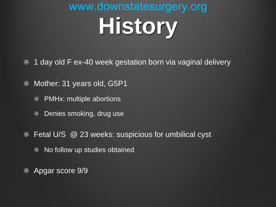
History 1 day old F ex-40 week gestation born via vaginal delivery
Mother: 31 years old, G5P1
PMHx: multiple abortions
Denies smoking, drug use
Fetal U/S @ 23 weeks: suspicious for umbilical cyst
No follow up studies obtained
Apgar score 9/9
www.downstatesurgery.org
Physical Exam 99.7 BP 72/37 HR 160; birth weight 3208 grams
No acute distress
S1/ S2 RR
Equal breath sounds b/l
Abd: midline defect at umbilicus containing viscera c sac
Genitourinary structures and anus grossly normal
Omphalocele
www.downstatesurgery.org

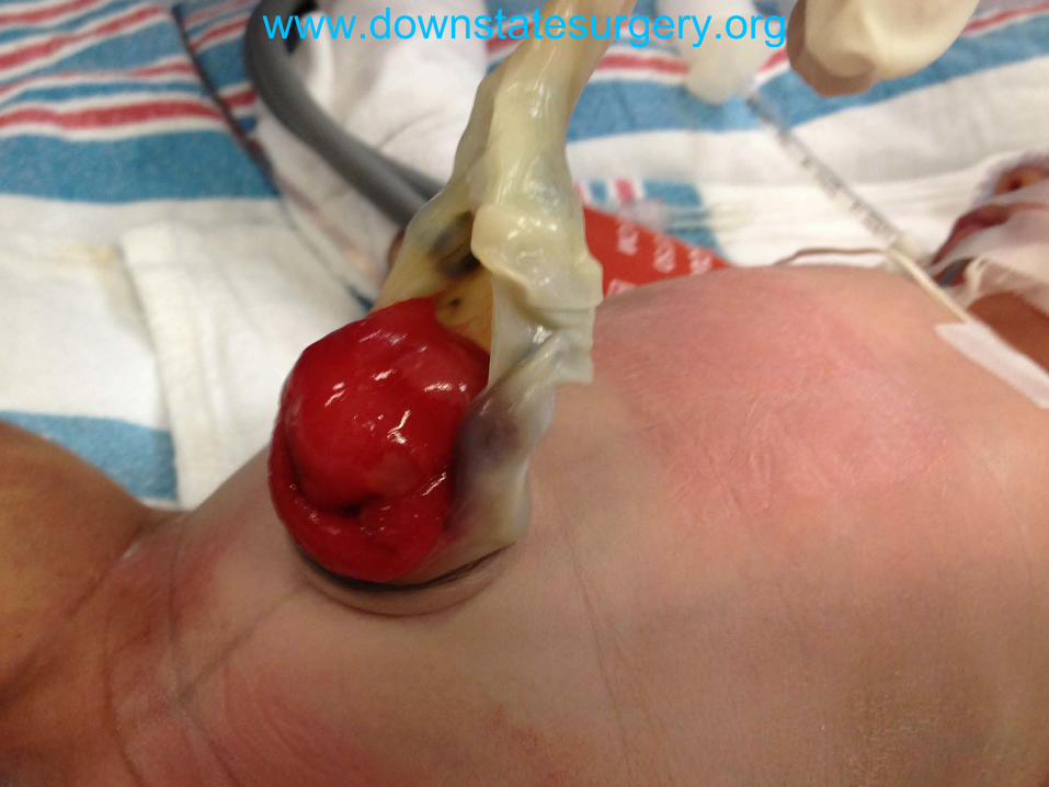
Imaging Upon arrival of pediatric team, omphalocele ruptured
Pediatric surgery was called
Echo: small PDA, normal function
Renal U/S: negative
Taken to OR urgently for exploration
www.downstatesurgery.org

Intraoperative Findings Ruptured omphalocele with everted ruptured bladder
Partial cystectomy and two layer repair
No other findings
Primary fascial closure achieved
Patient returned to NICU intubated
www.downstatesurgery.org
www.downstatesurgery.org

Postoperative Course Extubated POD#2
Voiding freely
Started on feeds POD#4, advanced as tolerated
Patient was discharged home on POD#7
www.downstatesurgery.org
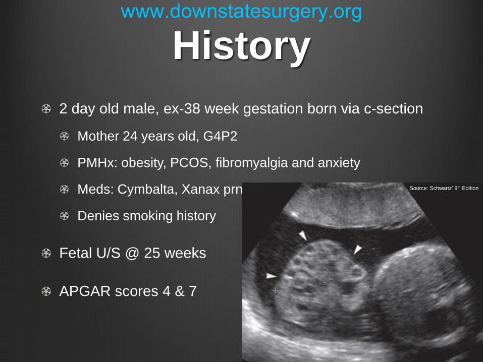
History 2 day old male, ex-38 week gestation born via c-section
Mother 24 years old, G4P2
PMHx: obesity, PCOS, fibromyalgia and anxiety
Meds: Cymbalta, Xanax prn
Denies smoking history
Fetal U/S @ 25 weeks
APGAR scores 4 & 7
Source: Schwartz’ 9th Edition
www.downstatesurgery.org
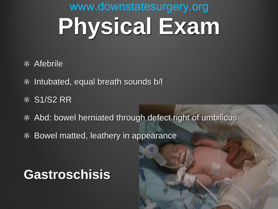
Physical Exam Afebrile
Intubated, equal breath sounds b/l
S1/S2 RR
Abd: bowel herniated through defect right of umbilicus
Bowel matted, leathery in appearance
Gastroschisis
www.downstatesurgery.org

Preoperative Workup Echo: normal function
Renal U/S: no abnormalities noted
PICC placed in NICU
TPN started DOL 2
Taken to OR for exploration on DOL 2
www.downstatesurgery.org
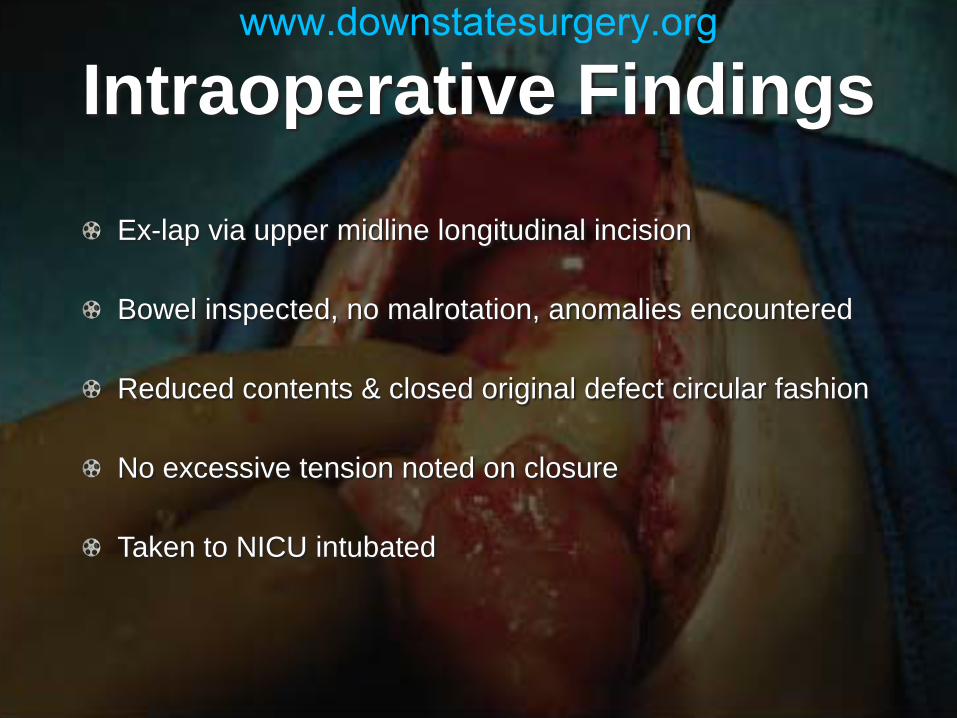
Intraoperative Findings
Ex-lap via upper midline longitudinal incision
Bowel inspected, no malrotation, anomalies encountered
Reduced contents & closed original defect circular fashion
No excessive tension noted on closure
Taken to NICU intubated
www.downstatesurgery.org
Postoperative Course
Extubated POD#4
POD# 10 Started on oral 10% dextrose solution
Return of bowel function
POD# 18 half-strength elemental feeds started
Remains in NICU tolerating feeds
www.downstatesurgery.org

Congenital Defects Omphalocele
Gastroschisis
Umbilical cord hernia
Ectopia cordis thoracis
Cloacal Exstrophy
Patent Urachus
www.downstatesurgery.org

Embryology Craniocaudal & mediolateral infolding at 4th week
Lateral folds meet at midline constricting yolk sac
By 6th week, intestinal growth leads to herniation
By week 10 midgut returns to abdomen; bowel fixation
Closure of the abdominal ring
Failure leads to abdominal wall defects
www.downstatesurgery.org
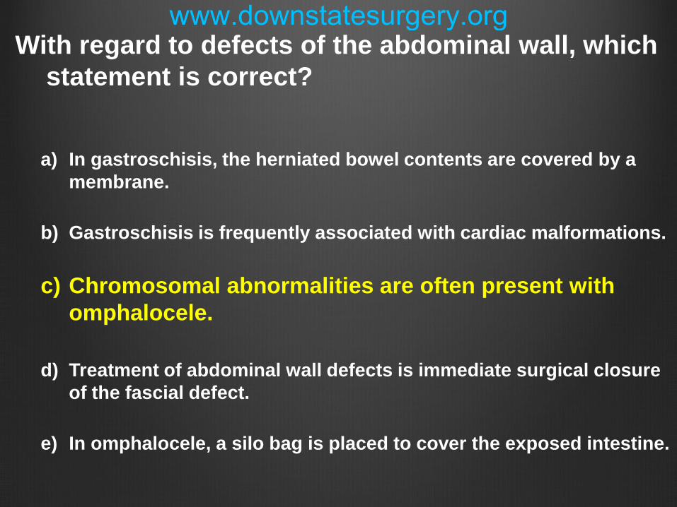
Omphalocele Incidence of 1 per 6-10,000, male predominance
Results from incomplete closure of abdominal wall
Central defect covered by sac (midgut & abd organs)
30-70% incidence of associated anomalies
Large for gestational age (> 4 kg)
www.downstatesurgery.org
Presenter
Presentation Notes
Defect > 4cm Second most common abdominal wall defect Ultrasonographically diagnosed at 18 weeks with 2D Can be diagnosed in first trimester with 3D u/s

Risk Factors Male sex
Twin or higher multiple order gestations
Increased prevalence among Black mothers
Both young and advanced maternal age
Alcohol consumption in first trimester
Mac Bird, T et al. Demographic and environmental risk factors for gastroschisis and omphalocele in the National Birth Defects Prevention Study, J Pediatr Surg 2009
www.downstatesurgery.org

Associated Abnormalities
Chromosomal abnormalities in ~50% (trisomy 13, 18, 21)
Cardiac abnormalities (ASD/VSD, coarctation of aorta)
Pulmonary hypoplasia, respiratory distress
www.downstatesurgery.org
Presenter
Presentation Notes
Patau, Edwards
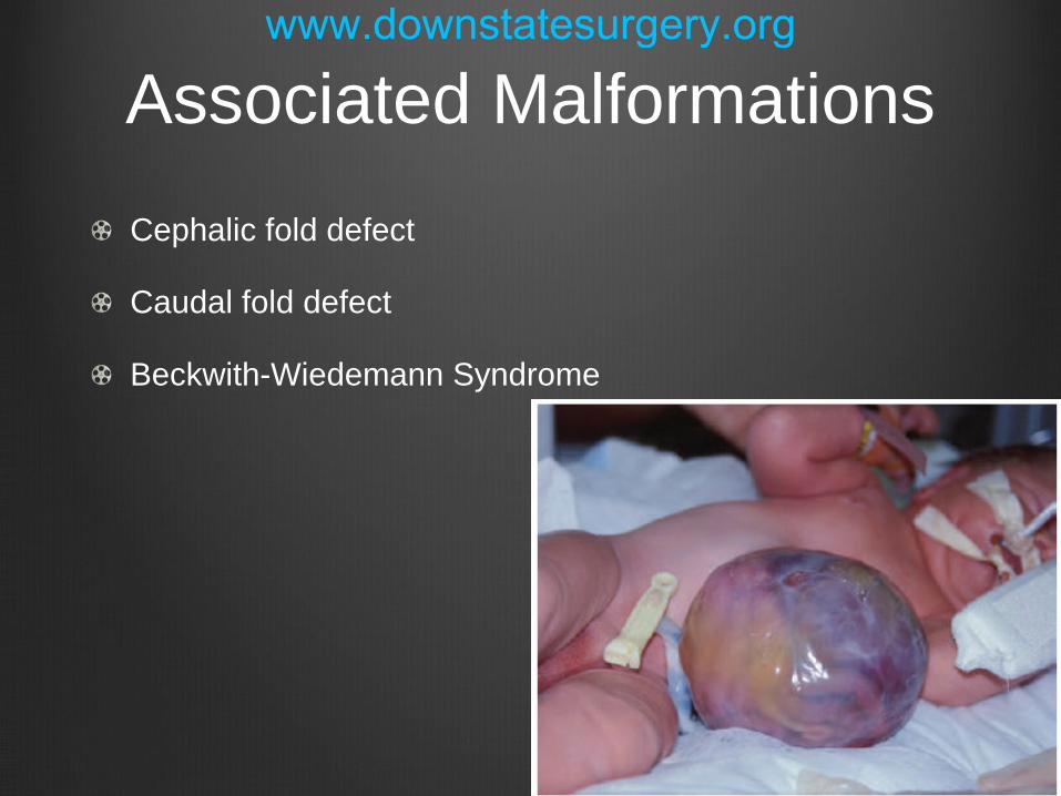
Associated Malformations Cephalic fold defect
Caudal fold defect
Beckwith-Wiedemann Syndrome
www.downstatesurgery.org

Cephalic Fold Defect Pentalogy of Cantrell
Thoracoabdominal ectopia cordis
Central tendon diaphragmatic defect
Upper midline omphalocele
Cardiac defect (VSD, LV diverticulum)
Apical pericardial defect
www.downstatesurgery.org
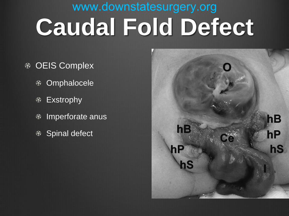
Caudal Fold Defect OEIS Complex
Omphalocele
Exstrophy
Imperforate anus
Spinal defect
www.downstatesurgery.org

Beckwith-Wiedemann Sx Macrosomia
Macroglossia
Omphalocele/Umbilical hernia
Ear pits or creases
Neonatal hypoglycemia
Increased likelihood of childhood cancers (Wilms, HB)
www.downstatesurgery.org
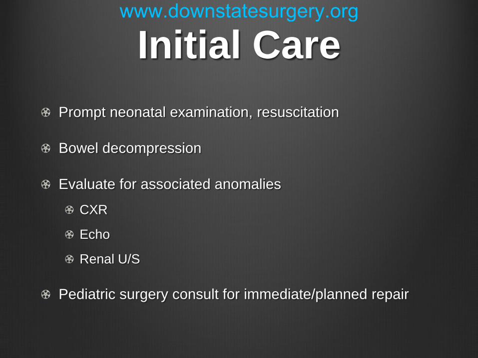
Initial Care Prompt neonatal examination, resuscitation
Bowel decompression
Evaluate for associated anomalies
CXR
Echo
Renal U/S
Pediatric surgery consult for immediate/planned repair
www.downstatesurgery.org

Surgery Small defects more amenable to primary closure
‘Giant’ defects managed with expected ventral hernia
Silvadene topically to promote granulation
Skin flaps and grafting
Tissue expanders increase abdominal domain
www.downstatesurgery.org
Gastroschisis 1 in 3-8,000 live births
Increasing incidence over past two decades
Right paramedian defect without sac
Prematurity, low birth weights in 40%
Increased α-FP
Classified as simple & complex gastroschisis Presence of associated abnormality
www.downstatesurgery.org

Etiology Theories:
Failure of mesoderm to form anterior abdominal wall
Failure of lateral folds to fuse midline leaves defect
In utero rupture of omphalocele
Resorption of right umbilical vein leads to weakness
www.downstatesurgery.org

Risk Factors Maternal age < 21 years
Stressed, under-nourished mothers
Smoking
Preterm delivery more common in gastroschisis
Use of vaso-constrictive drugs, ephedrine, cocaine
www.downstatesurgery.org
Presenter
Presentation Notes
Hispanic background also thought to be a risk factor… disputed in recent review

Associated Abnormalities
Concomittant bowel atresias in 6.9-28% (jejunal/ileal)
Cryptorchidism 10-15%
Amniotic Band Syndrome Thoracic Wall abnormalities Limb abnormalities Intestinal atresias Abnormal genitalia Meningocele Umbilical cord abnormalities
www.downstatesurgery.org
Presenter
Presentation Notes
Differentiate atresia from ‘vanishing bowel’ Intestinal atresia should be differentiated from “vanishing bowel” in infants with gastroschisis. This condition is usually associated with a very small abdominal wall defect and is characterized by necrosis and disappearance of some or all of the intestine. Although this is a rare finding, it usually results in short bowel syndrome.

Initial Care Prevent hypothermia & volume depletion; respiratory support
Inspect and cover exposed bowel
Position on side to prevent mesenteric kinking
Establish central venous access early
Rule out associated anomalies (rare)
Rapid transfer to OR for attempted primary closure
www.downstatesurgery.org

Surgical Management Return the bowel to the abdominal cavity
Minimize damage and intra-abdominal hypertension
Central venous access for TPN, dysmotility common
Surgical Techniques
Silo, serial reductions
Primary reduction with operative closure
Primary reduction with umbilical closure
www.downstatesurgery.org
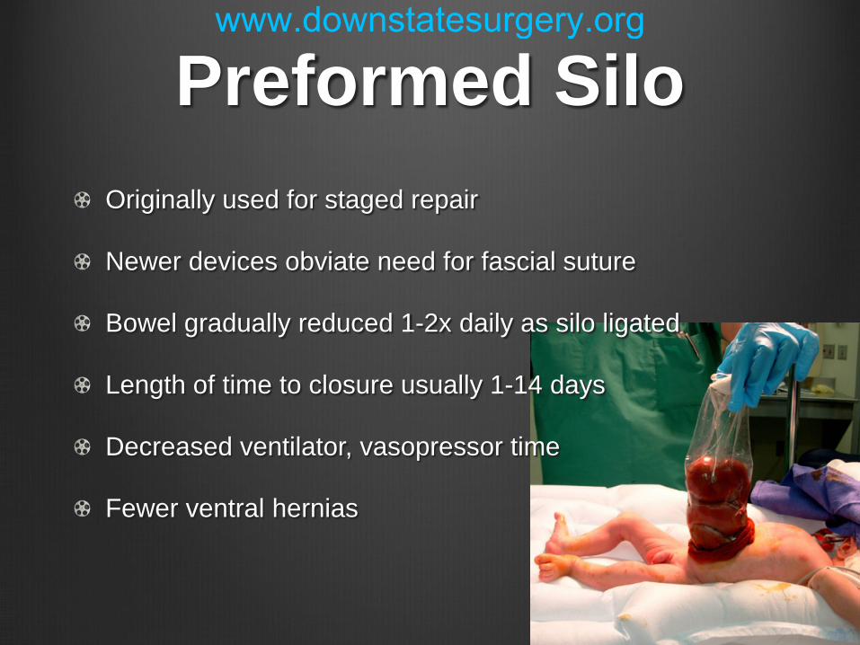
Preformed Silo Originally used for staged repair
Newer devices obviate need for fascial suture
Bowel gradually reduced 1-2x daily as silo ligated
Length of time to closure usually 1-14 days
Decreased ventilator, vasopressor time
Fewer ventral hernias
www.downstatesurgery.org
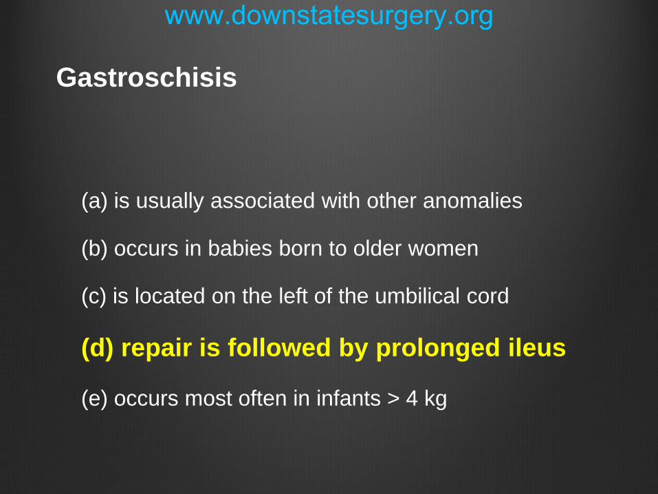
Postoperative Care Prolonged ileus is typical in gastroschisis
Benefit from early central venous access, TPN
Early oral stimulation; sucking-swallowing reflex lost
Small bowel series if no function after 6-8 weeks
60% of patients have social stress without umbilicus
www.downstatesurgery.org
Presenter
Presentation Notes
Cisapride may aid in prolonged dysmotility

Controversies Optimal mode of delivery has been debated
No difference in outcomes of c-section vs vaginal
Early delivery may prevent exposure to amniotic fluid
Literature is mixed, 1 RCT: shorter LOS, earlier feeding
Low birth weight (<2 kg) have longer course
Silo vs primary fascial closure?
www.downstatesurgery.org
Presenter
Presentation Notes
Early delivery of the fetus with gastroschisis has been advocated to limit exposure of the bowel to amniotic fluid in an attempt to reduce the inflammatory peel on the surface of the bowel. Poor motility of the bowel related to exposure to amniotic fluid IL6, IL8, and ferritin are elevated in the amniotic fluid in fetuses with gastroschisis when compared with controls. Amniotic fluid cytokines and other proinflammatory mediators damage the myenteric nerve plexus and interstitial cells of Cajal may contribute to the profound dysmotility and malabsorption seen in patients with gastroschisis. Bowel edema and peel formation increase as pregnancy progresses, most significantly if the gastroschisis defect constricts venous outflow from the herniated bowel.
Study Examined two cohorts: primary closure vs silo
190 patients in closure technique analysis
Lower Apgar score in preformed silo group (p=0.01)
Higher hernia rates among primary closure (p=0.0006)
NICU stay longer in silo group (p=0.002)
Retrospective cohort study, bias inevitable
Included skin only closures as part of primary closure cohort
To date, only 1 randomized prospective trial: no difference
www.downstatesurgery.org
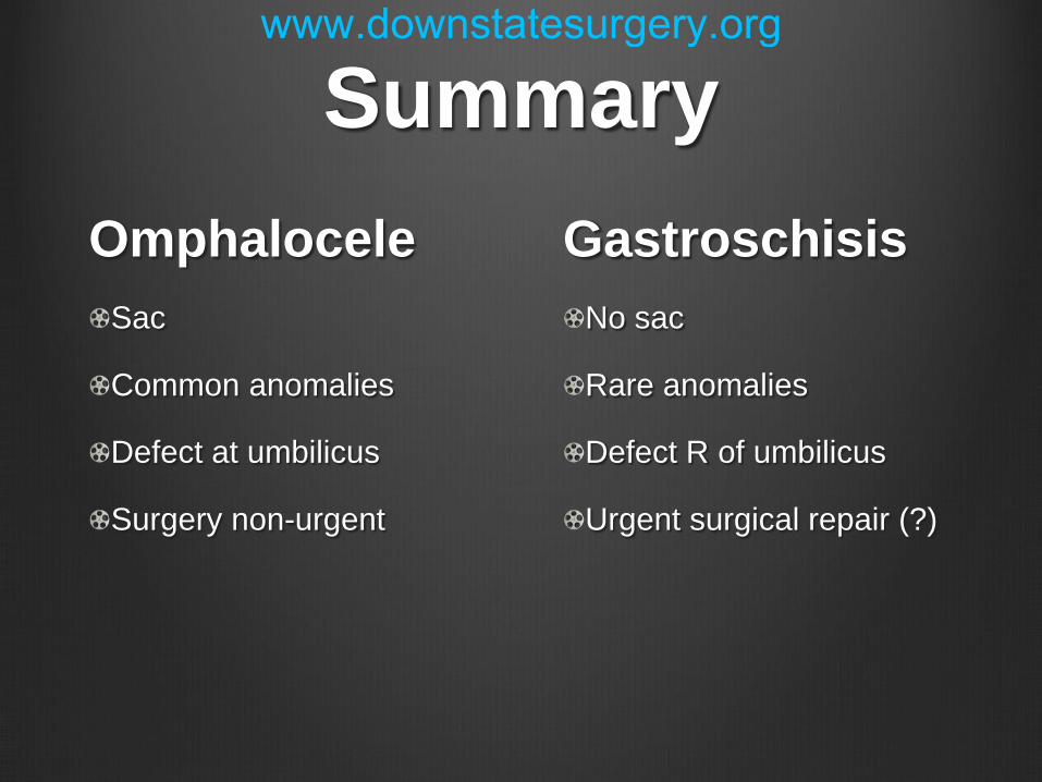
Summary Omphalocele
Sac
Common anomalies
Defect at umbilicus
Surgery non-urgent
Gastroschisis No sac
Rare anomalies
Defect R of umbilicus
Urgent surgical repair (?)
www.downstatesurgery.org
Summary Prevention of excessive heat and fluid loss is key
Evaluate all pts with abd wall defects for anomalies
Early primary fascial closure when possible
‘Giant’ omphaloceles require careful surgical planning
Early TPN in gastroschisis, expect prolonged ileus
Silo closure is also a reasonable option
www.downstatesurgery.org

References Coran: Pediatric Surgery, 7th ed.
Ashcraft's Pediatric Surgery, 5th ed.
Townsend: Sabiston Textbook of Surgery, 19th ed.
Greenfield's Surgery: Scientific Principles and Practice, 5th Edition
Schwartz’ Principles of Surgery 9th Edition
Weil B et al. The jury is still out: changes in gastroschisis management over the last decade are associated with both benefits and shortcomings. J Pediatr Surg 2012; 47: 119-124
www.downstatesurgery.org
With regard to defects of the abdominal wall, which statement is correct?
a) In gastroschisis, the herniated bowel contents are covered by a
membrane.
b) Gastroschisis is frequently associated with cardiac malformations.
c) Chromosomal abnormalities are often present with omphalocele.
d) Treatment of abdominal wall defects is immediate surgical closure of the fascial defect.
e) In omphalocele, a silo bag is placed to cover the exposed intestine.
www.downstatesurgery.org
Gastroschisis
(a) is usually associated with other anomalies
(b) occurs in babies born to older women
(c) is located on the left of the umbilical cord
(d) repair is followed by prolonged ileus
(e) occurs most often in infants > 4 kg
www.downstatesurgery.org
Gastroschisis
(a) is usually associated with other anomalies
(b) occurs in babies born to older women
(c) is located on the left of the umbilical cord
(d) repair is followed by prolonged ileus
(e) occurs most often in infants > 4 kg
www.downstatesurgery.org

You are a pediatric surgery consult resident called by NICU in regards to a 1 month old male infant presenting with flaccid abdominal wall, bilateral undescended testes and ureteral dilatation. Mom says she changes wet diapers several times a day. What is your plan?
a) “Call GU, they cover this sort of thing”
b) Bilateral orchiopexy and ureteral stent placement
c) Cystoscopy
d) “Observe, no acute surgical intervention at this time.”
www.downstatesurgery.org

Prune-Belly Syndrome Also known as Eagle-Barrett or Triad Syndrome
Extremely lax abdominal wall
Significant comorbidities; pulmonary hypoplasia
Higher incidence in males
Predisposed to UTIs
Abdominoplasty at 6-12 months
www.downstatesurgery.org
Presenter
Presentation Notes
Decreased smooth muscle and increased collagen in ureters, vesiculo-ureter reflux