c. ezike's presentations
25
-
Upload
dr-christopher-onyemaechi-ezike -
Category
Health & Medicine
-
view
101 -
download
0
Transcript of c. ezike's presentations


Fig. 4.1: Photomicrograph of kidney of control group of fish (C,K) and kidney exposed to 1.25ml/l for 10 weeks (T1,2-T1,10) showing progressive hyperplasia T1,2 - T1,4, blood vessel thickening T1,6, intercellular changes T1,8 - T 1,10.

Fig. 4.2: Photomicrograph of kidney of control group of fish (C,K) and (T2,2-T2,10) kidney exposed to 2.5ml/l for 10 weeks showing hypertrophic changes of kidney cells T2,2-T2,8, and infiltration/ blockage of blood vessel T2,10 ( x400).

Fig. 4.3: Photomicrograph of kidney of control group of fish (C,K) and kidney exposure for 10 weeks (T3,2-T3,10) to 5ml/l crude oil WSF showing cell depletion T3,2-T3,4, hypertrophy of kidney cells T3,6-T3,8 and intercellular shifts T3,10 (x400).

Fig. 4.4: Photomicrograph of kidney of control group of fish (C,K) and kidney exposure to 10ml/l WSF of crude oil for 10 weeks (T4,2-T4,10) showing moderate atrophy T4,2, vessel widening and constriction T4,4 and T4,6 and tubule wall thickening and atrophy inT4,8 and T4,10 respectively.

Fig.4.5: Photomicrograph of kidney of control group of fish (C,K) and kidney exposure to 20ml/l WSF of crude oil for 10 weeks (T5,2-T5,10) showing severe atrophy T5,2 - T5,4, blood vessel thickening T5,6, permanent dilation of capillaries or telangiectasia T5,8 and fragments of dead cells by apoptosis T5,10 (x400).

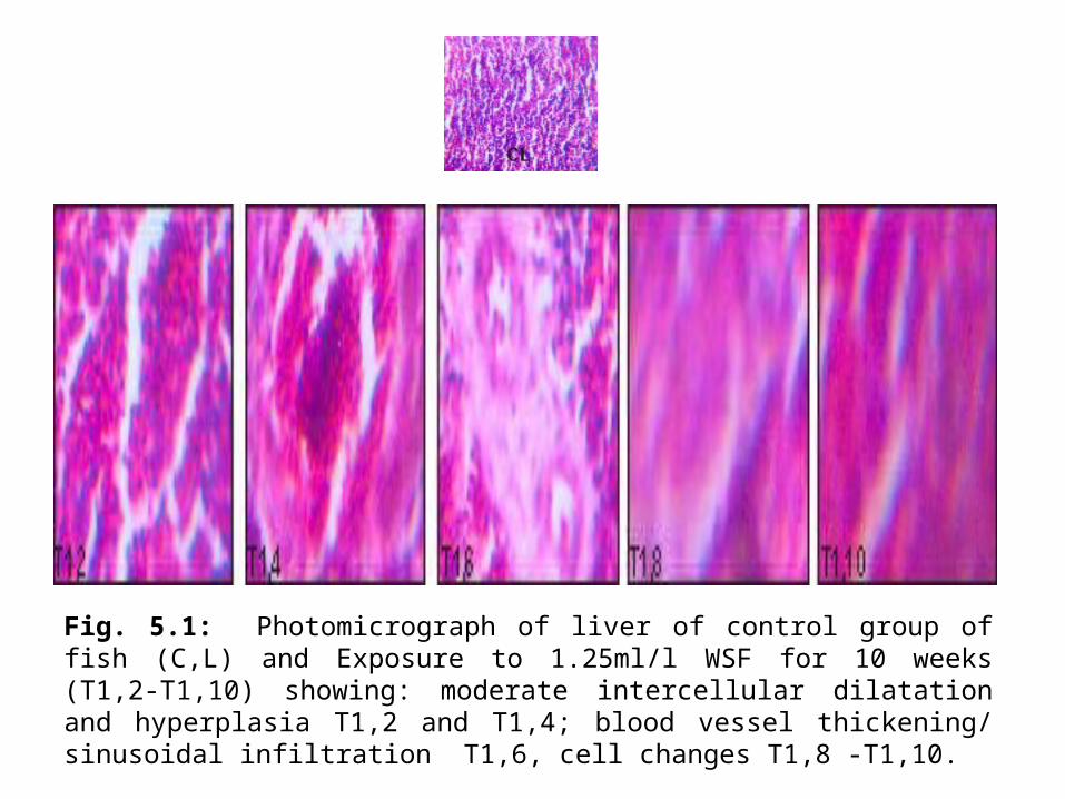
Fig. 5.1: Photomicrograph of liver of control group of fish (C,L) and Exposure to 1.25ml/l WSF for 10 weeks (T1,2-T1,10) showing: moderate intercellular dilatation and hyperplasia T1,2 and T1,4; blood vessel thickening/ sinusoidal infiltration T1,6, cell changes T1,8 -T1,10.
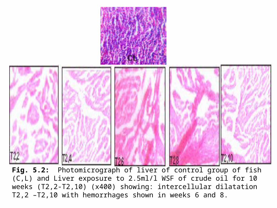
Fig. 5.2: Photomicrograph of liver of control group of fish (C,L) and Liver exposure to 2.5ml/l WSF of crude oil for 10 weeks (T2,2-T2,10) (x400) showing: intercellular dilatation T2,2 –T2,10 with hemorrhages shown in weeks 6 and 8.

Fig.5.3: Photomicrograph of liver of control group of fish (C,L) and Liver exposed to 5ml/l crude oil WSF for 10 weeks (T3,2 – T3,10). Hyperplasia T3,2 and hypertrophic changes T3,4– T3,10.

Fig. 5.4: Photomicrograph of liver of control group of fish (C,L) and Liver exposure to 10ml/l WSF of crude oil for 10 weeks (T4,2-T4,10) showing: fatty changes and vacuolation T4,2, T4,4 and T4,6, Expansion/ thickening of blood vessel T4,8,blockage of portal tract T4,10.

Fig. 5.5: Photomicrograph of liver of control group of fish (C,L) and Liver exposure to 20ml/l WSF of crude oil for 10 weeks (T5,2-T5,10) showing: cirrhosis with hepatocyte degeneration T5,2, severe cirrhosis T5,4, and T5,6, sinusoidal hemorrhage T5,8, and severe degeneration with necrosis T5,10.


Fig. 6.1: Photomicrograph of gill of control group (C,L) and Exposure of gill to 1.25ml/l WSF of crude oil for 10 weeks (T1,2-T1,10) showing: Degeneration of secondary lamellae T1,2-T2,6, clubbing of secondary lamellae T1,8 and T1,10).

Fig. 6.2: Photomicrograph of gill of control group (C,L) and gill of fish exposed to 2.5ml/l of crude oil for 10 weeks (T2,2-T2,10) showing varying damages; moderate lamellae degeneration T2,2-T2,4, fussion of secondary lamellae T2,6 and T2,8 and Curling of Secondary lamella T2,10.

Fig.6:3: Photomicrograph of gill of control group (C,L) and Gill exposure to 5ml/l of WSF of crude oil for 10 weeks (T3,2- T3,10) showing abnormal elongation of filament T3,2, and T3,4; shortening and oedema of lamellae T3,6 - T3,8 and oedema and lamellae rupture (T3,10).

Fig. 6.4: Photomicrograph of gill of control group (C,L) and Exposed gills to 10ml/l WSF of crude oil for 10 weeks (T4,2-T4,10) showing: Thickening of epithelial cell and forms of telangiectasis T4,2-T4,8 and rupture of primary lamellae T4,10.

Fig. 6.5: Photomicrograph of gill of control group (C,L) and exposed gills to 20ml/l of WSF of crude oil for 10 weeks showing: lamellae degeneration T5,2 and T5,4, progressive loss of secondary lamellae T5,6, blockage, complete erosion of secondary lamellae T5,8, and severe rupture of primary lamella wall T5,10.


Fig. 7.1: Photomicrograph of muscle of control group (C,M) and Muscle of fish exposed to 1.25ml/l of WSF crude oil for 10 weeks (T1,2-T1,10) showing: hypertrophy and cell degeneration T1,2 and T1,4 and splitting of muscle fibres T1,6-T1,10.

Fig. 7.2: Photomicrograph of gill of control group (C,L) and muscle exposed to 2.5ml/l crude oil WSF for 10 weeks (T2,2-T2,10) showing hyperplasia T2,2, muscle cell hypertrophy/ degeneration T2,4 and T2,6, cell splitting and necrosis T2,8 fibre splitting with hemorrhagic damages T2,10.

Fig. 7.3: Photomicrograph of gill of control group (C,L) and Muscle exposed to 5ml/l WSF of crude oil for 10 weeks (T3,2-T3,10): splitting of cell fibres T2,2 -T3,6, atrophy T3,8 and Vacuolation of muscle cells T3,10.

Fig. 7.4: Photomicrograph of gill of control group (C,L) and Muscle exposed to 10ml/l WSF of crude oil for 10 weeks (T4,2-T4,10) showing: swelling/oedema T4,2, and T4,4, cell degeneration T4,6, intercellular expansion and congestion T4,8, Massive splitting and degeneration of cell fibre T4,10.

Fig. 7.5: Photomicrograph of muscle of control group (C,M) and Muscle exposed to 20ml/l of WSF of crude oil for 10 weeks (T5,2-T5,10) showing various damages: Atrophic changesT5,2 and T5,4; massive splitting of cell fibres T5,6; Atrophy and oedema T5,8 and T5,10.

![Samsung C&T Presentations 4 Hour [Compatibility Mode]](https://static.fdocuments.us/doc/165x107/5527248e550346eb358b46f9/samsung-ct-presentations-4-hour-compatibility-mode.jpg)