(C Concordance of Endotoxemia Gram-Negative Bacteremia ... · icant heterogeneity indicates that...
8
JOURNAL OF CLINICAL MICROBIOLOGY, Sept. 1994, p. 2120-2127 Vol. 32, No. 9 0095-1137/94/$04.00+0 Copyright (C 1994, American Society for Microbiology Concordance of Endotoxemia with Gram-Negative Bacteremia in Patients with Gram-Negative Sepsis: a Meta-Analysis JAMES C. HURLEY* Division of Infectious Diseases, Children's Hospital & Medical Center, Seattle, Washington 98105-0371 Received 8 April 1994/Returned for modification 12 May 1994/Accepted 7 June 1994 The Limulus amebocyte lysate (LAL) assay is a sensitive method for detecting endotoxin. Using gram- negative (GN) bacteremia as the basis for comparison, concordance with endotoxemia in 45 studies could be expressed as an odds ratio. Calculation of summary odds ratios by the Mantel-Haenszel-Peto method indicated that the concordance of the results was no higher by the chromogenic LAL assay than by the gelation version, and the sensitivity was improved by only 11% (62 versus 51%). Endotoxemia was detected in 77 (68%) of 114 patients with bacteremia caused by an organism that was not a member of the family Enterobacteriaceae, whereas endotoxemia was detected in only 120 (45%) of 269 patients with bacteremia caused by a member of the family Enterobacteriaceae or an anaerobe (P < 0.001). This difference was also apparent for patients with GN bacteremia for whom a fatal outcome had been recorded. The prevalence of GN bacteremia in the tested population and the type of etiological agent are critical and previously unrecognized variables which affect the interpretation of the LAL test in patients with suspected sepsis. Endotoxin is a cell wall component found exclusively in gram-negative (GN) bacteria. Considerable experience has accumulated with the use of the Limulus amebocyte lysate (LAL) for the detection of endotoxin in various body fluids (20). For example, when applied to samples of cerebrospinal fluid for the detection of GN meningitis, the LAL assay has a sensitivity and a specificity each greater than 95%, in compar- ison with the sensitivity of Gram stain examination, which is only 81% (58). By contrast, its value as a diagnostic test when applied to plasma samples remains unclear, despite more than 45 studies. With the detection of GN bacteremia as the basis for compar- ison, its sensitivity and specificity are less than 85% (20). Several estimates made on the basis of experimental (65) and clinical (8, 11, 64) data suggest that the minimum amount of endotoxin required to induce a positive LAL test result would be equivalent to that contained in at least 100 GN bacteria per ml, a level that is seldom reached in patients with clinical bacteremia. In addition, plasma contains incompletely defined factors that interfere with the detection of endotoxin (32, 41, 57) or that confound the reading of the assay (32). To overcome these obstacles, the assay has been modified. In 1983, a chromogenic substrate (CLAL) was introduced as a more sensitive indicator of LAL activation by endotoxin. The CLAL assay is sensitive to endotoxin in plasma samples spiked in vitro at concentrations 10 times lower than those used in the previous gelation end point (GLAL) assay (28). However, there has been no direct comparative study of the two assays on samples from patients with GN bacteremia. Additional considerations have come to light more recently. It has long been assumed, and implied in the term itself, that endotoxin is released only on death and lysis of the bacterial cell. Recently, it has been recognized that endotoxin is shed from the surface of certain GN bacteria in the absence of lysis and that this property is not equally expressed by different GN bacteria (62). For example, it is observed more often with * Corresponding author. Mailing address: Division of Infectious Diseases, Children's Hospital & Medical Center, P.O. Box 5371/CH 32, Seattle, WA 98105-0371. Phone: (206) 528-2116. Fax: (206) 527-3890. Electronic mail address: [email protected]. invasive than with mucosal isolates of Neisseria meningitidis (2, 49). A third consideration relates to the patient population. The proportion of patients with GN bacteremia is a key indicator of the patient mix in studies which may or may not have been designed for the purpose of examining the diagnostic applica- tion of the assay. In large published series of studies of patients satisfying generally accepted criteria of suspected GN sepsis, this proportion is usually in the range of 13 to 40%. For example, in five such studies, the proportions were 13% (22), 25% (53), 30% (6), 37% (74), and 40% (71). The purpose of this review is to examine the diagnostic experience with endotoxemia detection by using GN bactere- mia as the basis for comparison. The value of the more sensitive CLAL assay, the etiological agents of the bactere- mias, and the prevalence of GN bacteremia in the patient population were three factors identified for special attention. The statistical techniques of meta-analysis are used. These techniques evolved from their application to the evaluation of retrospective studies of disease (24a, 45a). MATERMILS AND METHODS Study selection. A review of the literature was performed. The review included a search of the Medline database back to 1972, with the search headings being "septicemia" (prior to 1992), "bacteremia" (from 1992 on), "endotoxins," and "hu- man." The search was not limited to reports published in the English language (13, 34) and included conference proceed- ings (14, 19, 48, 69). This was supplemented with a manual search of the references from each report retrieved, review articles, and textbooks. The following criteria were used in selecting studies for inclusion: (i) study design, which included a direct comparison of blood culture and LAL (or Tachypleus amebocyte lysate [67]) assay methods applied to blood from patients suspected of having GN sepsis; (ii) size, with at least four patients studied; and (iii) data presentation, such that each patient could be classified into one of four possible categories: category 1, blood cultures, GN bacteria, and endo- toxin positive; category 2, blood cultures, GN bacteria, and endotoxin negative; category 3, blood cultures, no GN bacteria, 2120 on May 24, 2019 by guest http://jcm.asm.org/ Downloaded from
-
Upload
phamnguyet -
Category
Documents
-
view
214 -
download
0
Transcript of (C Concordance of Endotoxemia Gram-Negative Bacteremia ... · icant heterogeneity indicates that...

JOURNAL OF CLINICAL MICROBIOLOGY, Sept. 1994, p. 2120-2127 Vol. 32, No. 90095-1137/94/$04.00+0Copyright (C 1994, American Society for Microbiology
Concordance of Endotoxemia with Gram-Negative Bacteremia inPatients with Gram-Negative Sepsis: a Meta-Analysis
JAMES C. HURLEY*
Division of Infectious Diseases, Children's Hospital & Medical Center, Seattle, Washington 98105-0371
Received 8 April 1994/Returned for modification 12 May 1994/Accepted 7 June 1994
The Limulus amebocyte lysate (LAL) assay is a sensitive method for detecting endotoxin. Using gram-negative (GN) bacteremia as the basis for comparison, concordance with endotoxemia in 45 studies could beexpressed as an odds ratio. Calculation of summary odds ratios by the Mantel-Haenszel-Peto method indicatedthat the concordance of the results was no higher by the chromogenic LAL assay than by the gelation version,and the sensitivity was improved by only 11% (62 versus 51%). Endotoxemia was detected in 77 (68%) of 114patients with bacteremia caused by an organism that was not a member of the family Enterobacteriaceae,whereas endotoxemia was detected in only 120 (45%) of 269 patients with bacteremia caused by a member ofthe family Enterobacteriaceae or an anaerobe (P < 0.001). This difference was also apparent for patients withGN bacteremia for whom a fatal outcome had been recorded. The prevalence of GN bacteremia in the testedpopulation and the type of etiological agent are critical and previously unrecognized variables which affect theinterpretation of the LAL test in patients with suspected sepsis.
Endotoxin is a cell wall component found exclusively ingram-negative (GN) bacteria. Considerable experience hasaccumulated with the use of the Limulus amebocyte lysate(LAL) for the detection of endotoxin in various body fluids(20). For example, when applied to samples of cerebrospinalfluid for the detection of GN meningitis, the LAL assay has asensitivity and a specificity each greater than 95%, in compar-ison with the sensitivity of Gram stain examination, which isonly 81% (58).By contrast, its value as a diagnostic test when applied to
plasma samples remains unclear, despite more than 45 studies.With the detection of GN bacteremia as the basis for compar-ison, its sensitivity and specificity are less than 85% (20).Several estimates made on the basis of experimental (65) andclinical (8, 11, 64) data suggest that the minimum amount ofendotoxin required to induce a positive LAL test result wouldbe equivalent to that contained in at least 100 GN bacteria perml, a level that is seldom reached in patients with clinicalbacteremia. In addition, plasma contains incompletely definedfactors that interfere with the detection of endotoxin (32, 41,57) or that confound the reading of the assay (32).To overcome these obstacles, the assay has been modified.
In 1983, a chromogenic substrate (CLAL) was introduced as amore sensitive indicator of LAL activation by endotoxin. TheCLAL assay is sensitive to endotoxin in plasma samples spikedin vitro at concentrations 10 times lower than those used in theprevious gelation end point (GLAL) assay (28). However,there has been no direct comparative study of the two assayson samples from patients with GN bacteremia.
Additional considerations have come to light more recently.It has long been assumed, and implied in the term itself, thatendotoxin is released only on death and lysis of the bacterialcell. Recently, it has been recognized that endotoxin is shedfrom the surface of certain GN bacteria in the absence of lysisand that this property is not equally expressed by different GNbacteria (62). For example, it is observed more often with
* Corresponding author. Mailing address: Division of InfectiousDiseases, Children's Hospital & Medical Center, P.O. Box 5371/CH32, Seattle, WA 98105-0371. Phone: (206) 528-2116. Fax: (206)527-3890. Electronic mail address: [email protected].
invasive than with mucosal isolates of Neisseria meningitidis (2,49).A third consideration relates to the patient population. The
proportion of patients with GN bacteremia is a key indicator ofthe patient mix in studies which may or may not have beendesigned for the purpose of examining the diagnostic applica-tion of the assay. In large published series of studies of patientssatisfying generally accepted criteria of suspected GN sepsis,this proportion is usually in the range of 13 to 40%. Forexample, in five such studies, the proportions were 13% (22),25% (53), 30% (6), 37% (74), and 40% (71).The purpose of this review is to examine the diagnostic
experience with endotoxemia detection by using GN bactere-mia as the basis for comparison. The value of the moresensitive CLAL assay, the etiological agents of the bactere-mias, and the prevalence of GN bacteremia in the patientpopulation were three factors identified for special attention.The statistical techniques of meta-analysis are used. Thesetechniques evolved from their application to the evaluation ofretrospective studies of disease (24a, 45a).
MATERMILS AND METHODS
Study selection. A review of the literature was performed.The review included a search of the Medline database back to1972, with the search headings being "septicemia" (prior to1992), "bacteremia" (from 1992 on), "endotoxins," and "hu-man." The search was not limited to reports published in theEnglish language (13, 34) and included conference proceed-ings (14, 19, 48, 69). This was supplemented with a manualsearch of the references from each report retrieved, reviewarticles, and textbooks. The following criteria were used inselecting studies for inclusion: (i) study design, which includeda direct comparison of blood culture and LAL (or Tachypleusamebocyte lysate [67]) assay methods applied to blood frompatients suspected of having GN sepsis; (ii) size, with at leastfour patients studied; and (iii) data presentation, such thateach patient could be classified into one of four possiblecategories: category 1, blood cultures, GN bacteria, and endo-toxin positive; category 2, blood cultures, GN bacteria, andendotoxin negative; category 3, blood cultures, no GN bacteria,
2120
on May 24, 2019 by guest
http://jcm.asm
.org/D
ownloaded from

ENDOTOXEMIA WITH GRAM-NEGATIVE BACTEREMIA 2121
and endotoxin positive; and category 4, blood cultures, no GNbacteria, and endotoxin negative.
Unpublished data were sought directly from the authors ofstudies who had published results in a form which did not meetthe above criteria to seek a clarification that would haveenabled inclusion. The exclusion criteria were duplicate pub-lication and application of the assay to patients with conditionsother than suspected sepsis. Twenty-eight studies were ex-
cluded (the list is available on request). For one study (33),only the data from the examination of systemic blood were
included and the data for portal blood examination were
excluded.Data analysis. For each study, the concordance of the results
of the two tests was expressed as an odds ratio (OR), which wascalculated by using the formulae given in the Appendix(calculation of study-specific ORs).To enable a comparison of the studies, I used the meta-
analysis procedure of Yusuf et al. (73). Details of the calcula-tions are given in the appendix (calculation of summary ORs).In brief, for each study (i), the expected number (Ei) ofendotoxin-positive patients among those in whom GN bacte-remia was detected was calculated, assuming statistical inde-pendence from the marginal totals in a two-by-two table. Theobserved number (O) of endotoxin-positive patients minus theexpected number (Oi - Ej) and its variance (Vi) were calcu-lated. The values of Oi - Ei and Vi for each study were
summed to produce a grand total (GT) and total variance(VT), respectively. From these totals, the summary ORs andtheir 95% confidence intervals (95% CIs) were calculated.Data aggregation and sensitivity analysis. I began the
analysis with all of the studies divided into two groups on thebasis of which of the two versions of the assay, GLAL or
CLAL, was used. A subgroup analysis was conducted by usingonly studies in which the proportion of patients with GNbacteremia was in the range of 13 to 40% (as in several largeseries [6, 22, 53, 71, 74]), after it was found that many of thestudies in which this proportion was outside of this range were
designed for purposes other than to examine the use of LAL as
a diagnostic test for unscreened patients with suspected sepsis.The homogeneity of the concordance among the studies in
each analysis was tested as described by Yusuf et al. (73) (seeAppendix [calculation of summary ORs]). A finding of signif-icant heterogeneity indicates that the variation in concordanceamong studies is more than could be expected from randomvariation. Summary estimates may be inappropriate in cases ofsignificant heterogeneity.
Diagnostic utility. For each analysis, a calculation of sensi-tivity, specificity, and positive predictive value (PPV) was
derived from the formulae as given in the Appendix (calcula-tion of assay parameters).
RESULTS
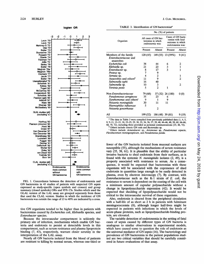
Forty-five studies were identified; those studies reported theresults for a total of 2,539 patients with suspected GN sepsis.There were only 43 separate publications because two publi-cations (45, 52) each provided the results of two studies. Atotal of 29 studies used the GLAL assay (Table 1) and 16studies used the CLAL assay (Table 2). The concordancebetween the detection of endotoxemia and GN bacteremia inthese studies is illustrated in Fig. 1.
In 24 studies sepsis was suspected on the basis of fever andclinical assessment. In nine studies blood for culture andplasma were drawn from patients with suspected sepsis in thesettings of specific illnesses such as malnutrition (37, 50),
pancreatitis (23), and obstructive jaundice (4, 44) or followingabdominal surgery (25, 26, 33) or liver transplantation (5).Nine studies were limited to patients with specific infectionssuch as typhoid fever (1, 9, 45, 48), salmonellosis (45), plaque(10, 11), or meningococcemia (8, 29). These patients wereusually selected on the basis of a clinical diagnosis supple-mented by confirmatory microbiological testing either by apositive culture for the specific pathogen from any site or byserology. One study (69) included only patients with microbi-ologically documented urological sepsis. The sensitivity limitfor GLAL studies was usually in the range of 0.5 to 10 ng/ml,and that for CLAL studies was in the range of 10 to 500 pg/ml.This variability in sensitivity limits reflects the nonuniformpotency among both the different reference endotoxin stan-dards and the LAL reagents used in each study. In addition,the method of plasma pretreatment was more commonly bythe dilution and heating method in the CLAL studies than inthe GLAL studies (10 of 16 versus 7 of 29 studies).
Diagnostic utility. As an aggregate result, of the 2,539patients from the 45 studies, GN bacteremia was detected in615 (24%) of them. However, there was a large range in thepretest probability for GN bacteremia in individual studies (4to 86%). Moreover, the derivation of a summary OR incorpo-rates a formal test for heterogeneity which was highly signifi-cant in the two series of studies (Table 1 and 2).Of 10 studies in which the prevalence of GN bacteremia was
50% or greater, microbiological documentation of GN sepsisby serology or cultural isolation of the organism from blood oranother site was either an elective or an obligate requirementfor patient inclusion in 7 studies (1, 8, 9, 11, 45, 48, 69). Ofeight studies in which the prevalence of GN bacteremia was10% or less, five (4, 5, 25, 33, 44) were studies of endotoxemiaoccurring in the setting of obstructive jaundice or abdominalsurgery.Comparison of the summary results for studies that used the
CLAL assay versus those that used the GLAL assay gavesimilar results for concordance (6.3, 4.4 to 9.1, and 8.4, 5.9 to11.8, respectively [values are summary ORs, 95% CIs]), sensi-tivity (54 and 53%, respectively), and PPV (40 and 49%,respectively). Further comparison of studies that used theCLAL assay versus those that used the GLAL assay waslimited to the 22 studies (9 used the CLAL assay and 13 usedthe GLAL assay) in which the percentage of patients havingGN bacteremia in each study was in the range 13 to 40%. Inthe 9 studies that used the CLAL assay, the assay sensitivitywas 62% (74 of 119 patients) and the PPV was 35% (74 of 214patients). Overall, in the 13 studies that used the GLAL assay,the assay sensitivity was 51% (82 of 161 patients) and the PPVwas 50% (82 of 163 patients). The summary ORs (with 95%CIs) indicate that the concordance with GN bacteremia wassignificantly less for the CLAL assay (3.6, 2.3 to 5.6) than forthe GLAL assay (20.1, 12.5 to 32.4). Recalculation of thechi-square test for heterogeneity yielded results for the twoseries that were not statistically significant, a finding that isconsistent with the absence of heterogeneity.
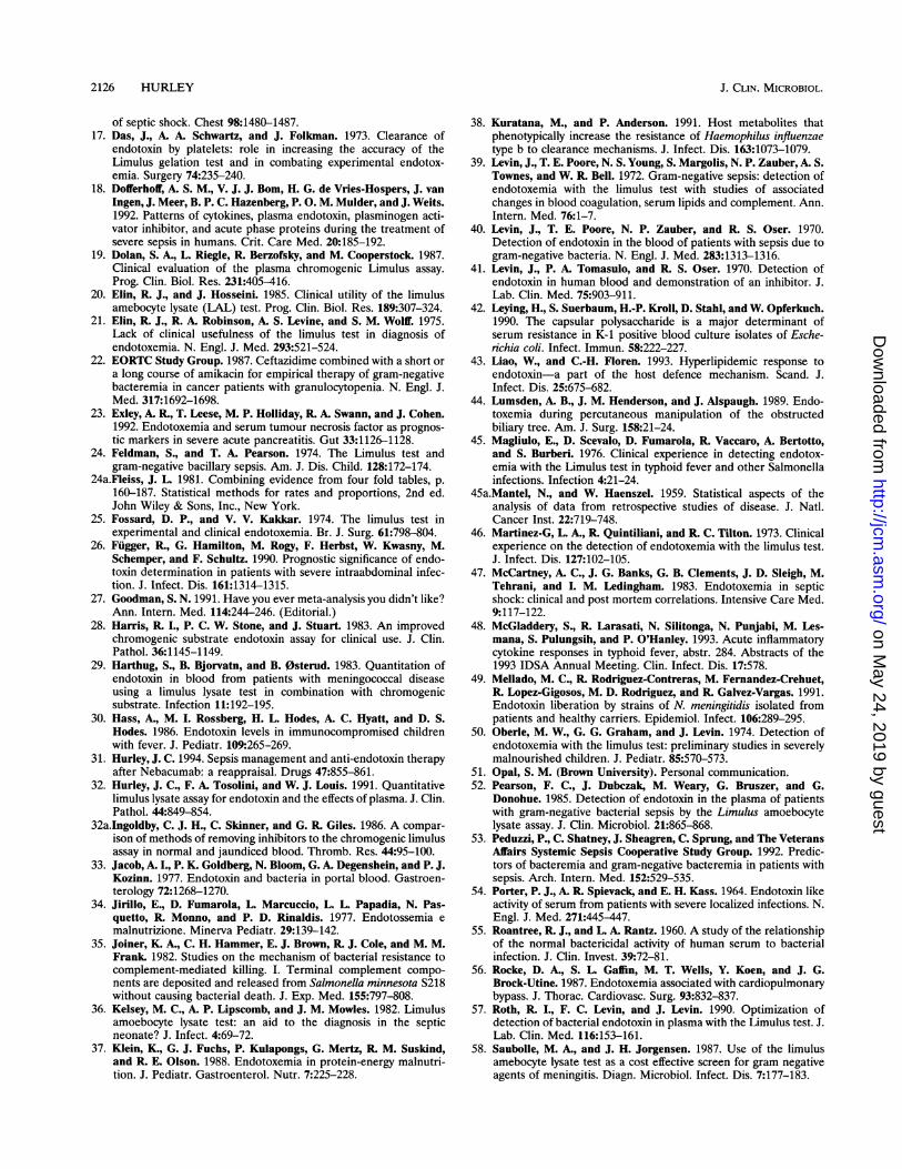
Bacteriology. The identifications of the isolates that causedbacteremia in 387 patients from 34 studies were tallied (Table3). Endotoxemia was detected in 79 (68%) of 116 patients withbacteremia caused by an organism that was not a member ofthe family Enterobacteriaceae (non-Enterobacteriaceae),whereas endotoxemia was detected in only 120 (45%) of 269patients with bacteremia caused by a member of the familyEnterobacteriaceae or an anaerobe (chi-square, 16.9; P < 0.001;degrees of freedom = 1; Table 3). This difference was alsoapparent for patients with GN bacteremias in whom a fataloutcome had been recorded; endotoxemia was found in 26 of
VOL. 32, 1994
on May 24, 2019 by guest
http://jcm.asm
.org/D
ownloaded from
2122 HURLEY
TABLE 1. Endotoxemia in the presence or absence of GN bacteremia in studies that used the GLAL assay
No. of patients in whomendotoxemia was detected/total
Author(s), yr (reference no.), Assay no. of patients (%) in whom GN % GNand diagnosis' bacteremia was or was bacteremiab Excluded'
not detected
Sensitivity limitd Codes' Detected Not detected
Levin et al., 1970 (40), Q 5 Ch 10/15 7/83 15Levin et al., 1972 (39), Q 5 Ch 17/31 19/187 14Martinez-G et al., 1973 (46), Q 5 pH 1/16 1/67 19Das et al., 1973 (17), Q 10 Ch 9/11 7/43 20Butler et al., 1973 (10), P1 0.5 Ch 2/2 7/8 20Stumacher et al., 1973 (65), Q 0.3 Ch 28/65 26/74 47 XFossard and Kakkar, 1974 (25) SS 1 Ch 2/2 22/23 8 XOberle et al., 1974 (50), N 0.5 Ch 3/3 6/20 13Feldman and Pearson, 1974 (24), Q 1 pH 2/36 0/53 40Jirillo et al., 1977 (34), Q 1 Ch 1/2 4/8 20Bailey, 1976 (4), J 5 Ch 1/1 12/23 4 XButler et al., 1976 (11), P1 1 Ch 3/5 0/5 50 XMagliulo et al., 1976 (45), T 1 pH 8/12 1/2 86 XMagliulo et al., 1976 (45), S 1 pH 4/4 9/21 16Clumeck et al., 1977 (13), Q 3 pH 9/12 2/36 25Jacob et al., 1977 (33), SS 1 DH 1/1 3/23 4 XButler et al., 1978 (9), T 1 Ch 0/13 0/8 62 XUsawattanakul et al., 1979 (67), Q 0.5 Ch 34/40 2/18 70 XScheifele et al., 1981 (59), Q 10 DH 5/8 10/55 13Kelsey et al., 1982 (36), Q ? pH 1/2 0/28 7 XTogari et al., 1983 (66), Q 0.5 pH 1/1 7/9 10 XMcCartney et al., 1983 (47), Q 0.1 Ch 15/15 16/16 48 XCooperstock and Riegle, 1985 (14), Q 1 DH 10/11 6/32 26Scheifele et al., 1985 (60), Q 0.2 DH 3/5 20/42 11 XPearson et al. (GLAL), 1985 (52), 0 0.1 DH 5/5 0/2 71 XAdinolfi et al., 1987 (1), T 0.3 DH 7/14 2/7 67 XKlein et al., 1988 (37), N 5 Ch 0/1 7/15 6 XShenep et al., 1988 (64), Q 0.01 DH 9/10 3/16 38McGladdery et al., 1993 (48), T 0.04 ? 0/16 0/6 72 X
All GLAL studies (n = 29) 191/359 (53) 199/930 (21) 28Excluding X (n = 13) 82/161 (51) 81/629 (13) 20
" Diagnosis codes; P1, plague; M, meningococcemia; Q, query sepsis; N, malnutrition; U, urosepsis; L, liver transplantation; T, typhoid; S, salmonellosis; P,pancreatitis; J, obstructive jaundice; SS postsurgical sepsis. For all studies that used the GLAL assay, chi-square for heterogeneity was 74.7 (degrees of freedom = 28;P = 1.0). Excluding X, chi-square for heterogeneity was 9.8 (degrees of freedom = 12; P = 0.37).
b Percentage of patients in whom GN bacteremia was detected.Excluded indicates studies in which the proportion of patients with GN bacteremia was outside the range of 13 to 40%.
dSensitivity limit of the LAL assay; (data are in nanograms per milliliter except those for the study of McGladdery et al. (48), which are in endotoxin in units permilliliter.
e Codes for method of plasma or serum pretreatment: Ch, chloroform-phenol extraction; pH, pH shift; DH, Dilution and heat treatment; PCA, perchloric acidtreatment; ?, not stated.
26 (100%) patients with fatal bacteremia caused by a non-Enterobacteriaceae, whereas it was found in 13 of 22 (59%)patients with fatal bacteremia caused by an Enterobacteriaceae(chi-square, 13.1; P < 0.001; degrees of freedom = 1).The more frequent association of endotoxemia with bacte-
remia for non-Enterobacteriaceae versus Enterobacteriaceaewas evident with the isolates stratified by type of test. That is,by the CLAL assay endotoxemia was found in 49 of 65 (75%)versus 28 of 47 (60%) patients with bacteremia caused bynon-Enterobacteniaceae versus Enterobacteriaceae, and by theGLAL assay endotoxemia was found in 30 of 51 (59%) versus92 of 222 (41%) patients with bacteremia caused by non-Enterobacteriaceae versus Enterobacteriaceae, respectively.Bacteremias caused by Yersinia pestis and Salmonella specieswere exceptions among the Enterobacteriaceae, being morecommonly found in association with endotoxemia than withoutendotoxemia.
DISCUSSION
I caution that this analysis does not attempt to establish theclinical significance of a positive LAL test result in any givenpatient group but, rather, attempts to examine how close theconcordance with GN bacteremia in the published experienceis. Were endotoxemia concordant with GN bacteremia, thismight be relevant to the application of new therapeutic mo-dalities for sepsis (31, 63).The 45 studies were heterogeneous with respect to diag-
noses, patient demographics, and pretest probability of thepatients having GN bacteremia, which limits any generaliza-tions that can be made on the basis of the full set of studies.This inconsistency, which is flagged by the finding of a statis-tically significant test for heterogeneity, would not have beenso readily appreciated from a review of the studies in thetraditional style without the statistical techniques of meta-analysis.
J. CLIN. MICROBIOL.
on May 24, 2019 by guest
http://jcm.asm
.org/D
ownloaded from
ENDOTOXEMIA WITH GRAM-NEGATIVE BACTEREMIA 2123
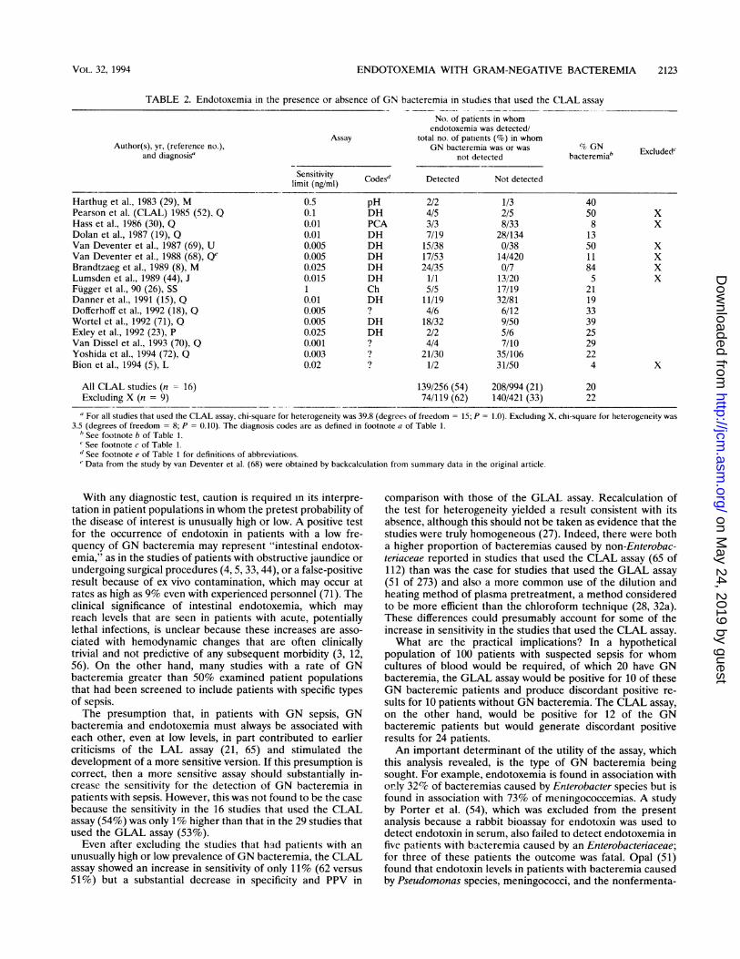
TABLE 2. Endotoxemia in the presence or absence of GN bacteremia in studies that used the CLAL assay
Author(s), yr, (reference no.),and diagnosis'
Assay
Sensitivity Codesdlimit (ng/ml)
No. of patients in whomendotoxemia was detected/
total no. of patients (%) in whomGN bacteremia was or was
not detected
Detected Not detected
Harthug et al., 1983 (29), MPearson et al. (CLAL) 1985 (52). 0Hass et al., 1986 (30), QDolan et al., 1987 (19), QVan Deventer et al., 1987 (69), UVan Deventer et al., 1988 (68), QeBrandtzaeg et al., 1989 (8), MLumsden et al., 1989 (44), JFugger et al., 90 (26), SSDanner et al., 1991 (15), QDofferhoff et al., 1992 (18), QWortel et al., 1992 (71), QExley et al., 1992 (23), PVan Dissel et al., 1993 (70), QYoshida et al., 1994 (72), QBion et al., 1994 (5), L
All CLAL studies (n = 16)Excluding X (n = 9)
0.50.10.010.010.0050.0050.0250.01510.010.0050.0050.0250.0010.0030.02
pHDHPCADHDHDHDHDHChDHD?DHDHI?I?I?
2/24/53/37/19
15/3817/5324/351/15/511/194/618/322/24/4
21/301/2
1/32/58/33
28/1340/3814/4200/71312017/1932/816/129/505/67/10
35/10631/50
139/256 (54) 208/994 (21)74/119 (62) 140/421 (33)
a For all studies that used the CLAL assay, chi-square for heterogeneity was 39.8 (degrees of freedom = 15; P = 1.0). Excluding X, chi-square for heterogeneity was3.5 (degrees of freedom = 8; P = 0.10). The diagnosis codes are as defined in footnote a of Table 1.
h See footnote b of Table 1.' See footnote c of Table 1.d See footnote e of Table 1 for definitions of abbreviations.' Data from the study by van Deventer et al. (68) were obtained by backcalculation from summary data in the original article.
With any diagnostic test, caution is required in its interpre-tation in patient populations in whom the pretest probability ofthe disease of interest is unusually high or low. A positive testfor the occurrence of endotoxin in patients with a low fre-quency of GN bacteremia may represent "intestinal endotox-emia," as in the studies of patients with obstructive jaundice orundergoing surgical procedures (4, 5, 33, 44), or a false-positiveresult because of ex vivo contamination, which may occur atrates as high as 9% even with experienced personnel (71). Theclinical significance of intestinal endotoxemia, which mayreach levels that are seen in patients with acute, potentiallylethal infections, is unclear because these increases are asso-ciated with hemodynamic changes that are often clinicallytrivial and not predictive of any subsequent morbidity (3, 12,56). On the other hand, many studies with a rate of GNbacteremia greater than 50% examined patient populationsthat had been screened to include patients with specific typesof sepsis.The presumption that, in patients with GN sepsis, GN
bacteremia and endotoxemia must always be associated witheach other, even at low levels, in part contributed to earliercriticisms of the LAL assay (21, 65) and stimulated thedevelopment of a more sensitive version. If this presumption iscorrect, then a more sensitive assay should substantially in-crease the sensitivity for the detection of GN bacteremia inpatients with sepsis. However, this was not found to be the casebecause the sensitivity in the 16 studies that used the CLALassay (54%) was only 1% higher than that in the 29 studies thatused the GLAL assay (53%).Even after excluding the studies that had patients with an
unusually high or low prevalence of GN bacteremia, the CLALassay showed an increase in sensitivity of only 11% (62 versus51%) but a substantial decrease in specificity and PPV in
comparison with those of the GLAL assay. Recalculation ofthe test for heterogeneity yielded a result consistent with itsabsence, although this should not be taken as evidence that thestudies were truly homogeneous (27). Indeed, there were botha higher proportion of bacteremias caused by non-Enterobac-teriaceae reported in studies that used the CLAL assay (65 of112) than was the case for studies that used the GLAL assay(51 of 273) and also a more common use of the dilution andheating method of plasma pretreatment, a method consideredto be more efficient than the chloroform technique (28, 32a).These differences could presumably account for some of theincrease in sensitivity in the studies that used the CLAL assay.What are the practical implications? In a hypothetical
population of 100 patients with suspected sepsis for whomcultures of blood would be required, of which 20 have GNbacteremia, the GLAL assay would be positive for 10 of theseGN bacteremic patients and produce discordant positive re-sults for 10 patients without GN bacteremia. The CLAL assay,on the other hand, would be positive for 12 of the GNbacteremic patients but would generate discordant positiveresults for 24 patients.An important determinant of the utility of the assay, which
this analysis revealed, is the type of GN bacteremia beingsought. For example, endotoxemia is found in association withonly 32% of bacteremias caused by Enterobacter species but isfound in association with 73% of meningococcemias. A studyby Porter et al. (54), which was excluded from the presentanalysis because a rabbit bioassay for endotoxin was used todetect endotoxin in serum, also failed to detect endotoxemia infive patients with bacteremia caused by an Enterobacteriaceae;for three of these patients the outcome was fatal. Opal (51)found that endotoxin levels in patients with bacteremia causedby Pseudomonas species, meningococci, and the nonfermenta-
% GNbacteremiab Excluded'
XX
XXXX
40508135011845
211933392529224 X
2022
VOL. 32, 1994
on May 24, 2019 by guest
http://jcm.asm
.org/D
ownloaded from
2124 HURLEY
logten OR
-3Levin '70Levin '72
Martinez-G '73Das '73-
Butler '73Stumacher '73-
Fossard '73Oberle '74
Feldman '74Jirillo '75 -
Bailey '76Butler '76-
Magliulo '76aMagliulo '76bClumeck '77-
Jacob '77-Butler '78-
Usawattanakul '79 -
Scheifele '81Kelsey '82Togari '83
McCartney '83Cooperstock '85
Scheifele '85Pearson (gel)'85
Adinolfi '87Klein '88
Shenep '88McGladdery '93
GLAL studies (all)(excluding 'x')
Harthug '83Pearson (chromo) '85
Hass '86Dolan '87
Van Deventer'87 -
Van Deventer'88Brandtzaeg '89Lumsden '89Fugger '90-Danner '91 -
Dofferhoff '92Wortel '92-Exley '92-
van Dissel '93-Yoshida '93-
Bion '94
CLAL studies (all)(excluding 'x')-
-2 -1I . I
Endotoxemiamore commonwithoutGN bacteremia
0 1a
2 3a .
Endotoxemiamore commonwithGN bacteremia
FIG. 1. Concordance between the detection of endotoxemia andGN bacteremia in 45 studies of patients with suspected GN sepsisexpressed as study-specific (open symbols and crosses) and groupsummary (closed symbols) ORs and 95% CIs. Studies which used theGLAL version of the LAL assay are grouped separately from thosethat used the CLAL version. Studies in which the incidence of GNbacteremia was outside the range of 13 to 40% are indicated by a cross.
tive GN organisms tended to be higher than in patients withbacteremias caused by Escherichia coli, Klebsiella species, andEnterobacter species.
Because the intravascular compartment is seldomly theprimary site of infection, mechanisms which enable GN bac-teria and endotoxin to persist at detectable levels in thiscompartment, such as serum resistance and plasma lipoproteinbinding (7, 43), respectively, warrant closer scrutiny in theinterpretation of the LAL assay.
Nearly all GN bacteria isolated from the blood of patientsare resistant to killing by normal serum, whereas one-third or
TABLE 3. Identification of GN bacteremiasa
No. (%) of patients
All cases of GN bac- Cases of GN bacte-Allanismeremiases of GNbc- remias with fatal
enrotoxemia was: outcome in whichendotoxemia was:
Present Absent Present Absent
Members of the family 120 (45) 149 (55) 13 (59%) 9 (41)Enterobacteriaceae and
anaerobesEschenichia coli 38 44 6 2Kiebsiella sp. 21 23 3 1Enterobacter sp. 8 17 3 1Proteus sp. 7 9Serratia sp. 4 2 1Anaerobes and othersb 11 9 2Salmonella typhi 16 40Salmonella sp. 10 3 2Yersinia pestis 5 2 1
Non-Enterobacteiiaceae 79 (68) 37 (32) 26 (100) 0 (0)Pseudomonas aeruginosa 35 23 6Xanthomonas and othersc 9 1 9Neisseria meningitidis 30 11 11Haemophilus influenzae 5Neisseria gonorrhoeae 2
All 199 (52) 186 (48) 39 (81) 9 (19)a The data in Table 3 were compiled from previously published data (1, 4, 5,
8, 9-11, 13-15, 18, 23-25, 29, 30, 33, 34, 36, 37, 39, 40, 4 44-6, 48, 50, 60, 64-66,68, 70, 72), including three provided as personal communications (15, 48, 72).
b Others include enteric GN rods and Bacteroides sp.c Others include Acinetobacter sp., Aeromonas sp., Pseudomonas cepacia,
Flavobacterium meningosepticum, and Pseudomonas putida.
fewer of the GN bacteria isolated from mucosal surfaces aresusceptible (55), although the mechanisms of serum resistancevary (35, 38, 61). It is plausible that the ability of particularinvasive bacteria to shed endotoxin from their surfaces, as isfound with the systemic N. meningitidis isolates (2, 49), is a
property associated with resistance to serum. As a conse-quence, it would be expected that bacteremias with theseorganisms will be associated with the copresence of shedendotoxin in quantities large enough to be easily detected inplasma, even by electron microscopy (7). By contrast, withEnterobacteriaceae such as the K-1 strain of E. coli, theresistance to serum is dependent on the coating of the cell witha minimum amount of capsular polysaccharide without a
change in lipopolysaccharide expression (42). It would beexpected that shedding of lipopolysaccharide would not becritical to the intravascular survival of these organisms.
Also, endotoxin is cleared from the peripheral circulationwith a half-life of as short as 2 h in patients with fulminantmeningococcemia (8), although longer half-lives might beexpected in patients with infections in which the levels ofacute-phase proteins, such as lipopolysaccharide-binding pro-tein, are elevated.The variable detection of endotoxemia in the setting of fatal
cases of sepsis caused by different types of GN bacteria isanalogous to similar observations in experimental models,which have caused some to question the role of endotoxin asthe universal mediator of GN sepsis (16). The bacteriology andprevalence of GN bacteremia in the patient populations stud-ied are two critical variables that should be carefully consid-ered in future evaluations of that assay.
0_
I. N.
,
_--
I -0-
A
J. CLIN. MICROBIOL.
on May 24, 2019 by guest
http://jcm.asm
.org/D
ownloaded from

ENDOTOXEMIA WITH GRAM-NEGATIVE BACTEREMIA 2125
TABLE Al. Format to which the selected studies conformed
EndotoxemiaaGN bacteremia
Positive Negative
Positive a* biNegative c; dia,a, number of true positives; bi, number of discordant negatives; ci, number of
discordant positives; di, number of true negatives.
APPENDIX
Selection of studies. Studies which conform to the formatshown in Table Al were included.
Calculation of study-specific ORs. The concordance of thetwo test results is expressed as an OR, where
OR (ai+ 0.5) x (di + 0.5)(bi+ 0.5) X(ci+ 0.5)
with
95 CI =1.96
\/(a,+0.5) + 0.5 ci + 0.5) (i 05
Calculation of summary ORs. Summary ORs were calcu-lated from all studies by the Peto modification of the MantelHaenszel method (45a), where
In OR = (°i - Ei)
with
1.96CI
with
chi-square (heterogeneity) = Y V ]
where
(Oi- Ei) = ai -
Vi = (ai + C
[E(O° - Ei)]2I vi
- (ai bi)
(ai + bi + Ci + di) X (a, + ci)
(ai bi)
'i) x (ai + bi + ci + di) x
11 (ai + bi) ll (bi + di)(ai + bi + ci + di) J (ai+bi+ci+di-1)
Calculation of assay parameters.
Sensitivity = (Xaj)/(Iaj + :bi)
Specificity = 1 - [(Y,cx)/(Ecj + 5;di)]
PPV = (Iaj)/(I;aj + Ici)
ACKNOWLEDGMENTS
I thank R. Danner, National Institutes of Health, Bethesda, Md., S.McGladdery and P. O'Hanley, Naval Medical Research Unit No. 2;and M. Yoshida, Jichi Medical School, Togichi, Japan; for unpublisheddata or the clarification of previously published data. The assistance ofthe staff of the medical library at Children's Hospital in accessing theliterature and the comments of Jack Levin (San Francisco VeteransAffairs Hospital) and Arnold L. Smith are gratefully acknowledged.
I am currently supported by the American Cystic Fibrosis Founda-tion and a traveling fellowship administered by The Royal AustralasianCollege of Physicians.
REFERENCES1. Adinolfi, L. E., R. Utili, G. B. Gaeta, P. Perna, and G. Ruggiero.
1987. Presence of endotoxemia and its relationship to liver dys-function in patients with typhoid fever. Infection 15:359-362.
2. Andersen, B. M., and 0. Solberg. 1984. Endotoxin liberation andinvasivity of Neisseria meningitidis. Scand. J. Infect. Dis. 16:247-254.
3. Andersen, L. W., L. Baek, H. Degn, J. Lehd, M. Krasnik, and J. P.Rasmussen. 1987. Presence of circulating endotoxins during car-diac operations. J. Thorac. Cardiovasc. Surg. 93:115-119.
4. Bailey, M. E. 1976. Endotoxin, bile salts and renal function inobstructive jaundice. Br. J. Surg. 63:774-778.
5. Bion, J. F., I. Badger, H. A. Crosby, P. Hutchings, K.-L. Kong, J.Baker, P. Hutton, P. McMaster, J. A. Buckels, and T. S. J. Elliot.1994. Selective decontamination of the digestive tract reducesgram negative pulmonary colonization but not systemic endotox-emia in patients undergoing elective liver transplantation. Crit.Care Med. 22:40-49.
6. Bone, R. C., C. J. Fisher, T. P. Clemmer, G. J. Slotman, C. A. Metz,R. A. Balk, and the Methylprednisolone Severe Sepsis StudyGroup. 1987. A controlled trial of high-dose methylprednisolonein the treatment of severe sepsis and septic shock. N. Engl. J. Med.317:653-658.
7. Brandtzaeg, P., K. Bryn, P. Kierulf, R. 0vstebo, E. Namork, B.Aase, and E. Jantzen. 1992. Meningococcal endotoxin in lethalseptic shock plasma studied by gas chromatography, mass-spec-trometry, ultracentrifugation, and electron microscopy. J. Clin.Invest. 89:816-823.
8. Brandtzaeg, P., P. Kierulf, P. Gaustad, A. Skulberg, J. N. Bruun,S. Halvorsen, and E. Sorensen. 1989. Plasma endotoxin as apredictor of multiple organ failure and death in systemic menin-gococcal disease. J. Infect. Dis. 159:195-204.
9. Butler, T., W. R. Bell, J. Levin, N. N. Linh, and K. Arnold. 1978.Typhoid fever. Studies of blood coagulation, bacteremia, andendotoxemia. Arch. Intern. Med. 138:407-410.
10. Butler, T., J. Levin, D. Q. Cu, and R. I. Walker. 1973. Bubonicplague: detection of endotoxemia with the limulus test. Ann.Intern. Med. 79:642-646.
11. Butler, T., J. Levin, N. N. Linh, D. M. Chau, M. Adickman, and K.Arnold. 1976. Yersinia pestis infection in Vietnam. II. Quantita-tive blood cultures and detection of endotoxin in the cerebrospinalfluid of patients with meningitis. J. Infect. Dis. 133:493-499.
12. Casey, W. F., G. J. Hauser, R. S. Hannallah, F. M. Midgley, andW. N. Khan. 1992. Circulating endotoxin and tumour necrosisfactor during pediatric cardiac surgery. Crit. Care Med. 20:1090-1096.
13. Clumeck, N., S. Lauwers, A. Kahn, M. Mommens, and J.-P.Butzler. 1977. Apport du "test limule" au diagnostic des endotox-inemies et des meningites a germes Gram-negatif. Nouv. PresseMed. 6:1451-1454.
14. Cooperstock, M., and L. Riegle. 1985. Plasma limulus gelationassay in infants and children: correlation with gram negativebacterial infection and evidence for "intestinal endotoxemia."Prog. Clin. Biol. Res. 189:329-345.
15. Danner, R. L., R. J. Elin, J. M. Hosseini, R. A. Wesley, J. M. Reilly,and J. E. Parillo. 1991. Endotoxemia in human septic shock. Chest99:169-175.
16. Danner, R. L., C. Natanson, R. J. Elin, J. M. Hosseini, S. Banks,T. J. MacVittie, and J. E. Parillo. 1990. Pseudomonas aeniginosacompared with Escherichia coli produces less endotoxemia butmore cardiovascular dysfunction and mortality in a canine model
and
VOL. 32, 1994
on May 24, 2019 by guest
http://jcm.asm
.org/D
ownloaded from
2126 HURLEY
of septic shock. Chest 98:1480-1487.17. Das, J., A. A. Schwartz, and J. Folkman. 1973. Clearance of
endotoxin by platelets: role in increasing the accuracy of theLimulus gelation test and in combating experimental endotox-emia. Surgery 74:235-240.
18. Dofferhoff, A. S. M., V. J. J. Bom, H. G. de Vries-Hospers, J. vanIngen, J. Meer, B. P. C. Hazenberg, P. O. M. Mulder, and J. Weits.1992. Patterns of cytokines, plasma endotoxin, plasminogen acti-vator inhibitor, and acute phase proteins during the treatment ofsevere sepsis in humans. Crit. Care Med. 20:185-192.
19. Dolan, S. A., L. Riegle, R. Berzofsky, and M. Cooperstock. 1987.Clinical evaluation of the plasma chromogenic Limulus assay.Prog. Clin. Biol. Res. 231:405-416.
20. Elin, R. J., and J. Hosseini. 1985. Clinical utility of the limulusamebocyte lysate (LAL) test. Prog. Clin. Biol. Res. 189:307-324.
21. Elin, R. J., R. A. Robinson, A. S. Levine, and S. M. Wolff. 1975.Lack of clinical usefulness of the limulus test in diagnosis ofendotoxemia. N. Engl. J. Med. 293:521-524.
22. EORTC Study Group. 1987. Ceftazidime combined with a short ora long course of amikacin for empirical therapy of gram-negativebacteremia in cancer patients with granulocytopenia. N. Engl. J.Med. 317:1692-1698.
23. Exley, A. R., T. Leese, M. P. Holliday, R. A. Swann, and J. Cohen.1992. Endotoxemia and serum tumour necrosis factor as prognos-tic markers in severe acute pancreatitis. Gut 33:1126-1128.
24. Feldman, S., and T. A. Pearson. 1974. The Limulus test andgram-negative bacillary sepsis. Am. J. Dis. Child. 128:172-174.
24a.Fleiss, J. L. 1981. Combining evidence from four fold tables, p.160-187. Statistical methods for rates and proportions, 2nd ed.John Wiley & Sons, Inc., New York.
25. Fossard, D. P., and V. V. Kakkar. 1974. The limulus test inexperimental and clinical endotoxemia. Br. J. Surg. 61:798-804.
26. Fugger, R., G. Hamilton, M. Rogy, F. Herbst, W. Kwasny, M.Schemper, and F. Schultz. 1990. Prognostic significance of endo-toxin determination in patients with severe intraabdominal infec-tion. J. Infect. Dis. 161:1314-1315.
27. Goodman, S. N. 1991. Have you ever meta-analysis you didn't like?Ann. Intern. Med. 114:244-246. (Editorial.)
28. Harris, R. I., P. C. W. Stone, and J. Stuart. 1983. An improvedchromogenic substrate endotoxin assay for clinical use. J. Clin.Pathol. 36:1145-1149.
29. Harthug, S., B. Bjorvatn, and B. 0sterud. 1983. Quantitation ofendotoxin in blood from patients with meningococcal diseaseusing a limulus lysate test in combination with chromogenicsubstrate. Infection 11:192-195.
30. Hass, A., M. I. Rossberg, H. L. Hodes, A. C. Hyatt, and D. S.Hodes. 1986. Endotoxin levels in immunocompromised childrenwith fever. J. Pediatr. 109:265-269.
31. Hurley, J. C. 1994. Sepsis management and anti-endotoxin therapyafter Nebacumab: a reappraisal. Drugs 47:855-861.
32. Hurley, J. C., F. A. Tosolini, and W. J. Louis. 1991. Quantitativelimulus lysate assay for endotoxin and the effects of plasma. J. Clin.Pathol. 44:849-854.
32a.Ingoldby, C. J. H., C. Skinner, and G. R. Giles. 1986. A compar-ison of methods of removing inhibitors to the chromogenic limulusassay in normal and jaundiced blood. Thromb. Res. 44:95-100.
33. Jacob, A. I., P. K. Goldberg, N. Bloom, G. A. Degenshein, and P. J.Kozinn. 1977. Endotoxin and bacteria in portal blood. Gastroen-terology 72:1268-1270.
34. Jirillo, E., D. Fumarola, L. Marcuccio, L. L. Papadia, N. Pas-quetto, R. Monno, and P. D. Rinaldis. 1977. Endotossemia emalnutrizione. Minerva Pediatr. 29:139-142.
35. Joiner, K. A., C. H. Hammer, E. J. Brown, R. J. Cole, and M. M.Frank. 1982. Studies on the mechanism of bacterial resistance tocomplement-mediated killing. I. Terminal complement compo-nents are deposited and released from Salmonella minnesota S218without causing bacterial death. J. Exp. Med. 155:797-808.
36. Kelsey, M. C., A. P. Lipscomb, and J. M. Mowles. 1982. Limulusamoebocyte lysate test: an aid to the diagnosis in the septicneonate? J. Infect. 4:69-72.
37. Klein, K., G. J. Fuchs, P. Kulapongs, G. Mertz, R. M. Suskind,and R. E. Olson. 1988. Endotoxemia in protein-energy malnutri-tion. J. Pediatr. Gastroenterol. Nutr. 7:225-228.
38. Kuratana, M., and P. Anderson. 1991. Host metabolites thatphenotypically increase the resistance of Haemophilus influenzaetype b to clearance mechanisms. J. Infect. Dis. 163:1073-1079.
39. Levin, J., T. E. Poore, N. S. Young, S. Margolis, N. P. Zauber, A. S.Townes, and W. R. Bell. 1972. Gram-negative sepsis: detection ofendotoxemia with the limulus test with studies of associatedchanges in blood coagulation, serum lipids and complement. Ann.Intern. Med. 76:1-7.
40. Levin, J., T. E. Poore, N. P. Zauber, and R. S. Oser. 1970.Detection of endotoxin in the blood of patients with sepsis due togram-negative bacteria. N. Engl. J. Med. 283:1313-1316.
41. Levin, J., P. A. Tomasulo, and R. S. Oser. 1970. Detection ofendotoxin in human blood and demonstration of an inhibitor. J.Lab. Clin. Med. 75:903-911.
42. Leying, H., S. Suerbaum, H.-P. Kroll, D. Stahl, and W. Opferkuch.1990. The capsular polysaccharide is a major determinant ofserum resistance in K-1 positive blood culture isolates of Esche-richia coli. Infect. Immun. 58:222-227.
43. Liao, W., and C.-H. Floren. 1993. Hyperlipidemic response toendotoxin-a part of the host defence mechanism. Scand. J.Infect. Dis. 25:675-682.
44. Lumsden, A. B., J. M. Henderson, and J. Alspaugh. 1989. Endo-toxemia during percutaneous manipulation of the obstructedbiliary tree. Am. J. Surg. 158:21-24.
45. Magliulo, E., D. Scevalo, D. Fumarola, R. Vaccaro, A. Bertotto,and S. Burberi. 1976. Clinical experience in detecting endotox-emia with the Limulus test in typhoid fever and other Salmonellainfections. Infection 4:21-24.
45a.Mantel, N., and W. Haenszel. 1959. Statistical aspects of theanalysis of data from retrospective studies of disease. J. Natl.Cancer Inst. 22:719-748.
46. Martinez-G, L. A., R. Quintiliani, and R. C. Tilton. 1973. Clinicalexperience on the detection of endotoxemia with the limulus test.J. Infect. Dis. 127:102-105.
47. McCartney, A. C., J. G. Banks, G. B. Clements, J. D. Sleigh, M.Tehrani, and I. M. Ledingham. 1983. Endotoxemia in septicshock: clinical and post mortem correlations. Intensive Care Med.9:117-122.
48. McGladdery, S., R. Larasati, N. Silitonga, N. Punjabi, M. Les-mana, S. Pulungsih, and P. O'Hanley. 1993. Acute inflammatorycytokine responses in typhoid fever, abstr. 284. Abstracts of the1993 IDSA Annual Meeting. Clin. Infect. Dis. 17:578.
49. Mellado, M. C., R. Rodriguez-Contreras, M. Fernandez-Crehuet,R. Lopez-Gigosos, M. D. Rodriguez, and R. Galvez-Vargas. 1991.Endotoxin liberation by strains of N. meningitidis isolated frompatients and healthy carriers. Epidemiol. Infect. 106:289-295.
50. Oberle, M. W., G. G. Graham, and J. Levin. 1974. Detection ofendotoxemia with the limulus test: preliminary studies in severelymalnourished children. J. Pediatr. 85:570-573.
51. Opal, S. M. (Brown University). Personal communication.52. Pearson, F. C., J. Dubczak, M. Weary, G. Bruszer, and G.
Donohue. 1985. Detection of endotoxin in the plasma of patientswith gram-negative bacterial sepsis by the Limulus amoebocytelysate assay. J. Clin. Microbiol. 21:865-868.
53. Peduzzi, P., C. Shatney, J. Sheagren, C. Sprung, and The VeteransAffairs Systemic Sepsis Cooperative Study Group. 1992. Predic-tors of bacteremia and gram-negative bacteremia in patients withsepsis. Arch. Intern. Med. 152:529-535.
54. Porter, P. J., A. R. Spievack, and E. H. Kass. 1964. Endotoxin likeactivity of serum from patients with severe localized infections. N.Engl. J. Med. 271:445-447.
55. Roantree, R. J., and L. A. Rantz. 1960. A study of the relationshipof the normal bactericidal activity of human serum to bacterialinfection. J. Clin. Invest. 39:72-81.
56. Rocke, D. A., S. L. Gaffin, M. T. Wells, Y. Koen, and J. G.Brock-Utine. 1987. Endotoxemia associated with cardiopulmonarybypass. J. Thorac. Cardiovasc. Surg. 93:832-837.
57. Roth, R. I., F. C. Levin, and J. Levin. 1990. Optimization ofdetection of bacterial endotoxin in plasma with the Limulus test. J.Lab. Clin. Med. 116:153-161.
58. Saubolle, M. A., and J. H. Jorgensen. 1987. Use of the limulusamebocyte lysate test as a cost effective screen for gram negativeagents of meningitis. Diagn. Microbiol. Infect. Dis. 7:177-183.
J. CLIN. MICROBIOL.
on May 24, 2019 by guest
http://jcm.asm
.org/D
ownloaded from

ENDOTOXEMIA WITH GRAM-NEGATIVE BACTEREMIA 2127
59. Scheifele, D. W., P. Melton, and V. Whitchelo. 1981. Evaluation ofthe limulus test for endotoxemia in neonates with suspected sepsis.J. Pediatr. 98:899-903.
60. Scheifele, D. W., E. M. Olsen, and M. R. Pendray. 1985. Endotox-inemia and thrombocytopenia during neonatal necrotizing entero-colitis. Am. J. Clin. Pathol. 83:227-229.
61. Schiller, N. L., and K. A. Joiner. 1986. Interaction of complementwith serum-sensitive and serum-resistant strains of Pseudomonasaeruginosa. Infect. Immun. 54:689-694.
62. Schlichting, E., T. Lyberg, 0. Solberg, and B. M. Andersen. 1993.Endotoxin liberation from Neisseria meningitidis correlates totheir ability to induce procoagulant and fibrinolytic factors inhuman monocytes. Scand. J. Infect. Dis. 25:585-594.
63. Schulman, K. A., H. A. Glick, H. Rubin, and J. M. Eisenberg. 1991.Cost-effectiveness of HA-1A monoclonal antibody for gram neg-ative sepsis. JAMA 266:3466-3471.
64. Shenep, J. L., P. M. Flynn, F. F. Barrett, G. L. Stidham, and D. F.Westenkirchner. 1988. Serial quantitation of endotoxaemia andbacteraemia during therapy for gram-negative bacterial sepsis. J.Infect. Dis. 157:565-568.
65. Stumacher, R. J., M. J. Kovnat, and W. R. McCabe. 1973.Limitations of the usefulness of the limulus assay for endotoxin. N.Engl. J. Med. 288:1261-1264.
66. Togari, H., M. Mikawa, T. Iwanaga, N. Matsumoto, A. Kawase, M.Hagisawa, T. Ogino, R Goto, L. Watanabe, H. Kito, Y. Ogawa, andY. Wada. 1983. Endotoxin clearance by exchange blood transfu-sion in septic shock neonates. Acta Paediatr. Scand. 72:87-92.
67. Usawattanakul, W., S. Tharavanij, and A. Limsuwan. 1979.Tachypleus lysate test for endotoxin in patients with gram negativebacterial infections. Southeast Asian J. Trop. Med. Public Health10:13-17.
68. van Deventer, S. J. H., H. R. BuIler, J. W. ten Cate, A. Sturk, andW. Pauw. 1988. Endotoxaemia: an early predictor of septicaemiain febrile patients. Lancet i:605-608.
69. van Deventer, S. J. H., I. de Vries, L W. Statius van Eps, W. Pauw,H. R. Buller, A. Sturk, and J. W. ten Cate. 1987. Endotoxemia,bacteremia and urosepsis. Prog. Clin. Biol. Res. 272:213-244.
70. van Dissel, J. T., R van Furth, B. A. Compier, H. D. M. Feuth, andM. Frolich. 1993. Survival in selected patients with gram-negativesepsis after adjunctive therapy with HA-1A. Lancet 341:959-960.
71. Wortel, C. H., M. A. M. von der Mohlen, S. J. H. Van Deventer,C. L. Sprung, M. Jastremski, M. J. Lubbers, C. R Smith, I. E.Allen, and J. W. ten Cate. 1992. Effectiveness of a humanmonoclonal anti-endotoxin antibody (HA-1A) in gram-negativesepsis: relationship to endotoxin and cytokine levels. J. Infect. Dis.166:1367-1374.
72. Yoshida, M., T. Obayashi, H. Tamura, S. Tanaka, T. Kawai, S.Sakamoto, and Y. Miura. 1994. Diagnostic and prognostic signif-icance of plasma endotoxin determination in febrile patients withhematological malignancies. Eur. J. Cancer 30A:145-147.
73. Yusuf, S., R. Peto, J. Lewis, R Collins, and P. Sleight. 1985.Beta-blockade during and after myocardial infarction: an overviewof the randomised trials. Prog. Cardiovasc. Dis. 27:335-371.
74. Ziegler, E. J., C. J. Fisher, C. L Sprung, R. C. Straube, J. C.Sadoff, G. E. Foulke, C. H. Wortel, M. P. Fink, R P. Dellinger,N. N. H. Teng, I. E. Allen, H. J. Berger, G. L. Knatterud, A. F.LoBuglio, C. R Smith, and the HA-lA Sepsis Study Group. 1991.Treatment of gram-negative bacteremia and septic shock withHA-1A human monoclonal antibody against endotoxin-a ran-domized, double blind, placebo-controlled trial. N. Engl. J. Med.324:429-436.
VOL. 32, 1994
on May 24, 2019 by guest
http://jcm.asm
.org/D
ownloaded from