by Streptococcus pneumoniae clones related to thediposit.ub.edu/dspace/bitstream/2445/112271/1/Yanik...
37
Trabajo Final de Grado Universidad de Barcelona, Facultad de Farmacia “Dynamics of invasive disease caused by Streptococcus pneumoniae clones related to the PCV13 serotypes not included in the PCV7.” Yanik Sierra Urueña Ámbito principal: Microbiología Ámbitos secundarios: Bioquímica y biología molecular, Salud pública Sección Departamental de Microbiología, Departamento de Biología, Sanidad y Medioambiente, Facultad de Farmacia, Universidad de Barcelona Departamento de Microbiología Clínica, Hospital Universitario de Bellvitge Centro de Investigación Biomédica en Red de Enfermedades Respiratorias (CIBERES) Institut d’ Investigació Biomèdica de Bellvitge (IDIBELL) Curso académico 2016/2017 Convocatoria de marzo-abril 2017 This work is licensed under a Creative Commons license
Transcript of by Streptococcus pneumoniae clones related to thediposit.ub.edu/dspace/bitstream/2445/112271/1/Yanik...
Trabajo Final de Grado
Universidad de Barcelona, Facultad de Farmacia
“Dynamics of invasive disease caused
by Streptococcus pneumoniae clones related to the
PCV13 serotypes not included in the PCV7.”
Yanik Sierra Urueña
Ámbito principal: Microbiología
Ámbitos secundarios: Bioquímica y biología molecular, Salud pública
Sección Departamental de Microbiología, Departamento de Biología, Sanidad y
Medioambiente, Facultad de Farmacia, Universidad de Barcelona
Departamento de Microbiología Clínica, Hospital Universitario de Bellvitge
Centro de Investigación Biomédica en Red de Enfermedades Respiratorias (CIBERES)
Institut d’ Investigació Biomèdica de Bellvitge (IDIBELL)
Curso académico 2016/2017
Convocatoria de marzo-abril 2017 This work is licensed under a Creative Commons license

2016-17 Dinámica de la enfermedad invasiva por clones de Streptococcus pneumoniae
relacionados con los serotipos de la vacuna PCV13 no incluidos en la PCV7.
INDEX
1. Abstract ............................................................................................................ 1
Resumen ........................................................................................................... 2
2. Justification of the study ................................................................................ 3
3. Introduction ..................................................................................................... 4
4. Objectives ...................................................................................................... 10
5. Material ........................................................................................................... 11
5.1. Instrumental..................................................................................................... 11
5.2. Equipment ....................................................................................................... 11
5.3. Reagents ......................................................................................................... 11
6. Methods ......................................................................................................... 13
6.1. DNA extraction and immobilization in agarose plugs ....................................... 14
6.2. Molecular typing by pulsed field gel electrophoresis (PFGE) ........................... 15
6.3. Molecular typing by multilocus sequence typing (MLST) ................................. 17
6.4. Serotyping by PCR .......................................................................................... 19
6.5. Resistance gene genotyping by PCR .............................................................. 21
6.6. Sequence results edited and analyzed ............................................................ 23
7. Results and discussion................................................................................. 25
7.1. Invasive Pneumococcal Disease (IPD) in 2011-2016 period ............................ 25
7.2. Genetic relatedness of Pneumococcal isolates belonging to PCV13 serotypes
not included in PCV7 (additional PCV13) .................................................................... 28
7.3. Macrolide and tetracycline resistance genes ................................................... 29
7.4. Genotypes associated to additional PCV13 serotypes ..................................... 30
8. Conclusions ................................................................................................... 32
9. References ..................................................................................................... 33

1
2016-17 Dynamics of invasive disease caused by Streptococcus pneumoniae clones related to the PCV13 serotypes not included in the PCV7.
1. Abstract
Streptococcus pneumoniae is an important cause of some infectious diseases
that affect the population such as community-acquired pneumonia or meningitis. The
diagnosis of Pneumococcal disease is based on a variety of laboratory tests that
confirm the presence of the bacteria as the causal agent, starting with a Gram staining
for the previous microscopic identification and the bacterial growth in a culture media.
Then, it is followed by the MALDI-TOF identification once the bacterial strain is
isolated; in the case of urine samples, it proceeds with an lateral flow immunoassay
test. Finally the antimicrobial susceptibility profile is required to establish an appropriate
treatment as well as other useful complementary properties in clinical studies.
After completing this process, the monitoring of the disease is established by
serotyping tests, genotyping tests, and characterization of resistance mechanisms,
especially transferable by Tn916 transposon in ermB and mef genes associated with
macrolide resistance and the tetM gene associated with tetracycline resistance. This
whole process is needed to confirm the existence of different multidrug resistant clones
emerged and spread throughout the world during the early use of antibiotics decades
ago and generated by selective adaptation while naturally residing within the human
upper respiratory pathways, also inhabited by other species of nosocomial
streptococcus able to generate and transmit antibiotic resistances.
Nowadays, the prevention of Pneumococcal disease is based on vaccination;
the capsular polysaccharide is the major determinant of virulence of S. pneumoniae
and it is considered the basis of the current vaccine development. The introduction of
the 7-valent Pneumococcal conjugate vaccine or PCV7 has changed the epidemiology
of Pneumococcal disease worldwide (2001), and later the PCV13 in 2010. However,
some serotypes of PCV13 not included in the PCV7 remained as a cause of invasive
disease.
2
2016-17 Dynamics of invasive disease caused by Streptococcus pneumoniae clones related to the PCV13 serotypes not included in the PCV7.
Resumen
Streptococcus pneumoniae es un causante importante de algunas de las
enfermedades infecciosas que afectan a la población como la neumonía adquirida en
la comunidad o la meningitis. El diagnóstico de la enfermedad neumocócica se basa
en una variedad de pruebas de laboratorio confirmatorias de la presencia de la
bacteria como agente causal, empezando con una tinción de Gram para la previa
identificación microscópica junto con la siembra de la muestra en medios de cultivo
enriquecido, y seguido posteriormente de la identificación por MALDI-TOF una vez
aislada la cepa bacteriana; en el caso de las muestras urinarias, se procede con una
prueba inmunocromatográfica en tira reactiva. Finalmente se requieren las pruebas de
sensibilidad a antimicrobianos para establecer un perfil adecuado de tratamiento, así
como otras propiedades complementarias útiles en estudios clínicos.
Una vez completado este procedimiento, se establece la vigilancia de la
enfermedad mediante pruebas de identificación del serotipo, del genotipo, y la
caracterización de los mecanismos de resistencia, especialmente los transferibles a
través de transposones Tn916 relacionados con los genes ermB y mef asociados a
resistencias a macrólidos, y al gen tetM asociado a resistencias a la tetraciclina. Todo
este proceso es necesario para confirmar la existencia de diferentes clones
multirresistentes que surgieron y se difundieron por todo el mundo durante los
primeros usos de antibióticos décadas atrás y los cuales se generan a través de la
adaptación selectiva mientras residen de forma natural dentro de las vías respiratorias
superiores humanas, habitadas también por otras especies de estreptococos
nosocomiales capaces de generar resistencias y transmitirlas.
Actualmente, la prevención de la enfermedad neumocócica se basa en la
vacunación; el polisacárido capsular es el principal determinante de virulencia de S.
pneumoniae y también es la base del desarrollo de la vacuna actual. La introducción
de la vacuna neumocócica conjugada heptavalente o PCV7 ha cambiado la
epidemiología de las enfermedades neumocócicas en todo el mundo (2001) y
posteriormente la PCV13 en el año 2010. Sin embargo, algunos serotipos de la PCV13
no incluidos en la vacuna PCV7 se mantuvieron como causa de la enfermedad
invasiva.

3
2016-17 Dynamics of invasive disease caused by Streptococcus pneumoniae clones related to the PCV13 serotypes not included in the PCV7.
2. Justification of the study
This project is focused on invasive Pneumococcal disease (IPD) on adults.
The incidence of IPD is higher at extreme ages of life (under 5 years and over 65 years
old), and previous underlying diseases diagnostics. In these age groups the mortality
may reach 30% in the severe bacteremic pneumonia. In recent decades, the structure
of the population has increased the average age, the number of immune-suppressed
people and the underlying diseases prevalence; these facts cause a major risk of
people getting IPD and also make necessary a new molecular approach. Therefore,
the epidemiological IPD analysis by sequencing the entire genome will be a valuable
test to improve knowledge in the respective field. The study of persistent majority
clones which have been adapted to the different antibiotic and vaccine pressure can
contribute to understand in a better way the adaptive mechanisms of Streptococcus
pneumoniae and also improve the current treatments or prophylaxis.
The work pretends to collaborate with the current CIBERES project, based on
the dynamics of Streptococcus pneumoniae invasive disease clones related to the
PCV13 serotypes not included in the PCV7 and then complete the data base.
Supported by the learning of laboratory techniques and the appropriate bibliography, I
will perform this project that is an interesting work to consolidate the knowledge
acquired in the disciplines of microbiology, molecular biology, and public health.

4
2016-17 Dynamics of invasive disease caused by Streptococcus pneumoniae clones related to the PCV13 serotypes not included in the PCV7.
3. Introduction
Pneumococcus is an alpha-hemolytic, facultative anaerobic, motionless,
catalase negative, non-endospore-forming bacteria that belongs to Streptococcaceae
family and Streptococcus genus, and is grouped by a small Gram-positive diplococcus
chains that can be recognized using an optical microscope (Figure I).
Figure I. Gram stain of a respiratory sample from a patient with pneumonia.
Streptococcus pneumoniae could be identified (blue) as gram-positive diplococcus.
At present time it is the cause of multiple invasive and serious diseases, such
as community-acquired pneumonia, meningitis, blood stream infections on children and
adults, and otitis (1,2,3). Globally, pneumonia remains the most common cause of death
in children under 5 years of age, approximately 1.6 million deaths per year; the most
risked population groups are children of 2 years and adults of 65 years old, people with
underlying chronic diseases and those with immune-suppression due to congenital
immunodeficiency, HIV, leukemia, or corticosteroid routine, exceeding 80-100 cases
per 100,000 population (4). The incidence of invasive Pneumococcal disease (IPD) is
also very variable and depends on many factors related to the patient, such as race,
socioeconomic conditions and geographical area (5,6,7,8).

5
2016-17 Dynamics of invasive disease caused by Streptococcus pneumoniae clones related to the PCV13 serotypes not included in the PCV7.
In addition, the capsule is the main virulence factor of S. pneumoniae and it
has been used for the Pneumococcal vaccines design. Although more than 93 capsular
types are known, there are less than 25 which cause more than 90% of worldwide IPD
cases (3,5,6,9,10,11). Nowadays there are three vaccines used for the prevention of
Pneumococcal disease: 23-valent polysaccharide (PPSV23), 10-valent (PCV10) and
13-valent (PCV13). Until 2010 was available 7-valent conjugate vaccine (PCV7) which
was replaced later by the PCV13, developed by the same pharmaceutical company.
The PPSV23 effectiveness in preventing Pneumococcal bacteremia on adults is
around 60% but this vaccine is not immunogenic on children under two years old, so in
this age group only PCV10 and PCV13 conjugate vaccines are used (3,8,11).
Besides this, the implementation of PCV7 in 2001 was associated with a
dramatic decrease of the IPD incidence caused by serotypes 4, 6B, 9V, 14, 18C, 19F
and 23F on children under 5 years old, and also a decrease of the IPD incidence on
non-vaccinated population (children over 5 years old and adults) due to the protecting
population group (3,5,7,11). However, it was described a worldwide increased of IPD
caused by non-PCV7 serotypes, especially 19A (3,11,12,13,14). Although in Spain there
was a significant decrease of IPD caused by PCV7 serotypes on children and adults,
the overall rate has not decreased due to a significant increase caused by the 1, 5, 6C,
7F and 19A non-PCV7 serotypes (15). The declining PCV7 serotypes, most with
antibiotic multi-resistances, were associated with a significant decrease of
pneumococcus antibiotic resistance (3,5,6,7,8).
Furthermore, the application of molecular typing techniques such as pulsed
field gel electrophoresis (PFGE) and the multi-locus sequence typing (MLST), have
demonstrated that only a few clones reach a successful worldwide spread and are
capable of causing IPD in population (5,16,17,18). Genetic diversity of Streptococcus
pneumoniae is very high due to their ability to acquire homologous DNA by genetic
recombination (16,17,19,20,21). However, this variability is not equal for all serotypes; strains
of the same serotype such as 14, 19A, 19F and 23F may be genotypically different
from each other and present different antibiotic resistance patterns, whereas the strains
of other serotypes such as 1, 5 and 7F have low genetic diversity and is usually
susceptible to antibiotics (19,22).

6
2016-17 Dynamics of invasive disease caused by Streptococcus pneumoniae clones related to the PCV13 serotypes not included in the PCV7.
Regarding antimicrobial resistances, the first is dated on 1912 describing
optoquine resistance in experimental mice. In 1939, it was reported the first resistance
to sulfonamides in a case of Pneumococcal meningitis, and in 1965 a strain was
isolated with reduced susceptibility to penicillin. During the 1970s and 1980s,
resistance to penicillin, erythromycin, and cotrimoxazole quickly spread throughout the
world, including Australia, Papua New Guinea, Israel, Spain, Poland, South Africa and
the United States. Resistance to chloramphenicol and tetracycline were also identified,
with variations depending on region and population. Fluoroquinolone resistance has
been described in relatively low levels compared with the other antibiotics.
For testing the antibiotics it is required to grow the bacterial strain being able
to recognize the colony by their greenish appearance, sometimes mucosa, and
distinctly susceptible to optoquine (Figure II). In addition, to perform the antibiotic
susceptibility tests there are different resources like disc-diffusion antibiogram which
has tabulated susceptibility or resistance criteria according to the bacterial diameter
inhibition halo (Figure III). Other methods are based on the MIC (minimum inhibitory
concentration) by agar diffusion as the epsilometry test also called e-test and
concentration gradients in liquid medium as the SensititreTM test (Figure IV).
Figure II. Optoquine susceptibility test used for Streptococcus pneumoniae identification
in Mueller-Hinton Fastidiosus medium..

7
2016-17 Dynamics of invasive disease caused by Streptococcus pneumoniae clones related to the PCV13 serotypes not included in the PCV7.
Figure III. Streptococcus pneumoniae Antibiotic susceptibility test by disc-diffusion
method in Mueller-Hinton Fastidiosus medium.
Figure IV. Streptococcus pneumoniae Antibiotic Susceptibility tests.
Quantitative diffusion method e-test on Mueller-Hinton Fastidiosus medium (left).
Commercially available microdilution method SensititreTM
(right).
8
2016-17 Dynamics of invasive disease caused by Streptococcus pneumoniae clones related to the PCV13 serotypes not included in the PCV7.
The polysaccharide capsule, as already mentioned before, is the major
virulence factor of the pneumococcus and has been attributed full role in the invasive
capacity of different serotypes (1,9,10,22,23). The pneumococcus has been classified as
invasive or non-invasive depending on their higher prevalence on IPD or
nasopharyngeal colonization. Some serotypes as 1, 5 and 7F are considered primary
IPD pathogens but rarely isolated in nasopharyngeal colonization. While other
serotypes as 15A, 19F and 23F, considered opportunistic pathogens found in
nasopharynx more frequently than IPD (22,23). However, a recent study based on
genotype analysis clarifies there are homogeneously invasive serotypes with its
capsule, regardless of its genotype, while other serotypes are mucosal or noninvasive
independently of the genotype. There is also a third group of serotypes with
heterogeneous invasive capacity which varies according to genotype (23). It is important
to note that the invasiveness is not always synonymous of virulence or increased
mortality. Thus, a recent meta-analysis has compared the mortality association with
different serotypes on adults with IPD, using serotype 14 as a reference. In the study,
serotypes 3, 6A, 6B, 9N, and 19F were associated with increased mortality while
serotypes 1, 7F and 8 were associated with lower mortality.
Serotypes associated with higher mortality were the colonizers; they can
behave as opportunistic pathogens causing IPD on patients with underlying diseases,
which can lead to a worse outcome (24,25,26). An important epidemiological
pneumococcus aspect is the appearance of new emerging clones or IPD outbreaks in
the community caused by the expansion of existing clones. In Spain, in the 80s, most
of the strains were serotype 9V, and a decade later, in the 90s, appeared the variant
serotype 14; recently (2003) also emerged the 11th variant (not included in the PCVs)
having high amoxicillin resistance, so its spread in Spain leads to advise against the
use of oral penicillin in the empirical treatment of community-acquired pneumonia. This
phenomenon of changing serotype, known as capsular switching, appears due to the
characteristics of the capsular Pneumococcal operon. This operon, regardless of
serotype, has a fixed location between dexB and aliA genes and is organized in a
cassette form, which includes non-homologous genes, responsible of differences in the
capsule genes location, and flanked by homologous genes present in most serotypes
(19,21,27). Homologous genes facilitate genetic recombination where non-homologous
genes are also switched and results in to the serotype changing (27).

9
2016-17 Dynamics of invasive disease caused by Streptococcus pneumoniae clones related to the PCV13 serotypes not included in the PCV7.
In addition, CIBERES group of Bellvitge has characterized the appearance in
Madrid and later to other communities of a new multi-resistant clone spread of serotype
8 result of homologous recombination between a strain of serotype 8 sensitive to
antibiotics (ST53) and another serotype 15A with penicillin, macrolides, tetracycline
and ciprofloxacin resistances (ST63). The resulting clone presents new multidrug
resistance of the recipient strain and the invasive capacity of the donor strain, provided
by the serotype 8 capsules (28,29,30).

10
2016-17 Dynamics of invasive disease caused by Streptococcus pneumoniae clones related to the PCV13 serotypes not included in the PCV7.
4. Objectives
I. Analysis of the impact of the PCV13 on the dynamics of Streptococcus
pneumoniae serotypes that cause IPD on adults.
Dynamic of the PCV7 serotypes.
Dynamic of the PCV13 serotypes not included in PCV7.
Dynamic of the non-PCV13 serotypes.
II. Molecular typing of invasive pneumococcus belonging to PCV13
serotypes and not included in PCV7.
Molecular typing by pulsed field gel electrophoresis (PFGE). Identification of the
majority clones in the medium.
Molecular typing by Multi Locus Sequence Typing (MLST). Identification of
international clones among major clones.
III. Detection of resistance genes associated with transposons of the
Tn916 family.
PCR detection of ermB and mefA/E genes associated with macrolide resistance
in macrolide resistant strains.
PCR detection of tetM gene associated with tetracycline resistance in
tetracycline resistant strains.

11
2016-17 Dynamics of invasive disease caused by Streptococcus pneumoniae clones related to the PCV13 serotypes not included in the PCV7.
5. Material
5.1. Instrumental
Inoculation loop of 1 and 10 μL.
Micropipettes with adjustable volume.
Sterile pipette tips.
Glass slides.
Plastic spacers.
Sterile eppendorf tubes of 1.5 ml and 50 μL (PCR tubes).
Sterile disposable plastic tubes 4-6 ml.
Cooler rack.
5.2. Equipment
CHEF-DRII equipment: Pulse controller, electrophoresis cell, refrigeration
system and buffer recirculation pump (Bio-Rad).
Electrophoresis equipment (Bio-Rad).
Vortex.
Water bath.
Camera and ultraviolet light transilluminator (Bio-Rad).
Centrifuge (Heraeus).
Precision balance.
Thermoblock.
PCR thermal cycler (Applied Biosystems).
5.3. Reagents
Solution for the DNA agarose disc lysis:
- ST (basic lysis solution): 6 mM Tris-HCl (pH 8); 1 M NaCl; 0.1 M EDTA
(pH 8); 0.2 % sodium desoxicolate; 0.5 % Sarcosyl.
- Brij-58: 10 % solution.
- Rnase-A: 10 mg/ml.
- Lisozime: 50 mg/ml.

12
2016-17 Dynamics of invasive disease caused by Streptococcus pneumoniae clones related to the PCV13 serotypes not included in the PCV7.
Gel agarose and low melting point agarose.
PIV: 0.01 mM Tris-HCl (pH 8); 1 M NaCl.
ES solution: 0.5 M EDTA (pH 9), 1 % Sarcosyl.
TE buffer: 10 mM Tris-HCl (pH 8), 1 mM EDTA (pH 8).
TBE solution (10x): 890 mM Tris, 890 mM Boric acid, 20 mM EDTA pH 8.
Syber®Safe.
Proteinase-K.
Restriction enzymes: SmaI and ApaI.
Load buffer 1/3 and log-ladder marker.
PCR reagents:
- Taq polymerase.
- MgCl2 (Mg2+).
- dNTPs.
- Buffer (10x) or Red buffer (which includes dNTP and MgCl2).
- Primers (forward and reverse).
- Distilled water.
Differentiation between mefA and mefE by restriction enzymes:
- Buffer (10x).
- BamHI restriction enzyme.
- Distilled water.

13
2016-17 Dynamics of invasive disease caused by Streptococcus pneumoniae clones related to the PCV13 serotypes not included in the PCV7.
6. Methods
Patients and strains: This is a prospective, laboratory-based study collecting
all episodes of invasive Pneumococcal disease occurred in adults patients (≥18 years
old) from 2011 until 2016 at Hospital Universitari de Bellvitge (Barcelona, Southern
Metropolitan Area). Demographics (age, sex), source of isolation and focus of infection
were recorded. The PCV7 (serotypes 4, 6B, 9V, 14, 18C, 19F and 23F) was licensed in
2001 and PCV13 in 2010 (PCV7 + serotypes 1, 3, 5, 6A, 7F and 19A). The incidence
of invasive Pneumococcal disease (IPD) was calculated using as denominator the total
number of people that can be obtained by the regional government publication (web de
l’Estadística de Catalunya). This study was approved by the Clinical Research Ethics
Committee of Hospital Universitari de Bellvitge. Invasive Pneumococcal isolates were
serotyped. All available isolates belonging to PCV13 serotypes not included in the
PCV7 (serotypes 1, 3, 6A, 7F and 19A) were selected for molecular typing by PFGE or
MLST. The presence of ermB and mefA/E genes and tetM gene was screened by PCR
in all erythromycin- and tetracycline-resistant strains, respectively.
Invasive Pneumococcal disease was defined as the isolation of
Streptococcus pneumoniae from a normally sterile fluid such as blood, cerebrospinal
fluid, joint fluid, pleural fluid, peritoneal fluid, etc. Strains were preserved frozen in
glycerol until its use for further studies. The method used is based on the following
techniques:
1. - DNA extraction
2. - Molecular Typing by PFGE
3. - Molecular typing by MLST
4. – Serotyping by PCR
5. - PCR detection of resistance genes

14
2016-17 Dynamics of invasive disease caused by Streptococcus pneumoniae clones related to the PCV13 serotypes not included in the PCV7.
6.1. DNA extraction and immobilization in agarose plugs
The objective of this procedure is to obtain DNA immobilized in agarose plugs in
order to avoid their fragmentation during the manipulation process. Firstly, strains were
streaked on TSA with 5% sheep blood medium and incubated overnight at 37°C in a
5% CO2 atmosphere. An optoquine disc (plug) was placed in order to check the
absence of other streptococci (resistant) than Streptococcus pneumoniae (susceptible).
Several colonies of this culture were re-suspended in 150 μL of PIV into a 1.5
ml eppendorf tube. This mixture was mixed with 150 μL of low melting point agarose
and several 20 μL drops were placed into a glass slide. After their solidification the
agarose plugs were treated with a lysis solution and incubated at 37°C for 5-6 hours.
Then, the lysis solution was removed and 1 ml of ES solution containing 1mg/ml of
proteinase-K was added. This solution was incubated at 50°C for 18 hours. Following
this, the solution was discharged and 1 ml of TE buffer was added and the tubes were
shaken for 30-45 minutes (the process was repeated for 3-5 times). Finally, the plugs
were preserved in 1 ml of TE buffer at 4°C.
Table I. Lysis solution composition for one plug.
ST lysis RNAse Lysozyme Bridj
1 ml 5 µl 2 µl 2 µl
15
2016-17 Dynamics of invasive disease caused by Streptococcus pneumoniae clones related to the PCV13 serotypes not included in the PCV7.
6.2. Molecular typing by pulsed field gel electrophoresis (PFGE)
The term clone or clonal group in epidemiology refers to group of isolates with a
common ancestor. Clonally related isolates maintain a higher genetic identity than
isolates arbitrarily selected without epidemiological relation. The molecular typing of
microorganisms by pulsed field gel electrophoresis aims to recognize the relationship
between epidemiologically linked isolates and, therefore, recent derivatives of a
common ancestral microorganism. At the same time, it must differentiate unrelated
isolates, regardless of whether they belong to the same microbiological species or
taxon. This process had several steps: i) bacterial chromosomal DNA extraction, ii)
DNA restriction by low frequency restriction enzymes, iii) fragments separation by
pulsed field gel electrophoresis. Through this technique the bacterial chromosome is
resolved into simple patterns (10-20 bands) that facilitate the comparison between
isolates, allowing the establishment of genetic similarities between the studied bacteria.
Currently, it is considered a reference method for molecular typing, although it has
limitations such as the high cost of the equipment and the laboriousness of the
procedure. To analyze the results, there was proposed a standardized system for the
interpretation of the results according to the band patterns: bacterial isolates presenting
differences from one to three bands would reflect a simple mutation and are considered
related. While those differing in four to six bands represent two independent mutations
and are considered possibly related. Finally, isolates with differences in more than six
bands represent three mutations and are considered unrelated. The DNA embedded in
agarose plug (or disc) was placed in a tub containing 40 μl of the restriction solution
and incubated according to the manufacturer recommendations (Table II).
Table II. Restriction solution composition to perform the PFGE typing.
Enzyme
H2O2 Buffer
BSA Tº
Restr.
Pulses
PFGE
Tº
PFGE
Hours
PFGE
Serotype
3
0.5 µl
ApaI
26.5 µl 3 µl 0.8 µl
(200
µl/ml)
25ºC
1-30 s
11 ºC
18 h
Other
serotypes
0.5 µl
SmaI
25.5 µl 4 µl

16
2016-17 Dynamics of invasive disease caused by Streptococcus pneumoniae clones related to the PCV13 serotypes not included in the PCV7.
For the electrophoresis, a 1% agarose gel in TBE 0.5x solution was prepared.
The components were mixed (Table III), boiled for 5-10 minutes and cooled for 30
minutes. Plugs were carefully placed into the agarose gel before solidification that
occurs after 30 minutes.
Table III. Composition for the gel electrophoresis used in PFGE technique.
The electrophoresis was done in a CHE-DRII apparatus. The electrophoresis
buffer (TBE 0.5x) was prepared by adding 100 ml TBE 10x in 1900 ml of distilled water.
After the electrophoresis, the gel was removed and stained with an ethidium bromide
solution during 30 minutes. Finally, the gel was visualized using the UV transilluminator
(Figure V).
Figure V. Band patterns of bacterial DNA separated by pulsed field gel electrophoresis
(PFGE) after restriction with SmaI enzyme.
Lanes 1-10: samples; Lane 11: Log-ladder marker.
Agarose TBE 10x Distilled water
100 ml (16 wells) 1 g 5 ml 95 ml
150 ml (30 wells) 1.5 g 7.5 ml 142.5 ml

17
2016-17 Dynamics of invasive disease caused by Streptococcus pneumoniae clones related to the PCV13 serotypes not included in the PCV7.
6.3. Molecular typing by multilocus sequence typing (MLST)
The MLST is a molecular typing method based in DNA sequencing that is
precise, reproducible and with a high discrimination power. This technique uses allele
combination of several housekeeping genes. The sequence type is an arbitrary number
given to a single allele combination. The Streptococcus pneumoniae MLST scheme
seven metabolic genes are amplified by PCR and sequenced:
- aroE: shikimate dehydrogenase.
- gdh: glucosa-6-phosphate dehydrogenase.
- gki: glucosa kinase.
- recP: transketolase.
- spi: signal peptidase I.
- xpt: xanthine phosphoribosyltransferase.
- ddl: D-alanine-D-alanine ligase.
To obtain a DNA solution the agarose plugs were melted re-suspended in 380
μl of TE solution and incubated for 15 minutes at 70°C. The PCR mix was prepared
according to Table IV, dispensed into PCR tubs and 2 μl of DNA sample was added.
The tubs were placed in the PCR thermal cycler using the following cycling conditions
for all reactions:
1. 94 ºC x 10’
2. Denaturation 94 ºC x 30”
Annealing 55 ºC x 30”
Elongation 72 ºC x 60”
x 35 cycles
3. 72 ºC x 10’
The PCR products were visualized after electrophoresis. After this, 100 ml of 1
% agarose in TBE gel was prepared (1 g of agarose in 100 ml of TE solution 0.5x)
which contained 5 μl of Syber®Safe. Samples were loaded after mixing 5 μl of sample
and 5 μl of loading buffer. A ladder DNA marker was included in each electrophoresis.
The electrophoresis was performed at 130 V during 25 minutes, and then the gel was
visualized under UV light (Figure VI). The sequence of primers used for PCR
amplification is described in Table V.

18
2016-17 Dynamics of invasive disease caused by Streptococcus pneumoniae clones related to the PCV13 serotypes not included in the PCV7.
Table IV. MLST mix solution composition for one reaction.
Table V. Primers required to perform the MLST genotyping.
TARGET PRIMERS SEQUENCE (5’ 3’) SIZE
aroE aroE-F
aroE-R
GCCTTTGAGGCGACAGC
TGCAGTTCA(G/A)AAACAT(A/T)TTCTAA
405 pb
gdh gdh-F
gdh-R
ATGGACAAACCAGC(G/A/T/C)AG(C/T)TT
GCTTGAGGTCCCAT(G/A)CT(G/A/T/C)CC
459 pb
gki gki-F
gki-R
GGCATTGGAATGGGATCACC
TTCTCCCGCAGCTGACAC
483 pb
recP recP-F
recP-R
GCCAACTCAGGTCATCCAGG
TGCAACCGTAGCATTGTAAC
448 pb
spi spi-F
spi-R
TTATTCCTCCTGATTCTGTC
GTGATTGGCCAGAAGCGGAA
472 pb
xpt xpt-F
xpt-R
TTATTAGAAGAGCGCATCCT
AGATCTGCCTCCTTAAATAC
486 pb
ddl ddl-F
ddl-R
TGC(C/T)CAAGTTCCTTATGTGG
CACTGGGT(G/A)AAACC(A/T)GGCAT
441 pb
Buffer (10x) 5 µL
MgCl2 (25 mM) 4 µL
dNTPs 0,5 µL
Primers (forward and reverse) 0.5 µL
Taq polymerase 0.5 µL
Distilled water 37.2 µL
19
2016-17 Dynamics of invasive disease caused by Streptococcus pneumoniae clones related to the PCV13 serotypes not included in the PCV7.
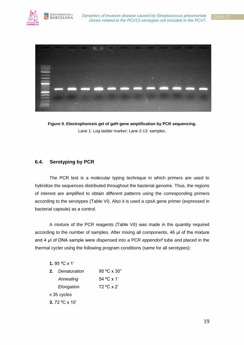
Figure II. Electrophoresis gel of gdH gene amplification by PCR sequencing.
Lane 1: Log-ladder marker; Lane 2-13: samples.
6.4. Serotyping by PCR
The PCR test is a molecular typing technique in which primers are used to
hybridize the sequences distributed throughout the bacterial genome. Thus, the regions
of interest are amplified to obtain different patterns using the corresponding primers
according to the serotypes (Table VI). Also it is used a cpsA gene primer (expressed in
bacterial capsule) as a control.
A mixture of the PCR reagents (Table VII) was made in the quantity required
according to the number of samples. After mixing all components, 46 μl of the mixture
and 4 μl of DNA sample were dispensed into a PCR eppendorf tube and placed in the
thermal cycler using the following program conditions (same for all serotypes):
1. 95 ºC x 1’
2. Denaturation 95 ºC x 30”
Annealing 54 ºC x 1’
Elongation 72 ºC x 2’
x 35 cycles
3. 72 ºC x 10’
20
2016-17 Dynamics of invasive disease caused by Streptococcus pneumoniae clones related to the PCV13 serotypes not included in the PCV7.
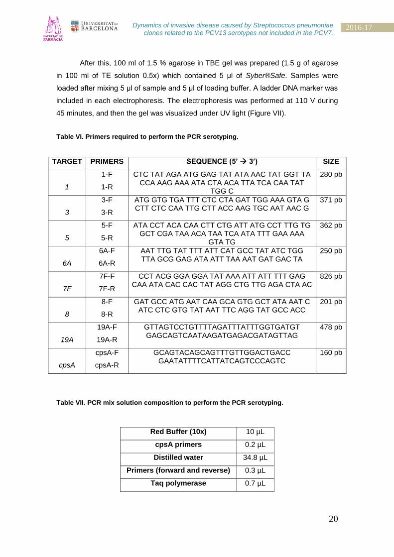
After this, 100 ml of 1.5 % agarose in TBE gel was prepared (1.5 g of agarose
in 100 ml of TE solution 0.5x) which contained 5 μl of Syber®Safe. Samples were
loaded after mixing 5 μl of sample and 5 μl of loading buffer. A ladder DNA marker was
included in each electrophoresis. The electrophoresis was performed at 110 V during
45 minutes, and then the gel was visualized under UV light (Figure VII).
Table VI. Primers required to perform the PCR serotyping.
TARGET PRIMERS SEQUENCE (5’ 3’) SIZE
1
1-F
1-R
CTC TAT AGA ATG GAG TAT ATA AAC TAT GGT TA CCA AAG AAA ATA CTA ACA TTA TCA CAA TAT
TGG C
280 pb
3
3-F
3-R
ATG GTG TGA TTT CTC CTA GAT TGG AAA GTA G CTT CTC CAA TTG CTT ACC AAG TGC AAT AAC G
371 pb
5
5-F
5-R
ATA CCT ACA CAA CTT CTG ATT ATG CCT TTG TG GCT CGA TAA ACA TAA TCA ATA TTT GAA AAA
GTA TG
362 pb
6A
6A-F
6A-R
AAT TTG TAT TTT ATT CAT GCC TAT ATC TGG TTA GCG GAG ATA ATT TAA AAT GAT GAC TA
250 pb
7F
7F-F
7F-R
CCT ACG GGA GGA TAT AAA ATT ATT TTT GAG CAA ATA CAC CAC TAT AGG CTG TTG AGA CTA AC
826 pb
8
8-F
8-R
GAT GCC ATG AAT CAA GCA GTG GCT ATA AAT C ATC CTC GTG TAT AAT TTC AGG TAT GCC ACC
201 pb
19A
19A-F
19A-R
GTTAGTCCTGTTTTAGATTTATTTGGTGATGT GAGCAGTCAATAAGATGAGACGATAGTTAG
478 pb
cpsA
cpsA-F
cpsA-R
GCAGTACAGCAGTTTGTTGGACTGACC GAATATTTTCATTATCAGTCCCAGTC
160 pb
Table VII. PCR mix solution composition to perform the PCR serotyping.
Red Buffer (10x) 10 µL
cpsA primers 0.2 µL
Distilled water 34.8 µL
Primers (forward and reverse) 0.3 µL
Taq polymerase 0.7 µL
21
2016-17 Dynamics of invasive disease caused by Streptococcus pneumoniae clones related to the PCV13 serotypes not included in the PCV7.
Figure VII. Electrophoresis gel of serotype amplification by PCR sequencing.
Lane 1: Log-ladder marker; Lane 2-10: samples; Lane 11: Positive control.
6.5. Resistance gene genotyping by PCR
The macrolide resistance may be occurs due to the acquisition of encoding
genes which usually belong to the 916 transposons family and often lead determinants
for tetracycline resistance. In particular, this technique was used to sequence the
macrolide resistance genes (ermB and mefA/E) and tetracycline (tetM). The protocol
begins by the selection of the resistant strains using the clinical breakpoints criteria
indicated in EUCAST website (Table VIII) and proceed with the same way as MLST
technique explained before. Subsequently, a PCR mixture is made according to the
number of samples (Table IX). In this case, a different buffer (Red Buffer) has been
used which includes the MgCl2 and the dNTPs already. The controls used in the
electrophoresis gel are ATCC England 14-9 as a positive control. The configuration of
the thermal cycler for all genes is:
1. 94 ºC x 1’
2. Denaturation 94 ºC x 30”
Annealing 58 ºC x 60”
Elongation 72 ºC x 90”
x 35 cycles
3. 72 ºC x 10’
22
2016-17 Dynamics of invasive disease caused by Streptococcus pneumoniae clones related to the PCV13 serotypes not included in the PCV7.
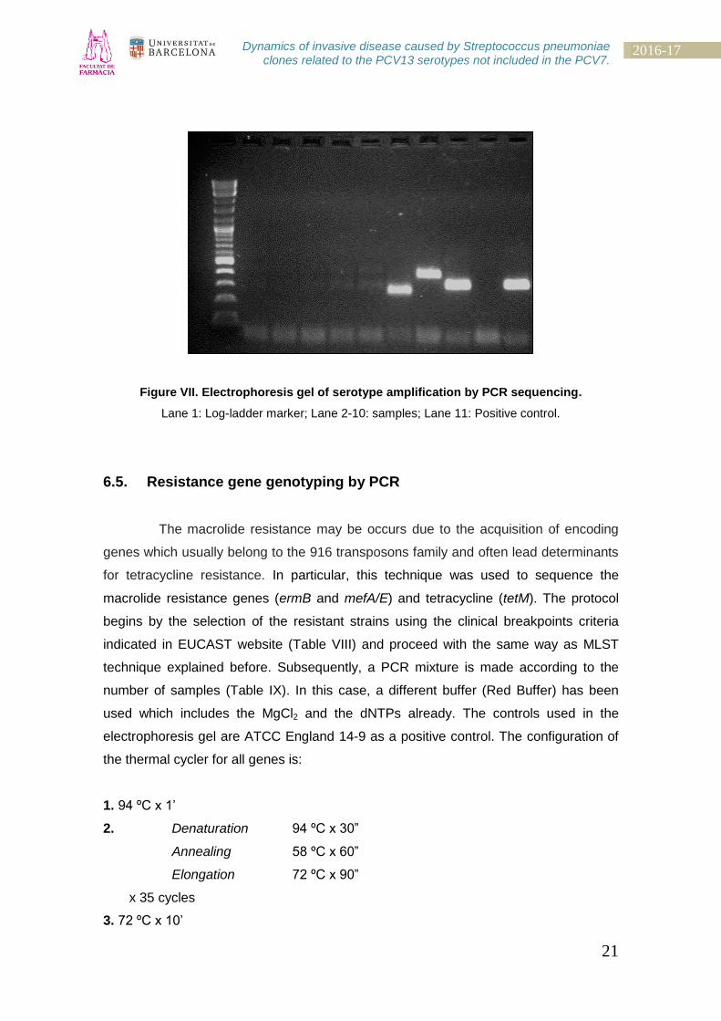
After this, 100 ml of 1.5 % agarose in TBE gel was prepared (1.5 g of agarose
in 100 ml of TE solution 0.5x) which contained 5 μl of Syber®Safe. Samples were
loaded after mixing 5 μl of sample and 5 μl of loading buffer. A ladder DNA marker was
included in each electrophoresis. The electrophoresis was performed at 120 V during
20 minutes, and then the gel was visualized under UV light (Figure VIII). Finally, it was
necessary to differentiate between mefA and mefE genes, performing a digestion with
bamHI for 1 hour at 37°C (Table X) and another electrophoresis at the same
conditions.
Figure VIII. Electrophoresis gel of tetM gene amplification by PCR sequencing.
Lane 1: Log-ladder marker; Lane 2-19: samples; Lane 20: Positive control
Table VIII. Pneumococcal susceptibility (CMI in mg/L) to macrolides and tetracycline
according to EUCAST clinical breakpoints criteria.
Table IX. PCR mix solution composition to perform the PCR resistance gene genotyping.
Red Buffer 10 µL
Primer-forward 0.4 µL
Primer-reverse 0.4 µL
Taq polymerase 0.3 µL
Distilled water 36.9 µL
Susceptible Intermediate Resistant
Tetracycline ≤1 1-2 >2
Erythromycin ≤0.25 0.25-0.5 >0.5
Clindamycin ≤0.25 0.25-0.5 >0.5

23
2016-17 Dynamics of invasive disease caused by Streptococcus pneumoniae clones related to the PCV13 serotypes not included in the PCV7.
Table X. Reagents required for the digestion of mefA and mefE genes.
Buffer (10x) 4 µL
bamHI 0.5 µL
Distilled water 16 µL
PCR product sample 20 µL
6.6. Sequence results edited and analyzed
Sequence edition: SeqScape v2.7 program.
The chromatogram (Figure IX) was the result of loading the Macrogen, Inc files
where the MLST genotyping products were sent to be sequenced. This sequencing is
made by the Sanger method, which is based on the presence of nucleotides labeled
with fluorophores and a capillary electrophoresis. To obtain the serotypes and the
antibiotic susceptibility of each strain, the samples were also sent to the Health Institute
Carlos III, Madrid.
Figure IX. Image of SeqScape v2.7 in which the nucleotide sequences can be edited.
24
2016-17 Dynamics of invasive disease caused by Streptococcus pneumoniae clones related to the PCV13 serotypes not included in the PCV7.
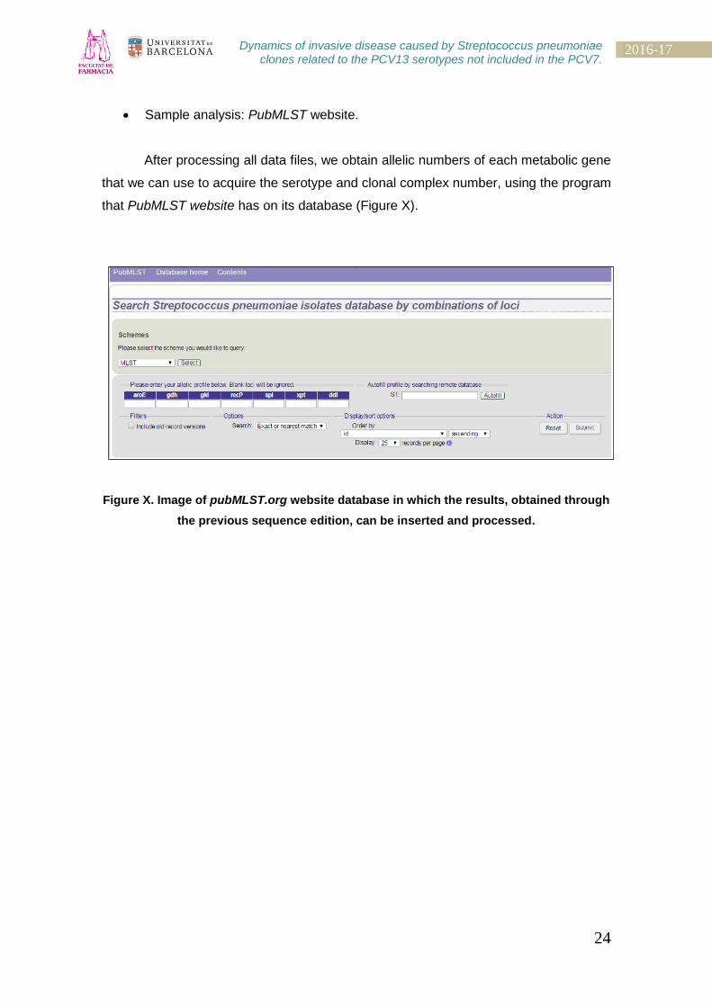
Sample analysis: PubMLST website.
After processing all data files, we obtain allelic numbers of each metabolic gene
that we can use to acquire the serotype and clonal complex number, using the program
that PubMLST website has on its database (Figure X).
Figure X. Image of pubMLST.org website database in which the results, obtained through
the previous sequence edition, can be inserted and processed.

25
2016-17 Dynamics of invasive disease caused by Streptococcus pneumoniae clones related to the PCV13 serotypes not included in the PCV7.
7. Results and discussion
7.1. Invasive Pneumococcal Disease (IPD) in 2011-2016 period
The results obtained have been analyzed by age groups (those over 65 years
old and those between 18 and 64 years) and sex (Figure XI). A total of 492 IPD strains
were isolated among the study period (2011-2016). The highest incidence was
observed in men older than 65 and this difference was statistically significant (p<0.01);
the remaining groups differences were not statistically significant (p>0.05).
Figure XI. Distribution of IPD episodes among age and sex group per year (2011-2016).
Figure XII shows the results of total number of IPD episodes analyzed by
serotype group. While IPD due to PCV7 serotypes and those due to the additional
PCV13 serotypes decrease, the IPD due to non-PCV13 serotypes did not change
significantly. Nevertheless, an increase in the IPD due to non-PCV13 serotypes was
observed in 2014 and onwards.
2011 2012 2013 2014 2015 2016
men - ≥65 25 24 26 33 16 27
men - 18-64 24 27 20 15 22 17
women - ≥65 17 25 18 34 21 15
women - 18-64 15 11 17 16 15 11
0
5
10
15
20
25
30
35
40
nº
of
ep
iso
de
s

26
2016-17 Dynamics of invasive disease caused by Streptococcus pneumoniae clones related to the PCV13 serotypes not included in the PCV7.
Figure XII. Total number of IPD episodes by serotype group (PCV7, non-PCV13 and
additional PCV13) per year (2011-2016).
In regard the vaccine group distribution, we observed a decrease in the IPD
among young adults (18-64) while in adults over 65 remains stable due to an increase
of the disease caused by non-PCV13 serotypes (Figure XIII). Despite this, if we
observe the results with absolute numbers we will notice how the total number of
isolates of invasive Pneumococcal strains has suffered a greater reduction per year;
this trend suggests that the impact of vaccines on the population is relevant and its
effectiveness can be evidenced by the total number of isolated invasive strains which
have decreased more than half in the last decade.
33 28 26 26
17 15
35
47 45
64
53 49
13 12 10 9 4 6
0
10
20
30
40
50
60
70
2011 2012 2013 2014 2015 2016
additional PCV13 non-PCV13 PCV7

27
2016-17 Dynamics of invasive disease caused by Streptococcus pneumoniae clones related to the PCV13 serotypes not included in the PCV7.
Figure XIII. Annual distribution of IPD (episodes/100.000 people) related to
vaccine serotypes and age groups in a Southern Barcelona Area. Top: Incidence of IPD in
people aged 18-64. Bottom: Incidence of IP in people aged over 65. Green: PCV7 serotypes (4,
6B, 9V, 14, 19F, 18C and 23F). Blue: additional PCV13 serotypes (1, 3, 5, 6A, 7F and 19A).
Orange: non-PCV13 serotypes (others).
0
5
10
15
20
25
30
35
40
45
2011 2012 2013 2014 2015 2016
nº
of
ep
iso
de
s (1
8-6
4 a
ge
gro
up
)
PCV7
additional PCV13
non-PCV13
0
10
20
30
40
50
60
70
80
2011 2012 2013 2014 2015 2016
nº
of
ep
iso
de
s (o
ve
r 6
5 a
ge
gro
up
)
PCV7
additional PCV13
non-PCV13

28
2016-17 Dynamics of invasive disease caused by Streptococcus pneumoniae clones related to the PCV13 serotypes not included in the PCV7.
7.2. Genetic relatedness of Pneumococcal isolates belonging to PCV13
serotypes not included in PCV7 (additional PCV13)
The IPD caused by additional PCV13 serotypes (1, 3, 5, 6A, 7F and 19A)
shows differences over the study period. In general, the IPD caused by the additional
serotypes decreases with the exception of serotypes 3 and 19A. In fact, the IPD
caused by serotype 3 remains stable over the study period. On the other hand, IPD
caused by serotype 19A remains stable between 2011 and 2014, decreasing in the last
two years (Figure XIV). These results show the impact of children vaccination among
adult IPD due to the herd protection, with exception of serotype 3 which is rarely found
in children.
Figure XIV. Incidence of PCV13 non-PCV7 serotypes (1, 3, 5, 6A, 7F and 19A)
episodes per year (2011-2016).
2
5
2 2
1 1
13
5
10 10
12
10
0 0 0 0 0 0
1
0 0 0 0
1
6
8
5
4
1
0
11
10
9
10
3 3
0
2
4
6
8
10
12
14
2011 2012 2013 2014 2015 2016
1 3 5 6A 7F 19A
29
2016-17 Dynamics of invasive disease caused by Streptococcus pneumoniae clones related to the PCV13 serotypes not included in the PCV7.
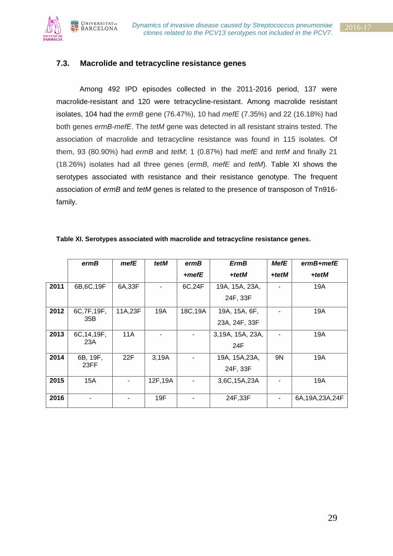
7.3. Macrolide and tetracycline resistance genes
Among 492 IPD episodes collected in the 2011-2016 period, 137 were
macrolide-resistant and 120 were tetracycline-resistant. Among macrolide resistant
isolates, 104 had the ermB gene (76.47%), 10 had mefE (7.35%) and 22 (16.18%) had
both genes ermB-mefE. The tetM gene was detected in all resistant strains tested. The
association of macrolide and tetracycline resistance was found in 115 isolates. Of
them, 93 (80.90%) had ermB and tetM; 1 (0.87%) had mefE and tetM and finally 21
(18.26%) isolates had all three genes (ermB, mefE and tetM). Table XI shows the
serotypes associated with resistance and their resistance genotype. The frequent
association of ermB and tetM genes is related to the presence of transposon of Tn916-
family.
Table XI. Serotypes associated with macrolide and tetracycline resistance genes.
ermB mefE tetM ermB
+mefE
ErmB
+tetM
MefE
+tetM
ermB+mefE
+tetM
2011 6B,6C,19F 6A,33F - 6C,24F 19A, 15A, 23A,
24F, 33F
- 19A
2012 6C,7F,19F, 35B
11A,23F 19A 18C,19A 19A, 15A, 6F,
23A, 24F, 33F
- 19A
2013 6C,14,19F, 23A
11A - - 3,19A, 15A, 23A,
24F
- 19A
2014 6B, 19F, 23FF
22F 3,19A - 19A, 15A,23A,
24F, 33F
9N 19A
2015 15A - 12F,19A - 3,6C,15A,23A - 19A
2016 - - 19F - 24F,33F - 6A,19A,23A,24F

30
2016-17 Dynamics of invasive disease caused by Streptococcus pneumoniae clones related to the PCV13 serotypes not included in the PCV7.
7.4. Genotypes associated to additional PCV13 serotypes
As can be observed in Table XII, within the additional PCV13 group
predominates the ST306 associated to serotype 1, ST180 and ST260 associated to
serotype 3 and finally the ST191 associated to serotype 7F; the clonal complex related
to serotypes 19A and 6A are varied.
Table XII. Clonal complex genotype related to additional PCV13 serotypes per year.
2011 2012 2013 2014 2015 2016
1
(n=13)
ST306 ST306 ST306 ST306 ST306 ST306
3
(n=60)
ST180
ST260
ST180
ST260
ST180
ST260
ST180
ST260
ST458
ST53
ST180
ST260
ST1377
ST180
ST260
5
(n=0)
- - - - - -
6A
(n=2)
ST473 - - - - -
7F
(n=24)
ST191 ST191 ST191 ST191 ST191
ST3544
-
19A
(n=46)
ST1201
ST320
ST3261
ST230
ST320 ST320
ST81
ST199
ST3259
ST450
ST1201
ST1201
ST320
ST7737
ST1201
ST230
ST320
ST230
ST320

31
2016-17 Dynamics of invasive disease caused by Streptococcus pneumoniae clones related to the PCV13 serotypes not included in the PCV7.
The Figure XV shows particularly the clonal complexes related to serotype 19A
in which multidrug-resistant caused by ST320 appears more frequently. Also, the
Figure XVI shows the clonal complex related to serotype 3, making evidence of the
greater clonal complex ST180 presence.
Figure XV. Clonal composition of serotype 19A (n=46).
Figure XVI. Clonal composition of serotype 3 (n=60) per years
ST199 2%
ST230 15%
ST320 55%
ST1201 13%
OTHERS 15%
0
2
4
6
8
10
12
14
16
18
2011-2012 2013-2014 2015-2016
ST180
ST260
ST53
ST458
ST1377

32
2016-17 Dynamics of invasive disease caused by Streptococcus pneumoniae clones related to the PCV13 serotypes not included in the PCV7.
8. Conclusions
The incidence of IPD in adults decreases over the study period demonstrating a
significant impact of children vaccination in adults due to herd protection.
The decrease on IPD was only remarkable in young adults (18-64) while in
adults over 65 remained stable.
In adults over 65 the decrease of IPD caused by PCV13 serotypes was
balanced by an increase of non-PCV13 serotypes.
There was not an impact of children vaccination on IPD due to serotype 3 that
is the major cause of invasive Pneumococcal disease in adults. The
maintenance of serotype 3 was due to the predominant clonal complex ST180.
Over the study period, a decrease of IPD due to macrolide resistant strains was
observed mainly linked to a felt in the IPD serotype 19A.
The association of macrolide and tetracycline resistances was frequent,
indicating the dissemination of Tn916-family transposon.

33
2016-17 Dynamics of invasive disease caused by Streptococcus pneumoniae clones related to the PCV13 serotypes not included in the PCV7.
9. References
(1) Austrian R. The pneumococcus at the millennium: not down, not out. J Infect Dis. 1999. 179:S338-41. (2) García-Vidal C, et al. Pneumococcal pneumonia presenting with septic shock: host- and pathogen-related factors and outcomes. Thorax 2010; 65(1):77-81. (3) Liñares J., et al. Changes in antimicrobial resistance, serotypes and genotypes in Streptococcus pneumoniae over a 30-year period. Clin Microbiol Infect. 2010. 16:402-10. (4) Kim, L., McGee, L., Tomczyk, S., & Beall, B. (2016). Biological and epidemiological features of antibiotic-resistant Streptococcus pneumoniae in pre- and post-conjugate vaccine eras: A United States perspective. Clinical Microbiology Reviews, 29(3), 525–552. (5) Ardanuy C., et al Epidemiology of invasive Pneumococcal disease among adult patients in Barcelona before and after pediatric 7-valent Pneumococcal conjugate vaccine introduction, 1997-2007. Clin Infect Dis. 2009; 48:57-64. (6) Ardanuy C., et al. Epidemiology of invasive Pneumococcal disease in older people in Spain (2007-2009): implications for future vaccination strategies. PLoS One. 2012; 7(8):e43619. (7) Muñoz-Almagro C., et al. Emergence of invasive Pneumococcal disease caused by nonvaccine serotypes in the era of 7-valent conjugate vaccine. Clin Infect Dis. 2008; 46:174-182. (8) Picazo J., Ruiz-Contreras J., Casado-Flores J., et al. Expansion of serotype coverage in the universal pediatric vaccination calendar: short-term effects on age- and serotype-dependent incidence of invasive Pneumococcal clinical presentations in Madrid, Spain. Clin Vaccine Immunol. 2013; 20:1524-30. (9) Dockrell D. H., Whyte MK, Mitchell T. J. Pneumococcal pneumonia: mechanisms of infection and resolution. Chest 2012; 142:482-91. (10) Mitchell A. M., Mitchell TJ. Streptococcus pneumoniae: virulence factors and variation. Clin Microbiol Infect. 2010; 16:411-8. (11) Pilishvili T., et al. Sustained reductions in invasive Pneumococcal disease in the era of conjugate vaccine. J Infect Dis. 2010; 201:32-41. (12) Ardanuy C., et al. Emergence of a multidrug-resistant clone (ST320) among invasive serotype 19A pneumococci in Spain. J Antimicrob Chemother. 2009; 64:507-10. (13) Fenoll A., et al. Temporal trends of invasive Streptococcus pneumoniae serotypes and antimicrobial resistance patterns in Spain from 1979 to 2007. J Clin Microbiol. 2009. 47:1012-20.

34
2016-17 Dynamics of invasive disease caused by Streptococcus pneumoniae clones related to the PCV13 serotypes not included in the PCV7.
(14) Hicks L. A., et al. Incidence of Pneumococcal disease due to non-Pneumococcal conjugate vaccine (PCV7) serotypes in the United States during the era of widespread PCV7 vaccination, 1998-2004. J Infect Dis. 2007; 196:1346-1354. (15) Liñares, J., Ardanuy, C., Pallares, R., & Fenoll, A. (2010). Changes in antimicrobial resistance, serotypes and genotypes in Streptococcus pneumoniae over a 30-year period. Clinical Microbiology and Infection, 16(5), 402–410. (16) Croucher N. J., et al. Rapid Pneumococcal evolution in response to clinical inteRentions. Science 2011 Jan 28; 331(6016):430-4. (17) Domenech A., et al. Evolution and genetic diversity of the Spain23F-ST81 clone causing adult invasive Pneumococcal disease in Barcelona (1990-2012). J Antimicrob Chemother. 2014 Apr; 69 (4):924-31. (18) McGee L., et al. Nomenclature of major antimicrobial-resistant clones of Streptococcus pneumoniae defined by the Pneumococcal molecular epidemiology network. J Clin Microbiol. 2001. 39:2565-71. (19) Coffey T. J., et al. Recombinational exchanges at the capsular polysaccharide biosynthetic locus lead to frequent serotype changes among natural isolates of Streptococcus pneumoniae. Mol Microbiol. 1998. 27:73-83. (20) Croucher N. J., et al. Dominant role of nucleotide substitution in the diversification of serotype 3 pneumococci over decades and during a single infection. PLoS Genet. 2013; 9(10):e1003868. (21) Donati C., et al. Structure and dynamics of the pan-genome of Streptococcus pneumoniae and closely related species. Genome Biol. 2010; 11(10):R107. (22) Brueggemann A. B., et al. Temporal and geographic stability of the serogroup-specific invasive disease potential of Streptococcus pneumoniae in children. J Infect Dis. 2004; 190:1203-11. (23) Sá-Leão R., et al. Analysis of invasiveness of Pneumococcal serotypes and clones circulating in Portugal before widespread use of conjugate vaccines reveals heterogeneous behavior of clones expressing the same serotype. J. Clin Microbiol. 2011; 49:1369-75. (24) Weinberger D. M., et al. Association of serotype with risk of death due to Pneumococcal pneumonia: a meta-analysis. Clin Infect Dis . 2010; 51: 692 – 699. (25) Rolo D., et al. Serotype 5 pneumococci causing invasive Pneumococcal disease outbreaks in Barcelona, Spain (1997 to 2011). J Clin Microbiol. 2013 Nov; 51(11):3585-90. (26) Rolo D., et al. Trends of invasive serotype 6C pneumococci in Spain: emergence of a new lineage. J Antimicrob Chemother. 2011; 66:1712-8. (27) López R., García E. Recent trends on the molecular biology of Pneumococcal capsules, lytic enzymes, and bacteriophage. FEMS Microbiol. 2004; 28:553-80. (28) Ardanuy, C., et al. Serotype 8 Streptococcus pneumoniae multidrug-resistant recombinant clone Sweden15A-ST63: dissemination in Spain. Emerg. Infect Dis. 2014 Nov.

35
2016-17 Dynamics of invasive disease caused by Streptococcus pneumoniae clones related to the PCV13 serotypes not included in the PCV7.
(29) Grau, I., Ardanuy, C., Cubero, M., Benitez, M. A., Liñares, J., & Pallares, R. (2016). Declining mortality from adult Pneumococcal infections linked to children’s vaccination. Journal of Infection, 72(4), 439–449. (30) Harboe, Z. B. Z., Dalby, T., Weinberger, D., Benfield, T., Mølbak, K., Slotved, H. C., Branth, P. V. (2014). Impact of 13-valent Pneumococcal conjugates vaccination in invasive Pneumococcal disease incidence and mortality. Clinical Infectious Diseases : An Official Publication of the Infectious Diseases Society of America, 59(8), 1066–1073.