By: Lawrence M. Parsons, Justine Sergent, Donald A. Hodges...
27
The brain basis of piano performance By: Lawrence M. Parsons, Justine Sergent, Donald A. Hodges, and Peter T. Fox Parsons, L., Sergent, J., Hodges, D. , & Fox, P. (2005) The brain basis of piano performance. Neuropsychologia, 43:2, 199-215. doi:10.1016/j.neuropsychologia.2004.11.007 Made available courtesy of ELSEVIER: http://www.elsevier.com/wps/find/journaldescription.cws_home/247/description#description ***Note: Figures may be missing from this format of the document Abstract: Performances of memorized piano compositions unfold via dynamic integrations of motor, perceptual, cognitive, and emotive operations. The functional neuroanatomy of such elaborately skilled achievements was characterized in the present study by using 150-water positron emission tomography to image blindfolded pianists performing a concerto by J.S. Bach. The resulting brain activity was referenced to that for bimanual performance of memorized major scales. Scales and concerto performances both activated primary motor cortex, corresponding somatosensory areas, inferior parietal cortex, supplementary motor area, motor cingulate, bilateral superior and middle temporal cortex, right thalamus, anterior and posterior cerebellum. Regions specifically supporting the concerto performance included superior and middle temporal cortex, planum polare, thalamus, basal ganglia, posterior cerebellum, dorsolateral premotor cortex, right insula, right supplementary motor area, lingual gyrus, and posterior cingulate. Areas specifically implicated in generating and playing scales were posterior cingulate, middle temporal, right middle frontal, and right precuneus cortices, with lesser increases in right hemispheric superior temporal, temporoparietal, fusiform, precuneus, and prefrontal cortices, along with left inferior frontal gyrus. Finally, much greater deactivations were present for playing the concerto than scales. This seems to reflect a deeper attentional focus in which tonically active orienting and evaluative processes, among others, are suspended. This inference is supported by observed deactivations in posterior cingulate, parahippocampus, precuneus, prefrontal, middle temporal, and posterior cerebellar cortices. For each of the foregoing analyses, a distributed set of interacting localized functions is outlined for future test. Keywords: Music performance; Piano performance; Functional neuroimaging; Music cognition Article: 1. Introduction Musical performance is very likely the domain in which humans produce the most intricate, complex integration of expert perceptual, motor, cognitive, and emotive skills. But although it may be the pinnacle of human central nervous system performance (and what space aliens most covet), its basis in the brain rarely has been investigated. Fortunately, musical ability and cognition appear to yield to fractionation (e.g., Peretz & Coltheart, 2003; Sergent, 1993), and components of musical performance have been studied with neurological, electrophysiological, and neuroimaging methods. These performance components include perception, sight- reading, motor-sensory processes, and attention. The most deeply studied component is the neural basis of perceptual aspects of musical performance. Researchers have demonstrated, for example, strong associations amongst the strength of neurophysiological responses to pure tones in the musical range (detected via magnetoencephlography, MEG), the volume of anterior-medial Heschl's gyrus from which the responses originate, and musical skill (Schneider et al., 2002). Others have demonstrated enhanced neural representation for the timbre of the instrument in which a musician specializes, as compared to others (Pantev, Roberts, Schultz, Egnelien, & Ross, 2001) and the differences in the neural representation of musical pitch and rhythm between musicians and individuals with very little musical performance experience or training (Evers, Dannert, Rodding, Rotter, & Ringelstein, 1999; Parsons & Thaut,
Transcript of By: Lawrence M. Parsons, Justine Sergent, Donald A. Hodges...

The brain basis of piano performance
By: Lawrence M. Parsons, Justine Sergent, Donald A. Hodges, and Peter T. Fox
Parsons, L., Sergent, J., Hodges, D., & Fox, P. (2005) The brain basis of piano performance. Neuropsychologia,
43:2, 199-215. doi:10.1016/j.neuropsychologia.2004.11.007
Made available courtesy of ELSEVIER:
http://www.elsevier.com/wps/find/journaldescription.cws_home/247/description#description
***Note: Figures may be missing from this format of the document
Abstract:
Performances of memorized piano compositions unfold via dynamic integrations of motor, perceptual,
cognitive, and emotive operations. The functional neuroanatomy of such elaborately skilled achievements was
characterized in the present study by using 150-water positron emission tomography to image blindfolded
pianists performing a concerto by J.S. Bach. The resulting brain activity was referenced to that for bimanual
performance of memorized major scales. Scales and concerto performances both activated primary motor
cortex, corresponding somatosensory areas, inferior parietal cortex, supplementary motor area, motor cingulate,
bilateral superior and middle temporal cortex, right thalamus, anterior and posterior cerebellum. Regions
specifically supporting the concerto performance included superior and middle temporal cortex, planum polare,
thalamus, basal ganglia, posterior cerebellum, dorsolateral premotor cortex, right insula, right supplementary
motor area, lingual gyrus, and posterior cingulate. Areas specifically implicated in generating and playing scales
were posterior cingulate, middle temporal, right middle frontal, and right precuneus cortices, with lesser
increases in right hemispheric superior temporal, temporoparietal, fusiform, precuneus, and prefrontal cortices,
along with left inferior frontal gyrus. Finally, much greater deactivations were present for playing the concerto
than scales. This seems to reflect a deeper attentional focus in which tonically active orienting and evaluative
processes, among others, are suspended. This inference is supported by observed deactivations in posterior
cingulate, parahippocampus, precuneus, prefrontal, middle temporal, and posterior cerebellar cortices. For each
of the foregoing analyses, a distributed set of interacting localized functions is outlined for future test.
Keywords: Music performance; Piano performance; Functional neuroimaging; Music cognition
Article:
1. Introduction
Musical performance is very likely the domain in which humans produce the most intricate, complex integration
of expert perceptual, motor, cognitive, and emotive skills. But although it may be the pinnacle of human central
nervous system performance (and what space aliens most covet), its basis in the brain rarely has been
investigated. Fortunately, musical ability and cognition appear to yield to fractionation (e.g., Peretz & Coltheart,
2003; Sergent, 1993), and components of musical performance have been studied with neurological,
electrophysiological, and neuroimaging methods. These performance components include perception, sight-
reading, motor-sensory processes, and attention.
The most deeply studied component is the neural basis of perceptual aspects of musical performance.
Researchers have demonstrated, for example, strong associations amongst the strength of neurophysiological
responses to pure tones in the musical range (detected via magnetoencephlography, MEG), the volume of
anterior-medial Heschl's gyrus from which the responses originate, and musical skill (Schneider et al., 2002).
Others have demonstrated enhanced neural representation for the timbre of the instrument in which a musician
specializes, as compared to others (Pantev, Roberts, Schultz, Egnelien, & Ross, 2001) and the differences in the
neural representation of musical pitch and rhythm between musicians and individuals with very little musical
performance experience or training (Evers, Dannert, Rodding, Rotter, & Ringelstein, 1999; Parsons & Thaut,
2001). Studies have also examined the relation between auditory perception and motor behavior. Thus, one
MEG study demonstrated that when pianists, but not singers, listen to familiar piano pieces to detect errors, they
exhibited involuntary activations in cerebral cortical motor systems (Haueisen & Knosche, 2001).
Fewer functional brain investigations have targeted activities more intimately related to the production aspects
of musical performances, which is the focus of the present paper. Various approaches have been used to
investigate the sight-reading of musical scores. Several neurological case studies examined musicians‘ acquired
impairments in sight-reading (Cappelletti, Waley-Cohen, Butterworth, & Kopelman, 2000; Judd, Gardner, &
Geschwind, 1983; Marin & Perry, 1992; Sergent, 1993; Stewart & Walsh, 2001). Positron emission tomography
(PET) has been used to study pianists sight-reading (Sergent, Zuck, Terriah, & McDonald, 1992), and to study
conductors sight-reading a score as they detected errors in its heard performance (Parsons, Hodges, & Fox,
1998). More recently, MEG was used to investigate musicians imagining the musical sounds of a score they
sight-read (Schurmann, Raij, Fujiki, & Hari, 2002). These studies and others (e.g., Nakada, Fujii, Suzuki, &
Kwee, 1998; Schon, Anton, Roth, & Besson, 2002; Stewart et al., 2003) suggest that a core distributed network
of areas in parietal, temporal, and occipital cortices support sight-reading, with other areas in frontal, sub-
cortical, and cerebellar areas, being recruited depending on whether the score is merely read, read and imagined
to be heard, or read while being performed.
A variety of research has focused on sensorimotor processes related to performing on musical instruments.
MEG studies indicate that the extent of cortical representations for musicians‘ digits is related to the degree of
skilled performance with those digits, as well as to the age at which the musicians started training on the
musical instrument (Elbert, Pantev, Wienbruch, Rockstroh, & Taub, 1995). Such functional differences are
complemented by anatomical MRI studies reporting increased size and specific structural differences in
musicians, as compared to non-musicians, in areas such as planum temporale, anterior corpus callosum, hand
primary motor cortex, anterior-medial Heschl's gyrus, and anterior cerebellum (see Gaser & Schlaug, 2003;
Munte, Altenmuller, & Jancke, 2002; Schneider et al., 2002).
Musicians performing rhythmic, sequential finger tapping tasks have been studied with functional
neuroimaging. When performing a novel, simple unimanual tapping task, a within-session increase in neural
activity in primary motor cortex was detected in musicians, but not in non-musicians, implicating adaptive
motor skill processes already acquired or present in musicians (Hund-Georgiadis & von Cramon, 1999). The
musicians exhibited at the same time a smaller extent of activation in supplementary motor area (SMA), pre-
SMA, and motor cingulate than did non-musicians, implying more efficient motor control processes. Similar
activation patterns were observed when musicians perform complex bimanual tapping tasks (Jäncke, Shah, &
Peters, 2000), with the exception that less activity is seen in primary motor cortex for musicians than non-
musicians. An fMRI study of complex sequences of unimanual finger tapping reported significantly reduced
activation for musicians as compared to non-musicians in primary motor and premotor cortices, SMA, and
superior parietal cortex (Krings et al., 2000). A metanalysis of neuroimaging studies examined the temporal and
sequence ordering involved in the foregoing kinds of tapping tasks (Janata & Grafton, 2003). The results
suggested that such tasks elicit responses in sensorimotor cortex, SMA, cerebellum, and premotor cortex.
Moreover, with increasing task complexity, other areas appear to be recruited in anterior cingulate, insula,
precuneus, intraparietal sulcus, basal ganglia, ventrolateral cortex, and thalamus.
A recent study of attentional states elicited by musical performance suggest that with the use of EEG-based
feedback training, musicians can improve the musical quality of their performances (Egner & Gruzelier, 2003).
In training sessions prior to musical performance, pianists learned to increase the theta over alpha band
amplitudes in their EEG. The enhancements in quality of musical performance appear to be a consequence of a
deep relaxed focus of attention, and may not be due to mere reductions in anxiety, as other methods of
relaxation training reduced anxiety but did not affect the quality of musical performance.
The brain basis of musical performance per se has been studied in the context of singing or piano playing. One
PET investigation studied non-musicians singing simple monotone sequences using a vowel (Perry et al., 1999)
and a second fMRI study examined non-musicians overtly or covertly singing a familiar melody without words
(Riecker, Ackermann, Wildergruber, Dogil, & Grodd, 2000). A more recent investigation employed PET to
examine amateur musicians who performed ‗listen and respond‘ tasks in which they either sang back repetitions
of novel melodies, sang back harmonizations to accompany novel melodies, or vocalized monotonically in
response (Brown, Parsons, Martinez, Hodges, & Fox, in press). Across these three studies, major singing-
specific activations were observed in primary and secondary auditory cortices, primary motor cortex, frontal
operculum, SMA, insula, posterior cerebellum, and basal ganglia. However, in the last study, melody singing
and harmonization, but not monotonic vocalization, activated planum polare (Brodmann area (BA) 38),
implicating it as an area supporting higher musical representations.
In an early, concerted effort to study the brain basis of musical performance (Sergent et al., 1992), pianists were
scanned with PET as they performed several conditions involving listening to scales, playing scales, sight-
reading a score, and sight-reading a score while playing it. The pianists always heard the sounds they produced
on the piano, and they sight-read or played an obscure partita by J.S. Bach. When the pianists played this piece,
they used only their right hand. As control conditions, the researchers included a task requiring a manual
response to indicate the location of a series of single visual dots within quadrants on a screen and a task with
fixation point rest. Of most interest at present is the pattern of activations detected for sight-reading when
playing (and hearing) the partita, as contrasted with a combination of the sight-reading and listening conditions.
This analysis revealed increases in left frontal operculum (BA 44) that were interpreted to support the patterning
of motor sequences of the right hand. There was also activity in left parietal cortex (supramarginal gyrus, BA
40), possibly involved in mapping visual and auditory representations of the melody. There were activations in
left occipitoparietal sulcus and bilateral superior parietal cortices (BA 7), which might subserve sensorimotor
transformations required for visually guided finger positioning.
Aspects of this early research have been pursued further in a recent study that used fMRI to compare the right
hand performance while sightreading the score of a Bartok piano piece to its imagined simulation (Meister et
al., 2004). In the baseline control the pianists read score with a single note repeated. Comparing the actual
performance to control, the authors observed activations in primarily left sensorimotor areas (BA 2–4), left
SMA (BA 6), bilateral precuneus (BA 7), bilateral inferior parietal (BA 40), left occipital (BA 37), left BA 5
(parietal), left posterior cerebellum (VI), midline anterior cerebellum (V), and left thalamus. Several of these
activations confirm those in the early PET study (Sergent et al., 1992). Comparing the imagined simulation to
control, they observed bilateral superior premotor (BA 6), left frontal (BA 9), bilateral parietal (BA 40 and 7),
bilateral occipital (BA 18 and 19), and left posterior cerebellum (VI). A direct comparison between performed
and imagined performance revealed performance-specific activations in left sensorimotor (BA 4, 2 and 3), left
SMA (BA 6), bilateral inferior parietal (BA 40), right anterior cerebellum (III), and left posterior cerebellum
(VI). Activation specific to imagined performance was limited to left occipital (BA 19). These findings are in
accord with prior research, such as that by Jeannerod, 1994 and Jeannerod, 1997, which demonstrated that
motor and sensory imagery involves psychological and neural processes similar to those for real motor and
sensory experiences (on auditory and musical imagery, see Halpern & Zatorre, 1999; Janata, 2001; Reiser,
1992).
Imagined musical performance was also examined in another recent study (Langheim, Callicott, Mattay, Duyn,
& Weinberger, 2002). This study combined fMRI data from imagined performance for different instruments
(cello, violin, piano) and different memorized compositions (various Vivaldi or Bach pieces) in order to localize
common, music-specific areas. Overall, the imagined musical performance (compared to rest) engaged right
SMA, right superior premotor cortex (BA 6), right superior parietal lobule (BA 7), right inferior frontal gyrus
(BA 47/45), left thalamus, left basal ganglia (caudate), and bilateral posterior cerebellum (VI). This
hemodynamic pattern was distinct from that for passive listening to musical pieces and for a self-paced
bimanual, finger-tapping task (both compared to rest). Thus, the results were taken to suggest that the foregoing
areas are involved in representing information for performing music. It is notable that neither of the two studies
just described of imagined musical performance observed activations in temporal cortical regions that support
auditory and musical information.

In sum, apart from brain areas for sensory-motor, attention, and executive control processes, three brain regions
have been identified so far that appear to be important for higher-level information processing aspects of music
performance. One area is the frontal operculum, which can be activated (left, right, or bilaterally) by sight-read
piano performance, by music singing, and by imagined string and piano performance of memorized music. This
area is often interpreted to be involved in sequence production and imitation learning. A second area is the
planum polare, which can be activated (either right or bilaterally) by singing and by sight-read piano
performance, but not apparently by imagined musical singing or imagined string or piano playing. This area
appears to represent musical representations of a higher order than, for example, is present in more posterior
superior temporal cortices (BA 22). A third region of interest in higher-level music is in rostromedial prefrontal
cortex, which responds to dissonance and consonance, and to changes in tonality (Blood, Zatorre, Bermudes, &
Evans, 1999; Janata et al., 2002; Peretz, Blood, Penhune, & Zatorre, 2001); however, this area has not yet been
studied in the context of musical performance.
The goal of the present study was to focus directly on musical performance as such, in order to complement and
clarify the foregoing findings. PET was employed to delineate brain areas subserving bimanual piano
performance of memorized music. This provides new information relative to prior studies since by recording
brain activity when both hands were equally and concurrently producing music, we examined neural systems
when both cerebral hemispheres were fully involved in a performance of a natural kind for musicians. This
approach goes beyond the Sergent et al. (1992) and Meister et al. (2004) studies in which only the right hand
was used to perform the music, a design that left unclear which particular right hemispheric areas may be
involved in music performance as such.
In addition, by asking pianists to perform a memorized composition, our design eliminated musical score
reading from scanned task performance. In this respect, we examined brain activation during a more purely
musical performance. Sight-reading a score during performance adds a considerable cognitive load, one
unrelated directly to music performance and cognition per se. Indeed, there is a common belief amongst
musicians that a fully memorized piece, one performed without score reading, engenders a distinctly deeper
understanding of the composition and more satisfying realization of the piece in performance (Aiello, 2001;
Chaffin & Imreh, 2002; Chaffin, Imreh, & Crawford, 2002; Mach, 1998). This belief is congruent with a
significant role of a deep focus of attention in the quality of musical performance, as discussed earlier.
Our design referenced the brain activity during the piano performance of a musical composition by J.S. Bach to
that during the two-handed performance of scales. The Bach and scales performances required movements of
approximately comparable frequency and complexity from each hand. This is a more comparable control
contrast than in the Langheim et al. (2002) study for (imagined) memorized musical performance. In this
design, real perceived musical sounds and similar executed motor behavior are compared across tasks of
varying musical structure to isolate the neural substrates of musical performance. In the Langheim et al. study,
imagined sounds and movements were compared to real ones, and the imagined motor behaviors were very
different than the control motor behavior (e.g., playing a cello piece versus finger tapping). Nonetheless, it was
recognized that studying a natural kind of musical performance, as compared to scales, entailed a number of
factors varying to influence brain activity, apart for musicality. Thus, there were intrinsic differences in
fingering complexity, independence of hands and melodic lines, complexity of memorized information to recall,
emotional content, and attentional demands. The outline of interactions observed here amongst these factors sets
the stage for more detailed, parametrically controlled studies of high-level performance skills.
2. Methods
2.1. Participants
After giving informed consent, eight professional musicians (five females and three males) volunteered to
participate in this study. All volunteers were right handed (Oldfield, 1971) and ranged from 27 to 54 years of
age. Each individual had from 14 to 20 years of training in piano performance, in addition to 10–18 years of
training and education on other instruments (either horn, voice, or string) and on other aspects of music
(composition and education).
2.2. Stimuli and tasks
Prior to the scanning session, the pianists practiced outside the laboratory in order to attain high quality on each
performance to be played from memory in the PET study: the third movement of the Italian Concerto in F
Major (BMV 971) by J.S. Bach, and two-handed, two-octave ascending and descending major scales beginning
in F major and progressing chromatically upward (e.g., F# major, then G major, etc.). The beginning of each
performance is illustrated in Fig. 1. The process of memorizing this movement of the Bach piece has been the
object of close psychological study (Chaffin & Imreh, 2002; Chaffin et al., 2002 R. Chaffin, G. Imreh and M.
Crawford, Practicing perfection: Memory and piano performance, Erlbaum, Mahway, NJ (2002).Chaffin et al.,
2002).
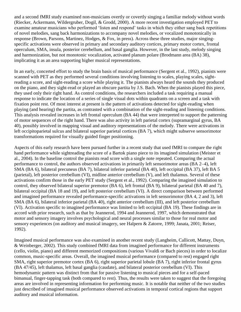
Fig. 1. Shown are the opening sections of stimuli for each performance task. The upper score shows the first
four measures of the third movement of the Bach Italian Concerto, and the lower score shows the two-octave F
major scale with hands in parallel motion. Note both the similarity of the scalar patterns in the Bach to the
scales below, and the comparative independence of the hands in the Bach. There are 68 finger strokes in the
concerto, 52 of which are not simultaneous, and 58 in the scales.
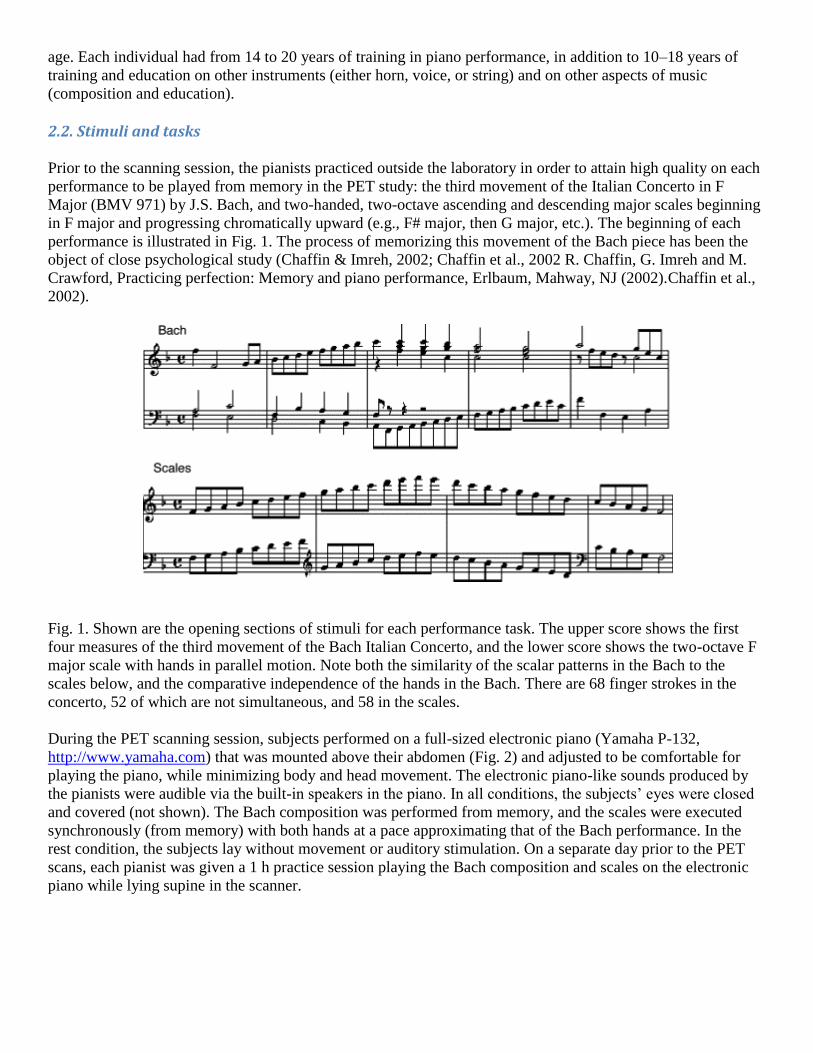
During the PET scanning session, subjects performed on a full-sized electronic piano (Yamaha P-132,
http://www.yamaha.com) that was mounted above their abdomen (Fig. 2) and adjusted to be comfortable for
playing the piano, while minimizing body and head movement. The electronic piano-like sounds produced by
the pianists were audible via the built-in speakers in the piano. In all conditions, the subjects‘ eyes were closed
and covered (not shown). The Bach composition was performed from memory, and the scales were executed
synchronously (from memory) with both hands at a pace approximating that of the Bach performance. In the
rest condition, the subjects lay without movement or auditory stimulation. On a separate day prior to the PET
scans, each pianist was given a 1 h practice session playing the Bach composition and scales on the electronic
piano while lying supine in the scanner.
Fig. 2. A pianist, viewed from above, lying in the PET scanner, as in the study. (photograph by Stephan
Elleringmann (laif photo agency)).
2.3. Procedure
The subjects completed each of three tasks (Bach, scales, rest) three times while being scanned. The three tasks
were conducted in pseudo-random order, such that PET scan trials 1 through 3 involved a trial (task) of each
kind, as did trials 4 through 6, and trials 7 through 9. Within each set of three trials, the order was completely
random.
During the PET session, subjects lay supine in the scanning instrument, with the head immobilized by a closely
fitted thermal-plastic facial mask with openings for the eyes, ears, nose, and mouth. The subjects began
performing the Bach composition or scales 30 s prior to injection of the bolus. The 15
0-water bolus uptake
required approximately 20 s to reach the brain at which time a 40 s scan was triggered by a sufficient rate of
coincidence-counts, as measured by the PET camera. At the end of the 40 s scan, the experimenter verbally
interrupted the performance to terminate the task, immediately after which the subject lay quietly without
moving during a second scan (50 s). From the initiation of the task until the start of the second scan on each
trial, each pianist played approximately 2 min. On trials with the Bach concerto, the pianists began at the
beginning of the third movement; on trials with scales, they began with F major.
The PET scans were performed on a GE 4096 camera, with a pixel spacing of 2.0 mm, and inter-plane, center-
to-center distance of 6.5 mm, 15 scan planes, and a z-axis field of view of 10 cm. Images were reconstructed
using a Hann filter, resulting in images with a spatial resolution of approximately 7 mm (full-width at half-
maximum). The data were smoothed with an isotropic 10 mm Gaussian kernel to yield a final image resolution
of approximately 12 mm. Anatomical MRI scans were acquired on an Elscint 1.9 T Prestige system with an in-
plane resolution of 1 mm2 and 1.5 mm slice thickness. The PET field of view was adjusted per subject such that
when the group mean functional PET blood flow image was spatially normalized, co-registered and

superimposed on anatomical MRI, activation was detectable at full axial plane for z height of 56 mm (see
illustration in center of Fig. 3).
Fig. 3. Significant blood flow changes as pianists play scales, and as pianists play the Bach composition.
Arrows on the upper images point to sensorimotor areas; arrows on the lower images point to auditory temporal
cortex and thalamus. Shown are group-averaged PET images, contrasted with rest, and overlaid onto a single
representative subject's anatomical MRI. PET data are z-scores displayed on a color scale ranging from 2.58
(yellow; P < 0.01) to 6.0 (red; P < 0.0001) for activations and −2.58 (green, P < 0.01) to −6.0 (blue;
P < 0.0001) for deactivations. Throughout, the z-values indicate the axial height of the brain volume relative to
Talairach and Tournoux (1988) stereotactic coordinates, and the left sides of the brain images are the left side of
the brain. The effective field of view for group mean functional PET data is illustrated by the central image.
maging procedures and data analysis were performed exactly as described in Parsons and Osherson (2001),
adhering to methods described in Raichle, Martin, Herskovitch, Mintun, and Markham (1983), Fox, Mintun,
Reiman, and Raichle (1988), and Mintun, Fox, and Raichle (1989). Briefly, local extrema were identified within
each image with a 3D search algorithm (Mintun et al., 1989) using a 125 voxel search cube (2 mm3 voxel). A
beta-2 statistic measuring kurtosis and a beta-1 statistic measuring skewness of the extrema histogram (Fox &
Mintun, 1989) were used as omnibus tests to assess overall significance (D‘Agostino, Belatner, & D‘Agostino,
1990). Critical values for beta statistics were chosen at P < 0.01. If the null hypothesis of omnibus significance
was rejected, then a post hoc (regional) test was done (Fox & Mintun, 1989; Fox et al., 1988). In this algorithm,
the pooled variance of all brain voxels is used as the reference for computing significance. This method is
distinct from methods that compute the variance at each voxel but is more sensitive in that results are more
reproducible across sample sizes of 1–8 (Strother et al., 1997), particularly for small samples, than the voxel-
wise variance methods of Friston, Frith, Liddle, and Frackowiak (1991) and others. The critical-value threshold
for regional effects (z > 2.58, P < 0.005, one-tailed) is not raised to correct for multiple comparisons since
omnibus statistics are established before post hoc analysis.
Gross anatomical labels were applied to the detected local maxima using a volume-occupancy-based,
anatomical labeling strategy as implemented in the Talaraich Daemon™ (Lancaster et al., 2000), except for
activations in the cerebellum which were labeled with reference to an atlas of the cerebellum (Schmahmann et
al., 1999).
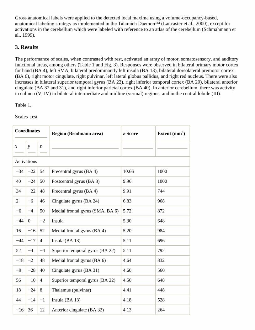
3. Results
The performance of scales, when contrasted with rest, activated an array of motor, somatosensory, and auditory
functional areas, among others (Table 1 and Fig. 3). Responses were observed in bilateral primary motor cortex
for hand (BA 4), left SMA, bilateral predominantly left insula (BA 13), bilateral dorsolateral premotor cortex
(BA 6), right motor cingulate, right pulvinar, left lateral globus pallidus, and right red nucleus. There were also
increases in bilateral superior temporal gyrus (BA 22), right inferior temporal cortex (BA 20), bilateral anterior
cingulate (BA 32 and 31), and right inferior parietal cortex (BA 40). In anterior cerebellum, there was activity
in culmen (V, IV) in bilateral intermediate and midline (vermal) regions, and in the central lobule (III).
Table 1.
Scales–rest
Coordinates
Region (Brodmann area) z-Score Extent (mm3)
x
y
z
Activations
−34 −22 54 Precentral gyrus (BA 4) 10.66 1000
40 −24 50 Postcentral gyrus (BA 3) 9.96 1000
34 −22 48 Precentral gyrus (BA 4) 9.91 744
2 −6 46 Cingulate gyrus (BA 24) 6.83 968
−6 −4 50 Medial frontal gyrus (SMA, BA 6) 5.72 872
−44 0 −2 Insula 5.30 648
16 −16 52 Medial frontal gyrus (BA 4) 5.20 984
−44 −17 4 Insula (BA 13) 5.11 696
52 −4 −4 Superior temporal gyrus (BA 22) 5.11 792
−18 −2 48 Medial frontal gyrus (BA 6) 4.64 832
−9 −28 40 Cingulate gyrus (BA 31) 4.60 560
56 −10 4 Superior temporal gyrus (BA 22) 4.50 648
18 −24 8 Thalamus (pulvinar) 4.41 448
44 −14 −1 Insula (BA 13) 4.18 528
−16 36 12 Anterior cingulate (BA 32) 4.13 264

Coordinates
Region (Brodmann area) z-Score Extent (mm3)
x
y
z
8 −22 46 Paracentral lobule (BA 24) 4.09 704
51 0 30 Precentral gyrus (BA 6) 3.71 488
−20 −8 2 Lateral globus pallidus 3.71 264
42 −34 18 Superior temporal gyrus (BA 41) 3.67 416
52 −24 22 Inferior parietal lobule (BA 40) 3.62 480
46 −22 2 Superior temporal gyrus (BA 22) 3.53 496
−46 −32 20 Insula 3.43 416
5 −24 −4 Red nucleus 3.29 280
−42 0 12 Insula (BA 13) 3.15 192
52 −54 −12 Inferior temporal gyrus (BA 20) 3.11 144
Anterior cerebellum activations
12 −54 −14 Culmen (V) 11.49 1000
22 −54 −22 Culmen (V) 11.21 1000
0 −60 −12 Culmen (V) (Vermis) 10.61 1000
−12 −52 −20 Culmen (IV) 10.33 1000
2 −38 −16 Central lobule (III) 4.32 536
Playing the Bach concerto, when contrasted with rest, likewise revealed a distributed pattern of activations in
motor, somatosensory, auditory, and other structures (Table 2 and Fig. 3). Responses were observed in bilateral
primary motor cortex for hand (BA 4), bilateral SMA, bilateral insula (BA 13), left dorsolateral premotor cortex
(BA 6), and right red nucleus. There was also strong subcortical activity in bilateral thalamus (left mammillary
body, right pulvinar) and bilateral basal ganglia (left globus pallidus, bilateral putamen). There were strong
responses in bilateral, predominantly right superior temporal cortex (BA 22), and right planum polare (BA 38).
Other activated areas included right cingulate gyrus (BA 24) and right occipitotemporal cortex (BA 37). In
anterior cerebellum, there was bilateral activity in intermediate and midline (vermal) regions of culmen (V). In
posterior cerebellum, there were responses in declive of vermis, right quadrangular lobule (VI), and left dentate
nucleus.
Table 2.
Bach–rest

Coordinates
Region (Brodmann area) z-Score Extent (mm3)
x
y
z
Activations
−32 −24 50 Precentral gyrus (BA 4) 11.95 1000
40 −24 52 Precentral gyrus (BA 4) 11.49 1000
6 −4 52 Medial frontal gyrus (BA 6) 7.80 1000
−6 −12 50 Medial frontal gyrus (BA 6) 6.97 992
−44 −2 −4 Insula (BA 13) 5.77 984
54 −12 2 Superior temporal gyrus (BA 22) 5.26 856
−42 −16 2 Insula (BA 13) 5.17 712
48 −30 4 Superior temporal gyrus (BA 22) 4.98 784
−14 −18 2 Thalamus (mammillary body) 4.89 696
52 0 −6 Superior temporal gyrus (BA 38) 4.66 880
16 −26 6 Thalamus (pulvinar) 4.52 592
26 4 −2 Lentiform nucleus (putamen) 4.29 504
16 −16 0 Thalamus 4.15 800
16 −10 0 Red nucleus 4.06 344
−18 0 48 Medial frontal gyrus (BA 6) 3.88 768
36 −2 18 Insula 3.88 456
6 −34 −10 Midbrain 3.88 472
−40 −2 12 Insula 3.79 248
4 −24 48 Paracentral lobule (BA 31) 3.74 512
32 −56 −9 Fusiform gyrus 3.69 304
50 −24 18 Insula 3.56 448
48 −44 −10 Inferior temporal gyrus (BA 37) 3.51 104
−24 −14 4 Lentiform nucleus (globus pallidus) 3.46 272
−40 −26 8 Superior temporal gyrus (BA 22/42) 3.32 432
8 4 35 Cingulate gyrus (BA 24) 3.28 312
−28 −2 4 Lentiform nucleus (putamen) 3.28 416

Coordinates
Region (Brodmann area) z-Score Extent (mm3)
x
y
z
Anterior cerebellum activations
14 −56 −14 Culmen (V) 14.07 1000
0 −62 −14 Culmen (V) 12.78 1000
−10 −56 −18 Culmen (V) 10.80 1000
−18 −54 −18 Culmen (V) 10.47 840
Posterior cerebellum activations
0 −66 −26 Declive (VI) (vermis) 11.30 1000
−12 −70 −30 Dentate nucleus 6.32 896
28 −73 −16 Quadrangular (VI) 3.79 376
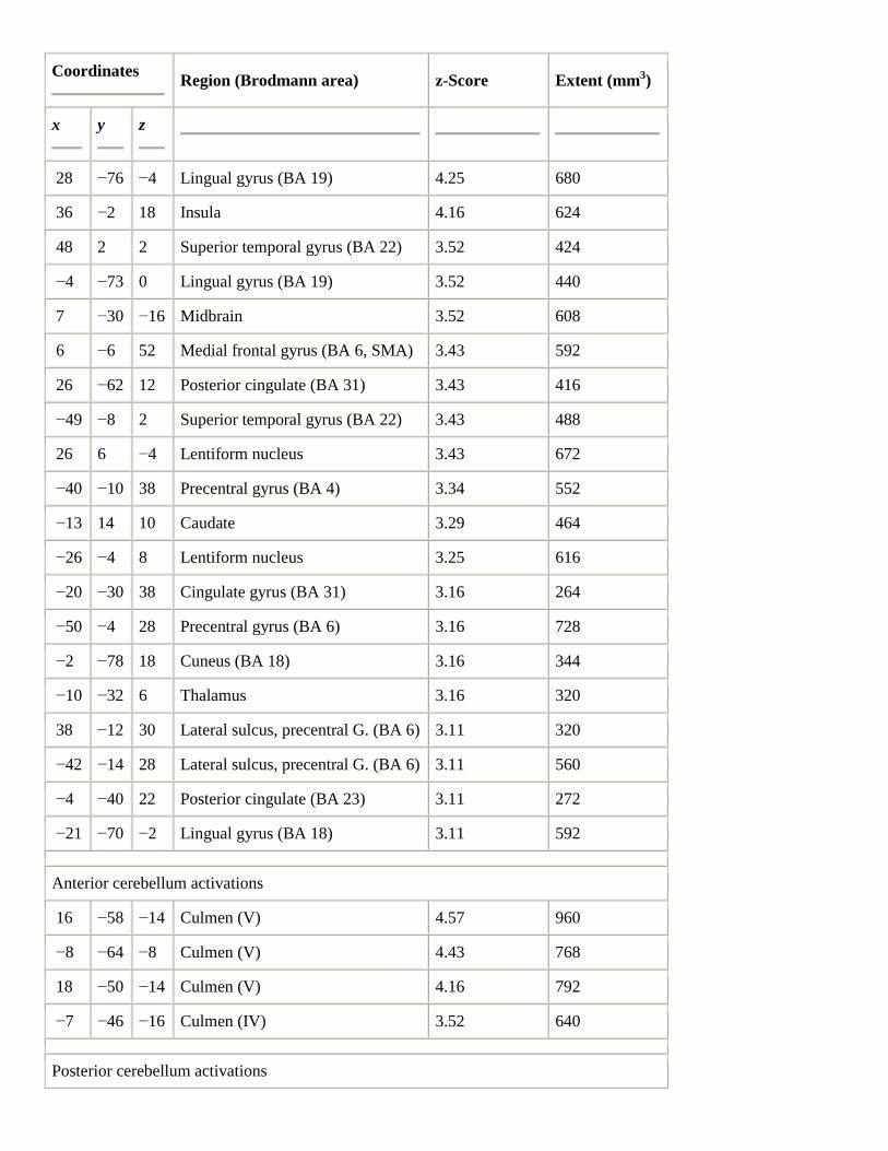
In the direct contrast between the performance of the Bach composition and scales, there were a variety of
activations specific to the Bach (Table 3). There were strong responses in subcortical areas, including bilateral,
predominantly right, thalamus, and bilateral basal ganglia (bilateral lentiform nucleus and left caudate nucleus).
There were activations in somatomotor-related regions, including bilateral dorsolateral premotor cortex (BA 6),
bilateral primary motor cortex (BA 4), right insula, and right SMA. There were strong increases detected in
bilateral superior temporal gyrus (BA 22) and planum polare (BA 38), as well as activity in bilateral lingual
gyrus (BA 19 and 18) and bilateral posterior cingulate (BA 31 and 23). Increases were detected in posterior
cerebellum (declive of vermis (VI) and quadrangular lobule (VI)), as well as in anterior cerebellum (left culmen
(IV)).
Table 3.
Bach–scales
Coordinates
Region (Brodmann area) z-Score Extent (mm3)
x
y
z
Activations
11 −18 16 Thalamus 4.29 656
16 −34 6 Thalamus 4.25 608
Coordinates
Region (Brodmann area) z-Score Extent (mm3)
x
y
z
28 −76 −4 Lingual gyrus (BA 19) 4.25 680
36 −2 18 Insula 4.16 624
48 2 2 Superior temporal gyrus (BA 22) 3.52 424
−4 −73 0 Lingual gyrus (BA 19) 3.52 440
7 −30 −16 Midbrain 3.52 608
6 −6 52 Medial frontal gyrus (BA 6, SMA) 3.43 592
26 −62 12 Posterior cingulate (BA 31) 3.43 416
−49 −8 2 Superior temporal gyrus (BA 22) 3.43 488
26 6 −4 Lentiform nucleus 3.43 672
−40 −10 38 Precentral gyrus (BA 4) 3.34 552
−13 14 10 Caudate 3.29 464
−26 −4 8 Lentiform nucleus 3.25 616
−20 −30 38 Cingulate gyrus (BA 31) 3.16 264
−50 −4 28 Precentral gyrus (BA 6) 3.16 728
−2 −78 18 Cuneus (BA 18) 3.16 344
−10 −32 6 Thalamus 3.16 320
38 −12 30 Lateral sulcus, precentral G. (BA 6) 3.11 320
−42 −14 28 Lateral sulcus, precentral G. (BA 6) 3.11 560
−4 −40 22 Posterior cingulate (BA 23) 3.11 272
−21 −70 −2 Lingual gyrus (BA 18) 3.11 592
Anterior cerebellum activations
16 −58 −14 Culmen (V) 4.57 960
−8 −64 −8 Culmen (V) 4.43 768
18 −50 −14 Culmen (V) 4.16 792
−7 −46 −16 Culmen (IV) 3.52 640
Posterior cerebellum activations

Coordinates
Region (Brodmann area) z-Score Extent (mm3)
x
y
z
−2 −66 −26 Declive (vermis) (VI) 4.25 760
38 −52 −20 Quadrangular lobule (VI) 3.93 576
−9 −73 −16 Quadrangular lobule (VI) 3.66 464
−13 −72 −31 Quadrangular lobule (VI) 3.61 384
16 −84 −16 Quadrangular lobule (VI) 3.48 288
26 −62 −23 Quadrangular lobule (VI) 3.34 464
−22 −75 −18 Quadrangular lobule (VI) 3.25 344
8 −62 −22 Quadrangular lobule (VI) 3.16 576
In the direct contrast between the performance of scales and the concerto, there were numerous increases
specific to scales (Table 4). Strong activations were seen in bilateral anterior cingulate gyrus (BA 31 and 32),
bilateral middle temporal cortex (BA 21), and right superior temporal cortex (BA 41). There were responses in
right frontal regions (BA 9 and 10), right temporoparietal regions (BA 39 and 40), right fusiform gyrus (BA
37), left inferior frontal gyrus (BA 47), right precuneus (BA 7), and right primary motor cortex (BA 4).
Increases were also detected in anterior and posterior cerebellum (culmen (IV) and quadrangular lobule (VI),
respectively).
Table 4.
Scales–Bach
Coordinates
Region (Brodmann area) z-Score Extent (mm3)
x
y
z
Activations
0 −56 30 Cingulate gyrus (BA 31) −4.17 920
−2 −56 28 Cingulate gyrus (BA 31) −4.17 208
−50 −29 −7 Middle temporal gyrus (BA 21) −3.85 648
0 32 34 Cingulate (BA 32) −3.80 608
2 −44 32 Cingulate gyrus (BA 31) −3.53 744

Coordinates
Region (Brodmann area) z-Score Extent (mm3)
x
y
z
57 −16 −10 Middle temporal gyrus (BA 21) −3.53 656
32 14 36 Middle frontal gyrus (BA 9) −3.49 744
4 −48 20 Posterior cingulate (BA 30) −3.49 608
42 −35 16 Superior temporal gyrus (BA 41) −3.44 344
4 −56 38 Precuneus (BA 7) −3.39 680
−48 20 2 Inferior frontal gyrus (BA 47) −3.30 216
46 −56 −12 Fusiform gyrus (BA 37) −3.30 432
−40 −64 26 Middle temporal gyrus (BA 39) −3.26 456
48 −52 30 Supramarginal gyrus (BA 40) −3.17 536
44 −64 30 Middle temporal gyrus (BA 39) −3.17 688
50 −6 32 Precentral gyrus (BA 4) −3.12 648
14 46 22 Medial frontal gyrus (BA 10) −3.12 504
Anterior cerebellum activations
20 −32 −14 Culmen (IV) −4.35 648
Posterior cerebellum activations
35 −63 −24 Quadrangular lobule (VI) −3.49 296
To visualize the increases in the two performances (Fig. 4), each voxel was characterized as responding for the
Bach only, for the scales only, or for both. Areas responding in common for playing Bach and scales included
motor and somatosensory cortices and SMA (Fig. 3 upper left and right), as well as superior and middle anterior
temporal cortex, preferentially on the right (Fig. 4), and anterior cerebellum (Fig. 4, bottom row). Other
activations (not shown) included bilateral inferior parietal cortex (BA 40), bilateral SMA (BA 6), bilateral
motor cingulate (BA 24), right thalamus, bilateral anterior cerebellum (III–V), and bilateral posterior
cerebellum (VI, vermus).
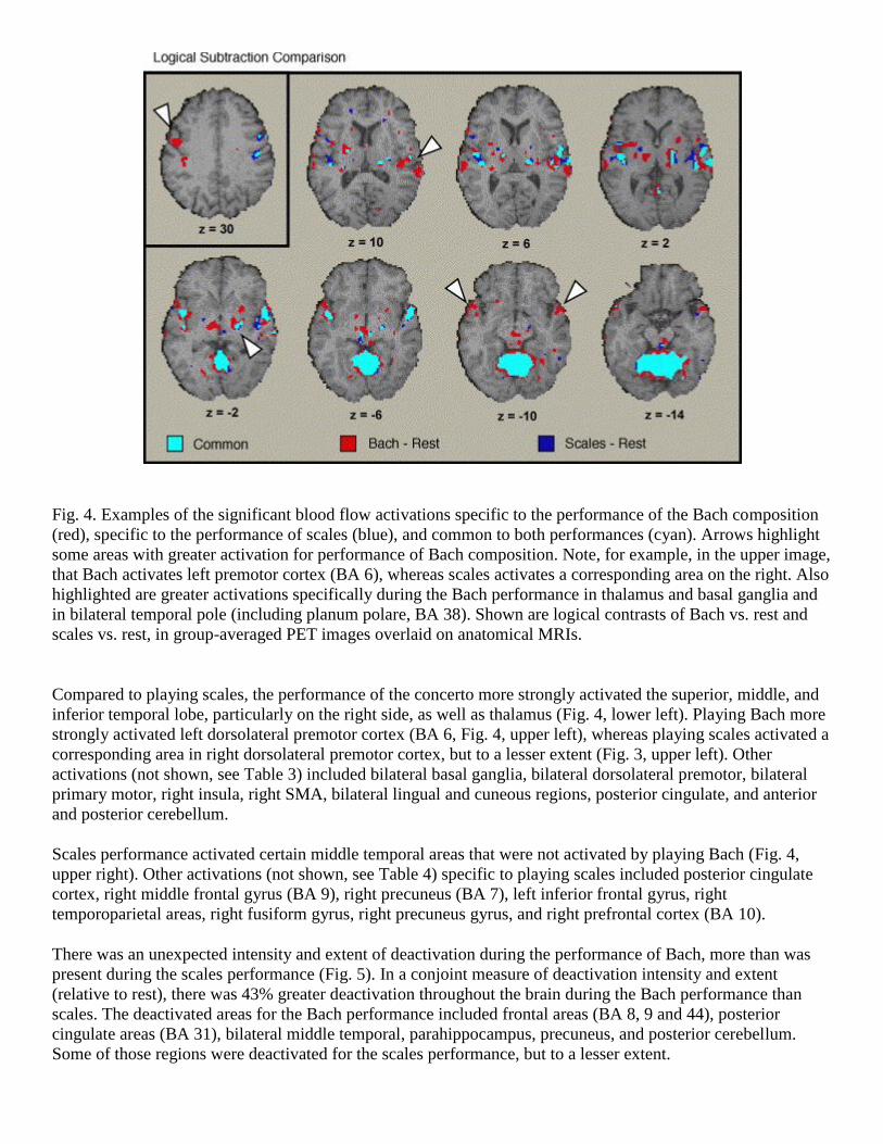
Fig. 4. Examples of the significant blood flow activations specific to the performance of the Bach composition
(red), specific to the performance of scales (blue), and common to both performances (cyan). Arrows highlight
some areas with greater activation for performance of Bach composition. Note, for example, in the upper image,
that Bach activates left premotor cortex (BA 6), whereas scales activates a corresponding area on the right. Also
highlighted are greater activations specifically during the Bach performance in thalamus and basal ganglia and
in bilateral temporal pole (including planum polare, BA 38). Shown are logical contrasts of Bach vs. rest and
scales vs. rest, in group-averaged PET images overlaid on anatomical MRIs.
Compared to playing scales, the performance of the concerto more strongly activated the superior, middle, and
inferior temporal lobe, particularly on the right side, as well as thalamus (Fig. 4, lower left). Playing Bach more
strongly activated left dorsolateral premotor cortex (BA 6, Fig. 4, upper left), whereas playing scales activated a
corresponding area in right dorsolateral premotor cortex, but to a lesser extent (Fig. 3, upper left). Other
activations (not shown, see Table 3) included bilateral basal ganglia, bilateral dorsolateral premotor, bilateral
primary motor, right insula, right SMA, bilateral lingual and cuneous regions, posterior cingulate, and anterior
and posterior cerebellum.
Scales performance activated certain middle temporal areas that were not activated by playing Bach (Fig. 4,
upper right). Other activations (not shown, see Table 4) specific to playing scales included posterior cingulate
cortex, right middle frontal gyrus (BA 9), right precuneus (BA 7), left inferior frontal gyrus, right
temporoparietal areas, right fusiform gyrus, right precuneus gyrus, and right prefrontal cortex (BA 10).
There was an unexpected intensity and extent of deactivation during the performance of Bach, more than was
present during the scales performance (Fig. 5). In a conjoint measure of deactivation intensity and extent
(relative to rest), there was 43% greater deactivation throughout the brain during the Bach performance than
scales. The deactivated areas for the Bach performance included frontal areas (BA 8, 9 and 44), posterior
cingulate areas (BA 31), bilateral middle temporal, parahippocampus, precuneus, and posterior cerebellum.
Some of those regions were deactivated for the scales performance, but to a lesser extent.
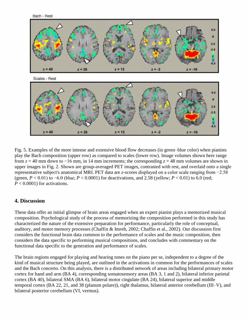
Fig. 5. Examples of the more intense and extensive blood flow decreases (in green–blue color) when pianists
play the Bach composition (upper row) as compared to scales (lower row). Image volumes shown here range
from z = 40 mm down to −16 mm, in 14 mm increments; the corresponding z = 48 mm volumes are shown in
upper images in Fig. 2. Shown are group-averaged PET images, contrasted with rest, and overlaid onto a single
representative subject's anatomical MRI. PET data are z-scores displayed on a color scale ranging from −2.58
(green, P < 0.01) to −6.0 (blue; P < 0.0001) for deactivations, and 2.58 (yellow; P < 0.01) to 6.0 (red;
P < 0.0001) for activations.
4. Discussion
These data offer an initial glimpse of brain areas engaged when an expert pianist plays a memorized musical
composition. Psychological study of the process of memorizing the composition performed in this study has
characterized the nature of the extensive preparation for performance, particularly the role of conceptual,
auditory, and motor memory processes (Chaffin & Imreh, 2002; Chaffin et al., 2002). Our discussion first
considers the functional brain data common to the performance of scales and the music composition, then
considers the data specific to performing musical compositions, and concludes with commentary on the
functional data specific to the generation and performance of scales.
The brain regions engaged for playing and hearing tones on the piano per se, independent to a degree of the
kind of musical structure being played, are outlined in the activations in common for the performances of scales
and the Bach concerto. On this analysis, there is a distributed network of areas including bilateral primary motor
cortex for hand and arm (BA 4), corresponding somatosensory areas (BA 3, 1 and 2), bilateral inferior parietal
cortex (BA 40), bilateral SMA (BA 6), bilateral motor cingulate (BA 24), bilateral superior and middle
temporal cortex (BA 22, 21, and 38 (planum polare)), right thalamus, bilateral anterior cerebellum (III–V), and
bilateral posterior cerebellum (VI, vermus).

Published research suggests pertinent functions that may underlie playing and hearing tones on the piano as
such. Motor sensory areas were engaged by requirements for finger and arm movement in bimanual piano
performance. These regions are known to be involved in the execution of planned motor output (e.g., Carpenter,
Georgopoulos, & Pellizzer, 1999; Graziano, Taylor, Moore, & Cooke, 2002; Zilles et al., 1995). SMA is likely
involved in memory and sequencing of self-generated, internally guided composite movements. Motor cingulate
has recently been observed to be active during bimanual tapping tasks for in-phase, but not for anti-phase or
polyrhythmic, coordination (Ullen, Forssberg, & Ehrsson, 2003), consistent with its activation here for the in-
phase coordination required in these tasks. The thalamus may be involved in linking sensory and motor
parameters (Connors, Landisman, & Reid, 1998; Guillery, 2003), in the present case, for the execution of fine
coordination of keyboard fingering and tone production. The anterior cerebellum is likely supporting sensory
processing associated with arm and finger movements (Bower & Parsons, 2003; Liu et al., 2000; Parsons &
Fox, 1997). The activated posterior cerebellar areas have been implicated in discrimination tasks involving pitch
and melody (Gaab, Gaser, Zaehle, Jancke, & Schlaug, 2003; Griffiths, Johnsrude, Dean, & Green, 1999;
Holcomb et al., 1998; Parsons, 2003). The inferior parietal areas activated in this analysis may support multi-
modal associations (tactile, motor, auditory), which are important to action planning and execution in this
context (Duhamel, Colby, & Goldberg, 1998; Faillenot, Toni, Decety, Gregoire, & Jeannerod, 1997; Milner &
Goodale, 1995; Sakata, 1996).
Also implicated in these components of performance were bilateral superior and middle temporal cortex,
wherein reside areas implicated in various music perception tasks, as described in Section 1 and later. Note that
in the Sergent (1993) study, the peak activation in this region for the condition of passively listening to scales
(minus visual fixation) was −56, −4, 2, which is homologous to the right hemispheric ones here (52, −4, −4, and
56, −10, 4) for playing and hearing scales (i.e., blindfolded and contrasted with eyes closed rest). This suggests
a hemispheric difference depending on the presence of an action–perception cycle like that when playing
bimanually, but not merely listening to, memorized scales.
There were a variety of hemodynamic changes specifically related to the performance of the Bach concerto.
Strong increases were evident in secondary auditory association areas, including bilateral superior and middle
temporal cortex and planum polare. Very active areas appeared as well in bilateral thalamus, bilateral basal
ganglia, and posterior cerebellum. Increases in motor-sensory areas were detected in bilateral dorsolateral
premotor, bilateral primary motor, right insula, right SMA, and anterior cerebellum. There were increases in
bilateral lingual regions and posterior cingulate. Each of these areas has been linked by data and hypotheses to
specific functions that could conceivably play a direct or indirect role in skilled piano playing of memorized
pieces.
The foregoing activated superior and middle temporal areas (BA 22 and 21) have been implicated in a variety of
music perception tasks, including tone and melody processing (Zatorre, 2003). These regions also appear to
process harmony, as when expert musicians sight-read a novel musical score to detect harmonic errors heard in
the performance of the score (Parsons et al., 1998) or when musically experienced listeners track a melody as it
changes keys (Janata et al., 2002). In the performance of the memorized concerto, it is likely that these regions
subserve the auditory anticipations of the recalled composition, as well as recognition of the produced piano
sounds.
The activation of the planum polare for the performance of Bach, but less so for scales, confirms other data
implicating this area in representing complex melody and harmony (Brown et al., in press; Griffiths, Buchel,
Frackowiak, & Patterson, 1998; Koelsch et al., 2002; Samson & Zatorre, 1988; Warrier & Zatorre, 2004;
Zatorre, 1985; Zatorre & Belin, 2001). The concerto performance activated a more extensive region of these
areas than did playing scales, often extending in the dorsal–ventral direction. This role of the planum polare
appears at current levels of resolution to be independent of features of the musical activities such as whether the
music is sung or played on piano (singing and playing music activate overlapping regions), and which of its
musical features one is closely attending to. The planum polare, as well as activated areas in superior and
middle temporal cortex, likely operate in conjunction with ventral and lateral frontal cortical areas implicated
for auditory working memory and monitoring in music tasks (Zatorre, 2003).
The thalamus, also active for music performance, may be involved in linking sensory and motor parameters
(Connors et al., 1998 and Guillery, 2003), and here may fill such a role in the execution of fine keyboard
fingering and precisely timed tone production (with eyes closed). This activation likely reflects the escalating
demands, different in kind and quantity from those for scales (i.e., interpretative, expressive, phrasing features
of performance), and related to the close coordination of kinesthetic, tactile, motor, auditory, and affect
information.
The basal ganglia, also strongly activated, have been associated with selecting and organizing segments of
action, including their timing (Houk, Davis, & Beiser, 1995; Jog, Kubota, Connolly, Hillegaart, & Graybiel,
1999; Kermadi & Joseph, 1995; Redgrave, Prescott, & Gurney, 1999). Their activation for playing the concerto
is probably in response to specific requirements for the selection and organizing of independent sequences and
phrases for each hand and each melodic voice.
The increased activity in right SMA likely reflects increased demand for independent coordination of the left
hand during the Bach concerto, as compared to scales. SMA is often associated with memory and sequencing of
self-generated, internally guided composite movements (Crammond & Kalaska, 1996; Passingham, 1996; Tanji
& Shima, 1994). In playing scales, the non-dominant left hand can be guided by the right hand, but in the
concerto the left hand is expected to achieve an autonomous melodic continuity.
Insula, which was activated on the right in the Bach performance, has been associated with higher levels of
somatic function (Schneider, Friedman, & Mishkin, 1993), and here could be involved in integrating
information from the whole body to support the coordination of bimanual performance. The specific increase
beyond that for scales, may be in response to the greater parity required in autonomy of the two sequences of
manual actions and melodic lines.
There were also increases in dorsal premotor cortex (e.g., seen in two left hemispheric foci in the upper left
image of Fig. 4). This region is often linked to planning, programming, initiation, guidance, and execution of
movements (see review by Passingham, 1993), and its activation here would accord with the escalating need for
motor planning and programming required for increased control and coordination in the Bach concerto.
Musical performance also specifically activated posterior regions of cerebellum in the vicinity of the primary
fissure. Although the weight of the evidence has now shifted in favor of the cerebellum (particularly the
posterior hemispheres) performing non-motor functions, there is no consensus as to exactly how to characterize
those function(s) (Bower & Parsons, 2003; Ivry & Fiez, 2000; Schmahmann et al., 1999 and Vokaer et al.,
2002). Indeed, recent neuroimaging studies of pitch or melody discrimination, as dissociated from motor
coordination or cerebral motor cortical activity, have implicated regions of posterior cerebellum (V and VI)
bilaterally (Gaab et al., 2003, Griffiths et al., 1999 and Parsons, 2003) or on the left (Holcomb et al., 1998).
Activations observed here specifically for the concerto are in these regions, thus suggesting that the activations
are supporting aspects of the perception of melody. Be that as it may, viable alternative candidate functions,
pertinent to musical performance, include the monitoring of errors (Fiez, Petersen, Cheney, & Raichle, 1992),
executive and attentional control (Akshoomoff, Courchesne, & Townsend, 1997; Hallett & Grafman, 1997),
perceptual motor timing and response preparation (Ivry, 1997), control of the acquisition of sensory information
(Bower, 1997; Bower & Parsons, 2003), and internal forward-inverse modelling of sensory-motor-spatial
aspects of the performance (Imamizu, Kuroda, Miyauchi, Yoshioka, & Kawato, 2003). The additional activity
in anterior cerebellum for the concerto may relate to sensory processing associated with bimanual arm and
finger movements (Bower & Parsons, 2003; Liu et al., 2000; Parsons & Fox, 1997) or alternatively to one of the
foregoing candidate cerebellar functions.

Regions of posterior cingulate cortex, also activated specifically for musical performance, have been implicated
in three functions: episodic memory (Grasby et al., 1993; Henson, Rugg, Shallice, Josephs, & Dolan, 1999;
Maddock, Garrett, & Buoncore, 2001), mediating interactions between emotional and memory processes
(Maddock, Garrett, & Buoncore, 2003), and monitoring stimuli in the environment (Raichle et al., 2001; Vogt,
Finch, & Olson, 1992). This region has luxuriant afferent projections from areas associated with emotion and
social information such as subgenual anterior cingulate, orbitofrontal, superior temporal, and dorsolateral
prefrontal cortices (Allison, Puce, & McCarthy, 2000; Carmichael & Price, 1995; Morris, Pandya, & Petrides,
1999; Vogt & Gabriel, 1993). In addition, the functional data implicating posterior cingulate in memory
processing (e.g., Grasby et al., 1993) is corroborated by reciprocal projections with memory structures in medial
temporal cortex and with thalamic nuclei (Bentovoglio, Kultas-Ilinsky, & Ilinsky, 1993; Suzuki & Amaral,
1994). In this context, it is conceivable that the role of these activated regions of posterior cingulate could be in
mediating and monitoring both the use of the memorized music composition and the unfolding contour of its
implied emotive, interpretative, and expressive features.
The activations in cuneus and bilateral lingual cortex, which occurred in spite of pianists being blindfolded,
suggest the use of visual imagery. Comparable activations in visual areas have been observed in other
neuroimaging studies of music experiences, including both imagined and actual performance (Brown et al., in
press and Langheim et al., 2002), as well as imagined and actual listening (Halpern & Zatorre, 1999; Janata et
al., 2002 and Platel et al., 1997; Satoh, Takeda, Nagata, Hatazawa, & Kuruhara, 2001; Zatorre, Evans, &
Meyer, 1994). The activations in visual areas in the present study may result if subjects visualize their own
hands playing piano (Jeannerod, 2004) or visualize notes on stave of the score (Bihan et al., 1993; Klein,
Paradis, Poline, Kosslyn, & LeBihan, 2000).
The pattern of increases for the musical performance here can be compared to those for right-handed piano
performance, singing, and imagined musical performance. As discussed earlier, the Sergent et al. (1992) study
of right handed piano performance (contrasted with sight reading and listening) found activations in left frontal
operculum, left dorsolateral premotor, left parietal cortex, as well as left occipitoparietal and bilateral superior
parietal regions. None of these activated areas, with the exception of the left dorsolateral premotor cortex,
responded during the bimanual performance of a memorized composition. These differences between the two
studies are very likely related to differences in experimental parameters, including contrast controls, bimanual
versus unimanual performance, blindfolded versus visually guided performance, and memorized versus not
memorized composition.
In the three neuroimaging studies of singing described earlier (Brown et al., in press, Perry et al., 1999 and
Riecker et al., 2000), singing-specific activations appeared in primary and secondary auditory cortices, primary
motor cortex, frontal operculum, SMA, insula, posterior cerebellum, and basal ganglia. Playing the piano
composition activated areas in each of these structures, with the exception of the frontal operculum. Note that
frontal operculum was active in the Sergent et al. analysis of playing a sight-read composition, contrasted with
sight-reading and listening. Thus, in combination, these studies indicate that activation in frontal operucular
areas can be elicited for either production modality (singing or piano playing). Moreover, as noted earlier,
musical piano playing, melody singing, and harmonization, all activated overlapping regions of the planum
polare, implicating it as a key area for the high level musical representation.
When musicians imagined playing memorized musical compositions (Langheim et al., 2002), there were
activations, combining over strings and piano performance and contrasting with rest, in right SMA, right
superior premotor cortex, right superior parietal lobule, right frontal operculum, left thalamus, left basal ganglia,
and bilateral posterior cerebellum. Playing the Bach concerto activated regions in each of these structures, with
exception of the frontal operculum and superior parietal cortex.
Thus, apart from sensory-motor, attention, and executive control processes, the three apparently key regions
emerging as important in higher-level aspects of music performance show the following tendencies. The
planum polare (BA 38) is activated (either right or bilaterally) by singing and by playing sight-read or

memorized music on piano, but is not appreciably activated by imagined musical singing or by imagined string
or piano performance of memorized music. The frontal operculum (typically BA 44) is activated (either left,
right, or bilaterally) by music singing and by imagined string and piano performance of memorized music, but
is not detectably engaged by the performance of memorized piano music. The rostromedial prefrontal cortex,
which responds to dissonance and consonance, and to changes in tonality, was not appreciably activated during
the Bach performance.
It is notable that areas subserving emotion and reward that are active during musical listening (Blood & Zatorre,
2001; Blood et al., 1999; Brown, Martinez, & Parsons, 2004; Peretz et al., 2001) were not appreciably active
during the performance of the concerto, which is associated by pianists with vibrant emotional structure
(Chaffin & Imreh, 2002; Chaffin et al., 2002). Indeed, no such activations were reported in other musical
performances, whether imagined or actual, that have been studied with neuroimaging (Brown et al., in press,
Langheim et al., 2002, Meister et al., 2004, Riecker et al., 2000 and Sergent et al., 1992). Additional research is
required to clarify this observation.
Surprisingly, there was a great deal of deactivation detected here, particularly during the performance of the
Bach concerto, which exhibited nearly fifty percent greater deactivation throughout the brain (compared to rest)
than did the scales performance. During performance of the musical composition, deactivation foci were
observed in frontal regions (BA 8, 9, and 44), posterior cingulate areas (BA 31), bilateral middle temporal
cortex, parahippocampus, precuneus, and posterior cerebellum. These deactivations, apparently in areas not
directly relevant to musical performance, may reflect a much deeper focused attention for performing the
musical piece than for playing scales.
The deactivations may be a consequence of inhibition of processes potentially able to distract the musician
during a sustained performance. This possibility could be consistent with the finding discussed earlier that
feedback training to enhance slow-wave EEG improves the quality of musical performances (Egner &
Gruzelier, 2003). Likewise, musicians report anecdotally that they ―lose themselves‖ in absorption during peak
musical performances (Chaffin & Imreh, 2002; Chaffin et al., 2002; Csikszentimihalyi & Csikszentimihalyi,
1988; Mach, 1998).
Some deactivated processes, perhaps those in executive function, cognitive association, and general memory
areas, etc., may be likely to be active during rest, so to some extent their appearance as deactivations in the task
state could be an artifact of a subtraction contrast. However, the possibility that the deactivations per se are
genuine suspensions of activity is consistent with a recent PET study of the resting state that used oxygen
extraction fraction both to begin characterizing brain areas that are active during rest (during an open attentive
state) and to document their deactivation or suspension during specific goal directed behaviors (Raichle et al.,
2001). These researchers identified areas in precuneus, posterior cingulate, and medial prefrontal cortex that
appear to be tonically active, for example, with the precuneus and posterior cingulate continuously collecting
information from the environment that can be evaluated for salience with assistance of areas in medial and
orbital frontal cortices. As shown in Fig. 5, some of these areas were deactivated here, particularly in the
performance of the Bach concerto. While such tonically active circuits may be suspended for any goal-driven
activity, deeply attentive states associated with intense musical performance, among other similarly intense
activities, may involve the suspension of a wider range of brain processes, as suggested by the overall
deactivations seen here.
Briefly then, here is a summary outline of the distributed set of areas and functions implicated by these data
specifically for the performance of memorized musical pieces, beyond those common to performing scales and
the musical piece. The activations in lingual and cuneus areas reflect subjects‘ visualization of their hands as
play piano blindfolded or of the notes on the score. The additional bilateral activations in temporal areas likely
hold the memorized representations of the melodic and harmonic structure of the concerto that subserve the
auditory anticipations of the recalled composition, as well as recognition of the produced piano sounds. The
responsive regions in posterior cingulate may mediate and monitor both the use of the memorized music
composition and the unfolding contour of its implied emotive, interpretative, and expressive features. Strong
thalamic activations may be instrumental in linking sensory and motor parameters for fine fingering and tone
production. Activated regions in basal ganglia are likely subserving the selection and organization of segments
of action, including timing. SMA, in which there is additional activation on the right beyond that for scales, is
probably instrumental in coordinating the independence of the left hand. Dorsolateral premotor cortex is likely
involved in the motor planning and programming for bimanual performance with increased control and
coordination required for the Bach concerto. The additional activations in primary motor areas are involved in
executing the more intricate and subtle plans for finger and sound production. The regions in posterior
cerebellum may support aspects of auditory processing. The anterior cerebellum is likely supporting sensory
processing associated with arm and finger movements. In addition, memorized musical performance is
associated with deactivation of a range of areas whose engagement may detract from a fully realized
performance. This network of areas and functions will need to be confirmed and refined by future studies.
Finally, a new view is also provided here of the brain areas specifically subserving the generation from memory
of musical scales during performance. The representation of information used to generate the sequence of notes
in a scale would appear to be very distinct from that for a musical composition, and should be reflected in the
neural structures activated. The strongest foci were in posterior cingulate cortex, bilateral middle temporal
cortex, right middle frontal gyrus (BA 9), and right precuneus (BA 7). Other activations were in right superior
temporal cortex, left inferior frontal gyrus, right temporoparietal areas, right fusiform gyrus, right precuneus
gyrus, and right prefrontal cortex (BA 10). Note that the Sergent et al. (1992) condition of playing (and hearing)
scales, contrasted with listening to scales, produced increases in left primary motor cortex (BA 4: −35, −26, 54),
left premotor cortex (BA 6: −4, −7, 57), and right (anterior) cerebellum (15, −62, −20). The two frontal
activations were indistinguishable from ones observed here (−34, −22, 54; −6, −4, −50). Moreover, the
cerebellar activation in the Sergent et al. study is reflected in bilateral activations here (culmen (V): −12, −52,
−20; 22, −54, −22) and is likely related to the auditory and somatomotor production of two, rather one, scale
melodies.
This distinctive network of activations suggests the following distributed functions. The cingulate may execute
a controlling influence over initiation of the ordered string of notes in each scale. The bilateral middle temporal
areas may hold auditory representations of scale information (a sequence of tone intervals and possibly a tonal
center). The right middle frontal and left inferior frontal cortical areas may be involved in programming the
sequential ordering of motor execution. The right precuneus, right temporoparietal, and fusiform areas may
represent abstract visual–spatial association amongst notes, staves, and keys. These speculations should be
assessed and refined in further experimentation.
The preceding comparisons of activated networks are delimited by the interplay of various differences in
playing scales and the concerto. First, the performance of the Bach piece requires recalling more complicated
information than does scales. Effects of this variable are probably apparent in the greater activation in auditory
association areas (BA 22 and 21), posterior cingulate, and cuneus and lingual areas. Second, the concerto
performance requires more intricate, controlled fingering and tone production. This factor is likely reflected in
increases in thalamus, basal ganglia, SMA, insula, dorsolateral premotor cortex, and anterior cerebellum. Third,
the concerto performance requires vividly conveying a comprehension of the musical structure (e.g., tonality,
rhythm, dynamics, interpretative features, etc.). Effects of this factor may be observed in auditory areas (BA 38,
22, and 21), posterior cerebellum, posterior cingulate, cuneus, and lingual areas. Fourth, musical performance is
associated with emotional responses, which are likely much more limited when playing scales. However, areas
known to activate during emotional responses to music were not appreciably active here. Nonetheless, it is
possible that activations related to emotional responses were in posterior cingulate, insula, and basal ganglia.
Fifth, the concerto performance demands more attention than scales. This factor may be reflected in different
ways here. There were no increases for the concerto in core attentional areas, but the effects may be more
diffuse because there was more detected activation overall than for scales: nearly twice as many distinct foci
and 72% greater overall extent of activation. Moreover, there was nearly 50% greater deactivation overall for
the concerto performance than scales, an effect that may be related to a deepened focus of attention, as
discussed earlier. It is conceivable then that both the greater activations and greater deactivations are related to
differences in attention in the two performances. Finally, interactions amongst these variables in the present
relatively naturalistic study need to be clarified by future studies aimed at comparing parametrically varied
conditions operationalizing each factor. For example, planned studies contrast performances varying in musical
complexity, while other factors are held relatively constant. These designs are analogous to studies of deduction
(Parsons, Monti, Martinez, & Osherson, 2004) and syntactical processing (Stromswold, Caplan, Alpert, &
Rauch, 1996).
Acknowledgments
We are grateful to Michael Martinez for expert assistance with data analysis and figures, and to an anonymous
reviewer for very helpful suggestions and comments.
References
Aiello, 2001 R. Aiello, Playing piano by heart: From behavior to cognition. In: R.J. Zatorre and I. Peretz,
Editors, The biological foundations of music, Annals of the New York Academy of Science, New York (2001),
pp. 389–393.
Akshoomoff et al., 1997 N.A. Akshoomoff, E. Courchesne and J. Townsend, Attention coordination and
anticipatory control. In: J.D. Schmahmann, Editor, The cerebellum and cognition, Academic Press, New York
(1997), pp. 575–598. Abstract
Allison et al., 2000 T. Allison, A. Puce and G. McCarthy, Social perception from visual cues: Role of the STS
region, Trends in Cognitive Science 4 (2000), pp. 267–278.
Bentovoglio et al., 1993 M. Bentovoglio, K. Kultas-Ilinsky and I. Ilinsky, Limbic thalamus: Structure, intrinsic
organization, and connections. In: B.A. Vogt and M. Gabriel, Editors, Neurobiology of cingulate cortex and
limbic thalamus, Birkhauser, Boston (1993), pp. 71–122.
Bihan et al., 1993 D.L. Bihan, R. Turner, T.A. Zeffiro, C.A. Cuenod, P. Jezzard and V. Bonnerat, Activation of
human primary visual cortex during visual recall: A magnetic resonance imaging study, Proceedings of the
National Academy of Sciences 90 (1993), pp. 11802–11805.
Blood and Zatorre, 2001 A.J. Blood and R.J. Zatorre, Intensely pleasurable responses to music correlate with
activity in brain regions implicated in reward and emotion, Proceedings of the National Academy of Science 98
(2001), pp. 11818–11823.
Blood et al., 1999 A.J. Blood, R.J. Zatorre, P. Bermudes and A. Evans, Emotional responses to pleasant and
unpleasant music correlate with activity in paralimbic brain regions, Nature Neuroscience 2 (1999), pp. 382–
387.
Bower, 1997 J.M. Bower, Control of sensory data acquisition. In: J.D. Schmahmann, Editor, The cerebellum
and cognition, Academic Press, New York (1997), pp. 490–513.
Bower and Parsons, 2003 J.M. Bower and L.M. Parsons, Rethinking the lesser brain, Scientific American 289
(2003), pp. 50–57.
Brown et al., 2004 S. Brown, M.J. Martinez and L.M. Parsons, Passive music listening spontaneously engages
limbic and paralimbic systems, NeuroReport 15 (2004), pp. 2033–2037.
Brown et al., in press Brown, S., Parsons, L. M., Martinez, M. J., Hodges, D., & Fox, P. T. (in press). The song
system of the human brain. Cognitive Brain Research, 20, 363–375.
Cappelletti et al., 2000 M. Cappelletti, H. Waley-Cohen, B. Butterworth and M. Kopelman, A selective loss of
the ability to read and write music, Neurocase 6 (2000), pp. 332–341.
Carmichael and Price, 1995 S.T. Carmichael and J.L. Price, Limbic connections of the orbital and medial
prefrontal cortex in macaque monkeys, Journal of Comparative Neurology 363 (1995), pp. 615–641.
Carpenter et al., 1999 A.F. Carpenter, A.P. Georgopoulos and G. Pellizzer, Motor control encoding of serial
order in a context-recall task, Science 283 (1999), pp. 1752–1757.
Chaffin and Imreh, 2002 R. Chaffin and G. Imreh, Practicing perfection: Piano performance as expert memory,
Psychological Science 13 (2002), pp. 342–349.

Chaffin et al., 2002 R. Chaffin, G. Imreh and M. Crawford, Practicing perfection: Memory and piano
performance, Erlbaum, Mahway, NJ (2002).
Connors et al., 1998 B.W. Connors, C.E. Landisman and R.C. Reid, Thalamus: Organization and function,
Trends in Neurosciences 21 (1998), pp. 539–540.
Crammond and Kalaska, 1996 D.J. Crammond and J.F. Kalaska, Differential relation of discharge in primary
motor cortex and premotor cortex to movement versus actively maintained postures during a reaching task,
Experimental Brain Research 108 (1996), pp. 45–61.
Csikszentimihalyi and Csikszentimihalyi, 1988 M. Csikszentimihalyi and I.S. Csikszentimihalyi, Optimal
experience: Psychological studies of flow in consciousness, Cambridge University Press, New York (1988).
D‘Agostino et al., 1990 R.B. D‘Agostino, A. Belatner and R.B. D‘Agostino Jr., A suggestion for using powerful
and informative tests of normality, American Statistician 44 (1990), pp. 316–321.
Duhamel et al., 1998 J.-R. Duhamel, C.L. Colby and M.E. Goldberg, Ventral intraparietal area of the macaque:
Congruent visual and somatic response properties, Journal of Neurophysiology 79 (1998), pp. 126–136.
Egner and Gruzelier, 2003 T. Egner and J.H. Gruzelier, Ecological validity of neurofeedback: Modulation of
slow wave EEG enhances musical performance, NeuroReport 14 (2003), pp. 1221–1224.
Elbert et al., 1995 T. Elbert, C. Pantev, C. Wienbruch, B. Rockstroh and E. Taub, Increased cortical
representation of the fingers of the left hand in string players, Science 270 (1995), pp. 305–307.
Evers et al., 1999 S. Evers, J. Dannert, D. Rodding, G. Rotter and E.-B. Ringelstein, The cerebral
hemodynamics of music perception: A trancranial Doppler sonography study, Brain 122 (1999), pp. 75–85.
Faillenot et al., 1997 I. Faillenot, I. Toni, J. Decety, M.C. Gregoire and M. Jeannerod, Visual pathways for
object-oriented action and object recognition, functional anatomy with PET, Cerebral Cortex 7 (1997), pp. 77–
85.
Fiez et al., 1992 J.A. Fiez, S.E. Petersen, M.K. Cheney and M.E. Raichle, Impaired nonmotor learning and
error-detection associated with cerebellar damage—A single case study, Brain 115 (1992), pp. 155–178.
Fox and Mintun, 1989 P.T. Fox and M. Mintun, Noninvasive functional brain mapping by change-distribution
analysis of averaged PET images of H215
O tissue activity, Journal of Nuclear Medicine 30 (1989), pp. 141–149.
Fox et al., 1988 P.T. Fox, M. Mintun, E. Reiman and M.E. Raichle, Enhanced detection of focal brain responses
using inter-subject averaging and change-distribution analysis of subtracted PET images, Journal of Cerebral
Blood Flow and Metabolism 8 (1988), pp. 642–653.
Friston et al., 1991 K.J. Friston, C.D. Frith, P.R. Liddle and R.S.J. Frackowiak, Comparing functional (PET)
images: The assessment of significant change, Journal of Cerebral Blood Flow and Metabolism 11 (1991), pp.
690–699.
Gaab et al., 2003 N. Gaab, C. Gaser, T. Zaehle, L. Jancke and G. Schlaug, Functional neuroanatomy of pitch
memory—An fMRI study with sparse temporal sampling, NeuroImage 19 (2003), pp. 1417–1426.
Gaser and Schlaug, 2003 C. Gaser and G. Schlaug, Brain structures differ between musicians and non-
musicians, Journal of Neuroscience 23 (2003), pp. 9240–9245.
Grasby et al., 1993 P.M. Grasby, C.D. Frith, K.J. Friston, C. Bench, R.S.J. Frackowiak and R.J. Dolan,
Functional mapping of brain areas implicated in auditory-verbal memory function, Brain 1 (1993) (16), pp. 1–
20.
Graziano et al., 2002 M.S.A. Graziano, C.S.R. Taylor, T. Moore and D.F. Cooke, The cortical control of
movement revisited, Neuron 36 (2002), pp. 1–20.
Griffiths et al., 1998 T.D. Griffiths, C. Büchel, R.S.J. Frackowiak and R.D. Patterson, Analysis of temporal
structure in sound by the human brain, Nature Neuroscience 1 (1998), pp. 422–427.
Griffiths et al., 1999 T.D. Griffiths, I. Johnsrude, J.L. Dean and G.G.R. Green, A common neural substrate for
the analysis of pitch and duration pattern in segmented sound?, NeuroReport 10 (1999), pp. 3825–3830.
Guillery, 2003 R.W. Guillery, Branching thalamic afferents link action and perception, Journal of
Neurophysiology 90 (2003), pp. 539–548.
Hallett and Grafman, 1997 M. Hallett and J. Grafman, Executive function and motor skill learning. In: J.D.
Schmahmann, Editor, The cerebellum and cognition, Academic Press, New York (1997), pp. 297–323. Abstract
Halpern and Zatorre, 1999 A.R. Halpern and R.J. Zatorre, When that tune runs through your head: A PET
investigation of auditory imagery for familiar melodies, Cerebral Cortex 9 (1999), pp. 697–704.

Haueisen and Knosche, 2001 J. Haueisen and T.R. Knosche, Involuntary motor activity in pianists evoked by
music perception, Journal of Cognitive Neuroscience 13 (2001), pp. 786–792.
Henson et al., 1999 R.N.A. Henson, M.D. Rugg, T. Shallice, O. Josephs and R.J. Dolan, Recollection and
familiarity in recognition memory: An event related functional magnetic resonance imaging study, Journal of
Neuroscience 19 (1999), pp. 3962–3972.
Holcomb et al., 1998 H.H. Holcomb, D.R. Medoff, P.J. Caudill, Z. Zhao, A.C. Lahti and R.F. Dannahs et al.,
Cerebral blood flow relationships associated with a difficult tone recognition task in trained normal volunteers,
Cerebral Cortex 8 (1998), pp. 534–542.
Houk et al., 1995 In: J.C. Houk, J.L. Davis and D.G. Beiser, Editors, Models of information processing in the
basal ganglia, MIT Press, Cambridge (1995).
Hund-Georgiadis and von Cramon, 1999 M. Hund-Georgiadis and D.Y. von Cramon, Motor-learning-related
changes in piano players and non-musicians revealed by functional magnetic-resonance signals, Experimental
Brain Research 125 (1999), pp. 417–425.
Imamizu et al., 2003 H. Imamizu, T. Kuroda, S. Miyauchi, T. Yoshioka and M. Kawato, Modular organization
of internal models of tools in the human cerebellum, Proceedings of the National Academy of Sciences of the
United States of America 100 (2003), pp. 5461–5466.
Ivry, 1997 R. Ivry, Cerebellar timing systems. In: J.D. Schmahmann, Editor, The cerebellum and cognition,
Academic Press, New York (1997), pp. 556–573.
Ivry and Fiez, 2000 R.B. Ivry and J.A. Fiez, Cerebellar contributions to cognition and imagery. In: M.S.
Gazzaniga, Editor, The new cognitive neurosciences (2nd ed.), MIT Press, Cambridge, MA (2000), pp. 999–
1011.
Janata, 2001 P. Janata, Brain electrical activity evoked by mental formation of auditory expectations and
images, Brain Topography 13 (2001), pp. 169–193.
Janata et al., 2002 P. Janata, J.L. Birk, J.D. Van Horn, M. Leman, B. Tillmann and J. Bharucha, The cortical
topography of tonal structures underlying western music, Science 298 (2002), pp. 2167–2170.
Janata and Grafton, 2003 P. Janata and S.T. Grafton, Swinging in the brain: Shared neural substrates for
behaviors related to sequencing and music, Nature Neuroscience 6 (2003), pp. 682–687.
Jäncke et al., 2000 L. Jäncke, N.J. Shah and M. Peters, Cortical activations in primary and secondary motor
areas for complex bimanual movements in professional pianists, Cognitive Brain Research 10 (2000), pp. 177–
183.
Jeannerod, 1994 M. Jeannerod, The representing brain: Neural correlates of motor intention and imagery,
Behavioral and Brain Sciences 17 (1994), pp. 187–245.
Jeannerod, 1997 M. Jeannerod, The cognitive neuroscience of action, Blackwell, Cambridge, MA (1997).
Jeannerod, 2004 M. Jeannerod, Visual and action cues contribute to the self-other distinction, Nature
Neuroscience 5 (2004), pp. 422–423.
Jog et al., 1999 M.S. Jog, Y. Kubota, C.I. Connolly, V. Hillegaart and A.M. Graybiel, Building neural
representations of habits, Science 286 (1999), pp. 1745–1749.
Judd et al., 1983 T. Judd, H. Gardner and N. Geschwind, Alexia without agraphia in a composer, Brain 106
(1983), pp. 435–457.
Kermadi and Joseph, 1995 I. Kermadi and J.P. Joseph, Activity in caudate nucleus of monkey during spatial
sequencing, Journal of Neurophysiology 74 (1995), pp. 911–933.
Klein et al., 2000 I. Klein, A.L. Paradis, J.B. Poline, S.M. Kosslyn and D. LeBihan, Transient activity in the
human calcarine cortex during visual–mental imagery: An event-related fMRI study, Journal of Cognitive
Neuroscience 12 (2000), pp. 15–23.
Koelsch et al., 2002 S. Koelsch, T.C. Gunter, D.Y. von Cramon, S. Zysset, G. Lohman and A.D. Friederici,
Bach speaks: A cortical ―language-network‖ serves the processing of music, NeuroImage 17 (2002), pp. 956–
966.
Krings et al., 2000 T. Krings, R. Tooper, H. Foltys, S. Erberich, R. Sparing and K. Willmes et al., Cortical
activation patterns during complex motor tasks in piano players and control subjects: A functional magnetic
resonance imaging study, Neuroscience Letters 278 (2000), pp. 189–193.
Lancaster et al., 2000 J.L. Lancaster, M.G. Woldorff, L.M. Parsons, M. Liotti, C.S. Freitas and L. Rainey et al.,
Automatic Talairach labels for functional brain mapping, Human Brain Mapping 10 (2000), pp. 120–131.
Langheim et al., 2002 F.J. Langheim, J.H. Callicott, V.S. Mattay, J.H. Duyn and D.R. Weinberger, Cortical
systems associated with covert music rehearsal, NeuroImage 16 (2002), pp. 901–908.
Liu et al., 2000 Y. Liu, Y. Pu, J.-H. Gao, L.M. Parsons, J. Xiong and M. Liotti et al., The human red nucleus
and lateral cerebellum in cooperative roles supporting sensory discrimination, Human Brain Mapping 10
(2000), pp. 147–159.
Mach, 1998 E. Mach, Great contemporary pianists speak for themselves, Dover, New York (1998).
Maddock et al., 2001 R.J. Maddock, A.S. Garrett and M.H. Buonocore, Remembering familiar people: The
posterior cingulate cortex and autobiographical memory retrieval, Neuroscience 104 (2001), pp. 667–676.
Maddock et al., 2003 R.J. Maddock, A.S. Garrett and M.H. Buonocore, Posterior cingulate activation by
emotional words: fMRI evidence from an emotional valence decision task, Human Brain Mapping 18 (2003),
pp. 30–41.
Marin and Perry, 1992 O.S.M. Marin and D.W. Perry, Neurological aspects of musical processing. In: D.
Deutsch, Editor, Psychology of music (2nd ed.), Academic Press, New York (1992), pp. 653–724.
Meister et al., 2004 I.G. Meister, T. Krings, H. Foltys, B. Boroojerdi, M. Muller and R. Topper et al., Playing
piano in the mind—An fMRI study on music imagery and performance in pianists, Cognitive Brain Research
19 (2004), pp. 219–228.
Milner and Goodale, 1995 A.D. Milner and M.A. Goodale, The visual brain in action, Oxford University Press,
Oxford (1995).
Mintun et al., 1989 M. Mintun, P.T. Fox and M.E. Raichle, A highly accurate method of localizing regions of
neuronal activity in the human brain with PET, Journal of Cerebral Blood Flow and Metabolism 9 (1989), pp.
96–103.
Morris et al., 1999 R. Morris, D.N. Pandya and M. Petrides, Fiber system linking the mid-dorsolateral frontal
cortex with the retrosplenial/presubicular region in the rhesus monkey, Journal of Comparative Neurology 407
(1999), pp. 183–192.
Munte et al., 2002 T.F. Munte, E. Altenmuller and L. Jancke, The musician's brain as a model of
neuroplasticity, Nature Reviews: Neuroscience 3 (2002), pp. 473–478.
Nakada et al., 1998 T. Nakada, Y. Fujii, K. Suzuki and I.L. Kwee, ‗Musical brain‘ revealed by high field (3
Tesla) functional MRI, NeuroReport 9 (1998), pp. 3853–3856.
Oldfield, 1971 R. Oldfield, The assessment and analysis of handedness: The Edinburgh inventory,
Neuropsychologia 9 (1971), pp. 97–113.
Pantev et al., 2001 C. Pantev, L.E. Roberts, M. Schultz, A. Egnelien and B. Ross, Timbre-specific enhancement
of auditory cortical representations in musicians, NeuroReport 12 (2001), pp. 169–174.
Parsons, 2003 L.M. Parsons, Exploring the functional neuroanatomy of music performance, perception and
comprehension. In: R.J. Zatorre and I. Peretz, Editors, The cognitive neuroscience of music, Oxford University
Press, Oxford, UK (2003), pp. 247–268.
Parsons and Fox, 1997 L.M. Parsons and P.T. Fox, Sensory and cognitive tasks. In: J.D. Schmahmann, Editor,
The cerebellum and cognition, international review of neurobiology vol. 41, Academic Press, San Diego
(1997), pp. 255–272.
Parsons et al., 1998 L.M. Parsons, D.A. Hodges and P.T. Fox, Neural basis of the comprehension of musical
harmony, melody, and rhythm, Abstracts for Cognitive Neuroscience Society Annual Meeting, Journal of
Cognitive Neuroscience (1998).
Parsons et al., 2004 L.M. Parsons, M. Monti, M.J. Martinez and D.A. Osherson, Brain basis of deduction:
Effects of argument validity and content, Society for Neuroscience Abstracts (2004).
Parsons and Osherson, 2001 L.M. Parsons and D.A. Osherson, New evidence for distinct right and left brain
systems for deductive and probabilistic reasoning, Cerebral Cortex 11 (2001), pp. 954–965.
Parsons and Thaut, 2001 L.M. Parsons and M.H. Thaut, Functional neuroanatomy of the perception of musical
rhythm in musicians and non-musicians., Proceedings of the International Conference on Functional Mapping
of the Human Brain, NeuroImage (2001).
Passingham, 1993 R.E. Passingham, Frontal lobes and voluntary action, Oxford University Press, New York
(1993).
Passingham, 1996 R.E. Passingham, Functional specialization of the supplementary motor area in monkeys and
humans, Advances in Neurology 70 (1996), pp. 105–166.

Peretz et al., 2001 I. Peretz, A.J. Blood, V. Penhune and R. Zatorre, Cortical deafness to dissonance, Brain 124
(2001), pp. 928–940.
Peretz and Coltheart, 2003 I. Peretz and M. Coltheart, Modularity of music processing, Nature Neuroscience 7
(2003), pp. 688–691.
Perry et al., 1999 D.W. Perry, R.J. Zatorre, M. Petrides, B. Alivisatos, E. Meyer and A.C. Evans, Localization
of cerebral activity during simple singing, NeuroReport 10 (1999), pp. 3979–3984.
Platel et al., 1997 H. Platel, C. Price, J.C. Baron, R. Wise, J. Lambert and R.S. Frackowiak et al., The structural
components of music perception, Brain 120 (1997), pp. 243–299.
Raichle et al., 2001 M.E. Raichle, A.M. MacLeod, A.Z. Snyder, W.J. Powers, D.A. Gusnard and G.L. Shulman,
A default mode of brain function, Proceedings of the National Academy of Sciences of the United States of
America 98 (2001), pp. 676–682.
Raichle et al., 1983 M.E. Raichle, M.R.W. Martin, P. Herskovitch, M.A. Mintun and J. Markham, Brain blood
flow measured with intravenous H215
O. II. Implementation and validation, Journal of Nuclear Medicine 24
(1983), pp. 790–798.
Redgrave et al., 1999 P. Redgrave, T.J. Prescott and K. Gurney, The basal ganglia: A vertebrate solution to the
selection problem?, Neuroscience 89 (1999), pp. 1009–1023.
Reiser, 1992 In: D. Reiser, Editor, Auditory imagery, Erlbaum, Mahwah, NJ (1992).
Riecker et al., 2000 A. Riecker, H. Ackermann, D. Wildgruber, G. Dogil and W. Grodd, Opposite hemispheric
lateralization effects during speaking and singing at motor cortex, insula and cerebellum, NeuroReport 11
(2000), pp. 1997–2000.
Sakata, 1996 H. Sakata, Coding 3-D features of objects used in manipulation by parietal neurons. In: R.
Caminiti, K.-P. Hoffmann, F. Lacquaniti and J. Altman, Editors, Vision and movement: Mechanisms in the
cerebral cortex, Human Frontier Science Program, Strasbourg (1996), pp. 55–63.
Samson and Zatorre, 1988 S. Samson and R.J. Zatorre, Melodic and harmonic discrimination following
unilateral cerebral excision, Brain and Cognition 7 (1988), pp. 348–360.
Satoh et al., 2001 M. Satoh, K. Takeda, K. Nagata, J. Hatazawa and S. Kuzuhara, Activated brain regions in
musicians during an ensemble: A PET study, Cognitive Brain Research 12 (2001), pp. 101–108.
Schmahmann et al., 1999 J.D. Schmahmann, J.A. Doyon, D. McDonald, C. Holmes, K. Lavoie and A. Hurwitz
et al., Three-dimensional MRI atlas of the human cerebellum in proportional stereotaxic space, NeuroImage 10
(1999), pp. 233–260.
Schneider et al., 2002 P. Schneider, M. Scherg, H.G. Dosch, H.J. Specht, A. Gutschalk and A. Rupp,
Morphology of Heschel's gyrus reflects enhanced activation in the auditory cortex of musicians, Nature
Neuroscience 5 (2002), pp. 688–694.
Schneider et al., 1993 R.J. Schneider, D.P. Friedman and M. Mishkin, A modality-specific somato-sensory area
within the insula of the rhesus monkey, Brain Research 621 (1993), pp. 116–120.
Schon et al., 2002 D. Schon, J.L. Anton, M. Roth and M. Besson, An fMRI study of music reading,
NeuroReport 13 (2002), pp. 2285–2289.
Schurmann et al., 2002 M. Schurmann, T. Raij, N. Fujiki and R. Hari, Mind's ear in a musician: Where and
when in the brain, NeuroImage 16 (2002), pp. 434–440.
Sergent, 1993 J. Sergent, Mapping the musician brain, Human Brain Mapping 1 (1993), pp. 20–38.
Sergent et al., 1992 J. Sergent, S. Zuck, Terriah and B. McDonald, Distributed neural network underlying
musical sight-reading and keyboard performance, Science 257 (1992), pp. 106–109.
Stewart et al., 2003 L. Stewart, R. Henson, K. Kampe, V. Walsh, R. Turner and U. Frith, Brain changes after
learning to read and play music, NeuroImage 20 (2003), pp. 71–83.
Stewart and Walsh, 2001 L. Stewart and V. Walsh, Music of the hemispheres, Current Biology 11 (2001), pp.
R125–R127.
Stromswold et al., 1996 K. Stromswold, D. Caplan, N. Alpert and S. Rauch, Localization of syntactic
comprehension by positron emission tomography, Brain and Language 52 (1996), pp. 452–473.
Strother et al., 1997 S.C. Strother, N. Lang, J.R. Anderson, K.A. Schaper, K. Rehm and L.K. Hansen et al.,
Activation pattern reproducibility: Measuring the effects of group size and data analysis models, Human Brain
Mapping 5 (1997), pp. 312–316.
Suzuki and Amaral, 1994 W.A. Suzuki and D.G. Amaral, Perirhinal and parahippocampal cortices of the
macaque monkey: Cortical afferents, Journal of Comparative Neurology 350 (1994), pp. 497–533.
Talairach and Tournoux, 1988 Talairach, J., & Tournoux, P. (1988). Co-planar stereotaxic atlas of the human
brain. 3-Dimensional proportional system: an approach to cerebral imaging. New York: Thieme.
Tanji and Shima, 1994 J. Tanji and K. Shima, Role for supplementary motor area cells in planning several
movements ahead, Nature 371 (1994), pp. 413–416.
Ullen et al., 2003 F. Ullen, H. Forssberg and H.H. Ehrsson, Neural networks for the coordination of the hands in
time, Journal of Neurophysiology 89 (2003), pp. 1126–1135.
Vogt et al., 1992 B.A. Vogt, D.M. Finch and C.R. Olson, Functional heterogeneity in cingulate cortex: The
anterior executive and posterior evaluative regions, Cerebral Cortex 2 (1992), pp. 435–443.
Vogt and Gabriel, 1993 In: B.A. Vogt and M. Gabriel, Editors, Neurobiology of cingulate cortex and limbic
thalamus, Birkhauser, Boston (1993).
Vokaer et al., 2002 M. Vokaer, J.C. Bier, S. Elincx, T. Claes, P. Paquier and S. Goldman et al., The cerebellum
may be directly involved in cognitive functions, Neurology 58 (2002), pp. 967–970.
Warrier and Zatorre, 2004 C.M. Warrier and R.J. Zatorre, Right temporal cortex is critical for utilization of
melodic contextual cues in a pitch constancy task, Brain 127 (2004), pp. 1616–1625.
Zatorre, 1985 R.J. Zatorre, Discrimination and recognition of tonal melodies after unilateral cerebral excisions,
Neuropsychologia 23 (1985), pp. 31–41.
Zatorre, 2003 R.J. Zatorre, Neural specialization for tonal processing. In: I. Peretz and R. Zatorre, Editors, The
cognitive neuroscience of music, Oxford University Press, Oxford (2003), pp. 231–246.
Zatorre and Belin, 2001 R.J. Zatorre and P. Belin, Spectral and temporal processing in human auditory cortex,
Cerebral Cortex 11 (2001), pp. 946–953.
Zatorre et al., 1994 R.J. Zatorre, A.C. Evans and E. Meyer, Neural mechanisms underlying melodic perception
and memory for pitch, Journal of Neuroscience 14 (1994), pp. 1908–1919.
Zilles et al., 1995 K. Zilles, G. Schlaug, M. Matelli, G. Luppino, A. Schleider and M. Qu et al., Mapping of the
human and macaque sensorimotor areas by integrating architectonic, transmitter receptor, MRI, and PET data,
Journal of Anatomy 187 (1995), pp. 515–537.