By Laurie Dickson Cardiac Rhythm Disorders. Electrical System Each beat that is generated from the...
94
By Laurie Dickson Cardiac Rhythm Disorders
-
Upload
damian-benson -
Category
Documents
-
view
214 -
download
0
Transcript of By Laurie Dickson Cardiac Rhythm Disorders. Electrical System Each beat that is generated from the...

By Laurie Dickson
Cardiac Rhythm Disorders

Electrical SystemEach beat that is generated from the same
pacemaker will look identicalImpulses from other cardiac cells are called
ectopic (PVC, PAC)This electrical activity produces mechanical
activity that is seen as waveforms.The ECG is the electrical activity of the heart.
Electrical precedes mechanicalWithout electricity, we have no pump!!

Action Potentials
Na K pumpCalcium channelsDepolarizationRepolarization
ECG waveforms are produced by the movement of charged ions across the semipermeable membranes of myocardial cells.


Normal Cardaic Cycle

Yellow is the isoelectric phase.
The purple is the "P"wave.
The purple and yellow split is the "PR" interval.
The red is the "Q" wave.
The light blue is the "R" wave.
The light green is the "S" wave.
The black is the "ST" segment.
The orange is the "T" wave.
Yellow again is isoelectric.
The dark blue is the "U" wave (seldom seen)..

Characteristics of Cardiac CellsCardiac cells are either contractile cells
influencing the pumping action or pacemaker cells influencing the electrical activity of the heart
AutomaticityExcitabilityConductivityContractility
Refractoriness- Refractory periodAbsolute/ Relative/ Full

Refractory Period

Pacemakers other than SA node
A pacemaker from another site can lead to dysrhythmias and may be discharged in a number of ways.
Secondary pacemakers may originate from the AV node or His-Purkinje system.
Secondary pacemakers can originate when they discharge more rapidly than the normal pacemaker of the SA node.
Triggered beats (early or late) may come from an ectopic focus (area outside the normal conduction pathway) in the atria, AV node, or ventricles.

Conduction systemSA node 60-100AV node 40-60Bundle of HisLeft and Right Bundle BranchPurkinge Fibers 20-40

Nervous System Control of the Heart Parasympathetic nervous system: Vagus
nerve Decreases rate Slows impulse conductionDecreases force of contraction
Sympathetic nervous systemIncreases rateIncreases force of contraction
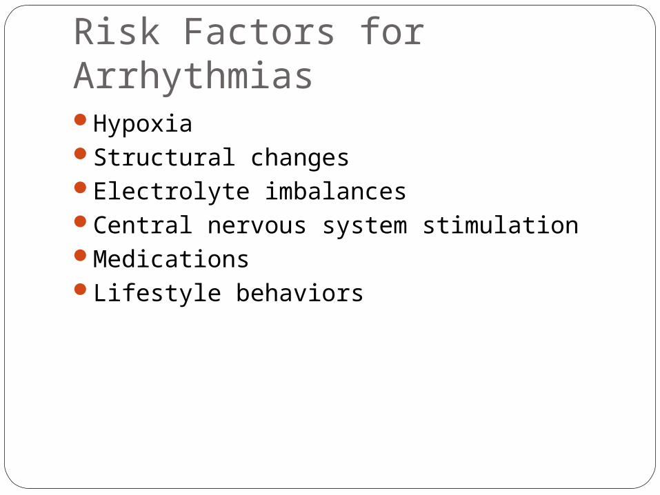
Risk Factors for ArrhythmiasHypoxiaStructural changesElectrolyte imbalancesCentral nervous system stimulationMedicationsLifestyle behaviors

ECG waveforms
P wave = Atrial depolarization (stimulation) QRS = Ventricular depolarization
(stimulation) T wave = Ventricular repolarization
(recovery) Atrial recovery wave hidden under QRS wave Stimulus causes atria to contract before
ventricles Delay in spread of stimulus to ventricles allows
time for ventricles to fill and for atrial kick

ECG Monitoring based on 12 lead ECG
Each lead has positive, negative and ground electrode.
Each lead looks at a different area of the heart.
This can be diagnostic in the case of an MIECG leads

Leads to monitorBest- lead II and MCL or V1 leads- lead II
easy to see P waves. MCL or V1 easy to see ventricular rhythms.
If impulse goes toward positive electrode complex is positively deflected or upright
If impulse goes away from positive electrode complex is negatively deflected or goes down form baseline
ECG leads

Lead II positive R arm looking to LL neg
3 lead placement: Depolarization wave
moving toward a positive lead will be upright.
Depolarization wave moving toward a negative lead will inverted.
Depolarization wave moving between negative and positive leads will have both upright and inverted components.

Lead II R arm looking to LL positive
Five lead placement allows viewing all leads within limits of monitor
Grass under clouds, smoke above fire
V1 is 2nd ICS right of sternum

ECG graph paperHorizontal measures timeVertical measures voltageHelps us determine rateWidth of complexesDuration of complexes

ECG graph paper

AssessmentCalculate rate
Big blockLittle blockNumber of R waves in 6 sec times 10
Calculate rhythm-reg or irregMeasure PR interval, <.20QRS interval .04-.12P to QRS relationship

1 lg box= .20 5 lg boxes =1 sec 30 lg boxes =6 secs
Therefore there are 300 lg boxes in 1 min.
Rate Calculation

Sinus Rhythm
Normal P wave- 0.06-0.12 secPR interval – 0.12-0.20QRS- 0.04-0.12T wave for every complex- 0.16Rate is regular 60-100

Sinus Tachycardia
Rate >100: Sinus TachycardiaCauses-anxiety, hypoxia, shock, pain,
caffeine, drugsTreatment-eliminate cause
Clinical significanceDizziness and hypotension due to
decreased COIncreased myocardial oxygen
consumption may lead to angina


Rate<60: Sinus Bradycardia- relative-symptomatic, absolute-normalCause-vagal stimulation, athlete, drugs
(Blockers and digoxin), head injuries, MIWatch for syncope
brady heart song

Sinus Bradycardia
Clinical significance-Dependent on symptoms
Hypotension , Weakness Pale, cool skinAngina, Shortness of breath Dizziness or syncopeConfusion or disorientation
Treatment- if symptomatic,
o atropine or pace maker

Sinus Arrhythmia (SA)
Rate 60-100Irregular rhythm- increases with inspiration,
decreases with expirationP, QRS,T wave normalCause- children, drugs(MS04), MITreatment- none

Sinus ArrestSee pausesMay see ectopic beats(PAC’s PVC’s) do not
treatCause MITreatment
atropinePacemaker

Medications used to treat atrial rhythms
diltiazem (Cardizem)digoxin (Lanoxin) amiodarone (Cordarone)dofetilide (Tikosyn)verapamil (Calan, Calan SR, Covera-HS,
Isoptin SR, Verelan, Verelan PM, Isoptin, Isoptin I.V.)
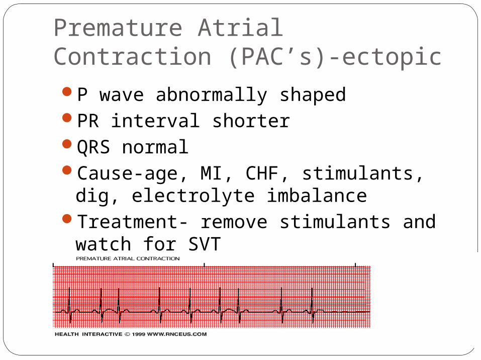
Premature Atrial Contraction (PAC’s)-ectopicP wave abnormally shapedPR interval shorterQRS normalCause-age, MI, CHF, stimulants, dig,
electrolyte imbalanceTreatment- remove stimulants and
watch for SVT

Paroxysmal Supraventricular Tachycardia (PSVT)Rate is 100-300, regular, p often hiddenEctopic foci in atrium above bundle of HISCause-SNS stimulation, MI, CHF,sepsis

Paroxysmal Supraventricular Tachycardia (PSVT)
Clinical significance -Prolonged episode and HR >180 bpm may precipitate ↓ CO
Palpitations, Hypotension, Dyspnea, Angina
Treatment- Vagal stimulation * adenosine, B blockers, Calcium channel
blockers, digoxin, amiodarone. Cardioversion

Atrial FlutterRate of atria is 250-300, vent rate variesRegular rhythmP waves saw tooth, one ectopi focusAV block in ratio 2:1, 3:1, 4:1Flutter waves- No PR intervalCause-diseased heart, drugs (digoxin)
3:1 flutter

Atrial FlutterClinical significance
High ventricular rates (>100) + loss of the atrial “kick” can decrease CO, precipitate HF, angina
Risk for stroke due to risk of thrombus formation in the atria
Treatment- Calcium channel blockers, Beta blockersamiodarone, CardioversionAblationwarfarin (Coumadin)

Atrial Fibrillation-most commonRate of atria 350-600- (disorganized
rhythm)Ventricular response irregularNo P waves, “garbage baseline”PR cannot measureQRS- normal
Cause-#1 arrhythmia in elderly, heart disease- CAD, rheumatic, CHF, alcohol

Atrial Fibrillation
Clinical significance Can result in decrease in CO due to ineffective
atrial contractions (loss of atrial kick) and rapid ventricular response
Thrombi may form in the atria as a result of blood stasis, travel to the brain, causing a stroke
Complications- dec. CO and thrombi stroke risk increases x5

Atrial Fibrillation-most common
Treatment- digoxin, Ca channel
blockers, Beta blockers
amiodorone, procainamaide (Pronestyl)
Cardioversion – warfarin + TEE
Ablation, Maze

AV Conduction Blocks
Arrhythmias of AV Node

First Degree AV BlockTransmission through AV node delayedPR interval >.20QRS normal and regular Cause- digoxin toxicity, MI, CAD, vagal, and
blocker drugs

First-Degree AV Block
Clinical significanceUsually asymptomaticMay be a precursor to higher degrees of AV block
Treatment Check medications Continue to monitor

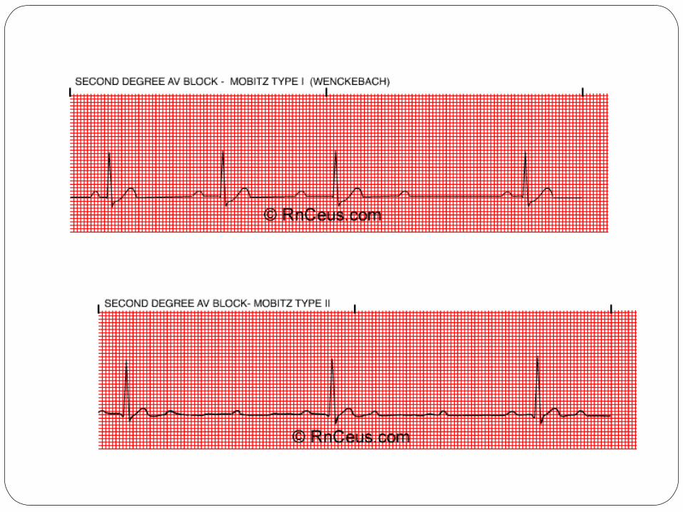
Second Degree AV Blockmore P’s than QRS’s
A. Mobitz I (Wenckebach) PR progressively longer then drops QRSCause- MI, drug toxicity
B. MobitzII More P’s but skips QRS in regular pattern
2:1,3:1, 4:1(QRS usually greater than .12-BBB)Constant PR interval- can be normal or
prolongedOccurs in HIS bundle with bundle branch block

Second-Degree AV Block, Type 1 (Mobitz I, Wenckebach)
Clinical significanceUsually a result of myocardial ischemia or
infarctionAlmost always transient and well toleratedMay be a warning signal of a more serious AV
conduction disturbance
Treatment- watch for type II and 3rd degreeIf symptomatic- atropine, pacer
Diagnosis Wenckebach

Second-Degree AV Block, Type 2 (Mobitz II)Clinical significance
Often progresses to third-degree AV block and is associated with a poor prognosis
Reduced HR often results in decreased CO with subsequent hypotension and myocardial ischemia
Treatment- pacemaker

3rd Degree AV BlockAtria and ventricles beat independently
Atrial rate- 60-100Slow ventricular rate 20-40P normalNo PR interval- no relationship with QRSWide or normal QRS (depends on where
block is)
Cause- severe heart disease, blockers, elderly, MI
Complications- dec. CO, ischemia, HF, shock, and syncope

Third-Degree AV Heart Block (Complete Heart Block)
Clinical significanceDecreased CO with subsequent ischemia,
HF, and shockSyncope may result from severe
bradycardia or even periods of asystole
Treatment- atropine, pacemaker

Bundle Branch Blocks
Left BBBRight BBBQRS.12 or greaterRabbit ears- RR’No change in rhythm

Right Bundle Branch Block

Junctional RhythmAV node is pacemaker- slow rhythm (40-60)
but very regular impulse goes to atria from AV node- backward
P wave patternsAbsentP wave precedes QRS inverted in II, III, and
AVFP wave hidden in QRSP wave follows QRS

Junctional RhythmPR interval
Absent or hiddenShort <.12Negative or RP interval
QRS normalNo treatment

Easy to recognize
Ventricular ArrythmiasMost serious

Premature Ventricular Contractions (PVC’s)-ectopic
No P wavesQRS wide and bizarreT opposite deflection of PVCCause- 90% with MI, stimulants, digoxin,
electrolyte imbalance

Premature Ventricular Contractions
Clinical significanceIn normal heart, usually benignIn heart disease, PVCs may decrease CO and
precipitate angina and HF Patient’s response to PVCs must be monitored
PVCs often do not generate a sufficient ventricular contraction to result in a peripheral pulse
Apical-radial pulse rate should be assessed to determine if pulse deficit exists

Premature Ventricular Contractions
Clinical significanceRepresents ventricular irritabilityMay occur:
After lysis of a coronary artery clot with thrombolytic therapy in acute MI—reperfusion dysrhythmias
Following plaque reduction after percutaneous coronary intervention
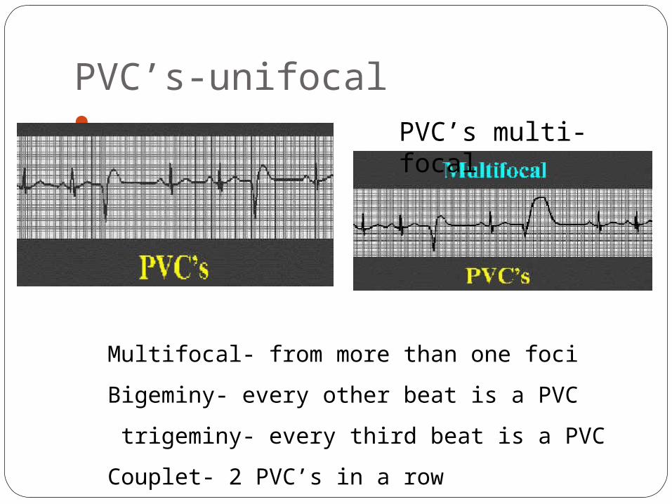
PVC’s-unifocal
Multifocal- from more than one foci
Bigeminy- every other beat is a PVC
trigeminy- every third beat is a PVC
Couplet- 2 PVC’s in a row
PVC’s multi-focal

Treat if:>5 PVC’s a minuteRuns of PVC’sMulti focal PVC’sR on T
Treatment- based on causeO2, lidocaine,

Ventricular Tachycardia (VT)Ventricular rate 150-250, regular or
irregularNo P wavesQRS>.12
Can be stable- pulse or unstable –no pulseCause- electrolyte imbalance, MI, CAD,
digoxinLife- threatening, decreased CO, watch for
V-fib
Ventricular Tachycardia Clinical significance
VT can be stable (patient has a pulse) or unstable (patient is pulseless)Sustained VT: Severe decrease in CO
HypotensionPulmonary edemaDecreased cerebral blood flowCardiopulmonary arrest

Ventricular Tachycardia
Clinical significanceTreatment for VT must be rapidMay recur if prophylactic treatment is not
initiatedVentricular fibrillation may develop
Treatment- same as for PVC’s and defibrillate for sustained

VT- Torsades de Pointes
French for twisting of the points

Ventricular FibrillationGarbage baseline-quiveringNo P’s No QRS’s No COCause-MI, CAD, CMP, shock, K+, hypoxia,
acidosis, and drugsTreatment- code situation, ACLS, CPR,
**defibrillate

Diagnostic Tests
Telemetry- 5 lead( lead II and V1)12 lead EKGHolter monitor- pt. keeps a diary Event monitoring- pt. records only when
having the event Exercise stress testElectrophysiology studies- induce
arrhythmias under controlled situation

Nursing AssessmentApical rate and rhythmApical/radial deficitBlood pressureSkinUrine outputSigns of decreased cardiac output

Nursing DiagnosesDecreased cardiac outputDecreased tissue perfusionActivity intoleranceAnxiety and FearKnowledge deficit

GoalsMaintain stable signs of effective cardiac
output and tissue perfusionAchieve a realistic program of activity
that balances physical activity with energy conserving activities
Report decreased anxiety and increased sense of self-control
Describe risk factors, the disease process, and treatment regimen

Medications
Classified by effect on action potential
Class I- fast Na blocking agents-ventricularquinidine, procainamide, lidocaine, disopyramide phosphate (Norpace),
propafenone (Rhythmol)
Class II- beta blockers SVT,Afib,flutter esmolol, atenolol (Tenormin),
propranolol(Inderal)

Medications
Class III- K blocking both atrial and ventricular amiodarone, dofetilide, sotalol
Class IV- Ca, channel blockers SVT,Afib,flutter verapamil, diltiazem
Other- adenosine, digoxin, atropine,
magnesium

AntiarrhythmicsRemembering that of all anti-arrhythmics "some block potassium channels" can help
you:
Class I "Some" = Sodium Class II "Block" = Beta blockers Class III "Potassium" = Potassium channel
blockers Class IV "Channels" = Calcium channel
blockers

Comfort Measures
RestO2Relieve fear and anxiety-
diazapam (Valium)

Invasive proceduresDefibrillation
Emergency- start at 200 watt/sec, go to 400Safety precautions
Synchronized Cardioversion- for vent. or SVTCan be planned- if stableGet permitStart at 50 watt/secAwake, give O2 and sedationHave to synchronize with rhythmcardioversion

My journey started July 13th 2008. Went to doctor thinking I had bronchitis. 2 days later went in because I got awoken during the night not being able to breath. Dr thought I had gone into pneumonia, gave chest xray,18th go back tells me I have congestive heart failure, starts me on water pills and something else has me scheduled for an echo on Monday, wait 2 days calls and wants me to come in on Friday and wants a
cardiologist to see me and the echo, go in tells me to go to a hospital north of us saying they have a room ready and will schedule a cath and the cardiologist can reveiw the echo. get up there doc reviews echo, while nurses are hooking me up with IVs, Dr comes in and says may have major heart damage but will wait until cath on Monday. Monday comes have cath a surgeon comes in with cardiologist telling us I have over half my heart damaged may need transplant, cardiologist says they would rather transport me to a major hospital that can handle transplant surgery if something goes wrong with bypass. EF is 15%. go to Indianapolis by ambulance,
Journal of Patient Needing Heart Transplant

I am in total shock by this point not being able to even comprehend what is going on 2 weeks from going from bronchitis or so I thought to maybe having heart transplant. My wife god bless her is having her own stress out of her mind over this. get to Indy Tues and Wed nuclear test, Friday high risk bypass surgery. Now its 6 weeks after surgery have had another echo EF went up a whopping 5% now getting defibbed Tuesday, today is Sunday and again my mind is wondering into the worst scenarios, it is getting harder and harder to grasp this stuff. hopefully sites like this will help, letting blow off steam, and learning.Dave

ICD Implanted defibrillator Device
Senses rate and width of QRSGoes off 3 times, then have to be resetCombined with pacemaker- overdrive pacing
or backup pacing
Indications for ICDSurvived SCDSpontaneous sustained V TachSyncope with V Tach/V FibHigh risk for life threatening dysrhythmia
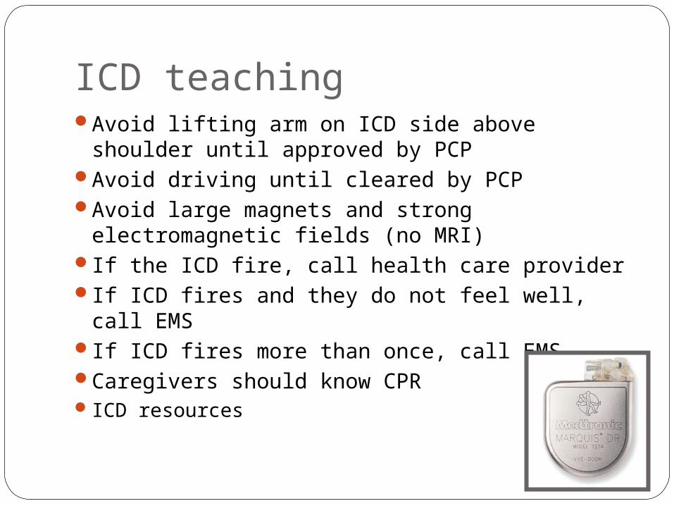
ICD teachingAvoid lifting arm on ICD side above shoulder
until approved by PCPAvoid driving until cleared by PCPAvoid large magnets and strong
electromagnetic fields (no MRI)If the ICD fire, call health care providerIf ICD fires and they do not feel well, call EMSIf ICD fires more than once, call EMSCaregivers should know CPRICD resources
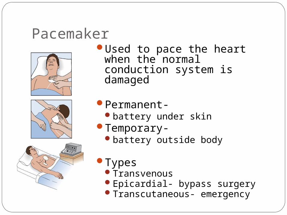
PacemakerUsed to pace the heart when
the normal conduction system is damaged
Permanent- battery under skin
Temporary- battery outside body
TypesTransvenousEpicardial- bypass surgeryTranscutaneous- emergency

PacemakerModes
Asynchronous- at preset time without failSynchronous or demand- when HR goes
below set rateClassifications

Indications for pacemakerAV blockA-Fib with slow ventricular responseBundle Branch BlockCardiomyopathy Heart failureSA node dysfunctionTachydysrhythmias (V Tach)Teaching-
Similar to ICDDaily Pulse

Pacemaker Problems:
Failure to sense
Failure to capture

AblationDone in special cardiac procedures labUse a laser to burn abnormal pathwayradiofrequency ablation

ECG Changes Associated with Acute Coronary Syndrome (ACS)
Ischemia ST segment depression and/or T wave inversion ST segment depression is significant if it is
at least 1 mm (one small box) below the isoelectric line

ECG Changes Associated with Acute Coronary Syndrome (ACS)
Copyright © 2007, 2004, 2000, Mosby, Inc., an affiliate of Elsevier Inc. All Rights Reserved.
Fig. 36-29 A

ECG Changes Associated with Acute Coronary Syndrome (ACS)
Injury/InfarctionST segment elevation is significant if >1 mm
above the isoelectric line If treatment is prompt and effective, may
avoid infarction If serum cardiac markers are present, an ST-
segment-elevation myocardial infarction (STEMI) has occurred

ECG Changes Associated with Acute Coronary Syndrome (ACS)
Injury/InfarctionNote: physiologic Q wave is the first
negative deflection following the P waveSmall and narrow (<0.04 second in duration)
Pathologic Q wave is deep and >0.03 second in duration

EKG changes in an acute MI

ECG Changes Associated with Acute Coronary Syndrome (ACS)
Copyright © 2007, 2004, 2000, Mosby, Inc., an affiliate of Elsevier Inc. All Rights Reserved.
Fig. 36-29 B

ECG Changes Associated with Acute Coronary Syndrome (ACS)
Copyright © 2007, 2004, 2000, Mosby, Inc., an affiliate of Elsevier Inc. All Rights Reserved.
Fig. 36-30

ECG changes with ACSThe 12-lead ECG is the primary diagnostic tool used
to evaluate patients presenting with ACS.There are definitive ECG changes that occur in
response to ischemia, injury, or infarction of myocardial cells and will be seen in the leads that face the area of involvement.
Typical ECG changes seen in myocardial ischemia include ST-segment depression and/or T wave inversion.
The typical ECG change seen during myocardial injury is ST-segment elevation.
An ST-segment elevation and a pathologic Q wave may be seen on the ECG with myocardial infarction
SyncopeBrief lapse in consciousness accompanied
by a loss of tone (fainting)Causes
CardiovascularVasovagal, Cardiac dysrhythmias,
hypertrophic cardiomyopathy , PENoncardiovascular
hypoglycemia, seizure, hysteria, TIA

Syncope
Diagnostic studiesEchocardiographyEPSHead-upright tilt table testing Holter monitor Subcutaneously implanted loop recording
device
1-year mortality rate as high as 30% for syncope from cardiovascular cause

Complications of ArrhythmiasHypotensionTissue ischemiaThrombi- low dose heparin, or ASAHeart failureShockDeath

Prioritization QuestionA client with atrial fibrillation is
ambulating in the hall on the coronary step-down unit and suddenly tells you, “I feel really dizzy.” which action should you take first?
A. Help the client sit down.B. Check the client’s apical pulseC. Take the client’s blood pressureD. Have the client breathe deeply

Prioritization questionA diagnosis of ventricular fibrillation is
identified for an unresponsive 50 year old client who has just arrived in the ED. Which action should be taken first?
A. Defibrillate at 200 joulesB. Begin CPRC. Administer epinephrine 1 mg IVD.Intubate and manually ventilate.

Prioritization question
Cardiac rhythms are being observed for clients in the CCU. Which client will need immediate intervention? A client:
A. admitted with heart failure who has atrial fibrillation with a rate of 88 while at rest.
B. with a newly implanted demand ventricular pacemaker, who has occasional periods of sinus rhythm, rate 90-100.
C. who has just arrived on the unit with an acute MI and has sinus rhythm, rate 76, with frequent PVC’s.
D. who recently started taking atenolol (Tenormin)) and has a first-degree heart block rate 58.

Video Acting Out Rhythmsmad german doctor dances to heart
rhythmsPractice-http://www.skillstat.com/Flash/
ECG_Sim_2004.htmlCasestudiesQuizzesDiscussionQuestions