


By 4-Hydroxynonenal · 2002-10-16 · 2 SUMMARY 4-Hydroxynonenal (4-HNE) is a cytotoxic...
51
Covalent Modification of Epithelial Fatty Acid Binding Protein By 4-Hydroxynonenal In vitro and In vivo: Evidence for a Role in Antioxidant Biology Assumpta Bennaars-Eiden 1 , LeeAnn Higgins 1 , Ann V. Hertzel 1 , Rebecca J. Kapphahn 2 , Deborah A. Ferrington 2 , and David A. Bernlohr 1* From the Departments of 1 Biochemistry, Molecular Biology and Biophysics, and 2 Ophthalmology, The University of Minnesota, Minneapolis, MN 55455 Running Title: Covalent Modification of E-FABP by 4-HNE * Author to whom correspondence should be addressed: Dr. David A. Bernlohr Department of Biochemistry, Molecular Biology and Biophysics The University of Minnesota 321 Church St. SE Minneapolis, MN 55455 T: 612-624-2712 [email protected] Copyright 2002 by The American Society for Biochemistry and Molecular Biology, Inc. JBC Papers in Press. Published on October 16, 2002 as Manuscript M209493200 by guest on May 18, 2020 http://www.jbc.org/ Downloaded from
Transcript of By 4-Hydroxynonenal · 2002-10-16 · 2 SUMMARY 4-Hydroxynonenal (4-HNE) is a cytotoxic...

Covalent Modification of Epithelial Fatty Acid Binding Protein
By 4-Hydroxynonenal In vitro and In vivo:
Evidence for a Role in Antioxidant Biology
Assumpta Bennaars-Eiden1, LeeAnn Higgins1, Ann V. Hertzel1, Rebecca J. Kapphahn2,
Deborah A. Ferrington2, and David A. Bernlohr1*
From the Departments of 1Biochemistry, Molecular Biology and Biophysics, and
2Ophthalmology, The University of Minnesota, Minneapolis, MN 55455
Running Title: Covalent Modification of E-FABP by 4-HNE
*Author to whom correspondence should be addressed: Dr. David A. Bernlohr Department of Biochemistry, Molecular Biology and Biophysics The University of Minnesota 321 Church St. SE Minneapolis, MN 55455 T: 612-624-2712 [email protected]
Copyright 2002 by The American Society for Biochemistry and Molecular Biology, Inc.
JBC Papers in Press. Published on October 16, 2002 as Manuscript M209493200 by guest on M
ay 18, 2020http://w
ww
.jbc.org/D
ownloaded from

2
SUMMARY
4-Hydroxynonenal (4-HNE) is a cytotoxic α,β-unsaturated acyl aldehyde that is naturally
produced from lipid peroxidation and cleavage in response to oxidative stress and
aging. Such reactive lipids covalently modify cellular target proteins, thereby affecting
biological structure and function. Herein we report the identification of the epithelial fatty
acid binding protein (E-FABP) as a molecular target for 4-HNE modification both in vitro
and in vivo. 4-HNE covalently modified (t1/2 < 60 seconds) E-FABP in vitro as revealed
by a combination of MALDI-TOF MS and immunochemical reactivity using antibodies
directed to 4-HNE-protein conjugates. Identification of C120 as the major site of
modification was determined through tandem mass spectral sequencing of tryptic
peptides as well as analysis of E-FABP mutants C120A, C127A, and C120A C127A.
The in-vitro modification of C120 by 4-HNE was relatively insensitive to pH (6.4 to 8.4),
and temperature (4° C to 37° C) but was markedly potentiated by non-covalently bound
fatty acids. 4-HNE-modified E-FABP was more stable than unmodified E-FABP to
chemical denaturation by guanidine hydrochloride, as assessed by changes in intrinsic
tryptophan fluorescence. Analysis of soluble protein extracts from rat retina with
antibodies directed to 4-HNE-protein conjugates revealed immunoreactivity with a 15
kDa protein that was identified by ESI and MALDI-TOF MS as E-FABP. Evaluation of
retinal pigment epithelial cell extracts derived from E-FABP null mice by two-
dimensional gel electrophoresis using anti-4-HNE antibodies revealed increased
by guest on May 18, 2020
http://ww
w.jbc.org/
Dow
nloaded from

3
modification in the null cells relative to those from wild type cells. These results indicate
that E-FABP is a molecular target for 4-HNE modification and the hypothesis that E-
FABP functions as an antioxidant protein by scavenging reactive lipids through covalent
modification of C120.
INTRODUCTION:
The mammalian intracellular lipid binding proteins (LBP) are expressed from a large
multigene family and encode ~15kDa proteins found dispersed within the cytoplasm and
nucleus (1). Due to their high affinity for hydrophobic molecules, they play a variety of
roles in intracellular fatty acid, retinoid, bile acid and sterol trafficking (2,3). Each protein
encoded by an LBP gene folds into a conserved structure consisting of 10 anti-parallel β
strands that form a large interior water-filled cavity that serves as the hydrophobic
ligand-binding site. Despite a wide variance in amino acid identity between family
members (20-70%), x-ray crystallographic and high-field NMR data demonstrates that
all LBP forms exhibit the same β-barrel fold and that their α-carbon backbones are
virtually superimposable (4,5). The epithelial fatty acid binding protein (E-FABP) is
unique amongst the LBPs due to the presence of six cysteine residues, two of which,
C120 and C127, are modeled to form a disulfide bond within the ligand binding cavity
(6,7). Importantly, the side chains of C120 and C127 lie within 4.5 Å of the C2-C4
methylene region of a bound fatty acid (8). The regulation of epithelial FABP gene
expression is unique for it is upregulated in a variety of cell types in response to
experimental challenges linked to oxidative stress and the generation of reactive oxygen
by guest on May 18, 2020
http://ww
w.jbc.org/
Dow
nloaded from

4
species. These include chemical, molecular (9) or viral transformation, disease states
associated with inflammation (psoriasis) (10,11), and physical or chemical damage
(nerve crush, kainic acid release) (12-14).
Reactive oxygen species can be produced exogenously or from a variety of intracellular
processes collectively linked to the generation of superoxide anions, hydroxyl radicals
and hydrogen peroxide (15). Such reactive oxidants chemically modify a variety of
biological molecules, including polyunsaturated acyl chains of membrane phospholipids
generating a family of lipid hydroperoxides (LOOH). In addition, enzymatic oxidation of
membrane phospholipids through the lipoxygenase enzyme systems also generates
bioactive lipid hydroperoxides. The combination of chemical and enzymatic LOOH
generating systems provide substrates for the Hock cleavage, thereby generating a
variety of α,β-unsaturated acyl aldehydes, the most well-studied being 4-
hydroxynonenal (4-HNE) (16,17). 4-HNE is a relatively stable, long-lived, diffusible lipid
that is generally considered to be cytotoxic due to its ability to covalently modify a
variety of biomolecules via Michael addition reactions across the double bond and Schiff
base formation at the carbonyl (18,19). 4-HNE-modified DNA is associated with defects
in replication (20,21), interference with cell cycle regulation (22), and the induction of
apoptosis (23,24). On proteins, 4-HNE reacts with a variety of amino acid side chains
through amine and/or sulfhydryl modification, often affecting the biological structure and
activity of target proteins (17,18). Since reactive oxygen species are linked to oxidative
stress and aging (25-27), the identification of cellular targets for 4-HNE modification has
been a subject of intense investigation. Moreover, analysis of cellular mechanisms
by guest on May 18, 2020
http://ww
w.jbc.org/
Dow
nloaded from

5
linked to the antioxidant response may provide mechanistic clues as to the regulation of
macromolecule turnover.
Given the lipid binding properties of E-FABP, its up-regulation in response to conditions
linked to oxidative stress, and structural positioning of redox-sensitive thiols within the
binding cavity near the bound lipid, we have pursued the hypothesis that the protein is
an endogenous target for 4-HNE modification. Herein we report that cysteine 120 is
covalently modified by 4-HNE in vitro as well as the properties of such modification.
Moreover, the protein is modified with 4-HNE in vivo and retinal epithelial cell lines
derived from E-FABP null mice exhibit an upregulation of 4-HNE modified proteins.
These results indicate that E-FABP is a molecular target for 4-HNE modification and the
hypothesis that the protein serves as an antioxidant by scavenging reactive lipids from
the cellular environment.
EXPERIMENTAL PROCEDURES:
Materials-4-HNE was purchased from Cayman Chemical Company, Ann Arbor, MI.
Polyclonal antibody against 4-HNE- protein adducts was obtained from Alpha
Diagnostics, San Antonio, TX. 1-Anilinonapthalene-8-sulfonate (1,8-ANS) was
purchased from Biomol, Inc. Modified sequence grade trypsin was obtained from
Promega. α-Cyano-4-hydroxycinnamic acid (4-HCCA) and dihydroxybenzoic (DHB) in
methanol were purchased from Agilent Technologies, Palo Alto, CA. Bio-Spin columns
loaded with Bio-Gel P-6 were obtained from Bio-Rad, Richmond, CA. Enhanced
by guest on May 18, 2020
http://ww
w.jbc.org/
Dow
nloaded from

6
chemiluminescence reagents were purchased from Amersham Pharmacia Biotech, Inc.,
Piscatawy, NJ.
Preparation of E. coli derived E-FABP- Bacterially expressed E-FABP was purified to
homogeneity as previously described (28) through a combination of acid fractionation
and gel filtration chromatography. Homogeneous protein eluting from the Sephadex G-
75 column was concentrated and dialyzed extensively against the standard buffer, 10
mM potassium phosphate, 150 mM NaCl (pH 7.4). The protein concentration was
determined using the bicinchoninic assay (Pierce Chemical Co). Using the standard
purification protocol, the pH 5.0 acidification step facilitates endogenous fatty acid
release and the subsequent gel filtration separates any lipid from protein. To evaluate
preparations of E-FABP for the presence of fatty acids, the protein sample was
extracted with chloroform/methanol (2:1, v/v) after the addition of C15:0 as an internal
standard. Extracted fatty acids were converted to methyl esters using 14% boron
trifluoride in methanol and subjected to gas chromatography analysis using a HP 5890
gas chromatograph equipped with a flame ionization detector and integrator. Native E-
FABP purified with less than 0.05 mol FA/mol protein. As such, the standard
purification yields protein essentially devoid of bound fatty acids and is considered apo
protein.
Construction of E-FABP Cysteine mutants-To prepare mutants of E-FABP with various
cysteine to alanine substitutions, the murine E-FABP cDNA was PCR amplified as a
BamH1 / EcoR1 fragment and cloned into the pRSET plasmid resulting in the
by guest on May 18, 2020
http://ww
w.jbc.org/
Dow
nloaded from

7
expression vector pRSET-6His-E-FABP. After induction of expression in E.coli, a
recombinant protein with an amino terminus containing a six histidine tract, a flexible
linker region and a protease cleavage site amino to the E-FABP coding region is
produced. The resulting protein has an amino terminus of:
MRGSHHHHHHGMASMTGGNNMGRDLYDDDDKSRWGSM where the first methionine
listed is the initiating residue and the last methionine listed initiates the E-FABP coding
region. To generate the C120A, C127A and C120A C127A mutants of E-FABP,
oligonucleotide-directed mutagenesis was performed (29) within the pRSET-6His-E-
FABP backbone. All expressed constructs were verified by DNA sequencing. The
cDNA constructs expressing the 6His-tagged proteins were transformed into BL21(DE3)
pLysS cells and expression was induced by the addition of 0.01mM isopropyl-β-D-
thiogalactopyranoside to the growth medium for 3 hr. Cultures were harvested by
centrifugation at 8000 rpm for 15 min at 4° C. The cell pellets were resuspended in ice
cold standard buffer containing a protease inhibitor cocktail of aprotinin, pepstatin A and
leupeptin, sonicated and centrifuged at 100,000 x g to generate a soluble protein
extract. The soluble protein extract containing various E-FABP forms was used for 4-
HNE modification reactions. Because the cysteine to alanine mutations have no effect
on fatty acid binding activity, wild type and E-FABP mutants associate with a variety of
endogenous E. coli fatty acids, the most prevalent being palmitic and oleic acid. On
average, recombinant FABPs expressed in bacteria have between 0.4 and 0.6 mol FA /
mol protein. As such, protein used in crude extracts or purified without acidification is
considered largely holo protein.
by guest on May 18, 2020
http://ww
w.jbc.org/
Dow
nloaded from

8
In vitro modification of E-FABP by 4-HNE - Purified E-FABP (10 µM) in 10 mM
potassium phosphate, 150 mM NaCl pH 7.4 (standard buffer) was incubated with 4-
HNE at 22° C for various times. Aliquots were removed, and immediately applied to a
P-6 Bio-Spin column and centrifuged for 30 seconds to separate modified protein
eluting in the void volume from any free unreacted 4-HNE remaining on the column.
Protein eluting from the Bio-Spin column was analyzed by a combination of MALDI-
TOF, electrospray MS including tandem mass spectrometry, or by immunochemical
methods. To evaluate the influence of non-covalently bound fatty acids on 4-HNE
modification, E-FABP (10 µM) was incubated with oleic acid (10 and 25 fold molar ratio
to E-FABP) and equilibrated for two minutes to saturate the protein (30) prior to addition
of 4-HNE then treated identically.
For immunochemical detection of 4-HNE bound to protein, the samples were subjected
to SDS-PAGE and transferred to nitrocellulose membranes. The membrane was
blocked in phosphate buffered saline containing 0.05% Tween and 5% non-fat dry milk
and incubated with rabbit polyclonal antibody directed towards 4-HNE protein adducts
(1:1000 dilution) at 4 °C for 16 hrs. The membranes were then washed in the same
buffer and incubated with HRP-conjugated goat anti-rabbit secondary antibody
(1:25,000 dilution) for one hour at room temperature. The immunoreactivity was
detected with enhanced chemiluminescence according to the manufacturer’s
instructions (31). For some experiments, goat anti-rabbit alkaline phosphatase-
conjugated secondary antibody (1:3000 dilution) was used in conjunction with the
substrate BCIP-NBT to visualize the immunoreaction.
by guest on May 18, 2020
http://ww
w.jbc.org/
Dow
nloaded from

9
Guanidine Hydrochloride induced protein denaturation- GdnHCL induced protein
unfolding was performed as previously described (28). Briefly, E-FABP (0.5 µM) in a
potassium phosphate buffer pH 7.4 was mixed with increasing concentrations of
guanidine hydrochloride from 0-3 M and the intrinsic tryptophan fluorescence measured
using a Spex Fluoromax II fluorometer. Tryptophan fluorescence was excited at 285
nm and denaturation was monitored by the red-shifting of the fluorescence emission
maximum from 336 nm to 356 nm.
Mapping the site of covalent modification of E-FABP by 4-HNE- 4-HNE modified protein
prepared as described from the P-6 column was dialyzed into 100 mM ammonium
bicarbonate buffer (pH 8) containing 6 M guanidine hydrochloride, 10 mM EDTA at 4 ° C
to denature the protein. The protein was incubated with 10 mM dithithreitol (DTT) for 1
hour at 56 °C and subsequently cooled to room temperature. To modify the reduced
thiol groups, iodoacetic acid was added to a final concentration of 20 mM and incubated
for 30 min at 25 °C in the dark. For rat retinal proteins, iodoacetamide was used for
cystein alkylations. The protein was then dialyzed exhaustively against 100 mM
ammonium bicarbonate (pH 8.0). After dialysis, 0.8 nmol of protein was digested with
0.016 nmol trypsin in 50 mM ammonium bicarbonate buffer pH 8.0 containing 1 mM
CaCl2 at 37 °C for 16 hr. The reaction was stopped by the addition of glacial acetic acid
and samples stored at –80 °C prior to mass spectral analysis. For some analyses, 4-
HNE-modified E-FABP was subjected to SDS-polyacrylamide gel electrophoresis and
silver stained to identify the samples of interest. The stained protein bands were
by guest on May 18, 2020
http://ww
w.jbc.org/
Dow
nloaded from

10
excised and processed for trypsin in-gel digestion as described (32). Prior to protease
digestion, the cysteine residues were reduced and alkylated using iodoacetic acid.
Peptides were extracted by sequential extraction with 50% ammonium bicarbonate
buffer and 45% acetonitrile/5% formic acid. The peptides were then evaporated to near
dryness and stored at –80° C prior to mass spectral analysis.
Matrix Assisted Laser Desorption Ionization Time of Flight Mass Spectrometry- (MALDI-
TOF)- Prior to MALDI-TOF analysis, a portion of the peptide mixture was desalted using
Millipore C18 ZipTips using the manufacturer’s protocol. Full scans from 500 – 3500
m/z of the tryptic peptide mixtures and intact protein data from 4000 – 22000 m/z were
collected on a Brüker Biflex III MALDI-TOF mass spectrometer equipped with a N2-laser
(337 nm, 3 nanosecond pulse length) and a microchannel plate (MCP) detector. The
peptide data was collected in the reflectron mode, positive polarity, with an accelerating
potential of 19 kV using α-cyano-4-hydroxycinnamic acid (4-HCCA) diluted 1:1 (v/v) with
50:50, acetonitrile: nanopure water, 0.1% trifluoroacetic acid (TFA). Whole protein
mass spectra were collected in linear mode using sinapinic acid (3,5-dimethoxy-4-
hydroxycinnamic acid) as the matrix with an accelerating potential of 19 kV. Each
spectrum was the accumulation of ~200 laser shots. External calibration for peptide
analysis was performed using human angiotensin II (monoisotopic [MH+] 1046.5 m/z;
Sigma) and adrenocorticotropin hormone (ACTH) fragment 18-39 (monoisotopic [MH+]
2465.2 m/z; Sigma); external calibration for intact protein analysis was performed with
myoglobin (average [MH+] 16952 m/z) and cytochrome C (average [MH+] 12361 m/z.
by guest on May 18, 2020
http://ww
w.jbc.org/
Dow
nloaded from
11
Full scan and tandem mass spectral data of select ions of the peptide mixture were
collected on a QSTAR Pulsar quadrupole time-of-flight mass spectrometer (Applied
Biosystems Inc., Foster City, California) with a MALDI source using dihydroxybenzoic
(DHB) as the matrix. The TOF region acceleration voltage was 4 kV and the injection
pulse repetition rate was 6.0 kHz. Laser pulses were generated with a nitrogen laser at
337 nm, 33 microJoules of laser energy using a laser repetition rate of 20 Hz. Mass
spectra were the average of approximately 50 laser shots collected in positive mode
from 500 – 3500 m/z. External calibration was performed with the same standards used
on the Biflex, described above.
Electrospray Mass Spectral Analysis: Ion Trap- The tryptic peptides in a portion of the
sample were separated on a 75 µm internal diameter C18 capillary column with a
ThermoFinnigan (San Jose, CA) LCQ Classic ion trap mass spectrometer equipped
with New Objective’s nanoelectrospray source (33). Tandem MS data were searched
against a subset protein database containing Mus mus entries using Sequest™,
ThermoFinnigan’s peptide MS/MS data-interpretation software (34). The variable
modification HNE (delta mass 156 if a Michael addition occurs on a cysteine, histidine
or lysine residue and 138 if a Schiff base reaction occurs on lysine residue (19) was
entered into the appropriate Sequest parameter field.
Electrospray Mass Spectral Analysis: quadrupole TOF. An aliquot of the tryptic
peptides was desalted using Poros R2 (ABI) in a glass purification capillary (Protana,
Denmark). Briefly, the peptide mixture was loaded onto the R2, washed three times with
by guest on May 18, 2020
http://ww
w.jbc.org/
Dow
nloaded from

12
7 µL of 95:5, H2O:ACN, 0.5% formic acid and eluted with 1.5 µL of 30:70, H2O:ACN,
0.5% formic acid into a coated nanoelectrospray capillary (Protana). Tandem mass
spectra were collected in an information dependent acquisition (IDA) scan mode during
nanospray infusion of peptides at approximately 5 nL/min flow rate with a spray voltage
of 1000 volts and TOF parameters as described above. Tandem mass spectra were
searched using BioAnalyst (ABI) and Mascot (www.matrixscience.com).
Isolation of rat retinal extracts and murine retinal pigment epithelial cell lines-
Experiments were performed on retinas obtained from 10 month old Fischer 344 Brown
Norway F1 hybrid male rats. Retinal isolates were prepared by homogenizing the
retinas from two rats in one mL of homogenization buffer (20% sucrose, 2 mM MgCl2 ,
10 mM glucose, 20 mM Tris-Acetate, pH 7.2, and 0.05% NP40) and centrifuged for 15
minutes at 100 x g. The supernatant was collected and the pellet re-homogenized in
one additional mL of homogenization buffer. The supernatants were combined and
centrifuged at 600 x g for 15 minutes. The supernatant containing soluble retina
proteins was retained and aliquots were stored at –80 ºC.
To prepare retinal pigment epithelial (RPE) cell lines, wild type C57Bl/6J mice or
animals harboring a targeted disruption of the FABP5 gene encoding E-FABP were
anesthetized with 5 mg ketamine and 1 mg xylazine per mouse and sacrificed with a
lethal injection of Euthasol (200µL). Epithelial cells from the retina were harvested and
cultured as described (35) with minor modifications. Briefly, the eyes were enucleated
and washed 2 times with Ca+2 , Mg+2-free phosphate buffered saline (CMF-PBS) and
by guest on May 18, 2020
http://ww
w.jbc.org/
Dow
nloaded from

13
placed in a digest solution containing 105 U/mL collagenase and 50 U/mL
hyaluronidase in CMF-PBS for 30 minutes at 37 °C. The globes were washed 2 times
with CMF-PBS and digested a second time with 0.05% (w/v) trypsin containing 0.5 mM
EDTA for 15 minutes at 37° C. Trypsin was inactivated by washing the globes with
Dulbecco’s Modified Eagle’s Medium containing 10% Fetal Bovine Serum (DMEM-10),
2 mM L-glutamine, 100 U/mL Penicillin, 100 ug/mL Streptomycin. Using a dissecting
microscope, the cornea and lens were aseptically excised from the eyes and the retina
with the layer of RPE cells still attached was gently lifted away from the choroid. The
connection between the retina and RPE was disrupted by incubating the tissue at 37° C
in trypsin/EDTA until the RPE cells dissociated from the retina. After removing the
retina, the remaining supernatant containing RPE cells was washed 2 times with
DMEM-10 and incubated as described above with the additions of 0.05% dimethyl
sulfoxide (DMSO) and 50 ng/mL mouse epidermal growth factor at 37° C. The RPE
cells were immortalized approximately 5 days after harvest with the papilloma virus E6a
and E7 genes via a retroviral vector produced in PA317 packaging cells.
Two-dimensional gel electrophoresis--To separate rat retinal or murine RPE proteins,
two-dimensional gel electrophoresis consisting of first dimension isoelectric focusing
(IEF) and second dimension SDS-PAGE was utilized. 100 µg of soluble protein was
organically precipitated according to the method of Wessel and Flügge (36), dried under
nitrogen, and rehydrated into an Immobilized pH Gradient (IPG) strip (pH 3-11) using
250 µL of 10 mM Tris pH 7.5, 2% Triton-X100, 8 M Urea, and 10 mM DTT for 14 hours.
Following hydration, samples were focused at 250V for 15 minutes at which time the
by guest on May 18, 2020
http://ww
w.jbc.org/
Dow
nloaded from

14
voltage was linearly increased to 8000V over 2.5 hours and held to reach a total of
35,000 V•hours. For the second dimension, the focused strips were equilibrated for 10
minutes in 6M urea, 2% SDS, 375 mM Tris-HCl (pH 8.8), 20% glycerol, 100 mM DTT,
rinsed and incubated for 10 minutes in 6 M Urea, 2% SDS, 375 mM Tris-HCl (pH 8.8),
20% glycerol, 135 mM iodoacetamide. The equilibrated strips were embedded in 0.5%
(w/v) agarose on the top of 10% acrylamide gel and second dimension SDS-PAGE was
performed (37). Proteins in gels were either stained (32) or transferred to PVDF
membranes for immunochemical analysis.
RESULTS:
High resolution NMR structures of E-FABP with a bound fatty acid have revealed that
the C2-C4 methylene region of the acyl chain is adjacent to the C120-C127 disulfide
bond (8). In addition, E-FABP is upregulated in response to a variety of stimuli
correlated with oxidative damage. Since oxidative damage is often mechanistically
linked to the generation of reactive oxygen species and lipid peroxidation with
subsequent production of reactive α,β-unsaturated medium chain acyl aldehydes, we
hypothesized that E-FABP may bind such lipids, even though their chain length is
generally considered too short to be bound by FABPs. Moreover, given the proximity of
the C120-C127 disulfide to the putative position of the bound unsaturated aldehyde, we
conjectured that if such binding were to occur, a Michael addition reaction may proceed
producing a covalent protein-lipid conjugate. To that end, we carried out preliminary
experiments by incubating 4-HNE, as a model α,β-unsaturated acyl aldehyde, and
by guest on May 18, 2020
http://ww
w.jbc.org/
Dow
nloaded from

15
homogeneous E-FABP and analyzed the product following separation using P-6 Bio-
Spin column chromatography by MALDI-TOF mass spectrometry. The calculated
molecular mass of murine E-FABP lacking the initiating methionine is 15,006 Da.
Figure 1 shows that three major mass forms of E-FABP were detected. An ion at m/z of
15,001 + 5 corresponds to the native protein and an ion observed at m/z of 15,155 + 5
is consistent with the calculated mass for E-FABP plus one 4-HNE molecule covalently
bound. The third ion with a m/z of 15,206 + 5 represents a sinapinic acid adduct of E-
FABP normally seen with high-resolution mass spectrometry. The indicated errors in
m/z value reflect the expected mass accuracy of the Biflex MALDI-TOF. Control
experiments carried out by incubating E-FABP with long chain fatty acids (oleic acid) did
not generate higher molecular mass forms suggesting that the 15,155 product
represented a covalent lipid-protein conjugate that survives mass spectral analysis as
opposed to non-covalently bound lipid.
As an alternate method to assess lipid-protein conjugate formation independently from
mass spectrometry, we employed immunochemical identification using antibodies
directed to 4-HNE protein conjugates (31). As shown in Figure 2, incubation of E-FABP
with either 4-HNE or with oleic acid followed by SDS-PAGE and immunoblotting shows
that the anti-HNE antibody is reactive only with the samples containing 4-HNE and not
with fatty acids, consistent with a covalent lipid-protein conjugate being formed.
To determine the time course of 4-HNE modification of E-FABP, the protein was
incubated with ten fold molar excess of the lipid at 22° C and at various times aliquots
by guest on May 18, 2020
http://ww
w.jbc.org/
Dow
nloaded from

16
removed, the reaction stopped, and protein analyzed by MALDI-TOF mass
spectrometry to detect the presence of 4-HNE covalently bound to E-FABP. Within the
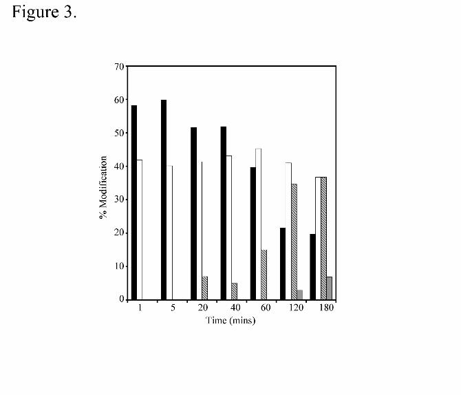
first minute, ~40% of the protein was modified with a single HNE molecule (Figure 3).
This suggests that the initial modification of E-FABP by 4-HNE is an extremely rapid
process (t1/2 of �������������� ��������������������������������������������������
addition of a second and third molecule of 4-HNE on E-FABP over 20 to 120 minutes.
Prolonged incubation (16 hr) resulted in the addition of up to 6 molecules of 4-HNE on
E-FABP (data not shown). These results suggest that the covalent modification of E-
FABP by 4-HNE is kinetically biphasic with the rapid incorporation of a single HNE
molecule on a seconds time scale followed by the slow addition of several other 4-HNEs
over the course of several hours.
To assess the concentration dependence of the 4-HNE modification, E-FABP was
incubated for 10 minutes at 22° C with increasing molar equivalents of 4-HNE and the
progress of the modification assessed immunochemically. As shown in Figure 4, the
modification of E-FABP with 4-HNE and the subsequent formation of lipid-protein
conjugates was concentration dependent. Combining the results of the time course and
the concentration dependence, standard conditions were adopted in which a 10-fold
molar excess of 4-HNE was incubated with E-FABP for 10 minutes at 22° C. Typically,
reactions were stopped by P-6 Bio-Spin chromatography or in some cases, by simple
addition of SDS-PAGE sample buffer.
by guest on May 18, 2020
http://ww
w.jbc.org/
Dow
nloaded from

17
Using the standard reaction protocol, the pH and temperature dependence of the
reaction was analyzed immunochemically using antibody directed to 4-HNE-protein
adducts. The modification of 4-HNE with E-FABP was unaffected by the pH of the
buffer over the pH range of 6.4 to 8.4 and similarly, the formation of 4-HNE protein
adducts was not affected by the temperature of the reaction from 4° C to 37° C (results
not shown).
Previous studies have shown that 4-HNE can covalently modify proteins at histidine,
lysine and cysteine residues (19). E-FABP contains six cysteine residues, two of which
(C120 and C127) are located within the ligand-binding cavity and form a disulfide bond.
In an effort to determine the site of 4-HNE modification of E-FABP, the lipid-protein
conjugate prepared using the standard protocol was digested with trypsin and analyzed
by MALDI-TOF MS. A theoretical digest of the protein with trypsin and no missed
cleavage sites yields 12 different peptide fragments with molecular masses ranging
from 537 Da to 2,390 Da. Peptide mass fingerprinting of the 4-HNE E-FABP conjugate
by MALDI-TOF MS identified peptides representing 83% of the primary sequence of the
protein (Table 1). The mass spectral analysis revealed similar coverage (73 %) of the
primary sequence of E-FABP. Incorporation of a single 4-HNE onto proteins yields a
mass addition of 156 and is consistent with a Michael addition reaction. Manual
interpretation of the MALDI-TOF data showed that two ions correspond to singly
charged m/z values of 4-HNE modified peptides. An abundant ion (85% relative
abundance by MALDI MS) with a m/z of 1798.8 corresponds to a single carboxymethyl
by guest on May 18, 2020
http://ww
w.jbc.org/
Dow
nloaded from
18
cysteine (delta 58) and a single 4-HNE (delta 156) adduct on the FABP peptide from
amino acids 116-129 (peptide #24- Table 1) with a mass accuracy of 5 ppm.
To verify the location of the 4-HNE modification on the 116-129 peptide, the ion with a
m/z of 1798.8 was selected for tandem spectral analysis by MALDI. The sequence of
the peptide (116-129) was confirmed as MIVECVMNNATCTR, which includes the
identification of a carboxymethylated cysteine at amino acid 127 and 4-HNE modified
cysteine at amino acid 120 (Figure 5). In the MS/MS spectrum, the observed
monoisotopic product ions y3 (m/z 437.23), y4 (m/z 538.22), y5 (m/z 609.25), y6 (m/z
723.31), y7 (m/z 837.32) and y9 (m/z 1067.47) correspond to carboxymethyl cysteine-
containing fragments generated from the precursor 1798.8, (see Papayannopoulos (39),
for fragment ion nomenclature). The precursor ion m/z 1798.8 as well as the
monoisotopic m/z value of 1642.74, which pertains to the carboxymethylated peptide
116-129 with the loss of 4-HNE (delta mass 156) each provide evidence for covalent
modification of cysteine by 4-HNE in a Michael addition reaction. The cysteine residue
that is modified is clearly indicated by the y10 (m/z 1326.60) and y10-H2O (m/z 1308.57)
fragment ions and the internal fragment ChneVMN-CO (m/z 576.25), which pertains to
peptide 120-124 with the loss of 28 Da, (CO = carbon monoxide). The mass difference
between the y9 and y10 ions, 259 Da, corresponds to the mass of a cysteine residue
(103 Da) + the delta mass value for 4-HNE (156). The average mass accuracy for all
fragment ion m/z values (all reported as monoisotopic values) is 16 ppm.
by guest on May 18, 2020
http://ww
w.jbc.org/
Dow
nloaded from

19
A second less abundant ion (15 % relative abundance by MALDI-MS) at 2156.97 m/z
corresponds to peptide from amino acids 113 – 129 with 2 carboxymethyl cysteines and
one 4-HNE (delta 156) (peptide #20 - Table 1). This ion was not observed using either
ESI or MALDI sources on the QSTAR mass spectrophometer therefore no tandem
mass spectral data could be acquired for sequence confirmation. Overall, tandem mass
spectra data (40 ppm mass accuracy) provided evidence for the confirmation of 13
peptide sequences. Consistent with only ~40% of the protein modified with 4-HNE,
MS/MS data revealed a mixture of peptides with both fully and partially
carboxymethylated cysteines (Table 1).
As an independent method to identify which cysteine was modified, and to determine if
both cysteine residues (Cys 120 and Cys 127) within the ligand binding cavity of E-
FABP were required for 4-HNE modification, a series of cysteine to alanine E-FABP
mutants were constructed. Soluble proteins from E. coli extracts expressing wild type
E-FABP, C120A, C127A and C120A C127A with bound fatty acids were incubated with
a ten fold molar equivalent of 4-HNE for 10 min at 22° C. Immunoblot analysis using the
antibody against 4-HNE protein adducts (normalized to total E-FABP expression)
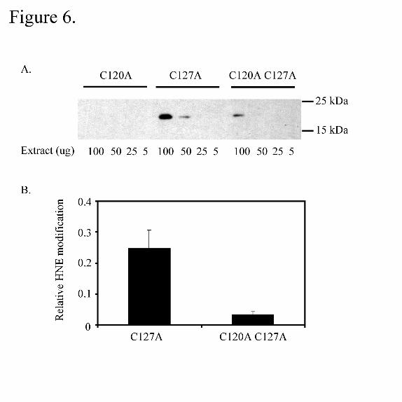
revealed that in the C120 A mutant, no modification was detected (Figure 6) whereas
modification was detected in the C127A mutant. Therefore, the presence of a nearby
second thiol is not necessary for 4-HNE covalent modification of E-FABP. Interestingly,
modification was also detected at very low levels (approximately 13% that of C127A
mutant) in the double mutant C120A C127 A. While not characterized, this trace
modification may correspond to the m/z 2156.97 ion corresponding to peptide 113-129
by guest on May 18, 2020
http://ww
w.jbc.org/
Dow
nloaded from

20
with two carboxymethylated cysteines and one HNE. We cannot rule out the possibility
that the poly histidine region of the recombinant protein also is the site of minor
modification. Moreover, the E-FABP mutant forms in crude bacterial extracts were
exclusively modified within the complex bacterial protein mixture by 4-HNE
demonstrating the specificity of the reaction. Therefore, mutational analysis is
consistent with mass spectral analysis in that C120 is the predominant site of 4-HNE
modification.
Since E-FABP is a fatty acid binding protein, and the recombinant proteins expressed in
E. coli bind endogenous fatty acids, we next addressed the potential effects of a bound
fatty acid on the covalent modification by 4-HNE in a purified system. To that end, E-
FABP was preincubated with either a 10 or 25 fold molar excess of oleic acid (Kd = 248
nM yielding an occupancy of 99.7% and 99.9% respectively) and then 4-HNE was
added to the same level as fatty acid. As shown in Figure 7, the modification was
markedly enhanced, relative to the apo protein, by the presence of fatty acids within the
lipid-binding cavity. This suggests that in crude extracts, the specific modification of E-
FABP cysteine mutants extracts (Figure 6) may be in part due to the presence of bound
endogenous fatty acids. Currently, we are not able to separate 4-HNE bound E-FABP
from unmodified E-FABP. As such, we cannot determine the fatty acid binding
properties of 4-HNE modified E-FABP.
To confirm that in the presence of bound FA the site of 4-HNE modification was C120,
peptide mass fingerprinting of the 4-HNE E-FABP conjugate by MALDI-TOF MS as well
by guest on May 18, 2020
http://ww
w.jbc.org/
Dow
nloaded from

21
as peptide sequencing by MS/MS was carried out. 82% coverage of E-FABP primary
sequence was identified as well as the site of modification in the presence of fatty acids.
Consistent with the apo-protein modification, C120 was the site of modification when
short-term incubations with 4-HNE were carried out (Table 2). At longer time points (60
minutes), minor secondary modifictions (< 5%) likely to be at C127 and/or K115, were
indicated. As such, both in the absence and presence of fatty acids, C120 is the site of
4-HNE modification.
Work with fatty acids has suggested that the presence of a non-covalently bound ligand
stabilizes the protein structure to chemical denaturation. However, this point cannot be
adequately addressed for the fatty acid rapidly exchanges from the protein during
analysis. To address this point with covalently bound 4-HNE, the protein prepared
using the standard protocol was subjected to increasing concentrations of guanidine
hydrochloride and the red-shifting of the emission maximum was used as an indicator of
protein unfolding (28). As shown in Figure 8, there is a shift in the progress curve to
increasing concentrations of guanidine hydrochloride when using the lipid-protein
conjugate relative to native protein. Since the sample contains a mixture of unmodified
and modified protein we cannot determine a true ���°’unfolding for the reaction.
However, it is clear that the presence of covalently bound 4-HNE stabilizes the protein
structure to chemical denaturation.
To determine if a lipid-protein conjugate could be detected in vivo, two-dimensional
polyacrylamide gel electrophoresis was used to separate proteins from crude rat retinal
by guest on May 18, 2020
http://ww
w.jbc.org/
Dow
nloaded from

22
tissue extracts where E-FABP is expressed to high levels. Despite the numerous
proteins present in the sample, only ~1 % of these spots were immunoreactive using an
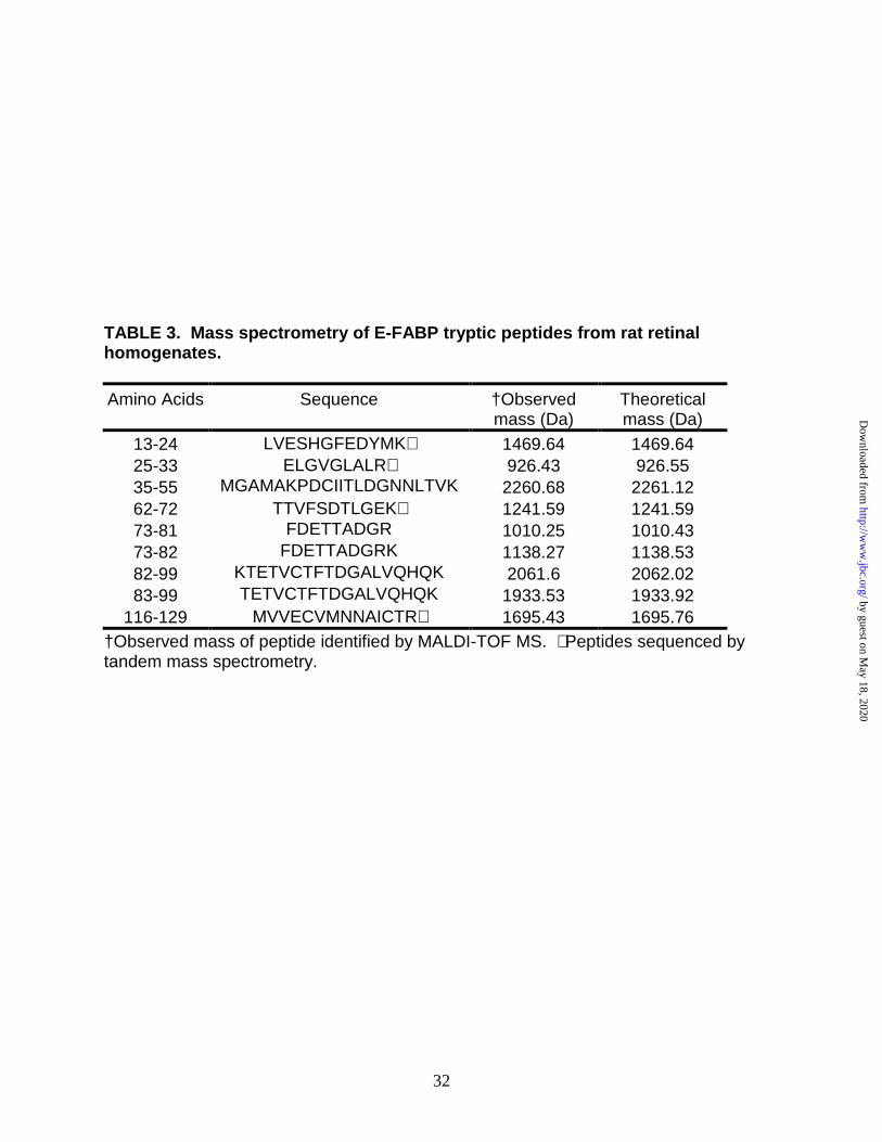
antibody directed towards 4-HNE-protein adducts (Figure 9). Using a combination of
MALDI-TOF and electrospray ionization MS/MS analysis of tryptic digests of the
resolved proteins, several spots were identified. The protein spot with a molecular
mass of 15kDa was identified as E-FABP. By combining the results obtained using
MALDI-TOF and ESI mass spectrometry, 70.4% of the primary sequence of E-FABP
was covered by a peptide map (Table 3) and four peptides were sequenced by MS/MS.
No peptide fragment corresponding to 4-HNE covalently bound to C120 was identified;
although peptide116-129 (without HNE) was sequenced by MS/MS. The data implies
that either the abundance of the modification was too low to detect the lipid-protein
conjugate or that the modification in vivo occurs on a residue(s) distinct from C120.
Our group (Maeda et al., submitted), as well as Owada et. al, (39), have independently
reported the development of E-FABP null mice. The phenotype of the null mice is
subtle, however Owada et al. have reported that in E-FABP null mice the skin epithelial
cell membranes are metabolically compromised such that there is a marked decrease in
transepidermal water loss. Using such E-FABP null mice and their wild type littermates,
we have developed immortalized lines of retinal pigment epithelial cells and analyzed
soluble cell extracts for 4-HNE-protein conjugates using two dimensional gel
electrophoresis followed by immunochemical analysis. Ferrington and colleagues (40)
have shown in retinal extracts that several proteins are 4-HNE modified and that their
abundance increases with increasing age. As shown in Figure 10, there are a number
by guest on May 18, 2020
http://ww
w.jbc.org/
Dow
nloaded from

23
of proteins endogenously modified by 4-HNE. Moreover, there is an upregulation of
both the number and intensity of the spots corresponding to 4-HNE modified proteins in
the E-FABP null cells (see white arrows). Overall, there was more than a doubling of
the number of 4-HNE modified proteins in the E-FABP null cells compared to wild type
epithelial cells. In such immortalized cells, the abundance of E-FABP is markedly down
regulated such that while E-FABP is a rather major protein in retinal extracts (Figure
9A), E-FABP in the immortalized epithelial cells is a quite minor protein (determined
from western blotting, results not shown). This may explain why we do not see a 15
kDa protein modified by 4-HNE in the immortalized cells as compared to the rat retinal
extracts. However, it is clear that the loss of E-FABP in the null cells results in an
increased level of 4-HNE modified cellular proteins.
DISCUSSION:
The upregulation of E-FABP in response to conditions linked to oxidative stress and the
presence of redox sensitive sulfhydryl residues within the lipid-binding cavity led to the
hypothesis that E-FABP maybe an endogenous target for 4-HNE modification and that
the protein functions to remove chemically reactive lipids from the cellular environment.
Three lines of evidence support the conclusion that E-FABP is a target for covalent
modification, both in vitro and in vivo, and that the protein may play a role in antioxidant
biology. Firstly, the properties of the in-vitro reaction of 4-HNE with E-FABP have been
by guest on May 18, 2020
http://ww
w.jbc.org/
Dow
nloaded from

24
characterized using a combination of mass spectrometry and immunochemical analysis.
Secondly, two dimensional electrophoresis of crude retinal lysates identified E-FABP as
a cellular target for 4-HNE modification in vivo. Thirdly, immortalized retinal pigment
epithelial cell lines originating from E-FABP null mice exhibit an upregulation of 4-HNE
modified proteins broadly in comparison to their wild type counterparts.
In-vitro, E-FABP is preferentially modified on C120. The in vitro reaction of 4-HNE with
E-FABP was concentration and time dependent, but largely pH and temperature
independent. Whereas long-term incubations with 4-HNE resulted in multiple
modifications, C120 was modified rapidly on a seconds time scale and fatty acids
potentiated this reaction. This led to the conclusion that the lipid-binding cavity of E-
FABP serves as a high affinity binding site for both fatty acids and 4-HNE and that both
ligands can be bound simultaneously, analogous to the binding of two lipids by the liver
FABP (41). Since in vivo, E-FABP is abundant in epithelial cells (42, 43) and 4-HNE is
present in only trace amounts, the molar ratio of 4-HNE to E-FABP is quite low and
leads to the consideration that only the high affinity cavity site is physiologically relevant.
As such, short-term incubations designed to modify E-FABP at only the high affinity site
were utilized to characterize the modification reaction.
Using MALDI-TOF MS and secondary sequencing of the1798.82 m/z peptide, C120
was identified as the major site of 4-HNE modification both in the presence and
absence of fatty acids. This result was corroborated with evidence obtained from the E-
FABP cysteine mutants where no modification with 4-HNE was detected with C120A,
by guest on May 18, 2020
http://ww
w.jbc.org/
Dow
nloaded from

25
only trace modification was revealed with C120A C127A relative to C127A modification.
In vitro, a second minor ion (2156.97 m/z) was detected whose mass was consistent
with amino acids 113-129 plus two carboxymethylations and one HNE modified lysine
residue (possibly K115). It is possible that the 4-HNE modification observed in the
C120A C127A mutant is on K115. This point has yet to be established but is consistent
with the binding data as well as NMR structure analysis of E-FABP with bound palmitic
acid.
The presence of six cysteine residues in E-FABP distinguishes it from the other lipid
binding protein family members (6,8). High resolution X-ray and NMR structures reveal
that the six cysteines are found as three clustered pairs: C43 and C47 found on the
surface of the protein, C67 and C87 buried in the hydrophobic core, and C120 and
C127 present in the ligand binding cavity as a disulfide bond (8). Analysis of fatty acid
binding (palmitic) within the cavity of E-FABP indicates that the lipid adopts a U-shape
comformation with the carboxyl head group coordinated between two arginine residues
(109 and 129) and Y131 in a hydrogen bonding network (7). The disulfide bond
between C120 and C127 of E-FABP is in close proximity to the C2-C4 region of the
bound fatty acid but is not directly involved in fatty acid binding. C120 of E-FABP is
homologous to C117 of A-FABP, a residue whose side chain also lies within close
proximity to the bound fatty acid. Covalent modification of C117 of A-FABP with small
adducts (methylmethane thiolsulfonate) has no effect on fatty acid binding, however
large adducts (N-ethylmaleimide) inhibit association (44). As such, steric factors around
C120 play a role in access to the H-bonding network involving the fatty acid carboxylate
by guest on May 18, 2020
http://ww
w.jbc.org/
Dow
nloaded from

26
and R109, R129 and Y131. In the absence of fatty acids, the carbonyl group of 4-HNE
could H-bond with Y131 providing some stabilization energy. Interestingly, the
observation that in-vitro, 4-HNE modification of E-FABP was stimulated by oleic acid
implies that both a fatty acid and 4-HNE are bound simultaneously. The presence of a
fatty acid adjacent to C120 could dynamically alter the electrostatic micro-environment
within the cavity thereby affecting the pKa of C120 thus making it more reactive towards
4-HNE modification. Alternatively, fatty acid bound may increase the generalized
hydrophobicity of the cavity by displacing water thereby increasing the binding of the
hydrophobic medium chain aldehyde, a reaction not normally carried out by FABPs.
The combination of C120 modification, potential for hydrogen bonding to Y131, and
stimulation by fatty acids suggests that the aldehyde is coordinated in a similar, but not
identical manner to fatty acids. Utilization of C120A mutant of E-FABP in future
experiments will facilitate dissection of the binding reaction in the absence of catalysis
and the structural organization of the binding cavity with two ligands present.
Given the homologous cysteine of A-FABP (C117) and its similar fatty acid binding
character, we have preliminarily evaluated if 4-HNE could covalently modify this protein.
Interestingly, attempts to covalently modify A-FABP in vitro with 4-HNE were
unsuccessful (results not shown) under a limited set of experimental conditions. This
may indicate that the covalent modification of E-FABP by 4-HNE is more complex than
simply thiol availability. The modification of E-FABP C127A by 4-HNE implies that
redox chemistry involving a structurally vicinyl thiol is not likely to be involved in the
modification, however, this point has not been analyzed in detail. As such, the
by guest on May 18, 2020
http://ww
w.jbc.org/
Dow
nloaded from

27
molecular determinants that define binding versus catalysis are obscure. It will be
interesting to analyze the potential covalent modification of other FABPs that have a
cysteine at a homologous position (myelin P2, testis lipid binding protein) to determine if
they too are reactive.
The role of E-FABP as an antioxidant protein is consistent with the cell and/or tissue
types in which it is expressed. E-FABP is found in cells exposed to high oxidative
stress, including the retina, lens, lung, and tongue (45). In these cells, E-FABP may
play a role in limiting the amount of oxidative damage to proteins by scavenging 4-HNE-
like molecules. Herein we report using crude rat retinal lysates the in vivo identification
of E-FABP as a molecular target for HNE modification. The rod outer segment
membranes of the retina in particular are highly enriched with long chain
polyunsaturated fatty acids including arachidonic acid (20:4) and docosahexaenoic acid
(22:6) thus making them highly susceptible to lipid peroxidation (46). A recent study
has demonstrated that in vitro induced lipid peroxidation of rod outer segment
membranes was reduced by the addition of increasing concentrations of retinal extracts
containing a low molecular mass FABP (46). The mechanism of this protection is
currently not clear but it is possible that the FABP acts by binding the free fatty acids
preventing their oxidation. Alternatively, as demonstrated herein, protection of rod outer
segments may be through the covalent modification (scavenging) of E-FABP by 4-HNE.
This point is currently under investigation.
by guest on May 18, 2020
http://ww
w.jbc.org/
Dow
nloaded from

28
Using retinal pigment epithelial cell lines derived from wild type and E-FABP null mice,
the abundance of soluble 4-HNE modified proteins was evaluated immunochemically.
Typically, FABPs are expressed to very high levels in cells in vivo (2) and generate a
cytoplasmic pool of fatty acid for metabolic utilization. However, in cultured epithelial
cells, E-FABP expression is markedly down regulated (unpublished observations) and
we did not observe any 4-HNE modified E-FABP in the culture system whereas we did
identify the protein-lipid conjugate in retinal extracts. The inability to detect 4-HNE
modified E-FABP in cultured cells may be due to any of several reasons. For example,
E-FABP with covalently bound 4-HNE could be a substrate for the proteasome (40)
such that the modified protein is cleared quickly from cells. An alternate possibility is
that the covalent modification of E-FABP by 4-HNE could be reversed within the cellular
context by protein glutathionylation. Indeed, Fratelli et al. (47) have reported that in
oxidatively stressed T lymphocytes E-FABP is glutathionylated, although the site of
modification was not reported. Current experiments are focussed on evaluating these
possibilities.
Even with a low level of E-FABP expression in the wild type cells, there was a marked
up-regulation in the modification of soluble proteins in cell lines derived from E-FABP
null mice. There are several potential mechanisms to explain the upregulation of 4-HNE
modified proteins in the E-FABP null cells. The most direct explanation is that the
increase in 4-HNE modification of epithelial cell proteins may be due to the lack of the
proposed protective effects of E-FABP via covalent modification by 4-HNE. The
absence of E-FABP may result in 4-HNE modification of alternate targets such as those
by guest on May 18, 2020
http://ww
w.jbc.org/
Dow
nloaded from

29
seen in Figure 10. Alternatively, increased modification by 4-HNE could be due to
increased lipid oxidation as a result of redistribution of endogenous polyenoic fatty acids
from the E-FABP soluble pool to the membrane where they could be more accessible to
peroxidation.
In sum, we have shown that HNE can rapidly modify C120 of E-FABP in vitro as shown
by a combination of mass spectral analysis and immunochemically with an antibody
directed towards 4-HNE protein adducts. E-FABP was also identified as a cellular
target for HNE modification in vivo as demonstrated by two-dimensional analysis of rat
retinal lysates. In vitro, the presence of fatty acids within the lipid-binding cavity greatly
enhanced the covalent modification of E-FABP by 4-HNE suggesting that the holo
protein is the target for 4-HNE modification in vivo. As such, the protein does not
undergo suicide inactivation since the non-covalent fatty acid binding function is not
affected. The absence of E-FABP in retinal pigment epithelial cell lines resulted in an
increase in the number and abundance of proteins modified by 4-HNE. Thus the role of
E-FABP can be expanded beyond simple fatty acid trafficking to a novel
protective/antioxidant function. This marks the first report, to our knowledge, of a role
for any FABP in the covalent binding of lipids and suggests the hypothesis that E-FABP
functions as an antioxidant protein, protecting the integrity of the cellular environment
through inactivation of reactive lipids.
by guest on May 18, 2020
http://ww
w.jbc.org/
Dow
nloaded from

30
Table 1. Tryptic Peptides from E-FABP Measured by Mass Spectrometry. 4-HNE modified E-FABP was digested with trypsin and the peptide fragments analyzed by MALDI-TOF (Biflex III and QSTAR Pulsar quadrupole; mass accuracies of <150 and 40 ppm, respectively) and ESI (unit mass accuracy) mass spectrometry.
Amino acid
position Identification
No. *MC From To Theoretical
[MH]+1 †Sequence Pep. m/z
MS/MS Spectrum
1 0 13 24 1500.6503 LMESHGFEEYMK X ‡2 0 13 24 1656.7653 LMESHGFEEYMK(hne) X 3 0 25 33 927.5627 ELGVGLALR X 4 1 25 34 1055.6577 ELGVGLALRK X 5 0 35 55 2209.0489 MAAMAKPDCIITCDGNNITVK X 6 0 35 55 2325.0593 MAAMAKPDC(cm)IITC(cm)DGNNITVK X 7 0 62 72 1198.5778 TTVFSCNLGEK X 8 0 62 72 1256.5833 TTVFSC(cm)NLGEK X 9 1 62 81 2190.9977 TTVFSCNLGEKFDETTADGR X 10 1 62 81 2249.0032 TTVFSC(cm)NLGEKFDETTADGR X 11 2 62 82 2319.0921 TTVFSCNLGEKFDETTADGRK X 12 2 62 82 2377.0976 TTVFSC(cm)NLGEKFDETTADGRK X 13 0 73 81 1011.4383 FDETTADGR X 14 1 82 103 2519.1989 KTETVCTFQDGALVQHQQWDGK X 15 1 82 103 2577.2044 KTETVC(cm)TFQDGALVQHQQWDGK X 16 0 83 103 2391.1039 TETVCTFQDGALVQHQQWDGK X 17 0 83 103 2449.1094 TETVC(cm)TFQDGALVQHQQWDGK X 18 1 83 109 3136.4640 TETVC(cm)TFQDGALVQHQQWDGKESTITR X 19 2 83 110 3264.5589 TETVC(cm)TFQDGALVQHQQWDGKESTITRK X ‡20 1 113 129 2156.9700 DGK(hne)MIVEC(cm)VMNNATC(cm)TR X 21 0 116 129 1584.7007 MIVECVMNNATCTR X 22 0 116 129 1642.7062 MIVECVMNNATC(cm)TR X 23 0 116 129 1700.7117 MIVEC(cm)VMNNATC(cm)TR X §24 0 116 129 1798.8212 MIVEC(hne)VMNNATC(cm)TR X
*MC-Number of trypsin missed cleavage sites in peptide. †Modifications indicated in parentheses, positioned C-terminal to the relevant amino acid; hne = 4-hydroxynonenal; cm = carboxymethyl. ‡Proposed HNE modification based on peptide m/z value. Signal/noise (S/N) for the monoisotopic peaks of peptide numbers 2 and 20 were 10 and 18, respectively. §Confirmed HNE modification based on MS/MS sequence information. Some peptides listed in the table were detected with oxidized methionines in addition to the forms detected above, but the m/z values are not shown.
by guest on May 18, 2020
http://ww
w.jbc.org/
Dow
nloaded from

31
TABLE 2. Tryptic peptides from holo E-FABP modified with 4-HNE measured by MALDI-TOF analysis. # Pep.
AA Observed
[M+H]+ Sequence �Abundance
1 116-129 1798.82 *MIVEC (hne) VMNNATC (cm) TR 0.3380 2 116-129 1896.91 †MIVEC (hne) VMNNATC (hne) TR 0.0318 3 113-129 2157.01 †DGK (hne) MIVEC (cm) VMNNATC (cm)TR 0.0190 *Peptide observed in all conditions- 10 min and 60 min time point with both 10 and 25 nmol of oleic acid and 4-HNE. †Peptides observed only at 60 min time point with 25 nmol of oleic acid and HNE. �Peptide abundance relative to peak intensities for complete field.
by guest on May 18, 2020
http://ww
w.jbc.org/
Dow
nloaded from
32
TABLE 3. Mass spectrometry of E-FABP tryptic peptides from rat retinal homogenates.
Amino Acids
Sequence
†Observed mass (Da)
Theoretical mass (Da)
13-24 LVESHGFEDYMK∗ 1469.64 1469.64 25-33 ELGVGLALR∗ 926.43 926.55 35-55 MGAMAKPDCIITLDGNNLTVK 2260.68 2261.12 62-72 TTVFSDTLGEK∗ 1241.59 1241.59 73-81 FDETTADGR 1010.25 1010.43 73-82 FDETTADGRK 1138.27 1138.53 82-99 KTETVCTFTDGALVQHQK 2061.6 2062.02 83-99 TETVCTFTDGALVQHQK 1933.53 1933.92
116-129 MVVECVMNNAICTR∗ 1695.43 1695.76 †Observed mass of peptide identified by MALDI-TOF MS. ∗ Peptides sequenced by tandem mass spectrometry.
by guest on May 18, 2020
http://ww
w.jbc.org/
Dow
nloaded from

33
ABBREVIATIONS:
LBP, lipid binding protein; FABP, fatty acid binding protein; LOOH, lipid hydroperoxide;
PCR, polymerase chain reaction; E-FABP, epithelial fatty acid binding protein; HRP,
horseradish peroxidase; 1,8-ANS, 1-Anilinonapthalene-8- sulfonate; 4-HNE, 4-
hydroxynonenal; DTNB, 5,5’-dinitrobis(2-nitrobenzoic acid); BCIP-NBT, 5-bromo-4-
chloro-3-indolyl phosphate/Nitro blue tetrazolium; PVDF, polyvinylidenedifluoride;
MALDI-TOF MS, matrix assisted laser desorption ionization time of flight mass
spectrometry; ESI, electrospray ionization; MS/MS, tandem mass spectrometry; SDS,
sodium dodecyl sulfate; ACN, acetonitrile.
ACKNOWLEDGMENTS: This work was funded by grants from the National Institute of
Aging (RO3 AG19024), Foundation Fighting Blindness, American Federation of Aging
Research, and an unrestricted grant to the Department of Ophthalmology from the
Research to Prevent Blindness Foundation to DAF and by a grant by the National
Science Foundation to DAB. All work with animals was approved by the University of
Minnesota IACUC prior to experimentation. The authors thank Lisa Smith for the
construction of the E-FABP mutants and Wesley Obritsch for the development of the
RPE cell lines. Dr. Christine Hunter of Applied Biosystems, Inc was particularly helpful
with initial MALDI-TOF MS/MS assignment. The authors would also like to thank Tom
Krick, Manager of the Mass Spectrometry Consortium for the Life Sciences, University
by guest on May 18, 2020
http://ww
w.jbc.org/
Dow
nloaded from

34
of Minnesota, for his help with MALDI-TOF MS as well as Dr. Michael Martinez for his
advice on protease digestions. Members of the Bernlohr, Ferrington, and Barry labs are
acknowledged for helpful suggestions.
REFERENCES:
1. Tan, N. S., Shaw, N. S., Vinckenbosch, N., Liu, P., Yasmin, R., Desvergne, B., Wahli, W., and Noy, N. (2002) Mol Cell Biol 22, 5114-5127
2. Coe, N. R., and Bernlohr, D. A. (1998) Biochim Biophys Acta 1391, 287-306 3. McArthur, M. J., Atshaves, B. P., Frolov, A., Foxworth, W. D., Kier, A. B., and
Schroeder, F. (1999) J Lipid Res 40, 1371-1383 4. Hertzel, A. V., and Bernlohr, D. A. (2000) Trends Endocrinol Metab 11, 175-180 5. Banaszak, L., Winter, N., Xu, Z., Bernlohr, D. A., Cowan, S., and Jones, T. A.
(1994) Adv Protein Chem 45, 89-151 6. Gutierrez-Gonzalez, L. H., Ludwig, C., Hohoff, C., Rademacher, M., Hanhoff, T.,
Ruterjans, H., Spener, F., and Lucke, C. (2002) Biochem J 364, 725-737 7. Hohoff, C., Borchers, T., Rustow, B., Spener, F., and van Tilbeurgh, H. (1999)
Biochemistry 38, 12229-12239 8. Kane, C. D., Coe, N. R., Vanlandingham, B., Krieg, P., and Bernlohr, D. A.
(1996) Biochemistry 35, 2894-2900 9. Ostergaard, M., Rasmussen, H. H., Nielsen, H. V., Vorum, H., Orntoft, T. F.,
Wolf, H., and Celis, J. E. (1997) Cancer Res 57, 4111-4117 10. Madsen, P., Rasmussen, H. H., Leffers, H., Honore, B., and Celis, J. E. (1992) J
Invest Dermatol 99, 299-305 11. Krieg, P., Finch, J., Fustenberger, G., Melber, K., Matrisian, L. M., and Bowden,
G. T. (1988) Carcinogenesis 9, 95-100 12. Owada, Y., Yoshimoto, T., and Kondo, H. (1996) Brain Res Mol Brain Res 42,
156-160 13. Owada, Y., Utsunomiya, A., Yoshimoto, T., and Kondo, H. (1997) Neurosci Lett
223, 25-28 14. De Leon, M., Welcher, A. A., Nahin, R. H., Liu, Y., Ruda, M. A., Shooter, E. M.,
and Molina, C. A. (1996) J Neurosci Res 44, 283-292 15. Finkel, T., and Holbrook, N. J. (2000) Nature 408, 239-247 16. Esterbauer, H., Schaur, R. J., and Zollner, H. (1991) Free Radic Biol Med 11, 81-
128 17. Van Kuijk, F. J., Holte, L. L., and Dratz, E. A. (1990) Biochim Biophys Acta 1043,
116-118 18. Esterbauer, H., and Zollner, H. (1989) Free Radic Biol Med 7, 197-203 19. Poli, G., and Schaur, R. J. (2000) IUBMB Life 50, 315-321 20. Hauptlorenz, S., Esterbauer, H., Moll, W., Pumpel, R., Schauenstein, E., and
Puschendorf, B. (1985) Biochem Pharmacol 34, 3803-3809 21. White, J. S., and Rees, K. R. (1984) Chem Biol Interact 52, 233-241
by guest on May 18, 2020
http://ww
w.jbc.org/
Dow
nloaded from

35
22. Pizzimenti, S., Barrera, G., Dianzani, M. U., and Brusselbach, S. (1999) Free Radic Biol Med 26, 1578-1586
23. Kruman, I., Bruce-Keller, A. J., Bredesen, D., Waeg, G., and Mattson, M. P. (1997) J Neurosci 17, 5089-5100
24. Herbst, U., Toborek, M., Kaiser, S., Mattson, M. P., and Hennig, B. (1999) J Cell Physiol 181, 295-303
25. Sohal, R. S., and Weindruch, R. (1996) Science 273, 59-63 26. Beckman, K. B., and Ames, B. N. (1998) Physiol Rev 78, 547-581 27. Stadtman, E. R. (1992) Science 257, 1220-1224 28. Simpson, M. A., and Bernlohr, D. A. (1998) Biochemistry 37, 10980-10986 29. US Patent 4873192. Licensed to Bio-Rad Laboratories. 30. Jenkins, A. E., Hockenberry, J. A., Nguyen, T., and Bernlohr, D. A. (2002)
Biochemistry 41, 2022-2027 31. Isacsson, U. a. W., G. (1974) Anal. Chim. Acta. 68, 339-362 32. Shevchenko, A., Wilm, M., Vorm, O., and Mann, M. (1996) Anal Chem 68, 850-
858 33. Singh, S. K., Miller, S. P., Dean, A., Banaszak, L. J., and LaPorte, D. C. (2002) J
Biol Chem 277, 7567-7573 34. Eng, J., McCormack, A. L., and Yates, J. R. (1994) J. Am. Soc. Mass Spectrom.
5, 976-989 35. Mayerson, P. L., Hall, M. O., Clark, V., and Abrams, T. (1985) Invest Ophthalmol
Vis Sci 26, 1599-1609 36. Wessel, D., and Flugge, U. I. (1984) Anal Biochem 138, 141-143 37. Laemmli, U. K. (1970) Nature 227, 680-685 38. Papayannopoulos, I.A. (1995) Mass Spec. Reviews. 14: 49-71. 39. Owada, Y., Takano, H., Yamanaka, H., Kobayashi, H., Sugitani, Y., Tomioka, Y.,
Suzuki, I., Suzuki, R., Terui, T., Mizugaki, M., Tagami, H., Noda, T., and Kondo, H. (2002) J Invest Dermatol 118, 430-435
40. Louie, J.K., Kapphahn, R.J., and Ferrington, D.A. (2002) Exp. Eye Res. 75; 271-284
41. Haunerland, N., Jagschies, G., Schulenberg, H., and Spener, F. (1984) Hoppe Seylers Z Physiol Chem 365, 365-376
42. Krieg, P., Feil, S., Furstenberger, G., and Bowden, G. T. (1993) J Biol Chem 268, 17362-17369
43. Watanabe, R., Fujii, H., Yamamoto, A., Yamaguchi, H., Takenouchi, T., Kameda, K., Ito, M., and Ono, T. (1996) Arch Dermatol Res 288, 481-483
44. Buelt, M. K., and Bernlohr, D. A. (1990) Biochemistry 29, 7408-7413 45. Kingma, P. B., Bok, D., and Ong, D. E. (1998) Biochemistry 37, 3250-3257 46. Terrasa, A., Guajardo, M., and Catala, A. (1998) Mol Cell Biochem 178, 181-186 47. Fratelli, M., Demol, H., Puype, M., Casagrande, S., Eberini, I., Salmona, M.,
Bonetto, V., Mengozzi, M., Duffieux, F., Miclet, E., Bachi, A., Vanderkerckhove, J., Gianazza, E., and Ghezzi, P. (2002) Proc. Natl. Acad. Sci. USA 99; 305-3510
by guest on May 18, 2020
http://ww
w.jbc.org/
Dow
nloaded from

36
FIGURE LEGENDS: Figure 1. MALDI-TOF linear mass spectrum of a mixture of native and 4-HNE-
modified E-FABP. E-FABP was modified with 4-HNE, separated from any unreactive
lipid by spin column chromatography, desalted, mixed with sinapinic acid and subjected
to MALDI-TOF MS analysis. The raw data of mass intensity versus m/z was smoothed
using the Golay-Savitzky formula with 15 smoothing points using Brüker’s Xmass
software package. The peak at m/z 15,001 corresponds to unmodified E-FABP, at
15,155 to singly 4-HNE modified E-FABP and at 15,206 to the sinapinic acid adduct of
FABP.
Figure 2. Immunoblotting analysis of 4-HNE modified E-FABP. E-FABP was
incubated with a 10 fold molar excess of either 4-HNE or oleic acid for 10 minutes at 22°
C and subjected to 15% SDS-PAGE. After electrophoresis, the protein was blotted and
probed with antibody towards 4-HNE protein adducts.
Figure 3. Time dependence of HNE modification of E-FABP analyzed by MALDI-
TOF. E-FABP was incubated with a ten fold molar excess of 4-HNE at 22° C and
aliquots removed at the indicated time points. The samples were applied to a BioGel-
P6 column to separate modified protein from free 4-HNE and the eluted proteins
subjected to MALDI-TOF MS to detect the presence of the 4-HNE-E-FABP conjugates.
by guest on May 18, 2020
http://ww
w.jbc.org/
Dow
nloaded from

37
The black bars represent unmodified E-FABP while the open, hatched and grey bars
represent singly, doubly, and triply modified E-FABP, respectively.
Figure 4. Concentration dependence of 4-HNE modification of E-FABP. Purified
E-FABP (1 nmol) was incubated with increasing molar amounts of 4-HNE for 10
minutes at 22° C. Following incubation, the samples were separated by 15% SDS-
PAGE, blotted and lipid-protein conjugates detected using the antibody against 4-HNE
protein adducts.
Figure 5. Tandem mass spectrum analysis of the 4-HNE containing peptide. The
m/z = 1798.8 peptide was subjected to fragmentation by MS/MS and the resultant
peptides analyzed. The spectrum contains peaks for 10/14 y ions and 4/14 b ions, 5
immonium ions, one internal fragment, and seventeen b and y fragments minus
ammonia or water. The data were plotted as a function of m/z using GraphPad Prism
after the raw mass spectral data was smoothed, centroided and labeled in BioAnalyst
(ABI, Foster City, CA). The fragments MNN-CO and OVMN-CO are internal fragment
ions minus carbon monoxide (28 Da); O = single letter code for HNE-containing
cysteine (chosen for labeling purposes only).
Figure 6. Western blot analysis of soluble protein extracts from E-FABP mutants
using anti-4-HNE antibodies. Panel A: Recombinant E-FABP protein containing either
C120A, C127A or C120A, C127A in crude E. coli extracts was incubated with 4-HNE,
separated by SDS-PAGE and analyzed by immunoblotting as described. Following
by guest on May 18, 2020
http://ww
w.jbc.org/
Dow
nloaded from
38
blotting with anti-4-HNE protein conjugate, the blot was stripped and re-probed with
anti-E-FABP antibody. Panel B; the normalized intensity of 4-HNE modification as
compared to E-FABP protein expression in the crude bacterial extracts.
Figure 7. Effect of fatty acids on the covalent modification of E-FABP by 4-HNE.
Purified E-FABP was incubated with 10 and 25 molar excess of oleic acid for 2 minutes
followed by the addition of 4-HNE to the same level for 10 minutes at 22° C. Following
modification, the protein was subjected to western blot analysis using primary antibody
against 4-HNE.
Figure 8. Guanidine HCl denaturation of native and 4-HNE modified E-FABP as
assessed by changes in intrinsic tryptophan fluorescence. Increasing
concentrations of guanidine hydrochloride (0 to 3 M) were added to either 0.5 µM E-
FABP or 4-HNE modified E-FABP. Tryptophan fluorescence was excited at 285nm and
emission maximum wavelengths were recorded and plotted versus Gdn-HCl
concentration. Each curve represents two independently collected data sets.
Figure 9. In vivo identification of 4-HNE modified E-FABP from rat retinal
homogenate.
Panel A: Silver-stained two dimensional gel of rat retinal homogenate. Soluble protein
(90 µg) was resolved by isoelectric focusing on an immobilized strip (pH 5-8) in the first
dimension, then by SDS-PAGE (10%) in the second dimension. The arrow shows the
spot identified as E-FABP by tandem mass spectral analysis. Panel B: Western blot
analysis of rat retinal homogenate resolved by two-dimensional PAGE using antibodies
by guest on May 18, 2020
http://ww
w.jbc.org/
Dow
nloaded from
39
against 4-HNE protein adducts. The arrow shows the spot identified as E-FABP
immunoreactive with 4-HNE antibody.
Figure 10. Western blot analysis of proteins derived from epithelial cells of wild
type and E-FABP null mice. Soluble proteins (100 ug) from epithelial cells derived
from wild type and E-FABP null mice were separated by 2-dimensional IEF/SDS-PAGE,
transferred to PVDF membrane and analyzed immunochemically with antibody against
4-HNE modified proteins. White arrows indicate 4-HNE modified proteins whose
abundance is increased in E-FABP null cells while a black arrow indicates a modified
protein that decreases in abundance. Numbers represent the molecular mass of
proteins in kDa.
by guest on May 18, 2020
http://ww
w.jbc.org/
Dow
nloaded from

Deborah A. Ferrington and David A. BernlohrAssumpta Bennaars-Eiden, LeeAnn Higgins, Ann V. Hertzel, Rebecca J. Kapphahn,
vitro and in vivo: Evidence for a role in antioxidant biologyCovalent modification of epithelial fatty acid binding protein by 4-hydroxynonenal in
published online October 16, 2002J. Biol. Chem.
10.1074/jbc.M209493200Access the most updated version of this article at doi:
Alerts:
When a correction for this article is posted•
When this article is cited•
to choose from all of JBC's e-mail alertsClick here
by guest on May 18, 2020
http://ww
w.jbc.org/
Dow
nloaded from