B.Sc. Biochem II Biomolecule I U 3.1 Structure of Proteins
39
Structure and Classification of Protein Course: B.Sc.(Biochem) II Subject: Biomolecules I Unit 3.1
-
Upload
rai-university -
Category
Science
-
view
47 -
download
0
Transcript of B.Sc. Biochem II Biomolecule I U 3.1 Structure of Proteins
Structure and Classification of Protein
Course: B.Sc.(Biochem) II
Subject: Biomolecules I
Unit 3.1
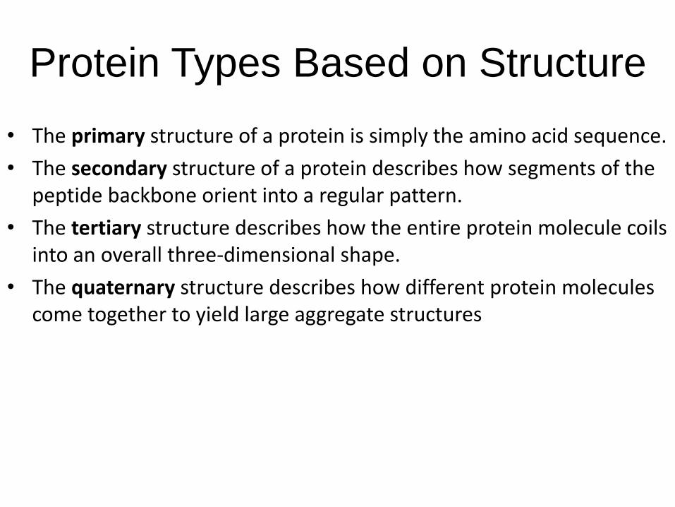
Protein Types Based on Structure
• The primary structure of a protein is simply the amino acid sequence.
• The secondary structure of a protein describes how segments of the peptide backbone orient into a regular pattern.
• The tertiary structure describes how the entire protein molecule coils into an overall three-dimensional shape.
• The quaternary structure describes how different protein molecules come together to yield large aggregate structures

2 polypeptides
1

Peptides and polypeptides
• Glycine and alanine can combine together with the
elimination of a molecule of water to produce a dipeptide.
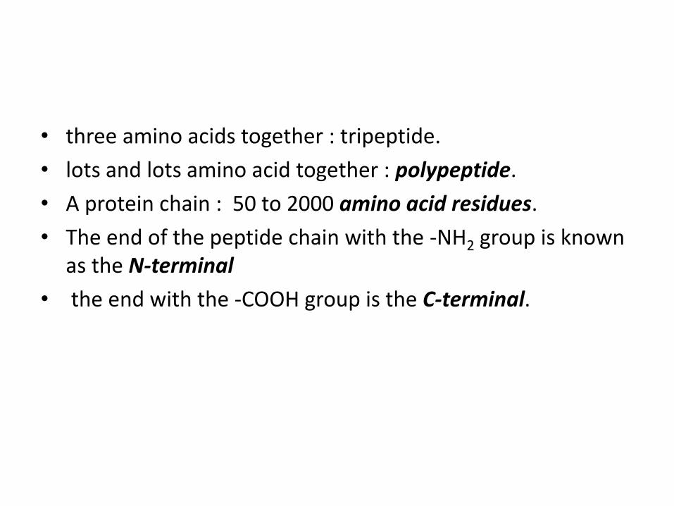
• three amino acids together : tripeptide.
• lots and lots amino acid together : polypeptide.
• A protein chain : 50 to 2000 amino acid residues.
• The end of the peptide chain with the -NH2 group is known as the N-terminal
• the end with the -COOH group is the C-terminal.

• A protein chain (with the N-terminal on the left)
The "R" groups come from the 20 amino acids which occur in proteins. The peptide chain : backbone, and the "R" groups are known as side chains.

Levels of protein structure
2

Levels of protein structure

The primary structure of proteins
• "primary structure" is used in two different ways.• describe the order of the amino acids joined
together to make the protein. • if you replaced the "R" groups in the last diagram
by real groups you would have the primary structure of a particular protein.
• This primary structure : abbreviations for the amino acid residues. These abbreviations commonly consist of three letters or one letter.
• Using three letter abbreviations, a bit of a protein chain might be represented by, for example:

Using three letter abbreviations, a bit of a protein chain might be represented by, for example:
• If you followed the protein chain all the way to its left-hand end, you would find an amino acid residue with an unattached -NH2 group.
• The N-terminal is always written on the left of a diagram for a protein's primary structure - whether you draw it in full or use these abbreviations.
• The wider definition of primary structure includes all the features of a protein which are a result of covalent bonds. Obviously, all the peptide links are made of covalent bonds, so that isn't a problem.
• But there is an additional feature in proteins which is also covalently bound. It involves the amino acid cysteine.

• If two cysteine side chains end up next to each other because of folding in the peptide chain, they can react to form a sulphur bridge.
• This is another covalent link and so some people count it as a part of the primary structure of the protein.
3
• Because of the way sulphur bridges affect the way the protein folds, other people count this as a part of the tertiary structure.

The secondary structure of proteins
• Within the long protein chains there are regions in which the chains are organised into regular structures known as alpha-helices (alpha-helixes) and beta-pleated sheets.
• held together by hydrogen bonds.
4

• hydrogen bonds are always between C=O and H-N groups, the exact pattern of them is different in an alpha-helix and a beta-pleated sheet.
• When you get to them below, take some time to make sure you see how the two different arrangements works.

a-helix features
• Coil direction – left handed or right handed• L- amino acids favor the right hand coil• One coil has about 3.6 aa residues; there can be several coils
with 650 aa residues(1000Å)
• Average length of helix in a globular protein is 15-20Å• H-bonds occur between 1st O of backbone C=O to 13th H atom
of backbone NH• The presence of the ff amino acids do not favor the helix
formation: Pro, adjacent basic or acidic amino acids, Asn, Tyr, Ser, Thr, Ile and Cys
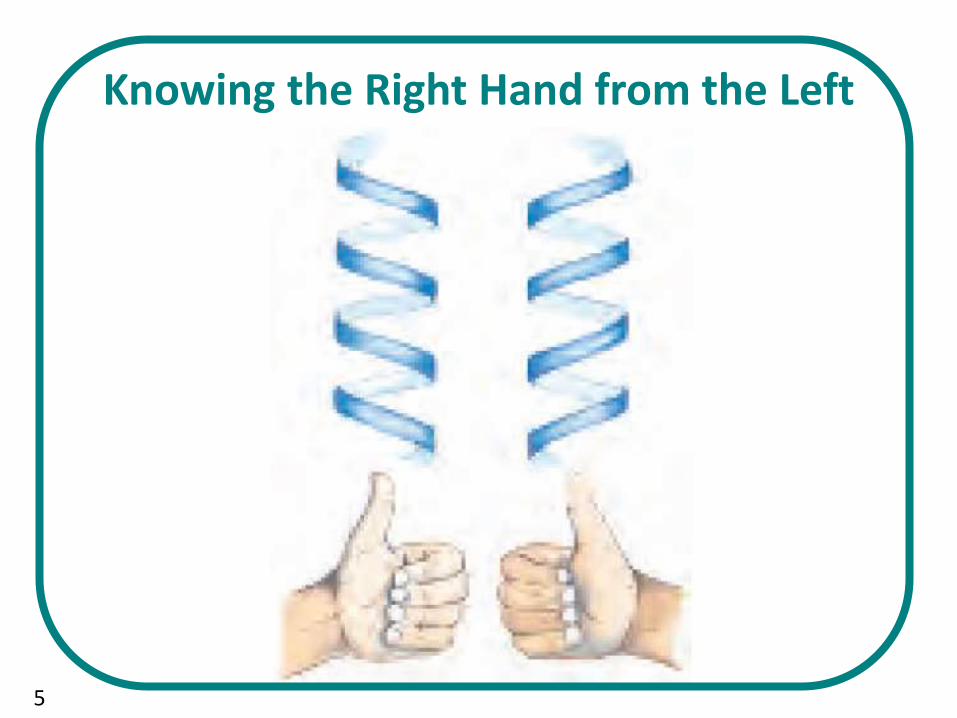
Knowing the Right Hand from the Left
5
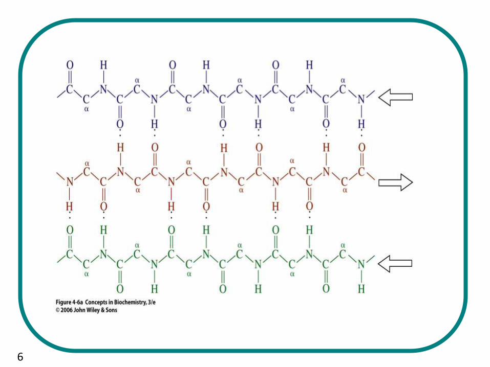
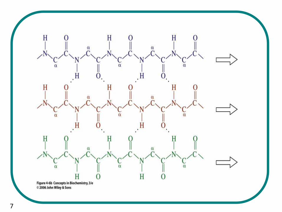
b-pleated sheet
• Two adjacent peptides
• Parallel (both NC or CN)
• Antiparallel (N to C running in opposite directions)
• Antiparallel more common in the structure of proteins
• Peptides with this structure are rich in alanine and glycine (silk fiber and spider web)
6
7

Supersecondary structuresor structural motifs
• The clusters are held together by favorable non covalent interactions
• Some structural motifs of folded proteins: aamotif; bb motif antiparallel; the Greek key (bbbb) motif; bab motif parallel

8

Tertiary structure
• Combination of several motifs of secondary structures into a compact arrangement
• Noncovalent forces bring about the interactions and stability; – H-bonds, – electrostatic, – hydrophobic, – Van Der Waal’s,– pi-pi complexation between R-side chains– Disulfide bonds occur between Cys residues

9

Tertiary protein structure
• determined by a variety of bonding interactions between the "side chains" on the amino acids.
• These bonding interactions may be stronger than the hydrogen bonds between amide groups holding the helical structure.
• bonding interactions between "side chains" : number of folds, bends, and loops in the protein chain.
• Four types of bonding interactions:• hydrogen bonding, • salt bridges, • disulfide bonds, • non-polar hydrophobic interactions.

Disulfide Bonds:- Oxidation of the sulfhydryl groups on cysteine.Different protein chains or loops within a single chain are held together
by the strong covalent disulfide bonds.

Hydrogen Bonding:
• occurs in a variety of
circumstances.
• 1. between two alcohols,
• 2. an alcohol and an acid,
• 3.two acids,
• 4. an alcohol and an amine or amide.
• In the prion protein, tyr 128 is hydrogen bonded to asp 178, which cause one part of the chain to be bonding with a part some distance away.
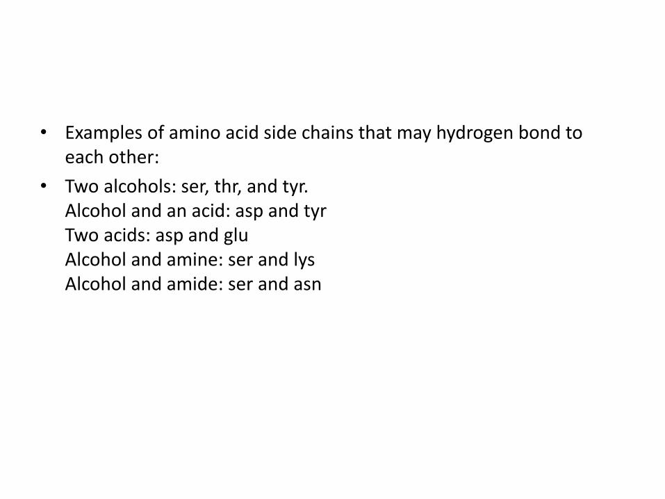
• Examples of amino acid side chains that may hydrogen bond to each other:
• Two alcohols: ser, thr, and tyr.Alcohol and an acid: asp and tyrTwo acids: asp and gluAlcohol and amine: ser and lysAlcohol and amide: ser and asn

Salt Bridges:
• neutralization of an acid and amine on side chains.
• interaction : ionic
• the positive ammonium group and the negative acid group.
• Any combination of the various acidic or amine amino acid side chains will have this effect.

The example on the left is from the prion protein with the salt bridge of glutamic acid 200 and lysine 204. In this case a very small loop is made because there are only three other amino acids between them. This salt bridge has the effect of straightening an alpha helix

Non-Polar Hydrophobic Interactions:
• The hydrophobic interactions of non-polar side chains are believed to contribute significantly to the stabilizing of the tertiary structures in proteins.
• The non-polar groups mutually repel water and other polar groups and results in a net attraction of the non-polar groups for each other.
• Hydrocarbon alkyl groups on ala, val, leu, and ileinteract in this way.
• In addition, benzene (aromatic) rings on phe and tyr can "stack" together.

Quaternary structure of proteins
• Oligomeric –two or more polypeptide chains; subunits
• Homotypic – almost identical subunits
• Heterotypic – different subunits
• Defines the arrangement and position of each subunit in an intact protein

quaternary protein structure• Involves the clustering of several individual peptide or protein
chains into a final specific shape.
• There are two major categories of proteins with quaternary structure - fibrous and globular.
• Fibrous Proteins:
• the final beta-pleated sheet structure of silk is the result of the interaction of many individual protein chains.
• Specifically, hydrogen bonding on amide groups on different chains is the basis of beta-pleated sheet in silk proteins.
• Other fibrous proteins such as the keratins in wool and hair are composed of coiled alpha helical protein chains with other various coils analogous to those found in a rope.
• Other keratins are found in skin, fur, hair, wool, claws, nails, hooves, horns, scales, beaks, feathers, actin and mysin in muscle tissues and fibrinogen needed for blood clots.

• Globular Proteins:• Globular proteins may have a combination of the above types of
structures and are mostly clumped into a shape of a ball. Major examples include insulin, hemoglobin, and most enzymes.
• Collagen:• found in tendons and other connective ligaments. • Collagens have a triple helix as the major structure.• The main differences in various keratins arises from their sulfur
content. • If there are many cysteine disulfide cross-links, then there is very
little flexibility as in horns, claws, hooves, or nails. • In wool, skin, and muscle proteins, there are fewer disulfide cross-
links which allows some stretching but returns to normal upon relaxation of tension.
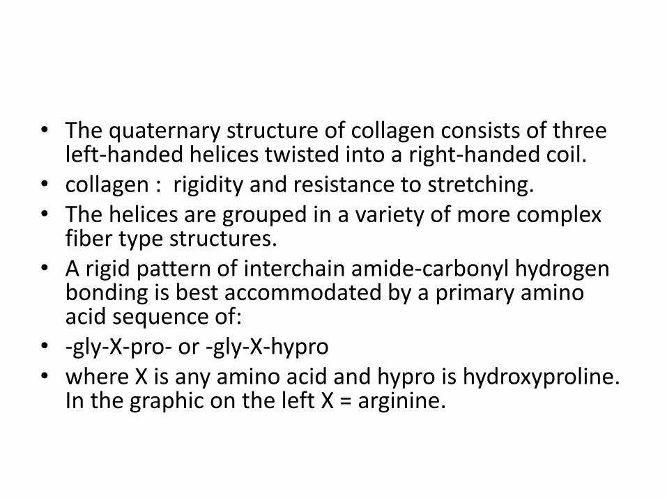
• The quaternary structure of collagen consists of three left-handed helices twisted into a right-handed coil.
• collagen : rigidity and resistance to stretching. • The helices are grouped in a variety of more complex
fiber type structures. • A rigid pattern of interchain amide-carbonyl hydrogen
bonding is best accommodated by a primary amino acid sequence of:
• -gly-X-pro- or -gly-X-hypro• where X is any amino acid and hypro is hydroxyproline.
In the graphic on the left X = arginine.
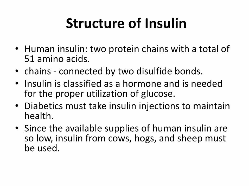
Structure of Insulin
• Human insulin: two protein chains with a total of 51 amino acids.
• chains - connected by two disulfide bonds. • Insulin is classified as a hormone and is needed
for the proper utilization of glucose. • Diabetics must take insulin injections to maintain
health. • Since the available supplies of human insulin are
so low, insulin from cows, hogs, and sheep must be used.
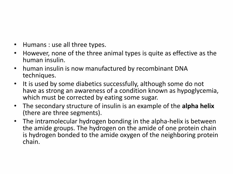
• Humans : use all three types. • However, none of the three animal types is quite as effective as the
human insulin.• human insulin is now manufactured by recombinant DNA
techniques. • It is used by some diabetics successfully, although some do not
have as strong an awareness of a condition known as hypoglycemia, which must be corrected by eating some sugar.
• The secondary structure of insulin is an example of the alpha helix(there are three segments).
• The intramolecular hydrogen bonding in the alpha-helix is between the amide groups. The hydrogen on the amide of one protein chain is hydrogen bonded to the amide oxygen of the neighboring protein chain.

References:
Image 1: Concepts in Biochemistry by Rodney F. BoyerImage 2: http://postimg.org/image/zc4dieawx/Image 3: http://postimg.org/image/3ys1bedvl/Image 4: http://postimg.org/image/7wfb0t0ox/Image 5: Concepts in Biochemistry by Rodney F. BoyerImage 6: Concepts in Biochemistry by Rodney F. BoyerImage 7: Concepts in Biochemistry by Rodney F. BoyerImage 8: Concepts in Biochemistry by Rodney F. BoyerImage 9: Concepts in Biochemistry by Rodney F. BoyerBooks: 1. Concepts in Biochemistry by Rodney F. Boyer2. Biochemistry, U. Satyanarayan, Elsevier India publication3. Principles of Biochemistry by Lehninger


![Natural Product Chemistry B.Sc. [Biochem] Course code 212 Section I.](https://static.fdocuments.us/doc/165x107/56649d415503460f94a1bd95/natural-product-chemistry-bsc-biochem-course-code-212-section-i.jpg)