BRS BASIC RADIOLOGICAL SYSTEM USER MANUAL...BRS BASIC RADIOLOGICAL SYSTEM USER MANUAL STEPHANIX 54...
34
BRS BASIC RADIOLOGICAL SYSTEM USER MANUAL STEPHANIX 54 bis, rue Jean-Baptiste David 42014 SAINT ETIENNE CEDEX 02 FRANCE E s te pr oduc to ost enta u na m ar c a C E d e ac uer do con la s d is pos ic io nes de l a D i r ecti v a 93/ 42/C E E d el 1 4 de Jun io de 19 93 s obr e Pr oduc tos Mé dic os . This product bears a CE marking in accordance with the provisions of the 93 / 42/ EEC MDD dated June 14, 1993 .
Transcript of BRS BASIC RADIOLOGICAL SYSTEM USER MANUAL...BRS BASIC RADIOLOGICAL SYSTEM USER MANUAL STEPHANIX 54...
BRS BASIC RADIOLOGICAL
SYSTEM USER MANUAL
STEPHANIX 54 bis, rue Jean-Baptiste David
42014 SAINT ETIENNE CEDEX 02 FRANCE
E s te pr oduc to ost enta u na m ar c a C E d e ac uer do con la s d is pos ic io nes de l a D i r ecti v a 93/ 42/C E E d el 1 4 de Jun io de 19 93 s obr e Pr oduc tos Mé dic os . This product bears a CE marking in accordance with the provisions of the 93 / 42/ EEC MDD dated June 14, 1993 .

BASIC RADIOLOGICAL SYSTEM BRS
STEPHANIX constantly strives to improve its products and, therefore, reserves the right to deliver,
without prior notice, machines whose characteristics differ from those described here: nonetheless, these
machines are still guaranteed to comply with regulations in force.
All rights reserved.
MDU BRS A January 2002 Page I / I

BASIC RADIOLOGICAL SYSTEM BRS
MDU BRS A January 2002 Page 1 / 30
TABLE OF CONTENTS
INTRODUCTION...........................................................................................................................................6 Preliminary Information .......................................................................................................................6
Explanations........................................................................................................................................6 GENERAL MANIPULATIONS......................................................................................................................7 Column and swivelling arm .................................................................................................................7 Cassette holder ...................................................................................................................................7 Light beam collimator ..........................................................................................................................7 X ray tube ............................................................................................................................................7 CONTROLS OF THE BRS ...........................................................................................................................8 Lungs and heart ..................................................................................................................................8 Ribs .....................................................................................................................................................9 Abdomen ...........................................................................................................................................10 Urinary tract survey ...........................................................................................................................11 Urinary bladder and inner pelvis .......................................................................................................12 Skull...................................................................................................................................................13 Sinuses and face...............................................................................................................................14 Mandible............................................................................................................................................15 Cervical spine....................................................................................................................................16 Thoracic and lumbar spine................................................................................................................17 Sacrum, lumbosacral junction and sacroiliac joints ..........................................................................18 Clavicle, scapula and shoulder joint..................................................................................................19 Humerus............................................................................................................................................20
Elbow.................................................................................................................................................21
Forearm.............................................................................................................................................22 Wrist, hand and fingers .....................................................................................................................23 Pelvis and hip joints ..........................................................................................................................24
Femur ................................................................................................................................................25
Knee ..................................................................................................................................................26
Leg ....................................................................................................................................................27
Foot and toes ....................................................................................................................................28 TABLE.........................................................................................................................................................29
The CE mark on the system indicates that the unit is in compliance with the European
Directive 93/42/CEE of the 14/06/93 concerning the medical devices.
Original language of document: FRENCH
BASIC RADIOLOGICAL SYSTEM BRS
Page 2 / 30 January 2002 MDU BRS A
GUARANTEE CONDITIONS
It is the responsibility of the user to ensure that the government regulations respecting installation and
operation of the equipment are observed.
Incorrect operation, or failure of the user to maintain the equipment in accordance with the schedule of
maintenance relieves STEPHANIX or his agent from all responsibility for consequent non-compliance,
damage, injury, defects and/or other malfunction.
The installation and equipment must not be used if any mechanical, electrical or radiation-emitting
component is defective, or if the procedures described in the schedule of maintenance have not been
carried out.
Changes and additions to the equipment may only be carried out by STEPHANIX or by third parties
expressly authorized by STEPHANIX to do so. Such changes must comply with local regulations and
accepted standards of good practice.
In case the power lines do not meet the specifications given in section A "Pre-installation" of the Service
Manual, the BRS cannot reach its maximum performance and the standard use cannot be guaranteed.
Technical files (diagrams, parts list, measurement procedure and so on...) of the
Basic Radiological System are available on request to STEPHANIX.
CARRIAGE CONDITIONS
Goods travel at owner's risk.
No contest shall be taken into account unless written reservations have been made on the consignment
note, face to face to the carrier, upon receipt of the goods in case of apparent loss or damage.
During transportation and storage, the black cap of the H.T. tank must be neither unscrewed nor
removed.
Crates and packing means made by STEPHANIX cannot be used for other purposes than carriage.
BASIC RADIOLOGICAL SYSTEM BRS
MDU BRS A January 2002 Page 3 / 30
SAFETY AND PROTECTION
X-rays are not innocuous and can be dangerous if used badly. You must, therefore, take precautions
even when following the instructions in this manual.
X-rays units manufactured by STEPHANIX comply with the strictest safety standards in force throughout
the world (Europe, Japan, USA, etc.). They guarantee optimum protection against radiation risks.
Nonetheless, you are handling a unit specifically designed to generate X-rays to allow medical diagnosis
on a film or using a digital imaging system. Consequently, despite the inherent safety of our equipment,
we recommend using conventional commercially available equipment to protect yourself and your patient
against scattered radiation risks.
The following instructions must be strictly observed:
- Use the smallest possible X-ray field
- Use the shortest possible fluoroscopy time
- Use the lowest input dose necessary for efficient working
- Maintain the X-ray tube in good condition
- Keep as far away from the beam as possible
- During exposure, personnel not directly involved with the patient must go behind lead or
lead-glass shielding
Should any part of the equipment be damaged, it must be repaired or replaced before the
equipment is returned to service.
Equipment described herein is unsuitable for operation where and when flammable gasses or
vapours are present.
The user must always electrically disconnect the equipment from the mains before cleaning or
disinfecting.
Do not allow water or other liquid to enter the equipment, as such liquid may cause short
circuits and /or some corrosion.
BASIC RADIOLOGICAL SYSTEM BRS
Page 4 / 30 January 2002 MDU BRS A
It must be noted that some disinfectants vaporise, forming potentially explosive mixtures.
Should such disinfectants be used, the vapours must first be allowed to disperse before the equipment is
returned to use.
In addition, it is vital that an authorized STEPHANIX distributor carries out the assembly,
extensions, adjustments, modifications and repairs. Also, your radiology unit must be installed in
premises that comply with IEC provisions and the standards in force.
Due care must be taken to prevent injury to the patient. Ensure that patient's clothing cannot
be trapped in the equipment. It is important that the patient hands remain on the tabletop.
It is important that all personnel dealing with X-radiation be fully acquainted with the radiation
protection.
In the event of failure to comply with these instructions, STEPHANIX shall not be held responsible for the
safety, reliability and characteristics of the machine.
Your distributor will be pleased to help you with the initial use of your unit and to answer any subsequent
questions you may have.
BASIC RADIOLOGICAL SYSTEM BRS
MDU BRS A January 2002 Page 5 / 30
NOTE TO THE USER
You have just purchased a Basic Radiological System. We congratulate you on your choice, and are sure
you will be fully satisfied with its use and diagnostic capabilities.
STEPHANIX radiology units offer high quality and advanced technology.
We recommend reading the user's manual carefully before using your unit, to become familiar with its
operation and make the most of its performance.
Keep this manual in a safe place so that you can easily refer to it in the future.
Thank you for placing your confidence in STEPHANIX.
BASIC RADIOLOGICAL SYSTEM BRS
INTRODUCTION
PRELIMINARY INFORMATION
The B.R.S. system has been design by the WHO in order to comply with a need for a small equipment,
easy to use and to manipulate for operator having a little knowledge in X ray units and techniques.
The B.R.S. system can be used with different X ray generators from STEPHANIX:
- N 32 HF, N 32 HFM, N 32 HFR
- N 40 HF
- N 50 HF
This manual explains shortly how to manipulate the B.R.S. system and gives a range of basic radiological
techniques sufficient to enable 90% of the problems diagnosable through radiography to be routinely
examined.
Concerning the X ray generator, the operator will have to refer to the relevant manual.
Since films, X ray cassettes and screens vary, the exposure required for each examination must be
adjusted to local conditions by a fully qualified radiographer / X ray technologist when the machine is
installed. If the film-cassette-screens combination is changed, readjustment of the exposure will be
necessary for the best results.
When used by trained radiographers, techniques more complex than those shown in this manual can de
carried out with the basic radiological system, serving the particular needs of specialists in various fields
of medicine.
EXPLANATIONS:
ERECT : standing or sitting up.
SUPINE : lying on the back.
PRONE : lying on the stomach.
OBLIQUE : turned a little, usually at a 40° angle.
LATERAL : lying with the side close to the cassette.
AP : antero-posterior
PA : postero-anterior
Page 6 / 30 January 2002 MDU BRS A
BASIC RADIOLOGICAL SYSTEM BRS
GENERAL MANIPULATIONS
COLUMN AND SWIVELLING ARM
- Up and down movement: the swivelling arm can go up and down just by pushing it in the right
direction. the up and down movement is fully counter balanced by a counterweight inside the column. A
manual brake, situated behind the column, on the up and down mechanism, can be used to block the
movement to the right position.
- Rotation movement: the swivelling arm is fully counter balanced and can rotate in the vertical
plane around its horizontal axis on the up and down mechanism. A big washer, with indication of the
rotation angle, is attached to the swivelling arm. A manual brake situated on the front of the up and down
mechanism is used to block the rotation movement.
CASSETTE HOLDER
The cassette holder is situated in the opposite position to the X ray tube ,and contain a fixed grid.
To insert a cassette inside pull the handle situated on the front of the tray and put the cassette between
the blocking parts which are also used to centre the cassette.
LIGHT BEAM COLLIMATOR
- Light: An halogen lamp can be switch on by pressing the button on the front of the collimator. An
integrated timer allows to switch off this light after around 30 seconds in order not to overheat the
collimator and the lead shutters.
- Lead shutters: the X ray beam can be reduced to the size of the cassette inserted in the cassette
holder, by turning the two buttons situated in front of the collimator, in the appropriate direction. Coloured
indications for positioning the shutters are also indicated on the cassette holder
X RAY TUBE
The X Ray tube is auto-centred on the cassette holder on the opposite side.
In some cases, it might be necessary to rotate the tube head in order to use the X Ray beam on an
other system, like a wall Bucky or a fluoroscopy stand. It is possible to do it, just by rotating the tube head
in the right direction. Adjustment of the surface of the beam can be made using the lead shutters of the
light beam collimator.
MDU BRS A January 2002 Page 7 / 30
BASIC RADIOLOGICAL SYSTEM BRS
CONTROLS OF THE B.R.S.
LUNGS AND HEART
- MATERIAL:
Cassette 36 x 43 cm with rare earth screens
- POSITION OF THE PATIENT:
Standing erect (chest PA)
Standing erect (chest lateral left or right)
Sitting erect (chest AP)
Supine (chest AP)
Lying on the right or left side ; horizontal beam (chest lateral decubitus AP or PA)
- PARAMETERS:
Adults: kV: from 90 to 110
mAs: from 2 to 12
Infants: kV: 70
mAs: 3
- OPERATING PROCEDURE:
1. Make sure the patient's shoulders are well pressed forward
2. Tell the patient to breathe in deeply and hold the breath
3. Expose
4. Tell the patient to breathe normally
5. For infants, use a 18 x 24 cm cassette, support the head and the legs and lie the infant on its
back
Page 8 / 30 January 2002 MDU BRS A
BASIC RADIOLOGICAL SYSTEM BRS
MDU BRS A January 2002 Page 9 / 30
RIBS
- MATERIAL:
Cassette 36 x 43 cm with rare earth screens
- POSITION OF THE PATIENT:
Standing or sitting erect ; left and right oblique (ribs oblique AP)
Supine ; right or left oblique (ribs oblique AP)
- PARAMETERS:
Adults: kV: from 60 to 80
mAs: from 20 to 60
Infants: kV: 70
mAs: 2
- OPERATING PROCEDURE:
1. Ask the patient to raise its arms
2. Tell the patient to breathe in deeply and hold the breath
3. Expose
4. Tell the patient to breathe normally
BASIC RADIOLOGICAL SYSTEM BRS
Page 10 / 30 January 2002 MDU BRS A
ABDOMEN
- MATERIAL:
Cassette 36 x 43 cm with rare earth screens
- POSITION OF THE PATIENT:
Supine (abdomen AP)
Standing erect (abdomen PA)
Lying left and right side (abdomen, lateral decubitus)
- PARAMETERS:
Adults: kV: from 75 to 85
mAs: from 40 to 120
Infants: kV: 70
mAs: 25
- OPERATING PROCEDURE:
1. Press the patient's abdomen against the cassette holder
2. Tell the patient to breath out and hold the breath
3. Expose
4. Tell the patient to breathe normally
5. For infants, use a 24 x 30 cm cassette, hold the child hanging by the upper arms
BASIC RADIOLOGICAL SYSTEM BRS
MDU BRS A January 2002 Page 11 / 30
URINARY TRACT SURVEY
- MATERIAL:
Cassette 36 x 43 cm with rare earth screens
- POSITION OF THE PATIENT:
Supine (urinary tract AP)
- PARAMETERS:
Adults: kV: from 65 to 75
mAs: from 75 to 180
Infants: kV: 70
mAs: 20
- OPERATING PROCEDURE:
1. Tell the patient to breath out and hold the breath
2. Expose
3. Tell the patient to breathe normally
5. For infants, use a 24 x 30 cm cassette
BASIC RADIOLOGICAL SYSTEM BRS
Page 12 / 30 January 2002 MDU BRS A
URINARY BLADDER AND INNER PELVIS
- MATERIAL:
Cassette 24 x 30 cm with rare earth screens
- POSITION OF THE PATIENT:
Supine ; vertical beam angled 20° toward the feet
- PARAMETERS:
Adults: kV: from 65 to 75
mAs: from 75 to 180
Infants: kV: 70
mAs: 20
- OPERATING PROCEDURE:
1. Tell the patient to breath out and hold the breath
2. Expose
3. Tell the patient to breathe normally
5. For infants, use a 24 x 30 cm cassette
BASIC RADIOLOGICAL SYSTEM BRS
MDU BRS A January 2002 Page 13 / 30
SKULL
- MATERIAL:
Cassette 24 x 30 cm with rapid screens
- POSITION OF THE PATIENT:
Supine ; horizontal beam (skull lateral)
Prone ; vertical beam angled 20° towards the feet (skull PA)
Supine ; vertical beam angled 20° towards the head (skull AP)
Supine ; vertical beam angled 30° towards the feet (skull semi-axial)
- PARAMETERS:
Adults: kV: from 65 to 75
mAs: from 32 to 80
Infants: kV: 65
mAs: 20
- OPERATING PROCEDURE:
1. Remove dentures, hair grips, or anything else in the hair
2. For supine position, the patient head should be raised on a foam rubber pad
3. For prone position, nose and forehead should be against the table and hands under the chest
4. Expose
BASIC RADIOLOGICAL SYSTEM BRS
Page 14 / 30 January 2002 MDU BRS A
SINUSES AND FACE
- MATERIAL:
Cassette 18 x 24 cm with rapid screens
- POSITION OF THE PATIENT:
Sitting erect (sinuses and face PA)
Sitting erect (sinuses or face semiaxial, or nose PA)
Sitting erect (sinuses, face, or nose lateral)
- PARAMETERS:
Adults: kV: from 70 to 90
mAs: from 25 to 50
Infants: kV: 65
mAs: 16
- OPERATING PROCEDURE:
1. Remove dentures, hair grips, or anything else in the hair
2. For face PA position, the patient nose should touch the cassette holder, face vertical
3. For face semiaxial position, tell the patient to open the mouth as widely as possible and place
the chin against the cassette holder and tilt the patient's head back 45°
4. Expose
BASIC RADIOLOGICAL SYSTEM BRS
MDU BRS A January 2002 Page 15 / 30
MANDIBLE
- MATERIAL:
Cassette 18 x 24 cm with rapid screens
- POSITION OF THE PATIENT:
Sitting erect (mandible PA)
Sitting erect (mandible oblique lateral)
Supine ; vertical beam angled 30° towards the feet (mandible AP)
Lying on the right (or left) side ; vertical beam angled 30° towards the head (mandible oblique
lateral)
- PARAMETERS:
Adults: kV: from 65 to 75
mAs: from 25 to 63
Infants: kV: 65
mAs: 16
- OPERATING PROCEDURE:
1. Remove dentures, hair grips, or anything else in the hair
2. For mandible PA position, tell the patient to open the mouth as widely as possible and place the
forehead and nose against the cassette holder
3. For mandible oblique lateral position, tilt the head inwards 15° so that the head and shoulder
rest against the cassette holder, patient sit with the side of the head to be X rayed nearest to the cassette
holder
3. For mandible AP position, tell the patient to open the mouth as widely as possible
4. For lying position in mandible oblique lateral position, tilt the patient head 15° towards the table
5. Expose
BASIC RADIOLOGICAL SYSTEM BRS
Page 16 / 30 January 2002 MDU BRS A
CERVICAL SPINE
- MATERIAL:
Cassette 18 x 24 cm or 24 x 30 cm with rapid screens
- POSITION OF THE PATIENT:
Sitting erect (cervical spine PA)
Sitting erect (cervical spine lateral)
Sitting erect (cervical spine oblique)
Supine ; vertical beam angled 15° towards the head (cervical spine AP)
Supine ; horizontal beam (cervical spine lateral)
- PARAMETERS:
Adults: kV: from 65 to 75
mAs: from 50 to 100
Infants: kV: 65
mAs: 32
- OPERATING PROCEDURE:
1. Remove dentures, hair grips, or anything else in the hair
2. For cervical spine PA position, tell the patient to place the chin against the cassette holder
3. Tell the patient to hold the breath
4. Expose
5. Tell the patient to breathe normally
BASIC RADIOLOGICAL SYSTEM BRS
MDU BRS A January 2002 Page 17 / 30
THORACIC AND LUMBAR SPINE
- MATERIAL:
Cassette 35 x 43 cm divided by two with, rare earth screens
- POSITION OF THE PATIENT:
Supine (thoracic or lumbar spine AP)
Lying on the left (or right) side (thoracic or lumbar spine lateral)
Supine ; horizontal beam (thoracic or lumbar spine lateral)
- PARAMETERS:
Adults: kV: from 65 to 80
mAs: from 63 to 120
Infants: kV: 65
mAs: 40
- OPERATING PROCEDURE:
1. For lumbar spine AP, the patient's knees should be bent so that the back is flat on the table
2. Tell the patient to hold the breath
3. Expose
4. Tell the patient to breathe normally
BASIC RADIOLOGICAL SYSTEM BRS
Page 18 / 30 January 2002 MDU BRS A
SACRUM, LUMBOSACRAL JUNCTION AND SACROILIAC JOINTS
- MATERIAL:
Cassette 24 x 30 cm with rapid screens
- POSITION OF THE PATIENT:
Supine ; vertical beam angled towards the head at 10° for men and 15° for women
- PARAMETERS:
Adults: kV: from 85 to 95
mAs: from 63 to 160
Infants: kV: 75
mAs: 63
- OPERATING PROCEDURE:
1. Tell the patient to hold the breath
2. Expose
3. Tell the patient to breathe normally
BASIC RADIOLOGICAL SYSTEM BRS
MDU BRS A January 2002 Page 19 / 30
CLAVICLE, SCAPULA AND SHOULDER JOINT
- MATERIAL:
Cassette 24 x 30 cm with rapid screens
- POSITION OF THE PATIENT:
Supine ; vertical beam angled 20° towards the head (clavicle AP)
Supine (scapula)
Sitting erect (scapula lateral)
Supine ; vertical beam angled 10° towards the feet (shoulder AP)
Supine (shoulder AP abducted)
Sitting erect (shoulder AP: after injury)
Sitting erect (shoulder lateral: after injury)
- PARAMETERS:
Adults: kV: from 60 to 75
mAs: from 40 to 120
Infants: kV: 55
mAs: from 6 to 20
- OPERATING PROCEDURE:
1. Put a pillow under the head for each supine position
2. For scapula lateral position raise the arm with the hand behind the head
3. For supine shoulder AP position, place a pillow under the normal shoulder and turn the patient
so that the shoulder to be X rayed lies flat on the table.
4. Expose
BASIC RADIOLOGICAL SYSTEM BRS
Page 20 / 30 January 2002 MDU BRS A
HUMERUS
- MATERIAL:
Cassette 35 x 43 cm divided by two, with rapid screens
- POSITION OF THE PATIENT:
Supine (humerus AP and lateral)
Sitting erect (humerus AP: after injury)
Sitting erect (humerus lateral: after injury)
- PARAMETERS:
Adults: kV: from 55 to 70
mAs: from 8 to 25
Infants: kV: 50
mAs: 4
- OPERATING PROCEDURE:
1. For supine position, rotate the arm outwards, palm of the hand facing up for a first picture, and
angle downwards for the second picture
2. For sitting erect, humerus AP ,the patient's forearm should be at right angle to the cassette
holder
3. For sitting erect, humerus lateral, the patient's forearm should be against the cassette holder
4. Expose
BASIC RADIOLOGICAL SYSTEM BRS
MDU BRS A January 2002 Page 21 / 30
ELBOW
- MATERIAL:
Cassette 18 x 24 cm, with rapid screens
- POSITION OF THE PATIENT:
Sitting with the arm in supine position (elbow AP)
Sitting (elbow lateral)
Sitting with the arm in supine position (elbow AP semi-flexed: after injury)
- PARAMETERS:
Adults: kV: from 50 to 60
mAs: from 6 to 10
Infants: kV: 45
mAs: 4
- OPERATING PROCEDURE:
1. For elbow AP the palm of the patient's hand should be facing up
2. Expose
BASIC RADIOLOGICAL SYSTEM BRS
Page 22 / 30 January 2002 MDU BRS A
FOREARM
- MATERIAL:
Cassette 24 x 30 cm, with rapid screens
- POSITION OF THE PATIENT:
Sitting with the arm in supine position (forearm AP)
Sitting (forearm lateral)
Sitting with the arm in prone position and the elbow flexed (forearm PA: after injury)
- PARAMETERS:
Adults: kV: from 50 to 60
mAs: from 4 to 12
Infants: kV: 45
mAs: 3
- OPERATING PROCEDURE:
1. For forearm AP the palm of the patient's hand should be facing up
2. For forearm lateral, the thumb should be uppermost
3. For forearm PA the palm of the patient's hand should be facing down
4. Add 15 kV if the arm is in a plastter
5. Expose
BASIC RADIOLOGICAL SYSTEM BRS
MDU BRS A January 2002 Page 23 / 30
WRIST, HAND AND FINGERS
- MATERIAL:
Cassette 18 x 24 cm or 24 x 30 cm, with rapid screens
- POSITION OF THE PATIENT:
Sitting (wrist PA)
Standing or sitting (wrist lateral)
Sitting (hand PA and oblique)
Standing (finger lateral)
- PARAMETERS:
Adults: kV: from 40 to 46
mAs: from 4 to 10
Infants: kV: 40
mAs: 2
- OPERATING PROCEDURE:
1. For wrist PA the hand should be straight with the palm facing down
2. For wrist lateral, the thumb should be uppermost
3. For a finger lateral, extend the finger to be X rayed horizontally
4. Expose
BASIC RADIOLOGICAL SYSTEM BRS
Page 24 / 30 January 2002 MDU BRS A
PELVIS AND HIP JOINTS
- MATERIAL:
Cassette 36 x 43 cm or 24 x 30 cm, with rare earth screens
- POSITION OF THE PATIENT:
Supine (pelvis AP)
Supine (hip joint AP)
Supine (hip joint lateral: no injury)
Supine ; X ray beam angled at 65° from the vertical position (hip joint lateral: if a fracture is
detected)
- PARAMETERS:
Adults: kV: from 65 to 75
mAs: from 63 to 100
Infants: kV: 50
mAs: 12
- OPERATING PROCEDURE:
1. For pelvis AP and hip joint AP, turn the feet heels apart and toes together
2. For hip joint lateral, no injury, turn the patient on the affected side so that the leg is flat on the
table
3. Expose
BASIC RADIOLOGICAL SYSTEM BRS
MDU BRS A January 2002 Page 25 / 30
FEMUR
- MATERIAL:
Cassette 36 x 43 cm divided by two, with rare earth screens
- POSITION OF THE PATIENT:
Supine (femur AP)
Lying on the right (or left) side (femur lateral: no injury)
Supine ; horizontal beam (femur lateral: after injury)
- PARAMETERS:
Adults: kV: from 55 to 60
mAs: from 12 to 24
Infants: kV: 46
mAs: 10
- OPERATING PROCEDURE:
1. For femur lateral, no injury place the patient with the leg to be X rayed underneath and bend the
normal leg
2. For femur lateral, after injury, place the patient so that the injured leg is nearest to the cassette
and bend the uninjured leg
3. Expose
BASIC RADIOLOGICAL SYSTEM BRS
Page 26 / 30 January 2002 MDU BRS A
KNEE
- MATERIAL:
Cassette 18 x 24 cm , with rapid screens
- POSITION OF THE PATIENT:
Supine (knee AP)
Lying on the right (or left) side (knee lateral: no injury)
Supine ; horizontal beam (knee lateral: after injury)
Supine ; vertical beam angled 30° towards the head (knee intercondylar space)
Standing (patella axial)
- PARAMETERS:
Adults: kV: from 52 to 58
mAs: from 12 to 24
Infants: kV: 46
mAs: 10
- OPERATING PROCEDURE:
1. For knee lateral, no injury place the patient with the leg to be X rayed underneath and bend the
normal leg
2. For knee lateral, after injury, place the patient so that the injured leg is nearest to the cassette
and raise the uninjured leg so that it is higher than the cassette
3. For intercondylar space, place the cassette under the knee on a firm support 18-20 cm high
4. For patella axial, place the cassette on top of the cassette holder on a stool
5. Expose
BASIC RADIOLOGICAL SYSTEM BRS
MDU BRS A January 2002 Page 27 / 30
LEG
- MATERIAL:
Cassette 36 x 43 cm divided by two or 18 x 24 cm, with rare earth screens
- POSITION OF THE PATIENT:
Supine (leg AP)
Lying on the right (or left) side (leg lateral: no injury)
Supine ; horizontal beam (leg lateral: after injury)
Sitting on the table (ankle joint: internal oblique and AP)
Sitting or lying on the table (ankle joint: lateral and external oblique)
- PARAMETERS:
Adults: kV: from 52 to 54
mAs: from 8 to 12
Infants: kV: 46
mAs: 6
- OPERATING PROCEDURE:
1. For leg lateral, no injury place the patient with the leg to be X rayed underneath and bend the
normal leg
2. For leg lateral, after injury, if there are splints on the injured leg, leave them on and bend the
uninjured leg
3. Expose
BASIC RADIOLOGICAL SYSTEM BRS
Page 28 / 30 January 2002 MDU BRS A
FOOT AND TOES
- MATERIAL:
Cassette 24 x 30 cm, with rare rapid screens
- POSITION OF THE PATIENT:
Supine ; vertical beam angled 10° towards the head (foot and toes AP)
Lying on the right (or left) side (foot lateral)
Sitting on the table ; vertical beam angled 10° towards the head (foot AP oblique)
Prone (foot PA oblique)
Supine ; vertical beam angled 30° towards the head (heel semi-axial)
- PARAMETERS:
Adults: kV: from 42 to 50
mAs: from 6 to 10
Infants: kV: 40
mAs: 4
- OPERATING PROCEDURE:
1. For foot and toes AP bend the patient's leg so that the foot rests flat on the table
2. For heel semi-axial the foot should be kept flexed by means of a sling held by the patient
3. Expose
BASIC RADIOLOGICAL SYSTEM BRS
MDU BRS A January 2002 Page 29 / 30
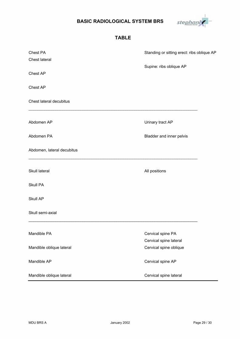
TABLE
Chest PA Standing or sitting erect: ribs oblique AP
Chest lateral
Supine: ribs oblique AP
Chest AP
Chest AP
Chest lateral decubitus
___________________________________________________________________________
Abdomen AP Urinary tract AP
Abdomen PA Bladder and inner pelvis
Abdomen, lateral decubitus
___________________________________________________________________________
Skull lateral All positions
Skull PA
Skull AP
Skull semi-axial
___________________________________________________________________________
Mandible PA Cervical spine PA
Cervical spine lateral
Mandible oblique lateral Cervical spine oblique
Mandible AP Cervical spine AP
Mandible oblique lateral Cervical spine lateral
BASIC RADIOLOGICAL SYSTEM BRS
Page 30 / 30 January 2002 MDU BRS A
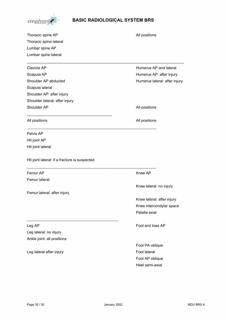
Thoracic spine AP All positions
Thoracic spine lateral
Lumbar spine AP
Lumbar spine lateral
__________________________________________________________________________
Clavicle AP Humerus AP and lateral
Scapula AP Humerus AP: after injury
Shoulder AP abducted Humerus lateral: after injury
Scapula lateral
Shoulder AP: after injury
Shoulder lateral: after injury
Shoulder AP All positions
________________________________________
All positions All positions
_____________________________________________________________
Pelvis AP
Hit joint AP
Hit joint lateral
Hit joint lateral: if a fracture is suspected
_____________________________________________________________
Femur AP Knee AP
Femur lateral
Knee lateral: no injury
Femur lateral: after injury
Knee lateral: after injury
Knee intercondylar space
Patella axial
___________________________________________
Leg AP Foot and toes AP
Leg lateral: no injury
Ankle joint: all positions
Foot PA oblique
Leg lateral after injury Foot lateral
Foot AP oblique
Heel semi-axial


![BRS Neuroanatomy [2nd Edition]](https://static.fdocuments.us/doc/165x107/55cf9c36550346d033a90bfd/brs-neuroanatomy-2nd-edition.jpg)