BRONCHIAL ASTHMA Dr. Maha Arafah, Dr. Ammar Al-Rikabi Department of Pathology KSU 2015 RESPIRATORY...
47
BRONCHIAL ASTHMA Dr. Maha Arafah, Dr. Ammar Al-Rikabi Department of Pathology KSU 2015 RESPIRATORY BLOCK
-
Upload
clifford-atkinson -
Category
Documents
-
view
220 -
download
1
Transcript of BRONCHIAL ASTHMA Dr. Maha Arafah, Dr. Ammar Al-Rikabi Department of Pathology KSU 2015 RESPIRATORY...

BRONCHIAL ASTHMA Dr. Maha Arafah, Dr. Ammar Al-RikabiDepartment of PathologyKSU2015
RESPIRATORY BLOCK

Objectives Define asthma.Compare and contrast immune-
mediated and nonimmune-mediated forms of asthma in terms of initiating factors and pathogenic mechanisms.
Understand the term bronchial hyperresponsiveness
Describe the morphologic changes in chronic asthma, and discuss the clinical course.

INTRODUCTION TO THE RESPIRATORY TRACT

NORMAL LUNG

The right and left main bronchi divide into lobar bronchi.
The lobar bronchi divide into tertiary/segmental bronchi, each of which supplies a bronchopulmonary segment.
The segmental bronchi divide into primary bronchioles which divide into terminal bronchioles and then divide into respiratory bronchioles, which go on to divide into alveolar ducts.
Each alveolar duct divides into five or six alveolar sacs.
The alveolar sacs are made up of alveoli. The alveolus is the basic anatomical unit of gas exchange in the lung.
Beyond terminal bronchiole gas exchange occurs
The distal airspaces are kept open by elastic tension in alveolar walls

Function of lungs….Gas exchange
(O2, CO2)◦ Depends on
compliance (stretchability) of lungs
◦ Can only occur in alveoli that are both ventilated and perfused

Spirometer is an equipments used for measuring
the volume of air inspired and expired by the lungs ( Pulmonary Function Tests)

Spirometry (pulmonary physiology) Forced expiratory volume (FEV1): volume of air blown
out forcibly in 1 second. A function of large airways. Dependent on body size.
Vital capacity (VC): total volume of expired air.
Diffusing capacity or Transfer factor of the lung for carbon monoxide (DLCO or TLCO): absorption of carbon monoxide in one breath (gas exchange). It is dependent on the concentration of blood haemoglobin, which has a strong affinity for CO and it assesses the ability of the lungs to exchange gas efficiently.

Classification of Lung Disease based on distinctive clinical and physiological features:
1. Obstructive lung diseases: Is a category of respiratory disease characterized by
airway obstruction due to partial or complete obstruction at any level from trachea to respiratory bronchioles. It is generally characterized by inflamed and easily collapsible airways, obstruction to airflow and problems exhaling.
Pulmonary function test: decreased FEV1 and decreased FEV1/VC.
2. Restrictive lung diseases: Are a category of diseases that restrict lung expansion,
resulting in a decreased total lung capacity, increased work of breathing, and inadequate ventilation and/or oxygenation.
Both forced expiratory volume in one second (FEV1) and forced vital capacity (FVC) are reduced with normal to high FEV1/VC and decreased Tco. The expiratory flow rate is near normal.

Types of (diffuse) Obstructive Lung Diseases
1) Bronchial Asthma
2) Chronic obstructive pulmonary disease (COPD). It is also known as Chronic obstructive airway disease (COAD) or Chronic obstructive lung disease (COLD).They are of two types:
a) Chronic bronchitis
b) Emphysema c) Bronchiectasis
Common symptoms in lung disease
– Dyspnea: difficulty with breathing
– Cough
– Hemoptysis

BRONCHIAL ASTHMA
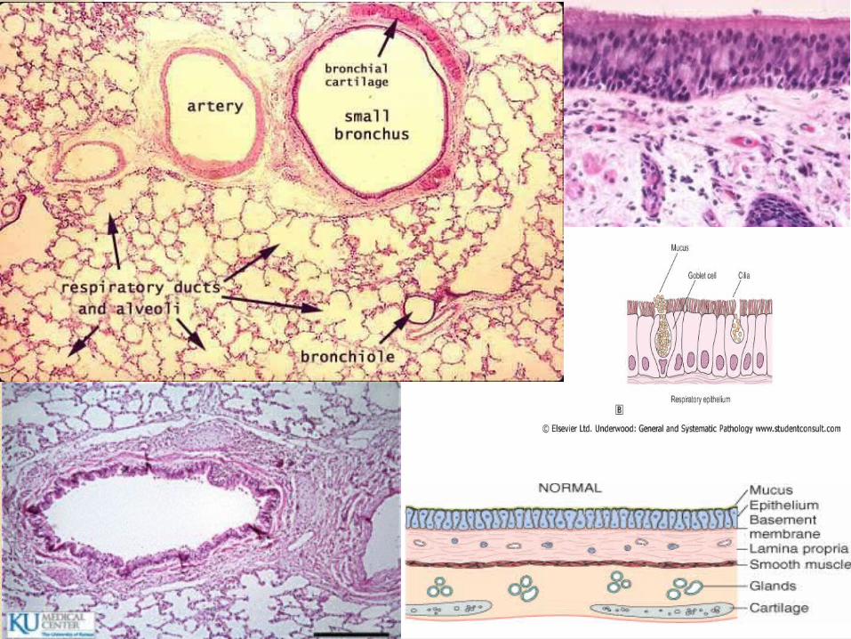
BRONCHIAL ASTHMA (BA)
• It is a chronic relapsing inflammatory obstructive lung disease characterized by hyper-reactive airways
• It is episiodic, reversible bronchoconstriction/spasm characterized by hyper-irratibility of the airways due to increased responsiveness of the tracheobronchial tree to various stimuli
• It results in bronchial constriction, odema, inflammation, excess mucus production and bronchial smooth muscle hypertrophy.

BRONCHIAL ASTHMA (BA)
• It is a triad of:1. intermittent and reversible airway
obstruction2. chronic bronchial inflammation with
eosinophils3. bronchial smooth muscle cell hypertrophy
and hyper-reactivity• Primarily targets the bronchi and terminal
bronchioles• Most common chronic respiratory disease in
children. More common in children than adultsAsthma animation:
http://link.brightcove.com/services/player/bcpid236059233?bctid=347806802

Bronchial asthma :
It has been divided into two basic types:1. Extrinsic asthma.2. Intrinsic asthma.
Sometimes extrinsic and intrinsic can co-exist in the same patient
Extrinsic (atopic, allergic) Asthma 70%
- Initiated by type 1 hypersensivity reaction induced by exposure to extrinsic antigen/allergens e.g. food, pollen, dust, etc.
- Subtypes include:
a) atopic (allergic)asthma.
b) occupational asthma.
c) allergic bronchopulmonary aspergillosis.
- Develop early in life
Intrinsic (non-atopic)Asthma 30%- Initiated by diverse, non-immune mechanisms e.g. infections, drugs like aspirin, pollutants, inhaled chemical irritants, cold, stress and exercise.
- No personal or family history of allergic reaction.
- Develop later in life

Extrinsic/ Allergic BA It is also known as allergic, immune mediated, atopic
or reaginic asthma. Bronchospasm is induced by inhaled antigens, usually
in children with a personal or family history of allergic disease (e.g., eczema, urticaria, or hay fever).
Atopic (allergic) asthma is the most common type and begins in childhood
Other allergic manifestation may be present: allergic rhinitis, urticaria, eczema.
Skin test with antigen is positive and results in an immediate wheel and flare reaction
Other family members are commonly also affected immune related with the involvement of TH2 subset of
CD4+ T cells

wheel and flare reaction

Pathogenesis of Bronchial Asthmacomplex and involves the following
components:1. Chronic airway inflammation2. Intermittent airflow obstruction.
Airflow obstruction can be caused by a variety of changes, including acute bronchoconstriction, airway edema, chronic mucous plug formation, and airway remodeling
3. Bronchial hyper-responsiveness causes exaggerated bronchoconstriction. The degree of airway hyper-responsiveness generally correlates with the clinical severity of asthma.

Pathogenesis of Bronchial AsthmaPrincipal cells in asthma: mast
cells, eosinophils, epithelial cells, macrophages, and activated T lymphocytes (TH2 subset) and neutrophils.
T lymphocytes play an important role in the regulation of airway inflammation through the release of numerous cytokines
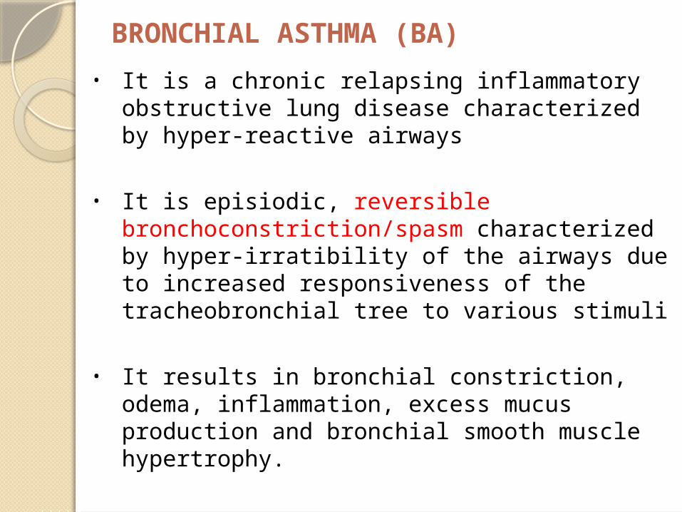
The pathogenetic mechanisms have been best studied in atopic asthma

Inhaled Allergens
House Dust Mites
Mold
Pollen
Animal Hair and Dander

Pathogenesis of Bronchial Asthma; type 1 IgE-mediated Atopic Asthma First there is initial sensitization or priming: first time
exposure to an inhaled allergen which stimulates induction of Th2-type T cells (CD4 TH2) to produce cytokines(interleukin IL- 4, IL-5 and IL-13)
IL-4 plays a role in cross-linking of immunoglobulins in B lymphocytes, promote production of IgE and mast cells.
IL-5 stimulates production and activation (recruitment) of eosinophils.
IL-13 is needed for IgE formation. And then there is subsequent re-exposure to the
allergen will leads to an IgE mediated reaction. This IgE-mediated reaction to inhaled allergens elicits:
1. an acute response (within minutes)
2. a late phase reaction (after 4-8 hours)

Acute-phase response • Begin 30 to 60 minutes after inhalation of
antigen/aeroallrgens(e.g. allergens, drugs, cold, exercise).
• The exposure results in the stimulation and degranulation of mast cells, eosinophils, and basophils with the release of inflammatory mediators from these cells and also from activated macrophages. The released mediators induce bronchoconstriction/spasm, increased vascular permeability, inflammation and injury of the bronchial walls and bronchial epithelium and excess mucous secretion.

Late phase reaction/ late asthmatic response: It is the airway edema which occurs 6-24 hours
following allergen exposure. The arrival of leukocytes at the site of mast cell
degranulation leads to release of more mediators to activate more mast cells
Discharge of eosinophil granules releases major basic protein, eosinophilic cationic protein and eosinophil peroxidase into the bronchial lumen. These substances are toxic to epithelial cells and cause epithelial cell damage and further impair mucociliary function.
Moreover, chemotactic factors like leukotriene B4, eosinophil chemotactic factor and PAF recruit more eosinophils, neutrophils and platelets to the bronchial wall, and so the vicious circle continues and prolongs and amplifies the asthmatic attack.
All these factors amplify and sustain injury without additional antigen.
Pathogenesis of Bronchial Asthma using Atopic Asthma as a model
Late phase reaction/ late asthmatic response: It is the airway edema which occurs 6-24 hours following allergen
exposure. The arrival of leukocytes at the site of mast cell degranulation
leads to release of more mediators to activate more mast cells Discharge of eosinophil granules releases major basic protein,
eosinophilic cationic protein and eosinophil peroxidase into the bronchial lumen. These substances are toxic to epithelial cells and cause epithelial cell damage and further impair mucociliary function.
Moreover, chemotactic factors like leukotriene B4, eosinophil chemotactic factor and PAF recruit more eosinophils, neutrophils and platelets to the bronchial wall, and so the vicious circle continues and prolongs and amplifies the asthmatic attack.
All these factors amplify and sustain injury without additional antigen.
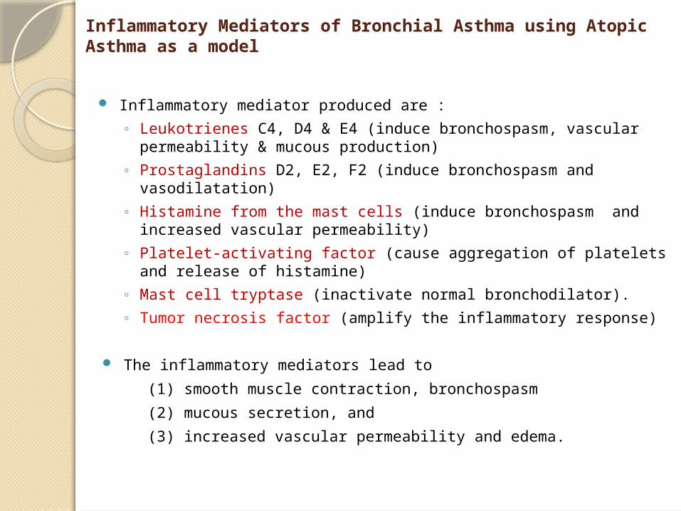
Inflammatory Mediators of Bronchial Asthma using Atopic Asthma as a model
Inflammatory mediator produced are :
◦ Leukotrienes C4, D4 & E4 (induce bronchospasm, vascular permeability & mucous production)
◦ Prostaglandins D2, E2, F2 (induce bronchospasm and vasodilatation)
◦ Histamine from the mast cells (induce bronchospasm and increased vascular permeability)
◦ Platelet-activating factor (cause aggregation of platelets and release of histamine)
◦ Mast cell tryptase (inactivate normal bronchodilator).
◦ Tumor necrosis factor (amplify the inflammatory response)
The inflammatory mediators lead to
(1) smooth muscle contraction, bronchospasm
(2) mucous secretion, and
(3) increased vascular permeability and edema.
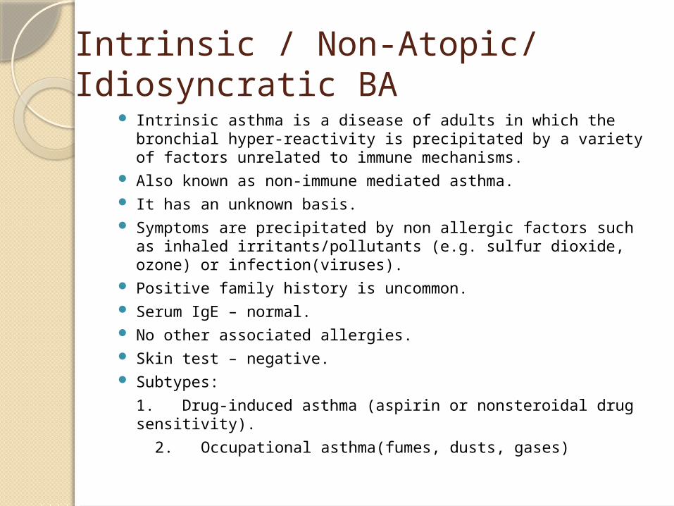
Intrinsic / Non-Atopic/ Idiosyncratic BA
Intrinsic asthma is a disease of adults in which the bronchial hyper-reactivity is precipitated by a variety of factors unrelated to immune mechanisms.
Also known as non-immune mediated asthma. It has an unknown basis. Symptoms are precipitated by non allergic factors such as
inhaled irritants/pollutants (e.g. sulfur dioxide, ozone) or infection(viruses).
Positive family history is uncommon. Serum IgE – normal. No other associated allergies. Skin test – negative. Subtypes:
1. Drug-induced asthma (aspirin or nonsteroidal drug sensitivity).
2. Occupational asthma(fumes, dusts, gases)

Morphology of Asthma: the pathologic findings are similar in both types BA
Grossly: - lung over distended (over inflation), occlusion of bronchi and bronchioles by thick mucous.
the bronchi have thickened walls with narrowed lumina and generally are filled with plugs of mucus in acute attack


Hyperinflated lung

Mucus plugs

Morphology of Asthma: the pathologic findings are similar in both types BA:
Histologic finding:
– Thick basement membrane– Infiltration of eosinophils. – Edema and inflammatory
infiltrate in bronchial wall.– Chronic mucous plug
formation. It consists of an exudate of serum proteins and cell debris that may take weeks to resolve
– Mucous contain Curschmann spirals, eosinophil and Charcot-Leyden crystals.
– Submucosal glands increased.
– Hypertrophy of the bronchial wall muscle.

Morphology of Asthma
Curschmann spiralsCoiled, basophilic
plugs of mucus formed in the lower airways and found in sputum and tracheal washings
Charcot-Leyden crystals.Eosinophilic
needle-shaped crystalline structures.
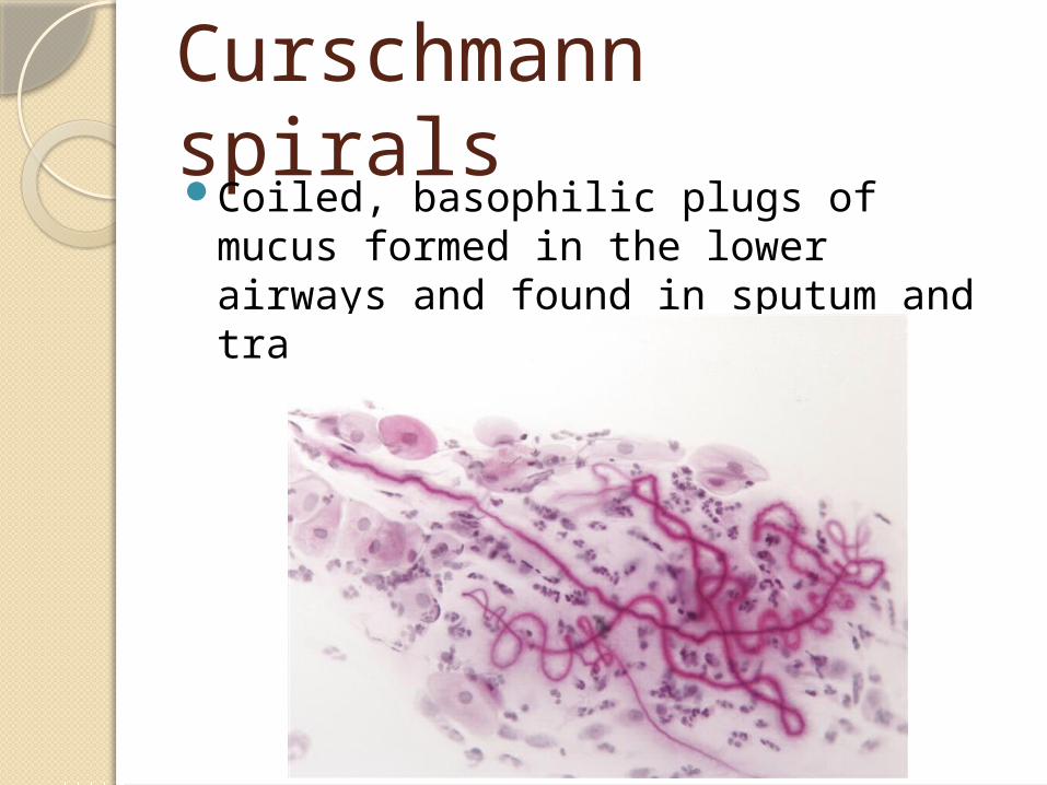
Curschmann spiralsCoiled, basophilic plugs of mucus
formed in the lower airways and found in sputum and tracheal washings


CLINICAL COURSE of BA The clinical manifestations vary from
occasional wheezing to paroxysms of dyspnea and respiratory distress.
In a classic asthmatic attack there is dyspnea, cough, difficult expiration, progressive hyperinflation of lung and mucous plug in bronchi. This may resolve spontaneously or with treatment.
Nocturnal cough Increased anteroposterior diameter, due to
air trapping and increase in residual volume
Status asthmaticus – severe cyanosis and persistent dyspnea, may be fatal

COMPLICATIONS OF ASTHMA
Airway remodeling: some persons with long standing asthma develop permanent structural changes in the airway with a progressive loss of lung function that increase airflow obstruction and airway responsiveness.
Superimposed infection i.e. pneumonia Chronic bronchitis (i.e.Asthmatic
bronchitis: chronic bronchitis with superimposed asthma)
Emphysema AND pneumothorax Bronchiectasis Respiratory failure requiring intubation
in severe exacerbations i.e. status asthmaticus
In some cases cor pulmonale and heart failure develop.
STATUS ASTHMATICUS• It is the most severe form of asthma. It refers to
severe bronchoconstriction that does not respond to the drugs that usually abort the acute attack. There is severe acute paroxysm of respiratory distress.
• This situation is potentially serious and requires hospitalization. Patients in status asthmaticus have hypoxemia and often hypercapnia.
• In particularly severe episodes the ventilatory functions may be so impaired so as to cause severe cyanosis and even death.
• They require oxygen and other pharmacologic interventions.
• It may persists for days and even weeks.
Prognosis Approximately half the children
diagnosed with asthma in childhood outgrow their disease by late adolescence or early adulthood and require no further treatment.
Patients with poorly controlled asthma develop long-term changes over time (i.e. with airway remodeling). This can lead to chronic symptoms and a significant irreversible component to their disease.
Many patients who develop asthma at an older age also tend to have chronic symptoms.
Prevention Control of factors contributing to
asthma severity as exposure to irritants or allergens has been shown to increase asthma symptoms and cause exacerbations.
Clinicians should evaluate patients with persistent asthma for allergen exposures and sensitivity to seasonal allergens. Skin testing results should be used to assess sensitivity to common indoor allergens.

Asthma: chronic relapsing, reversible inflammatory obstructive lung disease
• 1.Extrinsic asthma: Type 1 Hypersensitivity reaction, IgE, childhood, family Hx of allergy.
• 2.Intrinsic asthma: associated e bronchial asthma, aspirin, exercise, cold induced. No Hx of allergy
Types
•Hypertrophy of bronchial smooth muscle & hyperplasia of goblet cells e eosinophils
•Mucous plug e Curschmann spirals & Charcot-Leyden crystals.
Morphology
•Superimposed infection
•Chronic bronchitis
•Pulmonary emphysema
•Status asthmaticus
Complication

Case scenarioAn 18-year-old black man presented
with wheezing and difficulty breathing. These attacks occurred intermittently and were not related to any known circumstances. An x-ray of the chest was unremarkable, but lung function tests performed when he was symptomatic a markedly decreased FEV1, which improved significantly after he inhaled a few puffs of a β-adrenergic agonist.
The patient was prescribed a β-adrenergic inhaler. Although the inhaler provided some relief, the patient continued to experience episodes of breathlessness, and a steroid inhalant was prescribed, which provided much greater relief. Four years later, he presented to the emergency department with severe shortness of breath of 8 hours' duration, he started that he had stopped taking all medication several weeks before because of financial reasons.

On arrival at the emergency department, the patient was in considerable distress; he could barely talk, and his respirations were 30/min. Physical examination was remarkable for rare wheezing and markedly diminished breath sounds. Arterial blood gas values were pH 6.9, PCO2 88 mm Hg, and PO2 35 mm Hg. While awaiting therapy, the patient experienced a cardiac arrest and could not be resuscitated.
At autopsy, gross findings were limited to the respiratory tract. The lungs were overinflated, and had focal areas of atelectasis, and many of the bronchi were occluded by thick, tenacious, mucous plugs.
What triggers an attack of asthma? The following are triggers of an asthma attack: (1) environmental allergens, including dusts, pollens, dander, and foods; (2) respiratory irritants, including smoke and occupational exposures to fumes, and chemicals; (3) respiratory infections, especially viral; (4) medications, especially aspirin; and (5) other triggers, including exercise, stress, cold, and menses.

What is the mechanism of the early symptoms, and what are the causes of effects that appear several hours after exposure to an allergen? Acute early-phase reaction: Exposure of presensitized IgE-coated mast cells to the same or cross-reacting antigens results in an acute early-phase reaction from the release of histamine (which causes bronchospasm), leukotrienes (which attract leukocytes and eosinophils and release mucus), and platelet activating factor (which causes more release of histamine and serotonin from platelets).

Late-phase effects: Cytokines released by leukocytes, eosinophils, and basophils recruited during the early phase contribute to the late-phase effects. Histamine from basophils causes bronchoconstriction and edema; neutrophils cause inflammatory damage; and the major basic protein of eosinophils causes epithelial damage and shedding and contributes to bronchoconstriction.
Are all forms of asthma associated with type I hypersensitivity reactions? No. Intrinsic asthma is not triggered by type I hypersensitivity. The precise causes of hyperreactive airways in intrinsic asthma are not known.