
BROCCOLI ISOTHIOCYANATE CONTENT AND IN VITRO...
14
Macedonian Journal of Chemistry and Chemical Engineering, Vol. 32, No. 2, pp. 251–264 (2013) ISSN 1857-5552 UDC: 547.239:635.356 Original scientific paper MJCCA9 – 624 Received: May 30, 2013 Accepted: July 12, 2013 BROCCOLI ISOTHIOCYANATE CONTENT AND IN VITRO AVAILABILITY ACCORDING TO VARIETY AND ORIGIN M. Carmen Rodríguez-Hernández 1,‡ , Sonia Medina 2,‡ , Angel Gil-Izquierdo 2 , M. Carmen Martínez-Ballesta 1 , Diego A. Moreno 2 1 Plant Nutrition Department, 2 Food Science and Technology Department, Centro de Edafología y Biología Aplicada del Segura (CEBAS-CSIC), Campus de Espinardo, Edificio 25, E-30100, Murcia, Spain ‡ These two authors contributed equally to this work [email protected] Broccoli is considered one of the healthiest vegetables due to its high content of beneficial biologi- cally active compounds, i.e. the breakdown products of glucosinolates (GLSs), the isothiocyanates (ITCs). The aim of this work was to characterize the production of ITCs (sulforaphane and iberin and related metabolites) from different sources of GLSs by means of comparison between different broccoli cultivars and commercial samples in terms of their composition and in vitro bioavailability. Differences in the major intact GLSs were observed between the different experimental and commercial samples, showing lower concentrations of GLSs in the latter. The simulation of digestion reduced the concentration of the parent phytochemicals (GLSs), producing via hydrolysis the biologically active ITCs. The commercial samples showed lower levels of ITCs than the experimental broccoli cultivars. Measurement of GLSs by UPLC- QqQ-MS/MS allowed for exact quantification of these compounds, particularly ITCs, which will help in future cancer chemoprevention studies. Key words: sulforaphane; iberin; digestibility; maca; isothiocyanates СОДРЖИНА НА ИЗОТИОЦИЈАНАТИ вО бРОкулА И НИвНА IN VITRO РАСПОлОЖлИвОСТ вО ОДНОС НА вИД И ПОТЕклО Брокулата претставува еден од најздравите зеленчуци благодарејќи на високата содржина полезни биолошки активни компоненти, т.е. продукти на разложување на глукозинолати (GLSs), изотиоцијанати (ITCs). Целта на ова истражување беше да се изврши карактеризација на продуктите на ITCs (сулфорафан и иберин и други сродни метаболити) од различни извори на GLSs, со посебен акцент на споредбата на различни култури на брокула и комерцијални примероци од аспект на нивниот состав и in vitro биорасположливост. Разлики во доминантните непроменети GLSs беа забележани помеѓу различните експериментални и комерцијални примероци, покажувајќи пониска концентрација на GLSs кај комерцијалните. Симулацијата на дигестија ја намали концентрацијата на основните фитохемикалии (GLSs), создавајќп со хидролиза биолошко активни ITCs. Комерцијалните култури на брокула покажаа пониско ниво на ITCs во однос на експерименталните. Мерењата на GLSs со UPLC-QqQ-MS/MS овозможија целосна квантификација на овие компоненти, особено на ITCs, што во иднина ќе помогне во хемопревентивните студии за рак. клучни зборови: сулфорафан; иберин; варење (дигестија); мака; изотиоцијанати
Transcript of BROCCOLI ISOTHIOCYANATE CONTENT AND IN VITRO...

Macedonian Journal of Chemistry and Chemical Engineering, Vol. 32, No. 2, pp. 251–264 (2013) ISSN 1857-5552
UDC: 547.239:635.356
Original scientific paper
MJCCA9 – 624Received: May 30, 2013Accepted: July 12, 2013
BROCCOLI ISOTHIOCYANATE CONTENT AND IN VITRO AVAILABILITY ACCORDING TO VARIETY AND ORIGIN
M. Carmen Rodríguez-Hernández1,‡, Sonia Medina2,‡, Angel Gil-Izquierdo2, M. Carmen Martínez-Ballesta1, Diego A. Moreno2
1Plant Nutrition Department, 2Food Science and Technology Department,
Centro de Edafología y Biología Aplicada del Segura (CEBAS-CSIC), Campus de Espinardo, Edificio 25, E-30100, Murcia, Spain
‡ These two authors contributed equally to this [email protected]
Broccoli is considered one of the healthiest vegetables due to its high content of beneficial biologi-cally active compounds, i.e. the breakdown products of glucosinolates (GLSs), the isothiocyanates (ITCs). The aim of this work was to characterize the production of ITCs (sulforaphane and iberin and related metabolites) from different sources of GLSs by means of comparison between different broccoli cultivars and commercial samples in terms of their composition and in vitro bioavailability. Differences in the major intact GLSs were observed between the different experimental and commercial samples, showing lower concentrations of GLSs in the latter. The simulation of digestion reduced the concentration of the parent phytochemicals (GLSs), producing via hydrolysis the biologically active ITCs. The commercial samples showed lower levels of ITCs than the experimental broccoli cultivars. Measurement of GLSs by UPLC-QqQ-MS/MS allowed for exact quantification of these compounds, particularly ITCs, which will help in future cancer chemoprevention studies.
Key words: sulforaphane; iberin; digestibility; maca; isothiocyanates
СОДРЖИНА НА ИЗОТИОЦИЈАНАТИ вО бРОкулА И НИвНА IN VITRO РАСПОлОЖлИвОСТ вО ОДНОС НА вИД И ПОТЕклО
Брокулата претставува еден од најздравите зеленчуци благодарејќи на високата содржина полезни биолошки активни компоненти, т.е. продукти на разложување на глукозинолати (GLSs), изотиоцијанати (ITCs). Целта на ова истражување беше да се изврши карактеризација на продуктите на ITCs (сулфорафан и иберин и други сродни метаболити) од различни извори на GLSs, со посебен акцент на споредбата на различни култури на брокула и комерцијални примероци од аспект на нивниот состав и in vitro биорасположливост. Разлики во доминантните непроменети GLSs беа забележани помеѓу различните експериментални и комерцијални примероци, покажувајќи пониска концентрација на GLSs кај комерцијалните. Симулацијата на дигестија ја намали концентрацијата на основните фитохемикалии (GLSs), создавајќп со хидролиза биолошко активни ITCs. Комерцијалните култури на брокула покажаа пониско ниво на ITCs во однос на експерименталните. Мерењата на GLSs со UPLC-QqQ-MS/MS овозможија целосна квантификација на овие компоненти, особено на ITCs, што во иднина ќе помогне во хемопревентивните студии за рак.
клучни зборови: сулфорафан; иберин; варење (дигестија); мака; изотиоцијанати
252 M. C. Rodríguez-Hernández, S. Medina, A. Gil-Izquierdo, M. C. Martínez-Ballesta, D. A. Moreno
Maced. J. Chem. Chem. Eng. 32 (2), 251–264 (2013)
1. INTRODUCTION
Epidemiological studies have shown that a diet rich in fruits and vegetables such as cruci-ferous foods may reduce the risk of many can-cers [1–2]. In particular, cruciferous vegetables may provide greater protective benefits than many other vegetables or fruits [2]. Broccoli, the globally known immature flower vegetable of Brassicaceae (Brassica oleracea L. [Italica group]), is well-recognized as a health pro-moter owing to its high content of beneficial biologically active compounds [3], is rich in antioxidants which may play important roles in chemoprevention [4]. In addition to these com-pounds, shared with many other plant foods, broccoli contains glucosinolates (GLSs), which are considered to play the major role in health protection and the reduction of cancer risk by crucifers [5].
GLSs are a unique group of sulfur-con-taining plant secondary metabolites that re-quire enzymatic hydrolysis by myrosinase, a β-thioglucosidase present only in the plant and in the gut microflora, to form various metabo-lites [6] such as glucose, hydrogen sulfate, thio-cyanates, nitriles or isothiocyanates (ITCs), de-pending on the starting glucosinolate, reaction pH and availability of transition metal ions [7–8].
ITCs are the bioactive compounds of cruciferous foods [4, 9–10]. A large body of available data clearly demonstrates that ITCs are effective inhibitors of carcinogen-esis [11]. Sulforaphane (1-isothiocyanate-4-(methylsulfinyl)-butane, SFN), derived from glucoraphanin (GR), and iberin (1-isothiocy-anate-3-(methylsulfinyl)-propane, IB) derived from GIB, are major ITCs in broccoli, derived from methylsulfinyl GLSs which are found in high concentrations in broccoli heads or inflo-rescences [12, 13], as well as in the germinat-ing seeds and edible sprouts of broccoli of the green and purple varieties [14]. After inges-tion, ITCs are metabolized by the mercapturic acid pathway and are ultimately excreted in the urine, predominantly as N-acetyl-cysteine con-
jugates, as demonstrated for SFN [6]. Howev-er, little information is available about the var-iation in production of broccoli ITCs (SFN and IB) and other metabolites of GLS degrada-tion according to agronomical (genotypes, ge-ographical origins) or physiological (organs) factors. Moreover, the study of the selection of different varieties of brassica sprouts and the measurement of their phytochemical composi-tion is a valuable tool for the development of new fresh functional foods containing health-promoting compounds [15, 16].
The differential production of these me-tabolites may also result in changes during ab-sorption and metabolism in the animal/human body. For this reason, the aim of this work was to characterize the production of broccoli ITCs (SFN and IB, and related metabolites) from dif-ferent sources of GLSs by means of comparison between different samples from broccoli organs and commercial ingredient sources, in terms of their composition, in vitro availability, bioac-cesibility and metabolism.
2. MATERIALS AND METHODS
2.1. Plant material sourcing and growth conditions
Broccoli plants. Two varieties of broccoli (Brassica oleracea L. [Italica group]), a pur-ple-sprouting broccoli (‘Viola’, Thompson & Morgan Ltd., Cedar Lane, Ipswich, UK) and a green-sprouting broccoli (‘Naxos’, Sakata Seed Ibérica S.L., Valencia, Spain), were grown in an experimental field (latitude 38°06´N; 1º02´W) in a semi-arid Mediterranean climate in the winter season at La Matanza (Santomera, Mur-cia, SE Spain). First, broccoli seeds were pre-hydrated and germinated in vermiculite (28°C in the dark) for 3 days. Then, seedlings were transferred to a controlled-environment growth chamber with a 16 h light – 8 h dark cycle and air temperature of 25 and 18°C, respectively. The relative humidity (RH) was 60 % (day) and 80 % (night), and the photosynthetically active radiation (PAR) was 400 μmol m–2 s–1,
253Broccoli isothiocyanate content and in vitro availability according to variety and origin
Maced. J. Chem. Chem. Eng. 32 (2), 251–264 (2013)
provided by a combination of 44 fluorescent tubes (Philips TLD 36 W/83 and Sylvania F36 W/GRO) for every two metal halide lamps (Os-ram HQI.T 400 W). After 4 days, the seedlings were transplanted to hydroponic containers (15 liters) filled with Hoagland nutrient solution, which was completely replaced every week. After 24 days (days after transplanting (DAT) = 0), plants were transplanted to open-air cul-tivation and the same solution was used for ir-rigation. Daily mean temperature and relative humidity (RH) in the open-air cultivation were calculated from measurements taken every 30 min using dataloggers (AFORA SA, Barlow-orld Scientific, Murcia, Spain). The tempera-ture range was 2–10°C (minimum) and 18–20°C (maximum), the RH range was 70–100 % (maximum) and 20–40 % (minimum) and the PAR was 1000 μmol m–2 s–1 during the first 43 DAT with reductions until 500 μmol m–2 s–1 at 44 DAT followed by an increase to 1500 μmol m–2 s–1 at 72 DAT. A total of 20 broccoli plants were placed in a randomized design, using 10 plants per cultivar, resulting in a density of two plants m–2. All the plants were sampled at 95 DAT (~120-day old plants) by collecting whole aerial biomass (stalks, leaves and inflorescenc-es). After recording the weight, samples were flash-frozen using liquid nitrogen and kept at –80°C until being freeze-dried (Christ Alpha 1-4D, Christ, Osterode am Harz, Germany). The material was ground into a fine powder and stored at –20°C for further analysis.
Edible sprouts. Edible sprouts of ‘Naxos’ and ‘Viola’ broccolis were obtained from pre-hydrated and germinated seeds in cellulose trays kept for 8 days in a controlled-environment growth chamber, with a 16 h/8 h light/dark cy-cle, air temperature 25/18°C and relative humid-ity (RH) 60 %/80 %. Photosynthetically active radiation (PAR, 400 μmol m–2 s–1), was provided by a combination of fluorescent tubes (Philips TLD 36 W/83 and Sylvania F36 W/GRO) and metal halide lamps (Osram HQI.T 400 W). The edible sprouts and seed samples were then flash-frozen using liquid nitrogen and kept at –80°C until being freeze-dried (Christ Alpha 1-4D,
Christ, Osterode am Harz, Germany). Samples were ground to a fine powder and stored at –20°C until further analysis.
Broccoli commercial samples. Powdered broccoli from AQP&Ingredients SL (Murcia, Spain) and Draco (Draco Natural Products, Inc., Broccoli powdered extract, San Jose, Cali-fornia, USA) were used to compare with the ex-perimental samples.
Alternative source of GLSs. In order to use a sample without glucoraphanin (SFN pa-rental-GLS) or GIB (IB parental-GLS), com-mercial powders of Lepidium campestre L., namely ‘Energy-Mac’, ‘Harina’, and naturally produced maca (from a local grower) (Lima, Peru) were integrated into the experiment.
2.2. In vitro gastrointestinal digestion
This method allows for investigating the release of GLSs and ITCs and to study their behavior and stability upon simulated gastro-intestinal conditions. The experiment consisted of pepsin-HCl digestion for 2 h and pancreatic digestion with bile salts for 2.5 h, both at 37 °C. For the pepsin-HCl digestion, the sample (60 ml) was treated with 15,750 units of pepsin (EC 3.423.1; Sigma, Steinheim, Germany). The pH was adjusted to 2, and the samples were in-cubated in a 37°C shaking water bath (Selecta, Barcelona, Spain). The pepsin digests (20 ml) was transferred to a polyethylene tube contain-ing cellulose dialysis tubing (molecular weight cut-off of 12000 Da; Sigma) filled with 25 ml of water and the amount of NaHCO3 (Sigma) equivalent to the titratable acidity (equivalents of NaHCO3 required to titrate the combined pepsin-digest pancreatin-bile extract mixture to pH 7.5) [17]. Five milliliters of pancreatin-bile extract mixture was added when the pH value reached 5.0 and was left for 2 h to allow for enzyme activity and to reach an equilibri-um between the dialyzed (permeate) and non-dialyzed (retentate) fractions [17]. GLSs were analyzed by HPLC-DAD and ITCs were ana-lyzed by UPLC-MS/MS (Agilent technologies, Waldbronn, Germany).
254 M. C. Rodríguez-Hernández, S. Medina, A. Gil-Izquierdo, M. C. Martínez-Ballesta, D. A. Moreno
Maced. J. Chem. Chem. Eng. 32 (2), 251–264 (2013)
2.3. Analysis of GLSs
Undigested samples and samples of the in vitro gastrointestinal digestion, i.e. samples of the both dialyzed (permeate) and non-dialyzed (retentate) fractions (20 μl) were analyzed in a Waters HPLC-DAD system (Waters Cromato-grafía S.A., Barcelona, Spain) consisting of a W600E multisolvent delivery system, in-line degasser, W717 plus autosampler and a W2996 photodiode array detector at 330 nm. The com-pounds were separated on a Luna C18 column (25 cm × 0.46 cm, 5 μm particle size; Phenom-enex, Macclesfield, UK) with a security guard C18-ODS (4 × 30 mm) cartridge system (Phe-nomenex). The mobile phase was a mixture of water/trifluoroacetic acid (99.9:0.1, V/V) (A) and a mixture of acetonitrile/trifluoroacetic acid (99.9:0.1, V/V) (B). The flow rate was 1 ml min–1 in a linear gradient, starting with 1 % B for 5 min to reach 17 % B at 15 min and maintained for 2 min, 25 % B at 22 min, 35 % B at 30 min, 50 % B at 35 min and 99 % B at 40 min.
GLSs (227 nm) were eluted off the col-umn at 35 min, identified using a previously described LC-MS method [18] and quantified using sinigrin as the standard (sinigrin monohy-drate from Sinapis nigra, Phytoplan Diehm & Neuberger, Gmbh, Heidelberg, Germany). The
content of GLSs was expressed as milligrams per 100 g of fresh weight.
2.4. Analysis of ITCs
After in vitro gastrointestinal digestion, samples of the both dialyzed (permeate) and non-dialyzed (retentate) fractions, as well as the undigested samples, were analyzed by UPLC-MS/MS (UPLC-1290 Series and a 6460 QqQ-MS/MS; Agilent Technologies, Waldbronn, Germany). Chromatographic separation was carried out on a ZORBAX Eclipse Plus C18 column (2.1 × 50 mm, 1.8 μm) (Agilent Tech-nologies, Waldrom, Germany). The column temperature was held at 10°C (left and right). The Multiple Reaction Monitoring (MRM) was performed using positive ESI mode and the dwell time was 25 ms for all MRM transi-tions. The MS analysis was applied in MRM mode, assigning preferential MRM transition of the corresponding analytes. The mobile phases employed were solvent A (H2O/ammo-nium acetate 13 mM (pH 4) (with acetic acid); 99.99:0.01, V/V) and solvent B (ACN/acetic acid; 99.99:0.1, V/V). The flow rate was 0.3 ml min-1 using a linear gradient scheme (t; %B): (0.00; 60), (7.00; 60), (7.01; 73), (10.00; 73), (10.01; 100), (13.50; 100), and (13.51; 60).
T a b l e 1
Optimized dynamic MRM conditions for the analysis of the studied compounds and their metabolites by UPLC-QqQ-MS/MS
Analyte MRM (m/z) Fragmentor (V) CEb (V) Retention time (min)GRa 438 > 196 90 4 0.8SFN 178 > 114 70 4 1.6
SFN-CYS 299 > 178 115 0 0.9SFN-NAC 341 > 178 80 0 2.2SFN-GSH
GIB485 > 178
421.9 > 357.780100
00
0.92.2
IB 164 > 105 90 6 1.4aAnalyzed compounds, GR: glucoraphanin, SFN: sulforaphane, SFN-Cys: sulforaphane-cysteine, SFN-NAC: sulforaphane-N-acetylcysteine, SFN-GSH: sulforaphane-gluthatione, GIB: glucoiberin, IB: iberin. bCE: collision energy.
255Broccoli isothiocyanate content and in vitro availability according to variety and origin
Maced. J. Chem. Chem. Eng. 32 (2), 251–264 (2013)
The optimal ESI conditions for maximal detection of the analytes were: gas flow: 8 l min–1, nebulizer: 30 psi, capillary voltage: 2750 V, noz-zle voltage: 1500 V, gas temperature: 325°C, sheath gas temperature: 350°C, and jet stream gas flow: 12 l min–1. The acquisition time was 13.5 min for each sample with a post-run of 1.5 min for the column equilibration. The MS parameter fragmentor (ion optics capillary exit voltage) and collision energy were optimized for each com-pound to generate the most abundant product ions in MRM mode. Selected reaction monitor-ing (SRM) MS/MS transitions were developed for the analytes using collision induced dissocia-tion (CID) (Table 1). The MRM of the all com-pounds were measured using positive ESI mode, except for GIB which was quantified in negative ESI mode. Data acquisition was performed using MassHunter software version B.04.00 (Agilent, Waldrom, Germany).
GR was a gift from Professor Dr. Renato Iori (CRA-CIN, Rome, Italy). SFN was pur-chased from Sigma (St. Louis, MO, USA), and the standards SFN-gluthatione, SFN-cysteine and SFN-N-acetylcysteine (SFN-GSH, SFN-Cys and SFN-NAC, respectively) were from purchased Santa Cruz Biotech (Santa Cruz, CA, USA). GIB was purchased from Phytoplan Diehm & Neuberger Gmbh (Heidelberg, Ger-many) and IB from LKT Laboratories (Biomol Gmbh, Hamburg, Germany).
3. RESULTS AND DISCUSSION
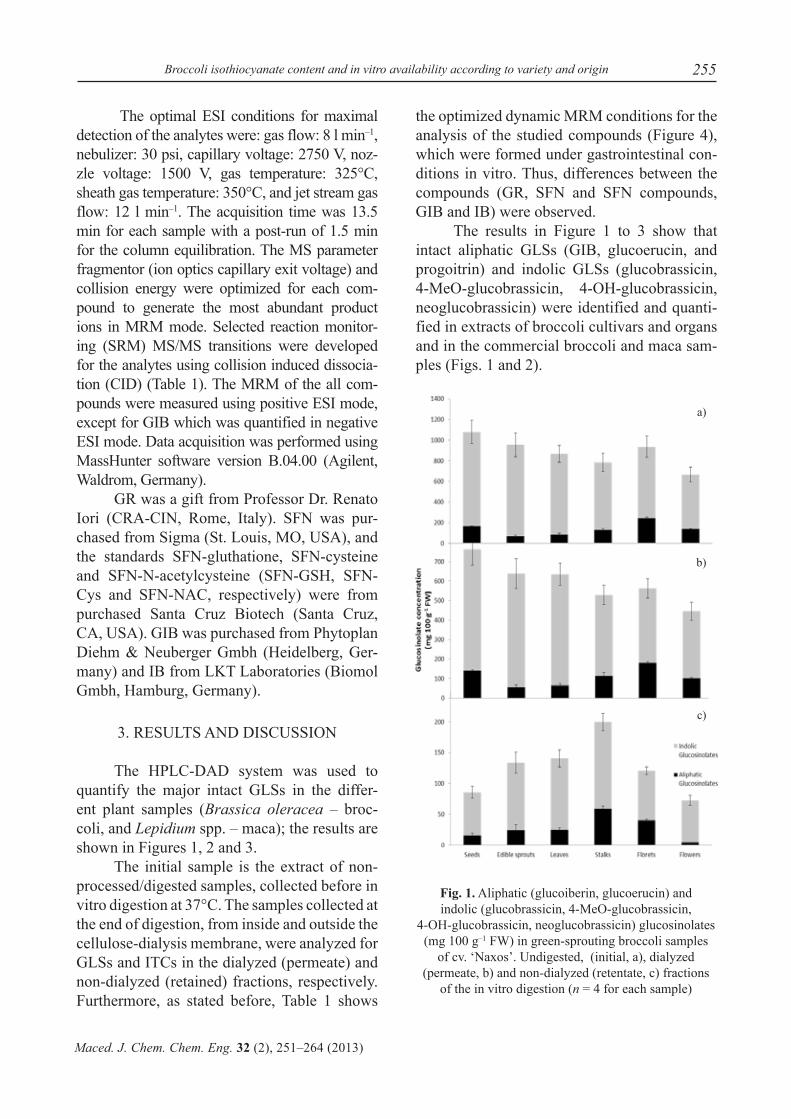
The HPLC-DAD system was used to quantify the major intact GLSs in the differ-ent plant samples (Brassica oleracea – broc-coli, and Lepidium spp. – maca); the results are shown in Figures 1, 2 and 3.
The initial sample is the extract of non-processed/digested samples, collected before in vitro digestion at 37°C. The samples collected at the end of digestion, from inside and outside the cellulose-dialysis membrane, were analyzed for GLSs and ITCs in the dialyzed (permeate) and non-dialyzed (retained) fractions, respectively. Furthermore, as stated before, Table 1 shows
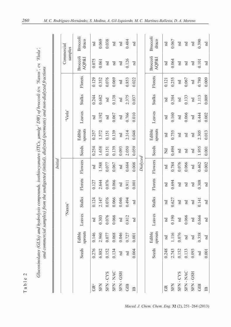
the optimized dynamic MRM conditions for the analysis of the studied compounds (Figure 4), which were formed under gastrointestinal con-ditions in vitro. Thus, differences between the compounds (GR, SFN and SFN compounds, GIB and IB) were observed.
The results in Figure 1 to 3 show that intact aliphatic GLSs (GIB, glucoerucin, and progoitrin) and indolic GLSs (glucobrassicin, 4-MeO-glucobrassicin, 4-OH-glucobrassicin, neoglucobrassicin) were identified and quanti-fied in extracts of broccoli cultivars and organs and in the commercial broccoli and maca sam-ples (Figs. 1 and 2).
Fig. 1. Aliphatic (glucoiberin, glucoerucin) and indolic (glucobrassicin, 4-MeO-glucobrassicin,
4-OH-glucobrassicin, neoglucobrassicin) glucosinolates (mg 100 g–1 FW) in green-sprouting broccoli samples
of cv. ‘Naxos’. Undigested, (initial, a), dialyzed (permeate, b) and non-dialyzed (retentate, c) fractions
of the in vitro digestion (n = 4 for each sample)
a)
b)
c)
256 M. C. Rodríguez-Hernández, S. Medina, A. Gil-Izquierdo, M. C. Martínez-Ballesta, D. A. Moreno
Maced. J. Chem. Chem. Eng. 32 (2), 251–264 (2013)
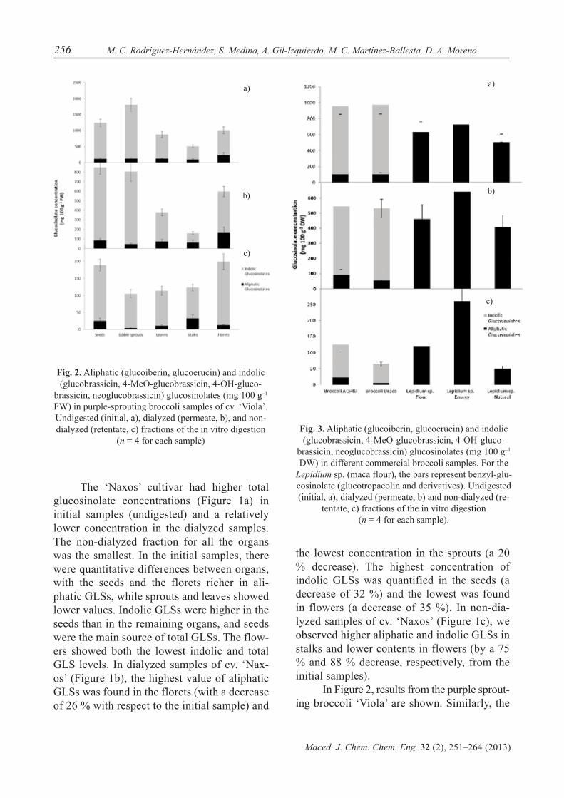
Fig. 2. Aliphatic (glucoiberin, glucoerucin) and indolic (glucobrassicin, 4-MeO-glucobrassicin, 4-OH-gluco-
brassicin, neoglucobrassicin) glucosinolates (mg 100 g–1 FW) in purple-sprouting broccoli samples of cv. ‘Viola’. Undigested (initial, a), dialyzed (permeate, b), and non-dialyzed (retentate, c) fractions of the in vitro digestion
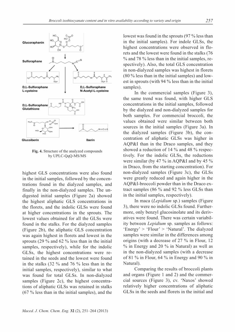
(n = 4 for each sample)Fig. 3. Aliphatic (glucoiberin, glucoerucin) and indolic (glucobrassicin, 4-MeO-glucobrassicin, 4-OH-gluco-
brassicin, neoglucobrassicin) glucosinolates (mg 100 g–1 DW) in different commercial broccoli samples. For the
Lepidium sp. (maca flour), the bars represent benzyl-glu-cosinolate (glucotropaeolin and derivatives). Undigested (initial, a), dialyzed (permeate, b) and non-dialyzed (re-
tentate, c) fractions of the in vitro digestion (n = 4 for each sample).
The ‘Naxos’ cultivar had higher total glucosinolate concentrations (Figure 1a) in initial samples (undigested) and a relatively lower concentration in the dialyzed samples. The non-dialyzed fraction for all the organs was the smallest. In the initial samples, there were quantitative differences between organs, with the seeds and the florets richer in ali-phatic GLSs, while sprouts and leaves showed lower values. Indolic GLSs were higher in the seeds than in the remaining organs, and seeds were the main source of total GLSs. The flow-ers showed both the lowest indolic and total GLS levels. In dialyzed samples of cv. ‘Nax-os’ (Figure 1b), the highest value of aliphatic GLSs was found in the florets (with a decrease of 26 % with respect to the initial sample) and
the lowest concentration in the sprouts (a 20 % decrease). The highest concentration of indolic GLSs was quantified in the seeds (a decrease of 32 %) and the lowest was found in flowers (a decrease of 35 %). In non-dia-lyzed samples of cv. ‘Naxos’ (Figure 1c), we observed higher aliphatic and indolic GLSs in stalks and lower contents in flowers (by a 75 % and 88 % decrease, respectively, from the initial samples).
In Figure 2, results from the purple sprout-ing broccoli ‘Viola’ are shown. Similarly, the
a)
b)
c)
a)
b)
c)
257Broccoli isothiocyanate content and in vitro availability according to variety and origin
Maced. J. Chem. Chem. Eng. 32 (2), 251–264 (2013)
highest GLS concentrations were also found in the initial samples, followed by the concen-trations found in the dialyzed samples, and finally in the non-dialyzed samples. The un-digested initial samples (Figure 2a) showed the highest aliphatic GLS concentrations in the florets, and the indolic GLSs were found at higher concentrations in the sprouts. The lowest values obtained for all the GLSs were found in the stalks. For the dialyzed samples (Figure 2b), the aliphatic GLS concentration was again highest in florets and lowest in the sprouts (29 % and 62 % less than in the initial samples, respectively), while for the indolic GLSs, the highest concentrations were re-tained in the seeds and the lowest were found in the stalks (32 % and 76 % less than in the initial samples, respectively), similar to what was found for total GLSs. In non-dialyzed samples (Figure 2c), the highest concentra-tions of aliphatic GLSs was retained in stalks (67 % less than in the initial samples), and the
Fig. 4. Structure of the analyzed compounds by UPLC-QqQ-MS/MS
lowest was found in the sprouts (97 % less than in the initial samples). For indole GLSs, the highest concentrations were observed in flo-rets and the lowest were found in the stalks (76 % and 78 % less than in the initial samples, re-spectively). Also, the total GLS concentration in non-dialyzed samples was highest in florets (80 % less than in the initial samples) and low-est in sprouts (with 94 % less than in the initial samples).
In the commercial samples (Figure 3), the same trend was found, with higher GLS concentrations in the initial samples, followed by the dialyzed and non-dialyzed samples for both samples. For commercial broccoli, the values obtained were similar between both sources in the initial samples (Figure 3a). In the dialyzed samples (Figure 3b), the con-centration of aliphatic GLSs was higher in AQP&I than in the Draco samples, and they showed a reduction of 14 % and 48 % respec-tively. For the indolic GLSs, the reductions were similar (by 47 % in AQP&I and by 45 % in Draco, from the starting concentration). For non-dialyzed samples (Figure 3c), the GLSs were greatly reduced and again higher in the AQP&I-broccoli powder than in the Draco ex-tract samples (86 % and 92 % less GLSs than in the initial samples, respectively).
In maca (Lepidium sp.) samples (Figure 3), there were no indolic GLSs found. Further-more, only benzyl glucosinolate and its deriv-atives were found. There was certain variabil-ity between Lepidium sp. samples as follows: ‘Energy’ > ‘Flour’ > ‘Natural’. The dialyzed samples were similar in the differences among origins (with a decrease of 27 % in Flour, 12 % in Energy and 20 % in Natural) as well as in the non-dialyzed samples (with a decrease of 81 % in Flour, 64 % in Energy and 90 % in Natural).
Comparing the results of broccoli plants and organs (Figure 1 and 2) and the commer-cial sources (Figure 3), cv. ‘Naxos’ showed relatively higher concentrations of aliphatic GLSs in the seeds and florets in the initial and

258 M. C. Rodríguez-Hernández, S. Medina, A. Gil-Izquierdo, M. C. Martínez-Ballesta, D. A. Moreno
Maced. J. Chem. Chem. Eng. 32 (2), 251–264 (2013)
dialyzed samples, whereas the stalks showed the highest content in the non-dialyzed sam-ples. In cv. ‘Viola’, the organ influence was different, with higher concentration of aliphat-ic GLSs in the florets and seeds for the initial and dialyzed samples, but similar to what hap-pened in green ‘Naxos’ broccoli, the stalks re-tained the highest levels of aliphatic GLSs in the non-dialyzed fraction. For the commercial samples, initial and dialyzed samples showed much lower concentrations of GLSs than the experimental broccoli samples.
The samples of Lepidium sp. were high-er in total GLSs than the commercial broccoli samples for the dialyzed and non-dialyzed samples, but not in the undigested starting material. However, for the maca samples, GLSs were represented only by benzyl-GLS (glucotropaeolin), not glucoiberin (GIB), glu-coerucin (GE) or glucoraphanin (GR).
The concentration of GLSs, mainly aliphatic GLSs, varies widely among differ-ent developmental stages of the plant, and also among different organs [19–21]. Also, aliphatic GLSs are clearly regulated by geno-type; in contrast, the effects of environment and genotype × environment interactions on the indolic GLS content appeared to be the main effects of variation [22]. The differences found between green and purple broccoli cul-tivars or between broccoli and maca samples agreed with this. Furthermore, processing pri-or to analysis could also affect GLSs, such as occurred in maca samples, which is in agree-ment with others reports [23].
Climatic conditions and ecophysiologi-cal factors, such as temperature and radiation, relative humidity and the degree of hydration, have an influential role on the phytochemical content of vegetables [24, 25]. Thus, Brown et al. [26] evaluated a subset of 10 broccoli varieties grown over four seasons, which al-lowed for determining the extent to which the GLS content varies with genotype and with environmental conditions [27]. Charron et al. [28] also found variations in the glucosi-
nolate content in cabbage leaves harvested in the spring and fall seasons; the highest con-centrations of total GLSs generally occurred when crops were harvested during periods of high temperature and long day length. Also, sprouts of most broccoli cultivars studied in recent years contain GR as the main thiofunc-tionalized glucosinolate, with its concentra-tion dependent on genotype and the duration of the sprouting period [23]. Oliviero et al. [29] showed that glucosinolate degradation increased with temperature. In our case, we showed a higher amount of GLSs in the origi-nal sample of plant material before in vitro digestion.
Should be noted that simulated diges-tion is far less complex than an animal feed-ing study, and a variety of factors could af-fect recovery in vivo, such as absorption of intact GLSs [30]. Thus, in this work, simu-lated digestion using major enzymes such as α-amylase, gastric pepsin and intestinal pan-creatin induced the differences found for the concentration of GLSs in the different steps of in vitro digestion.
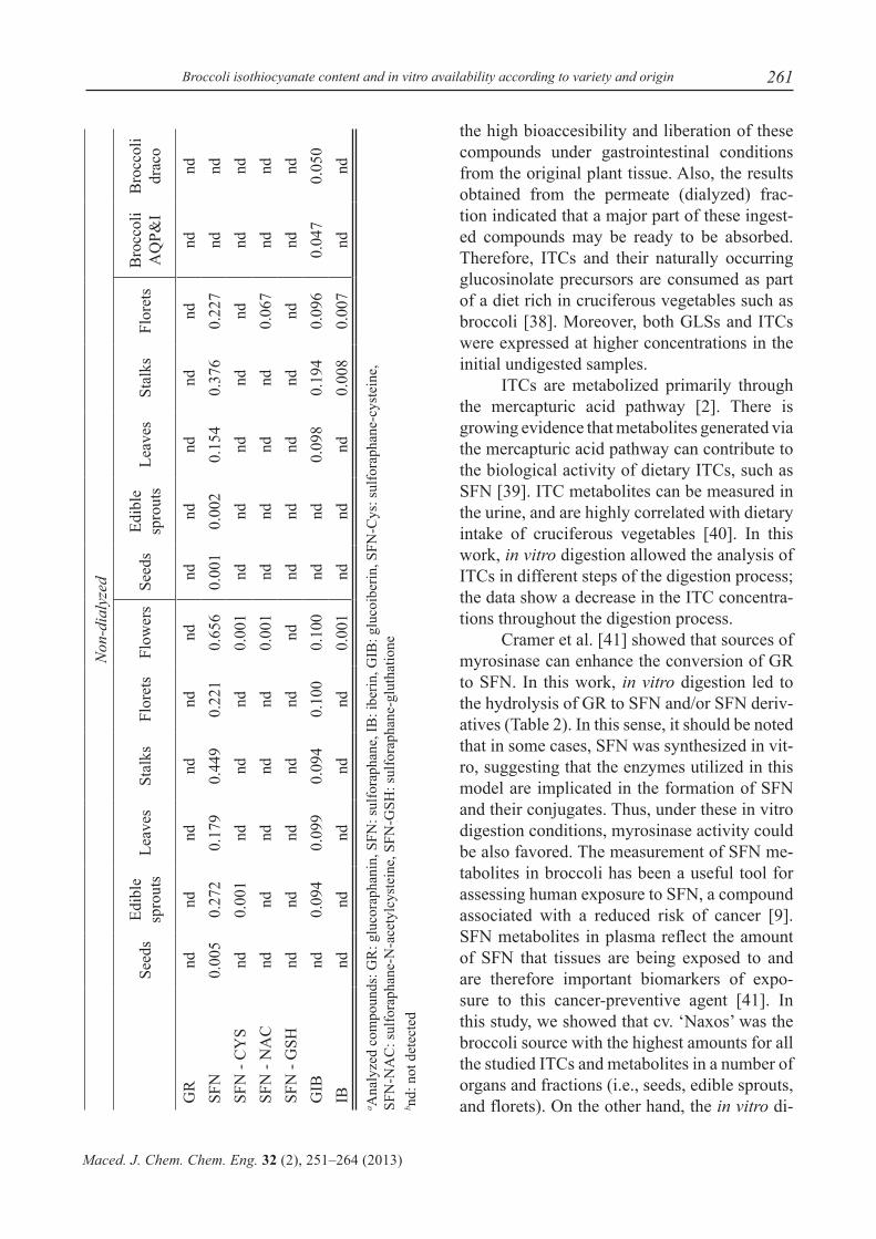
Existing methods to extract and prepare ITCs from biological samples are often com-plicated and time-consuming [31]. However, Dominguez-Perles et al. [32] developed a quantitative method for the direct determina-tion of intact ITCs in plant and other biologi-cal samples. Thus, ITCs were detected before and after the in vitro digestion of plant samples of different origins. We used UPLC-MS triple quadrupole technology to simultaneously ana-lyze SFN, IB and their mercapto-conjugates in a single injection (Table 2). The ‘Naxos’ cul-tivar showed higher GR concentrations in the initial samples for all the organs, except for the leaves and flowers, where GR was not de-tected. For the dialyzed samples, GR was only detected in seeds and, finally, in the non-dia-lyzed fractions, GR was absent. With respect to the SFN concentration, cv. ‘Naxos’ showed a similar trend for SFN contents in all organs: initial > dialyzed > non-dialyzed samples.

259Broccoli isothiocyanate content and in vitro availability according to variety and origin
Maced. J. Chem. Chem. Eng. 32 (2), 251–264 (2013)
For the ITC metabolites studied, SFN-CYS was present in undigested samples. The dialyzed and non-dialyzed samples presented SFN-CYS in the seeds, sprouts and flowers. SFN-NAC was detected in all the ‘Naxos’ or-gans in the initial samples, and was only pre-sent in seeds, leaves and flowers in the dialyzed samples and absent in almost all the non-dia-lyzed samples, detected only in flowers. SFN-GSH was detected only in seeds, sprouts and stalks in the initial samples, and present only in the dialyzed samples from seeds. In relation to GIB, the concentrations found in the differ-ent organs were much higher in the initial sam-ples, except in seeds which showed no GIB, followed by the dialyzed samples and, finally, the non-dialyzed samples. Finally, the IB con-centration was very low in the initial undigested samples of cv. ‘Naxos’ and very low levels (not quantifiable) were found in the dialyzed and non-dialyzed samples.
For the samples of ‘Viola’ broccoli (Ta-ble 2), the extracts of undigested material showed GR, except for the leaves. There was a great reduction in the content in the dialyzed samples, and GR was only detected in florets. Moreover, GR was absent in the non-dialyzed samples. For the SFN concentrations in undi-gested ‘Viola’ samples, all organs presented SFN in higher concentrations than GR. For the dialyzed samples, the SFN content was reduced and lower levels were also detected in the non-dialyzed samples. When analyzing SFN metab-olites in these ‘Viola’ samples, the SFN-CYS concentration was higher in the initial samples, except in both leaves and stalks where it was not detected. The dialyzed and non-dialyzed samples did not show any SFN-CYS. A simi-lar situation was found for SFN-NAC, with low concentrations in the initial samples, and with detection only in the leaves, stalks and florets of the dialyzed samples and in the florets of the non-dialyzed samples. SFN-GSH was de-tected only in the seeds of undigested samples of ‘Viola’, as in ‘Naxos’. GIB was found at a much higher concentration in ‘Viola’ samples
than in ‘Naxos’ for the undigested samples, as expected and also seen in literature [14, 33]. A very high proportion of GIB passed the mem-brane and was detected in the dialyzed fraction, and only a small amount of GIB remained in the non-dialyzed fraction. For IB, the content was also much higher in ‘Viola’ than in ‘Naxos’ samples, and again, a lower proportion was de-tected in the dialyzed samples, and scarce lev-els in the non-dialyzed samples. Looking into the commercial broccoli samples, for GR and GIB and their respective ITCs and related me-tabolites (Table 2), we only found GR, SFN and GIB in the AQP&I broccoli powder, with only SFN and GIB in the dialyzed extract, whereas the Draco broccoli presented a low amount of SFN and SFN-CYS, but a higher level of GIB. Similarly, only SFN and GIB were present in the dialyzed extracts and only GIB was found in the non-dialyzed extracts.
The Lepidium sp. (maca commercial flours) samples were analyzed for these GLSs and ITCs. Since benzyl-GLS and its cognate BITC were not available for setting the experi-ments in the UPLC-QqQ-MS study, and since the presence of GR (SFN and metabolites) or GIB (IB) was not expected, the results are not shown.
Taking all the data on the GLSs, GR and GIB and their hydrolysis products, we can say that cv. ‘Naxos’ showed higher ITC levels in all the tested samples. Both commercial broc-colis had a very low level of ITCs or related metabolites.
The GLSs are hydrolyzed by the enzyme myrosinase into a range of breakdown products such as thiocyanates, ITCs and nitriles [34-35]. In the gastrointestinal tract, the GLSs are hy-drolyzed by bacteria during digestion [36], but at a lower rate than in plant systems. ITCs must be absorbed to exert their associated cancer-preventive activity [37]. For this reason, the non-dialyzed fraction showed the lowest val-ues of GLSs and ITCs and their derivatives/metabolites, suggesting that the amount of GLSs or ITCs discarded was minimal, showing
260 M. C. Rodríguez-Hernández, S. Medina, A. Gil-Izquierdo, M. C. Martínez-Ballesta, D. A. Moreno
Maced. J. Chem. Chem. Eng. 32 (2), 251–264 (2013)
T a
b l e
2 Glu
cosi
nola
tes (
GLS
s) a
nd h
ydro
lysi
s com
poun
ds, i
soth
iocy
anat
es (I
TCs,
µmol
g-1 D
W) o
f bro
ccol
i (cv
. ‘N
axos
’, cv
. ‘Vi
ola’
, an
d co
mm
erci
al sa
mpl
es) f
rom
the
undi
gest
ed (i
nitia
l), d
ialy
zed
(per
mea
te) a
nd n
on-d
ialy
zed
frac
tions
Initi
al
‘Nax
os’
‘Vio
la’
Com
mer
cial
sa
mpl
es
Seed
sEd
ible
sp
rout
sLe
aves
Stal
ksFl
oret
sFl
ower
sSe
eds
Edib
le
spro
uts
Leav
esSt
alks
Flor
ets
Bro
ccol
i A
QP&
IB
rocc
oli
drac
o
GR
a0.
276
0.14
6nd
0.12
40.
127
nd0.
254
0.25
7nd
0.24
40.
129
0.07
5nd
SFN
6.88
22.
960
0.30
32.
147
2.64
41.
588
1.65
81.
572
0.19
20.
682
0.35
20.
061
0.06
9SF
N -
CY
S0.
152
0.07
70.
076
0.07
60.
076
0.07
70.
151
0.15
1nd
nd0.
076
nd0.
038
SFN
- N
AC
0.13
40.
068
0.06
60.
066
0.06
60.
067
0.13
90.
139
0.06
70.
138
0.06
9nd
ndSF
N -
GSH
nd0.
046
nd0.
046
ndnd
0.09
3nd
ndnd
ndnd
ndG
IBnd
0.72
70.
812
0.49
40.
911
0.68
42.
050
2.31
40.
766
2.37
50.
853
0.12
60.
484
IB0.
004
0.00
1nd
nd0.
001
0.00
80.
059
0.04
80.
010
0.03
70.
022
ndnd
Dia
lyze
d
Seed
sEd
ible
sp
rout
sLe
aves
Stal
ksFl
oret
sFl
ower
sSe
eds
Edib
le
spro
uts
Leav
esSt
alks
Flor
ets
Bro
ccol
iA
QP&
IB
rocc
oli
drac
o
GR
0.24
8nd
ndnd
ndnd
Nd
ndnd
nd0.
121
ndnd
SFN
2.74
31.
116
0.19
00.
627
0.69
40.
784
0.49
40.
755
0.16
00.
384
0.25
50.
064
0.06
7SF
N -
CY
S0.
152
0.07
6nd
ndnd
0.07
6nd
ndnd
ndnd
ndnd
SFN
- N
AC
0.13
3nd
0.06
6nd
nd0.
066
ndnd
0.06
60.
133
0.06
7nd
ndSF
N -
GSH
0.09
3nd
ndnd
ndnd
ndnd
ndnd
ndnd
ndG
IBnd
0.35
80.
666
0.14
10.
308
0.46
21.
955
2.30
50.
444
1.11
30.
780
0.10
10.
390
IB0.
001
ndnd
ndnd
0.00
20.
001
0.01
30.
002
0.00
90.
009
ndnd
261Broccoli isothiocyanate content and in vitro availability according to variety and origin
Maced. J. Chem. Chem. Eng. 32 (2), 251–264 (2013)
Non
-dia
lyze
d
Seed
sEd
ible
sp
rout
sLe
aves
Stal
ksFl
oret
sFl
ower
sSe
eds
Edib
le
spro
uts
Leav
esSt
alks
Flor
ets
Bro
ccol
i A
QP&
IB
rocc
oli
drac
o
GR
ndnd
ndnd
ndnd
ndnd
ndnd
ndnd
ndSF
N0.
005
0.27
20.
179
0.44
90.
221
0.65
60.
001
0.00
20.
154
0.37
60.
227
ndnd
SFN
- C
YS
nd0.
001
ndnd
nd0.
001
ndnd
ndnd
ndnd
ndSF
N -
NA
Cnd
ndnd
ndnd
0.00
1nd
ndnd
nd0.
067
ndnd
SFN
- G
SHnd
ndnd
ndnd
ndnd
ndnd
ndnd
ndnd
GIB
nd0.
094
0.09
90.
094
0.10
00.
100
ndnd
0.09
80.
194
0.09
60.
047
0.05
0IB
ndnd
ndnd
nd0.
001
ndnd
nd0.
008
0.00
7nd
nda A
naly
zed
com
poun
ds: G
R: g
luco
raph
anin
, SFN
: sul
fora
phan
e, IB
: ibe
rin, G
IB: g
luco
iber
in, S
FN-C
ys: s
ulfo
raph
ane-
cyst
eine
, SF
N-N
AC
: sul
fora
phan
e-N
-ace
tylc
yste
ine,
SFN
-GSH
: sul
fora
phan
e-gl
utha
tione
b nd:
not
det
ecte
d
the high bioaccesibility and liberation of these compounds under gastrointestinal conditions from the original plant tissue. Also, the results obtained from the permeate (dialyzed) frac-tion indicated that a major part of these ingest-ed compounds may be ready to be absorbed. Therefore, ITCs and their naturally occurring glucosinolate precursors are consumed as part of a diet rich in cruciferous vegetables such as broccoli [38]. Moreover, both GLSs and ITCs were expressed at higher concentrations in the initial undigested samples.
ITCs are metabolized primarily through the mercapturic acid pathway [2]. There is growing evidence that metabolites generated via the mercapturic acid pathway can contribute to the biological activity of dietary ITCs, such as SFN [39]. ITC metabolites can be measured in the urine, and are highly correlated with dietary intake of cruciferous vegetables [40]. In this work, in vitro digestion allowed the analysis of ITCs in different steps of the digestion process; the data show a decrease in the ITC concentra-tions throughout the digestion process.
Cramer et al. [41] showed that sources of myrosinase can enhance the conversion of GR to SFN. In this work, in vitro digestion led to the hydrolysis of GR to SFN and/or SFN deriv-atives (Table 2). In this sense, it should be noted that in some cases, SFN was synthesized in vit-ro, suggesting that the enzymes utilized in this model are implicated in the formation of SFN and their conjugates. Thus, under these in vitro digestion conditions, myrosinase activity could be also favored. The measurement of SFN me-tabolites in broccoli has been a useful tool for assessing human exposure to SFN, a compound associated with a reduced risk of cancer [9]. SFN metabolites in plasma reflect the amount of SFN that tissues are being exposed to and are therefore important biomarkers of expo-sure to this cancer-preventive agent [41]. In this study, we showed that cv. ‘Naxos’ was the broccoli source with the highest amounts for all the studied ITCs and metabolites in a number of organs and fractions (i.e., seeds, edible sprouts, and florets). On the other hand, the in vitro di-
262 M. C. Rodríguez-Hernández, S. Medina, A. Gil-Izquierdo, M. C. Martínez-Ballesta, D. A. Moreno
Maced. J. Chem. Chem. Eng. 32 (2), 251–264 (2013)
gestion showed that the ‘Viola’ broccoli had a higher concentration of the glucosinolate GIB than its breakdown product IB due to the high level of this glucosinolate in this cultivar [33]. However, Lepidium sp. samples showed no ali-phatic GLSs such as GR or GIB; for this reason, neither SFN nor IB was expected in the samples (data not shown).
Dietary GLSs, as ITC precursors, remain key to the current view of how a diet rich in Brassica vegetables is associated with a de-creased risk of cancer, particularly for cancers of the bladder, colon and lungs [37]. The ana-lytical methods presented here may assist in the analysis of GLS intake and bioavailability (through the study of ITCs and related metabo-lites). Furthermore, this study estimated SFN and related metabolites (-CYS, -NAC, and -GSH) separately in the same run and with a short analysis time, and found that most SFN was conjugated rather than free, showing that SFN metabolism takes place in the gastrointes-tinal lumen during absorption. This is in agree-ment with previous results [42].
In addition to the type of sample selected to analyze ITC bioavailability, the choice of the methodological procedure, i.e. UPLC-QqQ-MS/MS, used for this type of analysis constitutes an essential decision. The application of this meth-od to the qualitative and quantitative determi-nation of GLSs, ITCs and related metabolites, as well as the robustness and reproducibility of the analytical platform used exhibited utility for further assays focused on the characterization broccoli according to several factors.
4. CONCLUSIONS
In conclusion, we can say that the ITC concentration depends of the cultivar, the plant organ and sample processing. ITCs and their naturally occurring glucosinolate precursors are consumed as part of a diet rich in crucifer-ous vegetables such as broccoli. In this sense, we can conclude that cv. ‘Naxos’ produced higher ITC levels of all the studied ITCs and
metabolites in plant organs and fractions; there-fore, this dietary source may have enhanced chemopreventive properties. The freeze-dried plant samples grown for this study showed higher ITC concentrations than the commercial samples. Moreover, higher GLS and ITC con-centrations were found in the initial steps of the digestion process, followed by the concentra-tions found in the dialyzed samples, and finally in the non-dialyzed samples. These findings demonstrate the highest bioaccesibility and lib-eration of these compounds under gastrointes-tinal conditions from the original plant tissue, and also showed, on the other hand, a decrease in the GLS and ITC concentrations with the di-gestion process. Therefore, the concentration of ITCs decreased in accordance with the content of precursor GLSs. Measurement of ingested GLSs, particularly the major GLSs that give rise to cancer-preventive ITCs, will assist in quantifying the GLS input in dietary surveys and cancer chemoprevention studies. However, additional research concerning the availability of bioactive compounds in humans is necessary.
Acknowledgments. This work was funded by the Seneca Foundation – Regional Agency for Science and Technology of the Autonomous Community of the Mur-cia Region through Project Ref. CARM 08753/PI/08. Part of this work was also funded through the Excellence in Research funds of the Food Science and Technol-ogy Group, grant number 04486/GERM/06. The authors would also thank Dr. David J. Walker for correction of the English language and style.
REFERENCES
[1] D. T. Verhoeven, R. A. Goldbohm, G. van Poppel, H. Verhagen, Epidemiological studies on Brassica vegetables and cancer risk, Cancer Epidem. Bio-mar., 5, 733–748 (1996).
[2] J. V. Higdon, B. Delage, D. E. Williams, R. H. Dashwood, Cruciferous vegetables and human cancer risk: Epidemiologic evidence and mecha-nistic basis, Pharmacol. Res., 55, 224–236 (2007).
[3] D. A. Moreno, M. Carvajal, C. López-Berenguer, C. García-Viguera, Chemical and biological char-acterisation of nutraceutical compounds of brocco-li, J. Pharmaceut. Biomed., 41, 1508–1522 (2006).
263Broccoli isothiocyanate content and in vitro availability according to variety and origin
Maced. J. Chem. Chem. Eng. 32 (2), 251–264 (2013)
[4] J. W. Fahey, A. T. Zalcmann, P. Talalay, The chemi-cal diversity and distribution of glucosinolates and isothiocyanates among plants, Phytochemistry, 56, 5–51 (2001).
[5] E. H. Jeffery, M. Araya, Physiological effects of broccoli consumption, Phytochemistry, 8, 283–298 (2009).
[6] J. D. Clarke, K. Riedl, D. Bella, S. J. Schwartz, J. F. Stevens, E. Ho, Comparison of isothiocyanate metabolite levels and histone deacetylase activ-ity in human subjects consuming broccoli sprouts or broccoli supplement, J. Agr. Food Chem., 59, 10955–10963 (2011).
[7] A. M. Bones, J. T. Rossiter, The myrosinase–glu-cosinolate system, its organisation and biochemis-try, Physiol. Plantarum, 97, 194–208 (1996).
[8] C. D. Grubb, S. Abel, Glucosinolate metabolism and its control, Trends Plant. Sci., 11, 89–100 (2006).
[9] T. A. Shapiro, J. W. Fahey, A. T. Dinkova-Kostova, W. D. Holtzclaw, K. K. Stephenson, K. L. Wade, L. Ye, P. Talalay, Safety, tolerance, and metabolism of broccoli sprout glucosinolates and isothiocya-nates: A clinical phase I study, Nutr. Cancer, 55, 53–62 (2006).
[10] L. Mi, Z. Xiao, B. L. Hood, S. Dakshanamurthy, X. Wang, S. Govind, T. P. Conrads, T. D. Veenstra, F. L. Chung, Covalent binding to tubulin by isothiocy-anates. A mechanism of cell growth arrest and apop-tosis, J. Biol. Chem., 283, 22136–22146 (2008).
[11] H. Yuan, S. Yao, Y. You, G. Xia, Q. You, Antioxi-dant activity of isothiocyanate extracts from broc-coli, Chinese J. Chem. Eng., 18, 312–321 (2010).
[12] R.F. Mithen, Glucosinolates and their degradation products, Adv. Bot. Res., 35, 213–262 (2001).
[13] M.W. Farnham, J. W. Fahey, K. K. Stephenson, Selection for floret glucoraphanin concentration among inbred Broccoli, HortScience, 34, 448 (1999).
[14] S. Pérez-Balibrea, D. A. Moreno, C. García-Viguera, Genotypic effects on the phytochemi-cal quality of seeds and sprouts from commercial broccoli cultivars, Food Chem., 125, 348–354 (2011).
[15] N. Baenas, D. A. Moreno, C. García-Viguera, Se-lecting sprouts of Brassicaceae for optimum phy-tochemical composition, J. Agr. Food Chem., 60, 11409–11420 (2012).
[16] G. R. de Nicola, M. Bagatta, E. Pagnotta, D. An-gelino, L. Gennari, P. Ninfali, P. Rollin, R. Lori,
Comparison of bioactive phyrochemical content and release of isothiocyanates in selected brassica sprouts, Food Chem., 141, 297–303 (2013).
[17] F. Vallejo, A. Gil-Izquierdo, A. Pérez-Vicente, C. García-Viguera, In vitro gastrointestinal digestión study of broccoli inflorescence phenolic com-pounds, glucosinolates, and vitamin C, J. Agric. Food Chem. 52, 135-138 (2004).
[18] A. Martinez-Sanchez, A. Allende, R. N. Bennett, F. Ferreres, M. I. Gil, Microbial, nutritional and sensory quality of rocket leaves as affected by dif-ferent sanitizers, Postharvest Biol. Tec., 42, 86–97 (2006).
[19] L. Rask, E. Andréasson, B. Ekbom, S. Eriksson, B. Pontoppidan, J. Meijer, Myrosinase: gene family evolution and herbivore defense in Brassicaceae, Plant Mol. Biol., 42, 93–114 (2000).
[20] N. Clossais-Besnard, F. Larher, Physiological role of glucosinolates in Brassica napus. Concentra-tion and distribution pattern of glucosinolates among plant organs during a complete life cycle, J. Sci. Food Agr., 56, 25–38 (2006).
[21] G. Padilla, M. E. Cartea, P. Velasco, A. de Harob, A. Ordás, Variation of glucosinolates in vegetable crops of Brassica rapa, Phytochemistry, 68, 536–545 (2007).
[22] M. Francisco, M. E. Cartea, P. Soengas, P. Velasco, Effect of genotype and environmental conditions on health-promoting compounds in Brassica rapa, J. Agr. Food Chem., 59, 2421–2431 (2011).
[23] S. Piacente, V. Carbone, A. Plaza, A. Zampelli, C. Pizza, Investigation of the Tuber Constituents of Maca (Lepidium meyenii Walp.), J. Agr. Food Chem., 50, 5621–5625 (2002).
[24] J. M. Abercrombie, M. W. Farnham, J. W. Rush-ing, Genetic combining ability of glucoraphanin level and other horticultural traits of broccoli, Eu-phytica, 143, 145–151 (2005).
[25] J. W. Fahey, Y. Zhang, P. Talalay, Broccoli sprouts: An exceptionally rich source of inducers of enzymes that protect against chemical carcinogens, Proc. Natl. Acad. Sci. U.S.A., 94, 10367–10372 (1997).
[26] A. F. Brown, G. G. Yousef, E. H. Jeffery, B. P. Klein, M. A. Wallig, M. M. Kushad, J. A. Juvik, Glucosi-nolate profiles in broccoli: Variation in levels and implications in breeding for cancer chemoprotec-tion, J. Am. Soc. Hortic. Sci., 127, 807–813 (2002).
[27] M. W. Farnham, K. K. Stephenson, J. W. Fahey, Glucoraphanin level in broccoli seed in largely determined by genotype, HortScience, 40, 50–53 (2005).
264 M. C. Rodríguez-Hernández, S. Medina, A. Gil-Izquierdo, M. C. Martínez-Ballesta, D. A. Moreno
Maced. J. Chem. Chem. Eng. 32 (2), 251–264 (2013)
[28] C. S. Charron, A. M. Saxton, C. E. Sams, Rela-tionship of climate and genotype to seasonal vari-ation in the glucosinolate–myrosinase system. II. Myrosinase activity in ten cultivars of Brassica oleracea grown in fall and spring seasons, J. Sci. Food Agr., 85, 682–690 (2005).
[29] T. Oliviero, R. Verkerk, M. Dekker, Effect of water content and temperature on glucosinolate degrada-tion kinetics in broccoli (Brassica oleracea var. italica), Food Chem., 132, 2037–2045 (2012).
[30] R. M. Bheemreddy, E. H. Jeffery, The metabolic fate of purified glucoraphanin in F344 rats, J. Agr. Food Chem., 55, 2861–2866 (2007).
[31] C. C. Conaway, J. Krzeminski, S. Amin, F.L. Chung, Decomposition rates of isothiocyanate conjugates determine their activity as inhibitors of cytochrome p450 enzymes, Chem. Res. Toxicol., 14, 1170–1176 (2001).
[32] R. Dominguez-Perles, S. Medina, D. A. Moreno, C. García-Viguera, F. Ferreres, A. Gil-Izquierdo, Bio-availability of glucosinolates and isothiocyanates from ½ and 1 servings of broccoli sprouts by a new UHPLC/MS/MS, Food Chem., In Press, (2013).
[33] M. C. Rodríguez-Hernández, D. A. Moreno, M. Carvajal, C. García-Viguera, M. C. Martínez-Ball-esta, Natural antioxidants in purple sprouting broc-coli under Mediterranean climate, J. Food Sci., 77, C1058–C1063 (2012).
[34] M. Wielanek, H. Urbanek, Glucotropaeolin and mirosinase production in hairy root cultures of Tropaeolum majus, Plant Cell Tiss. Org., 57, 39–45 (1999).
[35] M. Traka, R. Mithen, Glucosinolates, isothio-cyanates and human health, Phytochem. Rev., 8, 269–282 (2009).
[36] J. D. Hayes, M. O. Kelleher, I.M. Eggleston, The cancer chemopreventive actions of phytochemi-cals derived from glucosinolates, Eur. J. Nutr., 47, 73–88 (2008).
[37] P. J. Thornalley, IARC Workgroup, Cruciferous veg-etables, isothiocyanates and indoles, IARC Hand-book of Cancer Chemoprevention, 9, 1–261 (2004).
[38] T. A. Shapiro, J. W. Fahey, K. L. Wade, K. K. Ste-phenson, P. Talalay, Human metabolism and excre-tion of cancer chemoprotective glucosinolates and isothiocyanates of cruciferous vegetables, Cancer Epidem. Biomar., 7, 1091–1100 (1998).
[39] M. C. Myzak, P. A. Karplus, F. L. Chung, R. H. Dashwood, A novel mechanism of chemoprotec-tion by sulforaphane inhibition of histone deacety-lase, Cancer Res., 64, 5767–5774 (2004).
[40] A. Seow, C. Y. Shi, F. L. Chung, D. Jiao, J. H. Hankin, H. P. Lee, G. A. Coetzee, M. C. Yu, Uri-nary total isothiocyanate (ITC) in a population-based sample of middle-aged and older Chinese in Singapore: relationship with dietary total ITC and glutathione S-transferase M1/T1/P1 genotypes, Cancer Epidem. Biomar., 7, 775–781 (1998).
[41] J. M. Cramer, M. Teran-Garcia, E. H. Jeffery, En-hancing sulforaphane absorption and excretion in healthy men through the combined consumption of fresh broccoli sprouts and a glucoraphanin-rich powder, Brit. J. Nutr., 13, 1–6 (2011).
[42] S. Agrawal, B. Winnik, B. Buckley, L. Mi, F. L. Chung, T. J. Cook, Simultaneous determination of sulforaphane and its major metabolites from bio-logical matrices with liquid chromatography–tan-dem mass spectroscopy, J. Chromatogr. B., 840, 99–107 (2006).


![Anticancer Effect of Natural Product Sulforaphane by ... · traditional treatments, so it is worthwhile to search for a novel effective treatment for cervical cancer [5]. Sulforaphane,](https://static.fdocuments.us/doc/165x107/5f4b8833be79ae7285757261/anticancer-effect-of-natural-product-sulforaphane-by-traditional-treatments.jpg)


![New Reactivity Involving N-Isothiocyanates ...ruor.uottawa.ca/bitstream/10393/36042/1/Ranasinghe...New Reactivity Involving N-Isothiocyanates: Aminothiocarbonylation and [3+2] Cycloadditions](https://static.fdocuments.us/doc/165x107/5b29122e7f8b9ab23d8b45e6/new-reactivity-involving-n-isothiocyanates-ruor-reactivity-involving-n-isothiocyanates.jpg)


