Brian Bill, ; Justin Pedersen, ; and Evan Call MS RSM … Bill,1; Justin Pedersen, 2; and Evan Call...
1
Human Cadaver Testing to Determine the Reliability of Heel Boot Positioning or “Grip” of Eight Commercially Available Pressure Relieving Heel Protector Boots. BACKGROUND Support devices, such as pressure relieving boots, reduce the risk of ulceration by reducing friction, shear, and pressure to the at-risk tissues of the heel; however, the functionality of these devices depends on the boot’s ability to retain correct positioning (Figure 1A and 1B) 3,4 . High “grip” boots increase the protective ability of the device by holding the limb in position relative to the pressure relieving zones of the device. The “grip” of eight commercially available heel protectors (Table 1) were evaluated by comparing the coefficient of friction between the boot and heel of a fresh female cadaver. Brian Bill, 1 ; Justin Pedersen, 2 ; and Evan Call MS RSM (NRM) 1 1 Weber State University, Ogden, UT, 2 University of Utah, Salt Lake City, UT. METHODS Testing was performed on a female cadaver provided by the University of Utah Body Donor Program. The cadaver was 78 years of age, 41.28 kg, 160 cm, with no notable medical history. Following each test run, the heel of the cadaver was inspected for signs of damage. Samples of each type of boot were modified to include a strap, sewn into pre- existing seams of the boot, positioned uni-axially about the calf of a 50 th percentile mannequin calf. For boots that would not allow stitching, a strong double sided adhesive was used to fix the strap to the boot. To reduce friction between the test bench and the outside of the boot, the cadaver was positioned above a thin sheet of Teflon. The left heel of the cadaver was positioned as per manufacturer’s instruction (Figure 2); however, the straps of the boots were left un-attached. This was to allow for the evaluation of the boot-cadaver interface material without the variable of the strap. Figure 2- The positioning of the calf and heel in the heel protector. The boot was allowed to receive the load for 60 seconds before being pulled from the cadaver at a rate of 1 cm/second (Figure 3). The mean force required to remove the boot from the limb was recorded, in Newton's. The normal force by the cadaver to the boot was calculated 1 , and the coefficient of friction for each of the trials was calculated. A total of three samples were tested for each type of boot. Figure 3- Pulling the boot from the cadaver with the force gauge. RESULTS The foam, deformable fluid, and air bladder heel protector boots performed better than most of the fabric and batting heel protectors; however, one of the fabric and batting heel protectors (Sample 1, Figure 4) had a coefficient of friction of almost double the next highest sample (Sample 6, Figure 4). CONCLUSIONS As patients shift, the ability of a heel protecting boot to grip the limb and retain optimal off-loading positioning is vital to the function of the device. Correct positioning of the limb is key in relieving pressure, shear, and friction to the heel 3,4 . Multiple factors influence the coefficient of friction on a non-flat surface, such as the heel protector boots. Factors such as bulk modulus and the friction of materials may have a positive effect in the retention of correct positioning in heel protecting boots. Evaluation of heel protector “grip” of the limb is necessary for determining the effectiveness of the device to reduce risk of ulceration to tissue. The results indicate that the architecture and material of heel protectors play a role in maintaining correct positioning. The coefficient of friction of the construction materials along with bulk modulus provide the “grip” necessary to effectively immobilize the boot in respect to the heel. ACKNOWLEDGEMENTS REFERENCES 1 Anthropometric Data, Introduction to Biomechanics, University of Rhode Island. January 26.2011. 2 BS 3424-10:1987 Testing Coated Fabrics. Method 12A. 3 Lyder, C.H. Getting serious about preventing heel pressure ulcers in hospitals. 2011. WCET Journal. 31(4); 6-13. 4 Salcido, R., Lee, A., Ahn, C. Heel Pressure Ulcers: Purple Heel and Deep Tissue Injury. 2011. Advances in Skin & Wound Care. 24(8); 374- 380. Figure 4- The coefficient of friction for each of the eight different commercially available heel protector boots. Each sample is also identified by the general construction material of the boot. The error bars represent the 95 th % confidence interval (alpha = 0.05). Refer to Table 1 for the construction material information of each sample. Figure 1A- Correct positioning of the limb decreases pressure, friction, and shear on the at-risk tissue of the heel. Figure 1B- Incorrect positioning of the limb creates pressure, shear, and friction on the at-risk tissue of the heel. No abrasion or damage to the skin of the cadaver was observed during testing (Figure 5). Figure 5- Inspection of the heel of the cadaver following each test run. This research was funded by an unrestricted research grant from Sage Products. The cadaver was provided by the University of Utah Body Donor Program Sample Construction 1 Fabric and Batting with Adjustable Velcro Strap 2 Fabric and Batting with Adjustable Velcro Strap 3 Fabric and Batting with Adjustable Velcro Strap 4 Fabric and Batting with Adjustable Velcro Strap 5 Fabric and Batting with Adjustable Velcro Strap 6 Inflatable Air Bladder with Adjustable Velcro Strap 7 Air and Gel Fluid Bladder with Adjustable Velcro Strap 8 Foam with adjustable Velcro Strap Table 1- The construction of each of the eight heel protector boots. 0.00 0.20 0.40 0.60 0.80 1.00 1.20 1.40 Sample 1 Sample 2 Sample 3 Sample 4 Sample 5 Sample 6 Sample 7 Sample 8 Coefficient of Friction
Transcript of Brian Bill, ; Justin Pedersen, ; and Evan Call MS RSM … Bill,1; Justin Pedersen, 2; and Evan Call...
Human Cadaver Testing to Determine the Reliability of Heel Boot Positioning or “Grip” of Eight Commercially Available
Pressure Relieving Heel Protector Boots.
BACKGROUND
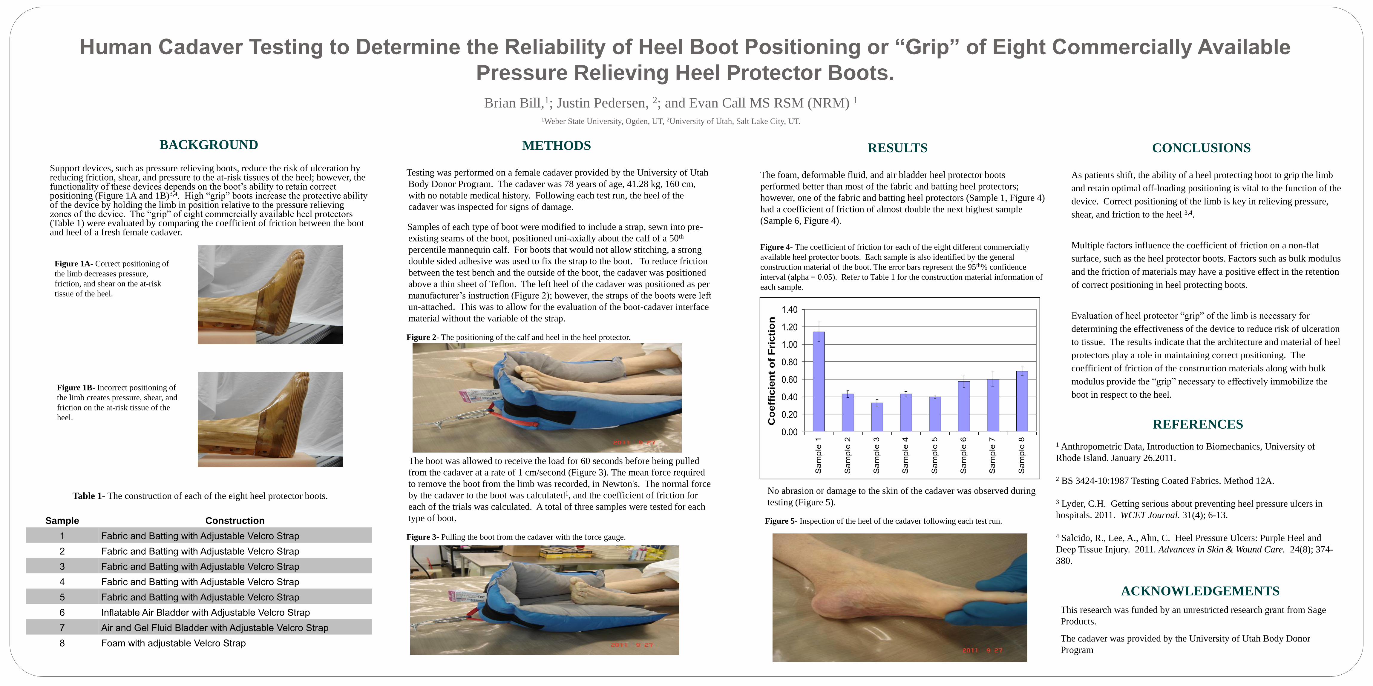
Support devices, such as pressure relieving boots, reduce the risk of ulceration by reducing friction, shear, and pressure to the at-risk tissues of the heel; however, the functionality of these devices depends on the boot’s ability to retain correct positioning (Figure 1A and 1B)3,4. High “grip” boots increase the protective ability of the device by holding the limb in position relative to the pressure relieving zones of the device. The “grip” of eight commercially available heel protectors (Table 1) were evaluated by comparing the coefficient of friction between the boot and heel of a fresh female cadaver.
Brian Bill,1; Justin Pedersen, 2; and Evan Call MS RSM (NRM) 1 1Weber State University, Ogden, UT, 2University of Utah, Salt Lake City, UT.
METHODS
Testing was performed on a female cadaver provided by the University of Utah
Body Donor Program. The cadaver was 78 years of age, 41.28 kg, 160 cm,
with no notable medical history. Following each test run, the heel of the
cadaver was inspected for signs of damage.
Samples of each type of boot were modified to include a strap, sewn into pre-
existing seams of the boot, positioned uni-axially about the calf of a 50th
percentile mannequin calf. For boots that would not allow stitching, a strong
double sided adhesive was used to fix the strap to the boot. To reduce friction
between the test bench and the outside of the boot, the cadaver was positioned
above a thin sheet of Teflon. The left heel of the cadaver was positioned as per
manufacturer’s instruction (Figure 2); however, the straps of the boots were left
un-attached. This was to allow for the evaluation of the boot-cadaver interface
material without the variable of the strap.
Figure 2- The positioning of the calf and heel in the heel protector.
The boot was allowed to receive the load for 60 seconds before being pulled
from the cadaver at a rate of 1 cm/second (Figure 3). The mean force required
to remove the boot from the limb was recorded, in Newton's. The normal force
by the cadaver to the boot was calculated1, and the coefficient of friction for
each of the trials was calculated. A total of three samples were tested for each
type of boot.
Figure 3- Pulling the boot from the cadaver with the force gauge.
RESULTS
The foam, deformable fluid, and air bladder heel protector boots
performed better than most of the fabric and batting heel protectors;
however, one of the fabric and batting heel protectors (Sample 1, Figure 4)
had a coefficient of friction of almost double the next highest sample
(Sample 6, Figure 4).
CONCLUSIONS
As patients shift, the ability of a heel protecting boot to grip the limb
and retain optimal off-loading positioning is vital to the function of the
device. Correct positioning of the limb is key in relieving pressure,
shear, and friction to the heel 3,4.
Multiple factors influence the coefficient of friction on a non-flat
surface, such as the heel protector boots. Factors such as bulk modulus
and the friction of materials may have a positive effect in the retention
of correct positioning in heel protecting boots.
Evaluation of heel protector “grip” of the limb is necessary for
determining the effectiveness of the device to reduce risk of ulceration
to tissue. The results indicate that the architecture and material of heel
protectors play a role in maintaining correct positioning. The
coefficient of friction of the construction materials along with bulk
modulus provide the “grip” necessary to effectively immobilize the
boot in respect to the heel.
ACKNOWLEDGEMENTS
REFERENCES 1 Anthropometric Data, Introduction to Biomechanics, University of
Rhode Island. January 26.2011.
2 BS 3424-10:1987 Testing Coated Fabrics. Method 12A.
3 Lyder, C.H. Getting serious about preventing heel pressure ulcers in
hospitals. 2011. WCET Journal. 31(4); 6-13.
4 Salcido, R., Lee, A., Ahn, C. Heel Pressure Ulcers: Purple Heel and
Deep Tissue Injury. 2011. Advances in Skin & Wound Care. 24(8); 374-
380.
Figure 4- The coefficient of friction for each of the eight different commercially
available heel protector boots. Each sample is also identified by the general
construction material of the boot. The error bars represent the 95th% confidence
interval (alpha = 0.05). Refer to Table 1 for the construction material information of
each sample.
Figure 1A- Correct positioning of
the limb decreases pressure,
friction, and shear on the at-risk
tissue of the heel.
Figure 1B- Incorrect positioning of
the limb creates pressure, shear, and
friction on the at-risk tissue of the
heel.
No abrasion or damage to the skin of the cadaver was observed during
testing (Figure 5).
Figure 5- Inspection of the heel of the cadaver following each test run.
This research was funded by an unrestricted research grant from Sage
Products.
The cadaver was provided by the University of Utah Body Donor
Program
Sample Construction
1 Fabric and Batting with Adjustable Velcro Strap
2 Fabric and Batting with Adjustable Velcro Strap
3 Fabric and Batting with Adjustable Velcro Strap
4 Fabric and Batting with Adjustable Velcro Strap
5 Fabric and Batting with Adjustable Velcro Strap
6 Inflatable Air Bladder with Adjustable Velcro Strap
7 Air and Gel Fluid Bladder with Adjustable Velcro Strap
8 Foam with adjustable Velcro Strap
Table 1- The construction of each of the eight heel protector boots.
0.00
0.20
0.40
0.60
0.80
1.00
1.20
1.40
Sa
mp
le 1
Sa
mp
le 2
Sa
mp
le 3
Sa
mp
le 4
Sa
mp
le 5
Sa
mp
le 6
Sa
mp
le 7
Sa
mp
le 8
Co
eff
icie
nt
of
Fri
cti
on
molly
Typewritten Text
molly
Typewritten Text
molly
Typewritten Text
21753
molly
Typewritten Text
molly
Typewritten Text
molly
Typewritten Text