Breast Lesion Segmentation in Ultrasound Images
19
Breast Lesion Segmentation in Ultrasound Images Group Members Ibrahim Sadek Mohamed Elawady Viktor Stefanovski Medical Imaging Analysis Module 1
-
Upload
mohamed-elawady -
Category
Engineering
-
view
294 -
download
2
Transcript of Breast Lesion Segmentation in Ultrasound Images

Breast Lesion Segmentation in
Ultrasound Images
Group Members
Ibrahim Sadek
Mohamed Elawady
Viktor Stefanovski
Medical Imaging Analysis Module 1

Outline
1. Introduction
2. Problem Definition
3. Framework
4. Results
5. Conclusion
Medical Imaging Analysis Module 2

Outline
1. Introduction
2. Problem Definition
3. Framework
4. Results
5. Conclusion
Medical Imaging Analysis Module 3

Introduction
Medical Imaging Analysis Module 4
Breast Lesion Segmentation
Digital Mammography
(DM)
Ultra-Sound (US) imaging
Magnetic Resonance Image (MRI)
• Harmless and painless examination method
• Perfect early-stage cancer detection
• Reduce the potential number of unnecessary
biopsies
Outline
1. Introduction
2. Problem Definition
3. Framework
4. Results
5. Conclusion
Medical Imaging Analysis Module 5

Problem Definition
Medical Imaging Analysis Module 6
Breast
Lesion Segmentation
In Ultrasound Images
Low Contrast
Inherent Speckle Noise

Outline
1. Introduction
2. Problem Definition
3. Framework
4. Results
5. Conclusion
Medical Imaging Analysis Module 7
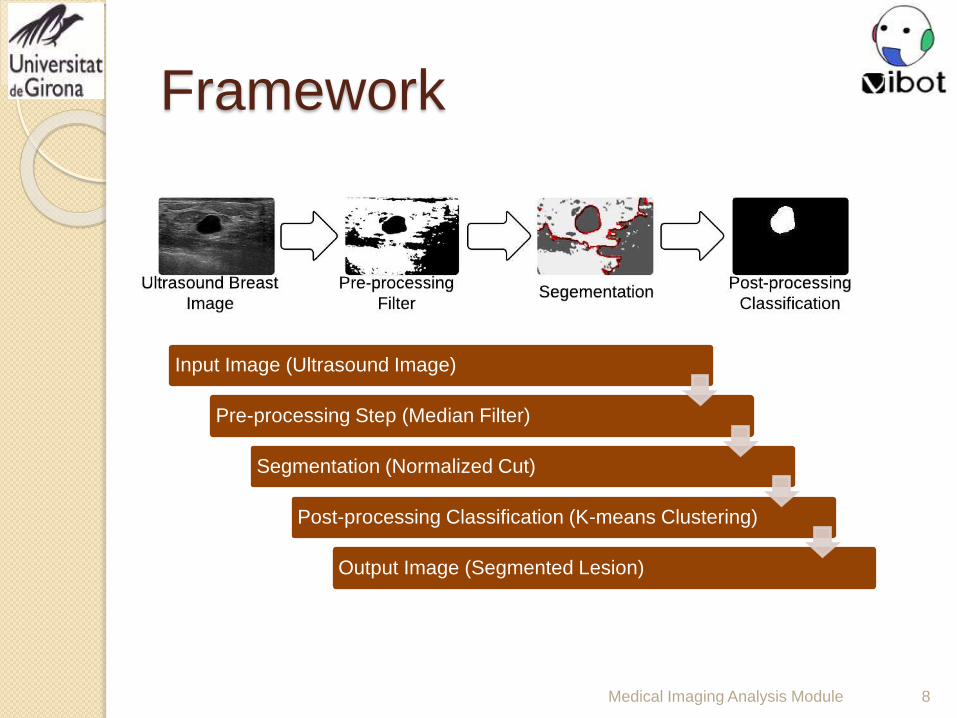
Framework
Medical Imaging Analysis Module 8
Input Image (Ultrasound Image)
Pre-processing Step (Median Filter)
Segmentation (Normalized Cut)
Post-processing Classification (K-means Clustering)
Output Image (Segmented Lesion)

Framework: Pre-processing Step
Medical Imaging Analysis Module 9
imadjust
Optional Intensity
Adjustment
0
500
1000
1500
2000
2500
3000
3500
imadjust
0 50 100 150 200 250
Input
Input Image
(Gray Scale)
0
500
1000
1500
2000
2500
3000
3500
Input
0 50 100 150 200 250
medfilt2
2D Median Filter
(7x7 Window Size)
histeq
0
500
1000
1500
2000
2500
3000
3500
4000
4500
histeq
0 50 100 150 200 250
Optional Histogram
Equalization
im2bw
Binary Thresholding
(Level=0.2)
Remove
Speckle
Noise
Enhance
Image
Quality

Framework: Segmentation
Medical Imaging Analysis Module 10
Enhanced Image
im2bw
20 40 60 80 100 120 140 160
20
40
60
80
100
120
140
160
Normalized Cut
Method
(4 Segments)
0
500
1000
1500
2000
2500
3000
3500
4000
0 0.1 0.2 0.3 0.4 0.5 0.6 0.7 0.8 0.9 1
Ncut eigenvectors of 1
Ncut eigenvectors of 2
0
200
400
600
800
1000
1200
1400
1600
1800
2000
0 0.1 0.2 0.3 0.4 0.5 0.6 0.7 0.8 0.9 1
Ncut eigenvectors of 3
0
500
1000
1500
2000
2500
3000
3500
0 0.1 0.2 0.3 0.4 0.5 0.6 0.7 0.8 0.9 1
Ncut eigenvectors of 4
0
500
1000
1500
2000
2500
0 0.1 0.2 0.3 0.4 0.5 0.6 0.7 0.8 0.9 1
Allocate
Region Of
Interest
(ROI)
Input Image

Framework: Classification
Medical Imaging Analysis Module 11
Norm
Input Image
One of
Segmented Images
kmeans
K-means Clustering
(2 clusters)
Contour Selection
Contour Selection with
Minimum Length
1st Approach
Main
2nd Approach
Backup
Contour Selection
Otsu Binary
Thresholding
Selection of
Best Classified
Image based on
Minimum Area
Extract
Lesion
Region
Outline
1. Introduction
2. Problem Definition
3. Framework
4. Results
5. Conclusion
Medical Imaging Analysis Module 12

Results
Medical Imaging Analysis Module 13
14 Dataset
Images
11 Correct
Segmentation
3 Incorrect
Segmentation
No Intensity
Adjustment
No Histogram
Equalization
Jaccard 0.8235
Dice 0.9032
FPR 0.0616
FNR 0.1257
Jaccard 0
Dice 0
FPR 75.488
FNR 100
Results GT

Results
Medical Imaging Analysis Module 14
Image Name Jaccard Dice RFP RFN
000018(F,F) 0.8235 0.9032 0.0616 0.1257
000032(F,F) 0.8107 0.8954 0.0017 0.1879
000031(T,F) 0.5857 0.7387 0.0294 0.3971
000025(T,F) 0.4410 0.6121 0.3541 0.4029
000023(T,F) 0.7143 0.8333 0.0687 0.2366
000011(F,F) 0.5338 0.6960 0.0207 0.4552
000001(F,F) 0.5206 0.6847 0 0.4794
000002(F,F) 0.7693 0.8696 0 0.2307
000022(F,F) 0.4869 0.6549 0.0034 0.5115
000010(T,T) 0.5061 0.6721 0.4162 0.2832
000007(T,T) 0.4365 0.6077 0 0.5635
000019
000014
000030

Outline
1. Introduction
2. Problem Definition
3. Framework
4. Results
5. Conclusion
Medical Imaging Analysis Module 15

Conclusion
Medical Imaging Analysis Module 16
Speckle noise reduction:
It is an important prerequisite , whatever ultrasound imaging
techniques is used for tissue characterization.
preprocessing step:
The median filter in the preprocessing step is not an effective method
to enhance the edges and lines in the images.

Bibliography
Medical Imaging Analysis Module 17
“Automated breast cancer detection and classification using
ultrasound images: A survey”, H.D. Cheng, J. Shan, W. Ju, Y. Guo,
and L. Zhang, Pattern Recognition, Volume 43, Issue 1, January
2010, Pages 299-317.
“Automated segmentation of breast lesions in ultrasound images”, X.
Liu, Z. M. Huo, and J. W. Zhang, IEEE Comput. Soc., Shanghai,
China, 2005, pp. 7433–7435.
“Image Segmentation with Normalized Cuts”, Jianbo Shi,
Department of Computer and Information Science, University of
Pennsylvania.

Medical Imaging Analysis Module 18
Thanks for
Listening!

Medical Imaging Analysis Module 19
Questions?!


![LI ET AL.: SEMI-SUPERVISED SKIN LESION SEGMENTATION …dermoscopy images [14,18]. For example, Jaisakthi et al. [14] proposed a semi-supervised skin lesion segmentation method using](https://static.fdocuments.us/doc/165x107/60658319b2024701434d8eca/li-et-al-semi-supervised-skin-lesion-segmentation-dermoscopy-images-1418-for.jpg)