
Branchial Cleft and Pouch Anomalies - Department of
32
Branchial Cleft and Pouch Anomalies Prof.Mohamed Hesham Alexandria Faculty of Medicine Alexandria, Egypt Emberyological Basis
Transcript of Branchial Cleft and Pouch Anomalies - Department of

Branchial Cleft and Pouch Anomalies
Prof.Mohamed HeshamAlexandria Faculty of Medicine
Alexandria, Egypt
Emberyological Basis

2nd
3rd
1st
Cervical sinus
4th
6th
PinnaEAC
Branchial Clefts
4th
2nd
3rd
1st
4th
6th
Branchial Pouches

•Sinus•External
•Internal
•Fistula
•Cyst
Types of Anomalies
Skin
Gut
Branchial cleft and pouches anomalies
• 2nd cleft&pouch 95%• 1st 5%• 3rd and 4th rarely reported
Incidence

First Branchial Cleft Anomalies• The first branchial cleft is the only cleft that
not become obliterated by the 8th week .• First cleft anomalies are a result of incomplete
closure of the cleft
First Branchial Cleft Anomalies
Classification
Arnot,1971
Work,1972
Olsen etal,1980

First Branchial Cleft Anomalies
• Preauricular and cervical region above hyoid bone
• The incidence of malformation is higher at the top of the triangle Trigalia et al,1998
Site
• Otological Manifestations
• Parotid Manifestations
• Neck Manifestations
Pochet’s triangle
First Branchial Cleft Anomalies
Presentation
•Cyst
•Sinus
•Fistula
Infection
Prior Surgery
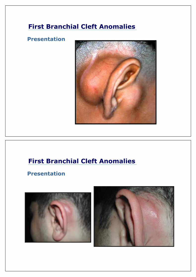
First Branchial Cleft Anomalies
Presentation
First Branchial Cleft Anomalies
Presentation

First Branchial Cleft Anomalies
Presentation
First Branchial Cleft Anomalies
Presentation

First Branchial Cleft Anomalies
Presentation
Branchio-Oto-renal syndromeAutosomal dominantRenal, 1st and 2nd arches•Hearing impairment 73%•Preauricular pits 70%•Renal abnormalities 67%•Branchial cleft cyst or sinus 60%
First Branchial Cleft AnomaliesPresentation

The relationship of the branchial anomaly to the facial nerve is variable
•The fate of branchial apparatus is complete by 6 or 7 week •Parotid gland appear at 6th week•Facial nerve and muscles migrate upward between 6-8 week
First Branchial Cleft Anomalies
Facial Nerve
First Branchial Cleft Anomalies
Facial Nerve
• In the Majority of cases, The anomaly is lateral to the nerve
• Miller et al , 1984; Sinuses lateral to the facial nerve
Fistulae medial to facial nerve
• Solares etal, 2003;
Complete fisulae are usually deep to the facial nerve
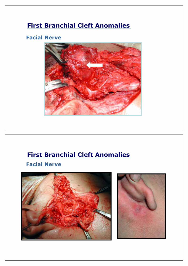
First Branchial Cleft Anomalies
Facial Nerve
First Branchial Cleft Anomalies
Facial Nerve
First Branchial Cleft Anomalies
Facial Nerve
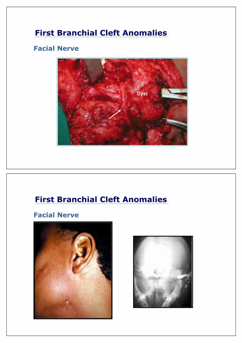
Cyst
First Branchial Cleft Anomalies
Facial Nerve
First Branchial Cleft Anomalies
Facial Nerve
First Branchial Cleft Anomalies
Facial Nerve
First Branchial Cleft Anomalies
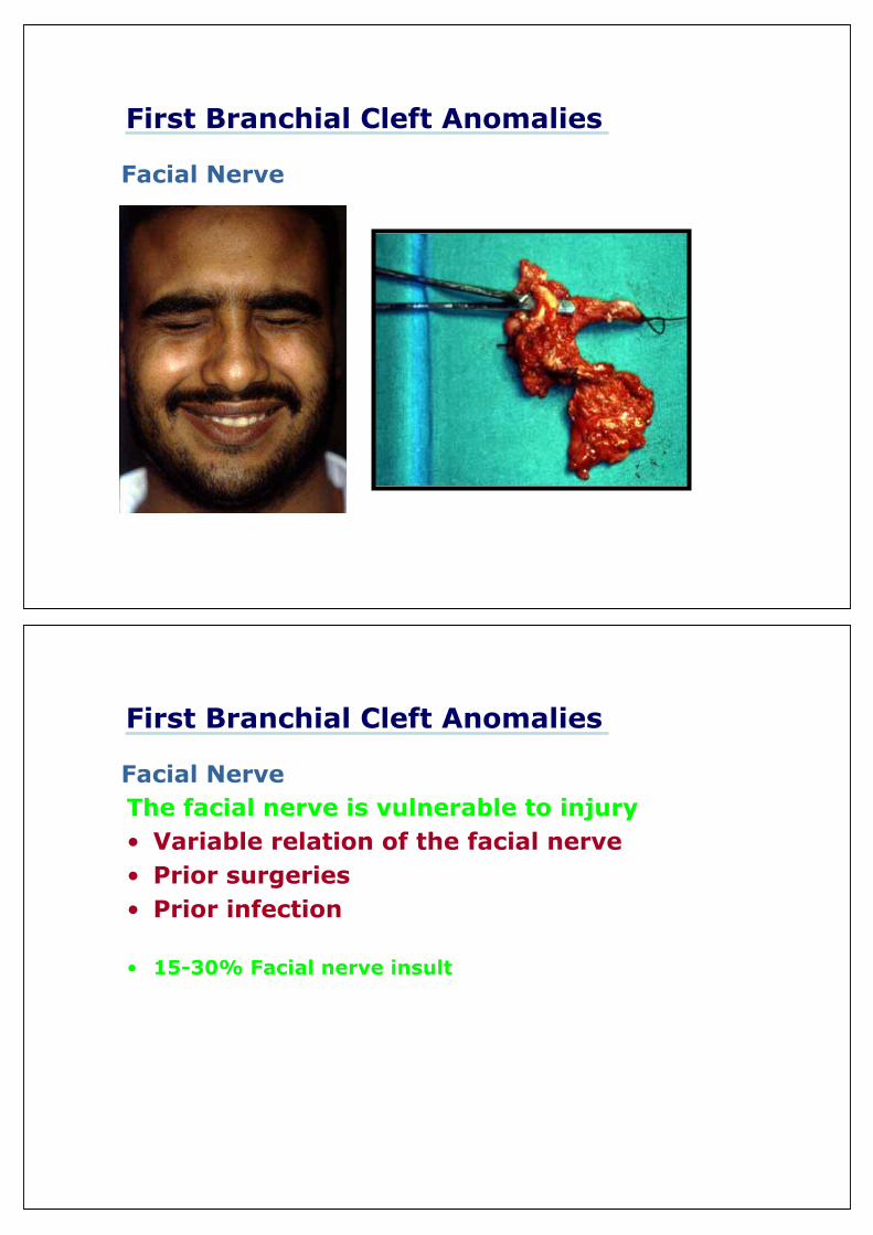
Facial Nerve
The facial nerve is vulnerable to injury• Variable relation of the facial nerve• Prior surgeries• Prior infection
• 15-30% Facial nerve insult
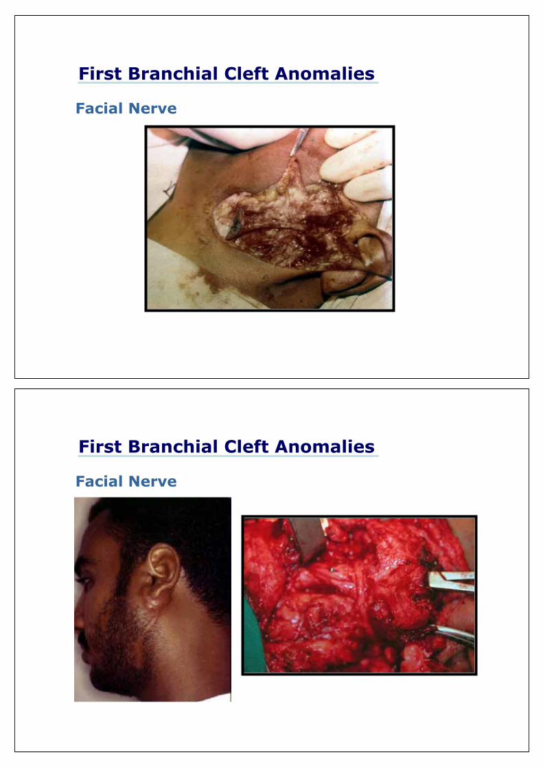
First Branchial Cleft Anomalies
Facial Nerve

First Branchial Cleft Anomalies
Second Branchial Cleft Anomalies
Tons. Skin
•Ext. opening: Ant. Border of sternomastoid muscle in the mid or lower neck.

Second Branchial Cleft Anomalies
Tons. Skin
•Course: Deep to the platysma, along the carotid sheath, then between 2 carotids, superficial to nerve XII, IX deep to stylohyoid ligament and post. Belly of the digastric muscle•Internal opening: Anterior aspect of the upper half of the post. pillar of the tonsil
Sinus or Fistula
How
Why
?Second Branchial Cleft Anomalies
Second Branchial Cleft Anomalies
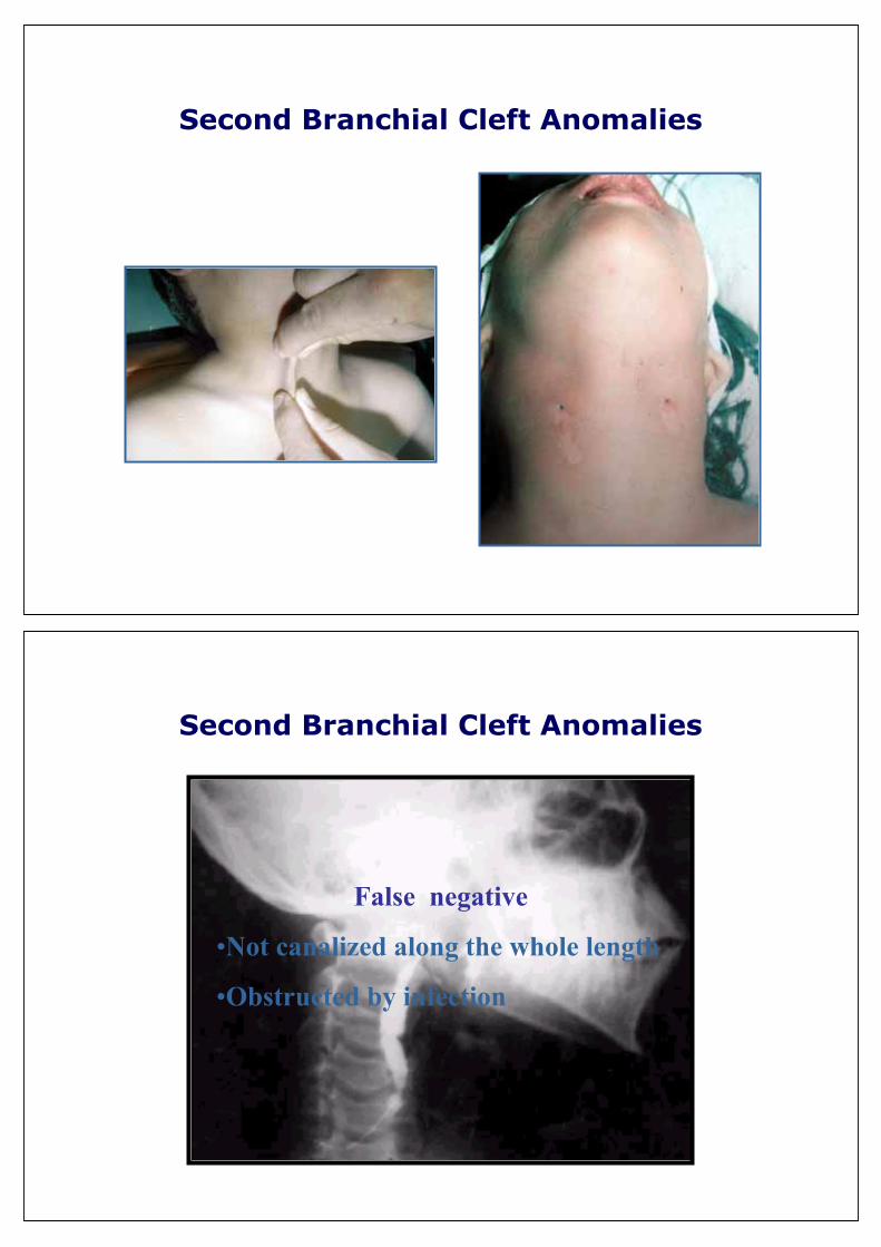
False negative
•Not canalized along the whole length
•Obstructed by infection
Second Branchial Cleft Anomalies
Second Branchial Cleft Anomalies
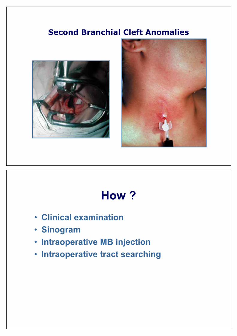
How ?
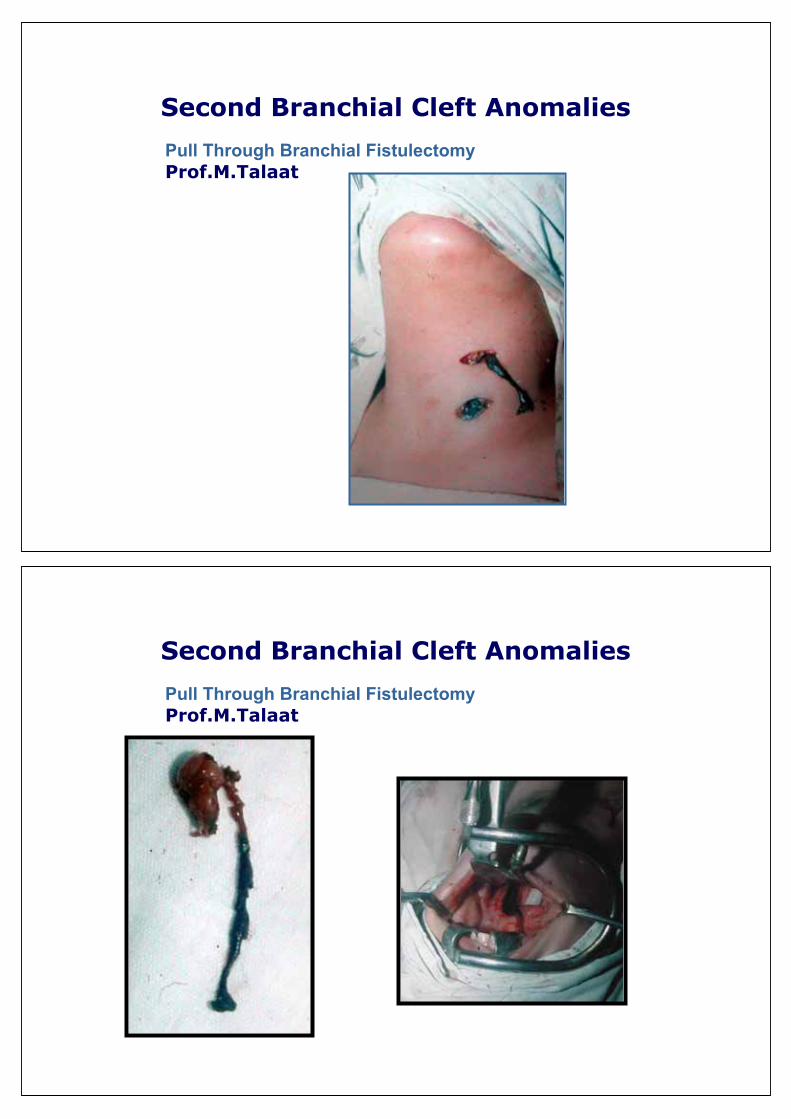
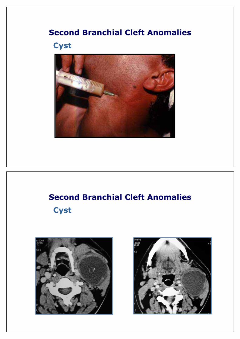
• Clinical examination• Sinogram• Intraoperative MB injection• Intraoperative tract searching
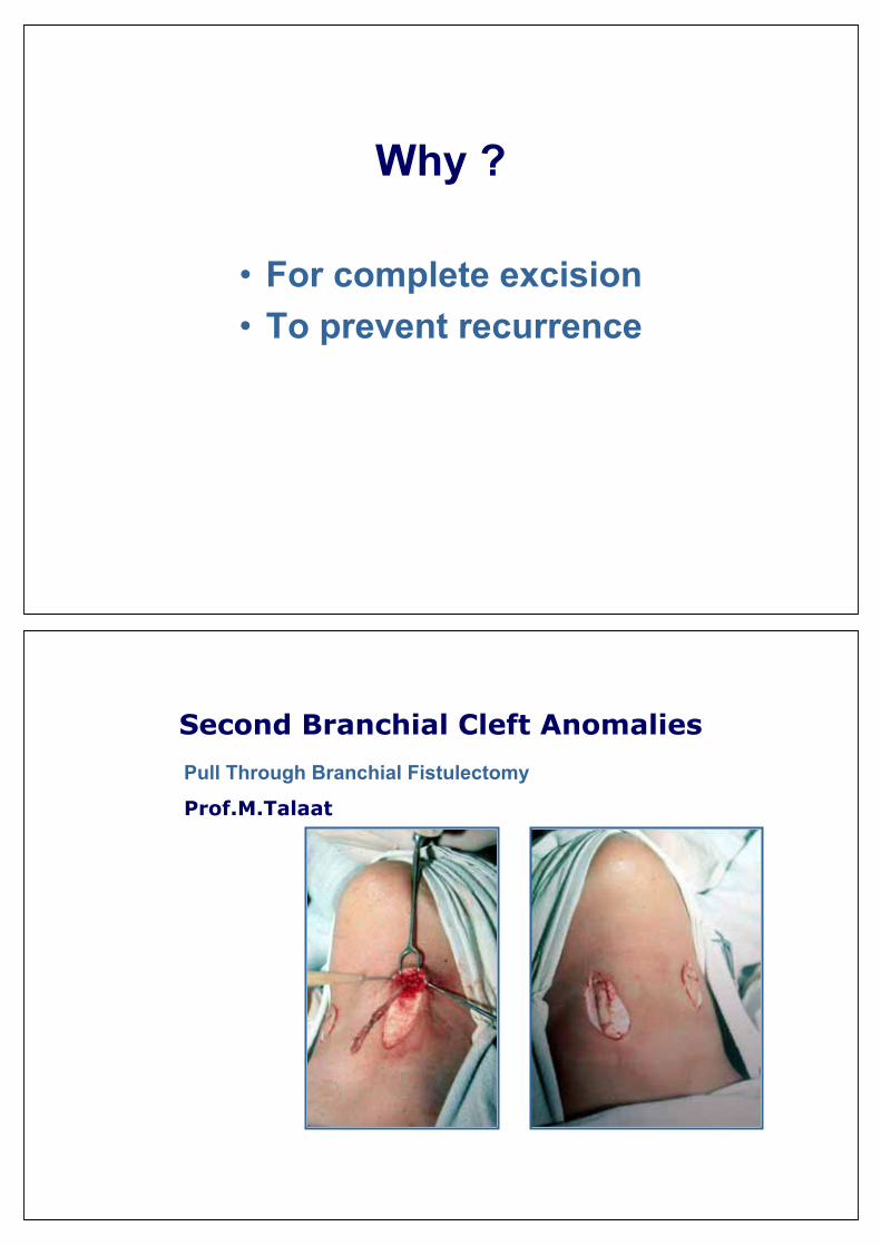
Why ?
• For complete excision• To prevent recurrence
Second Branchial Cleft Anomalies
Pull Through Branchial Fistulectomy
Prof.M.Talaat
Second Branchial Cleft Anomalies
Pull Through Branchial Fistulectomy Prof.M.Talaat
Second Branchial Cleft Anomalies
Pull Through Branchial Fistulectomy Prof.M.Talaat

Second Branchial Cleft Anomalies
Pull Through Branchial Fistulectomy Prof.M.Talaat
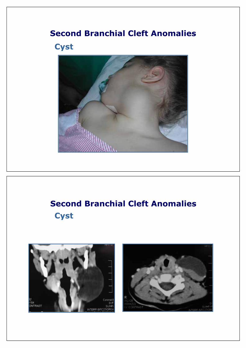
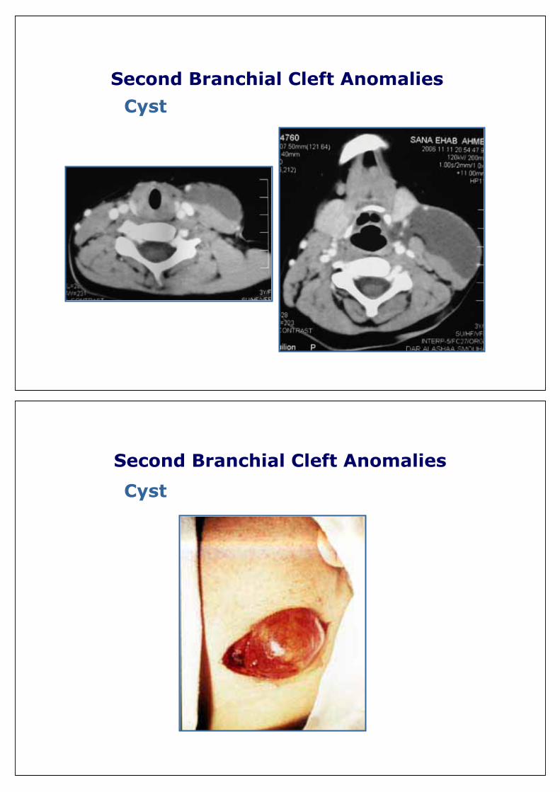
Second Branchial Cleft Anomalies
Cyst
Second Branchial Cleft Anomalies
Cyst
Second Branchial Cleft Anomalies
Cyst
Second Branchial Cleft Anomalies
Cyst
Second Branchial Cleft Anomalies
Cyst
Second Branchial Cleft Anomalies
Cyst
Second Branchial Cleft Anomalies
Cyst
Second Branchial Cleft Anomalies
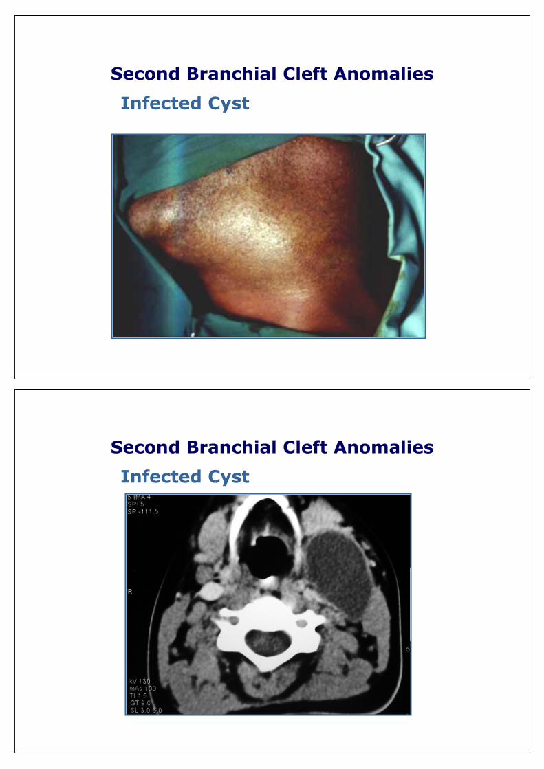
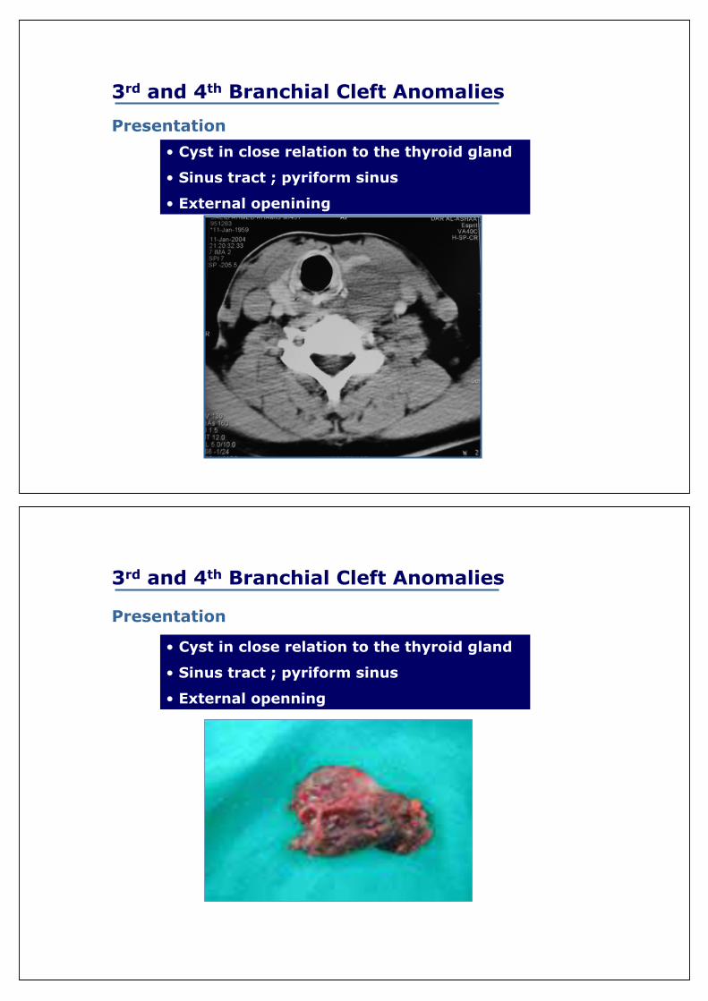
Infected Cyst
Second Branchial Cleft Anomalies
Infected Cyst

Second Branchial Cleft Anomalies
Infected Cyst
Second Branchial Cleft Anomalies
Infected Cyst

3rd and 4th Branchial Cleft Anomalies
• Rarely reported
• Failure of the pouches to obliterate
• 3rd : Inferior parathyroid, Thymus
• 4th: Superior Parathyroid, PFC
• Pharyngobranchial duct
3rd and 4th Branchial Cleft Anomalies
Presentation
• Cyst in close relation to the thyroid gland
• Sinus tract ; pyriform sinus
• External openining
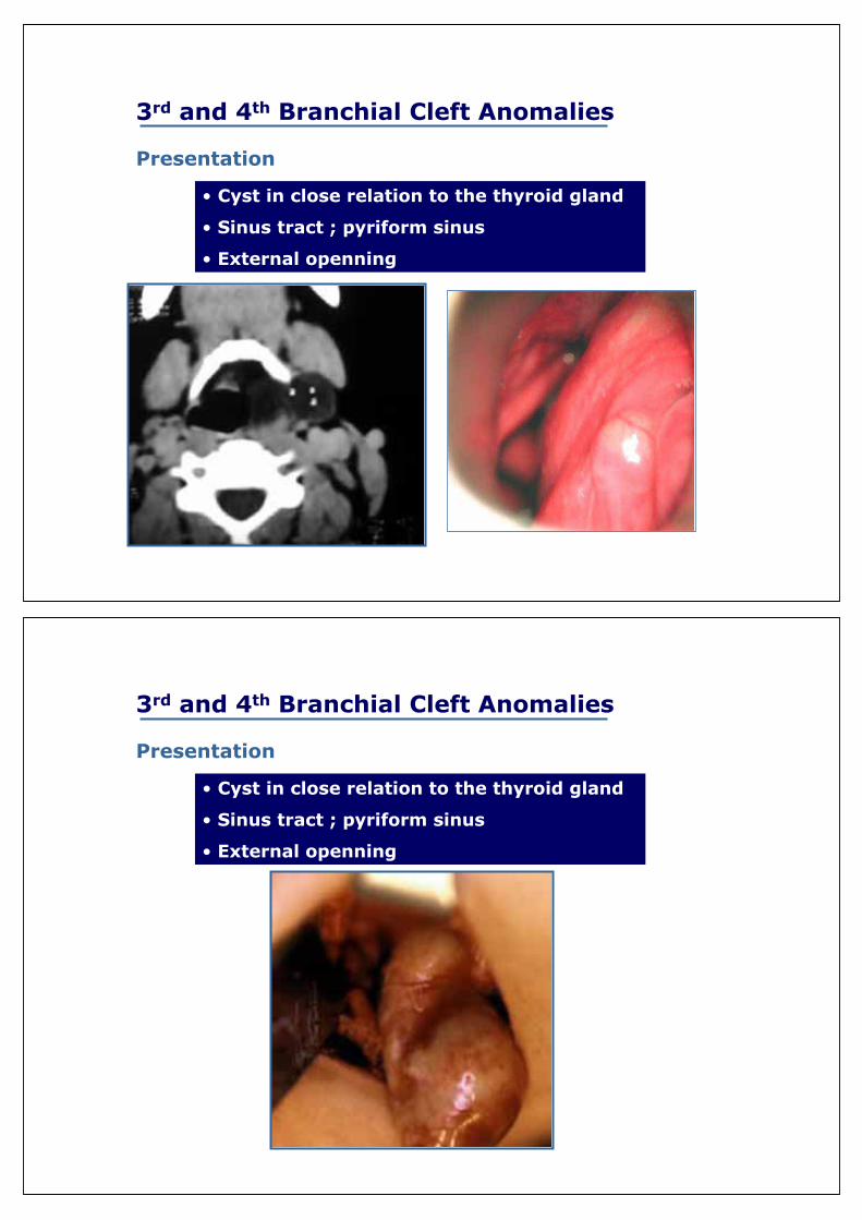
3rd and 4th Branchial Cleft Anomalies
Presentation
• Cyst in close relation to the thyroid gland
• Sinus tract ; pyriform sinus
• External openining
3rd and 4th Branchial Cleft Anomalies
Presentation
• Cyst in close relation to the thyroid gland
• Sinus tract ; pyriform sinus
• External openning

3rd and 4th Branchial Cleft Anomalies
Presentation
• Cyst in close relation to the thyroid gland
• Sinus tract ; pyriform sinus
• External openning
3rd and 4th Branchial Cleft Anomalies
Presentation
• Cyst in close relation to the thyroid gland
• Sinus tract ; pyriform sinus
• External openning
3rd and 4th Branchial Cleft Anomalies
Presentation
• Cyst in close relation to the thyroid gland
• Sinus tract ; pyriform sinus
• External openning
3rd and 4th Branchial Cleft Anomalies
Presentation
• Cyst in close relation to the thyroid gland
• Sinus tract ; pyriform sinus
• External openning

3rd and 4th Branchial Cleft Anomalies
Presentation
• Cyst in close relation to the thyroid gland
• Sinus tract ; pyriform sinus
• External openning
3rd and 4th Branchial Cleft Anomalies
Presentation
• Cyst in close relation to the thyroid gland
• Sinus tract ; pyriform sinus
• External openning

Midline Cervical Cleft
• Medial cleft • Median fissure of the neck • Midline cervical cord • Midline cervical webbing• Pterygium colli medianum
• It is not a "true" cleft, as it does not involve a gap between adjacent skin flaps and only extends partially through the skin layers.
•The cleft is often weeping at birth, then toughens and dries, healing with a scar
• A subcutaneous fibrous cord that originates from the deep layer of the skin tag and ends in the subcutaneous tissue of the chin
Midline Cervical Cleft

Conclusion� Branchial arch anomalies should be bared in mind in all cysts or sinuses located from the root of the helix to the clavicle
�Other congenital anomalies Should be excluded
�Complete fistula has to be diagnosed
� Pull through branchial fistulectomy for complete fistula of the 2nd arch
� 1st branchial arch anomalies should be excised with wide approach with care of the facial nerve