Brain Tumours
35
1 06 Brain Tumours J A S C A P JEET ASSOCIATION FOR SUPPORT TO CANCER PATIENTS, MUMBAI, INDIA
-
Upload
health-education-library-for-people -
Category
Documents
-
view
363 -
download
6
description
This booklet is for you if you have or someone close to you has a brain tumour.If you are a patient your doctor or nurse may wish to go through the booklet with you and mark sections that are particularly important for you.Primary brain tumours are tumours that start in the brain and have not spread there from somewhere else in the body.A secondary brain tumour is a tumour in the brain that has occurred because cancer cells from a cancer in another part of the body have spread to the brain.The incidence (newly diagnosed cases of cancer in a year) of Brain Tumours in India is about 2 patients per 1,00,000 population, while the morality rate (deaths due to brain cancer) is a little less than 2 patients per 1,00,000 population.In the year 2006 at TATA Memorial Hospital in Mumbai, India 372 people were diagnosed with Brain and Central Nervous System Tumours, out of which 250 (67%) were males and 122 (33%) were females.
Transcript of Brain Tumours

1
06
Brain Tumours
J A S C A P
JEET ASSOCIATION FOR SUPPORT TO CANCER PATIENTS, MUMBAI, INDIA

2
JASCAP JEET ASSOCIATION FOR SUPPORT TO CANCER PATIENTS
c/o. Abhay Bhagat & Co., Office No.4, “Shilpa”, 7th.Road, Prabhat Colony,
Santacruz (East), Mumbai – 400 055
Tel.: 2617 7543, 2616 0007. Fax: 91-22-2618 6162
E-mail :[email protected] & [email protected]
JASCAP is a charitable trust that provides information on various aspects of cancer.
This can help the patient and his family to understand the disease and its treatment
and thus cope with it better.
Registered under the Societies Registration Act, 1860 No.1359 / 1996 G.B.B.S.D., Mumbai and under the Bombay Public Trusts Act, 1950 No. 18751 (Mumbai). Donations to JASCAP qualify for deduction u/s 80G (1) of the Income Tax Act, 1961 vide Certificate No. DIT (E) / BC / 80G / 1383 / 96-97 dated 28.02.97 subsequently renewed.
Contact: Mr. Prabhakar K. Rao or Mrs. Neera P. Rao
Publisher: JASCAP, Mumbai 400 055
Printer: Surekha Press, Mumbai 400 019
Edition: March 2011___________
o © Cancerbackup 01 December 2009
o This booklet is an adaptation of “Understanding Brain Tumoursr” produced by
Cancerbackup and is reproduced with their kind permission.
o JASCAP gratefully acknowledges Cancerbackup‟s permission to reproduce this
booklet
Donation suggested ` 30.00

3
A model of the human brain.
Brain
A. pituitary gland B. cerebrum C. skull D. corpus callosum E. thalamus F.
hypothalamus G. pons H. cerebellum I. medulla J. spinal cord

4
Contents Brain Tumours............................................................................................. Introduction…………….……………………………………….…….......
What is cancer? ................................................................................. Types of cancers……………..…………………………………………... The brain - structure and function ……………..……….……………… Primary and secondary brain tumours…………….….….……………. Risk factors and possible causes of brain tumours…….………...……
Types of primary brain tumours………………………………………………
Gliomas**…………………………………………..……………………… Types of gliomas ………………………………..…………..…………… Medulloblastoma**……………………………………………...………… Central nervous system (CNS) lymphoma** .……….……….………… Pineal region tumours**………………………………………………….. Meningioma**…………………………..………….……………………… Acoustic neuroma**………………………………………………………. Haemangioblastoma**……………………………………..……………. Pituitary tumours**………………..……………………………………… Spinal tumours**…………………………………………………………..
Symptoms and Diagnosis..........................................................................
Symptoms of brain tumours …………………………………………….. How brain tumours are diagnosed ……………...………………………
Treating brain tumours…………..……………………………………………..
Mutlidisciplinary team…………………………………………………….. Treatment overview………………………..…………………………….. Surgery for brain tumours …………...………………………………….. Steroids and anticonvulsant drugs ………..…………………………… Radiotherapy for brain tumours …………..……………………………. Chemotherapy for brain tumours …………...…………………………..
Research - Clinical trials for brain tumours.............................................. Living with a brain tumour…………………………….…………….………… ** JASCAP has separate factsheets on these particular types of Brain Tumours.

5
Brain tumours
Introduction
This booklet is for you if you have or someone close to you has a brain tumour.
If you are a patient your doctor or nurse may wish to go through the booklet with you
and mark sections that are particularly important for you.
What is cancer?
The organs and tissues of the body are made up of tiny building blocks called cells. Cancer is a disease of these cells.
Cells in different parts of the body may look and work differently but most reproduce themselves in the same way. Cells are constantly becoming old and dying, and new cells are produced to replace them. Normally, cells divide in an orderly and controlled manner. If for some reason the process gets out of control, the cells carry on dividing, developing into a lump which is called a tumour.
Tumours can be either benign or malignant. Cancer is the name given to a malignant tumour. Doctors can tell if a tumour is benign or malignant by examining a small sample of cells under a microscope. This is called a biopsy.
In a benign tumour the cells do not spread to other parts of the body and so are not cancerous. However, if they continue to grow at the original site, they may cause a problem by pressing on the surrounding organs.
A malignant tumour consists of cancer cells that have the ability to spread beyond the original area. If the tumour is left untreated, it may spread into and destroy surrounding tissue. Sometimes cells break away from the original (primary) cancer. They may spread to other organs in the body through the bloodstream or lymphatic system.
The lymphatic system is part of the immune system - the body's natural defence against infection and disease. It is a complex system made up of organs, such as bone marrow, the thymus, the spleen, and lymph nodes. The lymph nodes (or glands) throughout the body are connected by a network of tiny lymphatic ducts.

6
When the cancer cells reach a new area they may go on dividing and form a new tumour. This is known as a secondary cancer or metastasis.
It is important to realise that cancer is not a single disease with a single type of treatment. There are more than 200 different kinds of cancer, each with its own name and treatment.
Types of cancers
Carcinomas
The majority of cancers, about 85% (85 in a 100), are carcinomas. They start in the
epithelium, which is the covering (or lining) of organs and of the body (the skin). The
common forms of breast, lung, prostate and bowel cancer are all carcinomas.
Carcinomas are named after the type of epithelial cell that they started in and the part
of the body that is affected. There are four different types of epithelial cells:
squamous cells - that line different parts of the body, such as the mouth, gullet (oesophagus), and the airways
adeno cells - form the lining of all the glands in the body and can be found in organs such as the stomach, ovaries, kidneys and prostate
transitional cells - are only found in the lining of the bladder and parts of the urinary system
basal cells - that are found in one of the layers of the skin.
A cancer that starts in squamous cells is called a squamous cell carcinoma. A cancer
that starts in glandular cells is called an adenocarcinoma. Cancers that start in
transitional cells are transitional cell carcinomas, and those that start in basal cells
are basal cell carcinomas.
Leukaemias and lymphomas
These occur in the tissues where white blood cells (which fight infection in the body)
are formed, i.e. the bone marrow and lymphatic system. Leukaemia and lymphoma
are quite rare and make up about 6.5% (6.5 in 100) of all cancers.
Sarcomas
Sarcomas are very rare. They are a group of cancers that form in the connective or
supportive tissues of the body such as muscle, bone and fatty tissue. They account
for less than 1% (1 in 100) of cancers.
Sarcomas are split into two main types:
bone sarcomas - that are found in the bones soft tissue sarcomas - that develop in the other supportive tissues of the
body.

7
Others forms of cancer
Brain tumours and other very rare forms of cancer make up the remainder of cancers.
The brain - structure and function
The brain, together with the spinal cord, makes up the central nervous system. This is the 'control centre' which coordinates the body‟s functions.
Between the surfaces of the brain and the skull there are three layers of membrane called the meninges, which completely cover the brain and spinal cord. Between two of these layers is a space called the subarachnoid space. The subarachnoid space contains a fluid called cerebrospinal fluid (CSF).
'>
Diagram showing the different structures of the brain
Types of brain cells
Like every other organ in the body, the brain is made up of cells. There are about 40 billion nerve cells, also called neurones, within the brain. Everyone is born with a similar amount. Unlike other cells, nerve cells are not able to replace themselves. In fact, as we get older there is a gradual decrease in their number.
The nerve cells communicate with each other and other parts of the body by sending messages (nerve impulses) through a system of nerve pathways or networks.
The nerve cells are held in place and supported by glial cells. There are different types of glial cells, including astrocytes, oligodendrocytes and ependymal cells.

8
Main structures and functions of the brain
The main parts of the brain are:
the cerebrum (the forebrain) made up of the right and left cerebral hemispheres
the cerebellum (the hindbrain)
the brain stem.
Cerebrum This is the largest area of the brain and is concerned with all higher mental functions, such as thinking and memory. It's made up of two halves or hemispheres. The right cerebral hemisphere controls the left side of the body and the left cerebral hemisphere controls the right side of the body.
Each cerebral hemisphere is divided into four areas, known as lobes: the frontal, parietal, temporal and occipital lobes. Each lobe controls a different range of activities.
Cerebellum This is the back part of the brain and is concerned with balance and coordination. These activities are carried out automatically (subconsciously) by this area of the brain and are not under a person's control.
Brain stem This controls the basic functions essential to maintaining life, including blood pressure, breathing, heartbeat, eye movements and swallowing. It's the bottom part of the brain and connects the cerebral hemispheres to the spinal cord.
'>
The lobes and functions of the brain

9
Primary and secondary brain tumours
Primary brain tumours
Primary brain tumours are tumours that start in the brain and have not spread there from somewhere else in the body.
Benign brain tumours
These are tumours that remain in the part of the brain in which they started and don't spread into and destroy other areas of the brain. They do not spread to other parts of the body. If a benign tumour can be removed successfully it should not cause any further problems.
However, sometimes it's difficult to remove the tumour because of its position within the brain, or because the surrounding brain tissue could be damaged by surgery. Some benign tumours will regrow slowly and, if this happens, treatment with radiotherapy or further surgery may be needed.
Malignant primary brain tumours
These are most likely to cause problems by spreading into the normal brain tissue which surrounds them and causing pressure and damage to the surrounding areas of the brain. These tumours rarely spread outside the brain to other parts of the body.
Secondary brain tumours
A secondary brain tumour is a tumour in the brain that has occurred because cancer cells from a cancer in another part of the body have spread to the brain.
Your doctor will be able to tell you if your brain tumour is a primary or secondary tumour.
Risk factors and possible causes of brain tumours
How common is Brain Tumour in India?
The incidence (newly diagnosed cases of cancer in a year) of Brain Tumours in India is about 2 patients per 1,00,000 population, while the morality rate (deaths due to brain cancer) is a little less than 2 patients per 1,00,000 population.
In the year 2006 at TATA Memorial Hospital in Mumbai, India 372 people were diagnosed with Brain and Central Nervous System Tumours, out of which 250 (67%) were males and 122 (33%) were females.
What Causes Primary Brain Tumours?
The cause of most primary brain tumours is unknown, research into this is ongoing. Brain tumours, like other tumours, are not infectious and can't be passed on to other people. They are slightly more common in men than in women.

10
Age
Although brain tumours can develop at any age, as with a lot of tumours, people are more likely to get them as they get older. Some types of brain tumour, however, are more common in younger adults. Children can also develop brain tumours. We have separate information about brain tumours in children.
Genetic conditions
Brain tumours are not caused by an inherited faulty gene that can be passed on to other family members.
A small number of brain tumours occur in people who have certain genetic conditions, such as neurofibromatosis type 1 and type 2, tuberous sclerosis, or the following syndromes:
Li-Fraumeni
Von Hippel-Lindau
Turcot's
Gorlin.
For more information about these conditions you can contact our cancer support specialists.
Previous radiotherapy treatment
People who've been exposed to radiation to their head, such as children who had radiotherapy to the head for leukaemia, are at a slightly higher risk of developing a brain tumour than others.
Other unproven causes
Other factors, such as mobile phones, power lines and certain viruses, have been suggested as possible causes of brain tumours. A lot of research has looked into these possible causes, especially mobile phones. However from the evidence to date we still can't say for sure that they cause or increase the risk of developing a brain tumour.
The cause of a secondary brain tumour is always a primary cancer somewhere else in the body.
Types of primary brain tumours
There are many different types of brain tumours. They are usually named after the type of brain cells from which they have developed.
This section gives brief details about the main types of malignant and benign brain tumours. You may find it helpful to look at the diagrams on the brain page when reading this section.

11
If you have any questions about the type and position of your tumour, your doctor can give you more information. Our cancer support specialists can also give you further information about the different types of brain tumours.
Gliomas
Most tumours develop from the supporting cells of the brain known as the glial cells. They may be named after the type of cell that they are made up of, or after the part of the brain in which they are found, such as a brain stem glioma. More than half of all primary brain tumours are gliomas.
Grading of gliomas
Grading is a term that refers to how the tumour cells look under the microscope. A pathologist will examine the cells and look at whether there are signs they are dividing, how abnormal they look and if there are any abnormal blood vessels.
The grade gives an idea of how quickly the tumour may develop. There are four grades: grade 1 tumours are benign and grow very slowly, whereas grade 4 tumours are malignant (cancerous) and grow faster. Sometimes grade 1 and 2 gliomas are called low-grade gliomas and grades 3 and 4 are called high-grade gliomas. Knowing the type and grade of your tumour will help your doctors plan the best treatment.
Types of gliomas
Astrocytic tumours − This is the most common type of glioma and develops from star-shaped cells called astrocytes. They are graded 1-4. Grade 4 astrocytic gliomas (sometimes called glioblastoma) is the commonest brain tumour found in adults. Grade 3 astrocytic glioma is also known as anaplastic astrocytoma.
Oligodendroglioma − These are tumours that are made up of cells known as oligodendrocytes, which produce the covering of nerve cells - the myelin sheath. They are usually slower-growing than astrocytic tumours.
Mixed glioma − Mixed gliomas are made up of a mixture of more than one type of cell. Oligo-astrocytomas are a type of mixed glioma and are made up of cells called astrocytes and oligodendrocytes.
Ependymoma − A rare type of glioma, ependymomas develop from the ependymal cells, which line the ventricles of the brain and the central canal of the spinal cord. These tumours can spread to other parts of the brain and spinal cord.
Medulloblastoma
Medulloblastomas are one of the most common malignant brain tumours in children, but rare in adults. They usually develop in the cerebellum at the back of the brain but may spread to other parts of the brain. Very occasionally, these tumours spread outside the brain to the lymph nodes or lungs.
Medullablastoma is a type of primitive neuroectodermal tumour (PNET). PNETs develop from cells which are left over from the earliest stages of a baby's development in the womb.

12
Central nervous system (CNS) lymphoma
A lymphoma is a malignant tumour of the lymphatic system, which is part of the body‟s immune system. In rare cases, these tumours only affect the brain. They are then called primary CNS lymphomas.
Pineal region tumours
The pineal gland is just below the area where the two cerebral hemispheres join. Tumours in this part of the brain are extremely rare. They can be made up of different types of cells.
The most common tumours found in the pineal gland are germinomas; others include teratomas, pineocytomas and pineoblastomas.
Meningioma
Meningiomas arise from the meninges, the covering of the brain. They can occur in any part of the meninges over the brain or spinal cord and usually grow very slowly. Most meningiomas are benign and do not spread from the area where they started. Malignant meningiomas are very rare.
Acoustic neuroma
Acoustic neuromas, (also called vestibular schwannoma or neurilemmoma) are benign tumours that develop in the acoustic or auditory nerve, which controls hearing and balance. The nerve is covered by schwann cells. The tumour arises from these cells, so it is also known as a schwannoma.
Acoustic neuromas are usually found only in adults and are more common in people who have a genetic condition called neurofibromatosis type 2 (NF2).
Haemangioblastoma
This is a rare type of tumour that develops from the cells that line the blood vessels. Haemangioblastomas are benign and grow slowly. It may take several years for symptoms to appear.
Pituitary tumours
The pituitary gland produces hormones that control and regulate the other hormone-producing glands of the body. Pituitary tumours are benign and are also called pituitary adenomas. Symptoms often occur due to disturbances in vision or hormone levels.
Spinal tumours
Spinal tumours often cause symptoms by pressing on the spinal nerves. These symptoms may include numbness, tingling, leg or arm weakness and pain in the back, neck and limbs.

13
Sometimes a tumour in the lower part of the spinal cord can lead to loss of control of the bladder or bowel.
** JASCAP has separate factsheets in more details on each of these
particular types of Brain Tumours.
Symptoms and diagnosis Symptoms of brain tumours
A brain tumour may cause symptoms because the space it takes up in the skull puts pressure on the brain or because it is disturbing the function of the part of the brain it's growing in.
Symptoms of brain tumour may occur due to following reasons:
Symptoms due to increased pressure in the skull Seizures Symptoms connected with the tumour's position Personality changes
Symptoms due to increased pressure in the skull
The brain is contained within the skull and has a fixed amount of space. If a tumour grows in the brain it will often cause an increase in pressure, which can cause symptoms to develop. An increase of pressure in the skull is called raised intracranial pressure (ICP). The most common symptoms of a rise in the pressure within the brain are headaches, feeling sick (nausea) and vomiting.
Of course, many other things can cause headaches or feelings of sickness, but if you have either of these for over a week with no sign of getting better, it's important to see your doctor.
A pressure headache may be most severe in the mornings, and can occasionally wake you. Usually this type of headache gets better during the day. However, it may get worse when you cough, sneeze, bend down or do any hard physical work. All of these tend to raise pressure in the brain. If the raised pressure makes you sick, it may be worse in the morning than during the day.
Another possible symptom of a brain tumour is drowsiness. This can happen as the pressure in the brain increases. You may find that you sleep more or that you drop off during the day when you wouldn‟t normally.
As well as the symptoms described here, raised intracranial pressure can also cause changes to your sight, such as blurred vision, 'floating objects' and tunnel vision. It may also make you confused or affect your balance.
Seizures
Seizures (fits) are another common symptom caused by brain tumours. Some people can may experience muscle spasms which could be twitching or jerking of an arm or

14
leg or sometimes the whole body. Occasionally they can cause moments of unconsciousness.
It's important to remember that a seizure can be caused by medical conditions other than a brain tumour. A seizure can be a frightening experience. if you have one you should seek medical help so that the cause can be diagnosed and treated.
Symptoms connected with the tumour's position
Some symptoms may be caused by tumours in particular parts of the brain. Sometimes a headache can feel worse on the same side of the tumour. In general, each area of the brain controls particular functions. A tumour in a particular part of the brain may prevent that area of the brain from working normally.
Some of these symptoms are listed below, grouped under the different parts of the brain. They are included only as a guide. Exact diagnosis can only be made by a doctor and confirmed by tests.
The diagram showing the lobes and functions of the brain shows the different functions of each area of the brain.
Frontal lobe tumours − Changes in personality and intellect. Uncoordinated walking or weakness of one side of the body, loss of smell, occasional speech difficulties.
Parietal lobe − Difficulty speaking or understanding words. Problems with writing, reading or doing simple calculations. Difficulty in coordinating certain movements, and finding your way around. Numbness or weakness on one side of the body.
Occipital lobe − Loss of vision on one side. The person may not notice this at first and it may sometimes be discovered during routine eye tests.
Temporal lobe − Fits, which may cause strange sensations: a feeling of fear or intense familiarity (déjà vu), strange smells or blackouts. Speech difficulties and memory problems.
Cerebellum − Lack of coordination which affects walking and speech (dysarthria), unsteadiness, flickering involuntary movement of the eyes (nystagmus). Vomiting and neck stiffness.
Brain stem − Unsteadiness and an uncoordinated walk. Facial weakness, a one-sided smile or drooping eyelid. Double vision. Vomiting or headache just after waking (this is rare). Difficulty speaking and swallowing. Symptoms may appear gradually.
All the above symptoms may be caused by conditions other than a brain tumour. If you have any of the symptoms described it is important that you go to see your GP.
Personality changes
Sometimes brain tumours may cause changes in personality or behaviour. These symptoms usually occur when the tumour is in the brain‟s cerebral hemispheres. This situation can be very unsettling for the person and their family. Sometimes a referral to a psychologist for assessment and support can help.

15
Driving
As brain tumours can cause changes in the way that your brain works, it may be dangerous to drive. In the UK, there are laws which restrict some people with brain tumours from driving for a time. The restrictions vary with the type of tumour you have, and the type of driving license you hold.
However, you will not usually be allowed to drive for at least a year after the condition has been diagnosed and in some circumstances you may not be able to drive again. With some types of benign tumours you may be able to drive again once you have recovered from your treatment.
It is your legal responsibility, not your doctor's, to tell the Drivers and Vehicle Licensing Authority (DVLA) about your illness. The DVLA will advise of you of any restrictions on your right to drive. You should also inform your car insurance company.
How brain tumours are diagnosed
Usually, if you have slowly developing symptoms you'll see your GP, who will examine you. If a brain tumour is suspected, they will refer you to a specialist doctor, either:
a neurologist (a specialist in brain and nerve disorders) or an oncologist (a doctor who specialises in cancer treatment).
Sometimes people with brain tumours have a sudden seizure (fit) or sudden onset of problems associated with the position of the tumour, and may be taken straight to hospital, where tests are carried out to diagnose the tumour. Brain tumours are often treated in specialist centres, so you may have to travel to your nearest centre.
At the hospital
At the hospital the doctor will ask you about your general health and any previous medical problems. You will then have a general physical examination. The doctor may listen to your chest and feel your abdomen to check your general health, and you will have a detailed examination to test your nervous system. The examination of your nervous system may include:
Mental exercises, eg basic arithmetic and simple questions.
An eye examination using an instrument that shines a light at the back of the eye (ophthalmoscope). This test is done to see if the optic disc at the back of your eye is swollen. This swelling is known as papilloedema and is a sign of raised pressure in your skull. Your eyesight will also be tested.
Hearing tests.
Facial muscle tests − smiling, grimacing etc.
Tongue movement, checking your swallow (gag) reflex.
Checking the strength of your arms and legs, and your reflexes.
A test of your ability to feel pinpricks on areas of skin, to tell the difference between hot and cold, and possibly to recognise the feeling and shape of familiar objects like coins.
Checking your balance and coordination, for example by asking you to walk a few steps or perform repeated movements.

16
Further tests for diagnosing brain tumours
At the hospital you may have to have some of the following tests. Your doctor will select the most suitable ones for your particular situation and symptoms.
Following tests are routinely used for diagnosing brain tumours:
Brain MRI (magnetic resonance imaging) scan Brain CT (computerised tomography) scan Biopsy Skull x-ray PET (positron emission tomography) scan SPECT (single photon emission computerised tomography) scan Blood tests and chest x-rays
Brain MRI (magnetic resonance imaging) scan
This test uses magnetic fields to build up a detailed picture of the inside of your head. Before the scan you may be asked to complete and sign a checklist. This is to make sure that it‟s safe for you to have an MRI scan, because the scanner is a powerful magnet. The checklist asks about any metal implants you may have, for example a pacemaker, surgical clips, bone pins etc.
You should also tell your doctor if you have ever worked with metal or in the metal industry as very tiny fragments of metal can sometimes lodge in the body. If you do have any metal in your body it‟s likely that you won‟t be able to have an MRI scan. In this situation another type of scan can be used.
Before having the scan, you‟ll be asked to remove any metal belongings including jewellery. You are usually given an injection of dye into a vein in the arm, which doesn‟t usually cause discomfort. This is called a contrast medium and can help the images from the scan to show up more clearly.
During the test you‟ll be asked to lie very still on a couch inside a long cylinder (tube) that is open at both ends. The whole test can take up to an hour. It‟s painless but can be slightly uncomfortable, and some people feel a bit claustrophobic during the scan. It‟s also noisy, but you‟ll be given earplugs or headphones. You will be able to hear, and speak to, the person operating the scanner.
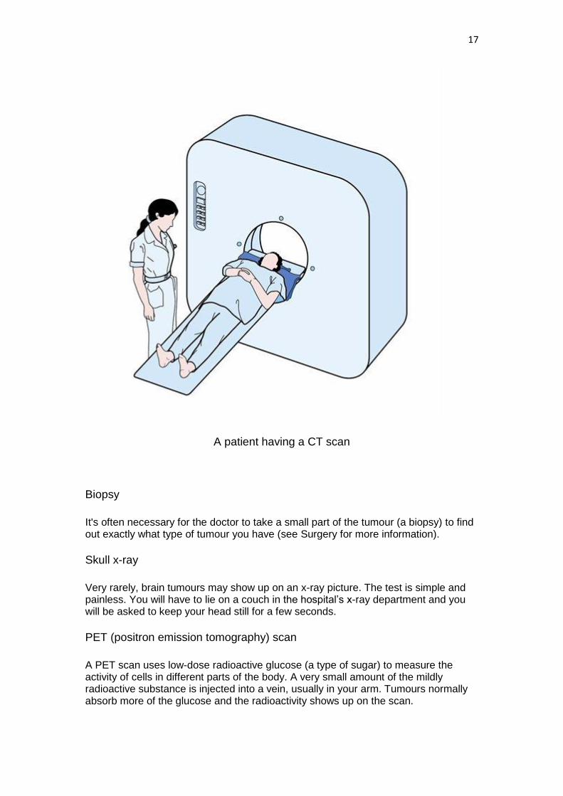
Brain CT (computerised tomography) scan
This is a series of x-rays, which builds up a three-dimensional picture of the inside of your head. During the test you will be asked to lie with your head inside an opening in the scanner. The scan is painless but takes longer than a normal x-ray (from 5 to 10 minutes). It may be used to identify the exact area and size of the tumour.
Most people who have a CT scan are given an injection of a liquid into a vein, to allow particular areas of the brain to be seen more clearly. The injection may make you feel hot all over for a few minutes. Before having the injection, it is important to tell your doctor and the person doing the scan if you are allergic to iodine or have asthma or diabetes. You'll probably be able to go home as soon as the scan is over.
17
A patient having a CT scan
Biopsy
It's often necessary for the doctor to take a small part of the tumour (a biopsy) to find out exactly what type of tumour you have (see Surgery for more information).
Skull x-ray
Very rarely, brain tumours may show up on an x-ray picture. The test is simple and painless. You will have to lie on a couch in the hospital‟s x-ray department and you will be asked to keep your head still for a few seconds.
PET (positron emission tomography) scan
A PET scan uses low-dose radioactive glucose (a type of sugar) to measure the activity of cells in different parts of the body. A very small amount of the mildly radioactive substance is injected into a vein, usually in your arm. Tumours normally absorb more of the glucose and the radioactivity shows up on the scan.

18
After the injection you may be asked to lie in a dark room with your eyes closed. You'll then be taken to the scanning room and asked to lie on a couch with the scanning ring around you. The dose of radiation you receive is no more than a normal x-ray.
A PET scan is not routinely used to diagnose a brain tumour but it may help to tell whether a tumour is growing and whether it is cancerous (malignant) or benign. PET scans aren‟t available in all hospitals, and you may have to travel to a hospital some distance away from your home to have one.
SPECT (single photon emission computerised tomography) scan
This test is similar to a PET scan. It can help to detect abnormalities in the blood brain barrier as it looks at blood flow through the brain.
You are given an injection of a very mild radioactive substance, usually in your arm. This susbstance travels in the blood to the brain. Then, in the scanning room, pictures (scans) of the brain are taken.
Blood tests and chest x-rays
There isn't a specific blood test that can detect brain tumours, but you may have blood tests to check your general health. A chest x-ray may also be done as part of a general health check.
Treating brain tumours
Treatments such as surgery, radiotherapy or chemotherapy may be used alone or in combination to treat brain tumours. The choice of treatment will depend on a number of factors, including the type of brain tumour and its size, the grade of the tumour, its position and your general health.
Mutlidisciplinary team
A team of doctors and other health professionals will plan your care. This is known as a multidisciplinary team (MDT) and may include:
a neurosurgeon – a doctor who specialises in operating on the brain or nervous system
a neurologist – a doctor who specialises in treating illnesses of the brain and nervous system
a clinical oncologist – a doctor who treats cancer with radiotherapy and chemotherapy
a specialist nurse who gives information and support to people with brain tumours.
The MDT may also include other health care professionals, including a:
dietitians physiotherapists occupational therapists

19
psychologists or counsellors.
Giving your consent
Before you have any treatment, your doctor will explain its aims. They will usually ask you to sign a form saying that you give your permission (consent) for the hospital staff to give you the treatment. No medical treatment can be given without your consent, and before you are asked to sign the form you should be given full information about:
the type and extent of the treatment you are advised to have
the advantages and disadvantages of the treatment
any other types of treatments that may be appropriate
any significant risks or side effects of the treatment.
If you do not understand what you've been told, let the staff know straight away so that they can explain again. Some cancer treatments can be very complex, so it's not unusual for people to need repeated explanations.
It is often a good idea to have a friend or relative with you when the treatment is explained, to help you remember the discussion more fully. You may also find it useful to write down a list of questions before you go to your appointment.
Patients often feel that the hospital staff are too busy to answer their questions, but it's important for you to be aware of how the treatment is likely to affect you. The staff should be willing to make time for you to ask questions.
You can always ask for more time to decide about the treatment if you feel that you can‟t make a decision when it is first explained to you.
You are also free to choose not to have the treatment. The staff can explain what may happen if you do not have it. It is essential to tell a doctor or the nurse in charge, so that they can record your decision in your medical notes. You do not have to give a reason for not wanting to have treatment, but it can be helpful to let the staff know your concerns so that they can give you the best advice.
Second opinion
Your MDT use national treatment guidelines to decide on the most suitable treatment for you. Even so, you may want another medical opinion. Either your specialist, or your GP, will be willing to refer you to another specialist for a second opinion, if you feel it will help. Getting a second opinion may cause a delay in the start of your treatment, so you and your doctor need to be confident that it will give you useful information.
If you do go for a second opinion, it may be a good idea to take a friend or relative with you, and have a list of questions ready; so that you can make sure your concerns are covered during the discussion.
The benefits and disadvantages of treatment
Often, people are concerned about the possible side effects associated with treatment. Many of the treatments for brain tumours can cause side effects.

20
However, improved ways of reducing or avoiding many of these problems have made most of the treatments much easier to cope with.
Treatment can be given for different reasons and the potential benefits will vary depending upon the individual situation. Surgery can be done with the aim of curing some types of brain tumour. Occasionally additional treatments are also given to reduce the risk of it coming back.
If the tumour is at a more advanced stage or in a part of the brain that‟s difficult to reach without risk of damage, the treatment may only be able to control it. This can lead to an improvement in symptoms and a better quality of life. However, for some people in this situation the treatment will have little effect on the tumour and they may get the side effects without any of the benefit.
If you‟ve been offered treatment that aims to cure your brain tumour, deciding whether to accept the treatment may not be difficult. However, if a cure is not possible and the treatment is to control the tumour for a period of time, it may be more difficult to decide whether to go ahead with treatment.
Making decisions about treatment in these circumstances is always difficult, and you may need to discuss in detail with your doctor whether you wish to have treatment. If you choose not to, you can still be given supportive (palliative) care, with medicines to control any symptoms.
Treatment overview
With most primary brain tumours, surgery is often the first treatment if the tumour can be removed without causing harm to the surrounding brain tissue. However, certain tumours may not need to be operated on immediately, or at all. Some low-grade gliomas, for example, may be carefully monitored if they are not causing problems, and others may be treated with radiotherapy alone. Rare tumours of the brain, such as germinomas or lymphomas, are sometimes treated without an operation, using radiotherapy and chemotherapy.
Surgery can range from having a biopsy (see Surgery for more information) to find out which type of tumour it is, to a major operation where the tumour is completely removed.
Radiotherapy is often used after surgery. It may be given if a tumour has not been completely removed, or if there is a chance that abnormal cells may be left behind following surgery. Or, in the case of high-grade tumours, radiotherapy may be recommended even if all of the tumour has been surgically removed.
When surgery is not possible, or not necessary, radiotherapy, with or without chemotherapy, is used as the main treatment.
Symptom control treatment
In both primary and secondary brain tumours, treatment for particular symptoms may be necessary. For example, anticonvulsant medicines to prevent seizures (fits) and steroids to reduce any inflammation and swelling around the tumour.

21
Surgery for brain tumours
Very often a biopsy is used to find out exactly what type of brain tumour you have. The biopsy may also be done as part of an operation to remove the tumour.
On this page
Biopsy Craniotomy
Biopsy
A biopsy is the removal of a small piece (sample) of the tumour. The sample is sent to a laboratory where it is examined by a pathologist (a doctor who specialises in how disease affects the body's tissue) who can tell what kind of cells are present.
Having a biopsy may mean a few days in hospital as it usually involves an operation under a general anaesthetic. An MRI brain scan or CT scan is done to find the position of the tumour to the nearest millimetre. During the operation a small hole, called a burr hole, is made in the skull. A fine needle is then passed down through the burr hole to remove a small piece of the tumour to be examined by the pathologist.
Guided biopsy
Sometimes, especially if the tumour is deep within the brain, you may have a specialist type of biopsy called a guided biopsy. This can be done either by stereotactic biopsy or by neuronavigation.
With stereotactic biopsy you will be fitted with a head frame either before or after you have been scanned. This helps the doctors to guide the biopsy needle to exactly the right part of the brain. Once the frame is in place the doctors will drill a small hole in the skull in much the same way as a normal biopsy. The frame will then guide the needle to the right place. A stereotactic biopsy is usually done under a local anaesthetic, but may involve a general anaesthetic.
With neuronaviagtion a frame is not needed. The biopsy is taken with a fine needle (in a similar way to a stereotactic biopsy). The surgeon uses the scan to help guide the needle to the right place. Before the scan you may have markers stuck to parts of your head (called fiduciary markers). These markers show up on the scan and help the surgeon guide the needle to the affected area.
Craniotomy
Once the type of brain tumour is known, a more extensive operation can be done to remove all or part of it. A craniotomy is an operation that involves opening the skull. For this operation, you will be given a general anaesthetic. However, you may be awake for at least part of the operation (with the surgical area numbed) if doctors need to check your brain function during surgery. This is called an awake craniotomy.

22
Some of your hair may need to be shaved off in the anaesthetic room before the operation (doctors try to shave only as much as is necessary). The surgeon will cut the scalp and the piece of skull over the tumour, remove the tumour itself, and replace the piece of skull. The flap of scalp is then stitched back in place.
If it‟s not possible, or advisable, to remove the whole tumour, only part of the tumour is removed. This is called partial resection or debulking.
Sometimes the only way for the surgeon to remove the tumour is to go through a healthy part of the brain, which may cause damage. The effects of this will vary depending on the area of the brain involved. Your surgeon will talk this over with you very carefully beforehand to make sure that you‟re fully aware of how the surgery may affect you.
In some situations it will be too difficult or dangerous to remove even a small part of the tumour, or the doctors may think that other treatments are more suitable. Your surgeon or doctor will discuss the most appropriate type of operation with you and, if you like, with a close relative or friend. Before any operation, do ask questions so that you know exactly what‟s involved. No operation or procedure will be done without your agreement.
After your operation
The length of your stay in hospital will depend on the extent of the operation and how well you are. For about the first 12 hours after the operation, you‟ll be closely observed, probably in the intensive care unit (ICU/ITU). You will probably have frequent observations taken to begin with. These are known as neurological observations, or „neuro obs‟. They include checking how alert you are, testing your reflexes, checking that your pupils react to light as well as checking your pulse, blood pressure, the amount of oxygen in your blood and number of breaths each minute. At first you may be cared for on a machine which maintains your breathing (a ventilator).
It‟s likely that your head will be bandaged and you may have a tube in the site of the operation, which drains into a bottle. This is used to drain excess blood from the head wound and is usually removed within a day or two. Your face and eyes may be swollen and bruised after the operation. The swelling should go down within 48 hours and the bruising within a few days. You may also have a drip of salt water (saline) to replace any fluids you may have lost and to keep you hydrated.
You may have a headache when you wake up after the operation and you‟ll be given painkillers to help relieve this.
It‟s unusual to get a lot of pain after surgery to the brain, but tell your nurse or doctor if you are in pain or the pain starts to get worse.
These descriptions may sound dramatic but the effects of the operation should settle fairly quickly and once staff are confident with your condition you‟ll go back to the ward to continue your recovery.
Shunts
Brain tumours can cause a wide variety of symptoms.

23
These are usually caused by a rise in pressure within the brain (raised intracranial pressure). This happens when the tumour blocks the flow of the cerebrospinal fluid (CSF) around the brain (this is called hydrocephalus). A shunt (also called a ventricular catheter) may be inserted to drain excess fluid from the brain. This will stop any further rise in intracranial pressure.
A shunt is a long, thin tube that is placed in the brain and then threaded under the skin to another part of the body, usually into the lining of the abdominal cavity (peritoneum). The tube allows excess fluid from the brain to drain into the abdominal cavity where the body reabsorbs it. The shunt has valves in place so that fluid can drain away from the brain but not back towards it.
The shunt is not visible outside of the body and you won‟t be able to feel it.
Steroids and anticonvulsant drugs
Before and after brain surgery, you'll be given steroid therapy and often medicines known as anticonvulsant drugs to help to prevent seizures.
Steroid therapy
Steroids are drugs that are used to reduce the swelling that often surrounds brain tumours. Although steroids don't treat the tumour itself, they help to improve symptoms and make you feel better. They may be used before or after surgery, or during or after radiotherapy.
Side effects of steroids
If you are taking steroids for some time, you may have temporary side effects. These can include putting on weight, indigestion, raised blood pressure and a slightly greater risk of getting infections, such as thrush (candida) in the mouth. Some people also have mood changes, feel low or depressed, find it difficult to get to sleep or feel 'hyper' or over-active.
You may also develop a higher than normal level of sugar in the blood. If this happens, your doctor will prescribe drugs which you will need to take every day to bring your blood sugar level back to normal. You may have to do a simple daily test to check for sugar in your urine. You'll be shown how to do this.
If you take steroids for a long time you may notice that you put on weight or that your thigh muscles are weaker. Your skin may bruise more easily and feel thinner.
These side effects may seem hard to bear at the time, but it's important to remember that they are temporary and will gradually disappear as the steroid dose is lowered. While you are having steroid treatment you should carry a steroid card (which your doctor or nurse will give you) to show the type of steroid and the dose you are taking.
It's important not to stop taking steroids suddenly as this can make you very ill. Your doctor will gradually reduce the dose.

24
Anticonvulsant medicines
If you have had seizures or fits you may also have to take anticonvulsants (drugs to prevent epileptic fits). These medicines are often used for people who have brain tumours and also after brain surgery.
There are several different types of anticonvulsants. Some commonly used types include phenytoin (Epanutin®), Levetiracetam (Keppra®), carbamazepine (Tegretol®), sodium valproate (Epilim®) and lamotrigine (Lamictal®). Let your doctor know if you have any side effects. Sometimes it's necessary to take more than one type of anticonvulsant tablet.
Radiotherapy for brain tumours
Radiotherapy treats cancer cells by using high-energy rays to destroy the cancer cells while doing as little harm as possible to normal cells. It's usually given using beams delivered from outside the body (external radiotherapy).
Radiotherapy is often used after surgery to treat any cancer cells that may have been left behind. It can also be given to treat secondary brain tumours, or when a primary brain tumour can‟t be removed or has come back after surgery. Radiotherapy may sometimes be given along with chemotherapy tablets to treat high-grade gliomas.
Radiotherapy is given in the hospital radiotherapy department. It's usually given as a series of short, daily sessions from Monday to Friday, with a rest at the weekend. The length of your treatment will depend on the type and size of brain tumour, but it is usually 2–6 weeks. Some people will have different treatment plans and may have treatment on only three days a week. Your doctor will discuss your treatment plan with you beforehand.
You may need to wear a radiotherapy mask that covers the whole of your face and front of your head. This mask is usually made from perspex (a type of plastic) or from a type of mesh plastic, which is moulded to fit the shape of your face.
A radiotherapy mask keeps your head as still as possible during treatment. This is to make sure that exactly the right area is treated. Your mask will be made before your treatment is planned. It allows you to see and breathe normally but it may make some people feel claustrophobic. You will only have it on for a few minutes at a time and most people soon get used to it.
For secondary brain tumours and some high-grade tumours, a smaller dose of radiotherapy is given to the whole head, so a mask may not be needed.

25
A radiotherapy mask being used during treatment
Planning your treatment
Radiotherapy has to be carefully planned to make sure that it's as effective as possible. This is done using a CT scanner (sometimes called a CT simulator), which takes CT scans of the area to be treated. These CT scans provide lots of images from different angles to build up a three-dimensional picture of the area to be treated.
At the same time, therapy radiographers will take measurements from you which are needed for treatment planning. The radiographer's measurements and the information from the scans are fed into the radiotherapy planning computer to help your doctors plan your treatment more precisely.
Treatment planning is a very important part of radiotherapy and it may take a few visits before the clinical oncologist (the doctor who plans and supervises your treatment) is satisfied with the result.
Having your treatment
Before each session of radiotherapy the radiographer, who gives you your treatment, will position you carefully on the couch and make sure you are comfortable.
During your treatment, which will only take a few minutes, you'll be left alone in the room but you can talk to the radiographer, who will watch you carefully from the next

26
room. Radiotherapy is not painful but you do have to be still for a few minutes during treatment.
Stereotactic radiotherapy and radiosurgery
These types of radiotherapy enable doctors to direct radiation more accurately at brain tumours. The aim is to deliver higher doses of radiotherapy to the tumour while doing as little harm as possible to surrounding brain tissue and to minimise the side effects of treatment. It is sometimes given after standard external radiotherapy or to treat small tumours that cannot be removed with surgery.
Stereotactic radiotherapy
This type of radiotherapy gives the treatment from a standard radiotherapy machine which has been adapted. The machine gives concentrated beams of radiation from several different angles which overlap at the brain tumour. This is done either by moving the machine during treatment or by aiming individual beams from a number of different directions.
The radiotherapy dose to the tumour is very high and the dose to surrounding healthy tissues is very low. Several doses are given.
Before treatment, several scans of the brain are taken. These scans are then analysed by computers to ensure that the radiotherapy is precisely targeted to the brain tumour. A special head frame will be made for you before you start treatment. This frame helps to keep your head still while having the radiotherapy.
This treatment is only available in specialist hospitals and is not suitable for everyone with a brain tumour. You could ask your clinical oncologist whether it would be appropriate in your particular situation.
Stereotactic radiosurgery (gamma knife)
This is a type of stereotactic radiotherapy. It's sometimes called gamma knife treatment, named after one of the machines that can be used to give this treatment. Unlike stereotactic radiotherapy, sterotactic radiosurgery is given over one session, as a single-dose treatment, taking about 4-5 hours.
It does not use a knife, but uses targeted beams of gamma radiotherapy given from many different angles, which cross at the point of the tumour. You will have several scans and x-rays to find the precise area for the treatment to be given. A special head frame will be made for you before your start treatment. Thos frame helps to keep your head still while having the radiosurgery.
Again, this treatment is only available in specialist hospitals and is not suitable for everyone with a brain tumour. It may be helpful to discuss with your clinical oncologist whether it's a suitable treatment for you.
Side effects of radiotherapy to the head
Side effects can be mild or more troublesome depending on the amount of radiotherapy given and the length of your treatment. Radiotherapy can cause general side effects such as tiredness, headaches, hair loss and feeling sick (nausea).

27
Tiredness As radiotherapy often makes you feel tired, try to get as much rest as you can, especially if you have to travel a long way for treatment each day. JASCAP booklet on fatigue has helpful tips on ways of saving energy and dealing with tiredness.
Headaches Some people have headaches while they are having their radiotherapy. These can be controlled with painkillers and sometimes steroids which will be prescribed by your doctors.
Hair loss You will lose hair within the area treated. Most hair loss is temporary but, unfortunately, it may be permanent for some people. This will depend on the dose of treatment you have had. Sometimes hair grows back with a slightly different colour and texture and perhaps not as thickly as before. It usually starts to grow back within 2–3 months of finishing treatment.
Skin changes Some people develop a skin reaction, similar to sunburn, while having radiotherapy. This normally happens 3–4 weeks after the start of treatment. People with pale skin may find that the skin in the treatment area becomes red and sore or itchy. People with darker skin may find that their skin becomes darker and can have a blue or black tinge. The amount of the reaction depends on the area being treated and the individual person‟s skin. Some people have no skin problems at all. Your radiographers will be looking for these reactions but you should also let them know if you feel any soreness. Staff at the radiotherapy department will be able to give you advice on skin care. As the skin is sensitive it is best not to over-expose it to the sun or cold winds. Try wearing a soft cotton or silk scarf or hat to cover the area when you go outside.
Somnolence (feeling drowsy) Four to eight weeks after finishing after radiotherapy, you may find that you generally slow down, have very little energy and feel much less active. You may also feel drowsy and spend more time sleeping. It gradually gets better over a few weeks.
Feeling sick Occasionally some people may feel sick but this can usually be treated effectively by anti-sickness drugs (called anti-emetics), which your doctor can prescribe. You may also find that food tastes different and you may have a metallic taste in your mouth. If you don‟t feel like eating, you can replace meals with nutritious, high-calorie drinks. These are available from most chemists and can also be prescribed by your GP. We have more information and helpful tips on how to cope with different eating problems in our diet and cancer section.
After your treatment
Some people find the symptoms of the brain tumour temporarily get worse after the treatment has finished. This can make them think their tumour is getting worse. In fact it is either a reaction to the radiotherapy treatment or may be because steroid treatment has been reduced or stopped.

28
If you find this happening to you it's important to discuss it with your doctor or nurse, who'll be able to provide the right treatment and support.
Chemotherapy for brain tumours
Chemotherapy is the use of special anti-cancer (cytotoxic) drugs, which work by disrupting the growth of cancer cells.
Chemotherapy is not used to treat all brain tumours. It may be used for people with high-grade primary brain tumours where the tumour has come back. In this situation chemotherapy is unlikely to be able to cure a brain tumour completely, but it can sometimes shrink a tumour down or slow its growth and so can reduce symptoms.
Chemotherapy drugs which may be used to treat primary brain tumours include: lomustine (CCNU), procarbazine, vincristine and temozolomide (Temodal®). Some are given as tablets or capsules, and some are given by injection into a vein (intravenously). Sometimes lomustine, procarbazine and vincristine are used together and this combination of chemotherapy drugs is known as PCV.
Another way of giving chemotherapy is by an implant which can be placed into the area of the brain tumour during surgery. These implants (called Gliadel® implants) are small gel wafers or discs which contain the chemotherapy drug carmustine. As the gel wafer dissolves, the drug is slowly released.
The type of chemotherapy you have will depend on the type and stage of the brain tumour. Chemotherapy to treat brain tumours can usually be given to you as an outpatient. Chemotherapy may sometimes be used after surgery, or with, and after radiotherapy in people who have just been diagnosed with a brain tumour.
The National Institute for Health and Clinical Excellence (NICE) is an independent body that was set up by the government. NICE assesses medicines and treatments and gives guidance to doctors on how they should be used in the NHS in England and Wales. The equivalent body in Scotland is the Scottish Medicines Consortium (SMC).
Current NICE guidance on the use of carmustine (Gliadel) implants and temozolomide recommends carmustine implants as a possible treatment for people with newly diagnosed high-grade glioma only if 90% or more of their tumour has been removed. People should have carmustine implants only at specialist treatment centres under the care of a team of experts. Temozolomide is recommended as a possible treatment for people with a type of high-grade glioma called a glioblastoma who are newly diagnosed and fit and well enough to have it.
Both of these drugs have been assessed by the SMC and have been approved for use in Scotland.
Side effects
Chemotherapy can cause side effects, which can be unpleasant, and for some people chemotherapy may have little effect on the tumour. So they will have the side effects without any noticeable benefits. The fitter a person is, the more likely they are to benefit from the chemotherapy and the less likely to have side effects.

29
Making decisions about treatment under these circumstances is always difficult. It will be helpful to discuss with your doctors the possible benefits and side effects of chemotherapy in your situation. You can also speak to our cancer support specialists.
Many people have few side effects and those that occur can often be well controlled with medicine. The main side effects are described here, together with some of the ways they can be reduced.
Lowered resistance to infection While the drugs are acting on the cancer cells in your body they also temporarily reduce the number of normal white blood cells. When these cells are reduced you're more likely to get an infection and you may tire more easily. During chemotherapy your blood will be tested regularly and, if necessary, you may be given antibiotics to treat any infections. If your temperature goes above 38°C (100.5°F), or you suddenly feel unwell even with a normal temperature, contact your doctor at the hospital straight away.
Anaemia If the level of red blood cells in your blood is low you will become very tired and lethargic. You may also become breathless. These are symptoms of anaemia. If you become very anaemic, you may be given a blood transfusion.
Bruising and bleeding Platelets are a type of cell that helps to clot the blood. If the number of platelets in your blood is low you will bruise very easily and may bleed heavily from even minor cuts or grazes. If you develop any unexplained bruising or bleeding, such as nosebleeds, blood spots or rashes on the skin, or bleeding gums, contact your doctor or the hospital immediately.
Feeling sick Some chemotherapy drugs may make you feel sick (nauseated) and can also make you be sick (vomit). There are now very effective anti-sickness drugs (anti-emetics) to prevent or greatly reduce nausea and vomiting. Your doctor can prescribe these for you. If you don‟t feel like eating during treatment, you could try replacing some meals with nutritious drinks or a soft diet - our section on eating well, has some useful tips on coping with eating problems.
Hair loss The chemotherapy drugs commonly used to treat brain tumours don't usually cause hair loss but some may cause hair-thinning. If your hair does fall out while you are having chemotherapy, it will grow back over a period of 3−6 months.
Tiredness It is important to remember that chemotherapy affects people in different ways. Some people find that they are able to lead a fairly normal life during their treatment, but many others find they become very tired and have to take things much more slowly. Just do as much as you feel like and try not to get too tired.

30
Although the side effects may be hard to bear at the time, they will gradually disappear once your treatment is over.
** JASCAP has separate information booklet on Chemotherapy.
Research - clinical trials for brain tumours
Cancer research trials are carried out to try to find new and better treatments for cancer. Trials that are carried out on patients are known as clinical trials.
Clinical trials may be carried out to:
test new treatments, such as new chemotherapy drugs, gene therapy or cancer vaccines
look at new combinations of existing treatments, or change the way they are given, to make them more effective or to reduce side effects
compare the effectiveness of drugs used to control symptoms find out how cancer treatments work see which treatments are the most cost-effective.
Trials are the only reliable way to find out if a different operation, type of chemotherapy, radiotherapy, or other treatment is better than what is already available.
Taking part in a trial
You may be asked to take part in a clinical trial. There can be many benefits in doing this. Trials help to improve knowledge about brain tumours and develop new treatments. You'll also be carefully monitored during and after the study. Usually, several hospitals around the country take part in these trials. It's important to bear in mind that some treatments that look promising at first are often later found not to be as good as existing treatments, or to have side effects that outweigh the benefits.
If you decide not to take part in a trial your decision will be respected and you don't have to give a reason. There will be no change in the way that you're treated by the hospital staff and you'll be offered the standard treatment for your situation.
Blood and tumour samples
Many blood and bone marrow or tumour samples may be taken to help make the right diagnosis. You may be asked for your permission to use some of your samples for research into cancer. If you are taking part in a trial you may also be asked to give other samples which may be frozen and stored for future use when new research techniques become available. These samples will have your name removed from them so you can't be identified.
The research may be carried out at the hospital where you are treated, or it may be at another hospital. This type of research takes a long time, so you are unlikely to hear the results. The samples will be used to increase knowledge about the causes of cancer and its treatment. This research will hopefully improve the outlook for future patients.

31
Living with a brain tumour
After treatment for a brain tumour
After your treatment is completed, you'll have regular check-ups and possibly scans or x-rays. These will probably continue for several years.
If you have any problems, or notice any new symptoms in between these times, let your doctor know as soon as possible.
For people whose treatment is over apart from regular check-ups, our section on life after cancer gives useful advice on how to keep healthy and adjust to life after cancer treatment.
If the brain tumour comes back
Attending follow-up clinics is often a worrying time as you're anxious to hear that everything is normal. In some cases a brain tumour can come back in the same area that it first started (recurrence). If this happens your doctor will explain the extent of this recurrence and how it will be treated. Recurrence is different from a secondary tumour, where a cancer has spread from a different part of the body.
Learning that your cancer has come back can feel devastating. You may find our section on coping with advanced cancer useful. You may also find it helpful to talk to our cancer support specialists. Your healthcare team can also provide a lot of support and can refer you to a counsellor if you feel this might help.
Living with and after Brain tumour
Cancer can affect many areas of your life such as your finances, work, your emotions and relationships. Find information and advice about what the effects might be, how to deal with them and how we can help.
Financial support
Find practical advice on the possible financial impact of a cancer diagnosis, including what benefits you might be entitled to.
Practical issues
Information on dealing with day-to-day problems, including work, travel, and travel insurance.
Emotional effects
Information on the emotions you might experience as a result of your cancer diagnosis, ways that you might manage them and other sources of support.

32
Relationships and communication
Advice on how to talk to other people, talking to children, relationships and sexuality.
How we can help
Find out about the ways in which JASCAP can offer you information and support.
33
Questions you might like to ask your doctor
You can fill this in before you see the doctor or surgeon, and then use it to remind yourself of the
questions you want to ask, and the answers you receive.
1. _______________________________________
Answer _______________________________________
_____________________________________________
2. _______________________________________
Answer _______________________________________
_____________________________________________
3. _______________________________________
Answer _______________________________________
_____________________________________________
4. _______________________________________
Answer _______________________________________
_____________________________________________
5. _______________________________________
Answer _______________________________________
_____________________________________________

34
JASCAP : We need your help
We hope that you found this booklet useful.
To help other patients and their families we need and intend to extend our Patient
Information Services in many ways.
Our Trust depends on voluntary donations. Please send your donation by Cheque or
D/D payable in Mumbai in favour of “JASCAP”.
Note for Reader
This JASCAP booklet is not designed to provide medical advice or professional
services and is intended to be for educational use only. The information provided
through JASCAP is not a substitute for professional care and should not be used for
diagnosing or treating a health problem or a disease. If you have, or suspect you may
have, a health problem you should consult your doctor.

35
JASCAP
JEET ASSOCIATION FOR SUPPORT TO CANCER PATIENTS,
C/O ABHAY BHAGAT & CO., OFFICE NO.4, “SHILPA”,
7TH. ROAD, PRABHAT COLONY,
SANTACRUZ (East),
MUMBAI - 400 055.
PHONE: 91-22-2617 7543 & 91-22-2616 0007
FAX: 91-22-2618 6162,
e-mail: [email protected], [email protected]
AHMEDABAD: MR. D.K.GOSWAMY,
1002, LABH, SHUKAN TOWER,
NEAR JUDGES‟ BUNGALOWS,
AHMEDABAD - 380 015.
PHONE : 91-79-6522 4287. Mob : 93270 10529
e-mail : [email protected]
BANGALORE: MS. SUPRIYA GOPI,
455, I CROSS,
HAL III STAGE,
BANGALORE – 560 075
PHONE : 91-80-2528 0309 .
e-mail : [email protected]
HYDERABAD: MS. SUCHITA DINAKER & DR. M. DINAKER, M.D.,
FLAT NO. G4, 1ST. FLOOR, “STERLING ELEGANZA”,
STREET NO.5, NEHRUNAGAR,
SECUNDERABAD – 500 026.
PHONE : 91-40-2780 7295.
e-mail : [email protected]