Interactions between DPAT and Optic Chiasm Stimulation in ...
Brain Tumors Involving the Optic Chiasm
AAPOS Workshop
March 27, 2015
Gena Heidary, M.D., Ph.D.
Director, Pediatric Neuro-ophthalmology Service Boston Children’s Hospital Harvard Medical School
Disclosures: None
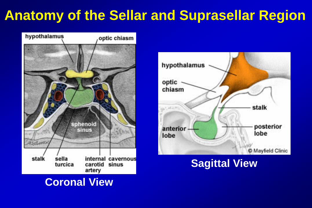
Anatomy of the Sellar and Suprasellar Region
Anatomy of the Sellar and Suprasellar Region
Coronal View
Sagittal View
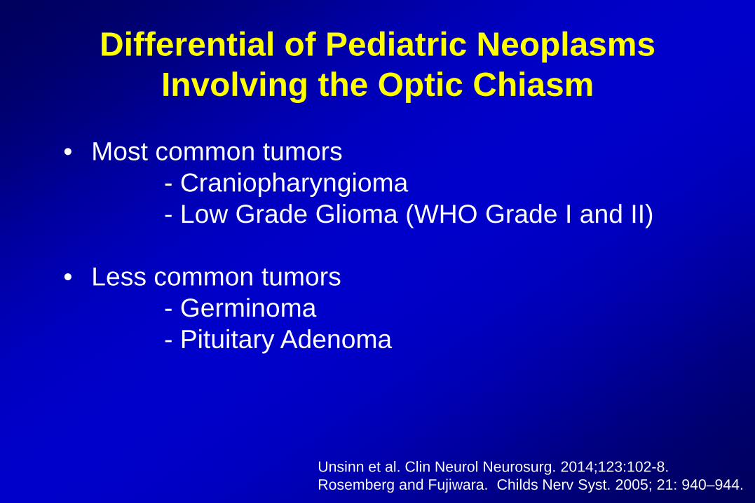
Differential of Pediatric Neoplasms Involving the Optic Chiasm
• Most common tumors - Craniopharyngioma - Low Grade Glioma (WHO Grade I and II)
• Less common tumors - Germinoma - Pituitary Adenoma
Unsinn et al. Clin Neurol Neurosurg. 2014;123:102-8. Rosemberg and Fujiwara. Childs Nerv Syst. 2005; 21: 940–944.
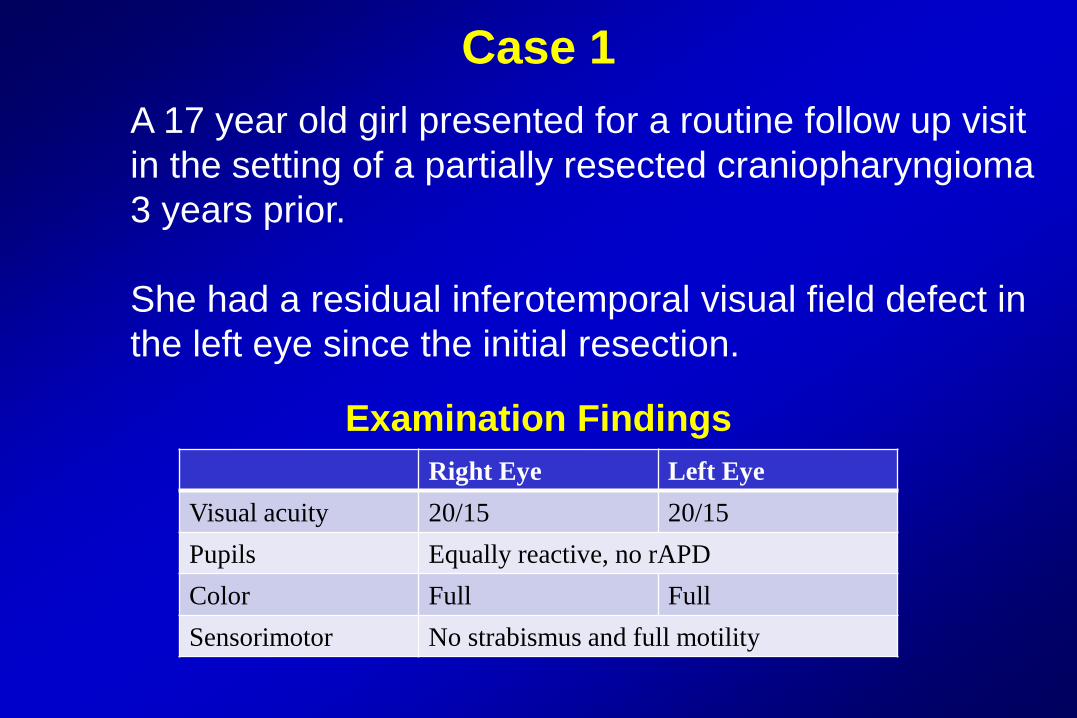
Case 1
A 17 year old girl presented for a routine follow up visit in the setting of a partially resected craniopharyngioma 3 years prior. She had a residual inferotemporal visual field defect in the left eye since the initial resection.
Right Eye Left Eye Visual acuity 20/15 20/15 Pupils Equally reactive, no rAPD Color Full Full Sensorimotor No strabismus and full motility
Examination Findings
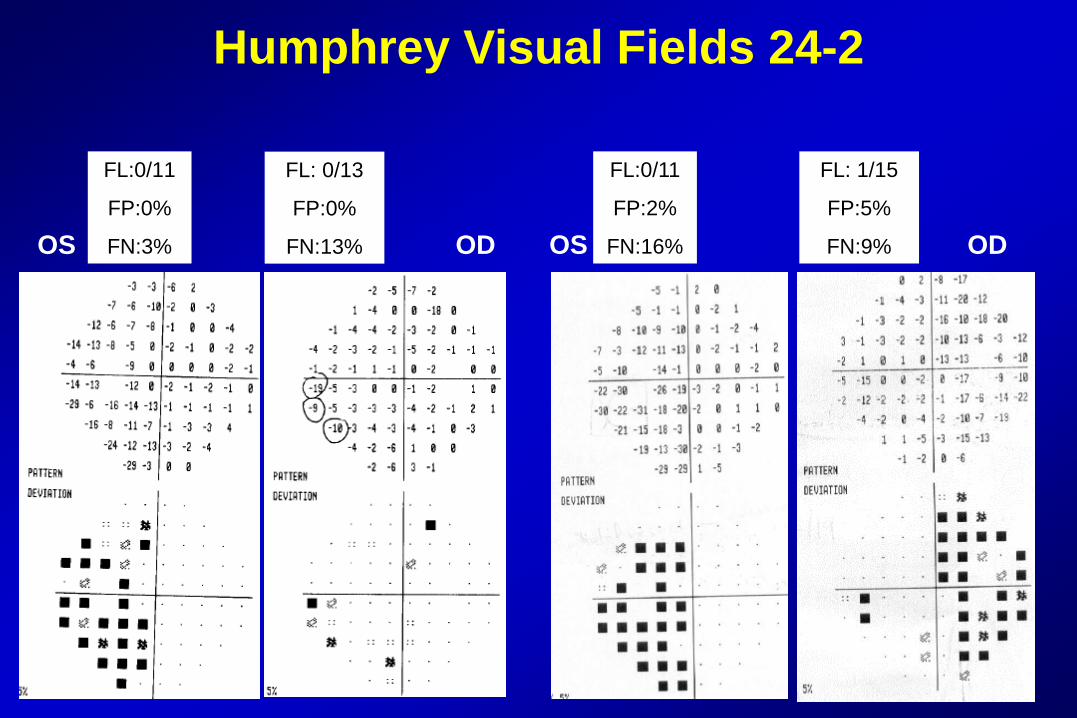
Humphrey Visual Fields 24-2
FL:0/11
FP:0%
FN:3%
FL: 0/13
FP:0%
FN:13% OD OS
FL:0/11
FP:2%
FN:16%
FL: 1/15
FP:5%
FN:9% OD OS
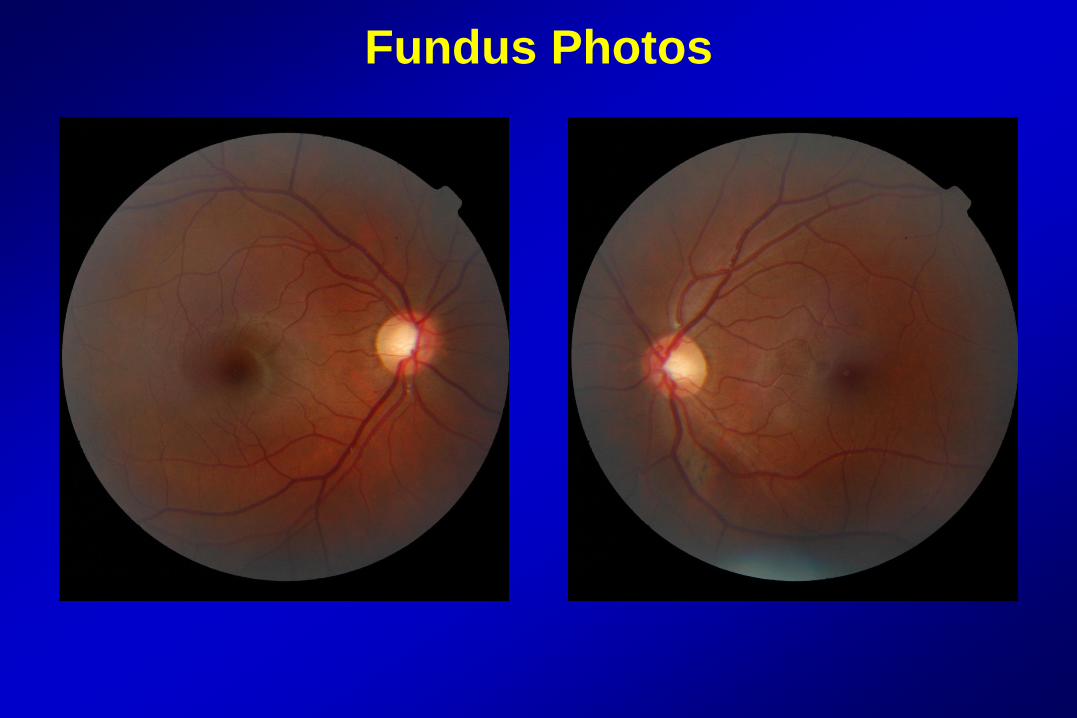
Fundus Photos
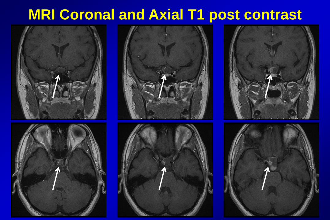
MRI Coronal and Axial T1 post contrast
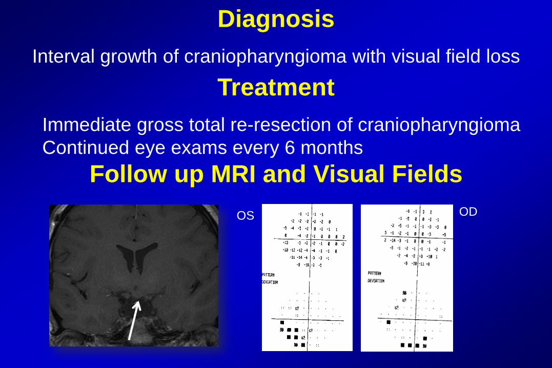
Diagnosis
Interval growth of craniopharyngioma with visual field loss
Treatment
Immediate gross total re-resection of craniopharyngioma Continued eye exams every 6 months
Follow up MRI and Visual Fields
OS
OD
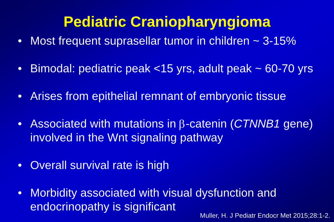
Pediatric Craniopharyngioma • Most frequent suprasellar tumor in children ~ 3-15%
• Bimodal: pediatric peak <15 yrs, adult peak ~ 60-70 yrs
• Arises from epithelial remnant of embryonic tissue
• Associated with mutations in β-catenin (CTNNB1 gene)
involved in the Wnt signaling pathway
• Overall survival rate is high
• Morbidity associated with visual dysfunction and endocrinopathy is significant Muller, H. J Pediatr Endocr Met 2015;28:1-2.
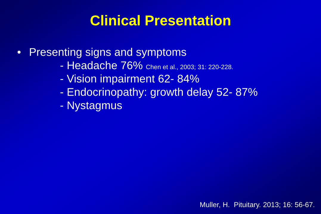
Clinical Presentation
• Presenting signs and symptoms - Headache 76% Chen et al., 2003; 31: 220-228.
- Vision impairment 62- 84% - Endocrinopathy: growth delay 52- 87% - Nystagmus
Muller, H. Pituitary. 2013; 16: 56-67.
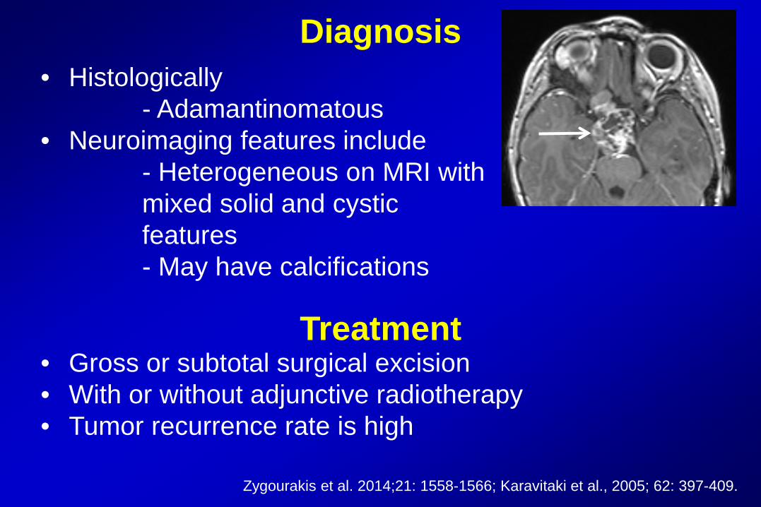
Diagnosis
• Histologically - Adamantinomatous • Neuroimaging features include - Heterogeneous on MRI with mixed solid and cystic features - May have calcifications Treatment • Gross or subtotal surgical excision • With or without adjunctive radiotherapy • Tumor recurrence rate is high
Zygourakis et al. 2014;21: 1558-1566; Karavitaki et al., 2005; 62: 397-409.
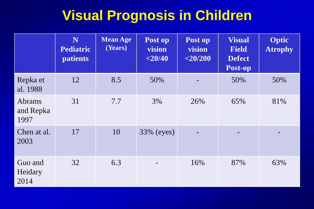
Visual Prognosis in Children N
Pediatric patients
Mean Age (Years)
Post op vision <20/40
Post op vision
<20/200
Visual Field
Defect Post-op
Optic Atrophy
Repka et al. 1988
12 8.5 50% - 50% 50%
Abrams and Repka 1997
31 7.7 3% 26% 65% 81%
Chen at al. 2003
17 10 33% (eyes) - - -
Guo and Heidary 2014
32 6.3 - 16% 87% 63%
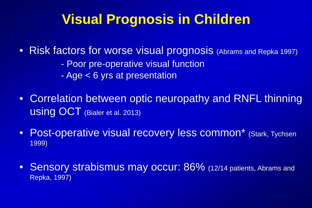
Visual Prognosis in Children
• Risk factors for worse visual prognosis (Abrams and Repka 1997)
- Poor pre-operative visual function - Age < 6 yrs at presentation • Correlation between optic neuropathy and RNFL thinning
using OCT (Bialer et al. 2013)
• Post-operative visual recovery less common* (Stark, Tychsen 1999)
• Sensory strabismus may occur: 86% (12/14 patients, Abrams and
Repka, 1997)
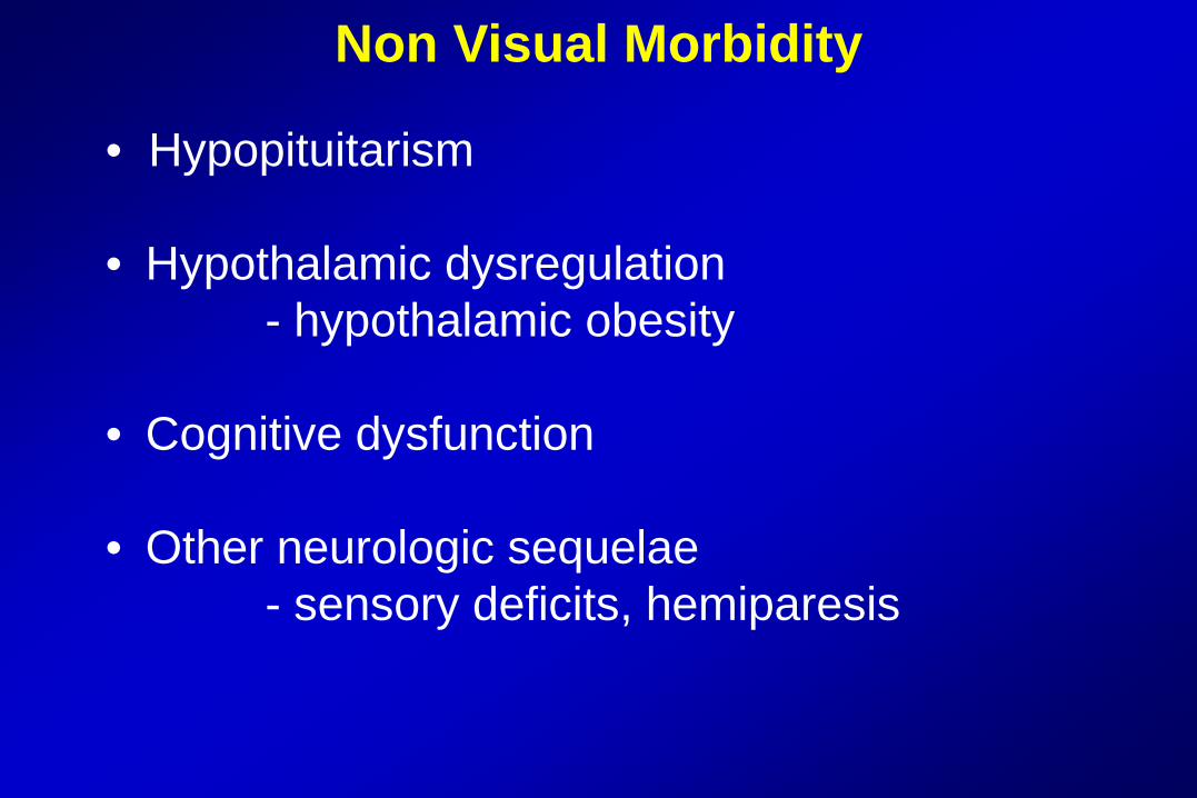
Non Visual Morbidity
• Hypopituitarism
• Hypothalamic dysregulation - hypothalamic obesity
• Cognitive dysfunction
• Other neurologic sequelae - sensory deficits, hemiparesis
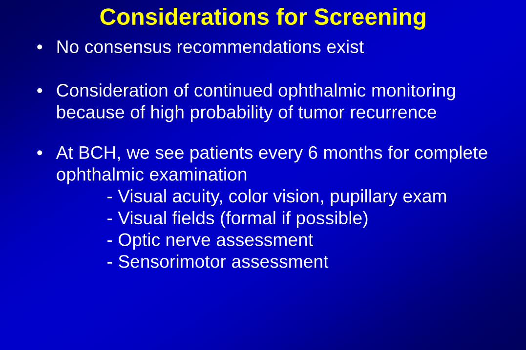
Considerations for Screening • No consensus recommendations exist
• Consideration of continued ophthalmic monitoring
because of high probability of tumor recurrence
• At BCH, we see patients every 6 months for complete ophthalmic examination
- Visual acuity, color vision, pupillary exam - Visual fields (formal if possible) - Optic nerve assessment - Sensorimotor assessment

Case 2
A 11 month old girl presented with failure to thrive and a newly noted nystagmus. Her medical history was uncomplicated until 6 months of age when she dropped from 21%ile to 8%ile in weight with normal height Concurrently she was noted to have a delay in motor milestones. Otherwise her neurologic examination was non-focal.
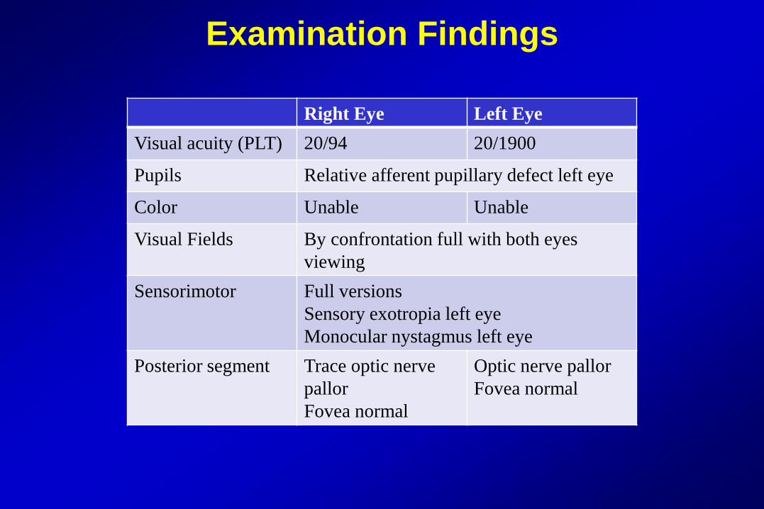
Examination Findings
Right Eye Left Eye Visual acuity (PLT) 20/94 20/1900
Pupils Relative afferent pupillary defect left eye
Color Unable Unable
Visual Fields By confrontation full with both eyes viewing
Sensorimotor Full versions Sensory exotropia left eye Monocular nystagmus left eye
Posterior segment Trace optic nerve pallor Fovea normal
Optic nerve pallor Fovea normal
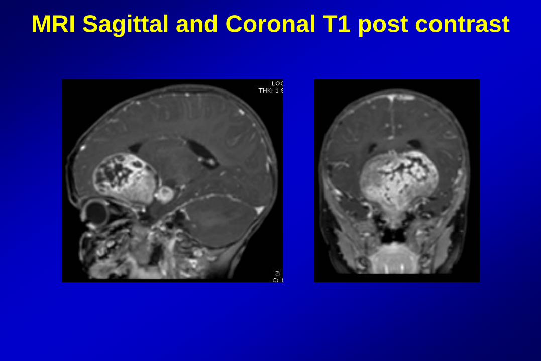
MRI Sagittal and Coronal T1 post contrast
Diagnosis and Management
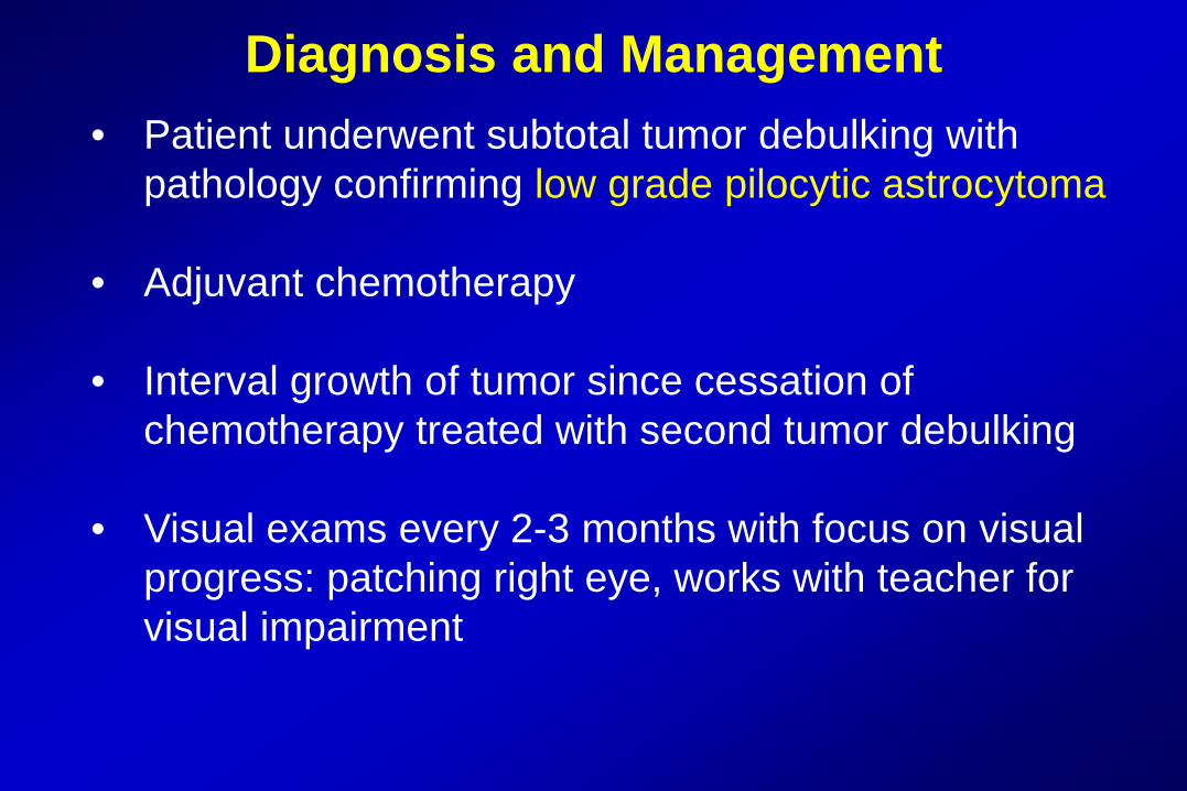
• Patient underwent subtotal tumor debulking with pathology confirming low grade pilocytic astrocytoma
• Adjuvant chemotherapy • Interval growth of tumor since cessation of
chemotherapy treated with second tumor debulking
• Visual exams every 2-3 months with focus on visual progress: patching right eye, works with teacher for visual impairment
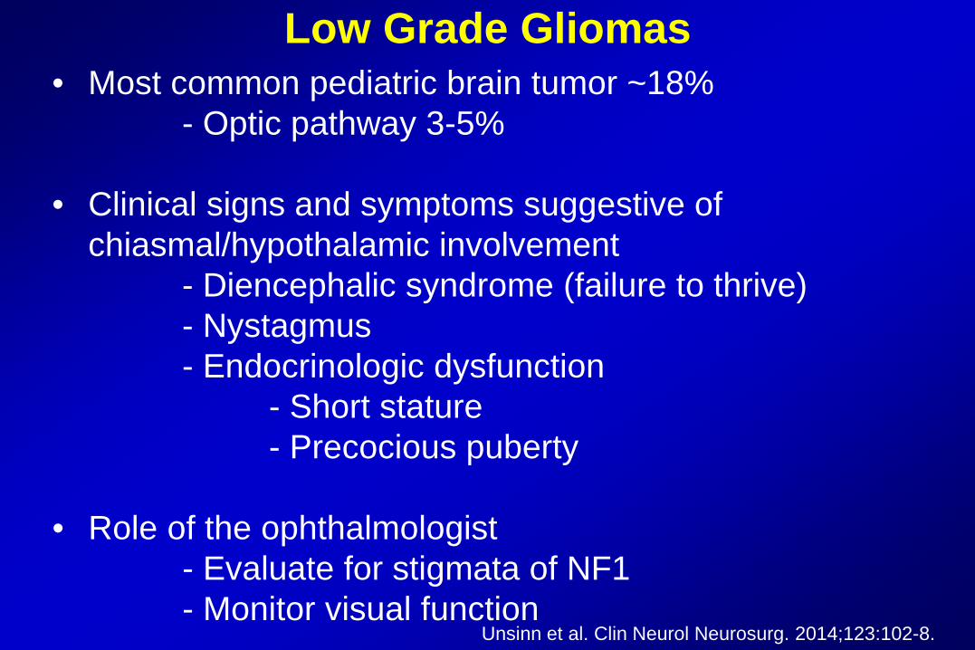
Low Grade Gliomas • Most common pediatric brain tumor ~18% - Optic pathway 3-5% • Clinical signs and symptoms suggestive of
chiasmal/hypothalamic involvement - Diencephalic syndrome (failure to thrive) - Nystagmus - Endocrinologic dysfunction - Short stature - Precocious puberty • Role of the ophthalmologist - Evaluate for stigmata of NF1 - Monitor visual function Unsinn et al. Clin Neurol Neurosurg. 2014;123:102-8.
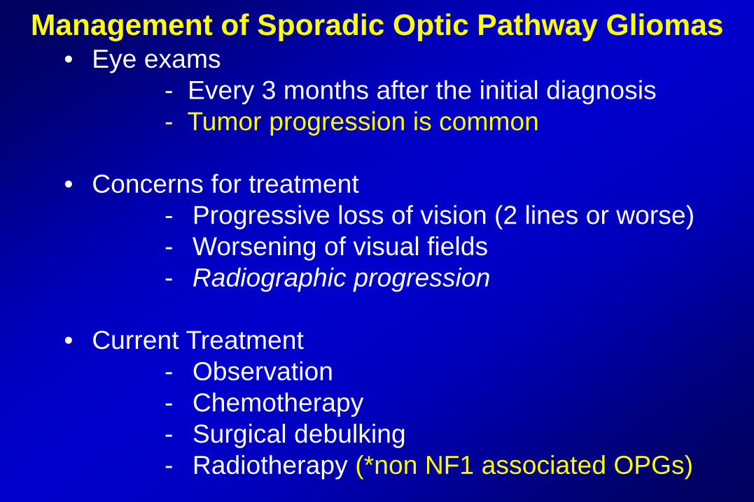
Management of Sporadic Optic Pathway Gliomas • Eye exams - Every 3 months after the initial diagnosis
- Tumor progression is common • Concerns for treatment
- Progressive loss of vision (2 lines or worse) - Worsening of visual fields - Radiographic progression
• Current Treatment - Observation - Chemotherapy - Surgical debulking - Radiotherapy (*non NF1 associated OPGs)
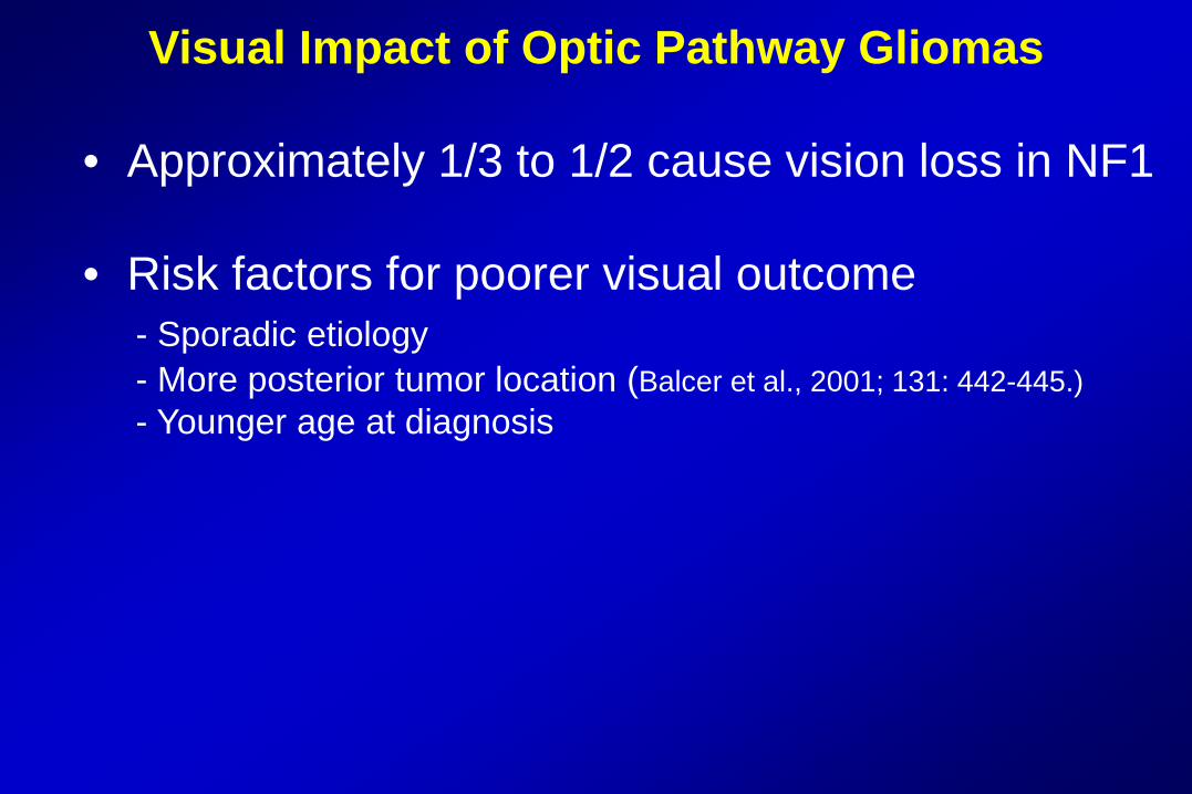
Visual Impact of Optic Pathway Gliomas
• Approximately 1/3 to 1/2 cause vision loss in NF1 • Risk factors for poorer visual outcome - Sporadic etiology - More posterior tumor location (Balcer et al., 2001; 131: 442-445.) - Younger age at diagnosis

Visual Impact of Optic Pathway Gliomas • Visual outcomes - Thiagalingam et al. 2004 of 54 children with NF1 OPGs, 31.5% of patients had profound visual impairment - Campagna et al. 2010 found that 56% of children with sporadic OPGs showed visual decline 6 yrs following dx • Treatment outcomes with chemotherapy
• Risk factors for worse prognosis in NF1-OPGs - Age <2 and > 5 years and optic atrophy Fisher et al. 2012;14:790-7.
N patients NF1 or Sporadic
Stabilized Vision
Improved Vision
Progressive Decline
Fisher et al. 2012
115 NF1 40% 32% 28%
Moreno et al. 2010
174 Both 47.1% 14.4% 38.5%
Role of the Pediatric Ophthalmologist
• Surveillance - Identification of children who harbor tumors affecting the visual pathways
• Management - Active monitoring of visual function and contribution towards treatment plan • Visual Prognosis