Brain Morphophysiology of Africanized Bee Apis mellifera Exposed … et al... · 2014-03-15 ·...
10
Brain Morphophysiology of Africanized Bee Apis mellifera Exposed to Sublethal Doses of Imidacloprid Caroline de Almeida Rossi • Thaisa Cristina Roat • Daiana Antonia Tavares • Priscila Cintra-Socolowski • Osmar Malaspina Received: 12 December 2012 / Accepted: 3 March 2013 / Published online: 7 April 2013 Ó Springer Science+Business Media New York 2013 Abstract Several synthetic substances are used in agri- cultural areas to combat insect pests; however, the indis- criminate use of these products may affect nontarget insects, such as bees. In Brazil, one of the most widely used insecticides is imidacloprid, which targets the nervous system of insects. Therefore, the aim of this study was to evaluate the effects of chronic exposure to sublethal doses of imidacloprid on the brain of the Africanized Apis mellifera. The organs of both control bees and bees exposed to insecticide were subjected to morphological, histochemical and immunocytochemical analysis after exposure to imidacloprid, respectively, for 1, 3, 5, 7, and 10 days. In mushroom bodies of bees exposed to imida- cloprid concentrations of LD 50/10 and in optic lobes of bees exposed to imidacloprid concentrations of LD 50/10 , LD 50/100 , and LD 50/50 , we observed the presence of con- densed cells. The Feulgen reaction revealed the presence of some cells with pyknotic nuclei, whereas Xylidine Ponceau stain revealed strongly stained cells. These characteristics can indicate the occurrence of cell death. Furthermore, cells in mushroom bodies of bees exposed to imidacloprid concentrations of LD 50/10 appeared to be swollen. Cell death was confirmed by immunocytochemical technique. Therefore, it was concluded that sublethal doses of imi- dacloprid have cytotoxic effects on exposed bee brains and that optic lobes are more sensitive to the insecticide than other regions of the brain. The value of insect pollination has been estimated at 212 billion dollars, representing approximately 9.5 % of the total value of agronomy production (Gallai et al. 2009). Bees are among the main pollinators, being responsible for 35 % of food crop pollination by insects (Klein et al. 2007; Willians 1995). In some places, including Europe and the United States, the loss of bee colonies is a large problem for beekeepers. The loss of bee colonies is known as ‘‘colony collapse disorder’’ (CCD). Although the causes of CCD are unclear, it has been speculated that the interaction of several factors, including bacterial diseases and mite infestations, may affect the health of the colony. In addition, the chemical intoxication of bees is of major concern. Due to defores- tation of their natural habitats, bees have increased their foraging activities in agricultural areas. Modern agriculture depends increasingly on the use of chemicals to control weeds, fungi, and arthropod pests in cultivated areas. As a consequence of foraging in agricultural areas, bees are increasingly being exposed to these chemicals (Croft 1990; Thompson 2003; Malaspina and Silva-Zacarin 2006; Des- neux et al. 2007). Neonicotinoid insecticides are the most commonly used on Brazilian crops. Imidacloprid is a neonicotinoid that is applied mainly to sugar cane, coffee, citrus, and cotton crops (Brasil 2001). This insecticide acts by binding to and inhibiting the function of nicotinic acetylcholine receptors in insects (Buckingham et al. 1997). These receptors are found in many regions of bee brain, including areas involved in learning and memory (Bicker 1999). Medrzycki et al. (2003) evaluated the effect of sublethal doses of imidacloprid on the behavior of the honeybee (A. mellifera). At both imidacloprid concentrations tested (100 and 500 ppb), they observed a decrease in bee mobility and communication, which can compromise the social behavior C. de Almeida Rossi Á T. C. Roat (&) Á D. A. Tavares Á P. Cintra-Socolowski Á O. Malaspina Departamento de Biologia, Centro de Estudos de Insetos Sociais, Instituto de Biocie ˆncias de Rio Claro, UNESP-Universidade Estadual Paulista, Av. 24A, 1515, Bela Vista, Rio Claro, SP 13500-900, Brazil e-mail: [email protected] 123 Arch Environ Contam Toxicol (2013) 65:234–243 DOI 10.1007/s00244-013-9897-1
Transcript of Brain Morphophysiology of Africanized Bee Apis mellifera Exposed … et al... · 2014-03-15 ·...

Brain Morphophysiology of Africanized Bee Apis melliferaExposed to Sublethal Doses of Imidacloprid
Caroline de Almeida Rossi • Thaisa Cristina Roat •
Daiana Antonia Tavares • Priscila Cintra-Socolowski •
Osmar Malaspina
Received: 12 December 2012 / Accepted: 3 March 2013 / Published online: 7 April 2013! Springer Science+Business Media New York 2013
Abstract Several synthetic substances are used in agri-cultural areas to combat insect pests; however, the indis-
criminate use of these products may affect nontarget
insects, such as bees. In Brazil, one of the most widely usedinsecticides is imidacloprid, which targets the nervous
system of insects. Therefore, the aim of this study was to
evaluate the effects of chronic exposure to sublethal dosesof imidacloprid on the brain of the Africanized Apis
mellifera. The organs of both control bees and bees
exposed to insecticide were subjected to morphological,histochemical and immunocytochemical analysis after
exposure to imidacloprid, respectively, for 1, 3, 5, 7, and
10 days. In mushroom bodies of bees exposed to imida-cloprid concentrations of LD50/10 and in optic lobes of
bees exposed to imidacloprid concentrations of LD50/10,
LD50/100, and LD50/50, we observed the presence of con-densed cells. The Feulgen reaction revealed the presence of
some cells with pyknotic nuclei, whereas Xylidine Ponceau
stain revealed strongly stained cells. These characteristicscan indicate the occurrence of cell death. Furthermore,
cells in mushroom bodies of bees exposed to imidaclopridconcentrations of LD50/10 appeared to be swollen. Cell
death was confirmed by immunocytochemical technique.
Therefore, it was concluded that sublethal doses of imi-dacloprid have cytotoxic effects on exposed bee brains and
that optic lobes are more sensitive to the insecticide than
other regions of the brain.
The value of insect pollination has been estimated at 212billion dollars, representing approximately 9.5 % of the
total value of agronomy production (Gallai et al. 2009).
Bees are among the main pollinators, being responsible for35 % of food crop pollination by insects (Klein et al. 2007;
Willians 1995).
In some places, including Europe and the United States,the loss of bee colonies is a large problem for beekeepers.
The loss of bee colonies is known as ‘‘colony collapse
disorder’’ (CCD). Although the causes of CCD are unclear,it has been speculated that the interaction of several factors,
including bacterial diseases and mite infestations, may
affect the health of the colony. In addition, the chemicalintoxication of bees is of major concern. Due to defores-
tation of their natural habitats, bees have increased their
foraging activities in agricultural areas. Modern agriculturedepends increasingly on the use of chemicals to control
weeds, fungi, and arthropod pests in cultivated areas. As a
consequence of foraging in agricultural areas, bees areincreasingly being exposed to these chemicals (Croft 1990;
Thompson 2003; Malaspina and Silva-Zacarin 2006; Des-neux et al. 2007).
Neonicotinoid insecticides are the most commonly used
on Brazilian crops. Imidacloprid is a neonicotinoid that isapplied mainly to sugar cane, coffee, citrus, and cotton
crops (Brasil 2001). This insecticide acts by binding to and
inhibiting the function of nicotinic acetylcholine receptorsin insects (Buckingham et al. 1997). These receptors are
found in many regions of bee brain, including areas
involved in learning and memory (Bicker 1999).Medrzycki et al. (2003) evaluated the effect of sublethal
doses of imidacloprid on the behavior of the honeybee (A.
mellifera). At both imidacloprid concentrations tested (100and 500 ppb), they observed a decrease in bee mobility and
communication, which can compromise the social behavior
C. de Almeida Rossi ! T. C. Roat (&) ! D. A. Tavares !P. Cintra-Socolowski ! O. MalaspinaDepartamento de Biologia, Centro de Estudos de Insetos Sociais,Instituto de Biociencias de Rio Claro, UNESP-UniversidadeEstadual Paulista, Av. 24A, 1515, Bela Vista, Rio Claro,SP 13500-900, Brazile-mail: [email protected]
123
Arch Environ Contam Toxicol (2013) 65:234–243
DOI 10.1007/s00244-013-9897-1
of these insects. Another study evaluated the effects of
imidacloprid at concentrations of 0.5 mg/L and 5 lg/L.The results of this study showed that imidacloprid is toxic
to bees, changing their driving frequency during pollen
foraging and altering the number of operculated cells in thehive (Faucon et al. 2005).
Many studies have been conducted to evaluate the tox-
icity of imidacloprid in bees. Small, sublethal doses ofinsecticides may not directly cause the death of bees but
instead may induce changes in bee behavior. Some effects
include a decrease in foraging activity or disorientation,which can affect the entire colony. The aforementioned
changes have been observed in some studies that evaluated
the acute toxicity of imidacloprid (Bortolotti et al. 2003;Medrzycki et al. 2003; Decourtye et al. 2004; Schmuck
2004). The objective of this study was to evaluate the
effects of sublethal doses of imidacloprid on brains ofworkers of Africanized Apis mellifera.
Materials and Methods
Chemicals
The analytical reference standard of imidacloprid (92.5 %
purity) was obtained from Bayer CropScience (Brazil).Sodium chloride (NaCl), dibasic sodium phosphate
(Na2HPO4), monobasic potassium phosphate (KH2PO4),paraformaldehyde, ethanol, hematoxylin, eosin, Xylidine
Ponceau, periodic acid, Schiff’s reagent, hydrochloric acid,
xylene, and polylysine were obtained from Sigma-Aldrich(Brazil). The terminal deoxynucleotidyl transferase-mediated
biotinylated UTP nick end-labeling (TUNEL) reaction kit
(ISCDDK in situ Cell Death Detection Kit) was obtained fromRoche Molecular Biochemicals. The historesin embedding kit
was purchased from Leica Microsystems (Germany). The
paraffin (Histosec) was obtained from Merck (Brazil).
Honeybee Collection
Newly emerged adult bees of Africanized A. mellifera were
collected directly from combs in the apiary of the Institute of
Biosciences of Universidade Estadual Paulista, Campus ofRio Claro, Sao Paulo State, Brazil. We verified the health and
physiological status of the colony according to the guidelines
of the Organisation for Economic Co-Operation andDevelopment (OECD 1998) for testing chemicals in bees.
Oral Acute Toxicity
We calculated the lethal dose 50 (LD50) of imidacloprid to
Africanized honey bees using an ingestion bioassay. Newlyemerged bees were placed in disposable cages (10 bees/
cage) tat had been previously lined with filter paper. Three
replicates of each dose and control group were performed.The experiments were conducted in an environmental
biochemical oxygen demand (BOD) chamber with tem-
peratures held at 32 ± 1 "C and relative humidity of 70 %.The treated and control groups received food and water
ad libitum.
The bees were starved for B2 h and then exposed to arange of doses of imidacloprid dispersed in sucrose solu-
tion in water (50 % w/v). The stock solution of imidaclo-prid was made in acetone 100 % (v/v) and then diluted in
the sucrose diet. Five doses of imidacloprid were prepared
in a geometric series with final range from 80 to 100 ggimidacloprid/lL diet. For each treatment, a dilution was
made to obtain the desired concentration in 1,000 lL of
sucrose solution. The control group was fed with the samediet free of the test substance. Two separate control groups
were used: a sucrose solution in water and a sucrose
solution with the solvent (acetone) at the concentrationused in dosing solutions.
Once consumed (usually within 3–4 h), the feeder was
removed from the cage and replaced with one containingsucrose solution alone. The sucrose solutions were then
provided ad libitum. Subsequently, mortality was recorded
at 4 h after start of the test and thereafter at 24 and 48 h(i.e., after given dose). Bees from the control groups did
not reach a 10 % mortality rate for the validation test
(OECD 1998).Data were analyzed using Probit regression by plotting
the dose–response curves for each observation period.
Corrections for control mortality were made using Abbott’smethod (OECD 1998).
Honeybee Intoxication Assay
Bees were placed in disposable cages (10 bees/cage) lined
with filter paper. Three replicate experiments were per-formed for each dose of imidacloprid or control treatment.
The experiments were conducted in an environmental BOD
chamber at a temperature of 32 ± 1 "C and relativehumidity of 70 %. The experimental and control groups
received the same water supply consisting of cotton soaked
in water and food (10 lL/bee). There were two controlgroups: (1) bees not exposed to solvent, which were fed a
diet consisting of sucrose and water 50 % (w/v); and (2)
bees exposed to solvent, which were fed the same dietsupplemented with acetone 1 % (v/v) (the solvent used in
the pesticide solution).
The sublethal doses used in this study were calculatedusing the LD50 obtained according to the procedure
described in Oral Acute Toxicity. A stock solution of
imidacloprid was prepared, dissolved in acetone, and
Arch Environ Contam Toxicol (2013) 65:234–243 235
123

mixed with sucrose solution. Three dilutions of this imi-
dacloprid stock solution were made to yield final concen-
trations of LD50/100, LD50/50, and LD50/10. Theexperimental groups were fed the contaminated diet con-
sisting of these sublethal doses. The periods of exposure to
the insecticide were 1, 3, 5, 7, and 10 days for each group.Six bees were used per treatment group for the assay.
Morphological and Histochemical Analysis
To extract the brain, dissection was carried out on bees in
Petri dishes containing saline solution (20 mM Na2HPO4/KH2PO4 [pH 7.4] and 130 mM NaCl) under a Zeiss
stereomicroscope using dissecting forceps and microscis-
sors. The organs were fixed in paraformaldehyde 4 % (w/v) in phosphate-buffered saline [0.1 M (pH 7.4)]. Subse-
quently, the material was dehydrated in a series of ethanol
solutions at 4 "C of the following concentrations: 15, 30,
50, 70, 85, 90, 95, and 100 % (v/v). Each dehydration
step lasted for 2 h. After dehydration, the material was
incubated in embedding historesin for 3 days. Subse-quently, 7-lm sections were cut using a Leica Microtome
(Germany).
Some sections of brain were stained with hematoxylinand eosin (H&E) for morphological analysis. Other brain
sections were stained with Xylidine Ponceau to detect total
protein (Junqueira and Junqueira 1983) or were subjectedto Feulgen reaction (counterstained with Fast Green) to
determine the level of chromatin compaction (Feulgen and
Rossenbeck 1924).Finally, sections were examined under a light micro-
scope (Olympus BX51; Olympus America). Photomicro-
graphs were obtained using a digital camera (Olympus DP-71) that was fitted to the microscope and coupled to a Dell
computer. DP Controller software (Olympus America) was
used for image acquisition.
Table 1 Summary of morphological, histochemical, and immunohistochemical analyses on mushroom bodies of A. meliffera exposed tosublethal doses of imidacloprid
Treatment Days H&E staining Fuelgen reaction Xylidine Ponceau staining TUNEL reaction
Condensedcells
Cells with swollenappearance
Nuclei with compactionof chromatin
Increase in the intensityof staining
Nucleipositive
Control without solvent 1 - - - - -
3 - - - - -
5 - - - - -
7 - - - - -
10 - - - - -
Control with solvent 1 - - - - -
3 - - - - -
5 - - - - -
7 - - - - -
10 - - - - -
LD50/100 1 - - - - -
3 - - - - -
5 - - - - -
7 - - - - -
10 - - - - -
LD50/50 1 - - - - -
3 - - - - -
5 - - - - -
7 - - - - -
10 - - - - -
LD50/10 1 - - - - ?
3 ? - ? ? ?
5 ? - ? ? ?
7 ? - ? ? ?
10 ? ? ? ? ?
? presence of alteration, ?? alteration moderately present, ??? alteration extremely present, - alteration absent, LD50 lethal dose
236 Arch Environ Contam Toxicol (2013) 65:234–243
123

Immunocytochemical Detection of DNA
Fragmentation by Endonucleases
Brains were dissected and fixed as described previously.
After dehydration in a series of ethanol solutions, organs
were diaphanized in xylene and embedded in paraffin(Histosec). Sections of 8-lm thickness were transferred to
histological slides that had been cleaned and treated with
polylysine. The Histosec wax was removed with several
xylene washes before sections were rehydrated and sub-jected to TUNEL reaction (using the ISCDDK in situ Cell
Death Detection Kit) according to the manufacturer’s
instructions. Negative and positive controls were includedfor the TUNEL reaction experiment. After incubation, the
slides were examined for the presence of fluorescein-
labeled 30-OH ends of fragmented DNA. Fluorescent
Fig. 1 Images of H&E-stained mushroom bodies of newly emergedworker bees of Africanized A. mellifera with or without exposure toimidacloprid. a, b General and magnified views of mushroom bodiesfrom the control group with 7 days showing no morphologicalalterations. c, d General and magnified views of mushroom bodiesfrom bees with exposure to an imidacloprid concentration of LD50/10
for 7 days showing the presence of intensely stained cells (arrow). e,f General and magnified views of mushroom bodies of bees withexposure to an imidacloprid concentration of LD50/10 for 10 days.Note the presence of cells with a swollen appearance (arrowhead inf). Br basal ring, Bo border, Co collar, Occ outer compact cells, Iccinner compact cells, Nc noncompact cells
Arch Environ Contam Toxicol (2013) 65:234–243 237
123

images were captured using an Olympus BX-51 fluores-
cence microscope (Olympus America) at a wavelength of450–500 nm.
Results
Acute Toxicity Assay
The LD50 of imidacloprid to A. mellifera was established at80.9 gg/bee (v2 = 56.778, slope = 2.781 ± 1.757). From
this value, we calculated the sublethal doses equivalent to
LD50/100 (0.809 gg/bee), LD50/50 (1.618 gg/bee), andLD50/10 (8.09 gg/bee) to be used in other experiments.
Morphological and Histochemical Analysis
Mushroom Bodies
The results obtained from mushroom bodies of bees
exposed to imidacloprid are listed in Table 1. Morpho-
logical and histochemical alterations in mushroom bodieswere not observed in the groups of bees exposed to imi-
dacloprid at concentrations of LD50/100 or LD50/50 and
those in he control groups (Fig. 1a, b). However, thepresence of condensed cells was detected in the group of
bees exposed to imidacloprid for C3 days at a concentra-
tion of LD50/10 using H&E staining technique (Fig. 1c, d).In the same treatment group (LD50/10, bees exposed for
C3 days), Feulgen reaction revealed the presence of
strongly stained nuclei in some cells, thus indicating theoccurrence of chromatin condensation (Fig. 2c, d). Stain-
ing with Xylidine Ponceau revealed strongly stained cells,
which may indicate the presence of cell compaction(Fig. 2g, h). In control groups, nuclei were weakly stained
by Feulgen reaction (Fig. 2a, b), and cells were weakly
stained after Xylidine Ponceau staining (Figs. 2e, f).The presence of strongly stained nuclei visualized dur-
ing histochemical and morphological analysis indicates the
occurrence of cell death in Kenyon cells of exposed bees.TUNEL assay revealed the presence of positive nuclei in
Kenyon cells of bees exposed to imidacloprid for all
periods at a concentration of LD50/10 (Fig. 4b). Positivenuclei were not observed in the control group using this
technique (Fig. 4a).
In addition to intense H&E staining of mushroom bodiesobserved after imidacloprid exposure, morphological
alterations were observed in some Kenyon cells in mush-
room bodies of bees exposed to an imidacloprid concen-tration of LD50/10 for 10 days (Figs. 1e, f). Kenyon cells in
this treatment group exhibited less staining and were
greater in size, indicating the occurrence of cell swelling.
Optic Lobes
Table 2 lists the results obtained from optic lobes of con-trol and imidacloprid-treated bees. H&E staining revealed
the presence of condensed cells in optic lobes of bees
exposed to imidacloprid at a concentration of LD50/10 forall exposure times tested and in bees exposed to concen-
trations of LD50/100 and LD50/50, for 5, 7, or 10 days. The
presence of condensed cells in these treatment groups wasindicated by intense cell staining and decreased cell vol-
ume (Fig. 3b). At the same imidacloprid concentrations
and exposure times, Feulgen reaction produced intenselystained nuclei, thus indicating the presence of chromatin
compaction (Fig. 3d). In addition, cells were intensely
stained by Xylidine Ponceau (Fig. 3f), which may indicateincreased protein synthesis or cell compaction. No signif-
icant alterations in H&E staining (Fig. 3a), Feulgen reac-
tion staining (Fig. 3c), and Xylidine Ponceau staining(Fig. 3e) were observed in optic lobes of control bees.
Immunocytochemical analysis of optic lobes using
TUNEL assay found positive nuclei in all groups of imi-dacloprid-treated bees at all exposure times, thus indicating
that they were undergoing cell death (Fig. 4d). Positive
nuclei were not detected in the control groups (Fig. 4c).
Discussion
Our results establish the LD50 of 80.9 gg/bee for the
insecticide imidacloprid in Africanized honeybees, thusshowing this insecticide is harmful to these bees. This
value is close to the one found by Nauen et al. (2001) in
European bees, which reported a 48-h oral LD50 of imi-dacloprid between 41 and 81 gg/bee. The small variation
found is the result of differences in both the conditions of
the experiment and the subspecies of A. mellifera used.Other factors that may contribute to this variation include
the age of bees (Guez et al. 2001), the colony used (Suchail
et al. 2001) and the season (Decourtye et al. 2003).There were detectable morphological, histochemical,
and immunocytochemical alterations in mushroom bodies
and optic lobes of bees exposed to imidacloprid comparedwith the control groups. Morphological alterations
observed in mushroom bodies and optic lobes of imida-
cloprid-treated bees included more intense H&E stainingindicating the presence of condensed cells. Chromatin
condensation in the nuclei of cells from these brain struc-
tures was also evident when these tissues were subjected toFeulgen reaction.
Cell condensation may indicate the occurrence of cell
death by apoptosis (Bowen et al. 1998). The presence ofchromatin compaction in nuclei of cells can indicate that
these cells are in a state of low transcriptional activity,
238 Arch Environ Contam Toxicol (2013) 65:234–243
123
Arch Environ Contam Toxicol (2013) 65:234–243 239
123

which occurs during the process of cell death (Willye 1981;
Hacker 2000; Silva-Zacarin et al. 2008).During cell death by apoptosis, homeostasis is main-
tained by controlling the number of antiapoptotic and
proapoptotic proteins. Stimuli, such as DNA damage, canevoke increased expression of proapoptotic proteins, and
this imbalance can induce apoptosis (Petros et al. 2004). In
the present study, the technique used to detect total protein(Xylidine Ponceau staining) showed more intense staining
in cells in mushroom bodies and optic lobes of bees
exposed to imidacloprid compared with tissues from con-trol bees. This result may indicate the occurrence of
increased protein synthesis in imidacloprid-treated bees.
This increase may reflect the activation of signaling path-ways that participate in cell death, or it could result from
cell condensation.Morphological and histochemical analysis revealed the
presence of condensed cells, cells containing nuclei with
condensed chromatin, and cells in which there wasincreased protein synthesis; these characteristics are typi-
cally observed in cells undergoing cell death. Cells with
these characteristics were present in mushroom bodies ofbees exposed to imidacloprid concentrations of LD50/10 for
C3 days, in optic lobes of bees exposed to imidacloprid
concentrations of LD50/10 for all exposure times tested, and
Table 2 Summary of morphological, histochemical, and immunohistochemical analyses of optical lobes in A. meliffera exposed to sublethaldoses of imidacloprid
Treatment Days H&E staining Fuelgen reaction Xylidine Ponceau staining TUNEL reactionCondensedcells
Nuclei with compactionof chromatin
Increase in the intensityof staining
Nuclei positive toTUNEL reaction
Control without solvent 1 - - - -
3 - - - -
5 - - - -
7 - - - -
10 - - - -
Control with solvent 1 - - - -
3 - - - -
5 - - - -
7 - - - -
10 - - - -
LD50/100 1 - - - ?
3 - - - ?
5 ? ? ? ?
7 ? ? ? ?
10 ? ? ? ?
LD50/50 1 - - - ?
3 - - - ?
5 ? ? ? ?
7 ? ? ? ?
10 ? ? ? ?
LD50/10 1 ? ? ? ?
3 ? ? ? ?
5 ? ? ? ?
7 ? ? ? ?
10 ? ? ? ?
? presence of alteration, ?? alteration moderately present, ??? alteration extremely present, - alteration absent, LD50 lethal dose
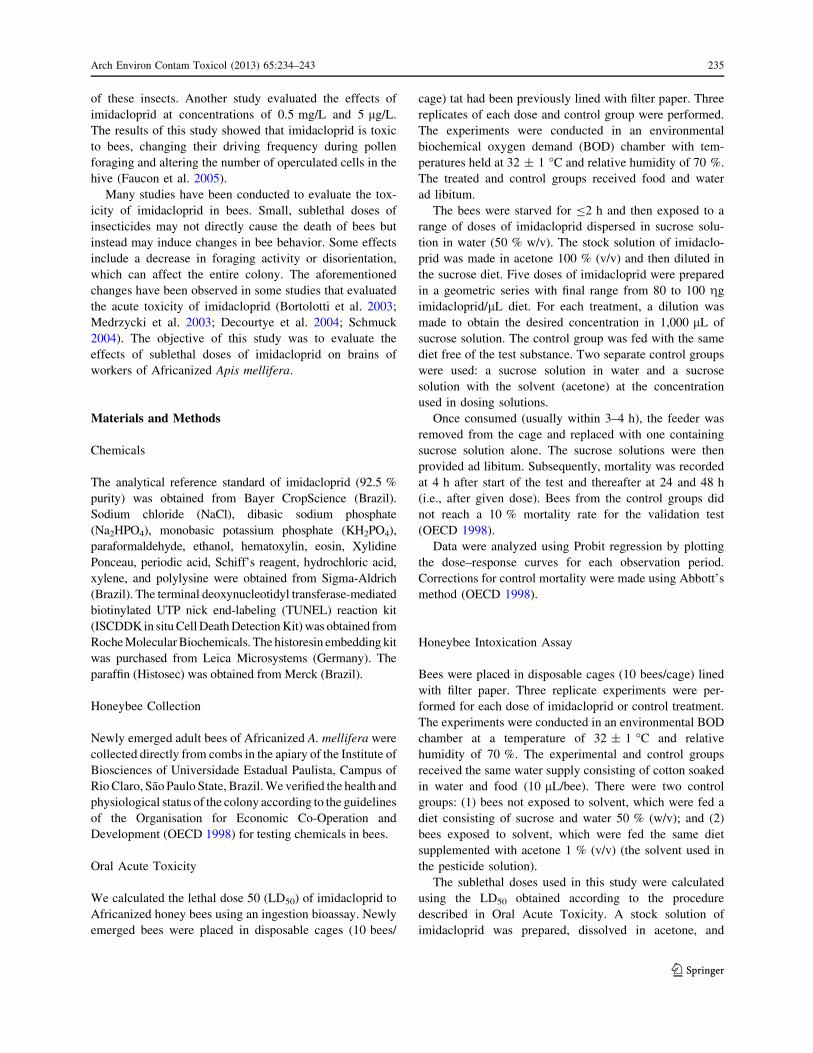
Fig. 2 Images of Feulgen reaction and Xylidine Ponceau staining ofmushroom bodies of newly emerged workers of Africanized A.mellifera with or without exposure to imidacloprid. a, b General andmagnified views of mushroom bodies from the control group for3 days, submitted to Fuelgen reaction, showing nuclei weakly stainedwith chromatin decondensed. c, d General view and detail ofmushroom bodies of bees exposed to LD50/10 for 10 days submitted toFuelgen reaction. Note the presence of intensely stained nuclei(arrows). e, f General view and detail of mushroom bodies fromcontrol group for 5 days submmited to Xylidine Ponceau stain. g,h General view and detail of mushroom bodies from bees exposed toLD50/10 for 10 days and exposed to LD50/50 for 3 and 7 days,respectively. Observed increased intensity of staining of some cells(arrowheads). Ba basal ring, Bo border, Co collar, Occ outer compactcells, Icc inner compact cells, Nc noncompact cells
b
240 Arch Environ Contam Toxicol (2013) 65:234–243
123

in optic lobes of bees exposed to imidacloprid concentra-
tions of LD50/100 and LD50/50 for 5, 7, or 10 days. In
contrast, TUNEL reaction produced positive nuclei inmushroom bodies of bees exposed to LD50/10 and optic
lobes of bees exposed to all doses of imidacloprid for all
exposure times tested. Such differences may reflect dif-ferences in the stage of cell death. TUNEL reaction may
detect positive nuclei when cells are in the initial stages of
cell death. In contrast, cell and chromatin condensation,
which were detected using morphological and histochem-
ical techniques, may occur during the final stages of celldeath. Alternatively, cell and chromatin condensation
might reflect the presence of autophagic or apoptotic cells,
in which endonucleases do not cause DNA fragmentation.A comparison of the results derived from the imida-
cloprid treatment groups indicates that optic lobes are more
Fig. 3 Images of optical lobes of newly emerged workers ofAfricanized A. mellifera with or without exposure to imidacloprid.a Optical lobes stained with H&E from control group for 5 dayswithout morphological alteration. b Optical lobes stained with H&Efrom bees exposed to LD50/50 for 7 days showing intensely stainedcells (arrow). c Optical lobes submitted to Fuelgen reaction fromcontrol group for 10 days without histochemical alteration. d Optical
lobes submitted to Fuelgen reaction from bee exposed to LD50/100 for10 days. Note the presence of intensely stained nuclei (arrowhead).e Optic lobes submitted to Xylidine Ponceau stain from control groupfor 5 days without alteration. f Optic lobes submitted to XylidinePonceau stain of bee exposed to LD50/10 for 7 days. Note the intenselystained cells (arrow outline). La lamina, Me medulla, Oc outerchiasmata, Ic inner chiasmata
Arch Environ Contam Toxicol (2013) 65:234–243 241
123
sensitive to insecticide relative to the other cerebral
structures analyzed. Changes in optic lobes were observedat all doses of imidacloprid for all exposure times tested. In
contrast, changes observed in mushroom bodies were only
observed in bees treated with imidacloprid concentrationsof LD50/10.
The visual system of an adult insect consists of the
retina (the eye itself), which contains photoreceptor cells,and the optic lobes, which comprise three layers: the
lamina, the medulla, and the lobula. The retinal cells form a
highly ordered arrangement of repeated units of ommatidia(Ready et al. 1976). Within the ommatidium, each retinal
unit (consisting of eight or nine photo-sensing cells) pro-
jects axonal beams toward the optic lobes to form synapsesin the lamina and possibly in the medulla. Neurons from
the lamina extend axons into the medulla and then to the
lobula (Horridge 1965). Thus, cell death in optic lobes canresult in fewer neurons and consequently decrease the
number of synapses between this structure and the com-
pound eyes. Therefore, the structural changes induced inoptic lobes of bees may lead to problems with the visual
acuity and consequently may negatively impact foraging
worker bees.
Bortolotti et al. (2003) evaluated the effects of sublethal
doses of imidacloprid on the foraging behavior and returnto the colony of A. mellifera. Bees from the control groups
returned to the feeder within 5 h. Bees fed syrup containing
100 ppb of insecticide returned to the feeder after 24 h.Bees exposed to 500 and 1,000 ppb of insecticide did not
return to the colony or to the feeder. In these studies, it is
possible that exposure of bees to imidacloprid caused celldeath in the optic lobes, as observed in the present study,
which may have impaired their visual acuity.
Our study also focused on mushroom bodies becausethis brain structure is involved in learning and memory in
bees (Daly et al. 1998; Cruz-Landim 2009). In mushroom
bodies of bees exposed to imidacloprid concentrations ofLD50/10 for 3, 5, 7 and 10 days, nuclei containing con-
densed chromatin were detected. In mushroom bodies of
bees exposed to imidacloprid for longer periods of time,swelling of cell bodies was also observed. Therefore, we
conclude that the concentration of insecticide to which the
bees were exposed is a more important factor in insecti-cide-induced damage to the brain than duration of insec-
ticide exposure.
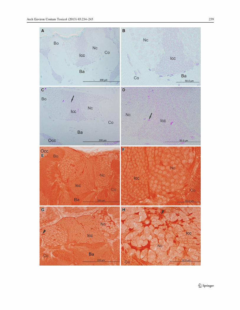
Fig. 4 Images of the cerebral structure of newly emerged workers ofAfricanized A. mellifera with or without exposure to imidaclopridsubmitted to TUNEL reaction. a Mushroom bodies from controlgroup for 7 days with no positive nuclei. b Mushroom bodies of LD50/
10 group for 1 day with positive nuclei in response to TUNELreaction. c Optical lobes of the control group for 5 days with no
positive nuclei. d Optical lobes of bees exposed to LD50/10 by 7 and3 days, respectively. Note the presence positive nuclei in response toTUNEL reaction (arrow). Br basal ring; Bo border, Co collar, Occouter compact cells, Icc inner compact cells, Nc non compact cells,La lamina, Me medulla, Oc outer chiasmata, Ic inner chiasmata
242 Arch Environ Contam Toxicol (2013) 65:234–243
123
Because of the contribution of mushroom bodies to
learning and memory processes in bees, damage to thesestructures can cause disorientation and impair their forag-
ing activity. As a result, the function of the entire colony
can be affected.The results of this study show that imidacloprid causes
morphological, histochemical, and immunocytochemical
alterations in optic lobes and mushroom bodies of bees.Therefore, sublethal doses of this insecticide can negatively
affect honeybee physiology, possibly by disrupting theirvisual system and impairing their learning capacity (El
Hassani et al. 2008). These changes could lead to abnormal
behavior and possibly to the death of the affected bees.
Acknowledgments The authors thank the Fundacao de Amparo aPesquisa do Estado de Sao Paulo/FAPESP for the financial support(Grants No. 2008/05018-7 and 2006/57122-7) and the BrazilianCouncil for Scientific and Technological Development/CNPq. Wethank Bibiana Monson de Souza for critical review of the manuscript.
References
Bicker G (1999) Histochemistry of classical neurotransmitters inantennal lobes and mushroom bodies of the honeybee. MicroscRes Tech 45:174–183
Bortolotti L, Montanari R, Marcelino J, Medrzycki P, Maini S, PorriniC (2003) Effects of sub-lethal imidacloprid doses on the homingrate and foraging activity of honey bees. Bull Insectology56:63–67
Bowen ID, Bowen SM, Jones AH (1998) Mitosis and apoptosis:matters of life and death. Chapman & Hall, New York
Brasil (2001) Ministerio da Agricultura, Pecuaria e Abastecimento.http://www.agricultura.gov.br. Accessed 7 Jan 7 2010
Buckingham SD, Lapied B, Corrong HLE, Grolleau F, Sattelle DB(1997) Imidacloprid actions on insect neuronal acetylcholinereceptors. J Exp Biol 200:2685–2692
Croft BA (1990) Arthropod biological control agents and pesticides.Wiley, New York
Cruz-Landim C (2009) Abelhas: morfologia e funcao de sistemas.Unesp, Sao Paulo
Daly HV, Doyen JT, Purcell AH (1998) Introduction to insect biologyand diversity. Oxford University Press, New York
Decourtye A, Lacassie E, Pham-Delegue M (2003) Learning perfor-mances of honeybees (Apis mellifera L) are differentiallyaffected by imidacloprid according to the season. Pest ManagSci 59(3):269–278
Decourtye A, Devillers J, Cluseau S, Charreton M, Pham-DelegueMH (2004) Effects of imidacloprid and deltamethrin on asso-ciative learning in honeybees under semi-field and laboratoryconditions. Ecotoxicol Environ Saf 57:410–419
Desneux N, Decourtye A, Delpuech JM (2007) The sublethal effectsof pesticides on beneficial arthropods. Annu Rev Entomol52:106–2007
El Hassani AK, Dacher M, Gary V, Lambin M, Gauthier M,Armengaud C (2008) Effects of sublethal doses of acetamipridand thiamethoxam on the behavior of the honeybee (Apismellifera). Arch Environ Contam Toxicol 54:653–661
Faucon J, Aurieres C, Drajnudel P, Mathieu L, Ribiere M, Martel Aet al (2005) Experimental study on the toxicity of imidaclopride
given syrup to honey bee (Apis mellifera) colonies. Pest ManagSci 61:111–125
Feulgen R, Rossenbeck H (1924) Mikroskopisch Chemischer Nac-hweis einer Nucleinsaure vom Typus der Thymonucleinsaureund Die Darauf Beruhend Elective Farbung von Zillkernen inMikroskopischen Praparaten Hoppe-Seylers. Hoppe Seylers ZPhysiol Chem 206:389–410
Gallai N, Salles J-M, Settele J, Vaissiere BE (2009) Economicvaluation of the vulnerability of world agriculture confrontedwith pollinator decline. Ecol Econ 68:810–821
Guez D, Suchail S, Gauthier M, Maleszka R, Belzunces LP (2001)Contrasting effects of imidaclopride on habituation in 7- and8-day old honeybees (Apis mellifera). Neurobiol Learn Mem76:183–191
Hacker G (2000) The morphology of apoptosis. Cell Tissue Res301:5–17
Horridge GA (1965) Arthropoda: receptors for light and optic lobes.In: Bullock TH, Horridge GA (eds) Structure and function of thenervous system of invertebrates, vol 2. Freeman, San Francisco,pp 1063–1113
Junqueira LCU, Junqueira LMMS (1983) Tecnicas basicas decitologia e histologia. Santos, Sao Paulo
Klein AM, Vaissiere BE, Cane JH, Steffan-Dewenter I, CunninghamSA, Kremen C et al (2007) Importance of pollinators in changinglandscapes for world crops. Proc R Soc Lond B Biol Sci274:303–313
Malaspina O, Silva-Zacarin ECM (2006) Cell makers for ecotoxico-logical studies in target organs of bees. Braz J Morphol Sci23:303–309
Medrzycki P, Montanari R, Bortolotti L, Sabatini AG, Maini S,Porrini C (2003) Effects of imidacloprid administered in sub-lethal doses on honey bee behaviour. Bull Insectology 56:59–62
Nauen R, Ebbinghaus-Kintscher U, Schmuck R (2001) Toxicity andnicotinic acetylcholine receptor interaction of imidacloprid andits metabolites in Apis mellifera (Hymenoptera: Apidae). PestManag Sci 57(7):577–586
Organisation for Economic Co-Operation and Development (1998)Guideline 214: guidelines for the testing of chemicals: Honey-bees, Acute contact toxicity test. http://puck.sourceoecd.org/vl=1619398/cl=20/nw=1/rpsv/ij/oecdjournals/1607310x/v1n2/s15/p1. Accessed 15 Feb 15 2010
Petros AM, Olejniczak ET, Fesik SW (2004) Structural biology of theBcl-2 family of proteins. Biochim Biophys Acta 1644:83–94
Ready DF, Hanson TE, Benzer S (1976) Development of theDrosophila retina, a neurocrystalline lattice. Dev Biol 53:217–240
Schmuck R (2004) Effects of a chronic dietary exposure of thehoneybee Apis mellifera (Hymenoptera: Apidae) to imidaclo-pride. Arch Environ Contam Toxicol 47:471–478
Silva-Zacarin ECM, Taboga SR, Silva de Moraes RL (2008) Nuclearalterations associated to programmed cell death in larval salivaryglands of Apis mellifera (Hymenoptera: Apidae). Micron 39:117–127
Suchail S, Guez D, Belzunces LP (2001) Discrepancy between acuteand chronic toxicity induced by imidaclopride and its metabo-lites in Apis mellifera. Environ Toxicol Chem 20(11):2482–2486
Thompson HM (2003) Behavioural effects of pesticides in bees: theirpotential for use in risk assessment. Ecotoxicology 12:317–330
Willians CS (1995) Conserving Europe’s bees: why all the buzz?Trends Ecol Evol 10:309–310
Willye AH (1981) Cell death: A new classification separatingapoptosis from necrosis. In: Bowen ID, Lockshin RA (eds) Celldeath in biology and pathology. Springer, New York
Arch Environ Contam Toxicol (2013) 65:234–243 243
123