Brain lipids: I. Quantification and fatty acid cerebral ... · Brain lipids: I. Quantification and...
8
Brain lipids: I. Quantification and fatty acid composition of cerebroside sulfate in human cerebral gray and white matter JOHN S. O’BRIEN, DOROTHY L. FILLERUP, and JAMES F. MEAD Departments of Pathology and Medicine, University of Southern California School of Medicine, and Departments of Biophysics and Nuclear Medicine, University of California School of Medicine, Los Angeles, California SUMMARY Cerebroside sulfate was isolated by column chromatography from infant whole brains and adult human cerebral grey and white matter. Fatty acid analyses, performed by gas-liquid chromatography (GLC), of normal and a- hydroxy fatty acids obtained after hydrolysis revealed that cerebroside sulfate contains long-chain fatty acids ranging from 14-26 carbons both in the normal and hydroxy series. In mature brains, 24 : 0 and 24 : 1 were the major fatty acids in both series. Odd-chain fatty acids were present, as well as monounsaturates of both odd- and even-chain fatty acids, but no polyenes were detected. Cerebroside sulfate in the infant brain contained smaller proportions of odd-chain and hydroxy fatty acids when compared with mature brains. Cerebroside sulfate contained smaller proportions of hydroxy acids than cerebroside in each tissue. The fatty acid compositions of cerebroside sulfate from grey and white matter were similar. Comparison of cerebroside and cerebroside sulfate isolated from the same source revealed a similar fatty acid composition for these glycolipids. A SYSTEMATIC STUDY of the fatty acid composition of human brain cerebroside sulfate has not been made previously. Blix (1) found the major cerebroside sulfate fatty acid to be cerebronic acid, while Jatzkewitz (2, 3) noted the presence of palmitic, stearic, behenic, lignoceric, and nervonic acids as well. O’Brien and Rouser (4), in a preliminary abstract, reported a heterogeneous fatty acid composition for beef brain cerebroside sulfate both in the normal and hydroxy fatty acid series. The present report represents the first complete quantitative fatty acid analysis of cerebroside sulfate from human brain. MATERIALS AND METHODS Materials. The solvents used were all A.C.S. reagent grade and were redistilled prior to use. One per cent methanol or ethanol was added to the chloroform as a preservative. 2,2-Dimethoxypropanel was distilled at atmospheric pressure (bp 80-82 ’) immediately before each column run. Nitrogen contained less than 5 parts per million of oxygen. All solvent ratios given in the text are on volume basis. In each of the cases studied, no cerebral pathology was evident on gross examination or after sectioning the cerebral cortex. Patient 1. A 3 month old infant, who died suddenly from probable viral pneumonia. Patient 2. An 11 month old Caucasian male, who died suddenly from asphyxiation. No information regarding past medical history was available (coroner’s case). Pathologic examination revealed no evidence of malnutrition nor chronic disease. A 55 year old Caucasian male, who died suddenly from an acute myocardial infarction. There was no history of dietary inadequacy or malnutrition. Neurological status was clinically normal prior to death. An 80 year old Caucasian male, who died of congestive heart failure due to hypertensive car- diovascular disease. No significant past history was elicited. Extraction of Tissue. The specimens were frozen immediately after removal and stored at -20’ prior to Patient 3. Patient 4. Obtained from Dow Co., Freeport, Texas. 109 by guest, on June 22, 2018 www.jlr.org Downloaded from
Transcript of Brain lipids: I. Quantification and fatty acid cerebral ... · Brain lipids: I. Quantification and...
Brain lipids: I. Quantification and fatty acid composition of cerebroside sulfate in human cerebral gray and white matter
JOHN S. O’BRIEN, DOROTHY L. FILLERUP, and JAMES F. MEAD
Departments of Pathology and Medicine, University of Southern California School of Medicine, and Departments of Biophysics and Nuclear Medicine, University of California School of Medicine, Los Angeles, California
SUMMARY Cerebroside sulfate was isolated by column chromatography from infant whole brains and adult human cerebral grey and white matter. Fatty acid analyses, performed by gas-liquid chromatography (GLC), of normal and a- hydroxy fatty acids obtained after hydrolysis revealed that cerebroside sulfate contains long-chain fatty acids ranging from 14-26 carbons both in the normal and hydroxy series. In mature brains, 24 : 0 and 24 : 1 were the major fatty acids in both series. Odd-chain fatty acids were present, as well as monounsaturates of both odd- and even-chain fatty acids, but no polyenes were detected. Cerebroside sulfate in the infant brain contained smaller proportions of odd-chain and hydroxy fatty acids when compared with mature brains. Cerebroside sulfate contained smaller proportions of hydroxy acids than cerebroside in each tissue. The fatty acid compositions of cerebroside sulfate from grey and white matter were similar. Comparison of cerebroside and cerebroside sulfate isolated from the same source revealed a similar fatty acid composition for these glycolipids.
A SYSTEMATIC STUDY of the fatty acid composition of human brain cerebroside sulfate has not been made previously. Blix (1) found the major cerebroside sulfate fatty acid to be cerebronic acid, while Jatzkewitz (2 , 3) noted the presence of palmitic, stearic, behenic, lignoceric, and nervonic acids as well. O’Brien and Rouser (4), in a preliminary abstract, reported a heterogeneous fatty acid composition for beef brain cerebroside sulfate both in the normal and hydroxy fatty acid series. The present report represents the first complete quantitative fatty acid analysis of cerebroside sulfate from human brain.
MATERIALS AND METHODS
Materials. The solvents used were all A.C.S. reagent grade and were redistilled prior to use. One per cent methanol or ethanol was added to the chloroform as a preservative. 2,2-Dimethoxypropanel was distilled at atmospheric pressure (bp 80-82 ’) immediately before each column run. Nitrogen contained less than 5 parts per million of oxygen. All solvent ratios given in the text are on volume basis.
In each of the cases studied, no cerebral pathology was evident on gross examination or after sectioning the cerebral cortex.
Patient 1. A 3 month old infant, who died suddenly from probable viral pneumonia.
Patient 2. An 11 month old Caucasian male, who died suddenly from asphyxiation. No information regarding past medical history was available (coroner’s case). Pathologic examination revealed no evidence of malnutrition nor chronic disease.
A 55 year old Caucasian male, who died suddenly from an acute myocardial infarction. There was no history of dietary inadequacy or malnutrition. Neurological status was clinically normal prior to death.
An 80 year old Caucasian male, who died of congestive heart failure due to hypertensive car- diovascular disease. No significant past history was elicited.
Extraction of Tissue. The specimens were frozen immediately after removal and stored at -20’ prior to
Patient 3.
Patient 4.
Obtained from Dow Co., Freeport, Texas.
109
by guest, on June 22, 2018w
ww
.jlr.orgD
ownloaded from
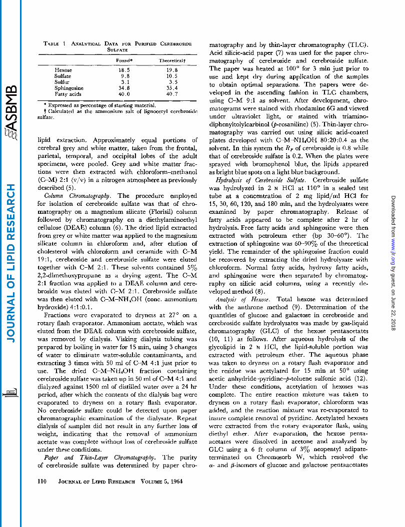
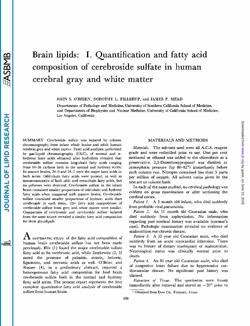
TABLE 1 ANALYTICAL DATA FOR PURIFIED CEREBROSIDE SULFATE
Found* Theoretical?
Hexose 18.5 1 9 . 8 Sulfate 9 .8 10.5 Sulfur 3.1 3 . 5
Fatty acids 40.0 40.7 Sphingosine 34.8 35.4
* Expressed as percentage of starting material. t Calculated as the ammonium salt of lignoceryl cerebroside
sulfate.
lipid extraction. Approximately equal portions of cerebral grey and white matter, taken from the frontal, parietal, temporal, and occipital lobes of the adult specimens, were pooled. Grey and white matter frac- tions were then extracted with chloroform-methanol (C-M) 2:l (v/v) in a nitrogen atmosphere as previously described (5).
Column Chromatography. The procedure employed for isolation of cerebroside sulfate was that of chro- matography on a magnesium silicate (Florid) column followed by chromatography on a diethylaminoethyl cellulose (DEAE) column (6). The dried lipid extracted from grey or white matter was applied to the magnesium silicate column in chloroform and, after elution of cholesterol with chloroform and ceramide with C-M 19 :1, cerebroside and cerebroside sulfate were eluted together with C-M 2:l . These solvents contained 5% 2,2-dimethoxypropane as a drying agent. The C-M 2:l fraction was applied to a DEAE column and cere- broside was eluted with C-M 2: l . Cerebroside sulfate was then eluted with C-M-NH40H (conc. ammonium hydroxide) 4:l:O.l.
Fractions were evaporated to dryness at 27' on a rotary flash evaporator. Ammonium acetate, which was eluted from the DEAE column with cerebroside sulfate, was removed by dialysis. Visking dialysis tubing was prepared by boiling in water for 15 min, using 3 changes of water to eliminate water-soluble contaminants, and extracting 3 times with 50 ml of C-M 4:l just prior to use. The dried C-M-NH,OH fraction containing cerebroside sulfate was taken up in 50 ml of C-M 4:l and dialyzed against 1500 ml of distilled water over a 24 hr period, after which the contents of the dialysis bag were evaporated to dryness on a rotary flash evaporator. No cerebroside sulfate could be detected upon paper chromatographic examination of the dialysate. Repeat dialysis of samples did not result in any further loss of weight, indicating that the removal of ammonium acetate was complete without loss of cerebroside sulfate under these conditions.
Paper and Thin-Layer Chromatography. The purity of cerebroside sulfate was determined by paper chro-
matography and by thin-layer chromatography (TLC). Acid silicic-acid paper (7) was used for the paper chro- matography of cerebroside and cerebroside sulfate. The paper was heated at 100' for 3 min just prior to use and kept dry during application of the samples to obtain optimal separations. 'The papers were de- veloped in the ascending fashion in TLC chambers, using C-M 9:l as solvent. After development, chro- matograms were stained with rhodamine 6G and viewed under ultraviolet light, or stained with triamino- diphenyltolylcarbinol @-rosaniline) (5). Thin-layer chro- matography was carried out using silicic acid-coated plates developed with C-M-NH40H 80:20:0.4 as the solvent. In this system the RF of cerebroside is 0.8 while that of cerebroside sulfate is 0.2. When the plates were sprayed with bromophenol blue, the lipids appeared as bright blue spots on a light blue background.
Cerebroside sulfate was hydrolyzed in 2 N HC1 at 110' in a sealed test tube at a concentration of 2 mg lipid/ml HC1 for 15, 30, 60, 120, and 180 min, and the hydrolysates were examined by paper chromatography. Release of fatty acids appeared to be complete after 2 hr of hydrolysis. Free fatty acids and sphingosine were then extracted with petroleum ether (bp 30-60'). The extraction of sphingosine was 60-900/, of the theoretical yield. The remainder of the sphingosine fraction could be recovered by extracting the dried hydrolysate with chloroform. Normal fatty acids, hydroxy fatty acids, and sphingosine were then separated by chromatog- raphy on silicic acid columns, using a recently de- veloped method (8).
Analysis of Hexose. Total hexose was determined with the anthrone method (9). Determination of the quantities of glucose and galactose in cerebroside and cerebroside sulfate hydrolysates was made by gas-liquid chromatography (GLC) of the hexose pentaacetates (10, 11) as follows. After aqueous hydrolysis of the glycolipid in 2 N HC1, the lipid-soluble portion was extracted with petroleum ether. The aqueous phase was taken to dryness on a rotary flash evaporator and the residue was acetylated for 15 min at 50' using acetic anhydride-pyridine-p-toluene sulfonic acid (1 2). Under these conditions, acetylation of hexoses was complete. The entire reaction mixture was taken to dryness on a rotary flash evaporator, chloroform was added, and the reaction mixture was re-evaporated to insure complete removal of pyridine. Acetylated hexoses were extracted from the rotary evaporator flask, using diethyl ether. After evaporation, the hexose penta- acetates were dissolved in acetone and analyzed by GLC using a 6 ft column of 3% neopentyl adipate- terminated on Chromosorb W, which resolved the CY- and @-isomers of glucose and galactose pentaacetates
Hydrolysis of Cerebroside Sulfate.
110 JOURNAL OF LIPID RESEARCH VOLUME 5,1964
by guest, on June 22, 2018w
ww
.jlr.orgD
ownloaded from
Wave numbers in CM- l S O 0 0 3000 1500 1300
1200 1100 1000 900 800 700 62 5 . ' . l ~ ' ~ ' . ' " ~ ~ " " " '
. . . . . . . . . . . .. . . . . . . L
. . . . - -. . - 4 0 J
W " I
- C . . . . I . - . . . . . - . . . . . . . . . . . . . .
2 0 L 1 0 . . - . . . . . . . . . - . - - ..... - - .... - -. . -. - __ . . . . . . - . . . -. . .. a
O 2 3 4 S 6 7 6 9 I O I I 12 13 14 I S 16 Wave length in microns
FIG. 1. of the sample pressed into a potassium bromide pellet.a
Infrared spectrum of cerebroside sulfate. Cerebroside sulfate was isolated from infant whole brain and a scan was made
into 4 peaks suitable for quantification (10, 11). These isomers were produced during acid hydrolysis of these hexoses. By comparison of the a-peaks of each hexose, it was possible to quantitate glucose and galactose at the 1% level. Using this method, the only hexose detected in cerebroside sulfate and cerebroside in grey and white matter from patient 3 was galactose.
Analysis of Sulfate and Sulfur. Sulfate was deter- mined after hydrolysis of cerebroside sulfate, using the barium chloranilate method (1 3). Sulfur determinations were made using an X-ray fluorescent method?
Nor- mal fatty acids and hydroxy fatty acids were converted to methyl esters using the boron trifluoride-methanol method (14). Methyl esters of hydroxy fatty acids were acetylated and the acetoxy methyl esters were analyzed by GLC as described (8). Stationary phases used 3% Apiezon L (w/w) on siliconized Chromosorb, and 10% diethyleneglycol succinate on Chromosorb W. Pure fatty acids ranging from 14-24 carbons were used as reference standards in quantification, since the detector response was not identical for each fatty acid. When standards were unavailable, the closest corresponding correction factor was used. Aliquots of normal and acetoxy fatty acid methyl esters were hydrogenated and chromatographed by GLC, to identify unsaturated acids. Peaks for which standards were not available were identified by carbon number (15) plotting log of retention time vs. chain length or degree of unsatura- tion. Only those compounds with carbon numbers corresponding exactly to known fatty acids are given
* Unpublished method of Mr. George Alexander, Department of Biophysics, U.C.L.A. School of Medicine, Los Angeles 24, Cali- fornia.
a Infrared analysis was kindly performed by Mr. George Alexander.
Gas-Liquid Chromatograflhy (GLC) of Fatty Acids.
positive identifications. The samples were chromato- graphed both on polar and non-polar columns to make identification more accurate. Quantification of each peak was carried out by measuring the peak area by triangulation, cutting out and weighing on an analytical
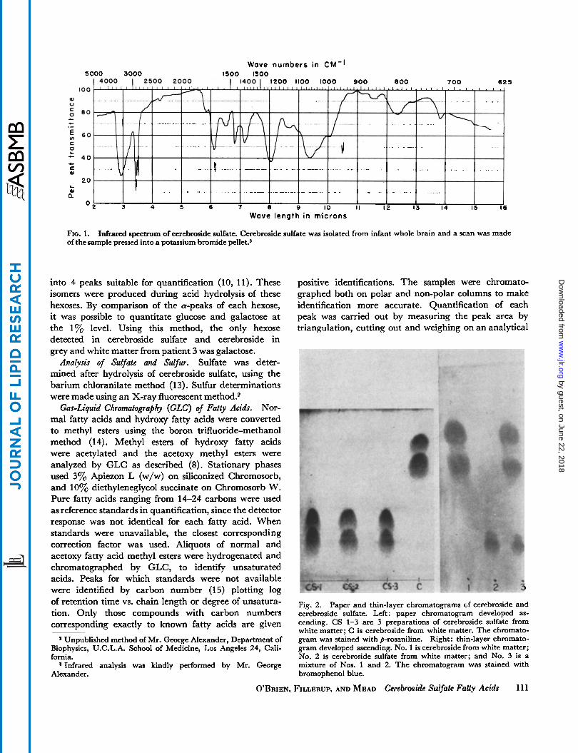
1
Fig. 2. Paper and thin-layer chromatograms cf cerebroside and cerebroside sulfate. Left : paper chromatogram developed as- cending. CS 1-3 are 3 preparations of cerebroside sulfate from white matter; C is cerebroside from white matter. The chromato- gram was stained with p-rosaniline. Right: thin-layer chromato- gram developed ascending. No. 1 is cerebroside from white matter; No. 2 is cerebroside sulfate from white matter; and No. 3 is a mixture of Nos. 1 and 2. The chromatogram was stained with bromophenol blue.
O'BRIEN, FILLERUP, AND MEAD Cerebroside Sulfate FaUy Acids 111
by guest, on June 22, 2018w
ww
.jlr.orgD
ownloaded from
it L!'
23 I 25 X
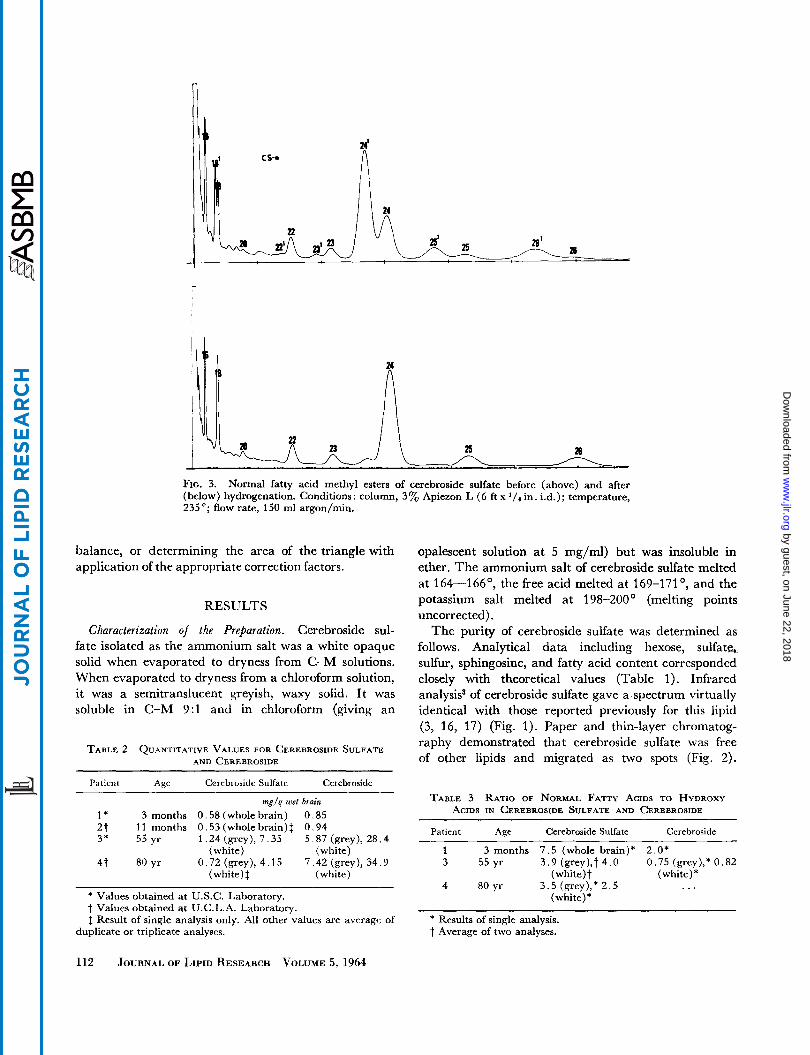
FIG, 3. (below) hydrogenation. Conditions: column, 3% Apiezon L (6 f t x 235 O ; flow rate, 150 ml argon/min.
Normal fatty acid methyl esters of cerebroside sulfate before (above) and after in. i.d.); temperature,
balance, or determining the area of the triangle with application of the appropriate correction factors.
RESULTS
characterization of the Preparation. Cerebroside sul- fate isolated as the ammonium salt was a white opaque solid when evaporated to dryness from C-M solutions. When evaporated to dryness from a chloroform solution, it was a semitranslucent greyish, waxy solid. It was soluble in C-M 9:l and in chloroform (giving an
TABLE 2 QUANTITATIVE VALUES FOR CEREBROSIDE SULFATE AND CEREBROSIDE
opalescent solution at 5 mg/ml) but was insoluble in ether. The ammonium salt of cerebroside sulfate melted at 164-166', the free acid melted at 169-171°, and the potassium salt melted at 198-200' (melting points uncorrected).
The purity of cerebroside sulfate was determined as follows. Analytical data including hexose, sulfate, sulfur, sphingosine, and fatty acid content corresponded closely with theoretical values (Table 1). Infrared analysis3 of cerebroside sulfate gave a spectrum virtually identical with those reported previously for this lipid (3, 16, 17) (Fig. 1). Paper and thin-layer chromatog- raphy demonstrated that cerebroside sulfate was free of other lipids and migrated as two spots (Fig. 2).
Patient Age Cerebroside Sulfate Cerebroside
mg/g wet brain 1 * 3 months 0.58 (whole brain) 0 .85 2 t 11 months 0.53(wholebrain)$ 0.94 3* 55yr 1.24(grey), 7.35 5.87(grey), 28.4
4 t 80 yr 0.72 (grey), 4.15 7.42 (grey), 34.9 (white) (white)
(white)$ (white)
* Values obtained at U.S.C. Laboratory. t Values obtained at U.C.L.A. Laboratory. $ Result of single analysis only. All other values are average of
duplicate or triplicate analyses.
TABLE 3 RATIO OF NORMAL FATTY ACIDS TO HYDROXY ACIDS IN CEREBROSIDE SULFATE AND CEREBROSIDE
Patient Age Cerebroside Sulfate Cerebroside
1 3 months 7 . 5 (whole brain)* 2.0* 3 55 yr 3 .9 (grey), t4.0 0.75 (grey*),* 0.82
(white) t (white)
(white) 4 80 yr 3.5 (grey!,* 2 . 5 ...
* Results of single analysis. t Average of two analyses.
112 JOURNAI. OF LIPID RESEARCH VOLUME 5, 1964
by guest, on June 22, 2018w
ww
.jlr.orgD
ownloaded from
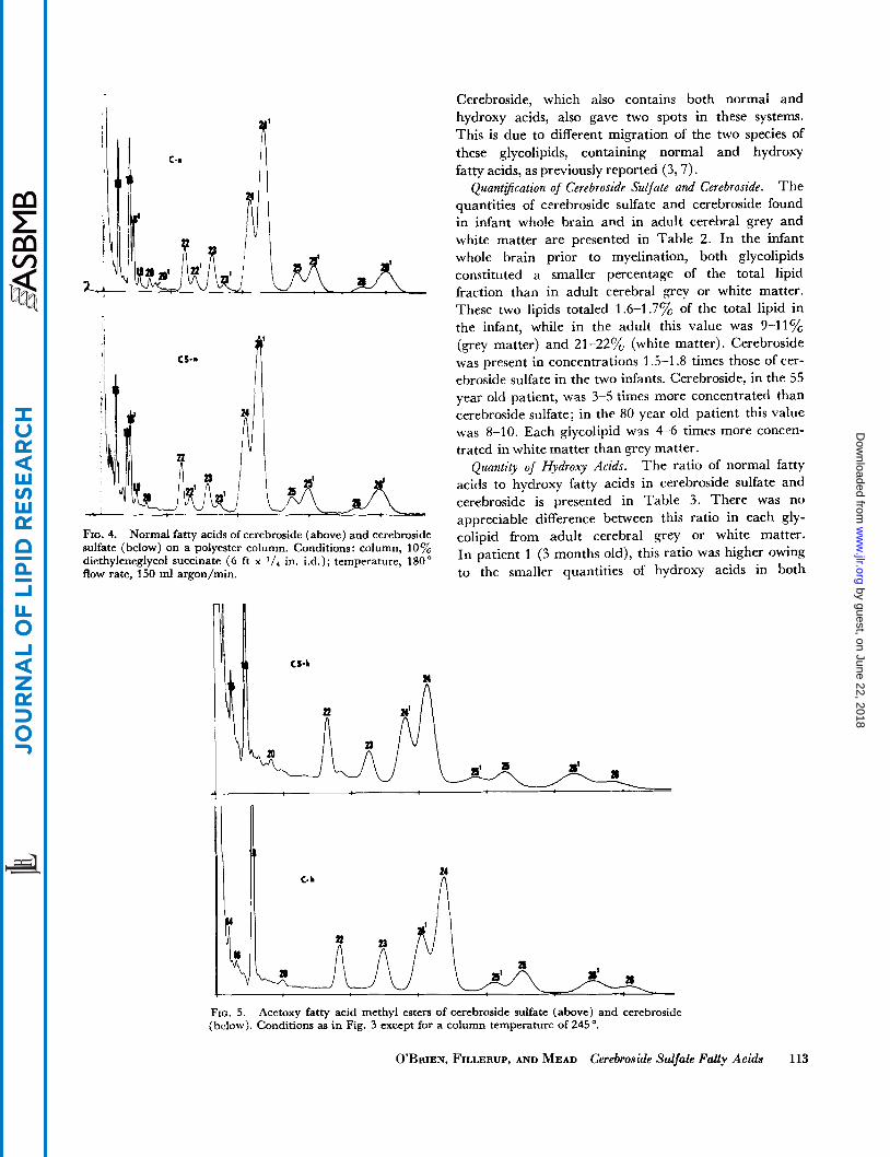
FIG. 4. Normal fatty acids of cerebroside (above) and cerebroside sulfate (below) on a polyester column. Conditions: column, 10 diethyleneglycol succinate (6 ft x 1/4 in. i.d.); temperature, 180" flow rate, 150 ml argon/min.
Cerebroside, which also contains both normal and hydroxy acids, also gave two spots in these systems. This is due to different migration of the two species of these glycolipids, containing normal and hydroxy fatty acids, as previously reported (3,7).
The quantities of cerebroside sulfate and cerebroside found in infant whole brain and in adult cerebral grey and white matter are presented in Table 2. In the infant whole brain prior to myelination, both glycolipids constituted a smaller percentage of the total lipid fraction than in adult cerebral grey or white matter. These two lipids totaled 1.6-1.7% of the total lipid in the infant, while in the adult this value was 9-11% (grey matter) and 21-220/;, (white matter). Cerebroside was present in concentrations 1.5-1.8 times those of cer- ebroside sulfate in the two infants. Cerebroside, in the 55 year old patient, was 3-5 times more concentrated than cerebroside sulfate; in the 80 year old patient this value was 8-10. Each glycolipid was 4-6 times more concen- trated in white matter than grey matter.
The ratio of normal fatty acids to hydroxy fatty acids in cerebroside sulfate and cerebrosidz is presented in Table 3. There was no appreciable difference between this ratio in each gly- colipid from adult cerebral grey or white matter. In patient 1 (3 months old), this ratio was higher owing to the smaller quantities of hydroxy acids in both
Quant8cation of Cerebroside Sulfate a n d cerebroside.
Quantity of Hydroxy Acids.
FIG. 5. (below), Conditions as in Fig. 3 except for a column temperature of 245 '.
Acetoxy fatty acid methyl esters of cerebroside sulfate (above) and cerebroside
O'BRIEN, FILLERUP, AND MEAD Cerebroside Sulfate Fatty Acids 113
by guest, on June 22, 2018w
ww
.jlr.orgD
ownloaded from
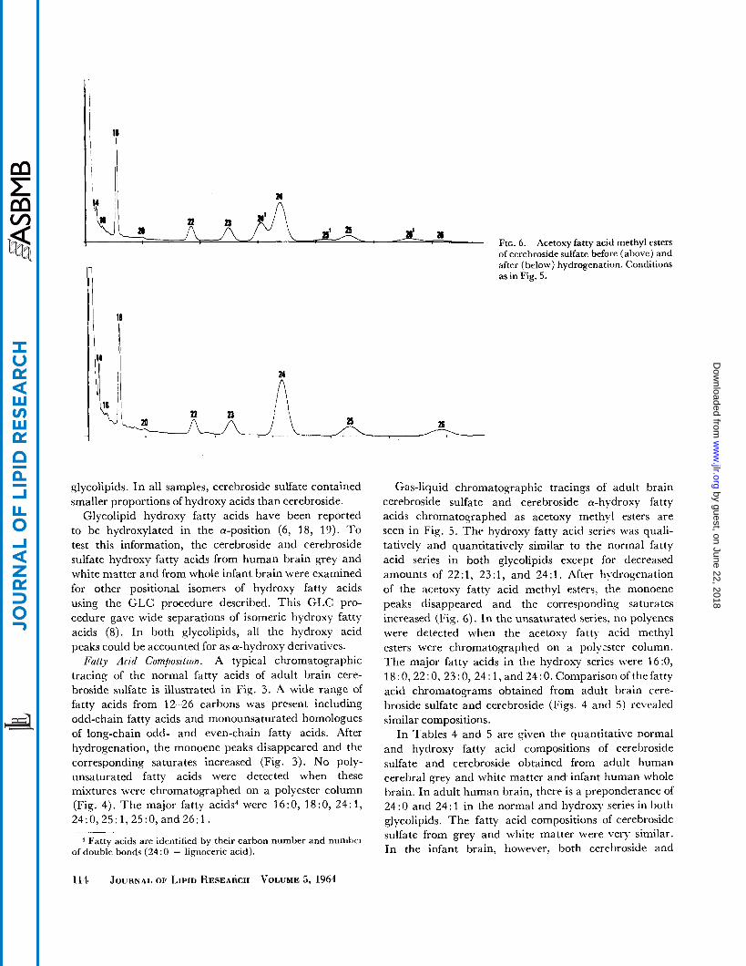
FIG. 6 . Acetoxy fatty acid methyl esters of cerebroside sulfate before (above) and after (below) hydrogenation. Conditions as in Fig, 5.
glycolipids. In all samples, cerebroside sulfate contained smaller proportions of hydroxy acids than cerebroside.
Glycolipid hydroxy fatty acids have been reported to be hydroxylated in the a-position (6, 18, 19). T o test this information, the cerebroside and cerebroside sulfate hydroxy fatty acids from human brain grey and white matter and from whole infant brain were examined for other positional isomers of hydroxy fatty acids using the GLC procedure described. This GLC pro- cedure gave wide separations of isomeric hydroxy fatty acids (8). In both glycolipids, all the hydroxy acid peaks could be accounted for as a-hydroxy derivatives.
Fatty Acid Composztzon. A typical chromatographic tracing of the normal fatty acids of adult brain cere- broside sulfate is illustrated in Fig. 3. A wide range of fatty acids from 12-26 carbons was present including odd-chain fatty acids and monounsaturated homologues of long-chain odd- and even-chain fatty acids. After hydrogenation, the monoene peaks disappeared and the corresponding saturates increased (Fig. 3). No poly- unsaturated fatty acids were detected when these mixtures were chromatographed on a polyester column (Fig. 4). The major fatty acids4 were 16:0, 18:0, 24:1, 24:0,25:1,25:0,and26:1.
Fatty acids are identified by their carbon number and numher of double bonds (24 : 0 = lignoceric acid).
Gas-liquid chromatoqraphic tracinys of adult brain cerebroside sulfate and cerebroside 0-hydroxy fatty acids chromatographed as acetoxy mrthyl esters are seen in Fig. 5. Thc hydroxy fatty acid series was quali- tatively and quantitatively similar to the normal fatty acid series in both glycolipids except for decreased amounts of 22 : 1, 23 : 1, and 24 : 1. After hydrogenation of the acrtoxy fatty acid methyl esters. the monoene peaks disappeared and the correspondiny saturates increased (Fig. 6). In the unsaturated series, no polyenes were detected when the acetoxy fatty acid methyl esters were chromatographed on a polycster column. The major fatty acids in the hydroxy series were 16:0, 18.0,22 : 0,23 : 0, 24: 1, and 24 : 0. Comparison of the fatty acid chromatograms obtained from adult brain cere- broside sulfate and cerebroside (Figs. 4 and 5) revealed similar compositions.
In Tables 4 and 5 are yiven the quantitative normal and hydroxy fatty acid compositions of cerebroside sulfate and cerebroside obtained from adult human cerebral grey and white matter and infant human whole brain. In adult human brain, there is a preponderance of 24 : 0 and 24 : 1 in the normal and hydroxy series in both glycolipids. The fatty acid compositions of cerebroside sulfate from grey and white matter were very similar. In the infant brain, however, both cerehroside and
114 JOURNAL OF LIPIL) KESEARCH VOLUME 5, 1964
by guest, on June 22, 2018w
ww
.jlr.orgD
ownloaded from
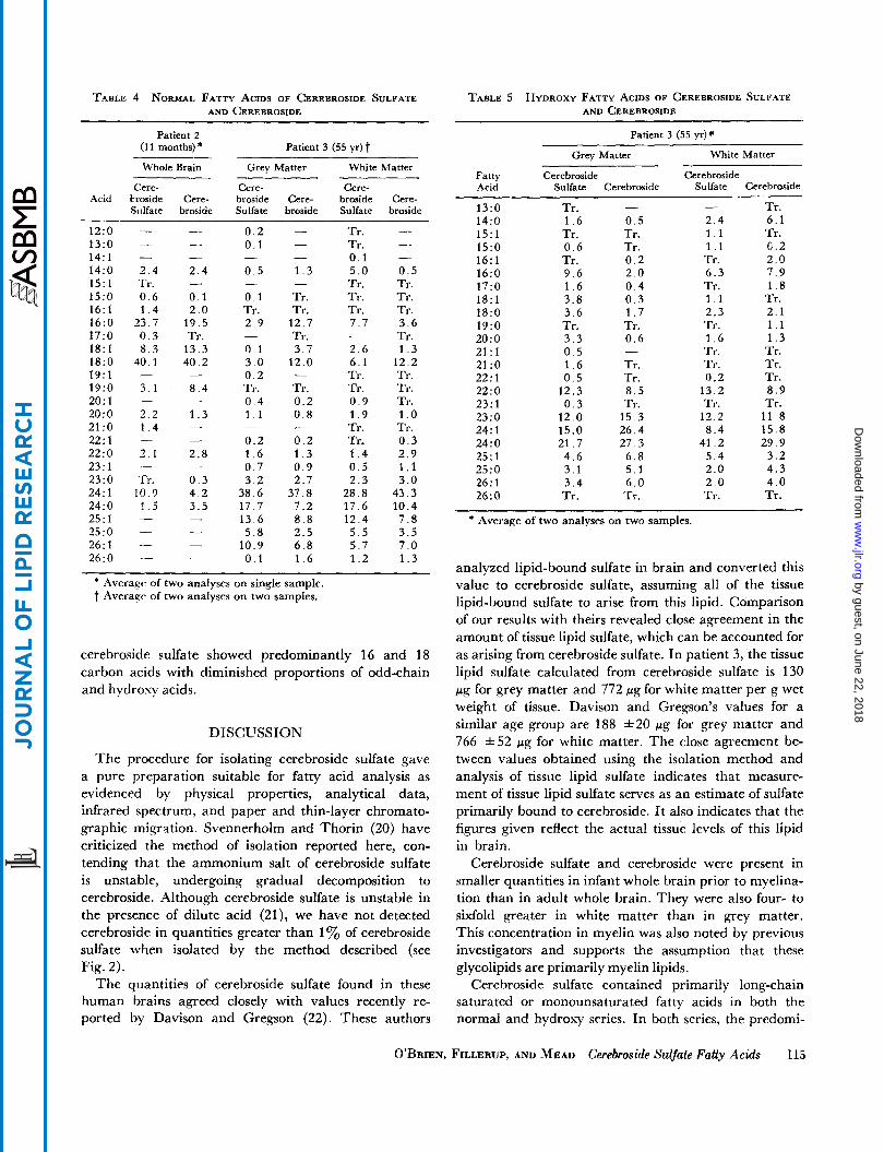
TABLE 4 NORMAL FATTY ACIDS OF CEREBROSIDE SULFATE AND CEREBROSIDE
Patient 2 (11 months)*
Whole Brain Grey Matter White Matter
Acid Cere-
broside Cere- Siilfate broside
12:o 13:O 14: 1 14:O 15:l 15:O 16: 1 16:O 17:O 18:l l8:O 19: 1 19:O 20: 1 20:o 21:o 22: 1 22:o 23: 1 23:O 24: 1 24: 0 25:l 25:O 26: 1 26:O
- - 2.4 2 . 4 Tr. 0.6 0 . 1 1 . 4 2 . 0
23.7 19.5 0 . 3 Tr. 8 . 3 13.3
40.1 40.2
3 .1 8 . 4
3 . 2 1 . 3 1 . 4 -
3.1 2.8
Tr. 0 . 3 10.9 4 .2
1 5 3 .5
-
- -
- -
- -
- -
- - - -
Cere- broside Cere- Sulfate broside
0 . 2 - 0.1 -
0 . 5 1 . 3
0 . 1 Tr. Tr. Tr. 2 . 9 12.7
Tr. 0 . 1 3.7 3.0 1’2.0 0 . 2 - Tr. Tr. 0 . 4 0.2 1 . 1 0 . 8
0 .2 0 . 2 1 . 6 1 . 3 0 . 7 0 . 9
- -
- -
-
- -
3.2 2.7 38.6 37.8 17.7 7.2 13.6 8.8 5.8 2.5
10.9 6 . 8 0 . 1 1 .6
Cere- broside Cere- Sulfate broside
- Tr. Tr. 0.1 - 5 . 0 0 . 5 Tr. Tr. Tr. Tr. Tr. Tr. 7 . 7 3 . 6
Tr . 2.6 1 . 3 6 . 1 12.2 Tr. Tr. Tr. Tr. 0 . 9 Tr. 1 . 9 1 . 0 Tr. Tr. Tr. 0 . 3 1 . 4 2 . 9 0 . 5 1 . 1 2 . 3 3.0
28.8 43.3 17.6 10.4 12 .4 7.8 5 . 5 3.5 5 . 7 7 . 0 1 . 2 1 . 3
-
-
* Averagr of two analyses on single sample. t Average of two analyses on two samples.
cerebroside sulfate showed predominantly 16 and 18 carbon acids with diminished proportions of odd-chain and hydroxy acids.
DISCUSSION
The procedure for isolating cerebroside sulfate gave a pure preparation suitable for fatty acid analysis as evidenced by physical properties, analytical data, infrared spectrum, and paper and thin-layer chromato- graphic migration. Svennerholm and Thorin (20) have criticized the method of isolation reported here, con- tending that the ammonium salt of cerebroside sulfate is unstable, undergoing gradual decomposition to cerebroside. Although cerebroside sulfate is unstable in the presence of dilute acid (21), wc have not detected cerebroside in quantities greater than 1 yo of cerebroside sulfate when isolated by the method described (see Fig. 2).
The quantities of cerebroside sulfate found in these human brains agreed closely with values recently re- ported by Davison and Gregson (22). These authors
TABLE 5 HYDROXY FATTY ACIDS OF CEREBROSIDE SULFATE AND CEREBROSIDE
Patient 3 (55 yr)*
White Matter Grey Matter
Fatty Cerebroside Cerebroside Acid Sulfate Cerebroside Sulfate Cerebroside
13:O 14:O 15: 1 15:O 16:l l6:O 17:O 18:l 18:O 19:O 20:o 21:l 21:o 22:l 22:o 23: 1 23:O 24: 1 24:O 25:l 25:O 26: 1 26:O
- Tr. 1 . 6 0 . 5 Tr . Tr. 0 . 6 Tr. Tr. 0.2 9.6 2 .0 1 . 6 0 .4 3.8 0 . 3 3 . 6 1 . 7 Tr. Tr. 3 . 3 0 .6 0 . 5 1 . 6 Tr. 0 . 5 Tr.
12.3 8 . 5 0 . 3 Tr.
12.0 15.3 15.0 26.4 21.7 27.3 4 .6 6 . 8 3.1 5.1 3 . 4 6 . 0 Tr. Tr.
-
Tr. 2.4 6.1 1 . 1 Tr. 1 . 1 0 .2 Tr . 2 . 0 6 .3 7.9 Tr. 1 . 8 1 . 1 Tr. 2.3 2 .1 Tr. 1 . 1 1 . 6 1 . 3 Tr. Tr. Tr . Tr. 0.2 Tr.
13.2 8.9 Tr. Tr.
12.2 11.8 8.4 15.8
41.2 29.9 5 .4 3.2 2.0 4 .3 2 .0 4 .0 Tr. Tr .
-
~~~~~~~~ ~ ~
* Average of two analyses on two samples.
analyzed lipid-bound sulfate in brain and converted this value to cerebroside sulfate, assuming all of the tissue lipid-bound sulfate to arise from this lipid. Comparison of our results with theirs revealed close ayreement in the amount of tissue lipid sulfate, which can be accounted for as arising from cerebroside sulfate. In patient 3, the tissue lipid sulfate calculated from cerebroside sulfate is 130 pg for grey matter and 772 fig for white matter per g wet weight of tissue. Davison and Gregson’s values for a similar age group are 188 *20 pg for ,grey matter and 766 *52 pg for white matter. The close agreement be- tween values obtained using the isolation method and analysis of tissue lipid sulfate indicates that measure- ment of tissue lipid sulfate serves as an estimate of sulfate primarily bound to cerebroside. I t also indicates that the figures given reflect the actual tissue levels of this lipid in brain.
Cerebroside sulfate and cerebroside were present in smaller quantities in infant whole brain prior to myelina- tion than in adult whole brain. They were also four- to sixfold greater in white matter than in grey matter. This concentration in myelin was also noted by previous investigators and supports the assumption that these glycolipids are primarily myelin lipids.
Cerebroside sulfate contained primarily long-chain saturated or monounsaturated fatty acids in both the normal and hydroxy series. In both series, the predomi-
O’BRIEN, FILLERUP, AND MEAD Cerebroside Sulfate Fatty Acids 115
by guest, on June 22, 2018w
ww
.jlr.orgD
ownloaded from
nant fatty acids were 24: 0 and 24: 1. Odd-chain fatty acids and monounsaturates of odd- and even-chain fatty acids from 14-26 carbons were present, but no poly- unsaturates were detected. Smaller quantities of 22 : 1, 23:1, and 24:l were found in the hydroxy series than in the normal series. This fatty acid composition is similar to that reported for cerehrosidr by Radin and co- workers (18, 23). The fatty acid compositions of cerebro- side sulfate from cerebral grey and white matter werequitesimilar. These findinys indicate that cerebroside sulfate has a similar fatty acid composition in a mem- brane-rich tissue (white matter) and in a tissue contain- ing a greatrr proportion of cytoplasmic organelles (qrey matter). The maintenance of a similar fatty acid composi- tion for both cerebroside and cerebroside sulfate in two different tissue types may indicate that this composition is essential for metabolic function of these glycolipids.
Cerebroside sulfate, like cerebroside (1 8), contained lesser proportions of odd-chain and hydroxy acids in the young-er brains. This finding indicates that these fatty acids accumulate in this glycolipid with aye, as they do in cerebroside (18). The accurmilation of hydrosy and odd-chain acids with age also indicates that cerebroside sulfate acids, likc cerebroside acids, may undergo a- oxidation and 1-carbon degradation (24).
The technical assistance of Lois Sampson, Ken Hardy, and Amy Toma is gratefully acknowledged. We thank Dr. David Blankenhorn for his critical review of the manuscript.
This investigation was supported in part by PHS Grant #62-56-H-7197 from the National Heart Institute, Los Angeles County Hospital Attending Staff Project #1065, and Contract AT (04-1) GEN-12 between the Atomic Energy Commission and the University of California.
Manuscript received June 77, 7963; accepted September 72, 7963.
REFERENCES 1. Blix, G. 2. Jatzkewitz, H. 3. Jatzkewitz, H. 4. O’Brien, J. S., and G. Rouser. Federation Proc. 21: 284,
1962. 5. Rouser, G., J. S. O’Brien, and D. Heller. J . Am. Oil
Chemists’ SOL. 38: 14, 1961. 6. Rouser, G., A. J. Baumann, G. Kritchevsky, D. Heller, and
J. S. O’Brien. 7. Rouser, G., A. J. Bauman, N. Nicolaides. and D. Heller.
J . Am. Oil Chemists’ SOC. 38 565, 1961. 8. O’Brien, J. S.: and G. Rouser. 9. Radin, N. S., F. B. Lavin, and J. R. Brown.
10. VandenHeuvel, W. J. A., and E. C. Horning.
11. Kircher, H. W. 12. Fritz, J. S., and G. H. Schenk. Anal. Chm. 31: 1808, 1959. 13. Bertolacini, R. J., and J. E. Barney. Anal. Chrm. 29: 281,
1957. 14. Metcalfe, L. D., and A. A. Schmitz. Anal. C‘hrm. 33: 363,
1961. 15. Woodford, F. P., and C. M. van Gent. ,J. Lipid Rrs. 1:
188, 1960. 16. Witmer, F. J., and J. H. Austin. Mikrochim. Acta no vol:
502, 1960. 17. Lloyd, A. G., and K. S. Dodgson. Biorhirn. Biophlls. Acta
46: 116, 1961. 18. Kishimoto, Y., and N. S. Radin. J . Lipid Res. 1: 79, 1959. 19. Kishimoto, Y., and N. S. Radin. .J. Lipid Res. 4: 130,
1963. 20. Svennerholm, L., and H. Thorin. J . Lipid Res. 3: 483,
1962. 21. Stoffyn, P., and A. Stoffyn. Biochim. Bioph!r. Acta 70: 107,
1963. 22. Davison, A. N., and N. A. Gregson. Biochem. J . 85: 558,
1962. 23. Radin, N. S., and Y. Akahori. J . Lipid Res. 2: 335. 1961. 24. Mead, J. F., and G. M. Levis. Biorhpni. BiophJs. Res.
Z . Physiol. Chem. 219: 82, 1933. Z. Physiol. Chem. 311: 279, 1958. Z. Physiol. Chem. 320: 134, 1960.
J . Am. Oil Chemists’ SOC. 38: 544. 1961.
4 n d . Biochern., in press. J . Bid.
Biochcm. Chem. 217: 789, 1955.
Biophys. Res. Commun. 4: 399, 1961. 7appi 45: 143, 1962.
Commun. 9: 231, 1962.
116 JOURNAL OF LIPID RESEARCH VOLUME 5: 1964
by guest, on June 22, 2018w
ww
.jlr.orgD
ownloaded from