Brain I: Telencephalon and Diencephalon * Cerebral Cortex and Basal Nuclei * Thalamus and...
21
Brain I: Telencephalon and Diencephalon * Cerebral Cortex and Basal Nuclei * Thalamus and Hypothalamus Nestor T. Hilvano M.D., M.P.H.
-
Upload
amy-lawrence -
Category
Documents
-
view
224 -
download
2
Transcript of Brain I: Telencephalon and Diencephalon * Cerebral Cortex and Basal Nuclei * Thalamus and...

Brain I: Telencephalon and Diencephalon
* Cerebral Cortex and Basal Nuclei
* Thalamus and Hypothalamus
Nestor T. Hilvano M.D., M.P.H.

Learning Objectives
1. Describe the meninges of the brain. 2. Identify the cerebrum as to major subdivisions, cortical
areas and their functions. 3. Discuss the blood supply of the brain and give its
significance. 4. Identify the components and functions of the limbic
system. 5. Discuss the significance of brain waves seen in EEG.6. Describe the components of basal nuclei. 7. List the components of diencephalon and discuss their
functions. 8. Correlate lesions affecting areas of cerebral cortex,
basal nuclei, thalamus, and hypothalamus.

Meninges (coverings)• Dura mater- outer
– outer periosteal layer – inner meningeal layer – dural sinuses in between
• Arachnoid- middle * Subarachnoid space filled w/ ___.
• Pia mater- innermost • Dural folds
– falx cerebri (contain sup. & inf. sagittal sinuses)– tentorium cerebelli (contain transverse sinus)– falx cerebelli
• Meningitis • Epidural hemorrhage – meningeal a.• Subdural hemorrhage – dural sinus

Cerebrum
• Brain consists of 6 regions: - Cerebrum. basal nuclei, diencephalon, midbrain, pons, medulla oblongata, and cerebellum• Cerebrum – largest part of brain; the outer part is _______, gray matter and inner part is __________, white matter. a. cerebral medulla b. cerebral cortex

Cerebral Cortex
• Neocortex (six-layered)• ___ What is the characteristic cells (motor neuron) of
cerebrum (cerebral cortex)?
a. stellate cells
b. pyramidal (Betz) cells
c. martinotti cells
d. cajal cells

Cerebral White MatterA. Association fibers - arcuate fibers - longitudinal fasciculusB. Commisural fibers - corpus callosum - anterior commissure - posterior commissureC. Projection fibers - internal capsuleExercise: ___ connections within one hemisphere___ connecting two hemispheres___ connect cerebrum with lower areas

Functional Regions of Cerebral Cortex
• Frontal lobe: motor lobe precentral gyrus (BA 4), premotor area (BA 6), frontal eye field (BA 8), broca’s area (BA 45, 44), prefrontal cortex (BA 9 – 12) • Parietal lobe: sensory lobe postcentral gyrus (BA 3, 1,2), gustatory center (BA 43), • Temporal lobe: auditory center, olfactory center, etc… primary auditory center (BA 41, 42), wernicke area (interpretive area, BA 22), uncus (olfactory center) • Occipital lobe: visual lobe calcarine area (BA 17), secondary visual area (BA 18,19)• Limbic lobe- visceral brain (autonomic function)Exercises:___ Primary motor cortex a. motor speech center ___ Frontal eye field b. personality, behavior, emotion ___ Prefrontal cortex c. conjugate eye movement ___ Broca’s area d. execution of movement

Limbic System• Emotion, Memory storage and retrieval, and Links
conscious functions of cerebral cortex with autonomic functions of brain stem
• Components - amygdala, limbic lobe ( hippocampus, parahippocampus, and cingulate gyrus), fornix, anterior thalamic nucleus, and reticular formation
• Anterograde amnesia - no new memories, injury of ___. • Retrograde amnesia – can’t remember old ones, injury of
___. a. Cerebrum (cerebral cortex)
b. hippocampus (limbic lobe)

Alzheimer Disease
• 100,000 deaths/year– 11% of population over 65; 47% by age 85
• Memory loss for recent events, moody, combative, lose ability to talk, walk, and eat
• Diagnosis confirmed at autopsy– atrophy of gyri (folds) in cerebral cortex– neurofibrillary tangles and senile plaques
• Degeneration of cholinergic neurons and deficiency of ACH and nerve growth factors
• Genetic connection confirmed

Cerebral Lateralization• Left hemisphere
– specialized for spoken and written language, math, reasoning
• Right hemisphere – perception of spatial
relationships, senses, recognition, imagination and insight, music and artistic skill
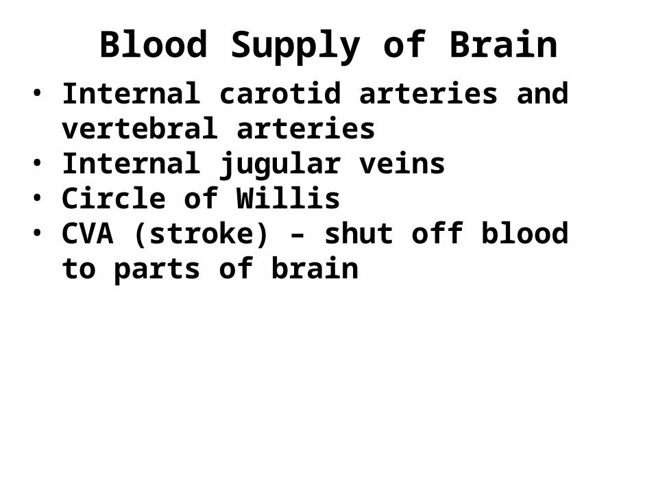
Blood Supply of Brain• Internal carotid arteries and vertebral
arteries• Internal jugular veins • Circle of Willis • CVA (stroke) – shut off blood to
parts of brain
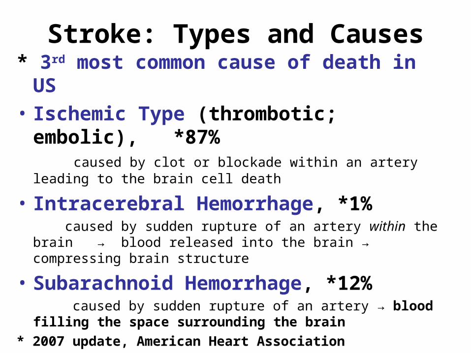
Stroke: Types and Causes* 3rd most common cause of death in US
• Ischemic Type (thrombotic; embolic), *87%
caused by clot or blockade within an artery leading to the brain cell death
• Intracerebral Hemorrhage, *1% caused by sudden rupture of an artery within the brain →
blood released into the brain → compressing brain structure
• Subarachnoid Hemorrhage, *12% caused by sudden rupture of an artery → blood filling the
space surrounding the brain* 2007 update, American Heart Association

EEG and Brain Waves
• Electrode placed on skull and monitor patterns of electrical activity
• alpha - Found in healthy, awake adults at rest with eyes closed
• beta - Found in adults concentrating or mentally stressed• theta – Found in children, in intensely frustrated adults, may
indicate brain disorder in adults • delta - During sleep, and in awake adults with brain damage

Basal Nuclei• Masses of gray matter deep to cortex; consists of
– c______________– p______________– g______________
• Subconscious control of muscle tone and posture for movements (coordination of learned movement like walking, lifting)
• Participates in automatic movements i.e., walking and in learning new motor behavior
• Lesion results to abnormal involuntary motor movements referred to as __________.
a. ataxia b. dyskinesia c. aphasia d. hemiplegia

Parkinson Disease
• Progressive loss of motor
function beginning in 50’s or 60’s– degeneration of dopamine-releasing neurons
• involuntary muscle contractions– pill-rolling motion, facial rigidity, slurred speech, – illegible handwriting, slow gait
• Treatment = drugs and physical therapy– dopamine precursor crosses brain barrier– MAO inhibitor slows neural degeneration– surgical technique to relieve tremors
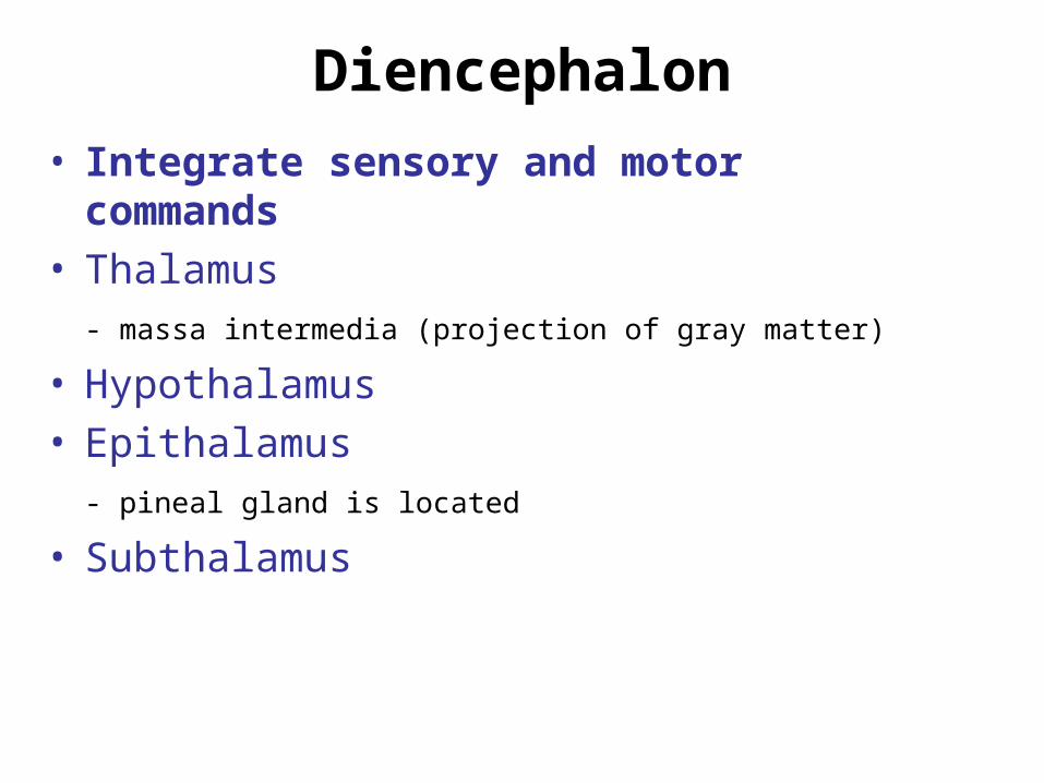
Diencephalon
• Integrate sensory and motor commands• Thalamus
- massa intermedia (projection of gray matter)
• Hypothalamus• Epithalamus
- pineal gland is located
• Subthalamus

Thalamus
• Relay information between basal nuclei and cerebral cortex
• Anterior nuclei- limbic, emotion & memory• Medial nuclei- awareness of emotion• Ventral nuclei- sensory to primary sensory area• Lateral nuclei- emotional stress• Posterior
- pulvinar = sensory to association areas of cortex- LGB = visual - MGB = auditory

Hypothalamus
• Functions – hormone secretions, autonomic control, and emotion
• Infundibulum
• Tuberal area
• Mamillary bodies

Hypothalamus• ADH and Oxytocin • Autonomic NS control
- anterior (PS); lat. & posterior (S) • Thermoregulation• Food and water intake
- ventromedial nucleus (satiety); lateral nucleus (feeding) • Sleep and circadian rhythms• Memory and emotional behavior (mammillary bodies)Exercises: A_______ hormone secreted by supraoptic nucleusO_______ hormone secreted by paraventricular nucleusS_______ controlled by suprachiasmatic nucleusL_______ feeding center; lesion results to anorexia nervosaV_______ satiety center; lesion results to obesityM_______ emotion

Epithalamus
• Epithalamus consists of pineal gland and the habenula (connects limbic system to midbrain)

Homework (Self Review) 1. Compare and contrast the cranial meninges and dural folds.2. Identify cortical areas affected: __ anopsia (loss of vision) a. Broca’s area (BA 45,44)
__ contralateral paralysis b. Postcentral gyrus (BA 3,1,2)
__ contralateral sensory loss c. Wernicke area (BA 22)
__ cortical deafness d. Calcarine area (BA 17)
__ expressive (motor) aphasia e. Precentral gyrus (BA 4)
__ sensory (interpretative) aphasia f. Temporal gyri of Heschl (BA 41,42)
3. Compare and contrast the dominant and non-dominant hemispheres. 4. Discuss retrograde and anterograde amnesia. 5. Discuss the blood supply of the brain. 6. Describe briefly thalamus and give the function of lateral and medial
geniculate bodies. 7. Name the hypothalamic nuclei involved in the following: secretion of
ADH and oxytocin, satiety center, feeding (hunger) center, memory and emotion, sleep rhythms.