
Brain (2001), 2528–2539 Brainstem gliomas in adults ......Brain (2001), 124, 2528–2539 Brainstem...
12
Brain (2001), 124, 2528–2539 Brainstem gliomas in adults: prognostic factors and classification Jean-Se ´bastien Guillamo, 1 Annick Monjour, 3 Luc Taillandier, 4 Bertrand Devaux, 2 Pascale Varlet, 2 Christine Haie-Meder, 5 Gilles-Louis Defer, 6 Patrick Maison, 7 Jean-Jacques Mazeron, 1 Philippe Cornu 1 and Jean-Yves Delattre 1 for the Association des Neuro-Oncologues d’Expression Franc ¸aise (ANOCEF) 1 Service de Neurologie, Service de Radiothe ´rapie, Service Correspondence to: Professor J.-Y. Delattre, Service de de Neurochirurgie, Ho ˆpital Pitie ´-Salpe ˆtrie `re and 2 Service Neurologie Mazarin, Ho ˆpital Pitie ´-Salpe ˆtrie `re, 75651 Paris de Neurochirurgie, Service d’Anatomopathologie, Ho ˆpital Cedex 13, France Saint-Anne, Paris, 3 Service de Neurologie, Ho ˆpital Pasteur, E-mail: jean-yves.delattre@psl.ap-hop-paris.fr Colmar, 4 Service de Neurologie, CHU Nancy, Nancy, 5 Service de Radiothe ´rapie, Institut Gustave Roussy, Villejuif, 6 Service de Neurologie Dejerine, CHU Co ˆte de Nacre, Caen and 7 Service de Pharmacologie clinique, CHU Henri Mondor, Cre ´teil, France Summary In contrast to childhood brainstem gliomas, adult brainstem gliomas are rare and poorly understood. The charts of 48 adults suffering from brainstem glioma were reviewed in order to determine prognostic factors, evaluate the effect of treatment and propose a classification of these tumours. Mean age at onset was 34 years (range 16–70 years). The main presenting symptoms were gait disturbance (61%), headache (44%), weakness of the limbs (42%) and diplopia (40%). Four patterns were identified on MRI, representing non- enhancing, diffusely infiltrative tumours (50%), contrast- enhancing localized masses (31%), isolated tectal tumours (8%) and other patterns (11%). Treatment consisted of partial resection (8%), radiotherapy (94%) and chemotherapy (56%). Overall median survival was 5.4 years. On univariate analysis, the following favourable prognostic factors were identified (P < 0.01): age of onset <40 years, duration of symptoms before diagnosis >3 months, Karnofski performance status >70, low-grade histology, absence of contrast enhancement and ‘necrosis’ on MRI. On multivariate analysis, the duration of symptoms, the appearance of ‘necrosis’ on MRI and the histological grade of the tumour remained significant and independent prognostic factors (P < 0.05). Eighty-five percent of the tumours could be classified into one of the following three groups on the basis of clinical, radiological Keywords: brainstem; glioma; adult; classification; prognostic factors Abbreviations: CI confidence interval; NF1 neurofibromatosis type 1 © Oxford University Press 2001 and histological features. (i) Diffuse intrinsic low-grade gliomas (46%) usually occurred in young adults with a long clinical history before diagnosis and a diffusely enlarged brainstem on MRI that did not show contrast enhancement. These patients were improved by radiotherapy in 62% of cases and had a long survival time (median 7.3 years). Anaplastic transformation (appearance of contrast enhancement, 27%) and relentless growth without other changes (23%) were the main causes of death. (ii) Malignant gliomas (31%) occurred in elderly patients with a short clinical history. Contrast enhancement and necrosis were the rule on MRI. These tumours were highly resistant to treatment and the patients had a median survival time of 11.2 months. (iii) Focal tectal gliomas (8%) occurred in young patients and were often revealed by isolated hydrocephalus. The course was indolent and the projected median survival period exceeded 10 years. In conclusion, adult brainstem gliomas are different from the childhood forms and resemble supratentorial gliomas in adults. Low-grade tumours have a clinicoradiological pattern that is so characteristic that the need for a potentially harmful biopsy is debatable. The optimum timing of treatment for supratentorial low- grade tumours remains unclear. In high-grade gliomas, the prognosis remains extremely poor despite aggressive treatment with radiotherapy and chemotherapy.
Transcript of Brain (2001), 2528–2539 Brainstem gliomas in adults ......Brain (2001), 124, 2528–2539 Brainstem...

Brain (2001), 124, 2528–2539
Brainstem gliomas in adults: prognostic factors andclassificationJean-Sebastien Guillamo,1 Annick Monjour,3 Luc Taillandier,4 Bertrand Devaux,2 Pascale Varlet,2
Christine Haie-Meder,5 Gilles-Louis Defer,6 Patrick Maison,7 Jean-Jacques Mazeron,1 Philippe Cornu1
and Jean-Yves Delattre1 for the Association des Neuro-Oncologues d’Expression Francaise (ANOCEF)
1Service de Neurologie, Service de Radiotherapie, Service Correspondence to: Professor J.-Y. Delattre, Service dede Neurochirurgie, Hopital Pitie-Salpetriere and 2Service Neurologie Mazarin, Hopital Pitie-Salpetriere, 75651 Parisde Neurochirurgie, Service d’Anatomopathologie, Hopital Cedex 13, FranceSaint-Anne, Paris, 3Service de Neurologie, Hopital Pasteur, E-mail: [email protected], 4Service de Neurologie, CHU Nancy, Nancy,5Service de Radiotherapie, Institut Gustave Roussy,Villejuif, 6Service de Neurologie Dejerine, CHU Cote deNacre, Caen and 7Service de Pharmacologie clinique, CHUHenri Mondor, Creteil, France
SummaryIn contrast to childhood brainstem gliomas, adultbrainstem gliomas are rare and poorly understood. Thecharts of 48 adults suffering from brainstem gliomawere reviewed in order to determine prognostic factors,evaluate the effect of treatment and propose aclassification of these tumours. Mean age at onset was34 years (range 16–70 years). The main presentingsymptoms were gait disturbance (61%), headache (44%),weakness of the limbs (42%) and diplopia (40%). Fourpatterns were identified on MRI, representing non-enhancing, diffusely infiltrative tumours (50%), contrast-enhancing localized masses (31%), isolated tectal tumours(8%) and other patterns (11%). Treatment consisted ofpartial resection (8%), radiotherapy (94%) andchemotherapy (56%). Overall median survival was5.4 years. On univariate analysis, the following favourableprognostic factors were identified (P < 0.01): age ofonset <40 years, duration of symptoms before diagnosis>3 months, Karnofski performance status >70, low-gradehistology, absence of contrast enhancement and ‘necrosis’on MRI. On multivariate analysis, the duration ofsymptoms, the appearance of ‘necrosis’ on MRI and thehistological grade of the tumour remained significant andindependent prognostic factors (P < 0.05). Eighty-fivepercent of the tumours could be classified into one of thefollowing three groups on the basis of clinical, radiological
Keywords: brainstem; glioma; adult; classification; prognostic factors
Abbreviations: CI � confidence interval; NF1 � neurofibromatosis type 1
© Oxford University Press 2001
and histological features. (i) Diffuse intrinsic low-gradegliomas (46%) usually occurred in young adults with along clinical history before diagnosis and a diffuselyenlarged brainstem on MRI that did not show contrastenhancement. These patients were improved byradiotherapy in 62% of cases and had a long survivaltime (median 7.3 years). Anaplastic transformation(appearance of contrast enhancement, 27%) and relentlessgrowth without other changes (23%) were the main causesof death. (ii) Malignant gliomas (31%) occurred in elderlypatients with a short clinical history. Contrastenhancement and necrosis were the rule on MRI. Thesetumours were highly resistant to treatment and thepatients had a median survival time of 11.2 months. (iii)Focal tectal gliomas (8%) occurred in young patients andwere often revealed by isolated hydrocephalus. The coursewas indolent and the projected median survival periodexceeded 10 years. In conclusion, adult brainstem gliomasare different from the childhood forms and resemblesupratentorial gliomas in adults. Low-grade tumours havea clinicoradiological pattern that is so characteristic thatthe need for a potentially harmful biopsy is debatable.The optimum timing of treatment for supratentorial low-grade tumours remains unclear. In high-grade gliomas,the prognosis remains extremely poor despite aggressivetreatment with radiotherapy and chemotherapy.

Adult brainstem glioma 2529
IntroductionIn children, brainstem gliomas constitute ~10% of braintumours and are usually classified in three main groups(Farwell et al., 1977; Freeman and Farmer, 1998; Walkeret al., 1999). The largest subgroup is diffuse intrinsic pontineglioma, which is characterized by a striking diffuseenlargement of the brainstem on MRI, with or withoutheterogeneous contrast enhancement, an aspect that obviatesthe need for biopsy according to many authors (Albrightet al., 1993; Constantini and Epstein, 1996). These tumourscarry the worst prognosis of any brain tumour in childhood,with a median survival of �1 year (Kaplan et al., 1996;Mandell et al., 1999). The second subgroup (10% of tumours)comprises slow-growing low-grade gliomas arising at thecervicomedullary junction or from the floor of the fourthventricle (Hoffman et al., 1980; Pollack et al., 1993). Thesetumours most often have a posterior contrast-enhancingexophytic development that may be amenable to surgicalresection (Bricolo et al., 1991; Constantini and Epstein,1996). Long survival is common and the median survivaltime is over 5 years. The third subgroup consists of indolentfocal tectal gliomas, which are often heralded byhydrocephalus (Squires et al., 1994; Bowers et al., 2000).
In contrast, brainstem gliomas in adults are poorlyunderstood because they are quite unusual, accounting for�2% of gliomas (White, 1963). Most reported studies belongto an era when MRI was not available at diagnosis (White,1963; Grigsby et al., 1989; Selvapandian et al., 1999).However, some data suggest that survival is much longer inadults than in children (Linstadt et al., 1991; Landolfi et al.,1998; Selvapandian et al., 1999).
To improve our understanding of the natural history ofthese tumours, to identify prognostic factors and to proposea scheme of classification for them, a retrospective study ofadult brainstem gliomas was undertaken.
Patients and methodsInclusion criteriaPatients were included if they met the following criteria: (i)they were older than 16 years at the first symptom; (ii) theepicentre of the tumour, defined as the centre of the tumourbulk, was located in the brainstem (midbrain, pons andmedulla oblongata) (this criterion excluded tumoursoriginating in the thalamus, the cerebellar peduncles orthe cervical spinal cord); (iii) the diagnosis was basedon histological confirmation or on clinical history withcharacteristic MRI appearance, consisting of an infiltrativeexpansive process, with or without contrast enhancement(when the lesion had contrast enhancement and when aninfectious process could not be ruled out, pathologicalexamination of a biopsy was mandatory); and (iv) a completemedical record, including clinical data, repeated MRI, anddetailed treatment data, was available.
Ependymomas, which constitute a distinct nosologicalgroup, were excluded from the study.
Data collectionThe following clinical data were collected: (i) at the time ofdiagnosis: age, sex, ethnic origin, time between the date ofthe first symptom and the date of the diagnosis (i.e. durationof symptoms), main symptoms and signs, Karnofskiperformance status, description of the tumour on MRI (T1-and T2-weighted images in at least two planes before andafter gadolinium contrast enhancement), and pathologicalreports when available; (ii) treatment administered atdiagnosis of the tumour; (iii) during follow-up: clinical andradiological course, complications (hydrocephalus, haemor-rhage, leptomeningeal dissemination, bulbar involvementwith swallowing impairment), treatment at recurrence, anddate and cause of death or date of the last visit if the patientwas alive.
The radiological response to radiotherapy and chemo-therapy was reported as: (i) a complete response, i.e.disappearance of all visible tumour; (ii) a partial response,i.e. a decrease of �50% in the axial cross-section of thegreatest surface area (contrast enhancement or T2 hyper-signal for non-enhancing tumours); (iii) progressive disease,i.e. �25% increase in axial cross-section of the greatestsurface area; or (iv) stable disease, i.e. all other situations(Macdonald et al., 1990; Bauman et al., 1999). The responsewas evaluated while the patients were receiving a stable ordecreasing dose of corticosteroids.
Statistical analysisSurvival time was measured from the date of symptom onsetto the date of last follow-up or death. Survival was estimatedby the Kaplan–Meier method and its 95% confidence interval(CI) by the Rothman method. Survival curves were comparedwith the log rank test. The following parameters wereevaluated for their association with survival: age of onset,sex, duration of symptoms, motor impairment, Karnofskiperformance status, location of the epicentre of the tumour,contrast enhancement on MRI after gadolinium infusion,MRI evidence of necrosis, histological grade and radiotherapyschedule (conventional versus hyperfractionated). The Coxproportional hazards model was used to test prognostic factorsin multivariate analysis. Results are expressed with relativerisk and its 95% CI.
ResultsPatient populationBetween 1985 and 1999, 48 patients from seven centresof the French association of neuro-oncologists [ANOCEF(Association des Neuro-Oncologues d’Expression Francaise)]fulfilled the criteria described above and were included inthe database.

2530 J.-S. Guillamo et al.
Fig. 1 Kaplan–Meier survival curve for the 48 patients. Themedian survival was 5.4 years and 3-year survival was 66% (95%CI 52–78%).
Overall survival and general characteristics ofthe populationMean follow-up was 5 years (range 3 months to 22 years)and 26 (54%) patients were dead at the time of analysis.Overall median survival for the entire group was 5.4 years(Fig. 1) and 3-year survival was 66% (95% CI 52–78%).Mean age at onset was 34 years (range 16–70, median29 years); there was a trend towards a biphasic agedistribution, with a first peak in the third decade and a secondpeak in the sixth decade. An age of �40 years at diagnosiswas associated with a significantly shorter survival timecompared with a younger age (P � 0.009; Fig. 2). Therewas a predominance of males (33 males/15 females) but sexwas not related to survival (P � 0.42). The medical historiesrevealed that three patients (6%) had neurofibromatosistype 1 (NF1) and one patient with a pathologically provenbrainstem oligodendroglioma had received cranialradiotherapy (15 Gy) 17 years earlier (at the age of 1 year)for histiocytosis X of the occipital bone.
Clinical presentationMedian Karnofski performance status at diagnosis was 80(range 50–100). Performance status �70 was associated witha shorter survival time (P � 0.002; Fig. 2). The mediansymptom duration before diagnosis was 4 months (range1 week to 7 years). The onset of the disease was sudden(stroke-like) for five patients, and was related in three ofthem to intratumoral haemorrhage. Among the other patients,20 (42%) had a short duration of symptoms (�3 months)and 23 (48%) had a long duration of symptoms (�3 months)before diagnosis, a feature strongly related to survival(P � 0.0001; Fig. 2). The main symptoms and signs atpresentation are presented in Table 1. Briefly, gait disturbance(61%), due to ataxia and/or weakness of the lower limbs,was the most frequent complaint, followed by diplopia (40%)and diffculty in swallowing (15%). Isolated facial paresis,
Fig. 2 Comparison of Kaplan–Meier survival curves according toclinical prognostic factors. Age of onset �40 years, symptomduration �3 months and Karnofski performance status �70 weresignificantly associated with longer survival (log-rank test).
lasting several months before diagnosis, occurred in sevencases (15%) and was associated with hemifacial spasm infive cases. Of interest is one patient who presented withfluctuating weakness of the upper limbs, with fatigability and

Adult brainstem glioma 2531
Table 1 Symptoms and signs at presentation (n � 48)
Symptoms (%)Gait disturbance 61Headache 44Weakness of the limbs 42Diplopia 40Dysphagia 15Vomiting 12Deafness 10Paraesthesia 6Tinnitus 6Nasal voice 6Hiccup 2
Signs (%)Cranial nerve involvement 87
VII 37VI 33VIII 33IX, X, XI 33V 21III 4IV 2XII 2
Pyramidal signs 58Cerebellar syndrome 37Nystagmus 31Sensory loss 25
improvement after rest. She also had intermittent nasal voice,swallowing impairment and a positive neostigmine test,which led to a tentative diagnosis of myasthenia gravis,and she was treated accordingly. However, 3 months later,cerebellar signs and nystagmus appeared and MRI showedan infiltrative brainstem tumour corresponding to a gliomaon pathological examination of a biopsy specimen.
MRI features at diagnosisThe main findings are summarized in Table 2. In an attemptto simplify the anatomical classification of these infiltratingtumours, their location was defined according to the site ofthe tumour epicentre. Using this criterion, 60% of tumourswere located in the pons, 25% in the medulla and 15% inthe mesencephalon. Most tumours extended outside theirmain location, and were most frequently pontomedullary(57%). Tumour site did not appear to affect survival (P �0.38).
Four patterns were identified on MRI, namely patternsrepresenting non-enhancing diffusely infiltrative tumours(50%) (Fig. 3), contrast-enhancing localized masses (31%)(Fig. 4), isolated tectal tumours (8%) (Fig. 5) and otherpatterns (11%). Forty-six per cent of tumours had contrastenhancement that was associated with a shorter survival time(P � 0.001; Fig. 6). Presumed ‘necrosis’ on MRI, definedas a zone of irregularly shaped T1 hyposignal surrounded bycontrast enhancement, was found in 20% of cases and wasstrongly associated with shorter survival (P � 0.0001; Fig. 6).
Table 2 MRI features at presentation (n � 48)
MRI characteristics (%)
T1-weighted imagesHyposignal 98Isosignal 2
T1-weighted images with gadolinium infusionContrast enhancement 46*Contrast enhancement with ‘necrosis’ 20*
T2-weighted imagesHypersignal 100
Heterogeneous 75Homogeneous 25
General patternsNon-enhancing, diffusely infiltrative 50Enhancing localized mass 31Isolated tectal tumour 8Others 11
Posterior exophytic 2Diffusely infiltrative with enhanced nodule 4Miscellaneous unclassified 5
Associated featuresMass effect 80Hydrocephalus 15Exophytic 12
Prepontine 10Posterior 2
Cystic component 12Haemorrhage 6
*Contrast enhancement and ‘necrosis’ seen on MRI wereassociated with worse prognosis (Fig. 6). ‘Necrosis’ was definedas a zone of irregularly shaped T1 hyposignal surrounded bycontrast enhancement.
Surgical proceduresThirty-four patients (71%) had surgery, including 22stereotactic biopsies and 12 craniotomies. In the latter group,partial resection was performed in only four patients. Minorpostoperative neurological complications were reported infour (12%) patients; these consisted of transient worseningof pre-existing cranial nerve palsies. CSF shunts wereperformed in four patients at presentation and in six patientsduring the course of the disease (total n � 10, 20%).Infiltration of the mesencephalon was a constant feature inpatients who developed symptomatic hydrocephalus thatrequired a shunt at diagnosis or during follow-up.
PathologyAmong 34 pathological specimens, two (6%) were non-diagnostic. The histological information (Table 3) fell intothree groups: astrocytic gliomas (56%), oligodendrocyticand oligoastrocytic gliomas (25%) and unspecified gliomas(19%). Low-grade tumour (grade I or II) was associated withlonger patient survival (P � 0.0001; Fig. 6).

2532 J.-S. Guillamo et al.
Fig. 3 MRI of a patient with non-enhancing, diffusely infiltrativetumour classified as an intrinsic diffuse low-grade brainstemglioma. (A) Sagittal T1-weighted image after gadolinium infusion.(B) Axial T1-weighted image after gadolinium infusion. (C) AxialT2-weighted image.
Fig. 4 MRI of a patient with a contrast-enhancing localized massclassified as a malignant brainstem glioma. (A) Sagittal T1-weighted image after gadolinium infusion. (B) AxialT1-weighted image after gadolinium infusion. (C) AxialT2-weighted image.

Adult brainstem glioma 2533
Fig. 5 Axial T1-weighted MRI of a patient with focal tectalbrainstem glioma.
Radiotherapy and chemotherapyForty-five patients out of 48 (94%) received radiotherapy.Two patients were not treated because they had minorsymptoms and non-progressive tumours during 3 and 6 yearsof follow-up, and one patient died before starting radiotherapy.Irradiation consisted of conventional focal radiotherapy in35 patients (mean dose 52 Gy, using fractions of 1.8–2 Gy)and hyperfractionated focal radiotherapy for 10 patients(mean dose 68 Gy, using fractions of 1–1.2 Gy twice daily).Survival did not seem to be affected by the radiation schedule(conventional versus hyperfractionated) (P � 0.671). Durablesymptomatic clinical improvement (defined as regression ofcranial nerve palsies or weakness of the limbs or cerebellarsyndrome for �6 months) was observed in 40% of patientsafter radiotherapy. After radiotherapy, a partial radiologicalresponse was noted in eight patients (18%), stable disease in27 (64%) and progressive disease in eight (18%). The bestresponse time was 14 � 10 months (mean � SD), with greatvariation between cases, ranging from 5 to 36 months aftercompletion of the radiotherapy. Tolerance of radiotherapywas generally good, although two patients died before theend of treatment and eight others required increased dosesof corticosteroids because of transient worsening of theirsymptoms during radiotherapy.
Chemotherapy was given to 27 patients (56%) at the timeof relapse or if radiotherapy failed. Regimens includednitrosourea-based chemotherapy for 12 patients BCNU [1,2-bis(2-chloroethyl)-1-nitrosourea], three patients; BCNU-procarbazine, one patient; CCNU [1-(2-chloroethyl)-3-cyclohexyl-1-nitrosourea]-procarbazine-vincristine, eightpatients, platin-based chemotherapy for 12 patients(carboplatin, five patients; carboplatin-VP16, four patients;carboplatin-VP16-ifosfamide, three patients) and miscellan-eous for three patients (ifosfamide, one patient; procarbazine-VP16, one patient; temozolomide, one patient). The meanduration of treatment was 4.5 months (range 1–24 months).Symptomatic clinical improvement lasting �6 months wasobserved in four (15%) patients after chemotherapy.Three months after the onset of chemotherapy, a partial
Fig. 6 Comparison of Kaplan–Meier survival curves according toradiological and histological prognostic factors. Absence ofcontrast enhancement and ‘necrosis’ on MRI and low-gradehistology were significantly associated with longer survival(log-rank test).
radiological response was seen in two patients (7%) (one ofthem had an anaplastic oligodendroglioma), stable disease innine (33%) and progressive disease in 16 (60%). Two patients

2534 J.-S. Guillamo et al.
Table 3 Histology of the tumours biopsied (n � 32)
Histological type (WHO classification) (n) (%)
Astrocytic gliomas 56%High-grade astrocytoma (III and IV) 11
Anaplastic astrocytoma (III) 7Glioblastoma (IV) 4
Low-grade astrocytoma (II) 6Pilocytic astrocytoma (I) 1
Oligodendrocytic or mixed gliomas 25%Anaplastic oligodendroglioma (III) 3Low-grade oligodendroglioma (II) 1Low-grade oligo-astrocytoma (II) 4
Unspecified gliomas 19%High-grade glioma (�III) 3Low-grade glioma (II) 3
died of chemotherapy-related causes (one patient died ofsepsis during aplasia and one of gastrointestinal bleedingduring thombocytopenia).
Evolution and complicationsDuring the follow-up, hydrocephalus occurred in eightpatients (16%); it required a shunt placement in six patients(13%). Tumour progression was characterized either bysteady deterioration or by rapid clinical worsening followinga long period of stable disease, a finding that we observedin six patients (13%), associated with the appearance of anenlarging, contrast-enhancing lesion on MRI suggestive ofanaplastic transformation.
Tumour extension was either intra- or extra-paren-chymatous. Intra-axial progression (particularly in themedulla oblongata, with swallowing difficulties in 42% ofpatients leading to severe aspiration pneumonia in 15%)eventually extended outside the brainstem in 12 (24%)patients, involving the diencephalon and the cerebralhemispheres (five patients), the cerebellum (four patients)and the cervical spinal cord (three patients). Extra-parenchymatous dissemination was due to leptomeningealdissemination, a finding observed in six (13%) patients andcharacterized on MRI by multiple disseminated contrast-enhancing nodules in the ventricles and subarachnoid spaces.Two patients had spontaneous intra-tumoral haemorrhagethat was rapidly fatal in both cases. These tumours wereanaplastic oligodendrogliomas.
Death was related to glioma in 92% of patients (24 of 26deaths) if we include tumour progression, intra-tumoralhaemorrhages and one patient who died of status epilepticussecondary to temporal extension of the tumour. Toxicity ofchemotherapy accounted for two deaths, as reported above.
Classification of adult brainstem gliomasUsing univariate analysis, six favourable prognostic factorswere identified: young age (�40 years); duration of symptoms
Table 4 Multivariate analyses of prognostic factors
Factor Relative 95% CIrisk
Clinical factors (n � 40)Duration of symptoms �3 months 9.3 2.9–29.4*Age at onset �40 years 2.0 0.7–5.8Karnofski performance status �70 1.3 0.4–3.7
Laboratory investigations (n � 32)Histologically high grade 5.1 1.4–18.2*Necrosis on MRI 3.5 1.01–12.4*Contrast enhancement 1.1 0.4–3.4
Laboratory investigations and clinical factors (n � 27)Necrosis on MRI 3.8 1.01–14.7*Histologically high grade 3.4 0.7–17.5Duration of symptoms �3 months 2.7 0.6–11.8
*P � 0.05.
Fig. 7 Comparison of Kaplan–Meier survival curves of the threemain subgroups of adult brainstem gliomas (log-rank test).
�3 months; Karnofski performance status �70; absence ofcontrast enhancement or ‘necrosis’ on MRI; and low-gradehistology. It is important to note that the prognostic value ofthese factors remained significant when patients withouthistology were removed from the analysis.
The main results of the multivariate analysis are shown inTable 4. Of the three clinical factors, duration of symptomswas the only one to be significant on multivariate analysis(P � 0.001). Of the three paraclinical factors, histologicalgrade and MRI ‘necrosis’ were significant prognostic factors(P � 0.05) but contrast enhancement was not. When theduration of symptoms, histological grade and MRI ‘necrosis’were introduced into multivariate analysis, the relative risksof these factors were similar (Table 4) but MRI ‘necrosis’was the only one to be significant (P � 0.05).
According to these variables and to the clinical andradiological patterns, we classified a posteriori 85% of cases(n � 41) in three main categories with significant survivaldifferences between them (P � 0.0001; Fig. 7).

Adult brainstem glioma 2535
Adult diffuse intrinsic low-grade brainstemgliomaThis group comprised 22 patients (46%). Onset occurred inyoung adults in their third decade (19 out of 22 were aged�40 years). Symptom duration was �3 months (18 out of22) and symptoms sometimes appeared several years beforediagnosis. In seven cases the presentation was remarkableand was characterized by prolonged isolated facial palsywith facial hemispasm in five cases. Most of the gliomasappeared as infiltrative, diffuse, pontomedullary (18 out of22) tumours without contrast enhancement (22 out of 22)and without necrosis (22 out of 22) on MRI. Pathologicalexamination showed a low-grade glioma (nine out of 11)(two tumours had one or a few mitoses and discretecytonuclear atypia without vascular proliferation and necrosisand were classified as grade III, but grading was probablyoverestimated because both patients had a long survivaltime (61 and 107 months). Radiation therapy significantlyimproved the clinical neurological status in 13 out of 21cases (one patient was not irradiated) and there were fourpartial radiological responses (19%). A presumed anaplastictransformation, characterized by contrast enhancement aftera long period of stable disease, occurred in 27% of patients.The overall median survival time of this group was 7.3 years.
Adult malignant brainstem gliomaFifteen cases (31%) were in this group, whose resultscontrasted with those for the previous group in most respects.The majority of patients were aged �40 years (10 out of15). Onset was rapidly progressive (14 out of 15) and therewas altered performance status (11 out of 15). At diagnosis,contrast enhancement (15 out of 15) and necrosis (10 out of15) were found on MRI. Pathological examination revealedastrocytic tumours (12 out of 14) or oligodendrocytic tumours(two out of 14), with evidence of anaplasia (14 out of 14)(one patient with NF1 was not biopsied). These tumourswere highly resistant to treatment (after radiotherapy, onlytwo patients had clinical and radiological improvement).Evolution was always rapidly fatal, with a median survivaltime of 11.2 months.
Focal tectal brainstem gliomaWe identified four cases (8%) of pure focal tectal tumourscharacterized by an indolent course. Hydrocephalus was theonly presenting syndrome in two of them. Pathologicalexamination was performed in two patients, both of whomwere found to have a low-grade mixed glioma. One patienthad partial resection of the tumour and all received radiationtherapy. All the patients survived (�5 years in one case and8 years in three cases).
Other tumoursSeven tumours (15%) could not be included in the threeprevious groups: one atypical extensively calcified oligo-
dendroglioma, one radiation-induced oligodendroglioma, oneglioma associated with an NF1 that had both diffuseinfiltrative patterns and focal-enhancing nodule, one cysticpilocytic astrocytoma, one dorsal exophytic contrast-enhancing glioma and two diffuse non-enhancing gliomasthat occurred in young patients (17 and 18 years old) andwere rapidly progressive and fatal.
DiscussionThis study confirms that adult brainstem gliomas are differentfrom the childhood subtypes, identifies prognostic factorsand proposes a classification of these tumours. Overall,brainstem gliomas are less aggressive in adults than inchildren. Our finding of a median survival time of 5.4 yearsis in agreement with previous reports (Table 5) (Linstadtet al., 1991; Shrieve et al., 1992; Guiney et al., 1993;Landolfi et al., 1998) and is clearly longer that the 10–12 months observed in children (Kaplan et al., 1996; Mandellet al., 1999). However, survival merely reflects the courseof the most frequent subtype of tumours, as brainstem gliomasin adults, as in children (Albright et al., 1986; Freeman andFarmer, 1998), do not constitute a homogeneous group.Analysis of this series indicates that brainstem gliomas inadults can be divided into at least three groups—diffuse,intrinsic, low-grade brainstem gliomas; malignant brainstemgliomas; and other gliomas (in particular tectal gliomas)—whose main characteristics are detailed below.
Diffuse intrinsic low-grade brainstem gliomaInterestingly, the most frequent type of of brainstem gliomain adults (representing 46% of the patients in this series)resembles the childhood diffuse gliomas of the pons in termsof clinical and radiological presentation but is radicallydifferent in course and survival. In both adults and children,the clinical picture is of a combination of cranial nerve andlong tract signs (Tokuriki et al., 1986; Maria et al., 1993).However, while the onset is rapid in children, the durationof symptoms is often long in adults, as illustrated by ourseven patients who experienced either long-lasting, isolated,discrete facial paresis or facial hemispasm. This finding hasbeen reported previously (Westra and Drummond, 1991;Gutmann et al., 1994) as the unique presenting symptom,and occurs up to 5 years before tumour recognition. Lessfrequent, and also reported previously (Dirr et al., 1989;Ragge and Hoyt 1992), is a pseudomyasthenic presentation.
In both children and adults, MRI at presentation reveals adiffuse infiltration of the pons, often increasing the size ofthe brainstem considerably. There is high signal on T2-weighted and low signal on T1-weighted images, whichusually do not show contrast enhancement (100% in adultsat diagnosis) (Fischbein et al., 1996; Freeman and Farmer,1998). It is worth noting that preferential location in the ponsis less striking in adults than in children since the epicentre
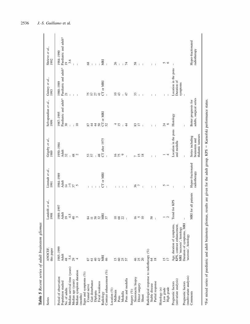
2536 J.-S. Guillamo et al.
Tab
le5
Rec
ent
seri
esof
adul
tbr
ains
tem
glio
mas
Seri
esA
NO
CE
F,L
ando
lfiet
al.,
Lin
stad
tet
al.,
Gri
gsby
etal
.,Se
lvap
andi
anet
al.,
Gui
ney
etal
.,Sh
riev
eet
al.,
this
pape
r19
9819
9119
8919
9919
9319
92
Peri
odof
obse
rvat
ion
1985
–199
919
89–1
997
1984
–198
919
50–1
984
1987
–199
519
80–1
989
1984
–199
0Po
pula
tion
stud
ied
Adu
ltA
dult
Adu
ltA
dult
Paed
iatr
ican
dad
ult*
Paed
iatr
ican
dad
ult*
Paed
iatr
ican
dad
ult*
No.
ofad
ults
4819
1432
3021
19M
edia
nsu
rviv
altim
e(y
ears
)5.
44.
5�
5–
––
3.6
Med
ian
age
(yea
rs)
2940
3748
––
–M
edia
nsy
mpt
omdu
ratio
n4
35
210
––
(mon
ths)
Sign
san
dsy
mpt
oms
(%)
Cra
nial
nerv
e87
84–
5387
7568
Gai
tdi
stur
banc
e61
11–
5744
57–
Dip
lopi
a40
58–
–44
27–
Foca
lw
eakn
ess
4232
–48
5041
–R
adio
logy
MR
IM
RI
CT
orM
RI
CT
afte
r19
75C
Tor
MR
IC
Tor
MR
IM
RI
Con
tras
ten
hanc
emen
t(%
)46
37–
–52
––
Loc
atio
n(%
)M
idbr
ain
1510
–16
410
26Po
ns60
68–
7515
43–
Med
ulla
2522
–9
––
–Po
nsan
dm
edul
la57
––
–44
4774
Surg
ery
(%)
Ster
eota
ctic
biop
sy46
1636
783
3358
Dir
ect
surg
ery
255
715
17–
–Sh
unt
2010
–18
––
–
Rad
iolo
gica
lre
spon
seto
radi
othe
rapy
(%)
Stab
ledi
seas
e64
58–
––
––
Part
ial
resp
onse
18–
––
––
–
His
tolo
gy(n
)L
owgr
ade
151
51
24–
5H
igh
grad
e17
21
46
–6
Prog
nost
icfa
ctor
sA
ge,
dura
tion
ofsy
mpt
oms,
Tre
ndfo
rK
PS–
Loc
atio
nin
the
pons
His
tolo
gyL
ocat
ion
inth
epo
ns–
(uni
vari
ate
anal
ysis
)K
PS,
cont
rast
enha
ncem
ent,
and
med
ulla
Dur
atio
nof
MR
I‘n
ecro
sis’
,hi
stol
ogy
sym
ptom
sPr
ogno
stic
fact
ors
Dur
atio
nof
sym
ptom
s,M
RI
––
––
–(m
ultiv
aria
tean
alys
is)
‘nec
rosi
s’,
hist
olog
yC
omm
ents
MR
Ifo
ral
lpa
tient
sH
yper
-fra
ctio
nate
dSe
ries
incl
udin
gB
ette
rpr
ogno
sis
for
Hyp
er-f
ract
iona
ted
radi
othe
rapy
brai
nste
man
dad
ults
,su
rgic
alse
ries
radi
othe
rapy
thal
amic
tum
ours
*For
mix
edse
ries
ofpa
edia
tric
and
adul
tbr
ains
tem
glio
mas
,re
sults
are
give
nfo
rth
ead
ult
grou
p.K
PS�
Kar
nofs
kipe
rfor
man
cest
atus
.

Adult brainstem glioma 2537
of the tumour was located in the pons in 15 out of 22 patientsand in the medulla in seven out of 22 patients in this study.
When a biopsy is performed, which is far from routinepractice in these diffuse intrinsic forms, a malignant glioma(grades III–IV) is found in many children (Albright et al.,1986, Franzini et al., 1988), whereas we found a benignhistology in 82% (nine out of 11) of the adults. It is likelythat lower grades of the tumours in the adult populationexplain, at least in part, their much better prognosis comparedwith children. Indeed, median survival was 7.3 years in ouradult group, which is similar to the survival of patients withlow-grade supratentorial gliomas (Mason and Macdonald,1997) but strikingly different from the survival of �1 yearin children with intrinsic brainstem gliomas. In addition,intrinsic diffuse glioma seems to be more responsive toradiotherapy in adults than in children since 62% (13 out of21) of adults improved for a long period after radiotherapy,although clinical improvement was often not correlated witha radiological response (19%). In this group, the optimal dateof radiotherapy remains unknown since several of our patientsdid well for many years without treatment.
We suggest that this subgroup of tumours be designatedin adults as ‘diffuse intrinsic low-grade brainstem gliomas’.Nevertheless, it is important to underline the possibility ofexceptions since the tumours of two of our youngest patients(aged 17 and 18 years), who had rapidly progressive cranialnerve deficits and a typical aspect of non-enhancing diffuseintrinsic glioma, behaved like the childhood subtype, and thepatients’ survival time was very short (14 and 16 months)despite vigorous treatment with radiotherapy andchemotherapy. These exceptions in adults seem to indicateoverlaps among tumour subtypes between the two age groups.
Malignant brainstem gliomasThe other common tumour type identified in this adult seriesis clearly different from those discussed above. It occurslater than the diffuse, intrinsic, low-grade type and affectsmainly older adults (most of them in their sixth decade). Theclinical picture is characterized by the rapid onset of cranialnerve palsies and long tract signs leading to an early alterationin performance status. MRI reveals a brainstem mass thatenhances after gadolinium infusion, often in a ring-likefashion. In our series, contrast enhancement was a pejorativefactor (particularly when the area of enhancement surroundeda low-signal area suggestive of necrosis) in contrast withchildren, in whom the prognostic value of contrastenhancement remains controversial (Albright et al., 1986;Fischbein et al., 1996). Pathologically, these tumourscorrespond to high-grade gliomas (grades III–IV) and mediansurvival time is short (11.2 months) despite treatment withradiotherapy and chemotherapy. Thus, the clinical–radiological pattern, pathology and course closely resemblethe common malignant supratentorial gliomas in adults andwe suggest that this group be designated ‘malignant brainstemgliomas’.
Focal tectal gliomasFocal tectal gliomas represent the third type of adult brainstemglioma and constitute a small subgroup (8%) that also existsin children (Squires et al., 1994; Bowers et al., 2000). Theclinical picture is dominated by hydrocephalus. A diagnosisof mixed glioma was made in two of our patients after apathological examination of the tumour. All our patientsreceived radiotherapy and experienced long-term survival ofgood quality. Nevertheless, the benefit of radiotherapy canbe questioned since paediatric patients with similar clinicaland radiological features have been managed with ventricularshunt or observation alone for long periods (Squires et al.,1994).
Other typesOther types of brainstem glioma can be observed in adults.Interestingly, we observed only one exophytic contrast-enhancing glioma arising from the floor of the fourth ventricle;this entity, which is associated with a good prognosis, is welldescribed in children (representing up to 10% of brainstemgliomas) (Hoffman et al., 1980; Pollack et al., 1993). Alikely explanation for this discrepancy between the two age-groups is that most of the exophytic gliomas correspond topilocytic astrocytoma, a very rare type of tumour in adults.
Three of our patients had NF1. The brainstem is the secondmost frequent location of brain tumours after the opticpathways in patients with NF1 (Molloy et al., 1995; Pollacket al., 1996). In contrast with children, in whom the courseis usually very long, the tumour behaviour that we observedin adults with NF1 was much more aggressive, but largerseries will be necessary to draw any conclusion on this point.
ComplicationsExcept for locoregional progression, two main complicationswere observed during the course of adult brainstem gliomas,namely hydrocephalus and leptomeningeal dissemination.Hydrocephalus was observed in 20% of cases. Whereas somepontine tumours may have an important mass effect on thefourth ventricle, hydrocephalus was always associated withmesencephalic involvement and blockage of the CSF at thelevel of the sylvian aqueduct. Leptomeningeal disseminationoccurred in 13% of cases and was the cause of a quarter ofthe deaths in our series. This complication has also beenreported with a high frequency in children (Packer et al.,1983; Donahue et al., 1998). Close proximity of the tumourand CSF pathways could explain such an increased trendfor leptomeningeal dissemination, but this remains to bedemonstrated.
The role of biopsyFinally, this classification may help in the selection of patientsfor biopsy. In children, MRI has become the reference for
2538 J.-S. Guillamo et al.
the diagnosis of brainstem glioma and is used for the currentclassification of these tumours (Barkovich et al., 1990;Epstein and Farmer, 1993; Fischbein et al., 1996). MRIhas replaced biopsy in the diagnosis of paediatric diffusebrainstem gliomas, for which most authors agree thatanticancer treatments can be administered withoutpathological confirmation if the clinical course is rapid, as isusual (Albright et al., 1993; Bouffet et al., 2000). In ourstudy, 71% of patients underwent a surgical biopsy, which isnowadays considered a relatively safe (transitory worseningoccurs in 12% of cases) and informative procedure (6% ofspecimens were non-contributory) (Franzini et al., 1988).However, we believe that biopsy is not useful in the diagnosisof intrinsic, diffuse, low-grade brainstem gliomas in adultswhen the clinical and radiological criteria described aboveare met. The issue is different in contrast-enhancing lesionsbecause several reports have underlined the limits of MRI indifferentiating tumours from infectious (e.g. tuberculomas)(Del Brutto and Mosquera, 1999) and inflammatory(sarcoidosis, Behcet’s disease) (Gizzi et al., 1993; Akman-Demir G et al., 1999) diseases. In this setting, a surgicalapproach is probably indicated in most cases.
AcknowledgementWe wish to thank Francois Doz for helpful criticism.
ReferencesAkman-Demir G, Serdaroglu P, Tasci B. Clinical patterns ofneurological involvement in Behcet’s disease: evaluation of 200patients. The Neuro-Behcet Study Group. Brain 1999; 122: 2171–82.
Albright AL, Guthkelch AN, Packer RJ, Price RA, Rourke LB.Prognostic factors in pediatric brain-stem gliomas. J Neurosurg1986; 65: 751–5.
Albright AL, Packer RJ, Zimmerman R, Rorke LB, Boyett J,Hammond GD. Magnetic resonance scans should replace biopsiesfor the diagnosis of diffuse brain stem gliomas: a report from theChildren’s Cancer Group. Neurosurgery 1993; 33: 1026–30.
Barkovich AJ, Krischer J, Kun LE, Packer R, Zimmerman RA,Freeman CR, et al. Brain stem gliomas: a classification systembased on magnetic resonance imaging. Pediatr Neurosurg 1990; 16:73–83.
Bauman G, Pahapill P, Macdonald D, Fisher B, Leighton C,Caincross G. Low grade glioma: a measuring radiographic responseto radiotherapy. Can J Neurol Sci 1999; 26: 18–22.
Bouffet E, Raquin M, Doz F, Gentet JC, Rodary C, Demeocq F,et al. Radiotherapy followed by high dose busulfan and thiotepa: aprospective assessment of high dose chemotherapy in children withdiffuse pontine gliomas. Cancer 2000; 88: 685–92.
Bowers DC, Georgiades C, Aronson LJ, Carson BS, Weingart JD,Wharam MD, et al. Tectal gliomas: natural history of an indolentlesion in pediatric patients. Pediatr Neurosurg 2000; 32: 24–9.
Bricolo A, Turazzi S, Cristofori L, Talacchi A. Direct surgery forbrainstem tumours. Acta Neurochir Suppl (Wien) 1991; 53: 148–58.
Constantini S, Epstein F. Surgical indication and technicalconsiderations in the management of benign brain stem gliomas.[Review]. J Neurooncol 1996; 28: 193–205.
Del Brutto OH, Mosquera A. Brainstem tuberculoma mimickingglioma: the role of antituberculous drugs as a diagnostic tool.Neurology 1999; 52: 210–11.
Dirr LY, Donofrio PD, Patton JF, Troost BT. A false-positiveedrophonium test in a patient with a brainstem glioma. Neurology1989; 39: 865–7.
Donahue B, Allen J, Siffert J, Rosovsky M, Pinto R. Patternsof recurrence in brain stem gliomas: evidence for craniospinaldissemination. [Review]. Int J Radiat Oncol Biol Phys 1998; 40:677–80.
Epstein FJ, Farmer JP. Brain-stem glioma growth patterns. JNeurosurg 1993; 78: 408–12.
Farwell JR, Dohrmann GJ, Flannery JT. Central nervous systemtumors in children. Cancer 1977; 40: 3123–32.
Fischbein NJ, Prados MD, Wara W, Russo C, Edwards MS,Barkovich AJ. Radiologic classification of brain stem tumors:correlation of magnetic resonance imaging appearance with clinicaloutcome. Pediatr Neurosurg 1996; 24: 9–23.
Franzini A, Allegranza A, Melcarne A, Giorgi C, Ferraresi S,Broggi G. Serial stereotactic biopsy of brain stem expanding lesions.Considerations on 45 consecutive cases. Acta Neurochir Suppl(Wien) 1988; 42: 170–6.
Freeman CR, Farmer JP. Pediatric brain stem gliomas: a review.[Review]. Int J Radiat Oncol Biol Phys 1998; 40: 265–71.
Gizzi MS, Lidov M, Rosenbaum D. Neurosarcoidosis presenting asa tumour of the basal ganglia and brainstem: sequential MRI. NeurolRes 1993; 15: 93–6.
Grigsby PW, Garcia DM, Simpson JR, Fineberg BB, Schwartz HG.Prognostic factors and results of therapy for adult thalamic andbrainstem tumors. Cancer 1989; 63: 2124–9.
Guiney MJ, Smith JG, Hughes P, Yang C, Narayan K. Contemporarymanagement of adult and pediatric brain stem gliomas. Int J RadiatOncol Biol Phys 1993; 25: 235–41.
Gutmann L, Hopf HC. Facial myokymia and contraction persisting20 years: a case of pontine glioma. Muscle Nerve 1994; 17: 1461–3.
Hoffman HJ, Becker L, Craven MA. A clinically and pathologicallydistinct group of benign brain stem gliomas. Neurosurgery 1980;7: 243–8.
Kaplan AM, Albright AL, Zimmerman RA, Rorke LB, Li H, BoyettJM, et al. Brainstem gliomas in children. A Children’s CancerGroup review of 119 cases. Pediatr Neurosurg 1996; 24: 185–92.
Landolfi JC, Thaler HT, DeAngelis LM. Adult brainstem gliomas.Neurology 1998; 51: 1136–9.
Linstadt DE, Edwards MS, Prados M, Larson DA, Wara WM.Hyperfractionated irradiation for adults with brainstem gliomas. IntJ Radiat Oncol Biol Phys 1991; 20: 757–60.
Macdonald DR, Cascino TL, Schold SC Jr, Cairncross JG. Responsecriteria for phase II studies of supratentorial malignant glioma.J Clin Oncol 1990; 8: 1277–80.

Adult brainstem glioma 2539
Mandell LR, Kadota R, Freeman C, Douglass EC, Fontanesi J,Cohen ME, et al. There is no role for hyperfractionated radiotherapyin the management of children with newly diagnosed diffuse intrinsicbrainstem tumors: results of a Pediatric Oncology Group phase IIItrial comparing conventional vs. hyperfractionated radiotherapy. IntJ Radiat Oncol Biol Phys 1999; 43: 959–64.
Maria BL, Rehder K, Eskin TA, Hamed LM, Fennell EB, QuislingRG, et al. Brainstem glioma: I. Pathology, clinical features, andtherapy. [Review]. J Child Neurol 1993; 8: 112–28.
Mason WP, Macdonald DR. Low-grade gliomas. In: Vinken PJ,Bruyn GW, editors. Handbook of clinical neurology, Vol. 68.Amsterdam: Elsevier; 1997. p. 33–62.
Molloy PT, Bilaniuk LT, Vaughan SN, Needle MN, Liu GT, ZackaiEH, et al. Brainstem tumors in patients with neurofibromatosistype 1: a distinct clinical entity. Neurology 1995; 45: 1897–902.
Packer RJ, Allen J, Nielsen S, Petito C, Deck M, Jereb B. Brainstemglioma: clinical manifestations of meningeal gliomatosis. AnnNeurol 1983; 14: 177–82.
Pollack IF, Hoffman HJ, Humphreys RP, Becker L. The long-termoutcome after surgical treatment of dorsally exophytic brain-stemgliomas. J Neurosurg 1993; 78: 859–63.
Pollack IF, Shultz B, Mulvihill JJ. The management of brainstemgliomas in patients with neurofibromatosis 1. [Review]. Neurology1996; 46: 1652–60.
Ragge NK, Hoyt WF. Midbrain myasthenia: fatigable ptosis, ‘lidtwitch’ sign, and ophthalmoparesis from a dorsal midbrain glioma.Neurology 1992; 42: 917–19.
Selvapandian S, Rajshekhar V, Chandy MJ. Brainstem glioma:comparative study of clinico-radiological presentation, pathologyand outcome in children and adults. Acta Neurochir (Wien) 1999;141: 721–7.
Shrieve DC, Wara WM, Edwards MS, Sneed PK, Prados MD,Cogen PH, et al. Hyperfractionated radiation therapy for gliomasof the brainstem in children and in adults. Int J Radiat Oncol BiolPhys 1992; 24: 599–610.
Squires LA, Allen JC, Abbott R, Epstein FJ. Focal tectal tumors:management and prognosis. Neurology 1994; 44: 953–6.
Tokuriki Y, Handa H, Yamashita J, Okumura T, Paine JT. Brainstemglioma: an analysis of 85 cases. Acta Neurochir (Wien) 1986; 79:67–73.
Walker DA, Punt JA, Sokal M. Clinical management of brain stemglioma. [Review]. Arch Dis Child 1999; 80: 558–64.
Westra I, Drummond GT. Occult pontine glioma in a patient withhemifacial spasm. Can J Ophthalmol 1991; 26: 148–51.
White HH. Brain stem tumors occurring in adults. Neurology 1963;13: 292–300.
Received April 30, 2001. Revised July 5, 2001.Accepted July 16, 2001


![Untitled-6 [] · tis 1227-2539 (1996) tis 1390-2539 (1996) tis 1227-2539 (1996) tis 1390-2539 (1996) tis 1227-2539 (1996)](https://static.fdocuments.us/doc/165x107/5e1a6a0f6b8d9f48bd19bcad/untitled-6-tis-1227-2539-1996-tis-1390-2539-1996-tis-1227-2539-1996-tis.jpg)

