Bradyarrhythmia Lecture PHRS 2015
77
Pusong Low Batt! Bradyarrhythmias
-
Upload
michael-joseph-agbayani -
Category
Health & Medicine
-
view
32 -
download
0
Transcript of Bradyarrhythmia Lecture PHRS 2015

Pusong Low Batt!Bradyarrhythmias


Image of heart: Public Domain. Image of Conduction system from Wikimedia user Madhero88: https://commons.wikimedia.org/wiki/File:Electrical_conduction_system_of_the_heart.svg

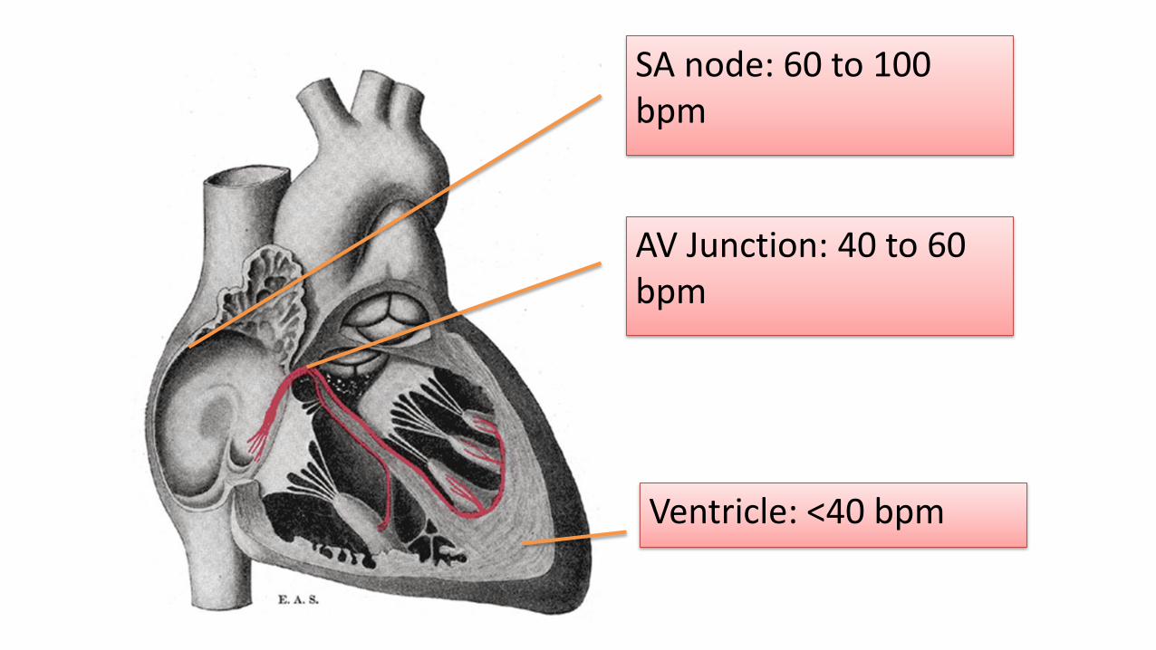
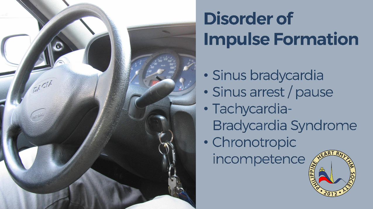
SA node: 60 to 100 bpm
AV Junction: 40 to 60 bpm
Ventricle: <40 bpm

Image from Ed Burns, Life in the Fast Lane: http://lifeinthefastlane.com/ecg-library/sinus-bradycardia/

•
•
•
Image from ECGpedia,.org: http://en.ecgpedia.org/wiki/File:Ecgfreq.png
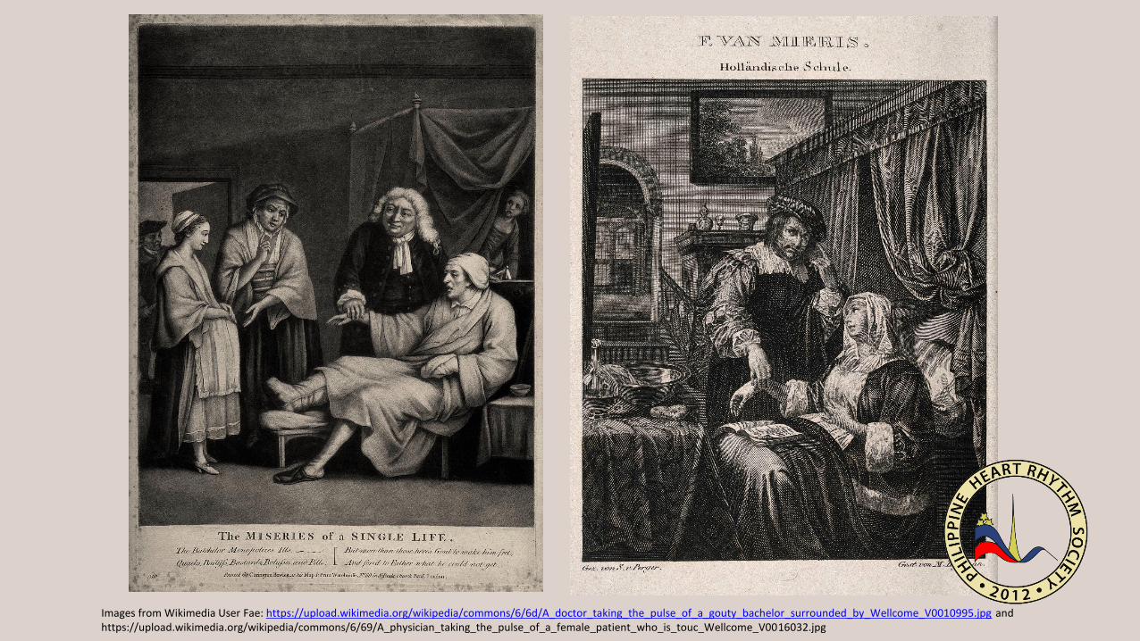
Images from Wikimedia User Fae: https://upload.wikimedia.org/wikipedia/commons/6/6d/A_doctor_taking_the_pulse_of_a_gouty_bachelor_surrounded_by_Wellcome_V0010995.jpg and https://upload.wikimedia.org/wikipedia/commons/6/69/A_physician_taking_the_pulse_of_a_female_patient_who_is_touc_Wellcome_V0016032.jpg
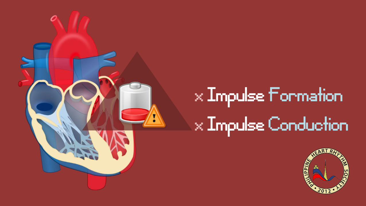
x Impulse Formationx Impulse Conduction
•
•
•
•
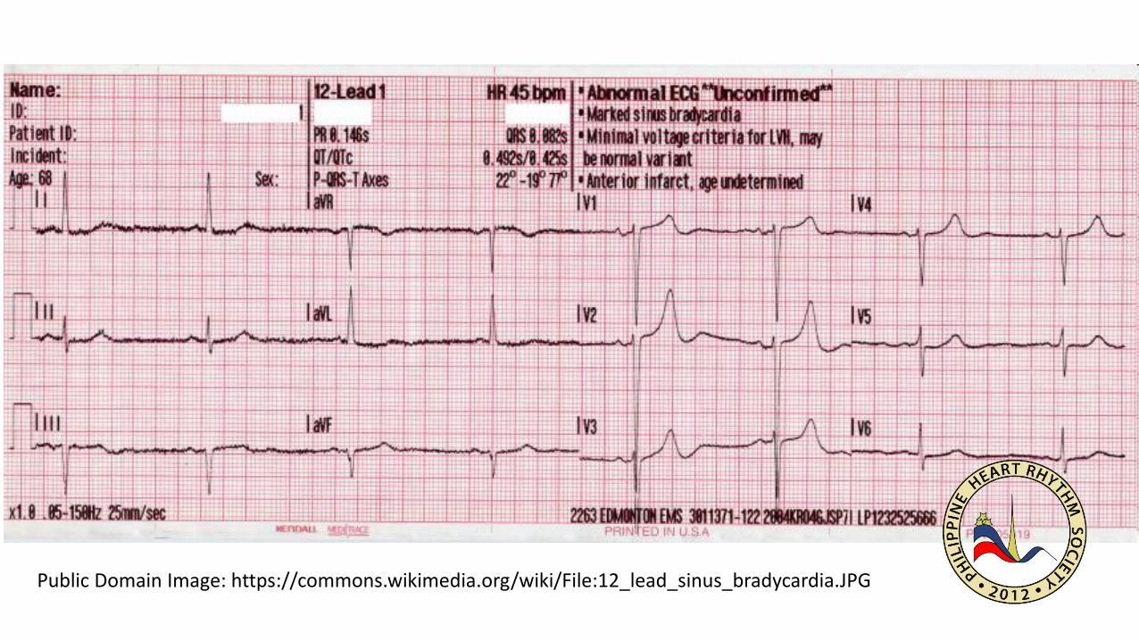
Public Domain Image: https://commons.wikimedia.org/wiki/File:12_lead_sinus_bradycardia.JPG
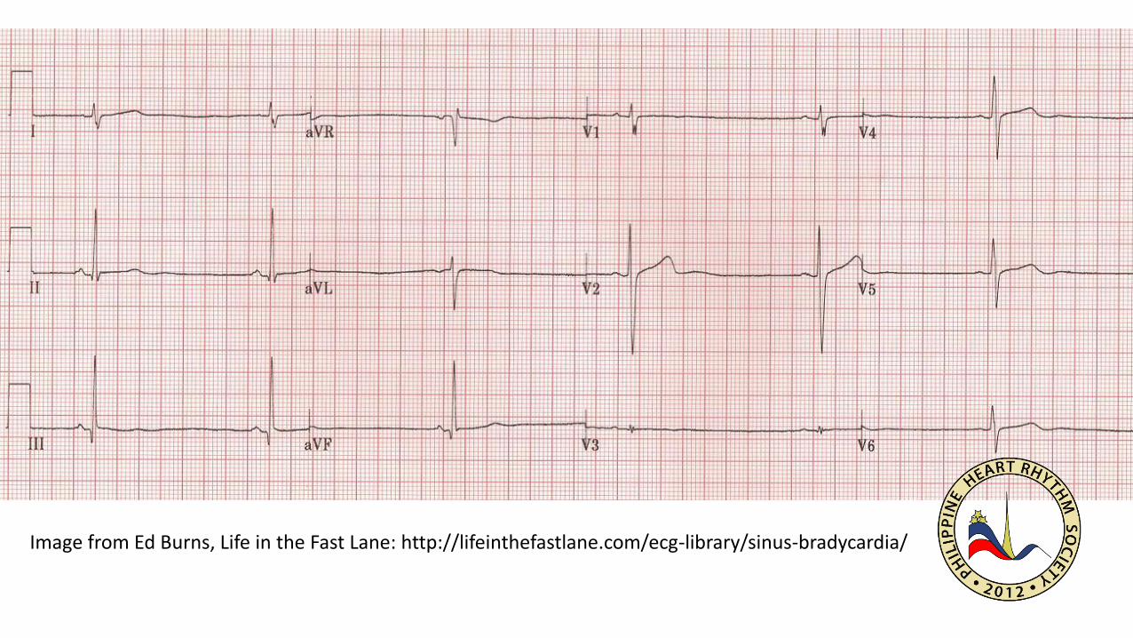
Image from Ed Burns, Life in the Fast Lane: http://lifeinthefastlane.com/ecg-library/sinus-bradycardia/

Image from Ed Burn, Life in the Fast Lane: http://lifeinthefastlane.com/ecg-library/sick-sinus/

Image from Ed Burn, Life in the Fast Lane: http://lifeinthefastlane.com/ecg-library/sick-sinus/

Public Domain Image from https://commons.wikimedia.org/wiki/File:Treadmills_at_gym.jpg
x Impulse Conduction
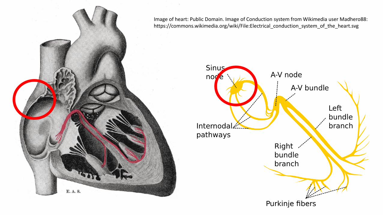
Image of heart: Public Domain. Image of Conduction system from Wikimedia user Madhero88: https://commons.wikimedia.org/wiki/File:Electrical_conduction_system_of_the_heart.svg
Sin
oat
rial
exi
t b
lock
: htt
p:/
/co
mm
on
s.w
ikim
edia
.org
/wik
i/U
ser:
Jer5
15
0
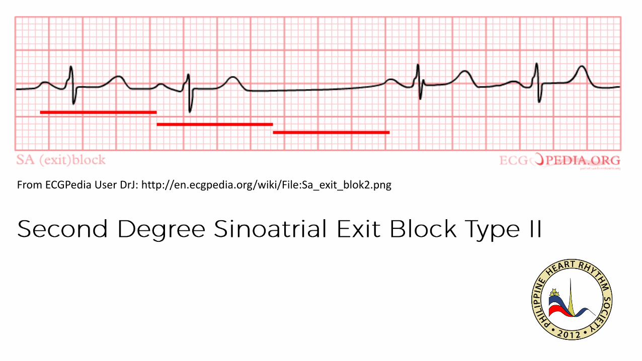
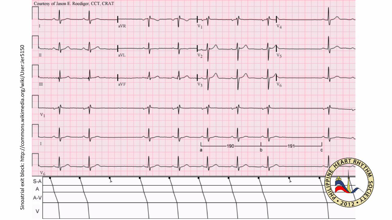
From ECGPedia User DrJ: http://en.ecgpedia.org/wiki/File:Sa_exit_blok2.png
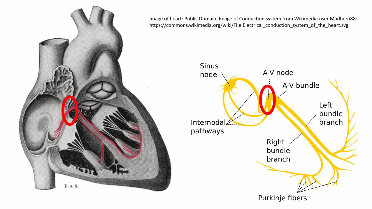
Image of heart: Public Domain. Image of Conduction system from Wikimedia user Madhero88: https://commons.wikimedia.org/wiki/File:Electrical_conduction_system_of_the_heart.svg
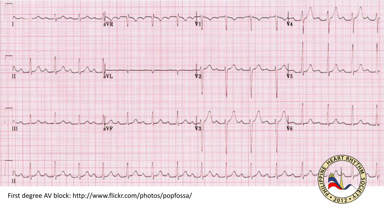
First degree AV block: http://www.flickr.com/photos/popfossa/

•
•
•

•
Image from Ed Burns: http://cdn.lifeinthefastlane.com/wp-content/uploads/2011/04/ECG_wenckebach.jpg

Second Degree AV block Mobitz Type 1: http://commons.wikimedia.org/wiki/User:Jer5150

•
•
Image from Ed Burns: http://cdn.lifeinthefastlane.com/wp-content/uploads/2011/04/Mobitz_II.gif
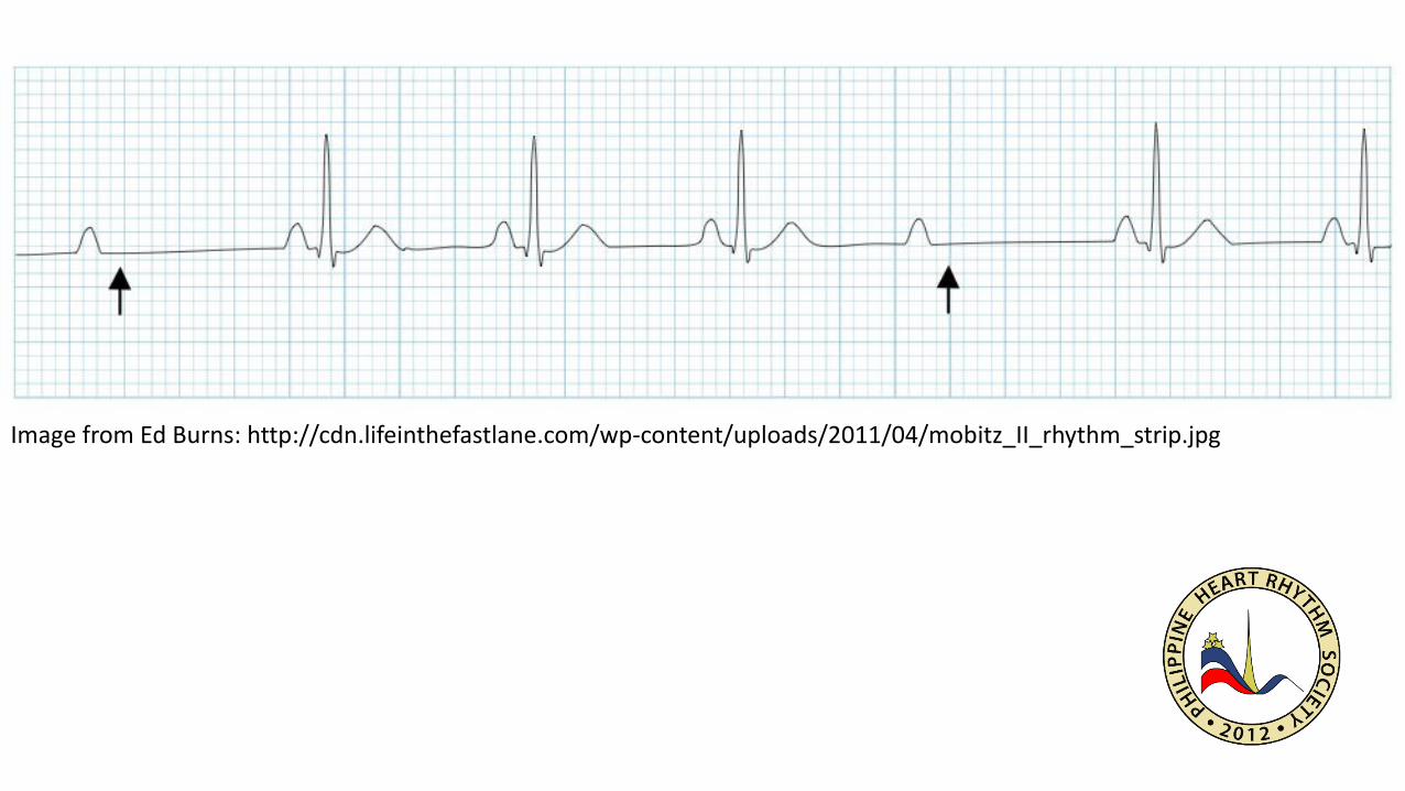
Image from Ed Burns: http://cdn.lifeinthefastlane.com/wp-content/uploads/2011/04/mobitz_II_rhythm_strip.jpg

Mobitz type 2 and 2:1: http://commons.wikimedia.org/wiki/User:Jer5150
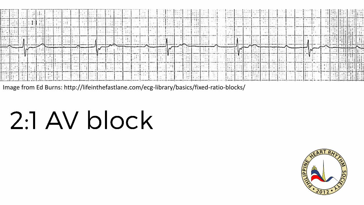
Image from Ed Burns: http://lifeinthefastlane.com/ecg-library/basics/fixed-ratio-blocks/
•
•
•

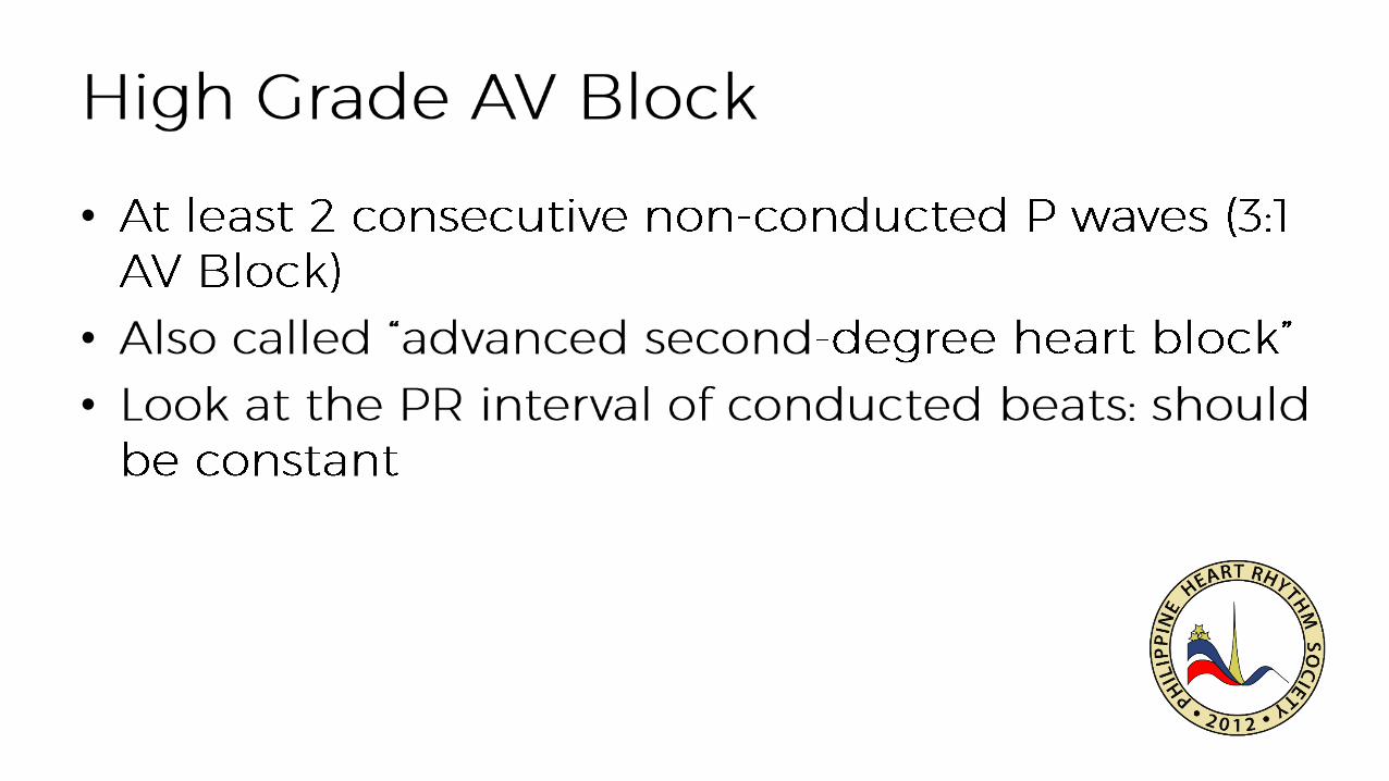
Image from Ed Burns: http://lifeinthefastlane.com/ecg-library/basics/high-grade-block/
•
•
•

Image of heart: Public Domain. Image of Conduction system from Wikimedia user Madhero88: https://commons.wikimedia.org/wiki/File:Electrical_conduction_system_of_the_heart.svg
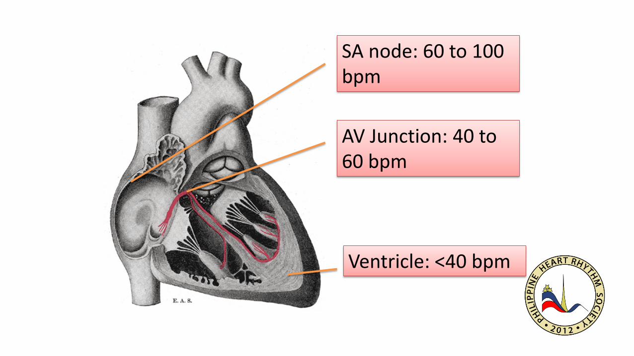
SA node: 60 to 100 bpm
AV Junction: 40 to 60 bpm
Ventricle: <40 bpm

Image from Ed Burns: http://lifeinthefastlane.com/ecg-library/basics/complete-heart-block/
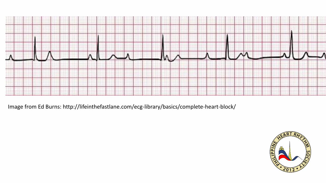
Image from Ed Burns: http://lifeinthefastlane.com/ecg-library/basics/complete-heart-block/

SA node: 60 to 100 bpm
AV Junction: 40 to 60 bpm
Ventricle: <40 bpm

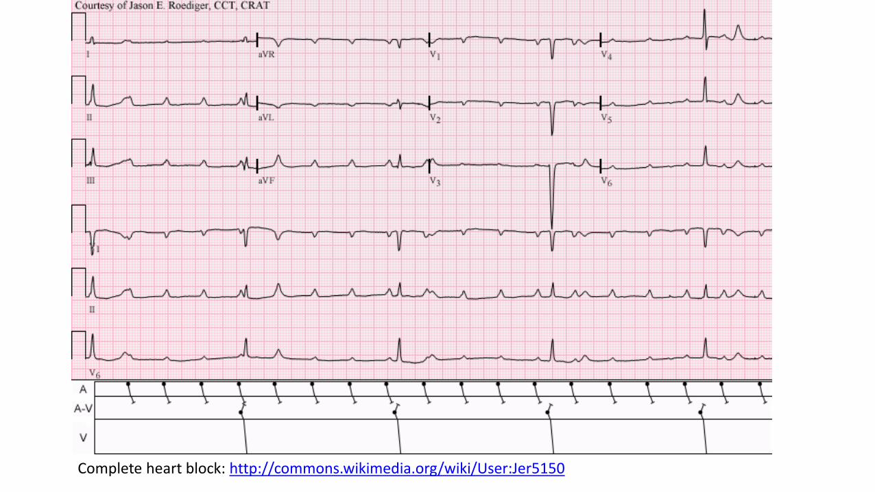
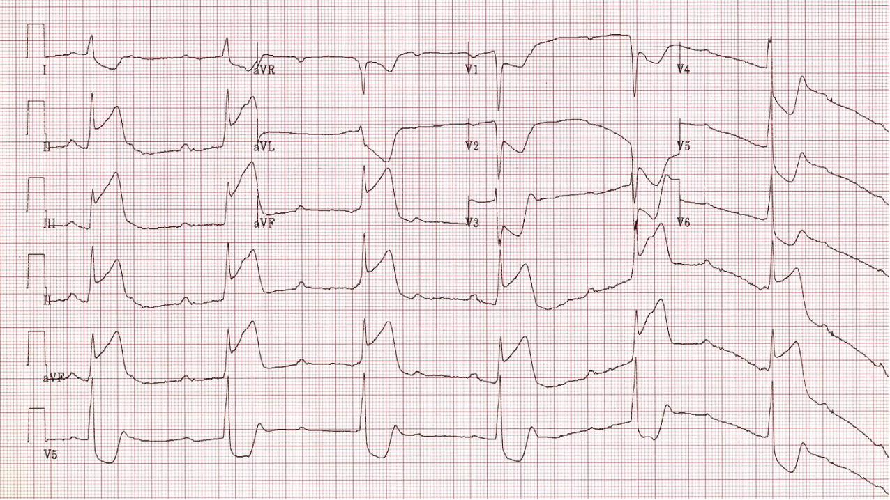
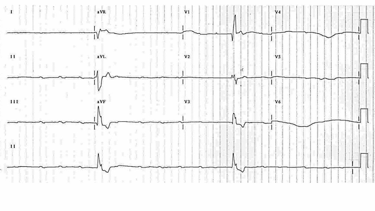
Complete heart block: http://commons.wikimedia.org/wiki/User:Jer5150

•
•
•
•
Complete heart block strip from Wikimedia user MoodyGroove: http://en.wikipedia.org/wiki/User:MoodyGroove

•
–
–
•
–

Image from Ed Burns: http://lifeinthefastlane.com/ecg-library/basics/complete-heart-block/
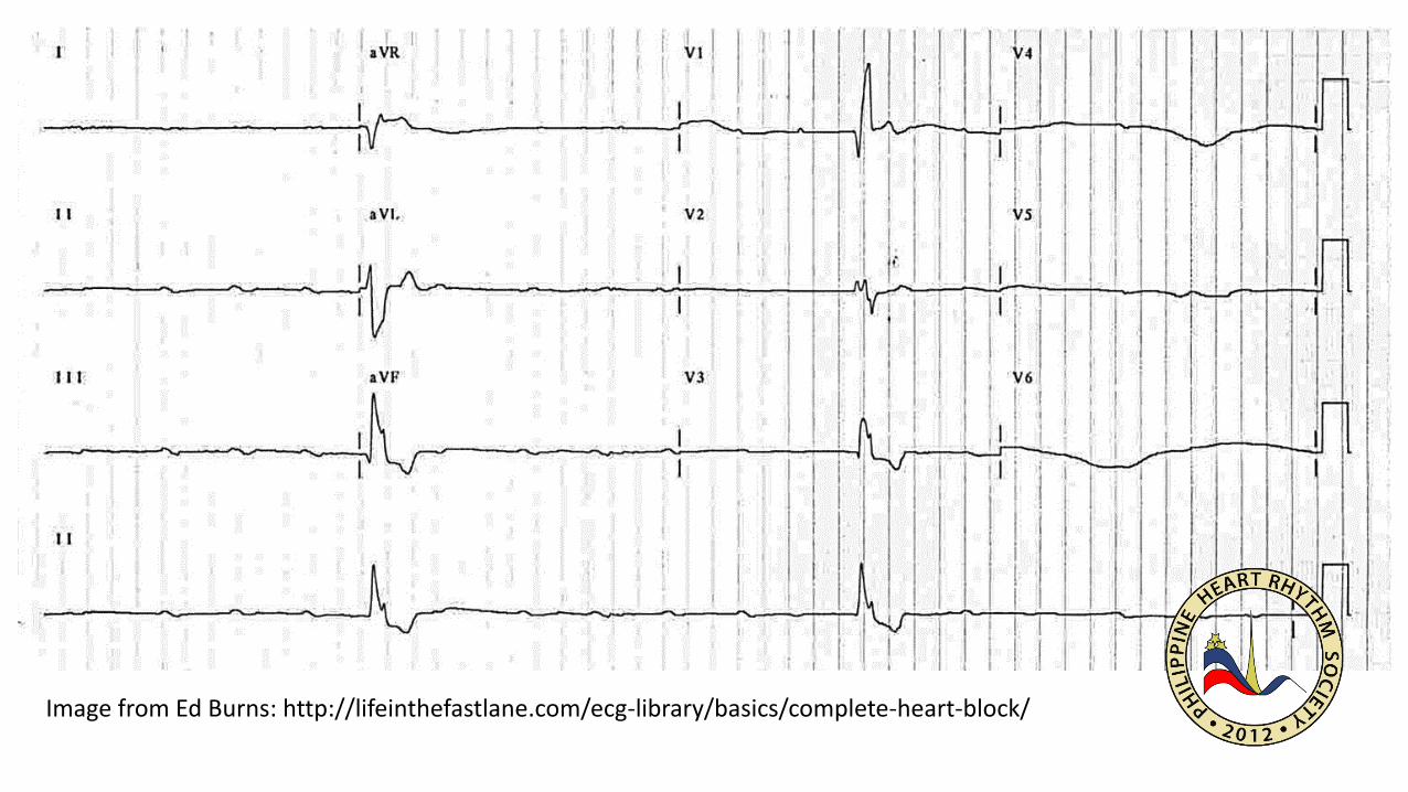
Image from Ed Burns: http://lifeinthefastlane.com/ecg-library/basics/complete-heart-block/

Images in Cardiovascular Medicine. From Bad to Worse. CirculationApril 18, 2006 vol. 113 no. 15 e707-e708

Image from Flickr User Helge V. Keltel: https://www.flickr.com/photos/17088227@N00/7147926407
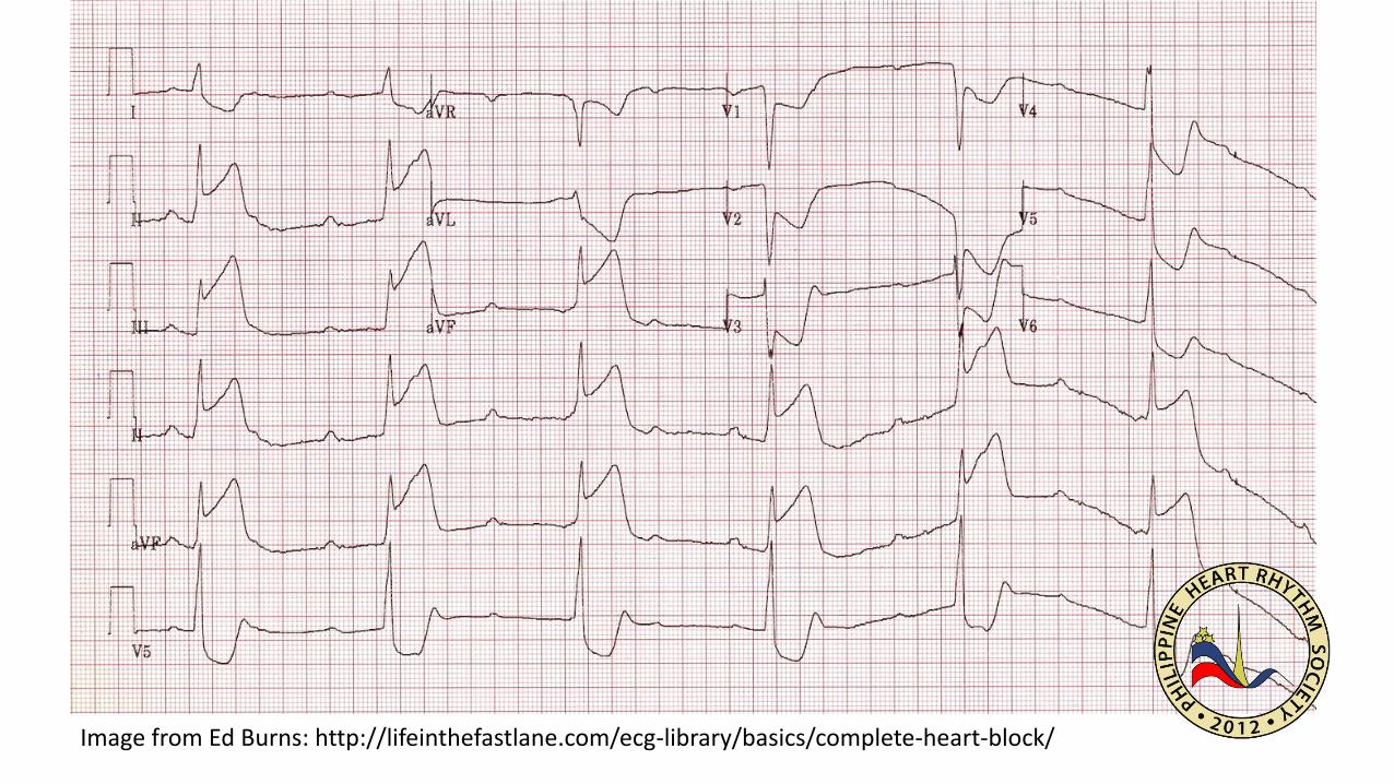
Image from Ed Burns: http://lifeinthefastlane.com/ecg-library/basics/complete-heart-block/

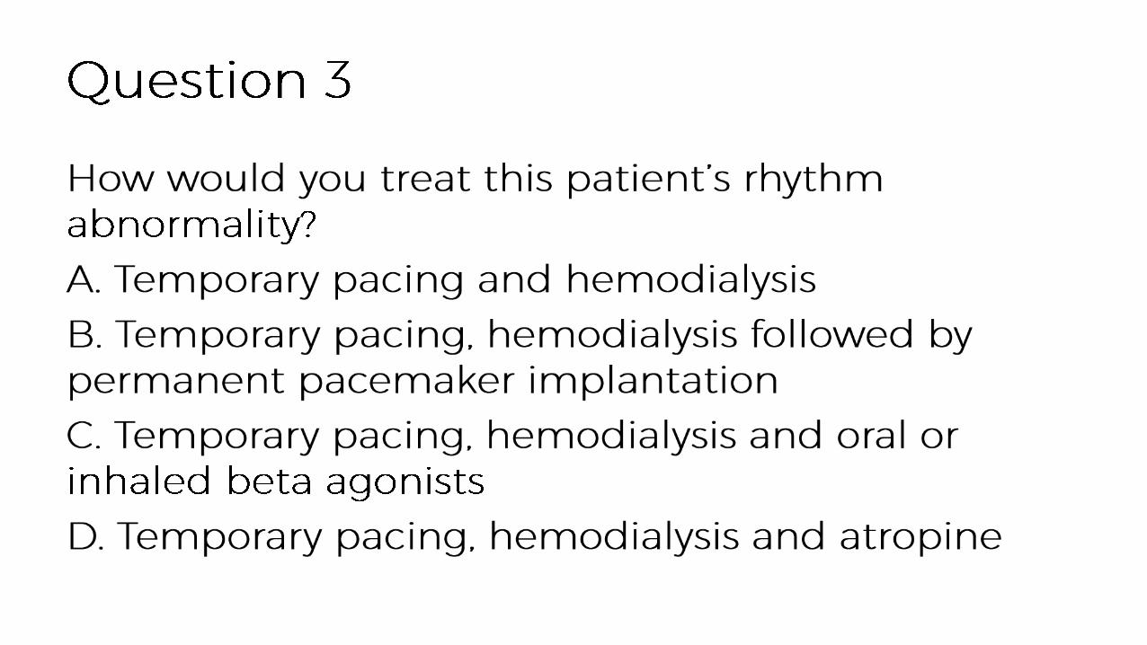
From https://en.wikipedia.org/wiki/Dialysis#/media/File:Patient_receiving_dialysis_03.jpg

Pills Image from https://www.flickr.com/photos/emagineart

Diagnosis and Management of Cardiac Amyloidoses. Circulation.2005; 112: 2047-2060
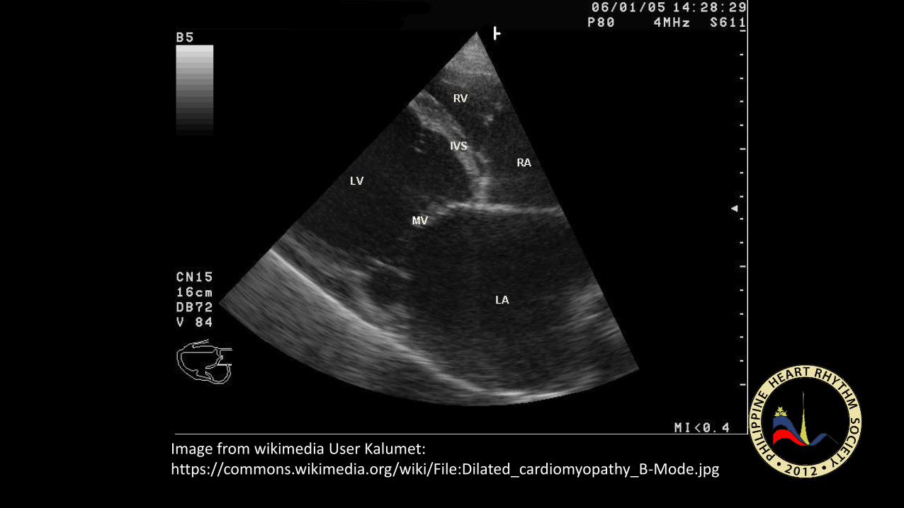
Image from wikimedia User Kalumet: https://commons.wikimedia.org/wiki/File:Dilated_cardiomyopathy_B-Mode.jpg

Holter image from Wikimedia user Misscurry: https://en.wikipedia.org/wiki/Holter_monitor#/media/File:Alex_CM4000.jpg

Brignole et al. J Am Coll Cardiol 2011;58:167–73

Image from Wikimedia user Intropin: https://commons.wikimedia.org/wiki/File:Atropine_(1).JPG

From Wikimedia User PhilippN. https://commons.wikimedia.org/wiki/File:Defibrillation_Electrode_Position.jpg

Image from From Wikimedia user: https://commons.wikimedia.org/wiki/User:Stevenfruitsmaak
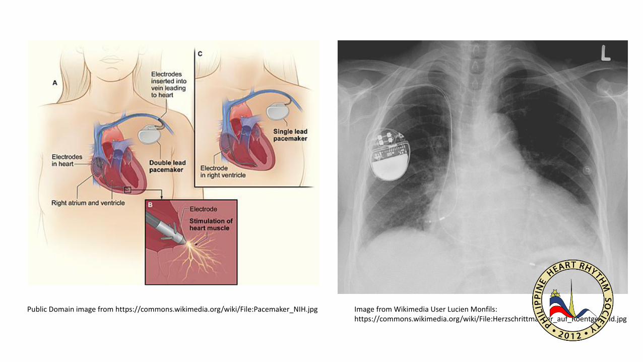
Public Domain image from https://commons.wikimedia.org/wiki/File:Pacemaker_NIH.jpg Image from Wikimedia User Lucien Monfils: https://commons.wikimedia.org/wiki/File:Herzschrittmacher_auf_Roentgenbild.jpg
•
•
•
•

•
•
–
–
–
–

•
•
•
•

•
•
•
•
•
•
•
•

Pusong Low Batt!Bradyarrhythmias

•
•
•
•


•
•
•
•
•
•
•
•
•


You are free to:Share — copy and redistribute the material in any medium or formatAdapt — remix, transform, and build upon the materialThe licensor cannot revoke these freedoms as long as you follow the license terms.
This slideshow is under a Attribution-NonCommercial-ShareAlike 4.0 InternationalCreative Commons license.
Under the following terms:Attribution — You must give appropriate credit, provide a link to the license, andindicate if changes were made. You may do so in any reasonable manner, but not in any way that suggests the licensor endorses you or your use.NonCommercial — You may not use the material for commercial purposes.ShareAlike — If you remix, transform, or build upon the material, you must distribute your contributions under the same license as the original.No additional restrictions — You may not apply legal terms or technological measures that legally restrict others from doing anything the license permits.