Brachial Plexus Birth Palsy - wesley ob/gyn · PDF fileBrachial Plexus Birth Palsy Joshua D....
34
Brachial Plexus Birth Palsy Joshua D. Linnell Via Christi Health Department of Orthopedics Hand and Microsurgery
Transcript of Brachial Plexus Birth Palsy - wesley ob/gyn · PDF fileBrachial Plexus Birth Palsy Joshua D....
Brachial Plexus Birth Palsy
Joshua D. LinnellVia Christi Health
Department of OrthopedicsHand and Microsurgery
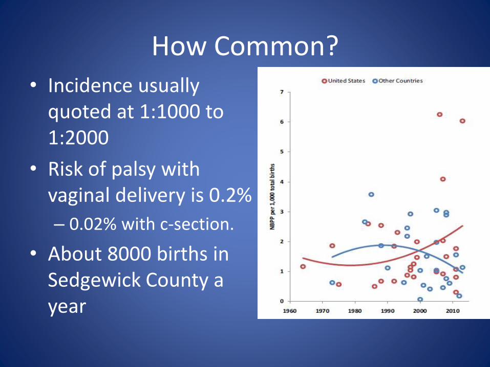
How Common?• Incidence usually
quoted at 1:1000 to 1:2000
• Risk of palsy with vaginal delivery is 0.2%
– 0.02% with c-section.
• About 8000 births in Sedgewick County a year
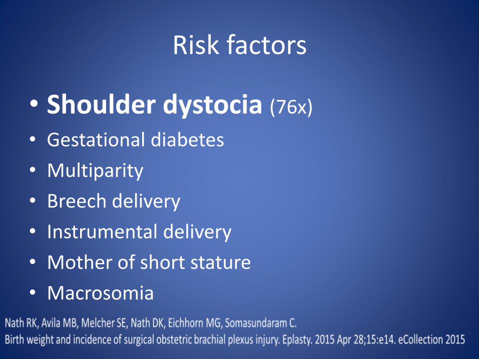
Risk factors
• Shoulder dystocia (76x)
• Gestational diabetes
• Multiparity
• Breech delivery
• Instrumental delivery
• Mother of short stature
• Macrosomia
Mechanism
• Has always been somewhat of a controversy
• Blamed it on instrumentation in the past
– Rate of instrumentation use went way down
• Blamed it on poor technique of delivery
– ACOG pressed for “gentleness” techniques in delivery
The Obstetricians Fault?
• Once thought that excessive lateral traction of the head with shoulder dystocia.
– No level 1 evidence to support
– The sacral arm is involved in only 1/3 of cases


Classification of Injury
• “Erb’s Palsy”
– upper root palsy of the C5-c6 (extended c7).
• Total palsy
– C5-T1
• “Klumpke’s Palsy” (C8 and T1) along with intermediate palsy (C7 alone)

Brachial Plexus Anatomy

Why Not a C-section
• The rate of c section has doubled in the past 25 years.
– Rates of BPPB have gone up in the US
Why Not C-Section
• Macrosomia:
– Ultrasound sensitivity 60%/Specificity 90%
– Reduce palsy by 31%
– Increase C section rate by 50%
– $5,000,000 per palsy prevented
– 1 maternal death per 3.2 cases palsy prevention
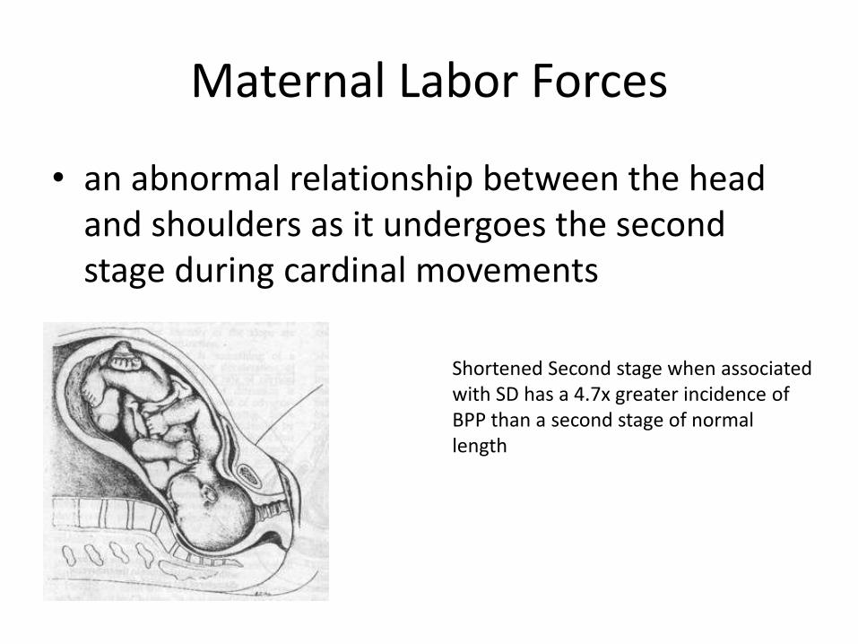
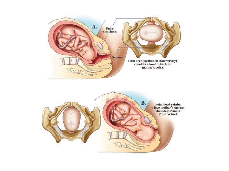
Maternal Labor Forces
• an abnormal relationship between the head and shoulders as it undergoes the second stage during cardinal movements
Shortened Second stage when associated with SD has a 4.7x greater incidence of BPP than a second stage of normal length

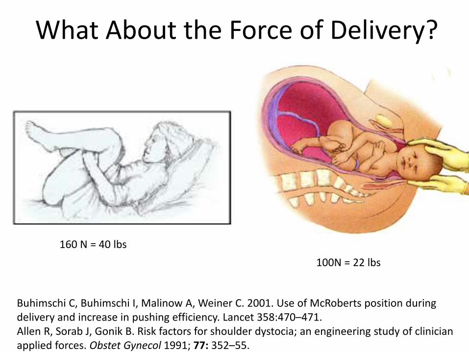
What About the Force of Delivery?
160 N = 40 lbs
100N = 22 lbs
Buhimschi C, Buhimschi I, Malinow A, Weiner C. 2001. Use of McRoberts position during delivery and increase in pushing efficiency. Lancet 358:470–471.Allen R, Sorab J, Gonik B. Risk factors for shoulder dystocia; an engineering study of clinician applied forces. Obstet Gynecol 1991; 77: 352–55.
Clavicle Fractures
• It has been labeled as “a natural risk of labor” (ACOG) and associated with the maternal force of labor.
• Presence of a clavicle fracture portends a greater risk of BPI.
• Clavicle fracture is not a prognostic indicator– Studies show that it is more common in affected
neonates. 71-96 per 1000 affected births.
– No difference in recovery
– No difference in secondary procedures

Incidence and prognosis of neonatal brachial plexus palsy with and without
clavicle fractures.Wall LB, Mills JK, Leveno K, Jackson G, Wheeler LC, Oishi SN, Ezaki M. Obstet Gynecol. 2014 Jun;123(6):1288-93.
• 288 BPPB from Parkland Hospital
– 74 also had ipsilateral clavicular fracture
• 72% resolved spontaneously (154/214);
– concomitant clavicular fracture, 74% resolved spontaneously (55/74).

Other Culprits in Disguise
• Pseudoparalysis: Fracture of the humerus or clavicle.
• Congenital aplasia of nerve roots: described by Gilbert
• Congenital varicella
• Umbilical cord palsy: Most commonly of the radial nerve, elbow extension and wrist drop. Caused by intrauterine compression of the upper arm.
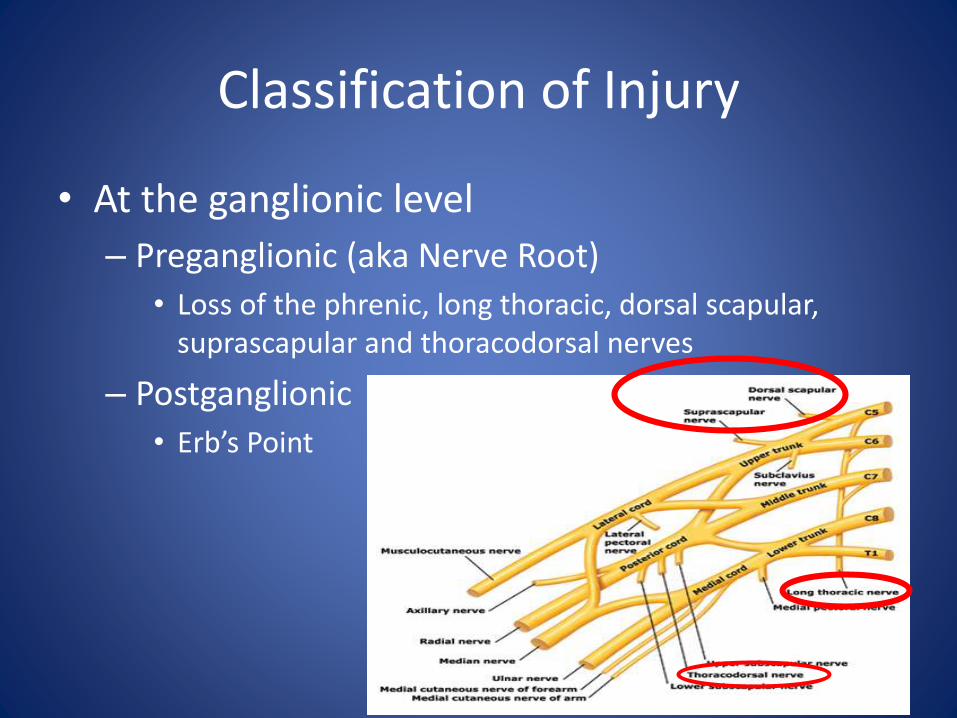
Classification of Injury
• At the ganglionic level
– Preganglionic (aka Nerve Root)
• Loss of the phrenic, long thoracic, dorsal scapular, suprascapular and thoracodorsal nerves
– Postganglionic
• Erb’s Point


Peripheral Branches
• Suprascapular Nerve (C5)


Peripheral Branches
• Axillary Nerve (C5,C6)
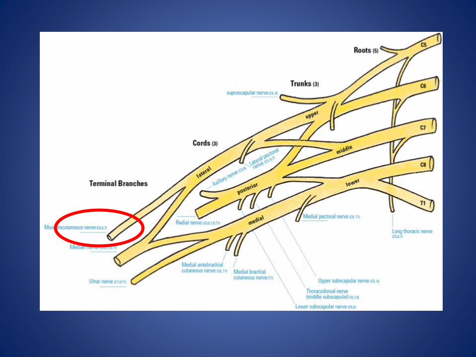

Peripheral Branches
• Musculocutaneous (C5-C6)
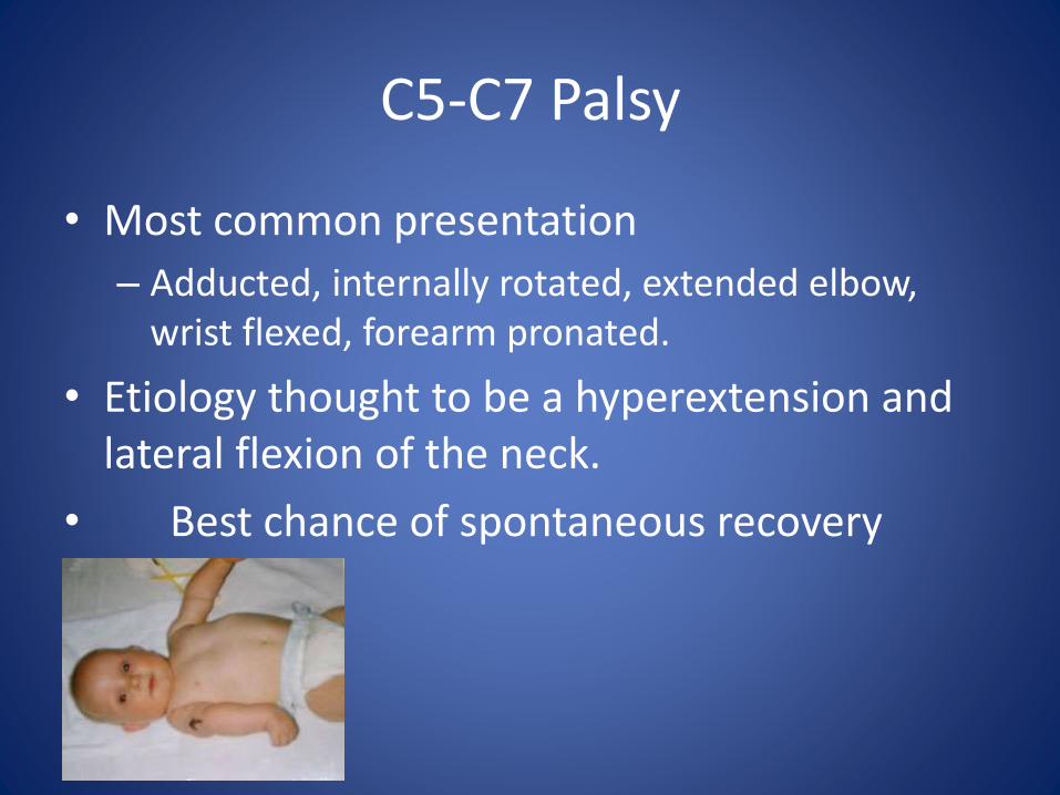
C5-C7 Palsy
• Most common presentation
– Adducted, internally rotated, extended elbow, wrist flexed, forearm pronated.
• Etiology thought to be a hyperextension and lateral flexion of the neck.
• Best chance of spontaneous recovery
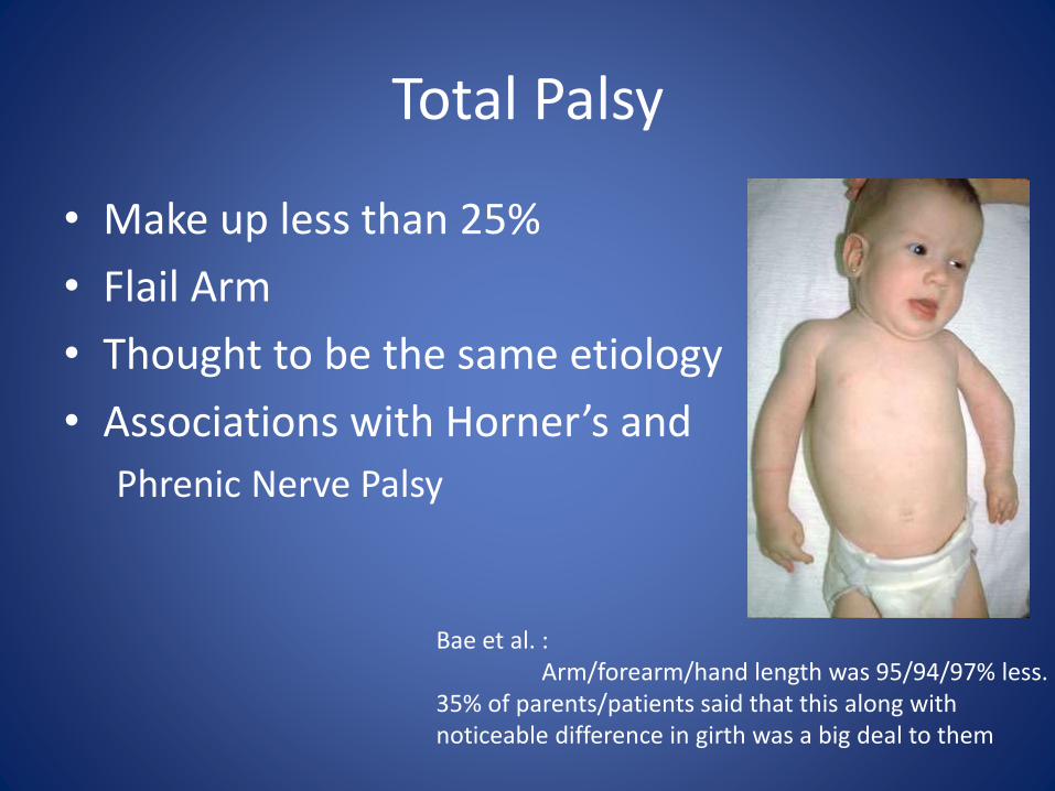
Total Palsy
• Make up less than 25%
• Flail Arm
• Thought to be the same etiology
• Associations with Horner’s and
Phrenic Nerve Palsy
Bae et al. :Arm/forearm/hand length was 95/94/97% less.
35% of parents/patients said that this along with noticeable difference in girth was a big deal to them
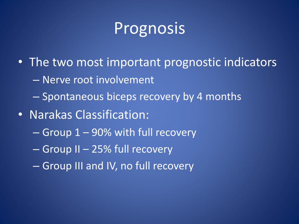
Prognosis
• The two most important prognostic indicators
– Nerve root involvement
– Spontaneous biceps recovery by 4 months
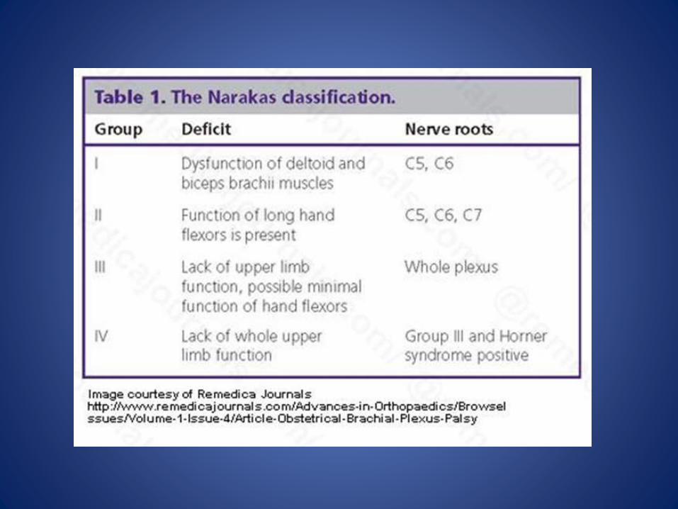
• Narakas Classification:
– Group 1 – 90% with full recovery
– Group II – 25% full recovery
– Group III and IV, no full recovery

What is the Role of the Upper Extremity Surgeon?
• Make the right diagnosis.
• Characterize the pattern
• Establish rapport with the family
• NOT to establish etiology!!!

Non-Surgical Intervention
• Physical Therapy
• Splinting and Casting
• Onabotulism Toxin Injection
Surgical Indications
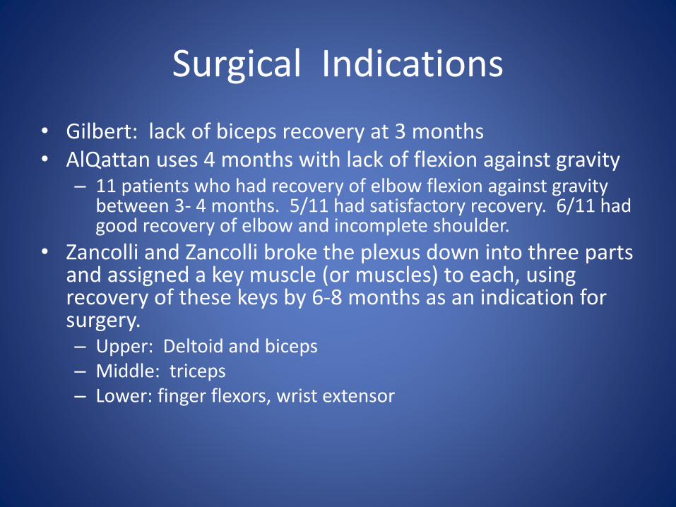
• Gilbert: lack of biceps recovery at 3 months • AlQattan uses 4 months with lack of flexion against gravity
– 11 patients who had recovery of elbow flexion against gravity between 3- 4 months. 5/11 had satisfactory recovery. 6/11 had good recovery of elbow and incomplete shoulder.
• Zancolli and Zancolli broke the plexus down into three parts and assigned a key muscle (or muscles) to each, using recovery of these keys by 6-8 months as an indication for surgery. – Upper: Deltoid and biceps– Middle: triceps– Lower: finger flexors, wrist extensor
Surgical Indications
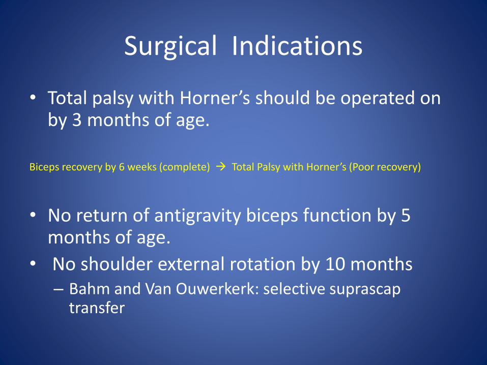
• Total palsy with Horner’s should be operated on by 3 months of age.
Biceps recovery by 6 weeks (complete) Total Palsy with Horner’s (Poor recovery)
• No return of antigravity biceps function by 5 months of age.
• No shoulder external rotation by 10 months– Bahm and Van Ouwerkerk: selective suprascap
transfer
Loss of External Rotation
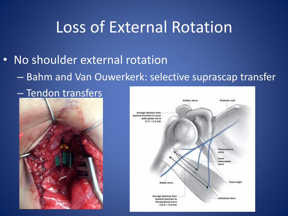
• No shoulder external rotation
– Bahm and Van Ouwerkerk: selective suprascap transfer
– Tendon transfers
Summary
• Uncommon but devastating injury
• Etiology still not completely clear and preventive measures unsuccessful
• Early establishment in therapy is key to outcomes
• A multimodality approach to treatment may be necessary
• Early recovery of nerve function (whether by natural history or surgical means) is paramount to functionality