BRACHIAL PLEXOPATHIES: CLASSIFICATION, … · the peripheral nervous system, ... Muscle Nerve 30:...
22
INVITED REVIEW ABSTRACT: The brachial plexus, which is the most complex structure of the peripheral nervous system, supplies most of the upper extremity and shoulder. The high incidence of brachial plexopathies reflects its vulnerabil- ity to trauma and the tendency of disorders involving adjacent structures to affect it secondarily. The combination of anatomic, pathophysiologic, and neuromuscular knowledge with detailed clinical and ancillary study evalua- tions provides diagnostic and prognostic information that is important to clinical management. Since most brachial plexus disorders do not involve the entire brachial plexus but, rather, show a regional predilection, a regional approach to assessment of plexopathies is necessary. Muscle Nerve 30: 547–568, 2004 BRACHIAL PLEXOPATHIES: CLASSIFICATION, CAUSES, AND CONSEQUENCES MARK A. FERRANTE, MD Department of Neurology, Tulane University Medical Center, New Orleans, Louisiana, USA Accepted 1 June 2004 The brachial plexus, which supplies most of the upper extremity and shoulder, is the most complex structure in the peripheral nervous system (PNS). Its vulnerability to trauma reflects its large size, super- ficial location, and position between two highly mo- bile structures (neck and upper extremity). 121,144 Also, it may be affected secondarily by pulmonary, vascular, or skeletal disorders involving neighboring structures. Hence, most physicians encounter pa- tients with brachial plexopathies. In addition to a comprehensive clinical evaluation, optimal assess- ment requires the performance of ancillary studies. Of these, electrodiagnostic examination is by far the most helpful. Although an extension of the neuro- logic examination, it has several advantages over the latter, including the ability to localize and character- ize the lesion, evaluate muscles not easily assessed clinically (e.g., anconeus), recognize minimally af- fected muscles that seem normal clinically, prove continuity when visible muscle movement is lacking, recognize remote lesions no longer appreciable clin- ically, and estimate lesion severity for current and future comparative studies. By integrating requisite anatomic, pathophysio- logic, and neuromuscular knowledge with detailed clinical assessment and the results of ancillary stud- ies, the examining physician can make an accurate diagnosis and prognosis. The lesion must be local- ized and characterized. This ability requires an un- derstanding of the relevant anatomy, as well as a familiarity with disorders affecting the brachial plexus. This review details a regional approach to assessment of the brachial plexus and discusses cer- tain plexopathies, especially those with a regional proclivity. Pertinent aspects of the anatomy, pathol- ogy, pathophysiology, electrodiagnosis, and injury classification of these disorders are reviewed. ANATOMY The brachial plexus is a triangular-shaped structure that extends from the spinal cord to the axilla. Its average extraforaminal length is 15.3 cm. 117 It is composed of connective and neural tissue in a 2 to 1 ratio, 9,117,154 and contains several elements: (1) five roots (classically, C5 through T1); (2) three trunks (upper, middle, and lower); (3) six divisions (three anterior, three posterior); (4) three cords (lateral, posterior, and medial); and (5) several terminal nerves (Fig. 1). The C6, C7, and C8 roots each Abbreviations: ADM, abductor digiti minimi; AHC, anterior horn cell; APB, abductor pollicis brevis; CMAP, compound muscle action potential; CSF, cerebrospinal fluid; CT, computerized tomography; DRG, dorsal root gan- glion; DUC, dorsal ulnar cutaneous; EDC, extensor digitorum communis; EIP, extensor indicis proprius; EPB, extensor pollicis brevis; FDI, first dorsal in- terosseous; LABC, lateral antebrachial cutaneous; MABC, medial antebra- chial cutaneous; MR, magnetic resonance; MUAP, motor unit action poten- tial; NA, neuralgic amyotrophy; NCS, nerve conduction study; NEE, needle electrode examination; PNS, peripheral nervous system; SNAP, sensory nerve action potential; TOS, thoracic outlet syndrome Key words: brachial plexus, classic postoperative paralysis, electrodiagnos- tic evaluation, iatrogenic plexopathy, medial brachial fascial compartment, neoplastic plexopathy, neuralgic amyotrophy, obstetric plexopathy, plexopa- thy, Pancoast syndrome, postmedian sternotomy, radiation plexopathy, root avulsion, rucksack, thoracic outlet syndrome, trauma Correspondence to: M. A. Ferrante, 1720-A Medical Park Drive, Suite 210, Biloxi, MS 39532, USA; e-mail: [email protected] © 2004 Wiley Periodicals, Inc. Published online 27 September 2004 in Wiley InterScience (www.interscience. wiley.com). DOI 10.1002/mus.20131 Brachial Plexopathies MUSCLE & NERVE November 2004 547
Transcript of BRACHIAL PLEXOPATHIES: CLASSIFICATION, … · the peripheral nervous system, ... Muscle Nerve 30:...
INVITED REVIEW ABSTRACT: The brachial plexus, which is the most complex structure ofthe peripheral nervous system, supplies most of the upper extremity andshoulder. The high incidence of brachial plexopathies reflects its vulnerabil-ity to trauma and the tendency of disorders involving adjacent structures toaffect it secondarily. The combination of anatomic, pathophysiologic, andneuromuscular knowledge with detailed clinical and ancillary study evalua-tions provides diagnostic and prognostic information that is important toclinical management. Since most brachial plexus disorders do not involvethe entire brachial plexus but, rather, show a regional predilection, a regionalapproach to assessment of plexopathies is necessary.
Muscle Nerve 30: 547–568, 2004
BRACHIAL PLEXOPATHIES: CLASSIFICATION,CAUSES, AND CONSEQUENCES
MARK A. FERRANTE, MD
Department of Neurology, Tulane University Medical Center, New Orleans, Louisiana, USA
Accepted 1 June 2004
The brachial plexus, which supplies most of theupper extremity and shoulder, is the most complexstructure in the peripheral nervous system (PNS). Itsvulnerability to trauma reflects its large size, super-ficial location, and position between two highly mo-bile structures (neck and upper extremity).121,144
Also, it may be affected secondarily by pulmonary,vascular, or skeletal disorders involving neighboringstructures. Hence, most physicians encounter pa-tients with brachial plexopathies. In addition to acomprehensive clinical evaluation, optimal assess-ment requires the performance of ancillary studies.Of these, electrodiagnostic examination is by far themost helpful. Although an extension of the neuro-logic examination, it has several advantages over thelatter, including the ability to localize and character-ize the lesion, evaluate muscles not easily assessed
clinically (e.g., anconeus), recognize minimally af-fected muscles that seem normal clinically, provecontinuity when visible muscle movement is lacking,recognize remote lesions no longer appreciable clin-ically, and estimate lesion severity for current andfuture comparative studies.
By integrating requisite anatomic, pathophysio-logic, and neuromuscular knowledge with detailedclinical assessment and the results of ancillary stud-ies, the examining physician can make an accuratediagnosis and prognosis. The lesion must be local-ized and characterized. This ability requires an un-derstanding of the relevant anatomy, as well as afamiliarity with disorders affecting the brachialplexus. This review details a regional approach toassessment of the brachial plexus and discusses cer-tain plexopathies, especially those with a regionalproclivity. Pertinent aspects of the anatomy, pathol-ogy, pathophysiology, electrodiagnosis, and injuryclassification of these disorders are reviewed.
ANATOMY
The brachial plexus is a triangular-shaped structurethat extends from the spinal cord to the axilla. Itsaverage extraforaminal length is 15.3 cm.117 It iscomposed of connective and neural tissue in a 2 to 1ratio,9,117,154 and contains several elements: (1) fiveroots (classically, C5 through T1); (2) three trunks(upper, middle, and lower); (3) six divisions (threeanterior, three posterior); (4) three cords (lateral,posterior, and medial); and (5) several terminalnerves (Fig. 1). The C6, C7, and C8 roots each
Abbreviations: ADM, abductor digiti minimi; AHC, anterior horn cell; APB,abductor pollicis brevis; CMAP, compound muscle action potential; CSF,cerebrospinal fluid; CT, computerized tomography; DRG, dorsal root gan-glion; DUC, dorsal ulnar cutaneous; EDC, extensor digitorum communis; EIP,extensor indicis proprius; EPB, extensor pollicis brevis; FDI, first dorsal in-terosseous; LABC, lateral antebrachial cutaneous; MABC, medial antebra-chial cutaneous; MR, magnetic resonance; MUAP, motor unit action poten-tial; NA, neuralgic amyotrophy; NCS, nerve conduction study; NEE, needleelectrode examination; PNS, peripheral nervous system; SNAP, sensorynerve action potential; TOS, thoracic outlet syndromeKey words: brachial plexus, classic postoperative paralysis, electrodiagnos-tic evaluation, iatrogenic plexopathy, medial brachial fascial compartment,neoplastic plexopathy, neuralgic amyotrophy, obstetric plexopathy, plexopa-thy, Pancoast syndrome, postmedian sternotomy, radiation plexopathy, rootavulsion, rucksack, thoracic outlet syndrome, traumaCorrespondence to: M. A. Ferrante, 1720-A Medical Park Drive, Suite 210,Biloxi, MS 39532, USA; e-mail: [email protected]
© 2004 Wiley Periodicals, Inc.Published online 27 September 2004 in Wiley InterScience (www.interscience.wiley.com). DOI 10.1002/mus.20131
Brachial Plexopathies MUSCLE & NERVE November 2004 547

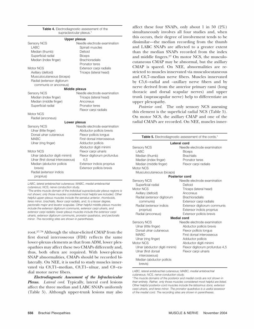
provide about 25% of its nerve fibers, and the C5and T1 roots provide the remainder.117 The percent-age of sensory and motor fibers composing each rootvaries. The largest percentage of motor fibers isfound in the C5 and C6 roots; C7 and T1 have theleast.46,154 The greatest number of sensory fibers isfound in the C7 root, followed, in descending order,by C6, C8, T1, and C5.154 The brachial plexus alsocarries sympathetic fibers.
Roots. The dorsal and ventral rootlets exit the spi-nal cord and fuse, forming the dorsal and ventralroots, respectively. The latter enter the interverte-bral foramen and fuse in the distal foramen, justbeyond the dorsal root ganglion (DRG), creating aspinal nerve. (The latter are also referred to asmixed spinal nerves because they contain both sen-sory and motor nerve fibers.) After exiting the fora-men, these nerves give off posteriorly directedbranches, the posterior primary rami, and continueas anterior primary rami (Fig. 2). The anterior pri-mary rami emerge from between the anterior andmiddle scalene muscles. The long thoracic nerve(serratus anterior) is derived via branches from theC5–C7 anterior primary rami, the C5 ramus contrib-utes to the phrenic (diaphragm) and dorsal scapular(levator scapulae; rhomboids) nerves, and theC5–C8 rami supply the scalene and longus colli mus-
cles. Preganglionic sympathetic fibers leave the spi-nal cord and exit the anterior primary rami, via whiterami communicantes, to reach the sympathetic gan-glia. The sympathetic ganglia send postganglionicfibers, via gray rami communicantes, to the C5through T1 spinal nerves. Although anatomists de-fine the anterior primary rami as the roots of thebrachial plexus, much of the surgical literature de-fines them as those PNS elements proximal to thetrunks.144 Because of its clinical utility, the latterapproach is used in this article.
Trunks. The trunks are located in the posteriorcervical triangle, behind the clavicle and sternoclei-domastoid. Trunk anomalies are infrequent.75 Typi-cally, the C5 and C6 anterior primary rami unite, theC7 anterior primary ramus continues, and the C8and T1 rami coalesce to become the upper, middle,and lower trunks, respectively (named for their re-lationship to each other). The upper trunk gives offthe suprascapular nerve and the nerve to the subcla-vius muscle. The lower trunk lies adjacent to thesubclavian artery and the apex of the lung.
Divisions. Each trunk divides into anterior and pos-terior divisions, all of which are retroclavicular. Theanterior and posterior divisions primarily supplyflexor and extensor muscles, respectively. Although
FIGURE 1. The brachial plexus.
548 Brachial Plexopathies MUSCLE & NERVE November 2004

the anterior and posterior divisions of the uppertrunk are similar in caliber, the posterior division ofthe middle trunk is much larger (C7 extensors) thanits anterior division,154 whereas the posterior divisionof the lower trunk is much smaller (C8–T1 exten-sors) than its anterior counterpart. When present,less than 5% of posterior cord fibers are T1-de-rived.52,117 Nerves usually do not arise from the divi-sions.
Cords. The cords are named for their relationshipto the second segment of the axillary artery, to whichtypically they are bound (Fig. 3). They form at or justbeyond the clavicle, below the pectoralis minor, andlie in the proximal region of the axilla, near theaxillary lymph node chain and major blood vessels tothe arm.16,20,47,138,148 The lateral cord, formed fromthe anterior divisions of the upper and middletrunks, contains C6–C7 sensory and C5–C7 motorfibers. No C5 sensory fibers exist in the lateral cord,since the C5 dermatome is subserved by the upperand lower lateral cutaneous nerves, which derivefrom the axillary and radial nerves, respectively;these nerves exit from the posterior cord. The lateralcord gives off the lateral pectoral and musculocuta-neous nerves and terminates as the lateral head ofthe median nerve. The posterior cord, formed byunion of the three posterior divisions, containsC5–C7 sensory and C5–C8 motor fibers; it does notcontain C8 sensory fibers.27 It gives off the subscap-ular and thoracodorsal nerves before terminating asthe axillary and radial nerves. The medial cord, adirect continuation of the anterior division of thelower trunk, contains C8 and T1 sensory and motorfibers. It gives off the medial pectoral, medial bra-chial cutaneous, medial antebrachial cutaneous(MABC), and ulnar nerves, and terminates as themedial head of the median nerve. When the lateralcord or C7 root sends nerve fibers to the ulnar nerve,C7 radiculopathies can produce abnormalities inulnar-innervated muscles (e.g., flexor carpi ul-naris).20,45,62,117
Terminal Nerves. These elements are located in thedistal axilla and, depending on the author, numberfrom three (median, ulnar, and radial) to five (in-clusion of musculocutaneous and axillary). Exceptfor the median nerve (derived from lateral and me-dial cords), these nerves originate from a singlecord: the ulnar nerve from the medial cord, theaxillary and radial nerves from the posterior cord,and the musculocutaneous nerve from the lateralcord. It is unclear at which point the terminal nervesof the brachial plexus become the peripheral nerves
of the upper extremity. Narakas defined that point at3 cm beyond the cord, but Wilbourn prefers toconsider the transition site as the point where theyexit the axilla.95,148
Classically, the brachial plexus is defined as con-sisting of sensory and motor nerve fibers derivedfrom neurons located in the C5–T1 DRG and ante-rior horn cells (AHCs), respectively.16,125 However,vertical variations in its composition are not uncom-mon. When adjacent roots contribute (e.g., C4, T2),it is “expanded.” Vertical shifts result when its for-mation is shifted one level upward or downward.When the C4 contribution is large and the T1 con-tribution is small, the brachial plexus is said to be“pre-fixed,” and when the C5 contribution is mini-mal and the T2 contribution is large it is “post-fixed”.Since these one-segment shifts do not affect theplexus arrangement itself, they do not affect lesionlocalization by either clinical or electrodiagnosticexamination.
CLASSIFICATION OF BRACHIAL PLEXOPATHIES
Brachial plexopathies can be classified in severalways. They are best classified according to the regioninvolved, such as supraclavicular (root and trunks),retroclavicular (divisions), and infraclavicular (cordsand terminal nerves) sites. (Isolated retroclavicularplexopathies are rare.) Although this approach isanatomically simple, it has considerable clinical util-ity because the incidence, severity, prognosis, andlesion type vary among these regions.144 In general,supraclavicular plexopathies are more common,more frequently due to closed traction (which canproduce lengthy lesions), usually more severe (sincegreater force is required to produce them), andtypically associated with a worse outcome.1,7,66,144
The supraclavicular plexus is further divided intoupper (C5 and C6 roots and upper trunk), middle(C7 root and middle trunk), and lower (C8 and T1roots and lower trunk) portions and, again, this is aclinically relevant distinction. Patients with upperplexopathies tend to recover more completely be-cause, in general, these lesions are more commonlydue to demyelinating conduction block, locatedcloser to the muscles they innervate, and extrafo-raminal (i.e., surgically accessible). This classifica-tion system facilitates communication among physi-cians since it is easier to discuss a patient with anupper plexopathy than to commit to one of its ele-ments before diagnostic testing has been performedor when there are examination limitations (e.g.,pain, mental status changes, or nonneural injuries,such as fractures or dislocations). The infraclavicular
Brachial Plexopathies MUSCLE & NERVE November 2004 549

plexus is not subdivided because lesions affecting itdo not show significant regional differences in inci-dence, severity, prognosis, or lesion type.
ASSESSMENT OF THE BRACHIAL PLEXUS
Clinical Assessment. A detailed clinical evaluationis vital for determining lesion localization (especially
FIGURE 2. The relationship between the more proximal elements of the brachial plexus and the spinal column.
FIGURE 3. The relationship between the brachial plexus and its neighboring arteries.
550 Brachial Plexopathies MUSCLE & NERVE November 2004

its proximal extent) and severity (complete or in-complete), both of which have diagnostic and prog-nostic implications that contribute to clinical man-agement. The initial and subsequent symptoms, thecircumstances surrounding their onset (e.g., back-pack usage; severe shoulder pain, followed by muscleweakness and wasting; postmedian sternotomy; axil-lary or scalene block anesthesia), and the past med-ical history are reviewed. In the setting of trauma,arm position at impact suggests the fibers most likelyaffected, as may concomitant injuries (e.g., scapular,clavicular, or humeral fracture; glenohumeral dislo-cation; scapulothoracic dissociation).156 Since mostbrachial plexopathies are axon loss in nature, neu-rologic examination frequently discloses weaknessand sensory loss. With supraclavicular lesions, thepattern of sensory and motor loss is segmental—dermatomal and myotomal, respectively—whereasinfraclavicular plexopathies produce nonsegmentalpatterns that resemble those observed with involve-ment of one or more terminal nerves. The presenceof a Horner’s syndrome or involvement of thephrenic, dorsal scapular, or long thoracic nerve in-dicates a proximal process and portends a worseprognosis. Dysautonomic features, such as cutaneoustrophic changes, sudomotor abnormalities, and va-somotor abnormalities, may also be noted. Clinicalfeatures strongly correlated with root avulsion in-clude severe pain in an anesthetic limb and Horner’ssyndrome. When traumatic plexopathies are en-countered, spinal accessory nerve, cervical plexus,and phrenic nerve function require assessment.
Radiologic Assessment. The radiologic proceduresemployed reflect the circumstances (e.g., urgency,suspected etiology and lesion site, availability). Plainfilms of the cervical spine, scapula, clavicle, hu-merus, shoulder, and chest assess for concomitantinjuries and, with open injuries, for foreign bodies.75
Signs of phrenic nerve dysfunction (e.g., elevateddiaphragm), vascular trauma (e.g., mediastinal wid-ening), or lung breach (e.g., pneumothorax, hemo-thorax) are sought. Radiologic features associatedwith brachial plexus injury include lateral tilt of thecervical spine and fractures of the transverse process,proximal first rib, or neighboring bones with rootavulsion injuries; nonunion or excessive callus for-mation with inadequately treated midshaft clavicularfractures; humeral fracture or glenohumeral disloca-tion with infraclavicular plexopathies; bone or lungabnormalities with neoplastic or radiation damage;and rudimentary cervical ribs or elongated C7 trans-verse processes with true neurogenic thoracic outletsyndrome (TOS).150
Despite its drawbacks (monoplanar imaging;beam-hardening artifacts; poor tissue differentia-tion), computerized tomography (CT) scanning isuseful for identifying bony changes and acute collec-tions of blood.35 Very thin slice (2-mm) CT-myelog-raphy images the axially oriented preganglionic rootelements when nerve root avulsion is suspected.119
When the meninges are pulled through the neuralforamen, a contrast-filled meningeal diverticulummay be observed. The width of the dye column in thecervical gutter is assessed for narrowing (spinal cordedema) and thickening (spinal cord atrophy), andthe intraspinal canal is assessed for masses. De-formed dural pouches, poor root sleeve filling, andcord edema or atrophy have strong correlations withroot avulsion.2,66,119,130 To lessen the chance ofarachnoiditis, these studies usually are performed4–6 weeks after symptom onset in those patientswith persistent deficits.119 As with other studies,falsely positive (e.g., extraforaminal injuries, menin-geal tearing without root damage) and negative(e.g., after healing and scarring of the dural pouch)results occur.20,56,63,86,93,107,108,130,156,158 The reliabilityof CT-myelography is greatest for C8 and T1 avul-sions.50
Its noninvasiveness, lack of radiation, multiplanarimaging, lack of degradation by bone, and, espe-cially, its tissue differentiating ability make magneticresonance (MR) imaging the modality of choice formore distal brachial plexus imaging. Although it isbecoming more widely used for proximal brachialplexus assessment, a recent study comparing it toCT-myelography found it less sensitive for avulsioninjuries.11 Unfortunately, when multiple slices andplanes are required, acquisition time can be consid-erable. Magnetic resonance myelography is a newertechnique that generates myelogram-like images ofthe intraspinal canal and intervertebral foramina viathe three-dimensional reconstruction of T2-weightedimages of the cerebrospinal fluid (CSF).70,129 Trau-matic meningoceles and injuries involving the C5 orC6 spinal nerves can be visualized. Since it is nonin-vasive, contrast-free, relatively quick, and multipla-nar, this technique could become a useful adjunctfor assessing proximal brachial plexus elements.150
Magnetic resonance neurography can image pe-ripheral nerves using diffusion neurography or T2-based neurography.31 With diffusion neurography,tissue differentiation reflects water diffusion differ-ences rather than T1 or T2 differences. Tissuebrightness is determined by the extent to whichprotons in that tissue are able to spin at exactly thesame rate and in phase with one another. Since thewater molecules within a nerve diffuse longitudi-
Brachial Plexopathies MUSCLE & NERVE November 2004 551
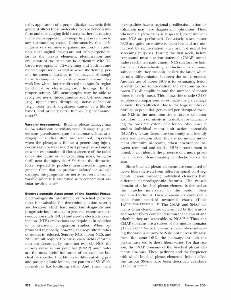
nally, application of a perpendicular magnetic fieldgradient allows these molecules to experience a uni-form and unchanging field strength, thereby causingthe nerve to appear increasingly bright in relation toany surrounding tissue. Unfortunately, this tech-nique is very sensitive to patient motion.31 In addi-tion, since sagittal images are not truly perpendicu-lar to the plexus elements, identification andevaluation of the latter can be difficult.85 With T2-based neurography, T2-weighting and both fat andblood suppression, as well as voxel shortening, per-mit intraneural fascicles to be imaged. Althoughthese techniques can localize neural lesions, theywork best when they are directed to a specific regionby clinical or electrodiagnostic findings. In theproper setting, MR neurography may be able torecognize nerve discontinuities and ball neuromas(e.g., upper trunk disruption), nerve deflections(e.g., lower trunk angulation caused by a fibrousband), and primary nerve tumors (e.g., schwanno-mas).31
Vascular Assessment. Brachial plexus damage mayfollow subclavian or axillary vessel damage (e.g., an-eurysms, pseudoaneurysms, hematomas). Thus, arte-riographic studies often are required, especiallywhen the plexopathy follows a penetrating injury,coexists with or was caused by a primary vessel injury,or when examination discloses absence of the radialor carotid pulse or an expanding mass, bruit, orthrill near the injury site.66,150 Since the distractiveforce required to produce neurovascular injury isgreater than that to produce isolated neurologicdamage, the prognosis for nerve recovery is less fa-vorable when it is associated with concomitant vas-cular involvement66
Electrodiagnostic Assessment of the Brachial Plexus.
Electrodiagnostic assessment of brachial plexopa-thies is invaluable for determining lesion severityand location, which have important diagnostic andprognostic implications. In general, extensive nerveconduction study (NCS) and needle electrode exam-ination (NEE) evaluations are required, in additionto contralateral comparison studies. When ap-proached regionally, however, the requisite numberof studies is reduced. Sensory NCS, motor NCS, andNEE are all required because each yields informa-tion not discerned by the other two. On NCS, thesensory nerve action potential (SNAP) amplitudesare the most useful indicators of an axon-loss bra-chial plexopathy. In addition to differentiating pre-and postganglionic lesions, the pattern of SNAP ab-normalities has localizing value. And, since many
plexopathies have a regional predilection, lesion lo-calization may have diagnostic implications. Thus,whenever a plexopathy is suspected, extensive sen-sory NCS are performed. Conversely, since motorNCS are quite insensitive to axon loss and are nor-malized by reinnervation, they are not useful forscreening purposes. During the first week, beforecompound muscle action potential (CMAP) ampli-tudes reach their nadir, motor NCS can localize bothaxonal and demyelinating conduction-block lesions;subsequently, they can only localize the latter, whichpermits differentiation between the two processes.Another use of motor NCS is for estimating lesionseverity. Before reinnervation, the relationship be-tween CMAP amplitude and the number of motorfibers is nearly linear. This allows side-to-side CMAPamplitude comparisons to estimate the percentageof motor fibers affected. Due to the large number offibrillation potentials generated per disrupted axon,the NEE is the most sensitive indicator of motoraxon loss. This sensitivity is invaluable for determin-ing the proximal extent of a lesion. Also, since itstudies individual motor unit action potentials(MUAPs), it can determine continuity and identifyearly reinnervation when there is no muscle move-ment clinically. Moreover, when discordance be-tween temporal and spatial MUAP recruitment isnoted, it can identify the presence of a more proxi-mally located demyelinating conduction-block le-sion.
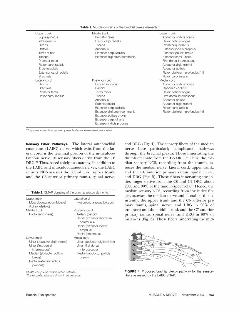
Since brachial plexus elements are composed ofnerve fibers derived from different spinal cord seg-ments, lesions involving individual elements havedifferent electrodiagnostic features. The muscledomain of a brachial plexus element is defined asthe muscles innervated by the motor fiberscontained within it. These domains are easily calcu-lated from standard myotomal charts (Table1).27,28,30,61,82,83,102,121,149 The CMAP and SNAP do-mains of an element are determined by the sensoryand motor fibers contained within that element andwhether they are assessable by NCS.27,30 Thus, theCMAP domains are a subset of the muscle domains(Table 2).28,30 Since the sensory nerve fibers subserv-ing the various sensory NCS do not necessarily arisefrom the same DRG, the pathways through theplexus traversed by these fibers varies. For that rea-son, the SNAP domains of the brachial plexus ele-ments also vary. These pathways and the frequencywith which brachial plexus elemental lesions affectthe various SNAPs have been described elsewhere(Table 3).27,28,30
552 Brachial Plexopathies MUSCLE & NERVE November 2004
Sensory Fiber Pathways. The lateral antebrachialcutaneous (LABC) nerve, which exits from the lat-eral cord, is the terminal portion of the musculocu-taneous nerve. Its sensory fibers derive from the C6DRG.27 Thus, based solely on anatomy, in addition tothe LABC and musculocutaneous nerves, the LABCsensory NCS assesses the lateral cord, upper trunk,and the C6 anterior primary ramus, spinal nerve,
and DRG (Fig. 4). The sensory fibers of the mediannerve have particularly complicated pathwaysthrough the brachial plexus. Those innervating thethumb emanate from the C6 DRG.27 Thus, the me-dian sensory NCS, recording from the thumb, as-sesses the median nerve, lateral cord, upper trunk,and the C6 anterior primary ramus, spinal nerve,and DRG (Fig. 5). Those fibers innervating the in-dex finger derive from the C6 and C7 DRG about20% and 80% of the time, respectively.27 Hence, themedian sensory NCS, recording from the index fin-ger, assesses the median nerve and lateral cord con-sistently; the upper trunk and the C6 anterior pri-mary ramus, spinal nerve, and DRG in 20% ofinstances; and the middle trunk and the C7 anteriorprimary ramus, spinal nerve, and DRG in 80% ofinstances (Fig. 6). Those fibers innervating the mid-
Table 1. Muscle domains of the brachial plexus elements.*
Upper trunk Middle trunk Lower trunkSupraspinatus Pronator teres Abductor pollicis brevisInfraspinatus Flexor carpi radialis Flexor pollicis longusBiceps Triceps Pronator quadratusDeltoid Anconeus Extensor indicis propriusTeres minor Extensor carpi radialis Extensor pollicis brevisTriceps Extensor digitorum communis Extensor carpi ulnarisPronator teres First dorsal interosseousFlexor carpi radialis Abductor digiti minimiBrachioradialis Adductor pollicisExtensor carpi radialis Flexor digitorum profundus 4,5Brachialis Flexor carpi ulnaris
Lateral cord Posterior cord Medial cordBiceps Latissimus dorsi Abductor pollicis brevisBrachialis Deltoid Opponens pollicisPronator teres Teres minor Flexor pollicis longusFlexor carpi radialis Triceps First dorsal interosseous
Anconeus Adductor pollicisBrachioradialis Abductor digiti minimiExtensor carpi radialis Flexor carpi ulnarisExtensor digitorum communis Flexor digitorum profundus 4,5Extensor pollicis brevisExtensor carpi ulnarisExtensor indicis proprius
*Only muscles easily assessed by needle electrode examination are listed.
Table 2. CMAP domains of the brachial plexus elements.*
Upper trunk Lateral cordMusculocutaneous (biceps) Musculocutaneous (biceps)Axillary (deltoid)
Middle trunk Posterior cordRadial (anconeus) Axillary (deltoid)
Radial (extensor digitorumcommunis)
Radial (extensor indicisproprius)
Radial (anconeus)Lower trunk Medial cord
Ulnar (abductor digiti minimi) Ulnar (abductor digiti minimi)Ulnar (first dorsal
interosseous)Ulnar (first dorsal
interosseous)Median (abductor pollicis
brevis)Median (abductor pollicis
brevis)Radial (extensor indicis
proprius)
CMAP, compound muscle action potential.*The recording sites are shown in parentheses.
FIGURE 4. Proposed brachial plexus pathway for the sensoryfibers assessed by the LABC SNAP.
Brachial Plexopathies MUSCLE & NERVE November 2004 553
dle finger arise from the C6, C7, and C8 DRG about10%, 70%, and 20% of the time, respectively.27 Thus,the median sensory NCS, recording from this finger,assesses the lateral cord in about 80% of instancesand the medial cord in about 20% of instances. Moreproximally, it assesses the upper trunk and the C6anterior primary ramus, spinal nerve, and DRG in10% of instances; the middle trunk and the C7 an-terior primary ramus, spinal nerve, and DRG in 70%of instances; and the lower trunk and the C8 anteriorprimary ramus, spinal nerve, and DRG in 20% ofinstances (Fig. 7).
The cell bodies of origin of the sensory nervefibers assessed by the superficial radial sensory NCSreside in the C6 and C7 DRG about 60% and 40% ofinstances, respectively.27 Thus, this study assesses thesuperficial radial nerve, radial nerve, and posteriorcord consistently; the upper trunk and the C6 ante-rior primary ramus, spinal nerve, and DRG in about60% of instances; and the middle trunk and the C7anterior primary ramus, spinal nerve, and DRG inabout 40% of instances (Fig. 8). Based on SNAP
abnormalities noted among patients with plexopa-thies related to median sternotomy, the cell bodiesof origin of the sensory fibers assessed by the ulnarsensory NCS, recording from the little finger—orfrom the dorsal aspect of the hand, as studied by thedorsal ulnar cutaneous (DUC) nerve—are primarilylocated in the C8 DRG.27,84,96 Thus, these SNAPsalways depend on the integrity of the ulnar nerve,medial cord, lower trunk, and the C8 anterior pri-mary ramus, spinal nerve, and DRG (Fig. 9). Basedon SNAP abnormalities noted among patients withtrue neurogenic TOS, as well as cadaver dissections,the cell bodies of origin of the sensory fibers assessedby the MABC sensory NCS reside predominantly inthe T1 DRG.27,37,84,96,117,138 Thus, this study assessesthe MABC nerve, medial cord, lower trunk, and theT1 anterior primary ramus, spinal nerve, and DRG(Fig. 10). The incidence of SNAP abnormalities as-sociated with individual trunk and cord lesions isshown in Table 3.
Electrodiagnostic Assessment of Individual Regions.
Typically, lesions involving the brachial plexus donot affect all of its elements (i.e., they are regional)and, consequently, the entire plexus does not re-quire exhaustive electrodiagnostic testing. Regard-ing the NCS, one approach is to screen the brachialplexus using just five sensory NCS (LABC; medianrecording from the thumb and index fingers; super-
FIGURE 5. Proposed brachial plexus pathway for the sensoryfibers assessed by the median SNAP recording from the thumb.
FIGURE 6. Proposed brachial plexus pathway for the sensoryfibers assessed by the median SNAP recording from the indexfinger.
FIGURE 7. Proposed brachial plexus pathway for the sensoryfibers assessed by the median SNAP recording from the middlefinger.
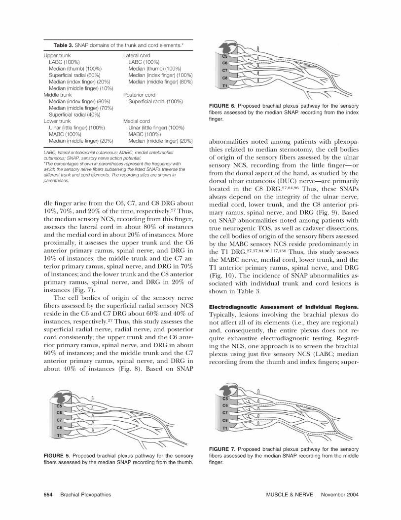
Table 3. SNAP domains of the trunk and cord elements.*
Upper trunk Lateral cordLABC (100%) LABC (100%)Median (thumb) (100%) Median (thumb) (100%)Superficial radial (60%) Median (index finger) (100%)Median (index finger) (20%) Median (middle finger) (80%)Median (middle finger) (10%)
Middle trunk Posterior cordMedian (index finger) (80%) Superficial radial (100%)Median (middle finger) (70%)Superficial radial (40%)
Lower trunk Medial cordUlnar (little finger) (100%) Ulnar (little finger) (100%)MABC (100%) MABC (100%)Median (middle finger) (20%) Median (middle finger) (20%)
LABC, lateral antebrachial cutaneous; MABC, medial antebrachialcutaneous; SNAP, sensory nerve action potential.*The percentages shown in parentheses represent the frequency withwhich the sensory nerve fibers subserving the listed SNAPs traverse thedifferent trunk and cord elements. The recording sites are shown inparentheses.
554 Brachial Plexopathies MUSCLE & NERVE November 2004

ficial radial; and ulnar, recording from the littlefinger). Whenever a specific region of the brachialplexus requires assessment, additional sensory NCS,motor NCS, and NEE of muscles belonging to thatparticular region are added (see Tables 4 and 5).
Electrodiagnostic Assessment of the SupraclavicularPlexus. Upper plexus. The upper plexus containsnerve fibers from C5 and C6. Table 4 details itselectrodiagnostic assessment. Regarding the sensoryNCS, although no studies assess the C5 DRG or itspostganglionic fibers, the other elements of the up-per plexus are assessable. The median NCS, record-ing from the thumb, and the LABC NCS both reli-ably assess the C6 DRG, its postganglionic fibers, andthe upper trunk. In general, upper plexopathiestend to affect these two studies equally. These studiesmay need to be performed contralaterally to identifyrelative abnormalities (i.e., side-to-side differencesexceeding 50%). The superficial radial NCS and themedian NCS, recording from the index finger, alsoassess these upper-plexus elements, albeit less reli-ably (i.e., in 60% and 20% of instances, respec-tively).27 The musculocutaneous (recording biceps)and axillary (recording deltoid) motor NCS assess allof the upper-plexus elements. To avoid relative ab-normalities, these studies are performed bilaterallyin the presence of upper-plexus SNAP abnormalitiesor whenever the recorded CMAP values are near orbelow their lower limit of normal. NEE of the shoul-der girdle, C5,6–radial, C5,6–axillary, and C6–me-dian innervated muscles is helpful, and evaluation oflevator scapulae, rhomboids, serratus anterior, andspinati muscles helps to define the proximal extentof the lesion.
Middle plexus. The middle plexus (Table 4) con-tains nerve fibers from C7. The sensory nerve fiberssubserving the median NCS, recording from the in-dex and middle fingers, traverse the middle plexusin approximately 80% and 70% of instances, respec-tively; whereas those subserving the superficial radialNCS traverse it in 40% of instances.27 Contralateralstudies help identify relative abnormalities. A radial
motor NCS, recording from extensor digitorumcommunis (EDC) or anconeus, can be added,though neither assesses solely the middle plexus.NEE of selected muscles (Table 4) is useful. Sinceisolated middle plexopathies are rare,1,27,75 theiridentification should always prompt screening of theadjacent upper and lower plexuses.
Lower plexus. The lower plexus (Table 4) con-tains fibers from C8 and T1. The ulnar sensory NCS,recording from the little finger, assesses the C8 DRG,its postganglionic fibers, and the lower trunk. TheMABC study assesses the corresponding T1 struc-tures. Thus, these two studies are complementary atthe pre-trunk level. Typically, with lower-trunk le-sions, both are equally affected, whereas their in-volvement is more discordant with more proximallysituated lesions. The DUC sensory NCS typically issuperfluous, since it assesses the same brachialplexus elements as the ulnar study.27 The ulnar [re-cording from the abductor digiti minimi (ADM)]and median [recording from the abductor pollicisbrevis (APB)] motor NCS assess the lower plexus, asdoes the radial motor NCS [recording from theextensor indicis proprius (EIP)]. Although the lattermay be spared with partial lower-trunk lesions, itsinvolvement excludes a medial cord lesion. Thesethree motor NCS assess the pre-trunk level of thelower plexus differentially—the radial NCS assessessolely the C8 root; the ulnar, the C8 root predomi-nantly; and the median, almost solely the T1
FIGURE 9. Proposed brachial plexus pathways for the sensoryfibers assessed by the ulnar SNAP recording from the little finger.
FIGURE 10. Proposed brachial plexus pathway for the sensoryfibers assessed by the MABC SNAP.
FIGURE 8. Proposed brachial plexus pathway for the sensoryfibers assessed by the superficial radial SNAP.
Brachial Plexopathies MUSCLE & NERVE November 2004 555
root.27,78 Although the ulnar-elicited CMAP from thefirst dorsal interosseous (FDI) reflects the samelower-plexus elements as that from ADM, lower plex-opathies may affect these two CMAPs differently and,thus, both often are required. With lower-plexusSNAP abnormalities, CMAPs should be recorded bi-laterally. On NEE, it is useful to study muscles inner-vated via C8,T1–median, C8,T1–ulnar, and C8–ra-dial motor nerve fibers.
Electrodiagnostic Assessment of the InfraclavicularPlexus. Lateral cord. Typically, lateral cord lesionsaffect the three median and LABC SNAPs uniformly(Table 5). Although upper-trunk lesions may also
affect these four SNAPs, only about 1 in 50 (2%)simultaneously involves all four studies and, whenthis occurs, their degree of involvement tends to bedissimilar—the median recording from the thumband LABC SNAPs are affected to a greater extentthan the median SNAPs recorded from the indexand middle fingers.27 On motor NCS, the musculo-cutaneous CMAP may be abnormal, but the axillaryCMAP is spared. On NEE, abnormalities are re-stricted to muscles innervated via musculocutaneousand C6,7–median nerve fibers. Muscles innervatedby C5,6–radial and –axillary nerve fibers and bynerve derived from the anterior primary rami (longthoracic and dorsal scapular nerves) and uppertrunk (suprascapular nerve) help to differentiate anupper plexopathy.
Posterior cord. The only sensory NCS assessingthis element is the superficial radial NCS (Table 5).On motor NCS, the axillary CMAP and one of theradial CMAPs are recorded. On NEE, muscles inner-
Table 4. Electrodiagnostic assessment of thesupraclavicular plexus.*
Upper plexusSensory NCS Needle electrode examination
LABC Spinati musclesMedian (thumb) DeltoidSuperficial radial BicepsMedian (index finger) Brachioradialis
Pronator teresMotor NCS Extensor carpi radialis
Axillary (deltoid) Triceps (lateral head)Musculocutaneous (biceps)Radial (extensor digitorum
communis or anconeus)Middle plexus
Sensory NCS Needle electrode examinationMedian (index finger) Triceps (lateral head)Median (middle finger) AnconeusSuperficial radial Pronator teres
Flexor carpi radialisMotor NCS
Radial (anconeus)Lower plexus
Sensory NCS Needle electrode examinationUlnar (little finger) Abductor pollicis brevisDorsal ulnar cutaneous Flexor pollicis longusMABC First dorsal interosseousUlnar (ring finger) Adductor pollicis
Abductor digiti minimiMotor NCS
Ulnar (abductor digiti minimi)Ulnar (first dorsal interosseous)Median (abductor pollicis
brevis)Radial (extensor indicis
proprius)
Flexor carpi ulnarisFlexor digitorum profundus
4,5Extensor indicis propriusExtensor pollicis brevis
LABC, lateral antebrachial cutaneous; MABC, medial antebrachialcutaneous; NCS, nerve conduction study.*The entire muscle domain of the individual supraclavicular plexus regions isnot shown; only those muscles considered most helpful are included. Otherhelpful upper-plexus muscles include the serratus anterior, rhomboids,teres minor, brachialis, flexor carpi radialis, and, to a lesser degree,pectoralis major and levator scapulae. Other helpful middle-plexus musclesinclude the extensor digitorum communis, extensor carpi ulnaris, andextensor carpi radialis; lower-plexus muscles include the extensor carpiulnaris, extensor digitorum communis, pronator quadratus, and pectoralisminor. The recording sites are shown in parentheses.
Table 5. Electrodiagnostic assessment of the cords.*
Lateral cordSensory NCS Needle electrode examination
LABC BicepsMedian (thumb) BrachialisMedian (index finger) Pronator teresMedian (middle finger) Flexor carpi radialis
Motor NCSMusculocutaneous (biceps)
Posterior cordSensory NCS Needle electrode examination
Superficial radialMotor NCS
Axillary (deltoid)Radial (extensor digitorum
communis)Radial (extensor indicis
proprius)Radial (anconeus)
DeltoidTriceps (lateral head)AnconeusBrachioradialisExtensor carpi radialisExtensor digitorum communisExtensor indicis propriusExtensor pollicis brevis
Medial cordSensory NCS Needle electrode examination
Ulnar (little finger) Abductor pollicis brevisDorsal ulnar cutaneous Flexor pollicis longusMABC First dorsal interosseousUlnar (ring finger)
Motor NCSUlnar (abductor digiti minimi)Ulnar (first dorsal
interosseous)Median (abductor pollicis
brevis)
Adductor pollicisAbductor digiti minimiFlexor digitorum profundus 4,5Flexor carpi ulnaris
LABC, lateral antebrachial cutaneous; MABC, medial antebrachialcutaneous; NCS, nerve conduction study.*The muscle domains of the posterior and medial cords are not shown intheir entirety. Rather, only those muscles considered most helpful are listed.Other helpful posterior-cord muscles include the latissimus dorsi, extensorcarpi ulnaris, and teres minor. The pronator quadratus is a useful assessorof the medial cord. The recording sites are shown in parentheses.
556 Brachial Plexopathies MUSCLE & NERVE November 2004
vated via axillary, radial, and thoracodorsal nervefibers are helpful. To differentiate a middle plexuslesion, median SNAPs from the index and middlefingers and NEE of muscles innervated via C6,7–median nerve fibers are included.
Medial cord. Ulnar and MABC SNAPs and ulnarand median CMAPs are recorded (Table 5). Theradial CMAP, recording from the EIP, is used toscreen for a more proximal process. NEE of musclesinnervated via C8,T1–median and C8,T1–ulnarnerve fibers is helpful. To screen for a more proxi-mal process, muscles innervated via C8–radial nervefibers [EIP; extensor pollicis brevis (EPB)] areadded. Importantly, their normalcy does not excludea more proximal process, as partial lower-trunk le-sions may spare them.
Terminal nerves. Reliable sensory and motorNCS are available to assess the median, radial, ulnar,musculocutaneous, and axillary nerves. On NEE,muscles innervated by these nerves are sampled. Thelack of proximal branches emanating from theseterminal nerves means that it is impossible, by clini-cal or electrodiagnostic means, to differentiate aterminal nerve lesion of the brachial plexus from amore distal, proximally located peripheral nerve le-sion.
Concluding Remarks. Typically, the five screen-ing sensory NCS listed in the introduction to thissection are generally required, with additional stud-ies based on the particular region under study or onany identified SNAP abnormalities. Patients with aflail arm due to diffuse involvement of the distalsupraclavicular plexus (pan-trunk plexopathy) ordiffuse involvement of the proximal infraclavicularplexus (pan-cord plexopathy) can be difficult to dif-ferentiate by electrodiagnostic studies. Regardingthe sensory and motor NCS, only an abnormal su-prascapular motor NCS can identify a diffuse supra-clavicular plexopathy. When normal, this study doesnot distinguish a distal supraclavicular plexus lesion(since the suprascapular nerve leaves the uppertrunk proximally) from a diffuse infraclavicular one.Although NEE of the muscles innervated by thedorsal scapular, long thoracic, and suprascapularnerves can be helpful, when normal the same distalpan-trunk versus pan-cord localization dilemma re-mains. In this setting, examination of the pectoralismajor muscle can be helpful, since it receives itsinnervation via motor nerve fibers exiting from theinfraclavicular plexus so proximally that it tends tobe affected by supraclavicular plexopathies andspared by infraclavicular ones. Clinically, diffuse su-praclavicular plexopathies are much more commonthan diffuse infraclavicular ones. In one report of 78
patients with flail arms, 75 were due to diffuse supra-clavicular lesions; only 3 reflected a diffuse infracla-vicular plexopathy.5
SELECTED SITE-SPECIFIC DISORDERS OF THEBRACHIAL PLEXUS
Supraclavicular Plexopathies with Regional Predilec-
tions. Upper Plexus. The most commonly injuredbrachial plexus region is the upper plexus, usuallyfrom closed traction.144,150 Brachial plexopathieswith a predilection for the upper plexus includeburner syndrome, rucksack paralysis, and classicpostoperative paralysis.
Burner syndrome. When sudden, forceful shoul-der contact produces separation of the shoulder andhead, upper plexus traction may occur; if associatedwith pain and paresthesias, the term burner or stingeris applied. As expected, these injuries are more com-mon among males involved in contact sports and arethe most common of all sports-related injuries. Inone report, they accounted for 38% of 190 sports-related injuries.69 In general, the pain is abrupt inonset, sharp and burning in quality, and, with theparesthesias, extends distally into the upper extrem-ity, often to the thumb. These symptoms usuallypersist for a few minutes, longer in the presence ofweakness. Their distribution implicates the C6 nervefibers, but whether they are affected at a pre- orpostganglionic level is debated. Although some play-ers experience numerous burners, permanent neu-rologic dysfunction is rare. Whenever the symptomsare prolonged, electrodiagnostic assessment is indi-cated. With burners, the electrodiagnostic abnormal-ities, when present, typically are limited to sparsefibrillation potentials in an upper-plexus distribu-tion.
Rucksack paralysis (cadet palsy; pack palsy). Thistypically unilateral upper plexopathy usually pre-sents with painless weakness following or during thecourse of wearing a rucksack or similar device (e.g.,backpack, child-carrying harness). Sensory involve-ment, in the same distribution, often is present. Itspathogenesis is likely nerve fiber compression re-lated to direct pressure from the rucksack. Risk fac-tors include the weight of the load transported, char-acteristics of the device itself (i.e., the presence of ametal frame or waist belt), and the duration worn.20
It may be more common in the presence of anunderlying abnormality (e.g., vertebral anomaly, cer-vical rib) or previous local injury.155 A history oftransient weakness following rucksack usage may beelicited.144 In two-thirds of patients, the lesions arepredominantly demyelinating conduction block.144
Brachial Plexopathies MUSCLE & NERVE November 2004 557

In this setting, the sensory NCS are normal andlesion localization and severity are determined bythe motor NCS and NEE. As expected with a demy-elinating process, treatment is conservative and re-covery occurs within a few months. In the one-thirdin whom axon loss predominates, recovery is moreprolonged and may be incomplete.
Classic postoperative paralysis. Classic postopera-tive paralysis, which was initially described in 1894, isa traction or pressure injury that characteristicallypresents in the immediate postoperative setting as aunilateral upper plexopathy or, much less fre-quently, as a more diffuse supraclavicular plexopathythat involves the upper plexus disproportionately.144
When involved, the middle and lower plexuses re-cover quicker, leaving an isolated upper plexopathy.Clinically, the primary complaint is painless weak-ness; paresthesias also may be noted. This entity isrelated to multiple factors, including patient posi-tioning, loss of muscle tone from anesthesia, andunconsciousness, which blocks weight-shifting abil-ity. Predisposing factors include the Trendelenburgposition; upper-extremity abduction beyond 90 de-grees; arm board restraint in an abducted, extended,and externally rotated position; and contralateraldeviation and rotation of the head.146 There is nogender or age group susceptibility. The underlyingpathophysiology typically is demyelinating conduc-tion block; much less frequently, axon loss predom-inates.144,146 Thus, SNAPs assessing the upper plexus(Table 4) usually are normal unless there is concom-itant axon loss. CMAP amplitude discrepancies be-tween axillary (normal) and supraclavicular fossa(absent or low amplitude) stimulation sites indicatea demyelinating conduction-block along upper-plexus fibers (musculocutaneous, recording from bi-ceps; radial, recording from brachioradialis). An ab-sent or low-amplitude axillary CMAP is seen withsupraclavicular fossa stimulation but, since this nervecannot be stimulated infraclavicularly, an amplitudediscrepancy cannot be sought. With demyelinatingconduction block lesions, rapid and full recovery isexpected and, thus, conservative treatment is em-ployed.144,146,150
Middle Plexus. Isolated middle plexopathies arerare.1,27,75 In one review of 417 brachial plexopa-thies, only one (surgically verified) was noted.27
More typically, middle-plexus involvement occurswith concomitant upper or lower plexus involve-ment. Similar to upper plexopathies, the most com-mon cause of middle plexopathies is closed trac-tion.144
Lower Plexus. Among supraclavicular plexopa-thies, lower plexopathies are less common than up-
per plexopathies.29 Disorders with a predilection forthe lower plexus include true neurogenic TOS, fol-lowing surgery for disputed neurogenic TOS, post-median sternotomy brachial plexopathy, and Pan-coast syndrome. The percentage of closed tractioninjuries involving the lower plexus is less than withupper and middle plexopathies.150 Avulsion injuriesare more common in this region of the supraclavic-ular plexus.
True neurogenic thoracic outlet syndrome. The bra-chial plexus and subclavian vessels traverse the tho-racic inlet, which lies between the first rib and clav-icle and is commonly referred to as the thoracicoutlet. When one of these structures is compressedin that space, the general term TOS is applied. Morespecific terms include arterial TOS, venous TOS,and neurogenic TOS. The latter is divided into trueneurogenic and disputed neurogenic TOS. Trueneurogenic TOS, which is more common amongyounger women and has an incidence of approxi-mately 1 per million, is also called the cervical riband band syndrome because the affected C8 and T1fibers of the lower plexus are stretched and angu-lated by a taut band that extends from a rudimentarycervical rib or elongated C7 transverse process to thefirst rib.37 Since the T1 fibers lie below the C8 fibers,they are deflected to a greater extent and, conse-quently, sustain greater injury.37,38 This has bothclinical and electrodiagnostic ramifications. Clini-cally, patients present with T1 more than C8 weak-ness and thenar muscle wasting, as well as paresthe-sias and pain along the medial aspects of the arm,forearm, and hand. Likewise, the MABC SNAP andmedian CMAP (i.e., studies that primarily assess T1fibers) are more affected than the ulnar SNAP andCMAPs (i.e., studies that primarily assess C8 fibers).The NEE indicates a slowly progressive axon-lossprocess with a lower-plexus distribution that is mostpronounced in the APB muscle. This pattern ofclinical and electrodiagnostic abnormalities is essen-tially pathognomonic for true neurogenic TOS.27
Although radiographic studies visualize associatedbony changes, they do not visualize the band. Surgi-cal division of the band typically relieves the painand paresthesias and arrests the muscle weaknessand wasting.38,150 A single case with manifestationssimilar to true neurogenic TOS was reported in acompetitive swimmer in whom the lower trunk wascompressed by a fibrous band located within a hy-pertrophied scalene muscle.59
Unlike true neurogenic TOS, which has a clearpathogenesis, objective clinical and electrodiagnos-tic features, and a good response to surgical inter-vention, disputed neurogenic TOS has an unclear
558 Brachial Plexopathies MUSCLE & NERVE November 2004
pathogenesis, lacks objective clinical and electrodi-agnostic features, and does not reliably respond tosurgical intervention.143 Although some of its propo-nents believe that disputed neurogenic TOS is acommon and underdiagnosed disorder,109,110 manyphysicians do not even consider it a distinct entity.Oddly, among 174 patients in Colorado undergoingsurgery for TOS in 1989, almost all of them hadeither private insurance or worker’s compensation;Medicaid patients almost never underwent surgery.13
Postoperative disputed neurogenic thoracic outlet syn-drome. When patients with disputed neurogenicTOS are treated surgically, especially by transaxillaryfirst rib resection, some develop severe brachial plex-opathies, a condition termed postoperative disputedneurogenic TOS.142 Most of these are incomplete,axon-loss lower plexopathies, though more extensivedamage may occur. These patients often presentwith severe causalgic hand pain and clinical deficitsin the distribution of the affected brachial plexuselements. Although the pain may improve aftersurgical repair of the surgically traumatized ele-ments, the hand weakness typically does not doso.14,66,81,141,142,146,147,150
Postmedian sternotomy plexopathy. This term ap-plies to brachial plexopathies following operationsrequiring median sternotomy, the most common ofwhich is coronary artery bypass surgery.72 The clini-cal and electrodiagnostic manifestations of this typeof plexopathy suggest C8 anterior primary ramusinvolvement; less commonly, adjacent elements alsoare affected. Postulated etiologies incriminate thefirst thoracic rib. Either chest wall retraction (1)pushes the clavicle into the retroclavicular space,rotating the first rib into the C8 anterior primaryramus, or (2) fractures the first rib and the fracturedsegment impinges upon the C8 anterior primaryramus.64,78,136 In either case, a lower-plexus tractioninjury results that, by clinical and electrodiagnosticassessments, affects the C8 anterior primary ramusdisproportionately. Although this ramus containsmotor fibers destined for the median, radial, andulnar nerves, its sensory fibers are destined solely forthe ulnar nerve. Thus, the associated paresthesiassuggest an ulnar neuropathy. Misdiagnosis is avoidedby clinical assessment of muscles supplied by C8–median nerve (e.g., flexor pollicis longus) and C8–radial nerve fibers (e.g., EIP, EPB). On sensory NCS,an absent or low-amplitude ulnar SNAP recordingfrom the little finger and a normal MABC SNAPusually are seen, implying an ulnar neuropathy or aganglionic or postganglionic C8 root lesion. (Al-though a fascicular process cannot be excluded, thenormal MABC SNAP argues against a lower trunk or
medial cord process, since lesions at these two sitestend to affect these two SNAPS more uniformly.) Onmotor NCS, isolated ulnar CMAP abnormalities maybe seen, accompanied infrequently by radial or me-dian CMAP abnormalities (recording from EIP andAPB, respectively). Thus, localization typically restson the NEE. When present, abnormalities in musclessupplied by C8–median nerve fibers indicate thatthe lesion lies at or proximal to the medial cord,whereas abnormalities in muscles innervated by C8–radial nerve fibers place it at or proximal to thelower trunk. Unless significant axon loss involves thedominant hand or causalgic pain develops, perma-nent disability is unexpected and, thus, conservativetreatment usually is employed.29,41,54,78,136,137,144,150
Pancoast syndrome. Since only the pleura sepa-rates the lung from the T1 anterior primary ramusand lower trunk, lung diseases may involve the lowerplexus. In 1924, Pancoast described the direct exten-sion of cancer from the lung apex to the lowerplexus.98,99 This syndrome occurs in about 3% oflung cancer patients and, thus, is most frequentlyobserved among men with a heavy smoking histo-ry.104 Pancoast syndrome may also be observedamong patients with lower plexopathies related toother tumors (both benign and malignant), tumorrecurrences, and infectious or inflammatory disor-ders.51,150 With lung cancer, shoulder-region paintypically is the initial and most pronounced symp-tom. It may reflect pleural, rib, spinal column, orbrachial plexus involvement. Interscapular pain maybe present when the cancer involves the posteriorprimary rami.51 The shoulder pain tends to be burn-ing or boring in character, worse at night, and tendsto radiate along the medial aspect of the arm to theelbow, and, less commonly, to the 4th and 5th dig-its.51 When present, clinical deficits are in a lowerplexus distribution. When the cancer involves the T1root or the inferior cervical sympathetic ganglion, aHorner’s syndrome may appear. Pancoast syndromeis often the first manifestation of the neoplasm, com-monly a non–small cell carcinoma; early recognitionand treatment are associated with a higher curerate.71 Electrodiagnostic studies can localize the pro-cess, thereby directing imaging studies. With severepain, narcotics and radiation therapy may be re-quired.68
Other Supraclavicular Plexopathies. Avulsions. Root-lets are not surrounded by connective tissue and,hence, are easily avulsed from the spinal cord bystretch (traction). Since the torn roots cannot regen-erate or be surgically repaired, avulsions representthe most serious complication of traction injuries.
Brachial Plexopathies MUSCLE & NERVE November 2004 559
The ventral roots are more easily avulsed becausethey are of lesser caliber, have thinner dural sacs,and are more dispersed along the spinal cord thanthe dorsal roots.124 Regarding the brachial plexus,the lower two roots more commonly avulse, whereasthe upper two roots more commonly rupture ex-traforaminally. This reflects anatomic differences intheir proximal anchorage sites, angles of exit fromthe intervertebral foramina, and lengths. The C5–T1spinal nerves traverse grooves in their respectivetransverse processes that lie between the intertrans-versalis muscles. Since the C5 and C6, and variablythe C7, spinal nerves are securely anchored by fasciaat this point, their anchorage sites are extraforami-nal, whereas the C8 and T1 spinal nerves are an-chored at the spinal cord.125 In addition, the obliquecourse of the upper cervical roots makes them morelikely to tear extraforaminally than to avulse,whereas the short length of the T1 root renders itmore susceptible to avulsion.1 Upper-extremity posi-tion at the time the traction force is applied alsoplays a role. The C5–C6 fibers are most susceptiblewith the upper extremity alongside the torso, the C7fibers when it is oriented parallel to the floor, andthe C8–T1 fibers when it is in an above-shoulderposition. Strong enough traction forces avulse allroots, regardless of limb position. Ruptures may beincomplete, with one or more fascicles remaining incontinuity. Approximately 15% of supraclavicular le-sions are two-level processes (preganglionic andpostganglionic), especially with upper cervical rootinvolvement.1 Concomitant axillary, musculocutane-ous, and suprascapular nerve injuries may occur attheir anchorage sites (i.e., quadrangular space, cor-acobrachialis, and suprascapular or spinoglenoidnotch, respectively). When the entire upper extrem-ity is paralyzed, including the long thoracic, dorsalscapular, thoracodorsal, and pectoral nerve–inner-vated muscles, especially in the presence of a Hor-ner’s syndrome, complete avulsion is likely. Otherindicators of possible avulsion include bony injury,especially a transverse process fracture (spinal nerveanchorage site); long-tract signs (damage severeenough to injure the spinal cord); and severe burn-ing pain, with shooting pain in the anesthetic ar-ea.155 Although extraforaminal ruptures may beamenable to surgical repair, root avulsions are not.Moreover, most avulsion injuries are associated withsevere pain (especially hand pain), the incidence ofwhich increases with the number of avulsed nerveroots.148
Obstetric Brachial Plexopathy. Obstetric brachialplexopathy follows a type of traction injury that typ-ically occurs when shoulder dystocia impedes vertex
delivery, thereby prompting excessive lateral devia-tion of the head and neck in order to free theshoulder.21,135 When this plexopathy follows abreech delivery, the risk of avulsion (usually of theC5 and C6 roots; less frequently of the C5–C7 roots)and bilateral involvement (22% in one series) isincreased.36 The fact that this type of plexopathy alsofollows deliveries by cesarean section implies that itdoes not simply follow poorly performed deliver-ies.36,57,58,133,135,146 Reported risk factors include in-fantile macrosomia (common with maternal diabe-tes), short mothers, low or midforceps delivery,vacuum extraction, second-stage labor exceeding 60min, passive head rotation with the shoulders fixed,multiparity, ethnic background, and delivery of aprevious infant with an obstetric brachial plexopa-thy; fetal growth restriction and prematurity are con-sidered protective.20,21,79,135,153 The incidence of thistype of plexopathy ranges from 0.5–2.6 per 1,000full-term live births 135 and reportedly is declining.20
Five patterns of nerve fiber involvement have beendescribed: (1) C5–C6 (Erb’s palsy; about 50%); (2)C5–C7 (Erb’s-plus palsy; waiter’s tip position, withadduction and internal rotation of arm, extensionand pronation of forearm, and flexion of wrists andfingers; about 35%); (3) C5–T1 with some fingerflexion sparing; (4) C5–T1 with flail arm and Hor-ner’s syndrome; and (5) C8–T1 with isolated paral-ysis of the hand and Horner’s syndrome (Klumpke’spalsy; almost never seen).135 Concomitant postgan-glionic lesions are more common with injuries in-volving the C5–C7 fibers (anchored extraforami-nally), whereas avulsion is more common withlesions involving the C8–T1 fibers (spinal cord an-choring).119 Although obstetric brachial plexopathywas first described in 1764, its management remainscontroversial.87 These lesions range from mixed de-myelination and axonal to pure axon loss (avulsion)but, in general, are less severe than traction injuriesoccurring among adults. Although many reviews sug-gest that some spontaneous recovery occurs in over90% of instances, its natural history is unknown.135
Two Swedish studies, in which surgical interventionwas not employed, reported that 20 to 25% of pa-tients are significantly impaired in later life.3,116,135
Unfortunately, neither clinical nor electrodiagnosticassessments can unequivocally identify this lattergroup. Hence, watchful waiting for evidence of re-covery usually is employed, though the duration ofsuch an approach is controversial. Since surgicalrepair yields the best results when performed withinthe 1st year, the observation period usually rangesfrom 3 to 9 months, or slightly longer.119 During thistime, physical therapy is employed. As expected, the
560 Brachial Plexopathies MUSCLE & NERVE November 2004

prognosis for C8–T1 nerve fiber involvement is poordue to the greater incidence of avulsion injury.
Infraclavicular Plexopathies with Regional Predilec-
tions. Disorders of the infraclavicular plexus havemuch less regional proclivity. At the cord level, radi-ation directed at the axillary lymph nodes (mostlywomen with breast cancer) tends to involve the in-fraclavicular plexus, especially the lateral cord,whereas midshaft clavicular fractures more com-monly affect the medial cord.18,24,60,88 At the termi-nal nerve level, the median terminal nerve charac-teristically is first and most affected with medialbrachial fascial compartment syndrome, and the ra-dial terminal nerve is more frequently affected withcrutch palsies. Of the five terminal nerves, the mus-culocutaneous terminal nerve is more often affectedby operative procedures to correct recurrent ante-rior shoulder dislocation or by other proceduresperformed near the coracoid process.12,32,150 Gleno-humeral dislocations and proximal humeral frac-tures most commonly involve the axillary terminalnerve, because of the short distance between its an-chorage site and its point of origin,76 yet the nerveinvolvement frequently goes unnoticed. In one elec-trodiagnostic study, performed 6 weeks after disloca-tion, 35 of 65 (55%) patients had axillary terminalnerve involvement, 8 of whom had a normal clinicalexamination.128 Suprascapular neuropathies are lessfrequent and, when present, often coexist with axil-lary neuropathies; musculocutaneous neuropathiesare least common.148 These traction injuries rangefrom focal demyelination to total axon loss (e.g.,nerve rupture, avulsion from the brachial plexus).
Other Infraclavicular Plexopathies. Staal et al. firstdescribed medial brachial fascial compartment syn-drome in 1966, and it has recently been re-viewed.120,132 The medial brachial fascial compart-ment, which extends from the clavicle to the elbowand houses the terminal nerves of the brachialplexus and the axillary vessels, is formed by themedial intermuscular septum, medially, when it di-vides into two fascial extensions that extend to thebrachial fascia that surrounds the arm. The five ter-minal nerves of the brachial plexus exit from thiscompartment in the following order: musculocuta-neous, axillary, radial, ulnar, and median. The me-dian nerve, characteristically, is affected first, mostseverely, and most often in isolation. Isolated ulnaror radial neuropathies are rare, and other mono-neuropathies have not been described.118,132 Whentwo nerves are involved, the median and ulnarnerves are the most common combination.132 The
radial, axillary, and musculocutaneous nerves areaffected less frequently. Reportedly, lesions locatedwithin the compartment (e.g., hematomas, aneu-rysms, pseudoaneurysms, and other lesions withmass effect) cause the intracompartmental pressureto rise, thereby impeding nerve fiber microcircula-tion and inducing clinical dysfunction. If the pres-sure increment were uniform, it should affect intra-compartmental nerves uniformly, whereas with thissyndrome, the median nerve is affected dispropor-tionately. However, compartment syndromes associ-ated with fractures are associated with pressure gra-dients that are greatest near the fracture site,48 andnerves located near a hematoma may be exposed togreater pressures.42 Regarding axillary arteriograms,since the median and ulnar nerves lie near the axil-lary artery at the point of cannulation,97 the occur-rence of a hematoma involves these two nerves out ofproportion to more distant ones, and the high pres-sures associated with the hematoma may account forthe high incidence of these neuropathies. Clinically,patients present with pain or paresthesias in thedistribution of the affected nerves, followed shortlythereafter by weakness in a similar or wider distribu-tion. Without prompt surgical intervention, thelikelihood of recovery is poor. Thus, these lesionsmust be recognized early and decompressed ur-gently.40,115,118,120,132,145,157 A recent study reportedthat complete recovery was 8.3 times more likelywhen surgical exploration occurred within the first4 h of symptom onset.15 Electrodiagnostic testing,although useful for localizing and characterizingneuropathies resulting from this syndrome, has norole in the acute setting.
SELECTED SITE-NONSPECIFIC BRACHIALPLEXOPATHIES
Neuralgic Amyotrophy (Parsonage-Turner Syndrome).
Although many names have been coined for thisdisorder, the term neuralgic amyotrophy (NA) con-veys its two quintessential features and thus is pre-ferred. Since NA has a predilection for predomi-nantly motor nerves, especially more proximallylocated ones, it frequently involves the long thoracic,suprascapular, and axillary nerves. The anterior in-terosseous and musculocutaneous nerves, and nervebranches to individual muscles (e.g., infraspinatus,pronator teres) also are frequently affected. Thephrenic and medullary cranial nerves (especially thespinal accessory), as well as individual nerve roots,may be involved.17,55,91,103,112 Proximally, the nervefibers have a somatotopic arrangement. Therefore,although NA most commonly presents as a mono-
Brachial Plexopathies MUSCLE & NERVE November 2004 561
neuropathy or a multiple mononeuropathy, this maysometimes reflect a proximal lesion within the bra-chial plexus.122 Within the brachial plexus, the up-per trunk is most commonly affected.23 When bicepsweakness is associated with musculocutaneousCMAP and LABC SNAP abnormalities and the me-dian SNAP recorded from the thumb is normal, amusculocutaneous neuropathy is more likely than anupper plexopathy.27 Conversely, when abnormalLABC and median (recording from the thumb)SNAPs are recorded and a median neuropathy isexcluded (by normal median SNAPs from the indexand middle fingers and a normal median CMAP), anupper plexopathy is more likely. Bilateral NA may besimultaneous or sequential and either symmetric orasymmetric. When it recurs in a previously affectedlimb, it may involve the same or different nerves.144,148
Clinically, abrupt and excruciating shoulder orupper-extremity pain, often with a nocturnal onset,is the presenting feature. Most commonly, the painis located at the lateral aspect of the shoulder or inthe periscapular region, but its location varies withthe involved nerve and can be most pronounced atthe shoulder (axillary nerve), scapula (suprascapularnerve), lateral thorax (long thoracic), antecubitalfossa (anterior interosseous nerve), or lateral armand forearm (musculocutaneous nerve). Althoughthe pain may extend proximally or distally, shouldermovement rather than neck movement intensifies it.When its nature is unrecognized, unnecessary pro-cedures may be performed.6 The severe pain typi-cally abates after 7–10 days or is replaced by a morepersistent dull ache. At this point, true weaknessbecomes apparent, as may significant muscle wast-ing. About 50% of affected individuals report ante-cedent events, such as recent infection, unaccus-tomed exertion, childbirth, trauma, or an invasivemedical or dental procedure. Although this triad(antecedent event, severe pain, and weakness andwasting) generally is observed, considerable individ-ual variation exists.22 Sensory NCS help to localizethe lesion. The motor NCS define the severity of theaffected nerves and thus are useful as baseline prog-nosticators and for subsequent comparative mea-surements. Consistent with an axon-loss process, theNEE shows findings indicative of acute and chronicmotor axon loss, the combination of which reflectsthe timing of the study.101,105,144 Rarely, early in itscourse, demyelinating conduction block may pre-dominate, as evidenced by full and rapid recoveryfrom severe weakness or by the electrodiagnosticstudy itself.139 Tsairis et al. reported recovery rates of36% by 1 year, 75% by 2 years, and 89% by 3 years.131
Recovery reflects lesion severity, lesion location, and
the degree of connective tissue involvement, and isbest determined by serial clinical and electrodiag-nostic assessments. Analgesics, including narcotics,may be required for the initial pain. At that time, ashort course of corticosteroids may be helpful.74
With chronic pain, neuropathic pain medications(e.g., gabapentin, tricyclics) are added. Strengthen-ing and stretching exercises are indicated. Unlikesporadic NA, the extremely rare familial form, whichhas been localized to chromosome 17, is associatedwith dysmorphic features (e.g., hypotelorism, high-arched palate, syndactyly) and commonly re-curs.19,134,140,152
Primary Neoplastic Brachial Plexopathies. Neoplasticbrachial plexopathies can be divided into primary(of brachial plexus origin) or secondary (originatingoutside the plexus). Primary brachial plexus tumorsare rare and usually benign. Of these, nerve sheathtumors predominate. Most are solitary schwannomasor neurofibromas involving the upper or middleplexus, proximally.148 Solitary schwannomas areslow-growing, encapsulated tumors that, at the rootlevel, more commonly affect the sensory roots. Whenthey grow through the neural foramen and expandat both ends, they appear dumbbell-shaped.43 Mostpatients present with a painless mass and may haveparesthesias, sometimes exacerbated by motion orpalpation. Motor symptoms follow ventral root orspinal cord compression. On MR imaging, these le-sions appear elliptical or spherical, isointense tomuscle on T1 and hyperintense on T2, brightly en-hance, and are often associated with entering, exit-ing, or displaced fascicles.80 Pathologically, they arisefrom a single fascicle (plexiform schwannomas arisefrom multiple fascicles) and are thickly encapsu-lated. Once enucleated, they seldom recur. Solitaryintraneural neurofibromas are benign, slowly grow-ing, nonencapsulated tumors that originate from theneural sheath. Following excision, recurrence is un-usual, even when incomplete.43 When these tumorsoccur as part of neurofibromatosis type 1, they donot have a regional predilection and more fre-quently are multiple and plexiform, recur followingexcision, and, like malignant nerve sheath tumors,present with pain or clinical deficits.119,150 On MRimaging, they appear fusiform or plexiform in shape,isointense to muscle on T1 and hyperintense on T2,and enhance. Unlike schwannomas, displaced fasci-cles are rare.80
Malignant nerve sheath tumors arise de novo orvia malignant transformation—usually from a plexi-form neurofibroma, less commonly from a solitaryintraneural neurofibroma, and rarely from a schwan-
562 Brachial Plexopathies MUSCLE & NERVE November 2004
noma.100 Patients often present with painful, enlarg-ing masses associated with appropriately distributedclinical deficits. Their highly malignant nature isreflected by their 5-year survival rate (10 to 50%).100
On MR imaging, these tumors are less circumscribedand may be observed to extend along fascialplanes.80 Complementary studies include CT scans,angiograms, and myelograms.80,100 In the future, MRneurography may be helpful in demonstrating theselesions.85
Secondary Neoplastic Brachial Plexopathies. Neo-plastic processes that involve the brachial plexus sec-ondarily (usually breast or lung cancers) do so bymeans of extrinsic compression or infiltration fromadjacent structures or spread from distant sites (me-tastases). When cancer involves the axillary lymphnodes, it may infiltrate the medial cord or nearbynerves (medial brachial cutaneous, MABC, ulnar, ormedian). Most patients present with severe and per-sistent shoulder and upper-extremity pain, followedby appropriate clinical deficits. With sympathetic in-volvement, the upper extremity may become warmand dry and a Horner’s syndrome sometimes devel-ops. With spread throughout the brachial plexus,symptom distribution increases.68,71,104 Althoughplain films, bone scans, and CT-myelography (whenepidural metastases are present) may demonstrateevidence of malignancy or metastatic disease, MRimaging is the radiographic procedure of choice forevaluating neoplastic plexopathies. Through lesionlocalization, electrodiagnostic testing can directthese studies. Since neoplastic processes frequentlyinvade the brachial plexus from below, electrodiag-nostic assessment of T1 fibers is mandatory. Initially,MABC SNAP abnormalities may be the only electro-diagnostic manifestation of T1 sensory fiber infiltra-tion.114
Radiation-Induced Brachial Plexopathy. Althoughthe PNS is relatively resistant to radiation damage,the incidence of damage increases with higher totaldose, larger fraction sizes, and application times ofshorter duration. Thus, lower doses administeredover longer periods are safer.43,77,123,155 The firstcases of radiation-induced brachial plexopathy werereported in 1964.92 These plexopathies are moreoften observed among women with breast cancerwho received axillary lymph node chain radiationtherapy months to decades earlier.126 Radiation-induced brachial plexopathies typically are painlesslesions that usually present with paresthesias involv-ing one or more of the lateral three digits (i.e.,lateral cord distribution), followed by extension of
the paresthesias and, later, weakness.151 Typically,these lesions are relentlessly progressive.39 Becausethe paresthesias involve the lateral digits, carpal tun-nel syndrome may be suspected unless electrodiag-nostic testing is performed. When related to radia-tion, the initial electrodiagnostic manifestationstypically include demyelinating conduction block onmotor NCS, and myokymic discharges and fascicula-tion potentials on NEE.25,39,68,73 The paraspinal mus-cles are studied, especially if tumor recurrence isbeing considered, because paraspinal fibrillation po-tentials are more common with radiation therapythan tumor recurrence.44 Unlike most demyelinat-ing conduction-block lesions, those associated withradiation therapy are prolonged and typically con-vert to axon loss, with electrodiagnostic features thatreflect the timing of the study and the severity of theloss. As axon loss progresses, the limb eventuallybecomes nonfunctional, accounting for the dismalprognosis of these lesions.151 The delayed effects ofradiation therapy likely reflect ischemic damage re-lated to microcirculatory impairment from radia-tion-induced fibrosis.20 Rarely, radiation therapygenerates nerve sheath tumors, typically malignantones.33
It is important to distinguish neoplastic fromradiation-induced plexopathy. Clinically, with neo-plastic plexopathies, the incidence of pain at onsetand Horner’s syndrome is much higher, whereaswith radiation plexopathies, isolated paresthesiaspredominate.44,68,73 Although the electrodiagnosticabnormalities typical of radiation therapy may beobserved, their presence does not exclude concom-itant tumor recurrence. On MR imaging, nodularenhancement is highly suggestive of a neoplasticprocess,100 as is the presence of a mass.127 Unfortu-nately, even surgical exploration with biopsy may benondiagnostic.68,73 In the future, it may be possibleto differentiate recurrent cancer from radiationchanges using MR neurography.31,85 Reversible bra-chial plexopathies also follow radiation therapy.111
Although there is no effective treatment for plexopa-thies following radiation, neurolysis may amelioratesevere pain, when present, although this may resultin greater clinical deficits.66,144,148
Traumatic Brachial Plexopathies. These typically re-sult from closed traction, most of which follow high-velocity injuries (e.g., motor vehicle accidents, espe-cially those involving motorcycles), or sports andoccupational injuries. Most closed-traction injuriesproduce supraclavicular plexopathies, typically up-per plexopathies that follow elongation of the bra-chial plexus by forceful separation of the head and
Brachial Plexopathies MUSCLE & NERVE November 2004 563

shoulder.34,89,90 Lower-plexus traction injuries areless common and usually follow forceful separationof the upper extremity from the torso. Traction in-juries involving the infraclavicular plexus, as withinfraclavicular plexopathies in general, more com-monly are associated with bone and blood vesselinjury.65 Open traction injuries are less common andusually result from gunshot or chainsaw accidents.148
Penetrating injuries most commonly involve the in-fraclavicular plexus. Low-velocity insults (e.g., knifeblade, low-velocity round) produce damage by directcontact, unless the injury occurs in a delayed man-ner (e.g., hematoma or pseudoaneurysm formationfollowing vessel injury).66,148 The pressure waves andcavitation created by the passage of a high-velocityround can indirectly damage the brachial plexus bycontusive and traction forces related to the cube ofits velocity.8,10,66 Although these forces infrequentlydisrupt the brachial plexus, they may be quitelengthy.66 Transecting injuries may be either sharp(knives, glass) or blunt (metal fragments, fan ormotor blades, chain saws, animal bites). Compres-sion injuries usually occur between the clavicle andfirst rib. Since the clavicle overlies the divisions,these injuries tend to involve the distal trunk, divi-sion, and proximal cord elements. In the com-pressed state, contralateral head movement pro-duces traction forces on the roots and trunks,whereas downward or lateral arm movement placestraction forces on the cords.
The treatment of traumatic brachial plexopa-thies reflects the type of injury, its location, its sever-ity, and its chronicity.119 Most closed injuries areassociated with lesions in continuity and are treatedconservatively since it is impossible at that time todetermine the likelihood of recovery or the type ofsurgical repair necessary.119 Conservative manage-ment includes physical therapy, and serial clinicaland electrodiagnostic assessments. When patientswith focal lesions show no signs of recovery after 2–3months (4–5 months for lengthier lesions), they arecandidates for surgical exploration.119 Shorter obser-vation periods (3 weeks to 3 months) may be con-sidered for high-energy injuries or those associatedwith total or near-total paralysis.49 Most infraclavicu-lar plexopathies following humeral fracture or gle-nohumeral dislocation (mostly axillary neuropa-thies) are treated conservatively, unless a severelesion in continuity or a rupture is present.150 Sharplacerations usually are repaired acutely (within72 h), by primary end-to-end neurorrhaphy, becausethey are easier to assess prior to the onset of scarringand because their length is not lost to retrac-tion.49,66,119 Other indications for acute intervention
include worsening pain or neurologic dysfunction,hematoma formation, concomitant bone or vascularinjuries, and compartment syndrome.119 With bluntlacerations, repair is delayed until the proximal anddistal extents of the neuroma are appreciable.66,119
Since the affected brachial plexus elements injuredby high-velocity rounds are infrequently disrupted,these lesions usually are initially (2–4 months) man-aged conservatively, with surgical intervention beingemployed for those individuals not showing im-provement. Resection with graft placement usually isrequired.66
Iatrogenic Brachial Plexopathy. Iatrogenic brachialplexopathies constitute about 7 to 10% of brachialplexopathies, the majority of which are blunt lesionsin continuity.66,67,94,146 Most follow operations andother medical procedures and are related to a num-ber of factors, including patient position (e.g., classicpostoperative paralysis) and the particular proce-dure performed (e.g., median sternotomy). Someiatrogenic brachial plexopathies have predisposi-tions for particular brachial plexus regions or ele-ments, as discussed earlier. The frequent location ofthe musculocutaneous nerve within the so-calledconjoint tendon renders it susceptible to proceduresperformed in this area.12,32,53 Like other traumaticplexopathies, iatrogenic brachial plexopathies mayaffect the plexus directly (e.g., suture or hardwaremisplacement, element transection, injection injury)or indirectly (e.g., pseudoaneurysm or hematomaformation) and, hence, do not always presentacutely. With midshaft clavicular fractures, theplexus can be acutely damaged by sharp bony edgesor subsequently damaged by hypertrophic callus,nonunion, or a subclavian pseudoaneurysm.4,18,22
When surgically placed screws subsequently loosen,detach, and damage nearby vascular structures, pro-ducing a hematoma or pseudoaneurysm, the plexusinjury can develop long after screw place-ment.26,106,146 Treatment, like that of other traumaticbrachial plexopathies, reflects the type of injury andits location, severity, and chronicity. With misplacedsutures or hardware, the offending item is removedand the element repaired; with sharp transections,acute end-to-end repair typically is employed; andwith blunt transections, treatment usually is delayedlong enough to permit neuroma formation.66 Inju-ries related to malpositioning or following proce-dures involving sternal splitting typically are treatedconservatively. With sharp injuries, surgical repairtypically is performed within the first 72 h, whereaswith blunt injuries, it typically is delayed for severalweeks.66
564 Brachial Plexopathies MUSCLE & NERVE November 2004
PROGNOSIS
The prognosis for a brachial plexus injury reflectslesion localization and severity. Disorders producingisolated demyelination recover following remyelina-tion, a process that typically occurs within 2 to 8weeks. However, in certain settings, demyelinationmore commonly converts to axon loss (e.g., radia-tion plexopathies) or is prolonged and frequentlyunresponsive to therapy (e.g., multifocal motor neu-ropathy). The majority of brachial plexopathies arerelated to isolated axon loss and the prognosis fortheir recovery reflects their potential for reinnerva-tion, which in turn reflects lesion completeness anddistance from the denervated muscle fibers, as wellas the degree of associated connective tissue involve-ment. Complete lesions are not amenable to rein-nervation via collateral sprouting because the latterrequire unaffected nerve fibers from which tosprout. Lesions located more than 20–24 inchesfrom the denervated muscle fibers are not amenableto reinnervation via proximodistal axon advance-ment, which occurs at a rate of 1 inch per month,because muscle fibers cannot survive in the dener-vated state for more than 20 to 24 months. Connec-tive tissue (endoneurium, perineurium, andepineurium) disruption at the lesion site leads tofibrosis, which impedes axon advancement.113,125
Unfortunately, there are no electrodiagnostic mani-festations by which to judge the degree of fibroticimpediment, and the only clinical manifestation isthe passage of time (i.e., failure to recover).
Even when sensory and motor functions are re-captured, persistent and unresponsive pain may oc-cur. Not infrequently, analgesics, neuropathic painmedications, transcutaneous electical nerve stimula-tion (TENS) units, stellate ganglion blocks, dorsalcolumn stimulation, and various surgical procedures(e.g., neurolysis, sympathectomies, stellate gan-glionectomies, amputation, cordotomies, and dorsalroot entry zone ablations) are ineffective.148
The operative outcomes associated with supracla-vicular lesions typically are most favorable for upperplexopathies (more likely extraforaminal; closer todenervated organs), least favorable for lower plex-opathies (more likely avulsion; further from dener-vated organs), and intermediate for mixed upperand middle plexopathies. Regarding infraclavicularprocesses, surgical outcomes are better for lateraland posterior cord repairs than for medial cordrepairs.66
The author thanks Asa J. Wilbourn, MD, for reviewing this manu-script, his post-fellowship mentoring, and his friendship.
REFERENCES
1. Alnot JY. Traumatic paralysis of the brachial plexus: preop-erative problems and therapeutic indications. In: Terzis JK,editor. Microreconstruction of nerve injuries. Philadelphia:WB Saunders; 1987. p 325–346.
2. Armington WG, Harnsberger HR, Osborn AG, Seay AR.Radiographic evaluation of brachial plexopathy. AJNR 1987;8:361–367.
3. Bager B. Perinatally acquired brachial plexus palsy: a persist-ing challenge. Acta Paediatr 1997;86:1214–1219.
4. Barbier O, Malghem J, Delaere O, Vande Berg B, RomboutsJJ. Injury to the brachial plexus by a fragment of bone afterfracture of the clavicle. J Bone Joint Surg 1997;79B:534–536.
5. Bentolila V, Nizard R, Bizot P, Sedel L. Complete traumaticbrachial plexus palsy. Treatment and outcome after repair.J Bone Joint Surg 1999;81A:20–28.
6. Beydoun SR, Rodriguez R. Neuralgic amyotrophy misdiag-nosed as diaphragmatic rupture. Muscle Nerve 1996;19:1181–1182.
7. Birch R, Bonney G, Wynn Parry CB. Surgical disorders ofperipheral nerves. London: Churchill Livingstone; 1998.539 p.
8. Black AN, Burns BD, Zuckerman S. An experimental studyof the wounding mechanism of high velocity missiles. BMJ1941;2:872–874.
9. Bonnel F. Microscopic anatomy of the adult human brachialplexus: an anatomical and histological basis for microsur-gery. Microsurgery 1984;5:107–117.
10. Callender GR. Wound ballistics: mechanism of productionof wounds by small arms, bullets, and shell fragments. WarMed 1943;3:337–342.
11. Carvalho GA, Nikkhah G, Matthies C, Penkert G, Sammi M.Diagnosis of root avulsions in traumatic brachial plexus in-juries: value of computerized tomography myelography andmagnetic resonance imaging. J Neurosurg 1997;86:69–76.
12. Caspi I, Exra E, Nerubay J, Horoszovski H. Musculocutane-ous nerve injury after coracoid process transfer for clavicularstability. Acta Orthop Scand 1987;58:294–295.
13. Cherington M, Cherington C. Thoracic outlet syndrome:reimbursement patterns and patient profiles. Neurology1992;42:943–945.
14. Cherington M, Happer I, Machanic B, Parry L. Surgery forthoracic outlet syndrome may be hazardous to your health.Muscle Nerve 1986;9:632–634.
15. Chitwood RW, Shepard AD, Shetty PC, Burke MW, ReddyDJ, Nypaver TJ, Ernest CB. Surgical complications of transax-illary arteriography: a case controlled study. J Vasc Surg1996;23:844–850.
16. Clemente CD. Gray’s Anatomy, 30th American ed. Philadel-phia: Lea & Febiger; 1985. 1466 p.
17. Comroe JH, Wood FC, Kay CF, Spoont EM. Motor neuritisafter tetanus antitoxin with involvement of muscles of respi-ration. Am J Med 1951;10:786–789.
18. Della Santa D, Narakas A, Bonnard C. Late lesions of thebrachial plexus after fracture of the clavicle. Ann Chir MainMemb Super 1991;10:531–540.
19. Dreschfeld J. On some of the rarer forms of muscular atro-phies. Brain 1886;9:178–195.
20. Dumitru D, Zwarts MJ. Brachial plexopathies and proximalmononeuropathies. In: Dumitru D, Amato AA, Zwarts MJ,editors. Electrodiagnostic medicine, 2nd ed. Philadelphia:Hanley & Belfus; 2002. p 777–836.
21. Eng GD, Binder H, Getson P, O’Donnell R. Obstetricalbrachial plexus palsy (OBPP) outcome with conservativemanagement. Muscle Nerve 1996;19:884–891.
22. England JD. The variations of neuralgic amyotrophy. MuscleNerve 1999;22:435–436.
23. England JD, Sumner AJ. Neuralgic amyotrophy: an increas-ingly diverse entity. Muscle Nerve 1987;10:60–68.
Brachial Plexopathies MUSCLE & NERVE November 2004 565

24. England JD, Tiel RL. Costoclavicular mass syndrome. MuscleNerve 1999;22:412–418.
25. Esteban A, Traba A. Fasciculation-myokymic activity and pro-longed nerve conduction block: a physiopathological rela-tionship in radiation-induced brachial plexopathy. Electro-encephalogr Clin Neurophysiol 1993;89:382–391.
26. Fee HJ, McAvoy JM, Dainko EA. Pseudoaneurysms of theaxillary artery following a modified Bristow operation: reportof a case and review. J Cardovasc Surg 1978;19:65–68.
27. Ferrante MA, Wilbourn AJ. The utility of various sensorynerve conduction responses in assessing brachial plexopa-thies. Muscle Nerve 1995;18:879–889.
28. Ferrante MA, Wilbourn AJ. Basic principles and practice ofEMG. In: Younger DS, editor. Motor disorders. Philadelphia:Lippincott, Williams, & Wilkins; 1999. p 19–44.
29. Ferrante MA, Wilbourn AJ. Plexopathies. In: Levin KH, Lud-ers HO, editors. Comprehensive clinical neurophysiology.Philadelphia: WB Saunders; 2000. p 201–214.
30. Ferrante MA, Wilbourn AJ. Electrodiagnostic approach tothe patient with suspected brachial plexopathy. Neurol Clin2002;20:423–450.
31. Filler A. Imaging of peripheral nerve. In: Katirji B, KaminskiHJ, Preston DC, Ruff RL, Shapiro BE, editors. Neuromuscu-lar disorders in clinical practice. Boston: Butterworth Hei-nemann; 2002. p 266–282.
32. Flatow EL, Bigiani LU, April EW. An anatomic study of themusculocutaneous nerve and its relationship to the coracoidprocess. Clin Orthop 1989;244:166–171.
33. Foley KM, Woodruff JM, Ellis FT, Posner JB. Radiation-induced malignant and atypical peripheral nerve sheathtumors. Ann Neurol 1980;7:311–318.
34. Frykholm R. The mechanism of cervical radicular lesionsresulting from stretch and forceful traction. Acta Chir Scand1957;102:93–96.
35. Fuller GN, Dick JPR, Colquhoun IR. Brachial plexus com-pression by hematoma following jugular puncture. Neurol-ogy 1994;44:775–776.
36. Geutjens G, Gilbert A, Helsen K. Obstetric brachial plexuspalsy associated with breech delivery: a different pattern ofinjury. J Bone Joint Surg [Br] 1996;78:303–306.
37. Gilliatt RW. Thoracic outlet syndromes. In: Dyck PJ, ThomasPK, Lambert EH, Bunge R, editors. Peripheral neuropathy,2nd ed. Philadelphia: Saunders; 1984. p 1409–1424.
38. Gilliatt RW, Le Quesne PM, Logue V, Sumner AJ. Wasting ofthe hand associated with a cervical rib or band. J NeurolNeurosurg Psychiatry 1970;33:615–624.
39. Glantz MJ, Burger PC, Friedman AH, Radtke RA, MasseyEW, Schold SC Jr. Treatment of radiation-induced nervoussystem injury with heparin and warfarin. Neurology 1994;44:2020–2027.
40. Groh GI, Gainor BJ, Jeffries JT, Brown M, Eggers GW Jr.Pseudoaneurysms of the axillary artery with median nervedeficit after axillary block anesthesia. J Bone Joint Surg1990;72A:1407–1408.
41. Hanson MR, Breuer AC, Furlan AJ, Lederman RJ, WilbournAJ, Cosgrove DM, Loop FD, Estafanous FG. Mechanism andfrequency of brachial plexus injury in open-heart surgery: aprospective analysis. Ann Thorac Surg 1983;36:675–679.
42. Hargens AR, Mubarak SJ. Current concepts in the patho-physiology, evaluation, and diagnosis of compartment syn-drome. Hand Clin 1998;14:371–383.
43. Harkin JC, Reed RJ. Tumors of the peripheral nervous sys-tem. Washington DC: Armed Forces Institute of Pathology;1969. 174 p.
44. Harper CM, Thomas JE, Cascino TL, Litchy WJ. Distinctionbetween neoplastic and radiation-induced brachial plexopa-thy, with emphasis on the role of EMG. Neurology 1989;39:502–506.
45. Harris W. Prefixed and postfixed types of brachial plexus.Brain 1903;26:613–615.
46. Harris W. The true form of the brachial plexus and its motordistribution. J Anat Physiol 1904;38:399–422.
47. Haymaker W, Woodhall B. Peripheral nerve injuries: princi-ples of diagnosis. Philadelphia; WB Saunders; 1953. 333 p.
48. Heckman MM, Whitesides TE Jr, Grewe SR, Rooks MD.Compartment pressure in association with closed tibial frac-tures. J Bone Joint Surg 1994;76A:1285–1292.
49. Hentz VR. Brachial plexus injuries. In: Omer GE Jr, SpinnerM, Van Beek AL, editors. Management of peripheral nerveproblems, 2nd ed. Philadelphia: WB Saunders; 1998. p 445–453.
50. Hentz VR, Narakas AO. The results of microneurosurgicalreconstruction in complete brachial plexus palsy. OrthopClin North Am 1988;19:107–114.
51. Hepper NGG, Herskovic T, Witten DM, Mulder DW, Wool-ner LB. Thoracic inlet tumors. Ann Intern Med 1966;64:979–989.
52. Hollinshead WH. Anatomy for surgeons, Vol 3: the back andlimbs, 3rd ed. Philadelphia: Harper & Row; 1982. 880 p.
53. Hoppenfeld S, deBoer P. Surgical exposures in orthopae-dics: the anatomic approach. Philadelphia: Lippincott; 1984.p 1–45.
54. Hotson JR. Neurological sequelae of cardiac surgery. In:Aminoff MJ, editor. Neurology and general medicine. NewYork: Churchill Livingstone; 1989. p 49–59.
55. James JL, Miles DW. Neuralgic amyotrophy: a clinical andelectromyographic study. BMJ 1966;2:1042–1043.
56. Jelasic F, Piepgrass U. Functional restitution after cervicalavulsion injury with typical myelographic findings. EurJ Neurol 1974;11:158–163.
57. Jennett RJ, Tarby TJ. Brachial plexus palsy: an old problemrevisited again. Am J Obstet Gynecol 1997;176:1354–1357.
58. Jones HR Jr. Pediatric electromyography. In: Brown WF,Bolton CF, editors. Clinical electromyography, 2nd ed. Bos-ton: Heinemann; 1993. p 695–758.
59. Katirji B, Hardy RW Jr. Classic neurogenic thoracic outletsyndrome in a competitive swimmer: a true scalenus anticussyndrome. Muscle Nerve 1995;18:229–233.
60. Kay SP, Eckardt JJ. Brachial plexus palsy secondary to clavic-ular nonunion. Clin Orthop 1986;206:219–222.
61. Kendall FP, McCreary EK, Provance PG. Muscles. Testingand function, 4th ed. Baltimore: Williams & Wilkins; 1993. p406–407.
62. Kerr AT. The brachial plexus of nerves in man: the variationsin its formation and branches. Am J Anat 1918;23:285–395.
63. Kewalramani LS, Taylor RG. Brachial plexus root avulsion:role of myelography. Review of diagnostic procedures.J Trauma 1975;15:603–608.
64. Kirsh MM, Magee KR, Gago O, Kahn DR, Sloan H. Brachialplexus injury following median sternotomy incision. AnnThorac Surg 1971;11:315–319.
65. Kline DG. Civilian gunshot wounds to the brachial plexus.J Neurosurg 1989;70:166–174.
66. Kline DG, Hudson AR. Nerve injuries. Philadelphia: WBSaunders; 1995. 611 p.
67. Kline DG, Judice DJ. Operative management of selectedbrachial plexus lesions. J Neurosurg 1983;58:631–649.
68. Kori SH, Foley KM, Posner JB. Brachial plexus lesions inpatients with cancer: 100 cases. Neurology 1981;31:45–50.
69. Krivickas LS, Wilbourn AJ. Sports and peripheral nerve in-juries: report of 190 injuries evaluated in a single electro-myographic laboratory. Muscle Nerve 1998;21:1092–1094.
70. Krudy AG. MR myelography using heavily T2-weighted fastspin-echo pulse sequences with fat presaturation. AJR Am JRoentgenol 1992;159:1315–1320.
71. Layzer RB. Neuromuscular manifestations of systemic dis-ease. Philadelphia: FA Davis; 1985. 434 p.
72. Lederman RJ, Breuer AC, Hanson MR, Furlan AJ, Loop FD,Cosgrove DM, Estafanous FG, Greenstreet RL. Peripheralnervous system complications of coronary artery bypass graftsurgery. Ann Neurol 1982;12:297–301.
73. Lederman RJ, Wilbourn AJ. Brachial plexopathy: recurrentcancer or radiation? Neurology 1984;34:1331–1335.
566 Brachial Plexopathies MUSCLE & NERVE November 2004

74. Lederman RJ, Paulson SM. Brachial neuritis. In: JohnsonRT, editor. Current therapy in neurology, 2nd ed. Philadel-phia: Decker; 1987. p 320–322.
75. Leffert RD. Brachial plexus injuries. New York: ChurchillLivingstone; 1985. 243 p.
76. Leffert RD, Seddon HJ. Infraclavicular brachial plexus inju-ries. J Bone Joint Surg 1965;47B:9–23.
77. Leibel SA, Sheline GE. Tolerance of the central and periph-eral nervous system to therapeutic irradiation. Adv Rad Biol1987;12:257–288.
78. Levin KH, Wilbourn AJ, Maggiano HJ. Cervical rib andmedian sternotomy-related brachial plexopathies: a reassess-ment. Neurology 1998;50:1407–1413.
79. Levine MG, Holroyde J, Woods JRJ, Siddiqi JA, Scott M,Miodovnik M. Birth trauma: incidence and predisposingfactors. Obstet Gynecol 1984;63:792–795.
80. Lin J, Martel W. Cross-sectional imaging of peripheral nervesheath tumors: characteristic signs on CT, MR imaging, andsonography. AJR Am J Roentgenol 2001;176:75–82.
81. Lindgren K. Reasons for failures in surgical treatment ofthoracic outlet syndrome. Muscle Nerve 1995;18:1484–1486.
82. Liveson JA. Peripheral neurology: case studies in electrodi-agnosis, 2nd ed. Philadelphia: Davis; 1992. p 36–42.
83. Liveson JA, Ma DM. Laboratory reference for clinical neu-rophysiology. Philadelphia: Davis; 1991. p 415–423.
84. Maggiano H, Levin KH, Wilbourn AJ. Relationship betweenthe medial antebrachial cutaneous sensory and median mo-tor responses in brachial plexopathies. Muscle Nerve 1993;16:1113–1114.
85. Maravilla KR, Bowen BC. Imaging of the peripheral nervoussystem: evaluation of peripheral neuropathy and plexopathy.AJNR 1998;19:1011–1023.
86. Mendelsohn RA, Weiner IH, Keegan JM. Myelographic dem-onstration of brachial plexus root avulsion. Arch Surg 1957;75:102–107.
87. Meyer R. Treatment of obstetrical palsy. In: Omer GE Jr,Spinner M, Van Beek AL, editors. Management of periph-eral nerve problems, 2nd ed. Philadelphia: WB Saunders;1998. p 454–458.
88. Miller DS, Boswick JA. Lesions of the brachial plexus associ-ated with fractures of the clavicle. Clin Orthop 1969;64:144–149.
89. Millesi H. Brachial plexus injury in adults: operative repair.In: Gelberman RH, editor. Operative nerve repair and re-construction, Vol 2. Philadelphia: Lippincott; 1991. p 1285–1301.
90. Milton GW. The mechanism of circumflex and other nerveinjuries in dislocation of the shoulder and the possiblemechanism of nerve injury during reduction of dislocation.Aust N Z J Surg 1953;23:25–30.
91. Mulvey DA, Aquilina RJ, Elliott MW, Moxham J, Green M.Diaphragmatic dysfunction in neuralgic amyotrophy: anelectrophysiologic evaluation of 16 patients presenting withdyspnea. Am Rev Respir Dis 1993;147:66–71.
92. Mumenthaler M. Armplexusparesen im anschluss an ront-genbestrahlung. Schweiz Med Wochenschr 1964;94:1069–1075.
93. Nagano A, Ochiai N, Sugioka H, Hara T, Tsuyama N. Use-fulness of myelography in brachial plexus injuries. J HandSurg 1989;14B:59–64.
94. Narakas AO. The surgical treatment of traumatic brachialplexus lesions. Int Surg 1980;6:521–527.
95. Narakas AO. Traumatic brachial plexus lesions. In: Dyck PJ,Thomas PK, Lambert EH, Bunge R, editors. Peripheral neu-ropathy, Vol 2. Philadelphia: WB Saunders; 1984. p 1393–1409.
96. Nishida T, Price SJ, Minieka MM. Medial antebrachial cutane-ous nerve conduction in true neurogenic thoracic outlet syn-drome. Electromyogr Clin Neurophysiol 1993;33:285–288.
97. O’Keefe DM. Brachial plexus injury following axillary arte-riography: a case report and review of the literature. J Neu-rosurg 1980;53:853–857.
98. Pancoast HK. Importance of careful roentgen-ray investiga-tions of apical chest tumors. JAMA 1924;83:1407.
99. Pancoast HK. Superior pulmonary sulcus tumor: tumor char-acterized by pain, Horner’s syndrome, destruction of boneand atrophy of hand muscles. JAMA 1932;99:1391–1396.
100. Park JK. Peripheral nerve tumors. In: Samuels MA, Feske SK,editors. Office practice of neurology, 2nd ed. Philadelphia:Churchill Livingstone; 2003. p 1118–1121.
101. Parsonage MJ, Turner AJW. Neuralgic amyotrophy: theshoulder girdle syndrome. Lancet 1948;1:973–978.
102. Perotto AO. Anatomical guide for the electromyographer:the limbs and trunk, 3rd ed. Springfield: Thomas; 1994. p305–307.
103. Pierre PA, Laterre CE, Van Den Bergh PY. Neuralgic amy-otrophy with involvement of cranial nerves IX, X, XI, andXII. Muscle Nerve 1990;13:704–707.
104. Posner JB. Neurologic complications of cancer. Philadel-phia: FA Davis; 1995. 482 p.
105. Rennels GD, Ochoa J. Neuralgic amyotrophy manifesting asanterior interosseous nerve palsy. Muscle Nerve 1980;3:160–164.
106. Richards RR, Hudson AR, Bertoia JT, Urbaniak JR, WaddellJP. Injury to the brachial plexus during Putti-Platt and Bris-tow procedures: a report of 8 cases. Am J Sports Med 1987;15:374–380.
107. Robles J. Brachial plexus avulsion: a review of diagnosticprocedures and report of six cases. J Neurosurg 1947;28:434–438.
108. Roger B, Travers V, Laval-Jeantet M. Imaging of posttrau-matic brachial plexus injury. Clin Orthop 1988;237:57–61.
109. Roos DB. The thoracic outlet syndrome is underrated. ArchNeurol 1990;47:327–328.
110. Roos DB. Thoracic outlet syndrome is underdiagnosed. Mus-cle Nerve 1999;22:126–129.
111. Salner AL, Botnick LE, Herzog AG, Goldstein MA, Harris JR,Levene MB, Hellman S. Reversible brachial plexopathy fol-lowing primary radiation therapy for breast cancer. CancerTreat Rep 1981;65:797–802.
112. Sanders EA, Van den Neste VM, Hoogenraad TU. Brachialplexus neuritis and recurrent laryngeal nerve palsy. J Neurol1988;235:323.
113. Seddon H. Surgical disorders of peripheral nerves. London:Churchill Livingstone; 1975. 336 p.
114. Seror P. Brachial plexus neoplastic lesions assessed by con-duction study of medial antebrachial cutaneous nerve. Mus-cle Nerve 2001;24:1068–1070.
115. Shoup D, Toffol GJ, Trahan TJ. Brachial plexopathy second-ary to anticoagulant-induced hematoma as a complication ofaxillary arteriography. J Am Osteopath Assoc 1997;97:102–105.
116. Sjoberg I, Erichs K, Bjerre I. Cause and effect of obstetric(neonatal) brachial plexus palsy. Acta Paediatr Scand 1988;77:357–364.
117. Slingluff CL Jr, Terzis JK, Edgerton MT. The quantitativemicroanatomy of the brachial plexus in man: reconstructiverelevance. In: Terzis JK, editor. Microreconstruction ofnerve injuries. Philadelphia: WB Saunders; 1987. p 285–324.
118. Smith DC, Mitchell DA, Peterson GW, Will AD, Mera SS,Smith LL. Medial brachial fascial compartment syndrome:anatomic basis of neuropathy after transaxillary arteriogra-phy. Radiology 1989;173:149–154.
119. Spinner RJ, Kline DG. Surgery for peripheral nerve andbrachial plexus injuries or other nerve lesions. Muscle Nerve2000;23:680–695.
120. Staal A, van Voorthuisen AE, van Dyck LM. Neurologiccomplication following arterial catheterization by the axil-lary approach. Br J Radiol 1966;39:115–116.
121. Stewart JD. Focal peripheral neuropathies, 2nd ed. NewYork: Raven Press; 1993. 506 p.
122. Stewart JD. Peripheral nerve fascicles: anatomy and clinicalrelevance. Muscle Nerve 2003;28:525–541.
Brachial Plexopathies MUSCLE & NERVE November 2004 567

123. Stoll BA, Andrews JT. Radiation-induced peripheral neurop-athy. Br Med J 1966;1:834–837.
124. Sunderland S. Meningeal-neural relations in the interverte-bral foramen. J Neurosurg 1974;40:756–763.
125. Sunderland S. Nerves and nerve injuries, 2nd ed. London:Churchill Livingstone; 1978. 1046 p.
126. Thomas JE, Colby MY. Radiation-induced or metastatic bra-chial plexopathy? A diagnostic dilemma. JAMA 1972;222:1392–1395.
127. Thyagarajan D, Cascino T, Harms G. Magnetic resonanceimaging in brachial plexopathy of cancer. Neurology 1995;45:421–427.
128. Toolanen G, Hildingsson C, Hedlund T, Knibestol M, ObergL. Early complications after anterior dislocation of the shoul-der in patients over 40 years: an ultrasonographic and elec-tromyographic study. Acta Orthop Scand 1993;64:549–552.
129. Toshiyasu N, Yabe Y, Horiuchi Y, Takayama S. Magneticresonance myelography in brachial plexus injury. J BoneJoint Surg 1997;79B:764–769.
130. Trojaborg W. Clinical, electrophysiological, and myelo-graphic studies of 9 patients with cervical spinal root avul-sions: discrepancies between EMG and X-ray findings. Mus-cle Nerve 1994;17:913–922.
131. Tsairis P, Dyck PJ, Mulder DW. Natural history of brachialplexus neuropathy: report on 99 patients. Arch Neurol 1972;27:109–117.
132. Tsao BE, Wilbourn AJ. The medial brachial fascial compart-ment syndrome following axillary arteriography. Neurology2003;61:1037–1041.
133. Ubachs JM, Slooff AC, Peeters LL. Obstetric antecedents ofsurgically treated obstetric brachial plexus injuries. Br J Ob-stet Gynaecol 1995;102:813–817.
134. van Arfen N, van Engelen BG, Reinders JW, Kremer H,Gabreels FJ. The natural history of hereditary neuralgicamyotrophy on the Dutch population: two distinct types?Brain 2000;123:718–723.
135. Van Dijk JG, Pondaag W, Malessy MJA. Obstetric lesions ofthe brachial plexus. Muscle Nerve 2001;24:1451–1461.
136. Vander Salm TJ, Cereda JM, Cutler BS. Brachial plexusinjury following median sternotomy. J Thorac CardiovascSurg 1980;80:447–452.
137. Vander Salm TJ, Cutler BS, Okike ON. Brachial plexusinjury following median sternotomy. Part 2. J Thorac Car-diovasc Surg 1982;83:914–917.
138. Walsh JF. The anatomy of the brachial plexus. Am J Med Sci1877;74:387–398.
139. Watson BV, Nicolle MW, Brown JD. Conduction block inneuralgic amyotrophy. Muscle Nerve 2001;24:559–563.
140. Watts GDJ, O’Briant KC, Borreson TE, Windebank AJ,Chance PF. Evidence for genetic heterogeneity in hereditaryneuralgic amyotrophy. Neurology 2001;56:675–678.
141. Wilbourn AJ. Electrodiagnosis of plexopathies. Neurol Clin1985;3:511–529.
142. Wilbourn AJ. Thoracic outlet syndrome surgery causing se-vere brachial plexopathy. Muscle Nerve 1988;11:66–74.
143. Wilbourn AJ. The thoracic outlet syndrome is overdiag-nosed. Arch Neurol 1990;47:328–330.
144. Wilbourn AJ. Brachial plexus disorders. In: Dyck PJ, ThomasPK, editors. Peripheral neuropathy, 3rd ed. Philadelphia:WB Saunders; 1993. p 911–950.
145. Wilbourn AJ. Nerve injuries caused by injections and tour-niquets. In: Syllabus on physical trauma to peripheral nerves(1996 AAEM plenary session). Minneapolis: American Asso-ciation of Electrodiagnostic Medicine; 1996. p 15–33.
146. Wilbourn AJ. Iatrogenic nerve injuries. Neurol Clin 1998;16:55–82.
147. Wilbourn AJ. Thoracic outlet syndrome is overdiagnosed.Muscle Nerve 1999;22:130–136.
148. Wilbourn AJ. Brachial plexopathies. In: Katirji B, KaminskiHJ, Preston DC, Ruff RL, Shapiro BE, editors. Neuromuscu-lar disorders in clinical practice. Boston: Butterworth Hei-nemann; 2002. p 884–906.
149. Wilbourn AJ, Aminoff MJ. Radiculopathies. In: Brown WF,Bolton CF, editors. Clinical electromyography, 2nd ed. Bos-ton: Butterworth Heinemann; 1993. p 175–205.
150. Wilbourn AJ, Ferrante MA. Plexopathies. In: Pourmand R,editor. Neuromuscular diseases: expert clinicians’ views. Bos-ton: Butterworth Heinemann; 2001. p 493–527.
151. Wilbourn AJ, Levin KH, Lederman RJ. Radiation-inducedbrachial plexopathy: electrodiagnostic changes over 13years. Muscle Nerve 1994;17:1108.
152. Windebank AJ. Inherited recurrent focal neuropathies. In:Dyck PJ, Thomas PK, Lambert EG, Bunge R, editors. Periph-eral neuropathy, Vol 2. Philadelphia: Saunders; 1984. p1656–1670.
153. Wolf H, Hoeksma AF, Oei SL, Bleker OP. Obstetric brachialplexus injury: risk factors related to recovery. Eur J ObstetGynecol Reprod Biol 2000;88:133–138.
154. Wolock B, Millesi H. Brachial plexus: applied anatomy andoperative exposure. In: Gelberman RH, editor. Operativenerve repair and reconstruction, Vol 2. Philadelphia: Lip-pincott; 1991. p 1255–1272.
155. Wynn Parry CB. Brachial plexus injuries. In: Vinken PJ,Bruyn GW, Klawans HL, editors. Handbook of clinical neu-rology, Vol 51. Amsterdam: Elsevier; 1987. p 143–155.
156. Zimmerman NB, Weiland AJ. Assessment and monitoring ofbrachial plexus injury in the adult. In: Gelberman RH, edi-tor. Operative nerve repair and reconstruction, Vol 2. Phil-adelphia: Lippincott; 1991. p 1273–1283.
157. Zipkin M, Backus WW, Scott B, Poppers PJ. False aneurysmof the axillary artery following brachial plexus block. J ClinAnesth 1991;3:143–145.
158. Zorub D, Nashold B, Cook W. Avulsion of the brachialplexus I: a review with implications on the therapy of intrac-table pain. Surg Neurol 1974;2:347–353.
568 Brachial Plexopathies MUSCLE & NERVE November 2004