Boswellia sacra essential oil induces tumor cell-specific
14
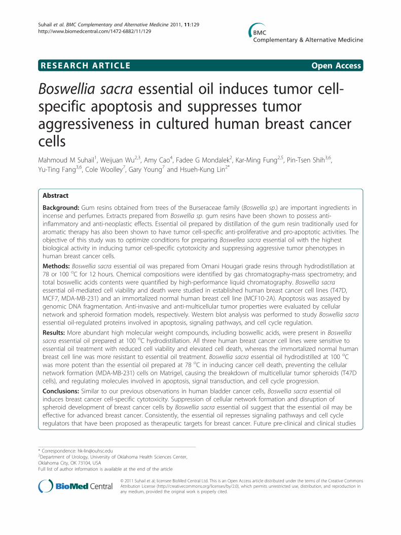
RESEARCH ARTICLE Open Access Boswellia sacra essential oil induces tumor cell- specific apoptosis and suppresses tumor aggressiveness in cultured human breast cancer cells Mahmoud M Suhail 1 , Weijuan Wu 2,3 , Amy Cao 4 , Fadee G Mondalek 2 , Kar-Ming Fung 2,5 , Pin-Tsen Shih 3,6 , Yu-Ting Fang 3,6 , Cole Woolley 7 , Gary Young 7 and Hsueh-Kung Lin 2* Abstract Background: Gum resins obtained from trees of the Burseraceae family (Boswellia sp.) are important ingredients in incense and perfumes. Extracts prepared from Boswellia sp. gum resins have been shown to possess anti- inflammatory and anti-neoplastic effects. Essential oil prepared by distillation of the gum resin traditionally used for aromatic therapy has also been shown to have tumor cell-specific anti-proliferative and pro-apoptotic activities. The objective of this study was to optimize conditions for preparing Boswellea sacra essential oil with the highest biological activity in inducing tumor cell-specific cytotoxicity and suppressing aggressive tumor phenotypes in human breast cancer cells. Methods: Boswellia sacra essential oil was prepared from Omani Hougari grade resins through hydrodistillation at 78 or 100 o C for 12 hours. Chemical compositions were identified by gas chromatography-mass spectrometry; and total boswellic acids contents were quantified by high-performance liquid chromatography. Boswellia sacra essential oil-mediated cell viability and death were studied in established human breast cancer cell lines (T47D, MCF7, MDA-MB-231) and an immortalized normal human breast cell line (MCF10-2A). Apoptosis was assayed by genomic DNA fragmentation. Anti-invasive and anti-multicellular tumor properties were evaluated by cellular network and spheroid formation models, respectively. Western blot analysis was performed to study Boswellia sacra essential oil-regulated proteins involved in apoptosis, signaling pathways, and cell cycle regulation. Results: More abundant high molecular weight compounds, including boswellic acids, were present in Boswellia sacra essential oil prepared at 100 o C hydrodistillation. All three human breast cancer cell lines were sensitive to essential oil treatment with reduced cell viability and elevated cell death, whereas the immortalized normal human breast cell line was more resistant to essential oil treatment. Boswellia sacra essential oil hydrodistilled at 100 o C was more potent than the essential oil prepared at 78 o C in inducing cancer cell death, preventing the cellular network formation (MDA-MB-231) cells on Matrigel, causing the breakdown of multicellular tumor spheroids (T47D cells), and regulating molecules involved in apoptosis, signal transduction, and cell cycle progression. Conclusions: Similar to our previous observations in human bladder cancer cells, Boswellia sacra essential oil induces breast cancer cell-specific cytotoxicity. Suppression of cellular network formation and disruption of spheroid development of breast cancer cells by Boswellia sacra essential oil suggest that the essential oil may be effective for advanced breast cancer. Consistently, the essential oil represses signaling pathways and cell cycle regulators that have been proposed as therapeutic targets for breast cancer. Future pre-clinical and clinical studies * Correspondence: [email protected] 2 Department of Urology, University of Oklahoma Health Sciences Center, Oklahoma City, OK 73104, USA Full list of author information is available at the end of the article Suhail et al. BMC Complementary and Alternative Medicine 2011, 11:129 http://www.biomedcentral.com/1472-6882/11/129 © 2011 Suhail et al; licensee BioMed Central Ltd. This is an Open Access article distributed under the terms of the Creative Commons Attribution License (http://creativecommons.org/licenses/by/2.0), which permits unrestricted use, distribution, and reproduction in any medium, provided the original work is properly cited.
Transcript of Boswellia sacra essential oil induces tumor cell-specific
RESEARCH ARTICLE Open Access
Boswellia sacra essential oil induces tumor cell-specific apoptosis and suppresses tumoraggressiveness in cultured human breast cancercellsMahmoud M Suhail1, Weijuan Wu2,3, Amy Cao4, Fadee G Mondalek2, Kar-Ming Fung2,5, Pin-Tsen Shih3,6,Yu-Ting Fang3,6, Cole Woolley7, Gary Young7 and Hsueh-Kung Lin2*
Abstract
Background: Gum resins obtained from trees of the Burseraceae family (Boswellia sp.) are important ingredients inincense and perfumes. Extracts prepared from Boswellia sp. gum resins have been shown to possess anti-inflammatory and anti-neoplastic effects. Essential oil prepared by distillation of the gum resin traditionally used foraromatic therapy has also been shown to have tumor cell-specific anti-proliferative and pro-apoptotic activities. Theobjective of this study was to optimize conditions for preparing Boswellea sacra essential oil with the highestbiological activity in inducing tumor cell-specific cytotoxicity and suppressing aggressive tumor phenotypes inhuman breast cancer cells.
Methods: Boswellia sacra essential oil was prepared from Omani Hougari grade resins through hydrodistillation at78 or 100 oC for 12 hours. Chemical compositions were identified by gas chromatography-mass spectrometry; andtotal boswellic acids contents were quantified by high-performance liquid chromatography. Boswellia sacraessential oil-mediated cell viability and death were studied in established human breast cancer cell lines (T47D,MCF7, MDA-MB-231) and an immortalized normal human breast cell line (MCF10-2A). Apoptosis was assayed bygenomic DNA fragmentation. Anti-invasive and anti-multicellular tumor properties were evaluated by cellularnetwork and spheroid formation models, respectively. Western blot analysis was performed to study Boswellia sacraessential oil-regulated proteins involved in apoptosis, signaling pathways, and cell cycle regulation.
Results: More abundant high molecular weight compounds, including boswellic acids, were present in Boswelliasacra essential oil prepared at 100 oC hydrodistillation. All three human breast cancer cell lines were sensitive toessential oil treatment with reduced cell viability and elevated cell death, whereas the immortalized normal humanbreast cell line was more resistant to essential oil treatment. Boswellia sacra essential oil hydrodistilled at 100 oCwas more potent than the essential oil prepared at 78 oC in inducing cancer cell death, preventing the cellularnetwork formation (MDA-MB-231) cells on Matrigel, causing the breakdown of multicellular tumor spheroids (T47Dcells), and regulating molecules involved in apoptosis, signal transduction, and cell cycle progression.
Conclusions: Similar to our previous observations in human bladder cancer cells, Boswellia sacra essential oilinduces breast cancer cell-specific cytotoxicity. Suppression of cellular network formation and disruption ofspheroid development of breast cancer cells by Boswellia sacra essential oil suggest that the essential oil may beeffective for advanced breast cancer. Consistently, the essential oil represses signaling pathways and cell cycleregulators that have been proposed as therapeutic targets for breast cancer. Future pre-clinical and clinical studies
* Correspondence: [email protected] of Urology, University of Oklahoma Health Sciences Center,Oklahoma City, OK 73104, USAFull list of author information is available at the end of the article
Suhail et al. BMC Complementary and Alternative Medicine 2011, 11:129http://www.biomedcentral.com/1472-6882/11/129
© 2011 Suhail et al; licensee BioMed Central Ltd. This is an Open Access article distributed under the terms of the Creative CommonsAttribution License (http://creativecommons.org/licenses/by/2.0), which permits unrestricted use, distribution, and reproduction inany medium, provided the original work is properly cited.

are urgently needed to evaluate the safety and efficacy of Boswellia sacra essential oil as a therapeutic agent fortreating breast cancer.
BackgroundFrankincense is an aromatic resin hardened from exudedgums obtained from trees of the genus Boswellia (Burser-aceae family). Boswellia sp. includes Boswellia sacra fromOman and Yemen, Boswellia carteri from Somalia, andBoswellia serrata from India and China. The resin hasbeen used in incense and fumigants, as well as a fixativein perfumes. Aroma from these resins is valued for itssuperior qualities for religious rituals since the time ofancient Egyptians [1]. Boswellia sp. resins have also beenconsidered throughout the ages to have a wealth of heal-ing properties. For example, resins of Boswellia sp. havebeen used for the treatment of rheumatoid arthritis andother inflammatory diseases [2] such as Crohn’s disease[3]. The anti-inflammatory activity has been attributed tothe resin’s ability in regulating immune cytokine produc-tion [4] and leukocyte infiltration [5,6]. Extracts fromBoswellia sp. have been shown to possess anti-bacterial,anti-fungal [7], anti-carcinogenic [8], and anti-neoplastic[9,10] properties. Clinically, extracts from the resin havebeen shown to reduce the peritumoral edema in glioblas-toma patients [9] and reverse multiple brain metastasesin a breast cancer patient [11]. These results suggest thatresins from Boswellia sp. contain active ingredients thatmodulate important biological and health supportingactivities.Boswellic acids have been identified as a major chemi-
cal component in Boswellia sp. extracts that provide theanti-inflammatory activity. Chevrier et al. reported thatethanol extracts of Boswellia carteri gum resins com-prise 7 boswellic acids [4]. Akihisa et al. reported thatmethanol extracts of Boswellia carteri resins consist of15 triterpene acids including boswellic acids [12].Acetyl-11-keto-b-boswellic acid (AKBA), being sug-gested as the most potent anti-inflammatory componentfrom the resins, selectively blocks leukotriene biosynth-esis through inhibiting 5-lipoxygenase activity [13].AKBA provides protective effects in a chemicallyinduced mouse ulcerative colitis model [14]. Boswellicacids including AKBA have also been proposed to pro-vide anti-neoploastic activity through their anti-prolif-erative and pro-apoptotic properties in multiple humancancer cell lines including meningioma cells [15], leuke-mia cells [16], hepatoma cells [17], melanoma cells,fibrosarcoma cells [18], colon cancer cells [19], andprostate cancer cells [20-22].Boswellia sp. essential oil, an extract prepared by distilla-
tion of frankincense gum resins, is one of the most com-monly used essential oils in aromatherapy. Considerable
amount of work has been attempted to identify chemicalcompositions of Boswellia sp. essential oils from differentcommercial brands. Chemical constituents of Boswellia sp.essential oils differ significantly due to climates, time ofharvest, storage conditions, geographical sources of resins[23], and methods of preparations. In this study, Boswelliasacra gum resins were collected in Oman; and essential oilwas prepared via hydrodistillation at 78 or 100 oC for 12hours. Chemical profiles of these essential oils were ana-lyzed. These essential oils were studied for their anti-tumor properties in a panel of human breast cancer celllines and an immortalized normal breast epithelial cellline. Boswellia sacra essential oil-regulated Akt and ERK1/2 activation, cyclin D1 and cdk4 expression, and caspasesactivation were also assessed.
MethodsReagents and chemicalsCell culture media (RPMI 1640, MEM, Leibovitz’s L-15,and DMEM/F-12), fetal bovine serum (FBS), horseserum, sodium pyruvate, MEM non-essential amino acids(NEAA), epidermal growth factor (EGF), cholera toxin,insulin, hydroxortisome, and penicillin-streptomycinwere purchased from Invitrogen (Grand Island, NY).XTT cell proliferation assay and lactate dehydrogenase(LDH) cytotoxicity detection kits were obtained fromRoche Applied Science (Indianapolis, IN). Matrigel™basement membrane matrix was purchased from BDBiosciences (Bedford, MA). NanoCulture® plate andmedia were obtained from SCIVAX Corp. (Kanagawa,Japan). Bicinchoninic acid (BCA) protein assay kit waspurchased from Thermo Scientific Pierce (Rockford, IL).Rabbit anti-phospho-Akt (protein kinase B; PKB)(Ser473) antibody, rabbit anti-phospho-p44/42 MAPkinase (ERK1/2) (Thr202/Tyr204) antibody, mouse anti-cyclin D1 monoclonal antibody, mouse anti-cdk4 mono-clonal antibody, mouse anti-human caspase-8 monoclo-nal antibody, rabbit anti-human caspase-9 polyclonalantibody, rabbit anti-cleaved caspase-3 (Asp175) mono-clonal antibody, and rabbit anti-poly (ADT-ribose) poly-merase (PARP) polyclonal antibody were purchased fromCell Signaling Technology (Danvers, MA). Mouse anti-human pro-caspase-3 monoclonal antibody was obtainedfrom abcam (Cambridge, MA). Mouse anti-b-actin anti-body was obtained from Sigma (St. Louis, MO).
Boswellia sacra essential oil preparationHougari grade Boswellia sacra gum resins were obtainedfrom the Hasik area to the east of Salalah City, Oman.
Suhail et al. BMC Complementary and Alternative Medicine 2011, 11:129http://www.biomedcentral.com/1472-6882/11/129
Page 2 of 14

The same batch of resins was equally divided into twoportions and hydrodistilled at two temperatures, 78 or100 oC, at roughly atmospheric pressure in Salalah City.Briefly, hydrodistillation was performed in a custommade hydrodistiller. Boswellia sacra resins were loadedinto 55 oC water with a ratio of 1:2.5 (w/v), and mixedwith an electromechanical agitator for 30-45 min oruntil a thick homogenous mucilage was formed. Tem-peratures of the hydrodistiller were monitored by aninfrared thermometer; and pressures were recorded atthe condenser terminal. To remove any residual water,collected Boswellia sacra essential oil was immediatelytransferred into a -20 oC freezer; and ice crystals wereseparated from the essential oil.
Chemical analysis of Boswellia sacra essential oil usinggas chromatography-mass spectrometry (GC-MS)Chemical components of the essential oils were analyzedwith a 7890 GC-MS (Agilent Technologies, Santa Clara,CA) equipped with an HP-1 column (50 m × 0.32 mm× 0.5 um). GC oven temperature was set and main-tained at 80 oC for 2 min and programmed to 250 oC ata rate of 3 oC/min. The oven temperature was then pro-grammed to 290 oC at a rate of 10 oC/min and main-tained for 15 min. An aliquot (1 µl) of essential oil wasinjected at a 1:125 split ratio with injector and detectortemperatures at 250 oC with He as the carrier gas at 1.3ml flow. MS was performed using a 5973 GC-MSD(Agilent Technologies) with an ionization voltage of 70eV. Comparison of mass spectra for identification pur-poses used an essential oil database from CNRS (Lyon,France) as well as Wiley and NIST mass spectrallibraries. Retention index calculations utilized C6 to C30alkanes.
Quantification of boswellic acids contentsAnalysis of total boswellic acids contents in the essentialoils was provided by San Rafael Chemical Services (SaltLake City, UT). Briefly, a weighed portion of the samplewas diluted in methanol, filtered, and then analyzed byhigh-performance liquid chromatography (HPLC) model1090 II/L (Hewlett Packard) with synergi hydro-RP, 150× 3.0 mm, 4 μm, 80 Å columns. Boswellic acids weredetected by a photodiode array detector, scanning from190 to 600 nm; and quantification was performed at205 nm.
Human breast cell linesHuman breast cancer T47D (HTB-133), MCF-7 (HTB-22), and MDA-MB-231 (HTB-26) cells as well asimmortalized normal breast epithelial MCF-10-2A(CRL-10781) cells were purchased from ATCC (Mana-ssas, VA). T47D cells were isolated from pleural effusionof a female patient with an infiltrating ductal carcinoma
of the breast [24]. This cell line is estrogen receptor(ER) positive and cultured in RPMI 1640 plus 10% FBSand 1% sodium pyruvate. MCF-7 cells were derivedfrom pleural effusion of breast adenocarcinoma from afemale patient [25]. This cell line is ER-positive and cul-tured in MEM supplemented with 1% MEM NEAA, 1mM sodium pyruvate, 10% FBS, and 10 ng/ml insulin.MDA-MB-231 cells were established from pleural effu-sion of a female patient diagnosed with adenocarcinoma[26], and maintained in Leibovitz’s L-15 medium supple-mented with 10% FBS. The immortalized normal MCF-10-2A breast cell line was derived from a patient withfibrocystic breast disease, and is non-tumorigenic inimmunodeficient mice [27]. MCF-10-2A cells is main-tained in DMEM/F-12 plus 5% horse serum, 20 ng/mlEGF, 100 ng/ml cholera toxin, 10 ng/ml insulin, and500 ng/ml hydroxortisome. All culture media also sup-plemented with 100 units/ml penicillin-100 µg/ml strep-tomycin. Cells were cultured in a humidified cellincubator at 37 oC and 5% CO2 and passaged every 3-4days or when cells reached about 80% confluence.
Cell growth and viability assayCell proliferation was determined in the breast cancercell lines and immortalized MCF10-2A cells in theirgrowth media. Cells (1x10³) were seeded into each wellof 96-well tissue culture plates in 200 µl growth media;and viable cells were quantified between 1 and 4 daysafter seeding using the XTT cell proliferation assay kit.Briefly, 100 µl culture medium was removed from eachwell at the time of assay, and an aliquot of 50 µl XTTlabeling mixture was added back to each well [28]. Reac-tions were performed at 37 oC for 4 hours. Absorbancewas read at 450 nm wavelength using a µQuant micro-plate reader (Bio-Tek; Winooski, VT). To determineBoswellia sacra essential oil-suppressed cell viability, thebreast cell lines were seeded at 5x10³ cells/well in 100µl growth medium in 96-well tissue culture plates. Fol-lowing overnight adherence, cells either received addi-tional 100 µl growth media (untreated controls) orvarying dilutions (1:200 to 1:2,700) of essential oil ingrowth media. Cell viability was determined using theXTT cell proliferation assay at 24 hours following essen-tial oil exposure. Numbers of viable cells were calculatedfrom standard curves with known numbers of cells runin parallel. Results were presented as average numbersof viable cells for cell growth and essential oil-sup-pressed cell viability.
Cell cytotoxicity assayBoswellia sacra essential oil-induced breast cell deathwas quantified by released LDH activity in culturemedia from damaged cells. Conditions for cell seedingand essential oil treatment were identical to the cell
Suhail et al. BMC Complementary and Alternative Medicine 2011, 11:129http://www.biomedcentral.com/1472-6882/11/129
Page 3 of 14
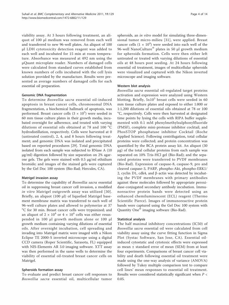
viability assay. At 3 hours following treatment, an ali-quot of 100 µl medium was removed from each welland transferred to new 96-well plates. An aliquot of 100µl LDH cytotoxicity detection reagent was added toeach well and incubated for 15 min at room tempera-ture. Absorbance was measured at 492 nm using theµQuant microplate reader. Numbers of damaged cellswere calculated from standard curves established fromknown numbers of cells incubated with the cell lysissolution provided by the manufacture. Results were pre-sented as average numbers of damaged cells for eachessential oil preparation.
Genomic DNA fragmentationTo determine Boswellia sacra essential oil-inducedapoptosis in breast cancer cells, chromosomal DNAfragmentation, a biochemical hallmark of apoptosis, wasperformed. Breast cancer cells (3 × 10⁵) were seeded in60 mm tissue culture plates in their growth media, incu-bated overnight for adherence, and treated with varyingdilutions of essential oils obtained at 78 and 100 oChydrodistillation, respectively. Cells were harvested at 0(untreated control), 2, 4, and 8 hours following treat-ment; and genomic DNA was isolated and precipitatedbased on reported procedures [29]. Total genomic DNAisolated from each sample was subjected to RNase A (10μg/ml) digestion followed by size separation on 2% agar-ose gels. The gels were stained with 0.5 µg/ml ethidiumbromide; and images of the stained gels were capturedby the Gel Doc 100 system (Bio-Rad; Hercules, CA).
Matrigel invasion assayTo determine the capability of Boswellia sacra essentialoil in suppressing breast cancer cell invasion, a modifiedin vitro Matrigel outgrowth assay was utilized [30].Briefly, an aliquot (100 µl) of liquefied Matrigel base-ment membrane matrix was transferred to each well of96-well culture plates and allowed to polymerize at 37oC for 30 min. Breast cancer cells were trypsinized; andan aliquot of 2 × 10⁴ or 4 × 10⁴ cells was either resus-pended in 100 µl growth medium alone or 100 µlgrowth medium containing varying dilutions of essentialoils. After overnight incubation, cell spreading andinvading into Matrigel matrix were imaged with a NikonEclipse TE 2000-S inverted microscope using a digitalCCD camera (Roper Scientific, Sarasota, FL) equippedwith NIS-Elements AR 3.0 imaging software. XTT assaywas then performed in the same wells to determine theviability of essential oil-treated breast cancer cells onMatrigel.
Spheroids formation assayTo evaluate and predict breast cancer cell responses toBoswellia sacra essential oil, multicellular tumor
spheroids, an in vitro model for simulating three-dimen-sional tumor micro-milieu [31], were applied. Breastcancer cells (1 × 10⁴) were seeded into each well of the96-well NanoCulture® plates in 50 µl growth mediumfor spheroids formation. Cells were then either leftuntreated or treated with varying dilutions of essentialoils at 48 hours post seeding. At 24 hours followingessential oil treatment, images of multicellular spheroidswere visualized and captured with the Nikon invertedmicroscope and imaging software.
Western blot analysisBoswellia sacra essential oil-regulated target proteinsactivation and expression were analyzed using Westernblotting. Briefly, 5x10⁵ breast cells were seeded in 60mm tissue culture plates and exposed to either 1:800 or1:1,200 dilutions of essential oils obtained at 78 or 100oC, respectively. Cells were then harvested at designatedtime points by lysing the cells with RIPA buffer supple-mented with 0.1 mM phenylmethylsulphonylfluoride(PMSF), complete mini-protease inhibitor cocktail, andPhosSTOP phosphatase inhibitor Cocktail (RocheApplied Science). Following centrifugation, total cellularproteins were collected; and protein concentration wasquantified by the BCA protein assay kit. An aliquot (30µg) of the total cellular proteins from each sample wasseparated on 10% Tris-HCl gel (Bio-Rad); and the sepa-rated proteins were transferred to PVDF membranes(Bio-Rad). Expression of caspase-8, caspase-9, pro andcleaved caspase-3, PARP, phospho-Akt, phospho-ERK1/2, cyclin D1, cdk4, and b-actin was detected by incubat-ing the PVDF membranes with primary antibodiesagainst these molecules followed by appropriate peroxi-dase-conjugated secondary antibody incubation. Immu-noreactive protein bands were detected using anenhanced chemiluminescent (ECL) reagent (ThermoScientific Pierce). Images of immunoreactive proteinbands were captured using the Gel Doc 100 system withQuantity One® imaging software (Bio-Rad).
Statistical analysisThe half maximal inhibitory concentrations (IC50) ofBoswellia sacra essential oil were calculated from cellviability assay using the curve fitting function in SigmaPlot (Systac Software, San Jose, CA). Essential oil-induced cytostatic and cytotoxic effects were expressedas mean ± standard error of mean (SEM) from at leastfour experiments. Comparisons of breast cancer cell via-bility and death following essential oil treatment weremade using the one-way analysis of variance (ANOVA)followed by Tukey multiple comparison test to comparecell lines’ mean responses to essential oil treatment.Results were considered statistically significant when P <0.05.
Suhail et al. BMC Complementary and Alternative Medicine 2011, 11:129http://www.biomedcentral.com/1472-6882/11/129
Page 4 of 14

ResultsChemical composition of Boswellia sacra essential oilChemical profiles for Boswellia sacra essential oilsobtained from different temperatures of hydrodistillationdemonstrated that a-pinene is the major compoundpresent in both temperatures preparations (AdditionalFile 1, Table S1). Contents of a-pinene decreased withhigher temperature distillation. In addition, essential oilsfrom both temperatures preparations were primarilycomposed of the major monterpene, including a-thu-jene, unidentified 1, b-pinene, and myrcene. In general,all compounds with higher retention indices, with a fewexceptions, were present in higher quantities in essentialoil distillated at 100 oC as compared to that obtained at78 oC.
Quantitatiion of boswellic acidsSince triterpene including boswellic acids could not bedetected by the current GC-MS protocol used in ourlaboratory, an HPLC method was applied to determinetotal boswellic acids in Boswellia sacra essential oils.Contents of boswellic acids in essential oils dependedupon temperature of hydrodistillation. Higher distillationtemperature produced higher quantities of boswellicacids; higher amounts of total boswellic acids weredetected in essential oil hydrodistilled at 100 oC as com-pared to 78 oC (Table 1).
Suppression of tumor cell-specific viability by Boswelliasacra essential oilsCell proliferation was determined in all four humanbreast cell lines in the absence of Boswellia sacra essen-tial oil. Under the culture condition used for each cellline, the immortalized normal breast epithelial cells pro-liferated faster than three breast cancer cell lines. Dou-bling time for MCF10-2A cells was 12 hours ascompared to 20, 22, and 27 hours for T47D, MCF7, andMDA-MB-231 cells, respectively; and MCF10-2A cellnumbers were significantly higher than T47D, MCF7, orMDA-MB-231 cells at days 2, 3, and 4 following cellseeding (Figure 1).Boswellia sacra essential oils were studied for their
capabilities in suppressing breast cancer cell viability incultures. For essential oil collected at 78 oC, 200 to1:1,600 dilutions were used, whereas a wider range ofdilutions (600 to 2,700) was used for essential oil col-lected at 100 oC. Although different cancer cell lines
varied in their sensitivities to essential oil treatment,both temperatures of essential oil preparations, in gen-eral, suppressed cell viability in all three human breastcancer cell lines (Figure 2). Boswellia sacra essential oil-suppressed cancer cell viability depended upon hydro-distillation temperatures. Essential oil obtained at 78 oCpossessed less potent anti-proliferative activity (Figure2A) as compared to that prepared at 100 oC (Figure 2B).More importantly, immortalized normal breast epithelialMCF10-2A cells were resistant to Boswellia sacra essen-tial oil-suppressed cell viability (Figure 2). MCF10-2Acell viability was significantly higher than three breastcancer cell lines when 600 to 1,200 dilutions of essentialoil (obtained at 100 oC) was administered; in contrast,results were less significant when essential oil preparedat 78 oC was used.IC50 values were calculated to provide a quantitative
assess of these essential oils. Results supported thatessential oil hydrodistilled at 100 oC produced morepotent cytotoxic effects. For example, IC50 values forT47D cells were 900 and 1,450 dilutions for essentialoils obtained at 78 and 100 oC, respectively (Table 2).Among the cancer cell lines, MCF7 cells were the mostsensitive to essential oil with suppressed cell viability.
Boswellia sacra essential oil-induced breast tumor cell-specific deathIn order to determine whether the reduced cell viabilityresulted from increased cell death and how cellsresponded at an early phase of treatment, essential oil-induced breast cell death was quantified by LDH release.Elevated cell death was observed in all three breast can-cer cell lines treated with Boswellia sacra essential oils(Figure 3). In contrast, essential oil-induced cytotoxicitywas significantly lower in immortalized MCF10-2A cellsat 3 hours after treatment. Consistent to results from cellviability assays, Boswellia sacra essential oil preparedfrom 78 oC hydrodistillation (Figure 3A) was less potentthan the essential oil obtained at 100 oC (Figure 3B) forinducing an early phase of tumor cell-specific death.
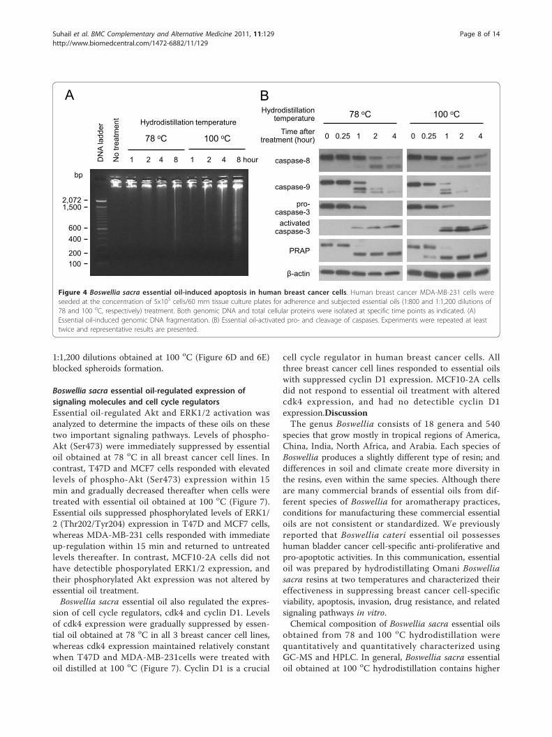
Boswellia sacra essential oil-induced apoptosisFragmentation of genomic DNA demonstrated that Bos-wellia sacra essential oil induced apoptosis in breastcancer cells. Essential oils prepared at 78 oC (at 800dilution) and 100 oC (at 1,200 dilution) induced geno-mic DNA fragmentation in a time-dependent manner;all three human breast cancer cell lines exhibited similarpatterns and visible fragmented genomic DNA within 8hour post-treatment (Figure 4A). In contrast, the sameconcentrations of essential oils treatment did not induceDNA fragmentation in MCF10-2A cells.Caspases, a family of cysteine proteases, play critical
roles in apoptosis. Cleaved caspase-8 p43/p41 and
Table 1 Boswellic acids contents in Boswellia sacraessential oils
Specific gravity Total boswellic acids (mg/ml)
78 oC distillation 0.852 19.6
100 oC distillation 0.847 30.1
Suhail et al. BMC Complementary and Alternative Medicine 2011, 11:129http://www.biomedcentral.com/1472-6882/11/129
Page 5 of 14

Time after cell seeding (day)
1 2 3 4
Cel
l num
ber
-2000
0
2000
4000
6000
8000
10000
12000
14000
16000MCF10-2A T47D MCF7 MDA-MB-231
*
*
*
Figure 1 Human breast cell growth. Human breast cancer cells and immortalized normal breast epithelial cells (1x10³) were seeded intriplicate in their culture media, and quantified for their growth between 1 and 4 days following cell seeding. Cell numbers were calculatedfrom standard curves with known numbers of cells. Results are presented as mean ± SEM from 4 independent experiments. * indicates statisticaldifferences between normal and malignant cells at P < 0.05.
Boswellia sacra essential oil dilution
Num
ber o
f via
ble
cells
0
2000
4000
6000
8000
10000
12000
14000
MCF10-2AT47D MCF7MDA-MB-231
1,60
0 1,
400
1,20
0 1,
000
800
600
400
200
2700
No
treat
men
t
2400
21
00
1800
1500
1200
900
600
A
* *
* *
Boswellia sacra essential oil dilution
Num
ber o
f via
ble
cells
0
2000
4000
6000
8000
10000
12000
14000
16000B
2,70
0 2,
400
2,10
0 1,
800
1.50
0
1,20
0
900
600
No
treat
men
t
* *
*
Figure 2 Breast cell viability in response to Boswellia sacra essential oil exposure. Human breast cells (5x10³) were seeded in each well of96-well tissue culture plates in triplicate. Following adherence, cells were treated with Boswellia sacra essential oil hydrodistilled at (A) 78 oC or(B) 100 oC for 12 hours. Cell viability was determined using the colometric XTT assay at 24 hours after essential oil treatment. Data are presentedas mean ± SEM from at least 4 independent experiments. * indicates statistical differences between essential oil-treated breast cancer cells andimmortalized breast cells (P < 0.05).
Suhail et al. BMC Complementary and Alternative Medicine 2011, 11:129http://www.biomedcentral.com/1472-6882/11/129
Page 6 of 14

caspase-9 p37/p35 were detected within 1 hour in essen-tial oil treated MDA-MB-231 cells (Figure 4B). Caspase-3 is a key enzyme either partially or totally responsiblefor the proteolytic cleavage of several target proteinsinvolved in executing apoptotic processes. Essential oil-induced activated (cleaved) caspase-3 expression showedprominent elevation of this protein corresponding todecreases of pro-caspase-3 levels within 1 hour post-sti-mulation by essential oils from both temperatures inMDA-MB-231 cells. Cleavage of PARP, involved inDNA repair following environmental stress [32] and amain cleavage target of caspase-3 [33], was also detectedin MDA-MB-231 cells within 1 hour and 15 min follow-ing treatment with essential oils obtained at 78 and 100oC, respectively. In contrast, the same concentrations ofBoswellia sacra essential oils did not induce detectiblecleavage of caspase-8, caspase-9, caspase-3, or PARP inT47D, MCF7, or MCF10-2A cells.
Anti-invasive activity of Boswellia sacra essential oil onMatrigelMDA-MB-231 cells were able to form networks of tubeson Matrigel (Figure 5A and 5B). When cells were trea-ted with essential oils at 1:800 (Figure 5C) and 1:1,500(Figure 5E) dilutions obtained at 78 and 100 oC hydro-distillation, respectively, the formation of cellular net-works was reduced without inducing cytotoxicity.Boswellia sacra essential oils completely blocked MDA-MB-231 cell tube formation when 1:600 dilution ofessential oil obtained at 78 oC (Figure 5D) and 1:1,200dilution of oil prepared at 100 oC (Figure 5F) wereapplied, while cells remained viable on Matrigel basedon the XTT assay. In contrast, higher concentrations ofboth essential oils (1:400 and 1:900 dilutions from 78and 100 oC hydrodistillation, respectively) suppressedboth tube formation and viability of MDA-MB-231 cellson Matrigel. In contrast, MCF10-2A cells did not formcapillary-like networks on Matrigel.
Boswellia sacra essential oil-suppressed multicellulartumor spheroids growthEssential oil was assessed for its capability in inducingtumor cell death in three-dimensional multicellularspheroids. Among the three breast cancer cell linestested, T47D was the only cell line that formed spher-oids on the NanoCluture® plates (Figure 6A). Treatmentof T47D cells with 1:800 dilution of essential oilobtained at 78 oC (Figure 6C) as well as 1:1,500 and
Table 2 IC50 values of Boswellia sacra essential oils onhuman breast cells
Breast cell line Temperature of hydrodistillation
78°C 100°C
MCF10-2A 1:680* NA**
T47D 1:900 1:1,450
MCF7 1:1,000 1:1,800
MDA-MD-231 1:950 1:1,300
*numbers represent dilutions of essential oil
**NA: value is not available
Boswellia sacra essential oil dilution
Num
ber o
f dea
d ce
lls
-500
0
500
1000
1500
2000
2500
3000 MCF10-2AT47D MCF7MDA-MB-231
Boswellia sacra essential oil dilution
Num
ber o
f dea
d ce
lls
0
500
1000
1500
2000
2500
3000
1,60
0 1,
400
1,20
0 1,
000
800
600
400
200
2,70
0
No
treat
men
t
No
treat
men
t
2,40
0 2,
100
1,80
0
1,50
0
1,20
0
900
600
A B
* * *
*
*
* *
Figure 3 Quantitative analysis of Boswellia sacra essential oil-induced human breast cancer cell death. Human breast cell lines wereseeded in each well of 96-well tissue culture plates at the concentration of 5x10³ cells/well in triplicate. Following overnight adherence, cellswere treated with either Boswellia sacra essential oil hydrodistilled at (A) 78 oC or (B) 100 oC. Cell death was determined at 3 hours followingessential oil exposure by the LDH cytotoxicity detection kit. Data were presented as mean average numbers of dead cells ± SEM from at least 3independent experiments. * indicates statistical differences of cell death between cancer cells and immortalized breast cells (P < 0.05).
Suhail et al. BMC Complementary and Alternative Medicine 2011, 11:129http://www.biomedcentral.com/1472-6882/11/129
Page 7 of 14
1:1,200 dilutions obtained at 100 oC (Figure 6D and 6E)blocked spheroids formation.
Boswellia sacra essential oil-regulated expression ofsignaling molecules and cell cycle regulatorsEssential oil-regulated Akt and ERK1/2 activation wasanalyzed to determine the impacts of these oils on thesetwo important signaling pathways. Levels of phospho-Akt (Ser473) were immediately suppressed by essentialoil obtained at 78 oC in all breast cancer cell lines. Incontrast, T47D and MCF7 cells responded with elevatedlevels of phospho-Akt (Ser473) expression within 15min and gradually decreased thereafter when cells weretreated with essential oil obtained at 100 oC (Figure 7).Essential oils suppressed phosphorylated levels of ERK1/2 (Thr202/Tyr204) expression in T47D and MCF7 cells,whereas MDA-MB-231 cells responded with immediateup-regulation within 15 min and returned to untreatedlevels thereafter. In contrast, MCF10-2A cells did nothave detectible phosporylated ERK1/2 expression, andtheir phosphorylated Akt expression was not altered byessential oil treatment.Boswellia sacra essential oil also regulated the expres-
sion of cell cycle regulators, cdk4 and cyclin D1. Levelsof cdk4 expression were gradually suppressed by essen-tial oil obtained at 78 oC in all 3 breast cancer cell lines,whereas cdk4 expression maintained relatively constantwhen T47D and MDA-MB-231cells were treated withoil distilled at 100 oC (Figure 7). Cyclin D1 is a crucial
cell cycle regulator in human breast cancer cells. Allthree breast cancer cell lines responded to essential oilswith suppressed cyclin D1 expression. MCF10-2A cellsdid not respond to essential oil treatment with alteredcdk4 expression, and had no detectible cyclin D1expression.DiscussionThe genus Boswellia consists of 18 genera and 540
species that grow mostly in tropical regions of America,China, India, North Africa, and Arabia. Each species ofBoswellia produces a slightly different type of resin; anddifferences in soil and climate create more diversity inthe resins, even within the same species. Although thereare many commercial brands of essential oils from dif-ferent species of Boswellia for aromatherapy practices,conditions for manufacturing these commercial essentialoils are not consistent or standardized. We previouslyreported that Boswellia cateri essential oil possesseshuman bladder cancer cell-specific anti-proliferative andpro-apoptotic activities. In this communication, essentialoil was prepared by hydrodistillating Omani Boswelliasacra resins at two temperatures and characterized theireffectiveness in suppressing breast cancer cell-specificviability, apoptosis, invasion, drug resistance, and relatedsignaling pathways in vitro.Chemical composition of Boswellia sacra essential oils
obtained from 78 and 100 oC hydrodistillation werequantitatively and quantitatively characterized usingGC-MS and HPLC. In general, Boswellia sacra essentialoil obtained at 100 oC hydrodistillation contains higher
78 oC 100 oC
0 0.25 1 2 4 Time after treatment (hour)
activated caspase-3
1 2 4 8 1 2 4 8 hour DN
A la
dder
No
treat
men
t
0 0.25 1 2 4
β-actin
Hydrodistillation temperature
78 oC 100 oC
Hydrodistillation temperature
A B
bp
2,072
600
1,500
400
100 200
pro- caspase-3
caspase-9
caspase-8
PRAP
Figure 4 Boswellia sacra essential oil-induced apoptosis in human breast cancer cells. Human breast cancer MDA-MB-231 cells wereseeded at the concentration of 5x10⁵ cells/60 mm tissue culture plates for adherence and subjected essential oils (1:800 and 1:1,200 dilutions of78 and 100 oC, respectively) treatment. Both genomic DNA and total cellular proteins were isolated at specific time points as indicated. (A)Essential oil-induced genomic DNA fragmentation. (B) Essential oil-activated pro- and cleavage of caspases. Experiments were repeated at leasttwice and representative results are presented.
Suhail et al. BMC Complementary and Alternative Medicine 2011, 11:129http://www.biomedcentral.com/1472-6882/11/129
Page 8 of 14

quantities of chemical compounds with retention timelonger than sabinene as compared to essential oil pre-pared at 78 oC. Boswellic acids from Boswellia sp. resinshave been suggested to be a major compound in med-iating various biological functions including anti-inflam-matory and anti-cancer activities. It has been shownthat b-boswellic acids from methanol extracts of Boswel-lia cateri gum resins exhibit potent cytotoxic activitiesagainst human neuroblastoma cell lines, IMR-32, NB-39,and SK-N-SH [12]. Shao et al. compared 4 triterpeneacids including b-boswellic acid, 3-O-acetyl-b-boswellicacid, 11-keto-b-boswellic acid, and AKBA isolated fromBoswellia serrata gum resins for their ant-cancer activityin vitro. AKBA is the most pronounced inhibitory effectsamong the 4 triterpene acids in suppressing human leu-kemia HL-60 cell growth as well as DNA, RNA, andprotein synthesis [16]. AKBA also exhibits anti-prolifera-tive and pro-apoptotic activities against human prostatecancer LNCaP and PC-3 cells in vitro and in animalmodels [21,34], and induces cytotoxicity in human
meningioma cells in culture [15]. Higher boswellic acidscontents are present in essential oil hydrodistilled athigher temperature. However, our results suggest thathigh molecular weight compounds other than boswellicacids may play significant roles in suppressing tumorcell viability and invasion. First, although shelf life con-tents of boswellic acids decreased, Boswellia sacra essen-tial oil-mediated tumor cell cytotoxicity remainedconstant during the same period of time. Second, hydro-sol, the aqueous phase of hydrodistilled products, con-tained up to 15.5% boswellic acids, but did not havedetectible cytoxicity against breast cancer cells evenwhen a 1:5 dilution was included in the cell cultures.Our results are in accordance with the report by Hos-tanska et al. that components other than AKBA fromsolvent extracts of Boswellia serrata gum resins caninduce cytotoxicity in malignant cells [10]. Additionally,Estrada et al. reported that tirucallic acids purified fromBoswellia carteri gum resins induce apoptosis in humanprostate cancer cell lines [35]. Although the active
A C
F B D
E
Figure 5 Assessment of anti-invasive activity of Boswellia sacra essential oil. Matrigel basement membrane matrix (100 μl) was transferredto each well of 96-well culture plates and allowed to polymerize at 37 oC. MDA-MB-231 cells (4 × 10⁴) were resuspended in 100 µl growthmedia and added on the top of the polymerized Matrigel (A and B). In separate wells, cells were either mixed with 100 µl of 78 oC essential oilat (C) 1:800 or (D) 1:600 dilution, or 100 oC essential oil at (E) 1:1,500 or (F) 1:1,200 dilution and layered on the top of Matrigel. Formation ofvascular-like networks was assessed at 24 hours following oil treatment. The capabilities of MDA-MB-231 cells in forming networks of tubes andBoswellia sacra essential oil in suppressing the cellular network formation on Matrigel are observed at 100× magnification. Experiments wererepeated at least 3 times and representative images are presented.
Suhail et al. BMC Complementary and Alternative Medicine 2011, 11:129http://www.biomedcentral.com/1472-6882/11/129
Page 9 of 14

compound(s) in Boswellia sacra essential oil responsiblefor anti-tumor activity cannot be identified immediatelydue the complexity of essential oils, chemical composi-tions and/or ratios of these components present in theoil obtained at 100 oC would play significant roles intumor cell-specific cytotoxicity.Commonly, cancer chemotherapy drugs, including
alkylating antineoplastic agent [36], antimetabolite [37],and anthracycline [38], act by impairing cell viability inrapidly dividing cells. Under the cell culture conditionsused in this report, immortalized normal breast epithe-lial MCF10-2A cells proliferate significantly faster thanbreast cancer T47D, MCF7, or MDA-MB-231 cells.However, all cancer cell lines are more sensitive to Bos-wellia sacra essential oil treatment as compared to theimmortalized normal cells. Consistently, human bladdercancer cells [39] and colonic cancer cells (data notshown) are more sensitive to Boswellia sp. essential oilwith elevated cytotoxicity and apoptosis as compared totheir normal counterparts. Boswellia sp. essential oilmay possess certain unique components that specificallytarget and induce programmed cell death in malignant
cells. The absence of activated caspase-3 expression inT47D and MCF7 cells suggests that essential oil-inducedapoptosis may activate a caspase-3-independent pathwaysimilar to taxol-induced apoptosis in breast carcinomaMCF7 cells [40].Boswellia sacra essential oil also suppresses important
malignant features of tumor cells, such as invasion andmulticellular tumor spheroids growth. Tumor cell plasti-city enables highly malignant tumor cells to expressendothelial cell-specific markers and form vessel-likenetwork structures on basement membranes. The invitro Matrigel-based tumor invasion model has beenshown to correlate with in vivo metastatic potential [41].This in vitro model has been used to study mechanismsof cancer aggressive behavior, metastasis, and poorprognosis [42], and has been used as a tool to screentherapeutic agents for their anti-metastatic property[30,43]. MDA-MB-231 cells grown on Matrigel aremore resistant to essential oil-suppressed cell viability ascompared to cells grown on tissue culture plates. Thesedifferences may result from protective effects of theMatrigel basement membrane matrix enriched with
A B
C
D
E
Figure 6 Capability of Boswellia sacra essential oil in suppressing multicellular spheroid growth. Breast cancer T47 cells (1 × 10⁴) wereseeded into each well of the 96-well NanoCulture® plates. Following the formation of spheroids, cells were either (A) left untreated or treatedwith 78 oC essential oil at (B) 1:800 or (C) 1:600 dilution, or 100 oC essential oil at (D) 1:1,500 or (E) 1:1,200 dilution. Spheroids images werecaptured at 24 hours following essential oil treatment at 100x magnification. Experiments were repeated at least 3 times and representativeimages are presented.
Suhail et al. BMC Complementary and Alternative Medicine 2011, 11:129http://www.biomedcentral.com/1472-6882/11/129
Page 10 of 14

T47D
pAkt
β-actin
pERK1/2
cdk4
cyclin D1
MC
F 7
pAkt
β-actin
pERK1/2
cdk4
cyclin D1
MD
A-M
B-2
31 pAkt
β-actin
pERK1/2
cdk4
cyclin D1
78 oC 100 oC
0 0.25 1 2 4 Time after oil treatment (hours) 0 0.25 1 2 4
Hydrodistillation temperature
Figure 7 Boswellia sacra essential oil-regulated signaling molecules activation and cell cycle-related proteins expression in humanbreast cancer cells. Breast cancer cells were seeded at the concentration of 5 × 10⁵ cells/60 mm tissue culture plate. After adherence, cellswere treated with either 1:800 dilution of 78 oC or 1:1,200 dilution of 100 oC essential oil. Total cellular proteins were isolated between 0(untreated control) and 4 hours following essential oils treatment. Western blot analysis was performed to determine levels of Akt and ERK1/2phosphorylation as well as cyclin D1 and cdk4 proteins expression. Expression of b-actin was also determined in parallel and used as a proteinloading control. Experiments were repeated at least twice for each cell line and representative results are presented.
Suhail et al. BMC Complementary and Alternative Medicine 2011, 11:129http://www.biomedcentral.com/1472-6882/11/129
Page 11 of 14

various growth factors. In addition, cancer cells canform multicellular spheroid aggregates, which affordprotection of cancer cells against some chemotherapeu-tic agents [44]. Multicellular tumor spheroids in culturehave been used as an in vitro model for screening andtesting anti-cancer drugs [45]. Similar to results fromcytotoxicity and apoptosis, Boswellia sacra essential oilobtained at 100 oC is more potent than essential oilobtained at 78 oC hydrodistillation in disruption cellularnetworks on Matrigel and spheroids. More importantly,observations obtained in the above described experi-mental models are consistent with clinical responses inhuman cancer cases; and clinical case studies will bereported separately. These results suggest that Boswelliasacra essential oil may represent an effective therapeuticagent for treating invasive breast cancer.Aberrant activations of Akt and ERK1/2 MAPK signal-
ing molecules have been identified in various cancersincluding breast cancer; and activations of Akt andERK1/2 have been suggested as independent cancerprognostic markers. The Akt pathway is found to be acti-vated in early stages of breast cancer development [46];and activation of Akt signaling protects breast cancercells from tamoxifen-induced apoptosis in vitro and con-fers poor prognosis in cancer patients [47,48]. Activationof ERK1/2 is also shown to be associated with the devel-opment of tamoxifen resistant and patient survival[49,50]. Both Akt and ERK1/2 have been proposed asmolecular targets for treating breast cancer particularlyin antiestrogen-resistant states [51,52]. Targeting Akt sig-naling by inhibiting mTRO signaling has been shown torestore cancer responses to chemotherapy drugs [53,54];and inhibition of both epidermal growth factor receptor(EGFR)/HER2 and MAPK signaling has been shown toresult in growth inhibition and apoptosis of EGFR-expressing breast cancer cells [55]. Studies have shownthat boswellic acids and AKBA activate the PI3K/Aktpathway in human colon cancer HT29 cells [56].Although AKBA was reported to rapidly and potentlyinhibit the phosphorylation of ERK1/2 in primary cul-tures of meningioma cells [15], other studies showed thatboswellic acids and AKBA activate ERK1/2 in humanpolymorphonuclear leukocytes and platelets [57,58]. Ourresults demonstrate that Boswellia sacra essential oil sup-presses Akt and ERK1/2 activation in human breast can-cer cell lines except MDA-MB-231 cells. The differencesbetween boswellic acids and Boswellia sacra essential oilmay result from different tumor cell types or componentsother than boswellic acids in the essential oil.The majority of human mammary carcinomas overex-
press cyclin D1 protein [59,60]; and overexpression ofcyclin D1 has been shown to be correlated to breastcancer development and progression, including meta-static lesions [61,62]. Among many different cyclin D1
interactors, the cdk4 gene is found to be amplified andthe protein to be overexpressed in a significant fractionof human breast cancer cases [63-65]; and the continuedpresence of cdk4-associated kinase activity is required tomaintain breast tumorigenesis [66]. Therefore, cyclinD1-cdk4 has been proposed as a target for therapeuticintervention in mammary carcinomas. A highly specificinhibitor of cdk4/6 activity (PD-0332991) has beendeveloped and evaluated for its efficacy in the treatmentof breast cancer [67]. Boswellic acids, including AKBA,have been shown to arrest cancer cells at the G1 phaseof cell cycle, suppresses cyclin D1 and E, cdk 2 and 4, aswell as phosphorylated Rb and increased p21 expressionthrough a p53-independent pathway [22,68,69]. Consis-tently, our results demonstrate that Boswellia sacraessential oil suppresses cyclin D1 and cdk4 expressionin almost all breast cancer cell lines examined. Biologi-cal significances between essential oil-regulated PI3K/Akt and ERK1/2 activation, cdk4 and cyclin D1 expres-sion, tumor cell cytotoxicity require further studies.Boswellia sacra essential oil-activated cell death path-
ways are still under intensive investigation. Pathways thatare activated by a mixture of chemical components inessential oil are expected to more complicate than theresults presented in this communication. In our previousreport, using Boswellia cateri essential oil, multiple genesand pathways that are associated with suppression of cellproliferation and cell cycle progression, as well as increaseof apoptosis in human bladder cancer J82 cells were char-acterized [39]. Using a comprehensive gene expressionanalysis in J82 cells treated with Boswellia cateri essentialoil, bioinformatics results suggest that the Nrf2-mediatedoxidative stress pathway appears is the most plausiblecause for selective cancer cell death (data not shown). Bos-wellia sp. essential oil may selectively eradicate cancercells via suppressing intracellular accumulation reactiveoxygen radicals as reported in other models [70,71].
ConclusionWe have demonstrated that Boswellia sacra essential oilprepared from hydrodistillation has tumor cell-specificcytotoxicity in multiple cancer cell types. Consistentwith anti-proliferative, pro-apoptotic, and anti-invasiveactivities in cultured breast cancer cells, Boswellia sacraessential oil is shown to induce tumor cell cytotoxicityin a drug resistant and metastasized breast cancer case.In addition to establishing standard procedures to pro-duce essential oil with consistent chemical composition,safety and toxicity studies of the oil and pre-clinical vali-dation of the in vitro results will be required. Moreover,formulation and standardization of clinical protocols tomonitor cancer progression following Boswellia sacraessential oil administration are needed immediately forprospective human clinical trials.
Suhail et al. BMC Complementary and Alternative Medicine 2011, 11:129http://www.biomedcentral.com/1472-6882/11/129
Page 12 of 14

Additional material
Additional file 1: Table S1. Chemical composition of Boswellia sacraessential oil. Chemical components of essential oil quantified by GC-MS.
AbbreviationsAKBA: acetyl-11-keto-β-boswellic acid; ER: estrogen receptor; GC: gaschromatography; HPLC: high performance liquid chromatography; MS: massspectrometry; PARP: poly (ADT-ribose) polymerase.
Author details1Al Afia Medical Complex, Salalah, Sultanate of Oman. 2Department ofUrology, University of Oklahoma Health Sciences Center, Oklahoma City, OK73104, USA. 3Department of Physiology, University of Oklahoma HealthSciences Center, Oklahoma City, OK 73104, USA. 4Department of BiologicalSciences, University of Southern California, Los Angeles, CA 90089, USA.5Department of Pathology, University of Oklahoma Health Sciences Center,Oklahoma City, OK 73104, USA. 6Department of Food Science, NationalPingtung University of Science and Technology, Pingtung 91207, Taiwan,ROC. 7Young Living Essential Oils, Lihi, UT 84043, USA.
Authors’ contributionsMS prepared Boswellia sacra essential oils. WW, AC, FGM, PTS, YTF, and HKLperformed molecular and cell biology studies of cultured breast cells. MS,KMF, CW, GY, and HKL conceived the idea, designed the experiments, andinterpreted the experimental and clinical results. All authors contributed tomanuscript preparations and approved the final manuscript.
Competing interestsCW and GY are affiliated with Young Living Essential Oils. The rest of authorsdeclare that they have no competing interests.
Received: 12 August 2011 Accepted: 15 December 2011Published: 15 December 2011
References1. Maloney GA: Gold, frankincense, and myrrh : an introduction to Eastern
Christian spirituality. New York: Crossroads Pub. Co; 1997.2. Banno N, Akihisa T, Yasukawa K, Tokuda H, Tabata K, Nakamura Y,
Nishimura R, Kimura Y, Suzuki T: Anti-inflammatory activities of thetriterpene acids from the resin of Boswellia carteri. J Ethnopharmacol2006, 107:249-253.
3. Langmead L, Rampton DS: Review article: complementary and alternativetherapies for inflammatory bowel disease. Aliment Pharmacol Ther 2006,23:341-349.
4. Chevrier MR, Ryan AE, Lee DY, Zhongze M, Wu-Yan Z, Via CS: Boswelliacarterii extract inhibits TH1 cytokines and promotes TH2 cytokines invitro. Clin Diagn Lab Immunol 2005, 12:575-580.
5. Sharma ML, Khajuria A, Kaul A, Singh S, Singh GB, Atal CK: Effect of salaiguggal ex-Boswellia serrata on cellular and humoral immune responsesand leucocyte migration. Agents Actions 1988, 24:161-164.
6. Singh GB, Atal CK: Pharmacology of an extract of salai guggal ex-Boswellia serrata, a new non-steroidal anti-inflammatory agent. AgentsActions 1986, 18:407-412.
7. Weckesser S, Engel K, Simon-Haarhaus B, Wittmer A, Pelz K, Schempp CM:Screening of plant extracts for antimicrobial activity against bacteria andyeasts with dermatological relevance. Phytomedicine 2007, 14:508-516.
8. Huang MT, Badmaev V, Ding Y, Liu Y, Xie JG, Ho CT: Anti-tumor and anti-carcinogenic activities of triterpenoid, beta-boswellic acid. Biofactors2000, 13:225-230.
9. Winking M, Sarikaya S, Rahmanian A, Jodicke A, Boker DK: Boswellic acidsinhibit glioma growth: a new treatment option? J Neurooncol 2000,46:97-103.
10. Hostanska K, Daum G, Saller R: Cytostatic and apoptosis-inducing activityof boswellic acids toward malignant cell lines in vitro. Anticancer Res2002, 22:2853-2862.
11. Flavin DF: A lipoxygenase inhibitor in breast cancer brain metastases. JNeurooncol 2007, 82:91-93.
12. Akihisa T, Tabata K, Banno N, Tokuda H, Nishimura R, Nakamura Y, Kimura Y,Yasukawa K, Suzuki T: Cancer chemopreventive effects and cytotoxicactivities of the triterpene acids from the resin of Boswellia carteri. BiolPharm Bull 2006, 29:1976-1979.
13. Safayhi H, Sailer ER, Ammon HP: Mechanism of 5-lipoxygenase inhibitionby acetyl-11-keto-beta-boswellic acid. Mol Pharmacol 1995, 47:1212-1216.
14. Anthoni C, Laukoetter MG, Rijcken E, Vowinkel T, Mennigen R, Muller S,Senninger N, Russell J, Jauch J, Bergmann J, et al: Mechanisms underlyingthe anti-inflammatory actions of boswellic acid derivatives inexperimental colitis. Am J Physiol Gastrointest Liver Physiol 2006, 290:G1131-1137.
15. Park YS, Lee JH, Bondar J, Harwalkar JA, Safayhi H, Golubic M: Cytotoxicaction of acetyl-11-keto-β-boswellic acid (AKBA) on meningioma cells.Planta Med 2002, 68:397-401.
16. Shao Y, Ho CT, Chin CK, Badmaev V, Ma W, Huang MT: Inhibitory activityof boswellic acids from Boswellia serrata against human leukemia HL-60cells in culture. Planta Med 1998, 64:328-331.
17. Liu JJ, Nilsson A, Oredsson S, Badmaev V, Duan RD: Keto- and acetyl-keto-boswellic acids inhibit proliferation and induce apoptosis in Hep G2cells via a caspase-8 dependent pathway. Int J Mol Med 2002, 10:501-505.
18. Zhao W, Entschladen F, Liu H, Niggemann B, Fang Q, Zaenker KS, Han R:Boswellic acid acetate induces differentiation and apoptosis in highlymetastatic melanoma and fibrosarcoma cells. Cancer Detect Prev 2003,27:67-75.
19. Liu JJ, Nilsson A, Oredsson S, Badmaev V, Zhao WZ, Duan RD: Boswellicacids trigger apoptosis via a pathway dependent on caspase-8activation but independent on Fas/Fas ligand interaction in coloncancer HT-29 cells. Carcinogenesis 2002, 23:2087-2093.
20. Pang X, Yi Z, Zhang X, Sung B, Qu W, Lian X, Aggarwal BB, Liu M: Acetyl-11-keto-b-boswellic acid inhibits prostate tumor growth by suppressingvascular endothelial growth factor receptor 2-mediated angiogenesis.Cancer Res 2009, 69:5893-5900.
21. Lu M, Xia L, Hua H, Jing Y: Acetyl-keto-β-boswellic acid induces apoptosisthrough a death receptor 5-mediated pathway in prostate cancer cells.Cancer Res 2008, 68:1180-1186.
22. Syrovets T, Gschwend JE, Buchele B, Laumonnier Y, Zugmaier W, Genze F,Simmet T: Inhibition of IκB kinase activity by acetyl-boswellic acidspromotes apoptosis in androgen-independent PC-3 prostate cancer cellsin vitro and in vivo. J Biol Chem 2005, 280:6170-6180.
23. Mikhaeil BR, Maatooq GT, Badria FA, Amer MM: Chemistry andimmunomodulatory activity of frankincense oil. Z Naturforsch C 2003,58:230-238.
24. Keydar I, Chen L, Karby S, Weiss FR, Delarea J, Radu M, Chaitcik S,Brenner HJ: Establishment and characterization of a cell line of humanbreast carcinoma origin. Eur J Cancer 1979, 15:659-670.
25. Soule HD, Vazguez J, Long A, Albert S, Brennan M: A human cell line froma pleural effusion derived from a breast carcinoma. J Natl Cancer Inst1973, 51:1409-1416.
26. Cailleau R, Young R, Olive M, Reeves WJ Jr: Breast tumor cell lines frompleural effusions. J Natl Cancer Inst 1974, 53:661-674.
27. Soule HD, Maloney TM, Wolman SR, Peterson WD Jr, Brenz R, McGrath CM,Russo J, Pauley RJ, Jones RF, Brooks SC: Isolation and characterization of aspontaneously immortalized human breast epithelial cell line, MCF-10.Cancer Res 1990, 50:6075-6086.
28. Yang Q, Titus MA, Fung KM, Lin HK: 5α-androstane-3α,17β-diol supportshuman prostate cancer cell survival and proliferation through androgenreceptor-independent signaling pathways: implication of androgen-independent prostate cancer progression. J Cell Biochem 2008, 104:1612-1624.
29. Mondalek FG, Lawrence BJ, Kropp BP, Grady BP, Fung KM, Madihally SV,Lin HK: The incorporation of poly(lactic-co-glycolic) acid nanoparticlesinto porcine small intestinal submucosa biomaterials. Biomaterials 2008,29:1159-1166.
30. Sasaki CY, Passaniti A: Identification of anti-invasive but noncytotoxicchemotherapeutic agents using the tetrazolium dye MTT to quantitateviable cells in Matrigel. Biotechniques 1998, 24:1038-1043.
31. Minchinton AI, Tannock IF: Drug penetration in solid tumours. Nat RevCancer 2006, 6:583-592.
32. Satoh MS, Lindahl T: Role of poly(ADP-ribose) formation in DNA repair.Nature 1992, 356:356-358.
33. Oliver FJ, de la Rubia G, Rolli V, Ruiz-Ruiz MC, de Murcia G, Murcia JM:Importance of poly(ADP-ribose) polymerase and its cleavage in
Suhail et al. BMC Complementary and Alternative Medicine 2011, 11:129http://www.biomedcentral.com/1472-6882/11/129
Page 13 of 14

apoptosis. Lesson from an uncleavable mutant. J Biol Chem 1998,273:33533-33539.
34. Buchele B, Zugmaier W, Estrada A, Genze F, Syrovets T, Paetz C,Schneider B, Simmet T: Characterization of 3α-acetyl-11-keto-α-boswellicacid, a pentacyclic triterpenoid inducing apoptosis in vitro and in vivo.Planta Med 2006, 72:1285-1289.
35. Estrada AC, Syrovets T, Pitterle K, Lunov O, Buchele B, Schimana-Pfeifer J,Schmidt T, Morad SA, Simmet T: Tirucallic acids are novel pleckstrinhomology domain-dependent Akt inhibitors inducing apoptosis inprostate cancer cells. Mol Pharmacol 2010, 77:378-387.
36. Zalupski M, Baker LH: Ifosfamide. J Natl Cancer Inst 1988, 80:556-566.37. Peters GJ, van der Wilt CL, van Moorsel CJ, Kroep JR, Bergman AM,
Ackland SP: Basis for effective combination cancer chemotherapy withantimetabolites. Pharmacol Ther 2000, 87:227-253.
38. Minotti G, Menna P, Salvatorelli E, Cairo G, Gianni L: Anthracyclines:molecular advances and pharmacologic developments in antitumoractivity and cardiotoxicity. Pharmacol Rev 2004, 56:185-229.
39. Frank MB, Yang Q, Osban J, Azzarello JT, Saban MR, Saban R, Ashley RA,Welter JC, Fung KM, Lin HK: Frankincense oil derived from Boswelliacarteri induces tumor cell specific cytotoxicity. BMC Complement AlternMed 2009, 9:6.
40. Ofir R, Seidman R, Rabinski T, Krup M, Yavelsky V, Weinstein Y, Wolfson M:Taxol-induced apoptosis in human SKOV3 ovarian and MCF7 breastcarcinoma cells is caspase-3 and caspase-9 independent. Cell Death Differ2002, 9:636-642.
41. Feng X, Wu Z, Wu Y, Hankey W, Prior TW, Li L, Ganju RK, Shen R, Zou X:Cdc25A regulates matrix metalloprotease 1 through Foxo1 andmediates metastasis of breast cancer cells. Mol Cell Biol 2011,31:3457-3471.
42. Gozgit JM, Pentecost BT, Marconi SA, Otis CN, Wu C, Arcaro KF: Use of anaggressive MCF-7 cell line variant, TMX2-28, to study cell invasion inbreast cancer. Mol Cancer Res 2006, 4:905-913.
43. Liang Z, Wu T, Lou H, Yu X, Taichman RS, Lau SK, Nie S, Umbreit J, Shim H:Inhibition of breast cancer metastasis by selective synthetic polypeptideagainst CXCR4. Cancer Res 2004, 64:4302-4308.
44. Sutherland RM, Eddy HA, Bareham B, Reich K, Vanantwerp D: Resistance toadriamycin in multicellular spheroids. Int J Radiat Oncol Biol Phys 1979,5:1225-1230.
45. West GW, Weichselbaum R, Little JB: Limited penetration of methotrexateinto human osteosarcoma spheroids as a proposed model for solidtumor resistance to adjuvant chemotherapy. Cancer Res 1980,40:3665-3668.
46. Lin HJ, Hsieh FC, Song H, Lin J: Elevated phosphorylation and activation ofPDK-1/AKT pathway in human breast cancer. Br J Cancer 2005, 93:1372-1381.
47. Cicenas J, Urban P, Vuaroqueaux V, Labuhn M, Kung W, Wight E,Mayhew M, Eppenberger U, Eppenberger-Castori S: Increased level ofphosphorylated akt measured by chemiluminescence-linkedimmunosorbent assay is a predictor of poor prognosis in primary breastcancer overexpressing ErbB-2. Breast Cancer Res 2005, 7:R394-401.
48. Perez-Tenorio G, Stal O: Activation of AKT/PKB in breast cancer predicts aworse outcome among endocrine treated patients. Br J Cancer 2002,86:540-545.
49. Svensson S, Jirstrom K, Ryden L, Roos G, Emdin S, Ostrowski MC,Landberg G: ERK phosphorylation is linked to VEGFR2 expression andEts-2 phosphorylation in breast cancer and is associated with tamoxifentreatment resistance and small tumours with good prognosis. Oncogene2005, 24:4370-4379.
50. Gee JM, Robertson JF, Ellis IO, Nicholson RI: Phosphorylation of ERK1/2mitogen-activated protein kinase is associated with poor response toanti-hormonal therapy and decreased patient survival in clinical breastcancer. Int J Cancer 2001, 95:247-254.
51. Frogne T, Jepsen JS, Larsen SS, Fog CK, Brockdorff BL, Lykkesfeldt AE:Antiestrogen-resistant human breast cancer cells require activatedprotein kinase B/Akt for growth. Endocr Relat Cancer 2005, 12:599-614.
52. Cui Y, Parra I, Zhang M, Hilsenbeck SG, Tsimelzon A, Furukawa T, Horii A,Zhang ZY, Nicholson RI, Fuqua SA: Elevated expression of mitogen-activated protein kinase phosphatase 3 in breast tumors: a mechanismof tamoxifen resistance. Cancer Res 2006, 66:5950-5959.
53. Beeram M, Tan QT, Tekmal RR, Russell D, Middleton A, DeGraffenried LA:Akt-induced endocrine therapy resistance is reversed by inhibition ofmTOR signaling. Ann Oncol 2007, 18:1323-1328.
54. deGraffenried LA, Friedrichs WE, Russell DH, Donzis EJ, Middleton AK,Silva JM, Roth RA, Hidalgo M: Inhibition of mTOR activity restorestamoxifen response in breast cancer cells with aberrant Akt Activity. ClinCancer Res 2004, 10:8059-8067.
55. Lev DC, Kim LS, Melnikova V, Ruiz M, Ananthaswamy HN, Price JE: Dualblockade of EGFR and ERK1/2 phosphorylation potentiates growthinhibition of breast cancer cells. Br J Cancer 2004, 91:795-802.
56. Liu JJ, Duan RD: LY294002 enhances boswellic acid-induced apoptosis incolon cancer cells. Anticancer Res 2009, 29:2987-2991.
57. Altmann A, Fischer L, Schubert-Zsilavecz M, Steinhilber D, Werz O: Boswellicacids activate p42MAPK and p38 MAPK and stimulate Ca2+ mobilization.Biochem Biophys Res Commun 2002, 290:185-190.
58. Poeckel D, Tausch L, Altmann A, Feisst C, Klinkhardt U, Graff J, Harder S,Werz O: Induction of central signalling pathways and select functionaleffects in human platelets by β-boswellic acid. Br J Pharmacol 2005,146:514-524.
59. Gillett C, Fantl V, Smith R, Fisher C, Bartek J, Dickson C, Barnes D, Peters G:Amplification and overexpression of cyclin D1 in breast cancer detectedby immunohistochemical staining. Cancer Res 1994, 54:1812-1817.
60. Bartkova J, Lukas J, Strauss M, Bartek J: Cyclin D1 oncoprotein aberrantlyaccumulates in malignancies of diverse histogenesis. Oncogene 1995,10:775-778.
61. Bartkova J, Lukas J, Muller H, Lutzhoft D, Strauss M, Bartek J: Cyclin D1protein expression and function in human breast cancer. Int J Cancer1994, 57:353-361.
62. Gillett C, Smith P, Gregory W, Richards M, Millis R, Peters G, Barnes D: CyclinD1 and prognosis in human breast cancer. Int J Cancer 1996, 69:92-99.
63. Samady L, Dennis J, Budhram-Mahadeo V, Latchman DS: Activation ofCDK4 gene expression in human breast cancer cells by the Brn-3b POUfamily transcription factor. Cancer Biol Ther 2004, 3:317-323.
64. An HX, Beckmann MW, Reifenberger G, Bender HG, Niederacher D: Geneamplification and overexpression of CDK4 in sporadic breast carcinomasis associated with high tumor cell proliferation. Am J Pathol 1999,154:113-118.
65. Takano Y, Takenaka H, Kato Y, Masuda M, Mikami T, Saegusa M, Okayasu I:Cyclin D1 overexpression in invasive breast cancers: correlation withcyclin-dependent kinase 4 and oestrogen receptor overexpression, andlack of correlation with mitotic activity. J Cancer Res Clin Oncol 1999,125:505-512.
66. Yu Q, Sicinska E, Geng Y, Ahnstrom M, Zagozdzon A, Kong Y, Gardner H,Kiyokawa H, Harris LN, Stal O, et al: Requirement for CDK4 kinase functionin breast cancer. Cancer Cell 2006, 9:23-32.
67. Dean JL, Thangavel C, McClendon AK, Reed CA, Knudsen ES: TherapeuticCDK4/6 inhibition in breast cancer: key mechanisms of response andfailure. Oncogene 2010, 29:4018-4032.
68. Liu JJ, Huang B, Hooi SC: Acetyl-keto-β-boswellic acid inhibits cellularproliferation through a p21-dependent pathway in colon cancer cells. BrJ Pharmacol 2006, 148:1099-1107.
69. Glaser T, Winter S, Groscurth P, Safayhi H, Sailer ER, Ammon HP, Schabet M,Weller M: Boswellic acids and malignant glioma: induction of apoptosisbut no modulation of drug sensitivity. Br J Cancer 1999, 80:756-765.
70. Ganapathy E, Su F, Meriwether D, Devarajan A, Grijalva V, Gao F,Chattopadhyay A, Gm A, Navab M, Fogelman AM, et al: D-4F, an apoA-Imimetic peptide, inhibits proliferation and tumorigenicity of epithelialovarian cancer cells by upregulating the anti-oxidant enzyme MnSOD.Int J Cancer 2011.
71. Dozio E, Ruscica M, Passafaro L, Dogliotti G, Steffani L, Marthyn P, Pagani A,Demartini G, Esposti D, Fraschini F, et al: The natural antioxidant α-lipoicacid induces p27(Kip1)-dependent cell cycle arrest and apoptosis inMCF-7 human breast cancer cells. Eur J Pharmacol 2010, 641:29-34.
Pre-publication historyThe pre-publication history for this paper can be accessed here:http://www.biomedcentral.com/1472-6882/11/129/prepub
doi:10.1186/1472-6882-11-129Cite this article as: Suhail et al.: Boswellia sacra essential oil inducestumor cell-specific apoptosis and suppresses tumor aggressiveness incultured human breast cancer cells. BMC Complementary and AlternativeMedicine 2011 11:129.
Suhail et al. BMC Complementary and Alternative Medicine 2011, 11:129http://www.biomedcentral.com/1472-6882/11/129
Page 14 of 14