Bortezomib Congeners Induce Apoptosis of Hepatocellular ... · Bortezomib Congeners Induce...
14
Molecules 2013, 18, 15398-15411; doi:10.3390/molecules181215398 molecules ISSN 1420-3049 www.mdpi.com/journal/molecules Article Bortezomib Congeners Induce Apoptosis of Hepatocellular Carcinoma via CIP2A Inhibition Duen-Ren Hou 1,† , Ann-Chi Huang 1,† , Chung-Wai Shiau 2,† , Chun-Yi Wang 1 , Hui-Chuan Yu 3,4 and Kuen-Feng Chen 3,4, * 1 Department of Chemistry, National Central University, Taoyuan 32001, Taiwan; E-Mail: [email protected] (D.-R.H.); [email protected] (A.-C.H.); [email protected] (C.-Y.W.) 2 Institute of Biopharmaceutical Sciences, National Yang-Ming University, Taipei 11221, Taiwan; E-Mail: [email protected] 3 Department of Medical Research, National Taiwan University Hospital, Taipei 10048, Taiwan; E-Mail: [email protected] 4 National Center of Excellence for Clinical Trial and Research, National Taiwan University Hospital, Taipei 10002, Taiwan † These authors contributed equally to this work. * Author to whom correspondence should be addressed; E-Mail: [email protected]; Tel.: +886-2-2312-3456 (ext. 63548); Fax: +886-2-2322-5329. Received: 25 August 2013; in revised form: 20 November 2013 / Accepted: 28 November 2013 / Published: 11 December 2013 Abstract: CIP2A is an oncoprotein that upregulates p-Akt and promotes cancer cell proliferation and survival. The proteasome inhibitor bortezomib has been shown to reduce CIP2A and lead to cell apoptosis. Here; we modified the functional group of bortezomib to generate a series of novel compounds and conducted a structure–activity relationship (SAR) study. The results showed that compound 1 was able to repress CIP2A expression and cell apoptosis in the same manner as bortezomib, but with less potency in inhibition of proteasome activity. This finding provides a new direction for the design of CIP2A inhibitors. Keywords: CIP2A; bortezomib; apoptosis OPEN ACCESS
Transcript of Bortezomib Congeners Induce Apoptosis of Hepatocellular ... · Bortezomib Congeners Induce...
Molecules 2013, 18, 15398-15411; doi:10.3390/molecules181215398
molecules ISSN 1420-3049
www.mdpi.com/journal/molecules
Article
Bortezomib Congeners Induce Apoptosis of Hepatocellular Carcinoma via CIP2A Inhibition
Duen-Ren Hou 1,†, Ann-Chi Huang 1,†, Chung-Wai Shiau 2,†, Chun-Yi Wang 1, Hui-Chuan Yu 3,4
and Kuen-Feng Chen 3,4,*
1 Department of Chemistry, National Central University, Taoyuan 32001, Taiwan;
E-Mail: [email protected] (D.-R.H.); [email protected] (A.-C.H.);
[email protected] (C.-Y.W.) 2 Institute of Biopharmaceutical Sciences, National Yang-Ming University, Taipei 11221, Taiwan;
E-Mail: [email protected] 3 Department of Medical Research, National Taiwan University Hospital, Taipei 10048, Taiwan;
E-Mail: [email protected] 4 National Center of Excellence for Clinical Trial and Research, National Taiwan University Hospital,
Taipei 10002, Taiwan
† These authors contributed equally to this work.
* Author to whom correspondence should be addressed; E-Mail: [email protected];
Tel.: +886-2-2312-3456 (ext. 63548); Fax: +886-2-2322-5329.
Received: 25 August 2013; in revised form: 20 November 2013 / Accepted: 28 November 2013 /
Published: 11 December 2013
Abstract: CIP2A is an oncoprotein that upregulates p-Akt and promotes cancer cell
proliferation and survival. The proteasome inhibitor bortezomib has been shown to reduce
CIP2A and lead to cell apoptosis. Here; we modified the functional group of bortezomib to
generate a series of novel compounds and conducted a structure–activity relationship (SAR)
study. The results showed that compound 1 was able to repress CIP2A expression and cell
apoptosis in the same manner as bortezomib, but with less potency in inhibition of proteasome
activity. This finding provides a new direction for the design of CIP2A inhibitors.
Keywords: CIP2A; bortezomib; apoptosis
OPEN ACCESS
Molecules 2013, 18 15399
1. Introduction
Cancerous inhibitor of protein phosphatase 2A (CIP2A) is a human oncoprotein which is
overexpressed in many cancers, including hepatocellular carcinoma (HCC), lung, colon, prostate,
ovarian, cervical, breast, head and neck cancers and leukemia [1]. Overexpression of CIP2A in
transformed human cells promotes anchorage-independent cell growth and in vivo tumor formation [2].
A mechanistic study showed that CIP2A inhibits protein phosphatase activity which reduces
PP2A-mediated dephosphorylation of c-myc or Akt. This inhibition prevents c-myc degradation,
maintains Akt activity and further promotes cell progression [3–6].
A natural product, rabdocoetsin B, has been reported to inhibit CIP2A level and reduce cancer cell
proliferation [7]. Although rabdocoetsin B shows CIP2A inhibition activity, the pure compound is not
easy to obtain and purify. In addition, designing a synthetic route for rabdocoetsin B is complicated by
the chiral centers in its structure. Moreover, this natural product may induce off-target effects and
activation or inactivation of other enzyme activity at high concentrations.
Botezomib (Velcade®, Millennium Pharmaceuticals, Inc., Cambridge, MA, USA, Figure 1) is a
proteasome inhibitor used clinically for relapsed multiple myeloma and mantle cell lymphoma [8].
Studies have shown that bortezomib is able to reduce the phosphorylation level of Akt in HCC [4].
Further study showed that bortezomib reduced p-Akt through activating PP2A phosphatase activity and
downregulating CIP2A expression in HCC cells [9,10]. To date, no correlation has been found between
proteasome inhibition and inhibition of the CIP2A/PP2A/Akt pathway by bortezomib. In addition, there
are no reports showing the relationship between CIP2A expression and proteasome activity induced by
bortezomib derivatives that inhibit CIP2A expression.
In light of the ability of bortezomib to reduce CIP2A level, here we synthesized some novel
bortezomib derivatives that retain the ability to reduce cell survival, but with less reduction of
proteasome activity than bortezomib. We provide data showing that these novel bortezomib derivatives
mediate apoptosis correlated with downregulation of CIP2A.
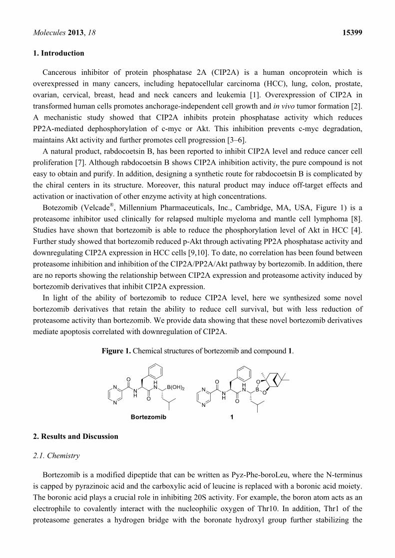
Figure 1. Chemical structures of bortezomib and compound 1.
2. Results and Discussion
2.1. Chemistry
Bortezomib is a modified dipeptide that can be written as Pyz-Phe-boroLeu, where the N-terminus
is capped by pyrazinoic acid and the carboxylic acid of leucine is replaced with a boronic acid moiety.
The boronic acid plays a crucial role in inhibiting 20S activity. For example, the boron atom acts as an
electrophile to covalently interact with the nucleophilic oxygen of Thr10. In addition, Thr1 of the
proteasome generates a hydrogen bridge with the boronate hydroxyl group further stabilizing the
Molecules 2013, 18 15400
whole complex. To elucidate the relationship between proteasome activity and downregulation of
CIP2A, we used a chemical approach to reduce the interaction between the boronic acid of bortezomib
and Thr1 of the proteasome by adding a bulky group to the boronic acid. The proteasome activity of the
resulting bortezomib derivative (1, Figure 1) was tested by ELISA. To explore the structure-activity
relationship of the downregulation of CIP2A, we replaced the boronic acid of bortezomib with various
functional groups yielding compounds 11–14. Moreover, we replaced the pyrazinoic ring with benzene
and methyl groups and used it as a platform to carry out structural modifications, which generated
compounds 15–20. These bortezomib derivatives were synthesized according to a general procedure
which is described in Scheme 1. The inhibition of CIP2A and the proteasome activity of these
compounds were tested by ELISA and western blot.
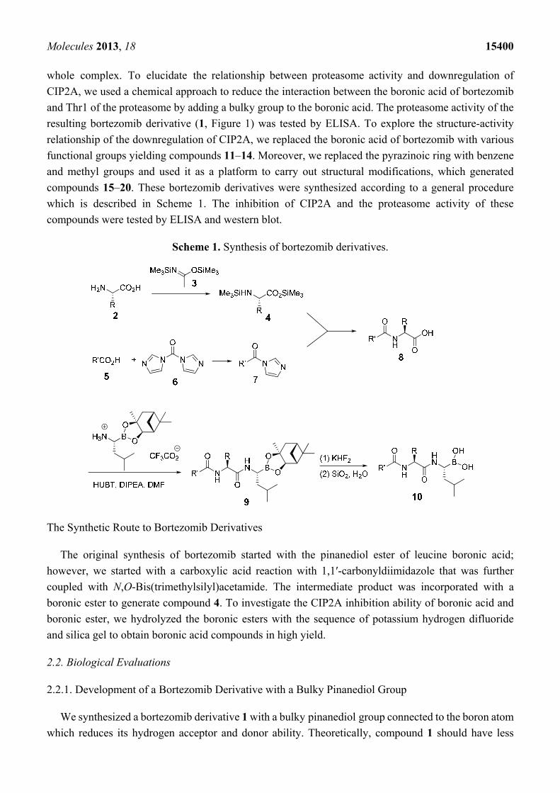
Scheme 1. Synthesis of bortezomib derivatives.
The Synthetic Route to Bortezomib Derivatives
The original synthesis of bortezomib started with the pinanediol ester of leucine boronic acid;
however, we started with a carboxylic acid reaction with 1,1′-carbonyldiimidazole that was further
coupled with N,O-Bis(trimethylsilyl)acetamide. The intermediate product was incorporated with a
boronic ester to generate compound 4. To investigate the CIP2A inhibition ability of boronic acid and
boronic ester, we hydrolyzed the boronic esters with the sequence of potassium hydrogen difluoride
and silica gel to obtain boronic acid compounds in high yield.
2.2. Biological Evaluations
2.2.1. Development of a Bortezomib Derivative with a Bulky Pinanediol Group
We synthesized a bortezomib derivative 1 with a bulky pinanediol group connected to the boron atom
which reduces its hydrogen acceptor and donor ability. Theoretically, compound 1 should have less
Molecules 2013, 18 15401
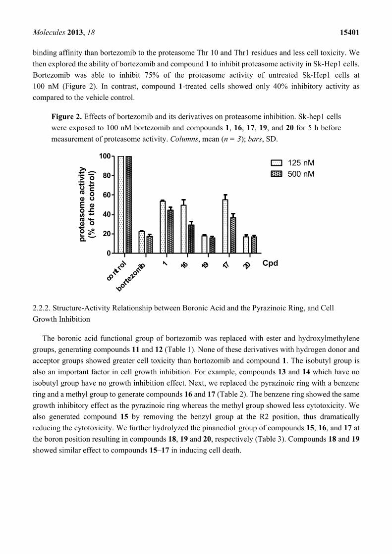
binding affinity than bortezomib to the proteasome Thr 10 and Thr1 residues and less cell toxicity. We
then explored the ability of bortezomib and compound 1 to inhibit proteasome activity in Sk-Hep1 cells.
Bortezomib was able to inhibit 75% of the proteasome activity of untreated Sk-Hep1 cells at
100 nM (Figure 2). In contrast, compound 1-treated cells showed only 40% inhibitory activity as
compared to the vehicle control.
Figure 2. Effects of bortezomib and its derivatives on proteasome inhibition. Sk-hep1 cells
were exposed to 100 nM bortezomib and compounds 1, 16, 17, 19, and 20 for 5 h before
measurement of proteasome activity. Columns, mean (n = 3); bars, SD.
2.2.2. Structure-Activity Relationship between Boronic Acid and the Pyrazinoic Ring, and Cell
Growth Inhibition
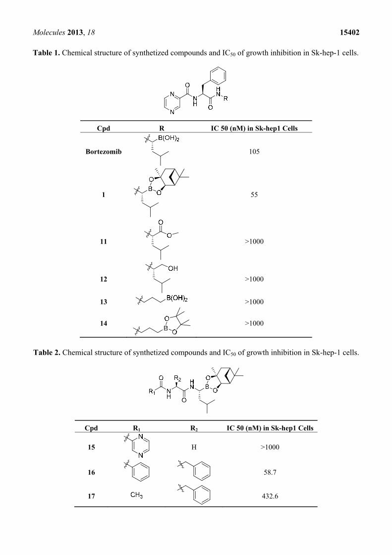
The boronic acid functional group of bortezomib was replaced with ester and hydroxylmethylene
groups, generating compounds 11 and 12 (Table 1). None of these derivatives with hydrogen donor and
acceptor groups showed greater cell toxicity than bortozomib and compound 1. The isobutyl group is
also an important factor in cell growth inhibition. For example, compounds 13 and 14 which have no
isobutyl group have no growth inhibition effect. Next, we replaced the pyrazinoic ring with a benzene
ring and a methyl group to generate compounds 16 and 17 (Table 2). The benzene ring showed the same
growth inhibitory effect as the pyrazinoic ring whereas the methyl group showed less cytotoxicity. We
also generated compound 15 by removing the benzyl group at the R2 position, thus dramatically
reducing the cytotoxicity. We further hydrolyzed the pinanediol group of compounds 15, 16, and 17 at
the boron position resulting in compounds 18, 19 and 20, respectively (Table 3). Compounds 18 and 19
showed similar effect to compounds 15–17 in inducing cell death.
cont
rol
bortezo
mib 1 16 19 17 20
0
20
40
60
80
100125 nM500 nM
Cpd
pro
teas
om
e ac
tivi
ty(%
of
the
con
tro
l)
Molecules 2013, 18 15402
Table 1. Chemical structure of synthetized compounds and IC50 of growth inhibition in Sk-hep-1 cells.
Cpd R IC 50 (nM) in Sk-hep1 Cells
Bortezomib 105
1 55
11 >1000
12 >1000
13 >1000
14 >1000
Table 2. Chemical structure of synthetized compounds and IC50 of growth inhibition in Sk-hep-1 cells.
Cpd R1 R2 IC 50 (nM) in Sk-hep1 Cells
15
H >1000
16
58.7
17 432.6
Molecules 2013, 18 15403
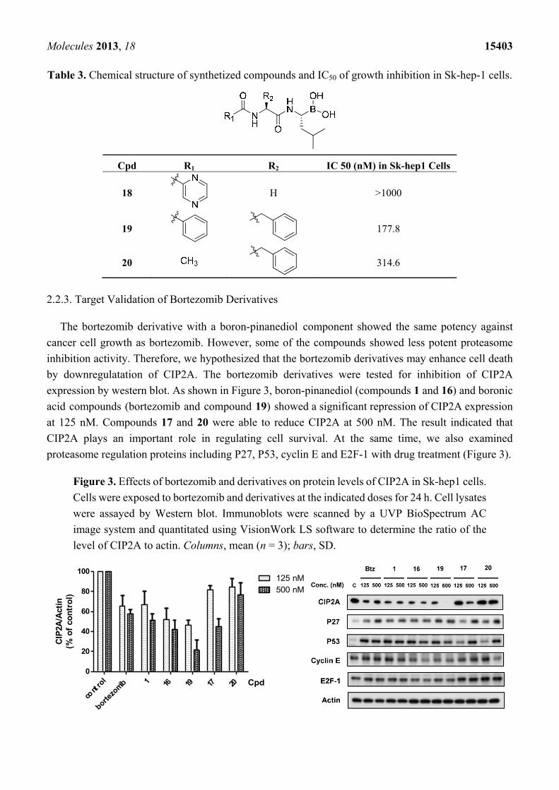
Table 3. Chemical structure of synthetized compounds and IC50 of growth inhibition in Sk-hep-1 cells.
Cpd R1 R2 IC 50 (nM) in Sk-hep1 Cells
18
H >1000
19
177.8
20 314.6
2.2.3. Target Validation of Bortezomib Derivatives
The bortezomib derivative with a boron-pinanediol component showed the same potency against
cancer cell growth as bortezomib. However, some of the compounds showed less potent proteasome
inhibition activity. Therefore, we hypothesized that the bortezomib derivatives may enhance cell death
by downregulatation of CIP2A. The bortezomib derivatives were tested for inhibition of CIP2A
expression by western blot. As shown in Figure 3, boron-pinanediol (compounds 1 and 16) and boronic
acid compounds (bortezomib and compound 19) showed a significant repression of CIP2A expression
at 125 nM. Compounds 17 and 20 were able to reduce CIP2A at 500 nM. The result indicated that
CIP2A plays an important role in regulating cell survival. At the same time, we also examined
proteasome regulation proteins including P27, P53, cyclin E and E2F-1 with drug treatment (Figure 3).
Figure 3. Effects of bortezomib and derivatives on protein levels of CIP2A in Sk-hep1 cells.
Cells were exposed to bortezomib and derivatives at the indicated doses for 24 h. Cell lysates
were assayed by Western blot. Immunoblots were scanned by a UVP BioSpectrum AC
image system and quantitated using VisionWork LS software to determine the ratio of the
level of CIP2A to actin. Columns, mean (n = 3); bars, SD.
cont
rol
bortezo
mib 1 16 19 17 20
0
20
40
60
80
100125 nM500 nM
Cpd
CIP
2A/A
ctin
(% o
f co
ntr
ol)
Molecules 2013, 18 15404
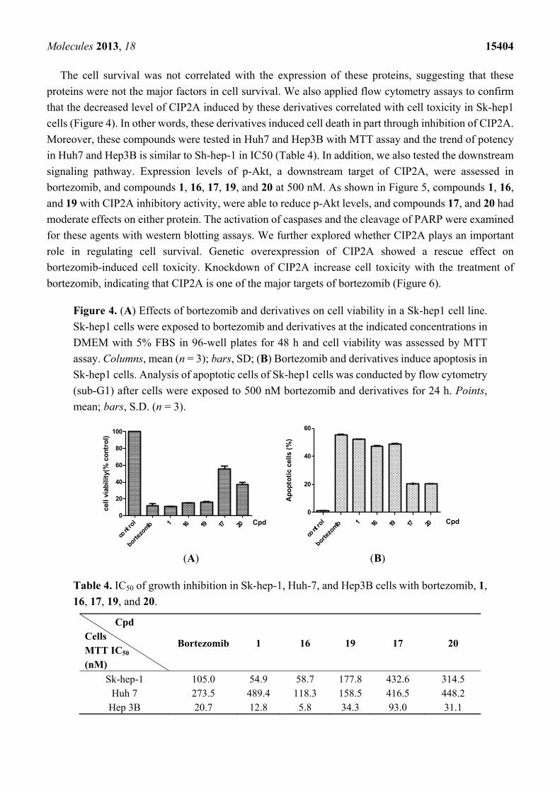
The cell survival was not correlated with the expression of these proteins, suggesting that these
proteins were not the major factors in cell survival. We also applied flow cytometry assays to confirm
that the decreased level of CIP2A induced by these derivatives correlated with cell toxicity in Sk-hep1
cells (Figure 4). In other words, these derivatives induced cell death in part through inhibition of CIP2A.
Moreover, these compounds were tested in Huh7 and Hep3B with MTT assay and the trend of potency
in Huh7 and Hep3B is similar to Sh-hep-1 in IC50 (Table 4). In addition, we also tested the downstream
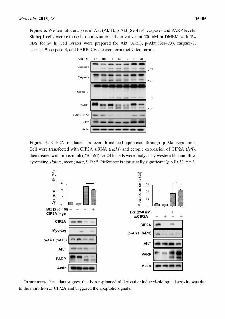
signaling pathway. Expression levels of p-Akt, a downstream target of CIP2A, were assessed in
bortezomib, and compounds 1, 16, 17, 19, and 20 at 500 nM. As shown in Figure 5, compounds 1, 16,
and 19 with CIP2A inhibitory activity, were able to reduce p-Akt levels, and compounds 17, and 20 had
moderate effects on either protein. The activation of caspases and the cleavage of PARP were examined
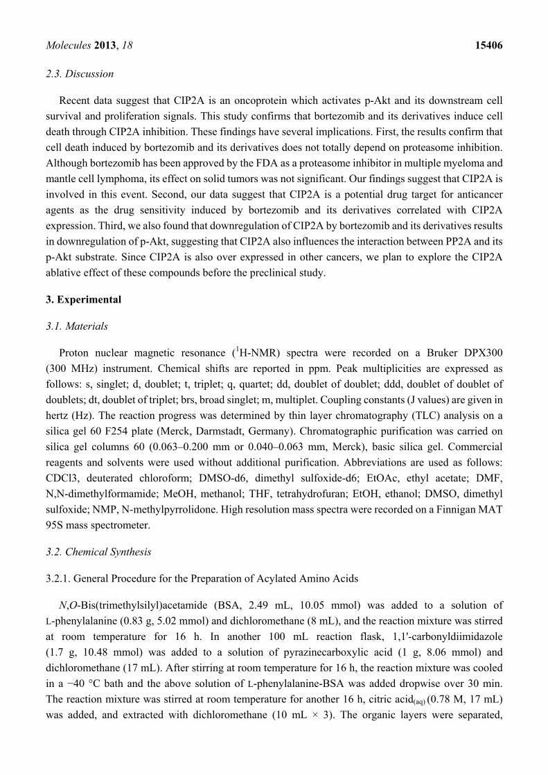
for these agents with western blotting assays. We further explored whether CIP2A plays an important
role in regulating cell survival. Genetic overexpression of CIP2A showed a rescue effect on
bortezomib-induced cell toxicity. Knockdown of CIP2A increase cell toxicity with the treatment of
bortezomib, indicating that CIP2A is one of the major targets of bortezomib (Figure 6).
Figure 4. (A) Effects of bortezomib and derivatives on cell viability in a Sk-hep1 cell line.
Sk-hep1 cells were exposed to bortezomib and derivatives at the indicated concentrations in
DMEM with 5% FBS in 96-well plates for 48 h and cell viability was assessed by MTT
assay. Columns, mean (n = 3); bars, SD; (B) Bortezomib and derivatives induce apoptosis in
Sk-hep1 cells. Analysis of apoptotic cells of Sk-hep1 cells was conducted by flow cytometry
(sub-G1) after cells were exposed to 500 nM bortezomib and derivatives for 24 h. Points,
mean; bars, S.D. (n = 3).
(A) (B)
Table 4. IC50 of growth inhibition in Sk-hep-1, Huh-7, and Hep3B cells with bortezomib, 1,
16, 17, 19, and 20.
Cpd
Cells
MTT IC50
(nM)
Bortezomib 1 16 19 17 20
Sk-hep-1 105.0 54.9 58.7 177.8 432.6 314.5 Huh 7 273.5 489.4 118.3 158.5 416.5 448.2
Hep 3B 20.7 12.8 5.8 34.3 93.0 31.1
cont
rol
bortezo
mib 1 16 19 17 200
20
40
60
80
100
Cpd
cell
via
bil
ity(
% c
on
tro
l)
cont
rol
bortezo
mib 1 16 19 17 20
0
20
40
60
Cpd
Ap
op
toti
c ce
lls
(%)
Molecules 2013, 18 15405
Figure 5. Western blot analysis of Akt (Akt1), p-Akt (Ser473), caspases and PARP levels.
Sk-hep1 cells were exposed to bortezomib and derivatives at 500 nM in DMEM with 5%
FBS for 24 h. Cell lysates were prepared for Akt (Akt1), p-Akt (Ser473), caspase-8,
caspase-9, caspase-3, and PARP. CF, cleaved form (activated form).
Figure 6. CIP2A mediated brotezomib-induced apoptosis through p-Akt regulation.
Cell were transfected with CIP2A siRNA (right) and ectopic expression of CIP2A (left),
then treated with brotezomib (250 nM) for 24 h. cells were analysis by western blot and flow
cytometry. Points, mean; bars, S.D.; * Difference is statistically significant (p < 0.05); n = 3.
In summary, these data suggest that boron-pinanediol derivative induced-biological activity was due
to the inhibition of CIP2A and triggered the apoptotic signals.
p-AKT (S473)
500 nM C Btz 1 16 19 17 20
Caspase 9
Caspase 3
Caspase 8
PARP
AKT
Actin
CF
CF
CF
CF
Btz (250 nM) CIP2A-myc
Actin
CIP2A
AKT
p-AKT (S473)
PARP
Myc-tag
Actin
CIP2A
AKT
p-AKT (S473)
PARP
0
10
20
30*
Ap
op
toti
c ce
lls
(%)
0
10
20
30
40*
Ap
op
toti
c ce
lls
(%)
Btz (250 nM) siCIP2A
Molecules 2013, 18 15406
2.3. Discussion
Recent data suggest that CIP2A is an oncoprotein which activates p-Akt and its downstream cell
survival and proliferation signals. This study confirms that bortezomib and its derivatives induce cell
death through CIP2A inhibition. These findings have several implications. First, the results confirm that
cell death induced by bortezomib and its derivatives does not totally depend on proteasome inhibition.
Although bortezomib has been approved by the FDA as a proteasome inhibitor in multiple myeloma and
mantle cell lymphoma, its effect on solid tumors was not significant. Our findings suggest that CIP2A is
involved in this event. Second, our data suggest that CIP2A is a potential drug target for anticancer
agents as the drug sensitivity induced by bortezomib and its derivatives correlated with CIP2A
expression. Third, we also found that downregulation of CIP2A by bortezomib and its derivatives results
in downregulation of p-Akt, suggesting that CIP2A also influences the interaction between PP2A and its
p-Akt substrate. Since CIP2A is also over expressed in other cancers, we plan to explore the CIP2A
ablative effect of these compounds before the preclinical study.
3. Experimental
3.1. Materials
Proton nuclear magnetic resonance (1H-NMR) spectra were recorded on a Bruker DPX300
(300 MHz) instrument. Chemical shifts are reported in ppm. Peak multiplicities are expressed as
follows: s, singlet; d, doublet; t, triplet; q, quartet; dd, doublet of doublet; ddd, doublet of doublet of
doublets; dt, doublet of triplet; brs, broad singlet; m, multiplet. Coupling constants (J values) are given in
hertz (Hz). The reaction progress was determined by thin layer chromatography (TLC) analysis on a
silica gel 60 F254 plate (Merck, Darmstadt, Germany). Chromatographic purification was carried on
silica gel columns 60 (0.063–0.200 mm or 0.040–0.063 mm, Merck), basic silica gel. Commercial
reagents and solvents were used without additional purification. Abbreviations are used as follows:
CDCl3, deuterated chloroform; DMSO-d6, dimethyl sulfoxide-d6; EtOAc, ethyl acetate; DMF,
N,N-dimethylformamide; MeOH, methanol; THF, tetrahydrofuran; EtOH, ethanol; DMSO, dimethyl
sulfoxide; NMP, N-methylpyrrolidone. High resolution mass spectra were recorded on a Finnigan MAT
95S mass spectrometer.
3.2. Chemical Synthesis
3.2.1. General Procedure for the Preparation of Acylated Amino Acids
N,O-Bis(trimethylsilyl)acetamide (BSA, 2.49 mL, 10.05 mmol) was added to a solution of
L-phenylalanine (0.83 g, 5.02 mmol) and dichloromethane (8 mL), and the reaction mixture was stirred
at room temperature for 16 h. In another 100 mL reaction flask, 1,1'-carbonyldiimidazole
(1.7 g, 10.48 mmol) was added to a solution of pyrazinecarboxylic acid (1 g, 8.06 mmol) and
dichloromethane (17 mL). After stirring at room temperature for 16 h, the reaction mixture was cooled
in a −40 °C bath and the above solution of L-phenylalanine-BSA was added dropwise over 30 min.
The reaction mixture was stirred at room temperature for another 16 h, citric acid(aq) (0.78 M, 17 mL)
was added, and extracted with dichloromethane (10 mL × 3). The organic layers were separated,
Molecules 2013, 18 15407
combined, dried over anhydrous Na2SO4 and concentrated. The crude product was recrystallized in
dichloromethane/hexanes to give N-(2-pyrazinylcarbonyl)-L-phenylalanine (8, R = benzyl; R' = pyrazinyl,
0.96 g, 3.54 mmol, 70%) as a colorless solid. [11] Mp 142–146 °C, 1H-NMR (CDCl3) δ 9.34 (s, 1H),
8.73 (s, 1H), 8.51 (s, 1H), 8.18 (d, 1H), 7.28–7.20 (m, 5H), 5.12–5.05 (m, 1H), 3.37–3.21 (m, 2H); 13C-NMR (DMSO-d6) δ 172.4, 162.6, 147.8, 144.1, 143.5, 143.4, 137.4, 129.1, 128.2, 126.5, 53.5, 36.3.
3.2.2. General Procedure for the Preparation of Dipeptide Boronates
N-[(1S)-1-[[[(1R)-1-[(3aS,4S,6S,7aR)-Hexahydro-3a,5,5-trimethyl-4,6-methano-1,3,2-benzodioxaboro
l-2-yl]-3-methylbutyl]-amino]carbonyl]-2-phenyl]-2-pyrazincarboxamide (1). Diethylisopropylamine
(0.2 mL, 0.15 g, 1.19 mmol) was added to the solution of N-(2-pyrazinylcarbonyl)-L-phenylalanine (75 mg,
0.26 mmol), (R)-BoroLeu-(+)-Pinanediol (100 mg, 0.28 mmol) and HBTU (110 mg, 0.29 mmol) in DMF
(2 mL) at 0 °C. The reaction mixture was stirred at room temperature for 16 h, the water (30 mL) was
added, and it was extracted with EtOAc (10 mL × 3). The organic layers were combined, washed with
citric acid(aq) (0.1 N, 30 mL), NaHCO3(aq) (0.1 N, 30 mL), sat. NaCl(aq) (30 mL), dried over Na2SO4,
filtered and conventrated. The crude product was purified by column chromatography (SiO2,
EtOAc/hexanes, 1:1; Rf 0.40) to give 1 [11] (100 mg, 0.19 mmol, 73%) as a colorless solid. 1H-NMR
(CDCl3) δ 9.31 (s, 1H), 8.71 (s, 1H), 8.49 (s, 1H), 8.35 (d, J = 8.4 Hz, 1H), 7.26–7.20 (m, 5H), 5.98
(m, 1H), 4.80–4.77 (m, 1H), 4.29–4.26 (d, J = 10.8 Hz, 1H), 3.17–3.08 (m, 3H), 2.33–2.25 (m, 1H),
2.18–2.12 (m, 1H), 2.03 (t, J = 5.4 Hz, 1H), 1.80–1.91 (m, 2H), 1.47–1.56 (m, 1H), 1.43 (s,3H). 1.35
(d, J = 10.7 Hz, 1H), 1.31–1.47 (m, 2H), 1.28 (s, 3H), 0.86 (s, 3H), 0.85 (d, J = 5.0 Hz, 3H), 0.84 (d,
J = 5.0 Hz, 3H); 13C-NMR (CDCl3) δ 170.5, 162.5, 147.1, 144.0, 142.5, 136.3, 129.2, 128.2, 126.6,
85.3, 77.5, 53.7, 51.3, 39.8, 39.4, 38.5, 38.3, 37.9, 35.7, 35.4, 28.4, 26.9, 26.1, 25.9, 23.8, 22.7, 21.8.
3.2.3. General Procedure for the Hydrolysis of Boronic Esters
(S)-(3-(3-Phenyl-2-(pyrazine-2-carboxamido)propanamido)propyl)boronic acid (13). Potassium
hydrogen difluoride (4.5 M, 0.41 mmol) was added to a solution of (S)-N-(1-oxo-3-phenyl-
1-((3-(4,4,5,5-tetramethyl-1,3,2-dioxaborolan-2-yl)propyl)amino)propan-2-yl)pyrazine-2-carboxamide
(19 mg, 0.043 mmol) and methanol (0.5 mL). The reaction mixture was stirred at room temperature for
4 h and concentrated. The residue was added with water (0.4 mL) and silica gel (8 mg, 0.13 mmol),
and the suspension was stirred for another 5 h at room temperature. The reaction mixture was diluted
with EtOAc (5 mL), filtered, dried over Na2SO4, and concentrated to give product 13 (10 mg,
0.028 mmol, 21%). 1H-NMR (acetone-d6) δ 9.18 (s, 1H), 8.82 (s, 1H), 8.65 (s, 1H), 8.47
(s, 1H), 7.50 (s, 1H), 7.28–7.15 (m, 5H), 4.90–4.83 (m, 1H), 3.24–3.13 (m, 4H), 1.55 (t, J = 7.2 Hz,
2H), 0.68 (t, J = 7.8 Hz, 2H); 13C-NMR (acetone-d6) δ 170.4, 162.3, 147.7, 144.5, 143.7, 143.2, 137.3,
129.4, 128.2, 126.5, 54.3, 41.3, 38.5, 29.6, 24.4; HRMS (ESI) [M−H]− calcd for C17H20BN4O4
355.1578, found 355.1575.
(S)-Methyl 4-methyl-2-((S)-3-phenyl-2-(pyrazine-2-carboxamido)propanamido)pentanoate (11): 1H-NMR
(CDCl3) δ 9.31 (s, 1H), 8.71 (s, 1H), 8.50 (s, 1H), 8.32 (d, J = 8.4 Hz, 1H), 7.28–7.15 (m, 5H), 6.21 (d,
J = 7.2 Hz, 1H), 4.81 (dt, J = 7.2 Hz, J = 7.2 Hz, 1H), 4.49, (m, 1H), 3.66 (s, 3H), 3.17, (d, J = 7.2 Hz,
Molecules 2013, 18 15408 2H), 1.54–1.42 (m, 3H), 0.82 (d, J = 5.5 Hz, 6H); 13C-NMR (CDCl3) δ 172.3, 170.2, 162.9, 147.4,
144.2, 144.1, 142.7, 136.2, 129.3, 128.5, 126.9, 54.4, 52.2, 50.9, 41.2, 38.4, 24.7, 22.5, 21.8 [11].
N-((S)-1-(((S)-1-Hydroxy-4-methylpentan-2-yl)amino)-1-oxo-3-phenylpropan-2-yl)pyrazine-2-carboxa
mide (12): 1H-NMR (CDCl3) δ 9.29 (d, J = 1.5 Hz, 1H), 8.71 (d, J = 2.4 Hz, 1H), 8.50 (d,
J = 2.4 Hz, 1H), 8.42 (d, J = 8.1 Hz, 1H), 7.28–7.15 (m, 5H), 6.10 (br, 1H), 4.83 (m, 1H), 3.88
(m, 1H), 3.91-3.31 (m, 2H), 3.24–3.06 (m, 2H), 2.30–2.00 (br, 2H), 1.45–1.38 (m, 1H), 1.41 (dd,
J = 6.3 Hz, J = 6.3 Hz, 2H), 0.75 (dd, J = 6.3 Hz, J = 1.9 Hz, 6H); 13C-NMR (CDCl3) δ 170.4, 163.0,
147.5, 144.2, 143.9, 142.8, 136.5, 129.4, 128.7, 127.1, 65.1, 55.0, 50.0, 39.7, 38.8, 24.6, 22.8, 22.1;
HRMS (ESI) [M−H]− calcd for C20H25N4O3 369.1927, found 369.1922.
(S)-N-(1-Oxo-3-phenyl-1-((3-(4,4,5,5-tetramethyl-1,3,2-dioxaborolan-2-yl)propyl)amino)propan-2-yl)
pyrazine-2-carboxamide (14): 1H-NMR (CDCl3) δ 9.30 (s, 1H), 8.70 (s, 1H), 8.51 (s, 1H), 8.41 (m,
1H), 7.28–7.18 (m, 5H), 4.76–4.70 (m, 1H), 3.22–3.07 (m, 4H), 1.50–1.43 (m, 2H), 1.19 (s, 12H),
0.84–0.77 (m, 2H); 13C-NMR (CDCl3) δ 169.9, 162.7, 147.3, 144.0, 142.7, 136.5, 129.2, 128.6, 126.9,
83.1, 54.9, 41.5, 38.8, 29.6, 24.7, 23.4, 14.1; HRMS (ESI) [M+Na]+ calcd for C23H31BN4O4Na
461.2336, found 461.2331.
N-(2-(((R)-3-methyl-1-((3aR,4R,6R,7aS)-5,5,7a-trimethylhexahydro-4,6-methanobenzo[d][1,3,2]dioxa
borol-2-yl)butyl)amino)-2-oxoethyl)pyrazine-2-carboxamide (15): 1H-NMR (CDCl3) δ 9.33 (s, 1H),
8.68 (s, 1H), 8.49 (s, 1H), 7.85 (br, s, 1H), 4.33 (d, 2H), 3.98–3.92 (m, 2H), 2.82–2.92 (m, 1H),
2.43–2.35 (m, 1H), 2.28–2.22 (m, 1H), 2.13 (t, J = 5.4 Hz, 1H), 1.91–1.80 (m, 2H), 1.56–1.47 (m, 1H),
1.43 (s,3H). 1.35 (d, J = 10.7 Hz, 1H), 1.47–1.31 (m, 2H), 1.28 (s, 3H), 0.86 (s, 3H), 0.85 (d, J = 5.0 Hz,
3H), 0.84 (d, J = 5.0 Hz, 3H); 13C-NMR (CDCl3) δ 171.0, 162.7, 147.0, 144.3, 142.6, 86.2, 78.0, 53.9,
51.2, 40.5, 40.1, 39.5, 38.9, 38.4, 35.7, 29.2, 27.8, 27.0, 25.6, 24.4, 23.3, 22.3.
N-((S)-1-(((R)-3-Methyl-1-((3aR,4R,6R,7aS)-5,5,7a-trimethylhexahydro-4,6-methanobenzo[d][1,3,2]di
oxaborol-2-yl)butyl)amino)-1-oxo-3-phenylpropan-2-yl)benzamide (16): 1H-NMR (CDCl3) δ 7.71–7.70 (d,
2H), 7.48–7.45 (d, 2H), 7.26–7.20 (m, 6H), 4.84–4.75 (m, 1H), 4.30–4.27 (d, J = 6.9 Hz, 1H),
3.17–3.08 (m, 3H), 2.33–2.25 (m, 1H), 2.18–2.12 (m, 1H), 2.03 (t, J = 5.4 Hz, 1H), 1.91–1.80 (m, 2H),
1.56–1.47 (m, 1H), 1.43 (s,3H). 1.35 (d, J = 10.7 Hz, 1H), 1.47–1.31 (m, 2H), 1.28 (s, 3H), 0.86 (s,
3H), 0.85 (d, J = 5.0 Hz, 3H), 0.84 (d, J = 5.0 Hz, 3H); 13C-NMR (CDCl3) δ 170.4, 162.8, 137.2, 133.9,
131.6, 129.4, 128.5, 128.4, 127.1, 126.7, 55.2, 37.5, HRMS (FAB) [M+H]+ calcd for C31H42O4N2B
517.3238, found 517.3232.
(S)-2-Acetamido-N-((R)-3-methyl-1-((3aR,4R,6R,7aS)-5,5,7a-trimethylhexahydro-4,6-methanobenzo[d
][1,3,2]dioxaborol-2-yl)butyl)-3-phenylpropanamide (17): 1H-NMR (CDCl3) δ 7.24–7.16 (m, 5H),
4.58–4.63 (m, 1H), 4.21–4.20 (d, J = 5.1 Hz, 1H), 3.05–2.98 (m, 3H), 2.33–2.25 (m, 1H), 2.18–2.12
(m, 1H), 2.03 (t, J = 5.4 Hz, 1H), 1.97 (s,3H) 1.80–1.91 (m, 2H), 1.47–1.56 (m, 1H), 1.43 (s,3H). 1.35
(d, J = 10.7 Hz, 1H), 1.31–1.47 (m, 2H), 1.28 (s, 3H), 0.86 (s, 3H), 0.85 (d, J = 5.0 Hz, 3H), 0.84 (d,
J = 5.0 Hz, 3H); 13C-NMR (CDCl3) δ 171.3, 169.9, 136.6, 129.3, 128.4, 126.7, 85.5, 77.6, 53.8, 51.3,
Molecules 2013, 18 15409 39.9, 39.5, 38.5, 38.4, 38.0, 35.5, 28.5, 27.0, 26.2, 25.2, 23.9, 22.9, 21.9; HRMS (ESI) [M+Na]+ calcd
for C26H39BN2O4Na 477.2901, found 477.2892.
(R)-(3-Methyl-1-(2-(pyrazine-2-carboxamido)acetamido)butyl)boronic acid (18): 1H-NMR (acetone-d6)
δ 9.28 (s, 1H), 8.88 (s, 1H), 8.67 (s, 1H), 5.80–5.60 (m, 2H), 2.60–2.53 (m, 1H), 1.89 (s,3H),
1.65–1.51 (m, 1H), 1.40–1.12 (m, 2H), 0.88–0.77 (m, 6H); 13C-NMR (CDCl3) δ 171.0, 162.7, 147.0,
144.3, 142.6, 142.1, 77.4, 50.3, 29.6, 27.3, 27.0.
((R)-1-((S)-2-Benzamido-3-phenylpropanamido)-3-methylbutyl)boronic Acid (19): 1H-NMR
(acetone-d6) δ 7.85–7.81 (d, 2H), 7.48–7.45 (d, 2H), 7.26–7.20 (m, 6H), 4.89–4.79 (m, 1H), 3.31–3.21
(m, 2H), 3.14–3.11 (m, 1H), 1.68–1.55 (m, 1H), 1.40–1.12 (m, 2H), 0.88–0.77 (m, 6H); 13C-NMR
(CDCl3) δ 171.0, 162.7, 141.2, 134.7, 133.4, 132.6, 77.4, 56.5, 30.7, 28.0, 27.8; HRMS (ESI) [M+Na]+
calcd for C21H27BN2O4Na 405.1962, found 405.1965.
((R)-1-((S)-2-Acetamido-3-phenylpropanamido)-3-methylbutyl)boronic Acid (20): 1H-NMR (acetone-d6)
δ 7.29–7.23 (m, 5H), 4.95–4.87 (m, 1H), 3.12–3.09 (d, J = 7.8 Hz, 2H), 2.60–2.53 (m, 1H), 1.89 (s, 3H),
1.65–1.51 (m, 1H), 1.40–1.12 (m, 2H), 0.88–0.77 (m, 6H); 13C-NMR (CDCl3) δ 171.3, 169.9, 136.6,
129.3, 128.4, 126.7, 53.8, 51.3, 29.6, 27.9, 27.3, 27.0.
3.3. Biological Assay
3.3.1. Cell Culture
The Sk-Hep1 cell line was obtained from American Type Culture Collection (Manassas, VA, USA).
Cells were maintained in Dulbecco’s modified Eagle’s medium supplemented with 10% fetal bovine
serum, 100 units/mL, penicillin G, 100 mg/mL streptomycin sulfate and 25 mg/mL amphotericin B in a
37 °C humidified incubator and an atmosphere of 5% CO2 in air.
3.3.2. Proteasome Activity Assay
A 20S Proteasome Activity Assay kit (Chemicon, Billerica, MA, USA) was used to determine the
inhibition of proteasome in drug-treated Sk-Hep1 cells. Cell lysates were prepared, and the fluorogenic
peptide substrate, Suc-Leu-Leu-Val-Tyr-AMC was used according to the manufacturer’s instructions. In
brief, control or compound-treated cells were broken in a lysis buffer without protease inhibitors. Total
cell lysate (50 μg) was incubated with 20 μmol/L of fluorogenic substrate Suc-Leu-Leu-Val-Tyr-AMC
at 45 °C in 100 μL of assay buffer. Free AMC liberated by the substrate hydrolysis was quantified for
90 min at 1-min intervals on a microtiter plate fluorometrer (excitation, 355 nm; emission, 460 nm). The
percentage of proteasome activity values (% control) were derived by dividing the slope obtained in the
presence of bortezomib or compounds by the slope obtained in its absence ×100 [12].
3.3.3. Western Blot
Lysates of Sk-hep1 treated with bortezomib and derivatives at the indicated concentrations for 24 h
were prepared for immunoblotting of CIP2A, Akt (Akt1), p27, Cyclin E and PARP (Santa Cruz
Molecules 2013, 18 15410
Biotechnology, Santa Cruz, CA, USA), p53, E2F-1, p-Akt (Ser473), caspase-8, caspase-9 and caspase-3
(Cell Signaling Technology, Danvers, MA, USA), -Actin (Sigma, Steinheim, Germany).
3.3.4. Cell Viability Analysis
The effect of bortezomib and its derivatives on Sk-hep1, Huh7, and Hep3B cell viability was assessed
by the 3-(4,5-dimethylthiazol-2-yl)-2,5-diphenyltetrazolium bromide (MTT) assay in 12 replicates.
3.3.5. Apoptosis Analysis
Apoptotic cells were measured by flow cytometry (sub-G1). After treatment with compounds, cells
were trypsinized, collected by centrifugation and resuspended in PBS. After centrifugation, the cells
were washed in PBS and resuspended in potassium iodide (PI) staining solution. Specimens were
incubated in the dark for 30 min at 37 °C and then analyzed with an EPICS Profile II flow cytometer
(Coulter Corp., Hialeah, FL, USA). All experiments were performed in triplicate.
4. Conclusions
We synthesized a series of bortezomib derivatives that showed CIP2A inhibition activity, and the
inhibition of CIP2A-Akt signaling pathway correlated with the cytotoxicity of these derivatives in HCC
cells. Several compounds were found equally as potent as bortezomib in inhibition of CIP2A and cell
growth. A mechanistic study showed that compound 1 also reduced expression of p-Akt after repressing
the CIP2A signaling cascade. In addition, compound 1 provides a useful pharmacological tool to the
study therapeutic relevance of HCC with unregulated CIP2A expression and drug resistance. These
compounds will be tested in other cancers. Moreover, a series of new derivatives provide structure
activity relationship which provide a direction for future lead optimization of CIP2A inhibitors.
Therefore, these data suggest the potential value of anti-CIP2A agents in the treatment of cancers.
Testing of compound 1 in an in vivo HCC model and pre-clincal studies are currently being pursued.
Supplementary Material
Supplementary materials can be accessed at: http://www.mdpi.com/1420-3049/18/12/15398/s1.
Acknowledgments
We thank the National Science Council, Taiwan (NSC99-2314-B-002-017-MY2,
NSC101-2325-B-002-032, NSC-98-2119-M-008-001-MY-3, NSC98-2320-B-010-005-MY3 and
NSC-101-2325-B-010-007) for financial support. We are grateful to Ping-Yu Lin at the Institute of
Chemistry, Academia Sinica, and Valuable Instrument Central University for obtaining mass analysis.
Conflicts of Interest
The authors declare no conflict of interest.
Molecules 2013, 18 15411
References
1. Junttila, M.R.; Puustinen, P.; Niemela, M.; Ahola, R.; Arnold, H.; Bottzauw, T.; Ala-aho, R.;
Nielsen, C.; Ivaska, J.; Taya, Y.; et al. CIP2A inhibits PP2A in human malignancies. Cell 2007,
130, 51–62.
2. Sablina, A.A.; Hahn, W.C. SV40 small T antigen and PP2A phosphatase in cell transformation.
Cancer Metastasis Rev. 2008, 27, 137–146.
3. Khanna, A.; Bockelman, C.; Hemmes, A.; Junttila, M.R.; Wiksten, J.P.; Lundin, M.; Junnila, S.;
Murphy, D.J.; Evan, G.I.; Haglund, C.; et al. MYC-dependent regulation and prognostic role of
CIP2A in gastric cancer. J. Natl. Cancer Inst. 2009, 101, 793–805.
4. Chen, K.F.; Liu, C.Y.; Lin, Y.C.; Yu, H.C.; Liu, T.H.; Hou, D.R.; Chen, P.J.; Cheng, A.L. CIP2A
mediates effects of bortezomib on phospho-Akt and apoptosis in hepatocellular carcinoma cells.
Oncogene 2010, 29, 6257–3266.
5. Chen, K.F.; Yeh, P.Y.; Hsu, C.; Hsu, C.H.; Lu, Y.S.; Hsieh, H.P.; Chen, P.J.; Cheng, A.L.
Bortezomib overcomes tumor necrosis factor-related apoptosis-inducing ligand resistance in
hepatocellular carcinoma cells in part through the inhibition of the phosphatidylinositol
3-kinase/Akt pathway. J. Biol. Chem. 2009, 284, 11121–11133.
6. Tseng, L.M.; Liu, C.Y.; Chang, K.C.; Chu, P.Y.; Shiau, C.W.; Chen, K.F. CIP2A is a target of
bortezomib in human triple negative breast cancer cells. Breast Cancer Res. 2012, 14, R68.
7. Ma, L.; Wen, Z.S.; Liu, Z.; Hu, Z.; Ma, J.; Chen, X.Q.; Liu, Y.Q.; Pu, J.X.; Xiao, W.L.;
Sun, H.D.; et al. Overexpression and small molecule-triggered downregulation of CIP2A in lung
cancer. PLoS One 2011, 6, e20159.
8. Nencioni, A.; Grunebach, F.; Patrone, F.; Ballestrero, A.; Brossart, P. Proteasome inhibitors:
Antitumor effects and beyond. Leukemia 2007, 21, 30–36.
9. Chen, K.F.; Yu, H.C.; Liu, C.Y.; Chen, H.J.; Chen, Y.C.; Hou, D.R.; Chen, P.J.; Cheng, A.L.
Bortezomib sensitizes HCC cells to CS-1008, an antihuman death receptor 5 antibody, through
the inhibition of CIP2A.. Mol. Cancer Ther. 2011, 10, 892–901.
10. Chen, K.F.; Yu, H.C.; Liu, T.H.; Lee, S.S.; Chen, P.J.; Cheng, A.L. Synergistic interactions
between sorafenib and bortezomib in hepatocellular carcinoma involve PP2A-dependent Akt
inactivation. J. Hepatol. 2010, 52, 88–95.
11. Zhu, Y.; Yao, S.; Xu, B.; Ge, Z.; Cui, J.; Cheng, T.; Li, R. Design, synthesis and biological
evaluation of tripeptide boronic acid proteasome inhibitors. Bioorgan. Med. Chem. 2009, 17,
6851–6861.
12. Codony-Servat, J.; Tapia, M.A.; Bosch, M.; Oliva, C.; Domingo-Domenech, J.; Mellado, B.;
Rolfe, M.; Ross, J.S.; Gascon, P.; Rovira, A.; et al. Differential cellular and molecular effects of
bortezomib, a proteasome inhibitor, in human breast cancer cells. Mol. Cancer Ther. 2006, 5,
665–675
Sample Availability: Samples of the compounds are available from the authors.
© 2013 by the authors; licensee MDPI, Basel, Switzerland. This article is an open access article
distributed under the terms and conditions of the Creative Commons Attribution license
(http://creativecommons.org/licenses/by/3.0/).