Bordetella parapertussis PagP Mediates the Addition of Two ... · the chloramphenicol resistance...
9
Bordetella parapertussis PagP Mediates the Addition of Two Palmitates to the Lipopolysaccharide Lipid A L. E. Hittle, a J. W. Jones, b A. M. Hajjar, c R. K. Ernst, a A. Preston d Department of Microbial Pathogenesis, University of Maryland School of Dentistry, Baltimore, Maryland, USA a ; Department of Pharmaceutical Sciences, University of Maryland School of Pharmacy, Baltimore, Maryland, USA b ; Department of Comparative Medicine, School of Medicine, University of Washington, Seattle, Washington, USA c ; Department of Biology and Biochemistry, University of Bath, Bath, United Kingdom d Bordetella bronchiseptica PagP (PagP BB ) is a lipid A palmitoyl transferase that is required for resistance to antibody-dependent complement-mediated killing in a murine model of infection. B. parapertussis contains a putative pagP homolog (encoding B. parapertussis PagP [PagP BPa ]), but its role in the biosynthesis of lipid A, the membrane anchor of lipopolysaccharide (LPS), has not been investigated. Mass spectrometry analysis revealed that wild-type B. parapertussis lipid A consists of a heterogeneous mixture of lipid A structures, with penta- and hexa-acylated structures containing one and two palmitates, respectively. Through mutational analysis, we demonstrate that PagP BPa is required for the modification of lipid A with palmitate. While PagP BB trans- fers a single palmitate to the lipid A C-3= position, PagP BPa transfers palmitates to the lipid A C-2 and C-3= positions. The addi- tion of two palmitate acyl chains is unique to B. parapertussis. Mutation of pagP BPa resulted in a mutant strain with increased sensitivity to antimicrobial peptide killing and decreased endotoxicity, as evidenced by reduced proinflammatory responses via Toll-like receptor 4 (TLR4) to the hypoacylated LPS. Therefore, PagP-mediated modification of lipid A regulates outer mem- brane function and may be a means to modify interactions between the bacterium and its human host during infection. T he genus Bordetella contains nine species, three of which have been studied in detail: B. pertussis, B. parapertussis, and B. bronchiseptica. B. pertussis and B. parapertussis are the causative agents of whooping cough. While long considered a disease of infants and children, it is now recognized that infections occur in adults, although they are milder than the classic whooping cough described in children (1–5). B. pertussis is considered the major pathogen of whooping cough; however, the prevalence of B. parapertussis in disease is not well understood but may be a signif- icant contributor to the overall burden (6). Symptoms of B. parap- ertussis and B. pertussis disease are clinically indistinguishable and include a prolonged cough, whooping, paroxysms, vomiting, and cyanosis (7), leading to increased mortality in infected children; however, B. parapertussis is often considered to cause milder dis- ease than B. pertussis (7). In many countries, whooping cough is controlled by vaccination against B. pertussis. However, many countries have recently suffered major outbreaks despite contin- uous high levels of vaccine coverage, initiating renewed interest in whooping cough vaccinology. The acellular pertussis vaccines (DTaP and Tdap) used in most developed countries do not result in protection against B. parapertussis, likely due to the B. paraper- tussis lipopolysaccharide (LPS) O antigen inhibiting the binding of antibodies to the bacterium (8). LPS is the major component of the outer leaflet of the outer membrane of Gram-negative bacteria. The lipid A region imparts the endotoxin activity of LPS (9) that arises when lipid A binds to a host membrane complex that includes and activates signaling by Toll-like receptor 4 (TLR4) (10), resulting in the expression of many proinflammatory cytokines and chemokines in cells of the innate immune system. TLR4 signaling is also important for the activation of adaptive immunity, particularly through the activa- tion of dendritic cells (DCs), which act as antigen-presenting cells (APCs) for T cells. Thus, lipid A-TLR4 interactions are central to the host immune response to Gram-negative bacteria, including Bordetella species. Bordetella subspecies exhibit extensive heterogeneity in their lipid A structure, arising from both species-specific differences in and species-variable expression of specific lipid A-modifying en- zymes (11, 12). Previously, we demonstrated that B. bronchiseptica pagP (pagP BB ) encodes a lipid A palmitoyl transferase that is re- sponsible for the presence of palmitate in lipid A. The expression of pagP BB is regulated by the Bvg two-component system (13) that regulates the expression of many Bordetella genes that are involved in pathogenesis (14). pagP BB is maximally expressed in the Bvg- plus (Bvg ) phase that is adopted when the system is active and includes the expression of many of the toxins and adhesins con- sidered important for infection. When the Bvg system is inactive, the bacteria adopt the Bvg-minus (Bvg ) phase, and the expres- sion of these virulence genes is downregulated. Thus, the Bvg and Bvg phases are regarded as the states that the bacteria adopt upon entering the host respiratory tract and the environment, respectively (14). We also showed that pagP BB is required for the persistence of B. bronchiseptica within the mouse respiratory tract through resistance to antibody-dependent complement-medi- ated killing (13, 15). This represented the first description of a Received 20 August 2014 Accepted 14 November 2014 Accepted manuscript posted online 24 November 2014 Citation Hittle LE, Jones JW, Hajjar AM, Ernst RK, Preston A. 2015. Bordetella parapertussis PagP mediates the addition of two palmitates to the lipopolysaccharide lipid A. J Bacteriol 197:572–580. doi:10.1128/JB.02236-14. Editor: V. J. DiRita Address correspondence to R. K. Ernst, [email protected], or A. Preston, [email protected]. Supplemental material for this article may be found at http://dx.doi.org/10.1128 /JB.02236-14. Copyright © 2015, American Society for Microbiology. All Rights Reserved. doi:10.1128/JB.02236-14 572 jb.asm.org February 2015 Volume 197 Number 3 Journal of Bacteriology on August 18, 2019 by guest http://jb.asm.org/ Downloaded from
Transcript of Bordetella parapertussis PagP Mediates the Addition of Two ... · the chloramphenicol resistance...
Bordetella parapertussis PagP Mediates the Addition of Two Palmitatesto the Lipopolysaccharide Lipid A
L. E. Hittle,a J. W. Jones,b A. M. Hajjar,c R. K. Ernst,a A. Prestond
Department of Microbial Pathogenesis, University of Maryland School of Dentistry, Baltimore, Maryland, USAa; Department of Pharmaceutical Sciences, University ofMaryland School of Pharmacy, Baltimore, Maryland, USAb; Department of Comparative Medicine, School of Medicine, University of Washington, Seattle, Washington,USAc; Department of Biology and Biochemistry, University of Bath, Bath, United Kingdomd
Bordetella bronchiseptica PagP (PagPBB) is a lipid A palmitoyl transferase that is required for resistance to antibody-dependentcomplement-mediated killing in a murine model of infection. B. parapertussis contains a putative pagP homolog (encoding B.parapertussis PagP [PagPBPa]), but its role in the biosynthesis of lipid A, the membrane anchor of lipopolysaccharide (LPS), hasnot been investigated. Mass spectrometry analysis revealed that wild-type B. parapertussis lipid A consists of a heterogeneousmixture of lipid A structures, with penta- and hexa-acylated structures containing one and two palmitates, respectively. Throughmutational analysis, we demonstrate that PagPBPa is required for the modification of lipid A with palmitate. While PagPBB trans-fers a single palmitate to the lipid A C-3= position, PagPBPa transfers palmitates to the lipid A C-2 and C-3= positions. The addi-tion of two palmitate acyl chains is unique to B. parapertussis. Mutation of pagPBPa resulted in a mutant strain with increasedsensitivity to antimicrobial peptide killing and decreased endotoxicity, as evidenced by reduced proinflammatory responses viaToll-like receptor 4 (TLR4) to the hypoacylated LPS. Therefore, PagP-mediated modification of lipid A regulates outer mem-brane function and may be a means to modify interactions between the bacterium and its human host during infection.
The genus Bordetella contains nine species, three of which havebeen studied in detail: B. pertussis, B. parapertussis, and B.
bronchiseptica. B. pertussis and B. parapertussis are the causativeagents of whooping cough. While long considered a disease ofinfants and children, it is now recognized that infections occur inadults, although they are milder than the classic whooping coughdescribed in children (1–5). B. pertussis is considered the majorpathogen of whooping cough; however, the prevalence of B.parapertussis in disease is not well understood but may be a signif-icant contributor to the overall burden (6). Symptoms of B. parap-ertussis and B. pertussis disease are clinically indistinguishable andinclude a prolonged cough, whooping, paroxysms, vomiting, andcyanosis (7), leading to increased mortality in infected children;however, B. parapertussis is often considered to cause milder dis-ease than B. pertussis (7). In many countries, whooping cough iscontrolled by vaccination against B. pertussis. However, manycountries have recently suffered major outbreaks despite contin-uous high levels of vaccine coverage, initiating renewed interest inwhooping cough vaccinology. The acellular pertussis vaccines(DTaP and Tdap) used in most developed countries do not resultin protection against B. parapertussis, likely due to the B. paraper-tussis lipopolysaccharide (LPS) O antigen inhibiting the bindingof antibodies to the bacterium (8).
LPS is the major component of the outer leaflet of the outermembrane of Gram-negative bacteria. The lipid A region impartsthe endotoxin activity of LPS (9) that arises when lipid A binds toa host membrane complex that includes and activates signaling byToll-like receptor 4 (TLR4) (10), resulting in the expression ofmany proinflammatory cytokines and chemokines in cells of theinnate immune system. TLR4 signaling is also important for theactivation of adaptive immunity, particularly through the activa-tion of dendritic cells (DCs), which act as antigen-presenting cells(APCs) for T cells. Thus, lipid A-TLR4 interactions are central tothe host immune response to Gram-negative bacteria, includingBordetella species.
Bordetella subspecies exhibit extensive heterogeneity in theirlipid A structure, arising from both species-specific differences inand species-variable expression of specific lipid A-modifying en-zymes (11, 12). Previously, we demonstrated that B. bronchisepticapagP (pagPBB) encodes a lipid A palmitoyl transferase that is re-sponsible for the presence of palmitate in lipid A. The expressionof pagPBB is regulated by the Bvg two-component system (13) thatregulates the expression of many Bordetella genes that are involvedin pathogenesis (14). pagPBB is maximally expressed in the Bvg-plus (Bvg�) phase that is adopted when the system is active andincludes the expression of many of the toxins and adhesins con-sidered important for infection. When the Bvg system is inactive,the bacteria adopt the Bvg-minus (Bvg�) phase, and the expres-sion of these virulence genes is downregulated. Thus, the Bvg�
and Bvg� phases are regarded as the states that the bacteria adoptupon entering the host respiratory tract and the environment,respectively (14). We also showed that pagPBB is required for thepersistence of B. bronchiseptica within the mouse respiratory tractthrough resistance to antibody-dependent complement-medi-ated killing (13, 15). This represented the first description of a
Received 20 August 2014 Accepted 14 November 2014
Accepted manuscript posted online 24 November 2014
Citation Hittle LE, Jones JW, Hajjar AM, Ernst RK, Preston A. 2015. Bordetellaparapertussis PagP mediates the addition of two palmitates to thelipopolysaccharide lipid A. J Bacteriol 197:572–580. doi:10.1128/JB.02236-14.
Editor: V. J. DiRita
Address correspondence to R. K. Ernst, [email protected], or A. Preston,[email protected].
Supplemental material for this article may be found at http://dx.doi.org/10.1128/JB.02236-14.
Copyright © 2015, American Society for Microbiology. All Rights Reserved.
doi:10.1128/JB.02236-14
572 jb.asm.org February 2015 Volume 197 Number 3Journal of Bacteriology
on August 18, 2019 by guest
http://jb.asm.org/
Dow
nloaded from
direct role for the pagP gene in virulence for Bordetella. B. parap-ertussis also contains a pagP gene, but B. parapertussis lipid A con-tains two secondary palmitates, compared to a single palmitateresidue in B. bronchiseptica (16). Here we present characterizationof B. parapertussis pagP (pagPBPa), its role in lipid A biosynthesis,and the role of lipid A palmitoylation in B. parapertussis outermembrane function.
MATERIALS AND METHODSBacterial strains and plasmids. Escherichia coli XL1-Blue (Agilent Tech-nologies, Wokingham, United Kingdom) or Top10 (Invitrogen Life Tech-nologies, Paisley, United Kingdom) cells were used for cloning and main-tenance of plasmids. E. coli SM10�pir (17) or CC118�pir was used as thedonor strain in conjugations. The pCR2.1 Topo or pCR8 Gateway entryvector (Invitrogen Life Technologies) was used to clone PCR products.pEX100T (18) was used as a suicide vector for allelic-exchange mutagen-esis in B. parapertussis. pBBR1kan is a derivative of pBBR1MCS in whichthe chloramphenicol resistance cassette has been replaced with a kanamy-cin resistance cassette. pBBR1MCS is a broad-host-range vector that iscapable of replication in Bordetella (19). pBBR1kan was used to introducea plasmid-borne pagP gene into the B. parapertussis wild type (WT) andthe pagP mutant.
Bacterial growth media and conditions. B. parapertussis was grownon charcoal agar (BD, Oxford, United Kingdom) at 37°C. E. coli wasgrown on LB agar at 37°C. For liquid culture, B. parapertussis was grown inStainer-Scholte broth (20) modified by the addition of Casamino Acids(50g liter�1) at 37°C with shaking, and E. coli was grown in LB broth at37°C with aeration. For growth of B. parapertussis in the Bvg� phase,MgSO4 was added to growth medium to a concentration of 50 mM. Strep-tomycin (200 �g ml�1), ampicillin (100 �g ml�1), erythromycin (10 �gml�1), and kanamycin (50 �g ml�1) were used where appropriate.
Chemicals and reagents. Chemicals and reagents were obtained fromBD, Fisher Scientific (Loughborough, United Kingdom), or Sigma-Al-drich (Gillingham, United Kingdom).
DNA purification. Plasmid DNA was purified by using an Invitrogenplasmid DNA purification kit according to the manufacturer’s instruc-tions. Genomic DNA was purified by using the agarose plug method (21).
DNA manipulation. DNA manipulations were performed accordingto standard methods. DNA restriction and modifying enzymes were ob-tained from New England BioLabs (Hitchin, United Kingdom), Roche(Burgess Hill, United Kingdom), or Invitrogen Life Technologies.
PCR and primers. A genomic DNA template was made by resuspend-ing plate-grown bacteria in 0.5 ml of water, boiling in a water bath for 5min, spinning at top speed in a bench-top microcentrifuge for 2 min, andtaking 0.2 ml of the supernatant. One microliter of supernatant was usedper PCR. Each 50-�l PCR mixture was comprised of the genomic DNAtemplate, buffer as directed by the manufacturer, deoxynucleosidetriphosphates (dNTPs) (25 �M each), 20 ng of each primer, 5% (vol/vol)dimethyl sulfoxide (DMSO), 5 mM MgCl2, and 2.5 units of Taq DNApolymerase (Promega, Southampton, United Kingdom). Primers used toamplify the B. parapertussis pagP locus were PagPF (5=-CTCCGCATTCCAGGTAGGCC-3=) and PagPR (5=-TTAGAACTCCCAGCGGCCAAAC-3=). PCR mixtures were incubated at 94°C for 5 min followed by 30 cyclesof 94°C for 75 s, 60°C for 75 s, and 72°C for 90 s, followed by a final step at72°C for 7 min.
Conjugations. Conjugations were performed as described previously(22).
Complementation of the pagP mutation. The region encompassingthe putative pagP promoter and the pagP coding sequence was amplifiedfrom either B. parapertussis or B. bronchiseptica by PCR, and the PCRproduct was cloned into the pCR8/GW/Topo cloning vector. The insertwas subcloned into pBBR1kan that had been modified to a Gateway des-tination vector by using the Invitrogen Gateway cloning system. This con-struct was introduced into B. parapertussis by conjugation.
SDS-PAGE/silver stain analysis of LPS. Whole-cell-lysate LPS sam-ples were prepared and analyzed by sodium dodecyl sulfate (SDS)-PAGE/silver staining, as previously described (23).
Peptide killing assays. Bacteria were grown on agar plates for 3 daysand then resuspended in phosphate-buffered saline (PBS). The suspen-sions were diluted to 104 bacteria/190 �l. Ten microliters of PBS contain-ing C18G at a final concentration of 0, 1, 2.5, 5, or 10 �g ml�1 was addedto each suspension, which was then incubated at 37°C for 30 min. Eachpeptide concentration was tested in triplicate. Suspensions were seriallydiluted in PBS, and 100 �l of dilutions was plated onto agar. Plates wereincubated at 37°C for 3 days, and colonies were counted to enumerate thebacteria that had survived the assay. Differences in the numbers of surviv-ing WT and mutant bacteria were compared by using a Student t test. Theexperiment was performed three times.
TLR4 stimulation assays. Reporter cell lines and stimulation assayswere performed as described previously (20).
Lipid A structural analyses. (i) Matrix-assisted laser desorption ion-ization–time of flight/time of flight mass spectrometry. LPS was purifiedfrom 1 liter of cultures grown overnight, as described previously (24).Further treatment of LPS with RNase A, DNase I, and proteinase K en-sured the removal of contaminating nucleic acids and proteins (25). Hy-drolysis of LPS to isolate lipid A was accomplished with 1% SDS at pH 4.5,as described previously (26).
Lipid A was analyzed by matrix-assisted laser desorption ionization(MALDI) in the negative-ion mode on a 4700 proteomics analyzer (Ap-plied Biosystems, Framingham, MA). Samples were dissolved in 10 �l of amixture of 5-chloro-2-mercaptobenzothiazole (20 mg ml�1) in chloro-form-methanol-water (4:4:1, vol/vol/vol), and 0.5 �l of the sample wasanalyzed by MALDI-time of flight/time of flight mass spectrometry (TOF/TOF MS). Both MS and tandem MS (MS/MS) data were acquired in thereflectron mode with a neodymium-doped yttrium aluminum garnet(Nd:YAG) laser with a 200-Hz repetition rate, and up to 4,000 shots wereaccumulated for each spectrum. The precursor isolation window was setto �5 Da. Tandem MS spectra were acquired with collision energies of 1keV, and air was used as the collision gas. Instrument calibration and allother tuning parameters were optimized by using HP Calmix (Sigma-Aldrich, St. Louis, MO). Data were acquired and processed by using DataExplorer (Applied Biosystems, Framingham, MA).
(ii) Electrospray ionization linear ion trap Fourier transform ioncyclotron resonance mass spectrometry. Lipid A was analyzed by elec-trospray ionization (ESI) in the negative-ion mode on an linear trap qua-drupole Fourier transform (LTQ-FT) mass spectrometer (Thermo Fisher,San Jose, CA). Samples were diluted to �0.5 to 1.0 mg/ml in chloroform-methanol (1:1) and infused at a rate of 1.0 �l/min via a fused silica capil-lary (75-�m internal diameter [i.d.]; 360-�m outside diameter [o.d.])with an �30-�m spray tip (New Objective, Woburn, MA). Instrumentcalibration and tuning parameters were optimized by using a solutionof Ultramark 1621 (Lancaster Pharmaceuticals, PA). For experimentsacquired in the ion cyclotron resonance (ICR) cell, resolution was setto 100K, and ion populations were held constant by automatic gaincontrol at 1.0 � 106 and 5.0 � 105 for MS and MS/MS, respectively.For tandem mass spectra, the precursor ion selection window was setto 5 Da, and the collision energy was set to 30% on the instrumentscale. Data from the collision-induced dissociation (CID) MSn analysis(where n represents the number of times an ion was subjected to MS)in the linear ion trap were acquired with an ion population of 1.0 � 104
and a maximum fill time of 200 ms. The subsequent MS3 and MS4
events had an isolation window of 4 Da with a collision energy of 25%.All spectra were acquired over a time period of 1 min and averaged. MSand MS2 events were mass analyzed in the ICR cell, and MS3 and MS4
events were mass analyzed in the LTQ instrument. Data were acquiredand processed by using Xcalibur, version 1.4 (Thermo Fisher), utiliz-ing 7-point Gaussian smoothing.
B. parapertussis pagP
February 2015 Volume 197 Number 3 jb.asm.org 573Journal of Bacteriology
on August 18, 2019 by guest
http://jb.asm.org/
Dow
nloaded from
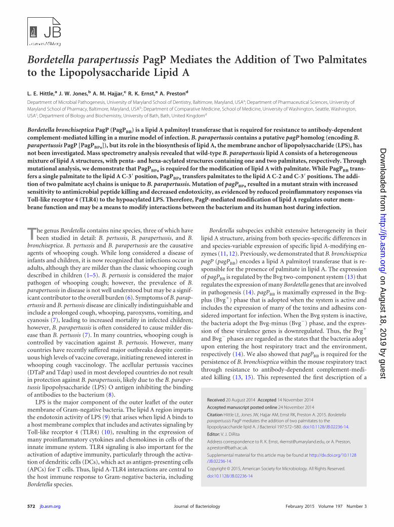
RESULTSIdentification of pagPBPa. pagPBPa was identified by genome se-quence analysis using the PagPBB sequence (Swiss-Prot databaseaccession number Q7WFT9.1). The B. parapertussis and B. pertus-sis genes were found to be nearly identical at the DNA level in boththe promoter and coding sequence. A single G:A base change inthe middle position of the 24th codon was observed, which re-sulted in an aspartate in PagPBPa, compared to a glycine in PagPBB.To investigate the role of pagP in overall LPS biosynthesis in B.parapertussis, a B. parapertussis pagP mutant was constructed andanalyzed by using SDS-PAGE to determine the LPS profile com-pared to that of the WT (Fig. 1).
In these analyses, two main LPS types are visible: lipid A-coreLPS and lipid A-core that is further substituted with O polysac-charide (O PS). The WT LPS profile differed between the Bvg�
and Bvg� phases. In the plus phase, core-lipid A LPS migrated as adiffuse smear, indicative of a heterogeneous nature of LPS struc-tures being present in this region. The O PS-containing LPS sep-arated as a series of bands, demonstrating that LPS molecules thatdiffer in the number of O PS repeats were well separated (Fig. 1,lane 1). In the Bvg� phase, the O PS migrated as a compactedseries of bands due to the action of the Bvg�-phase enzymeWbmE, which deaminates some of the O PS uronamide sugars(27). In addition, the core-lipid A LPS was present as a singleintense band and just one or two other very faint bands, suggestingthat a restricted number of core-lipid A structures were expressedin the minus phase compared to the plus phase (Fig. 1, lane 2).
The LPS profile of the pagPBPa mutant differed from that of theWT in the core-lipid A region. In the Bvg� phase, the mutantprofile comprised a doublet with near-equal-intensity bands,whereas in the minus phase, a single band was observed (Fig. 1,lanes 3 and 4). This suggests that PagPBPa expression is requiredfor some of the core-lipid A structures observed in the WT andthat Bvg activity affects the expression of PagP-dependent LPSstructures. Importantly, these analyses demonstrate that mutantstructures are incorporated into mature LPS and are likely incor-porated into the outer membrane. This is important for interpret-ing the phenotype of the pagPBPa mutant (see below).
To confirm the role of pagPBPa in LPS biosynthesis, the pagPmutation was complemented in trans by expressing pagP from aplasmid using the native pagP promoter. The LPS profile of thecomplemented mutant in the Bvg� phase comprised three well-resolved core-lipid A bands (Fig. 1, lane 6). This is different fromthe WT profile. We hypothesized that the multicopy nature of
carrying the complementing gene on a plasmid was responsiblefor this difference. Thus, the complemented mutant profile wascompared to that of the WT carrying the plasmid copy of pagP(Fig. 1, lane 5) and found to be the same. These data support theconclusion that the loss of core-lipid A bands from the profile ofthe pagPBPa mutant was due to a mutation of pagP (see also be-low).
Mass spectrometric analysis of lipid A isolated from WT B.parapertussis LPS. To define the specific structural differencesbetween WT and mutant LPS structures, lipid A was purified fromboth strains and analyzed by mass spectrometry (MS). MALDITOF/TOF and ESI LTQ-FT MS platforms were used to gain com-prehensive structure determination. The negative-ion-modeMALDI-TOF mass spectrum of lipid A isolated from WT B.parapertussis LPS is shown in Fig. 2A. The most abundant ion, atm/z 1,332 (Fig. 2A), corresponded to a singly deprotonated lipid Astructure that contained two phosphate groups and four acylchains (i.e., diphosphoryl tetra-acylated lipid A). Two otherprominent singly charged ions were recorded at m/z values of1,570 (diphosphoryl penta-acylated lipid A) and 1,808 (diphos-phoryl hexa-acylated lipid A). The ions at m/z 1,570 and 1,808differed from the ion at m/z 1,332 by the addition of one and twosecondary palmitate (C16) acyl chains, respectively. The proposedlipid A structures for the ions at m/z 1,332, 1,570, and 1,808 aredisplayed in Fig. 2. Structure determination was based on the tan-dem mass spectrometry experiments discussed in detail below. Inaddition to the m/z values mentioned above, a number of otherm/z values were observed, which demonstrated the extensive het-erogeneity of lipid A structures present in lipid A isolated fromWT B. parapertussis LPS (Fig. 2A). These m/z values correspondedto differing phosphorylation and acylation patterns (see Table S1in the supplemental material for proposed structures of these ad-ditional ions).
Mass spectrometric analysis of lipid A isolated from B. para-pertussis pagP mutant LPS. The negative-ion-mode MALDI-TOFmass spectrum of lipid A isolated from B. parapertussis pagP mu-tant LPS is shown in Fig. 2B. This lipid A also displayed the mostabundant ion, at m/z 1,332 (Fig. 2B), which corresponded to asingly deprotonated diphosphoryl tetra-acylated lipid A structure.Of particular note, ions at m/z 1,570 and 1,808, corresponding tothe addition of one and two secondary C16 acyl chains, respec-tively, were not present in mutant lipid A. All m/z values for mu-tant lipid A were also observed for lipid A from WT LPS andcorresponded to identical structures (see Table S1 in the supple-mental material).
Determination of acyl chain positioning in lipid A isolatedfrom WT and B. parapertussis pagP mutant LPS using tandemmass spectrometry. In order to confidently assign structures tospecific m/z values, a series of tandem MS experiments using aMALDI-TOF/TOF mass spectrometer were performed. In addi-tion, multistage tandem MS (MSn) experiments were performedon an ESI LTQ-FT MS platform to further validate structure as-signment. The gas-phase dissociation of the precursor ion at m/z1,332 determined via the MALDI-TOF/TOF platform revealedstructure-specific, diagnostic product ions that allowed confidentassignment of the four acyl chains along the lipid A glucosaminedisaccharide (Fig. 3A). Gas-phase dissociation for structure deter-mination of lipid A via tandem mass spectrometry, whereby struc-ture-specific, diagnostic product ions are used for structure deter-mination, was reported previously (12, 28). Of note, the collective
FIG 1 SDS-PAGE LPS profiles of the WT and a B. parapertussis pagP mutantgrown in either the Bvg-plus or Bvg-minus phase and these strains carrying aplasmid-borne copy of either pagPBPa or pagPBB (Bvg-plus phase only). Lane 1,WT in the Bvg-plus phase; lane 2, WT in the Bvg-minus phase; lane 3, B.parapertussis pagP mutant in the Bvg-plus phase; lane 4, B. parapertussis pagPmutant in the Bvg-minus phase; lane 5, WT in the Bvg-plus phase withpBBRpagPBPa; lane 6, B. parapertussis pagP mutant in the Bvg-plus phase withpBBRpagPBPa; lane 7, WT in the Bvg-plus phase with pBBRpagPBB; lane 8, B.parapertussis pagP mutant in the Bvg-plus phase with pBBRpagPBB.
Hittle et al.
574 jb.asm.org February 2015 Volume 197 Number 3Journal of Bacteriology
on August 18, 2019 by guest
http://jb.asm.org/
Dow
nloaded from
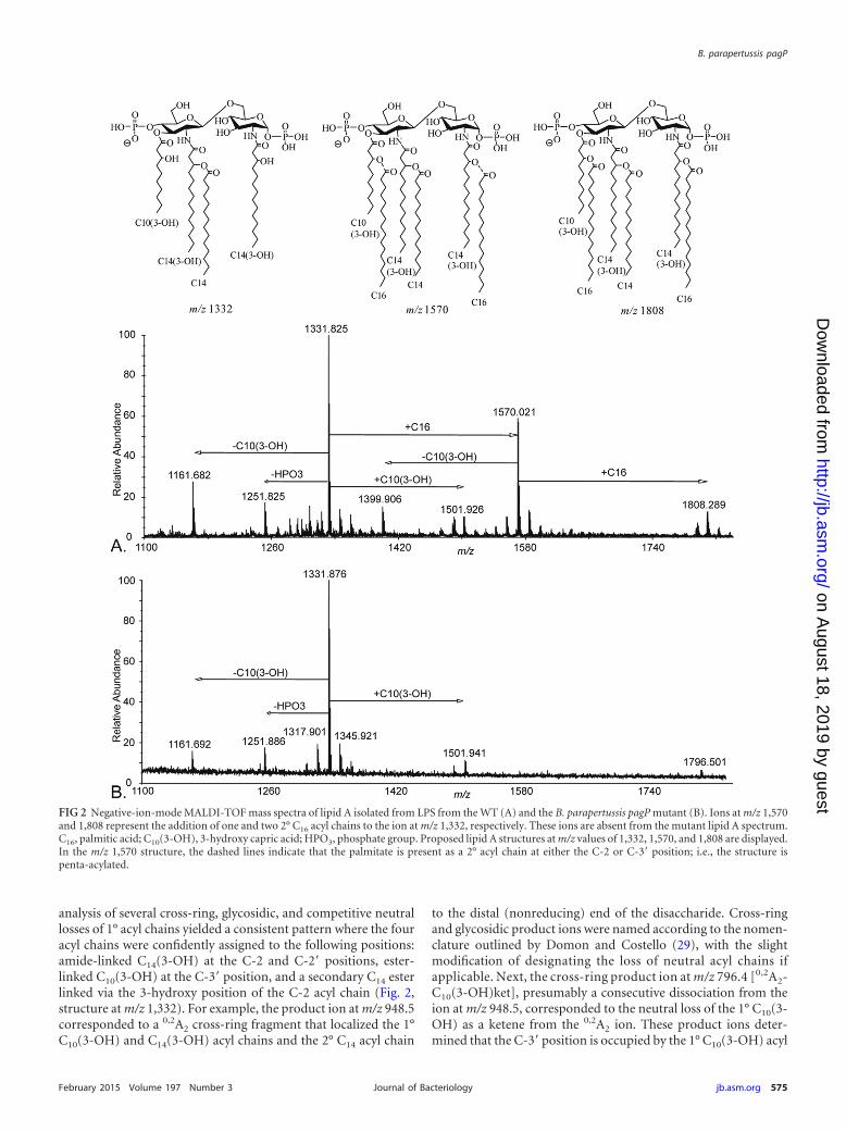
analysis of several cross-ring, glycosidic, and competitive neutrallosses of 1° acyl chains yielded a consistent pattern where the fouracyl chains were confidently assigned to the following positions:amide-linked C14(3-OH) at the C-2 and C-2= positions, ester-linked C10(3-OH) at the C-3= position, and a secondary C14 esterlinked via the 3-hydroxy position of the C-2 acyl chain (Fig. 2,structure at m/z 1,332). For example, the product ion at m/z 948.5corresponded to a 0,2A2 cross-ring fragment that localized the 1°C10(3-OH) and C14(3-OH) acyl chains and the 2° C14 acyl chain
to the distal (nonreducing) end of the disaccharide. Cross-ringand glycosidic product ions were named according to the nomen-clature outlined by Domon and Costello (29), with the slightmodification of designating the loss of neutral acyl chains ifapplicable. Next, the cross-ring product ion at m/z 796.4 [0,2A2-C10(3-OH)ket], presumably a consecutive dissociation from theion at m/z 948.5, corresponded to the neutral loss of the 1° C10(3-OH) as a ketene from the 0,2A2 ion. These product ions deter-mined that the C-3= position is occupied by the 1° C10(3-OH) acyl
FIG 2 Negative-ion-mode MALDI-TOF mass spectra of lipid A isolated from LPS from the WT (A) and the B. parapertussis pagP mutant (B). Ions at m/z 1,570and 1,808 represent the addition of one and two 2° C16 acyl chains to the ion at m/z 1,332, respectively. These ions are absent from the mutant lipid A spectrum.C16, palmitic acid; C10(3-OH), 3-hydroxy capric acid; HPO3, phosphate group. Proposed lipid A structures at m/z values of 1,332, 1,570, and 1,808 are displayed.In the m/z 1,570 structure, the dashed lines indicate that the palmitate is present as a 2° acyl chain at either the C-2 or C-3= position; i.e., the structure ispenta-acylated.
B. parapertussis pagP
February 2015 Volume 197 Number 3 jb.asm.org 575Journal of Bacteriology
on August 18, 2019 by guest
http://jb.asm.org/
Dow
nloaded from
chain and that the C-2= position contained the 1° C14(3-OH) and2° C14 acyl chains. In addition, the glycosidic product ion at m/z484.2 (Y1) squarely indicated that the reducing end of the lipid Adisaccharide contained a 1° C14(3-OH) at the C-2 position. Fur-ther confirmation of the structure assignment for the ion at m/z1,332 was demonstrated via the use of tandem mass spectrometryusing an ESI LTQ-FT mass spectrometer, where MSn experimentswere performed (see Fig. S1 in the supplemental material). Theprecursor ions at m/z 1,332 from both the WT and the pagP mu-tant had identical structures.
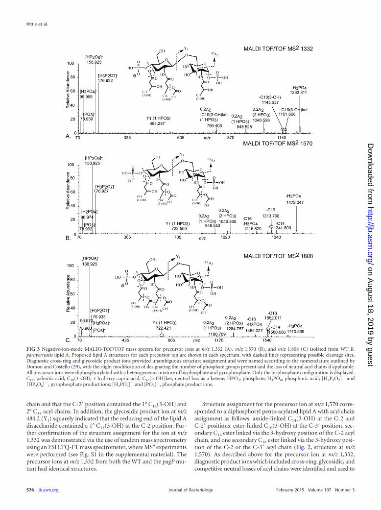
Structure assignment for the precursor ion at m/z 1,570 corre-sponded to a diphosphoryl penta-acylated lipid A with acyl chainassignment as follows: amide-linked C14(3-OH) at the C-2 andC-2= positions, ester-linked C10(3-OH) at the C-3= position, sec-ondary C14 ester linked via the 3-hydroxy position of the C-2 acylchain, and one secondary C16 ester linked via the 3-hydroxy posi-tion of the C-2 or the C-3= acyl chain (Fig. 2, structure at m/z1,570). As described above for the precursor ion at m/z 1,332,diagnostic product ions which included cross-ring, glycosidic, andcompetitive neutral losses of acyl chains were identified and used to
FIG 3 Negative-ion-mode MALDI-TOF/TOF mass spectra for precursor ions at m/z 1,332 (A), m/z 1,570 (B), and m/z 1,808 (C) isolated from WT B.parapertussis lipid A. Proposed lipid A structures for each precursor ion are shown in each spectrum, with dashed lines representing possible cleavage sites.Diagnostic cross-ring and glycosidic product ions provided unambiguous structure assignment and were named according to the nomenclature outlined byDomon and Costello (29), with the slight modification of designating the number of phosphate groups present and the loss of neutral acyl chains if applicable.All precursor ions were diphosphorylated with a heterogeneous mixture of bisphosphate and pyrophosphate. Only the bisphosphate configuration is displayed.C16, palmitic acid; C10(3-OH), 3-hydroxy capric acid; C10(3-OH)ket, neutral loss as a ketene; HPO3, phosphate; H3PO4, phosphoric acid; [H3P2O7]� and[HP2O6]�, pyrophosphate product ions; [H2PO4]� and [PO3]�, phosphate product ions.
Hittle et al.
576 jb.asm.org February 2015 Volume 197 Number 3Journal of Bacteriology
on August 18, 2019 by guest
http://jb.asm.org/
Dow
nloaded from

confidently assign the structure of the ion at m/z 1,570 (see Fig. 3B foridentities and m/z values of several diagnostic product ions). Of par-ticular note, the 2° C16 acyl chain appeared to be localized to thereducing end (C-2 position), yet conclusive evidence for the exclusivepositioning of the C16 acyl chain was not obtained via MALDI-TOF/TOF experiments. Further experiments using an ESI LTQ-FT massspectrometer capable of higher-order MSn revealed that the 2° C16
acyl chain was localized to either the C-2 or C-3= position (see Fig. S2in the supplemental material). Given the inherent difficulty of deter-mining even relative amounts of isomeric structures during ESI massspectrometry experiments, determination of the abundance of struc-tures having a secondary C16 at either the C-2 position or the C-3=position was not attempted.
Structure assignment for the precursor ion at m/z 1,808 corre-sponded to a diphosphoryl hexa-acylated lipid A with acyl chainassignment as follows: amide-linked C14(3-OH) at the C-2 andC-2= positions, ester-linked C10(3-OH) at the C-3= position, sec-ondary C14 ester linked via the 3-hydroxy position of the C-2 acylchain, and two secondary C16 acyl chains ester linked via the 3-hy-droxy position of the C-2 and C-3= acyl chains (Fig. 2, structure atm/z 1,808). Evidence and reasoning analogous to those for theprecursor ions at m/z 1,332 and 1,570 were used to determine thestructure of this ion (Fig. 3C; see also Fig. S3 in the supplementalmaterial). In effect, diagnostic product ions were identified toconfidently assign the positions of the two 2° C16 acyl chains.
These data demonstrate that B. parapertussis lipid A containsmolecules with either one or two palmitates and that the presenceof palmitate is dependent on pagPBPa. In turn, this strongly sug-gests that B. parapertussis PagP transfers two palmitate moieties tolipid A. This is different from B. bronchiseptica PagP, which trans-fers only a single palmitate. It was possible that the two enzymeshave different transferase abilities, presumably arising from thesingle-amino-acid difference between the two enzymes, or thatthe different lipid A substrates encountered by the enzymes in thetwo hosts direct different transferase activities. To investigate this,PagPBB was expressed in the B. parapertussis pagP mutant. The B.bronchiseptica gene was carried on a plasmid and expressed fromits natural promoter (the promoter regions of B. bronchiseptica andB. parapertussis are identical). This construct was previously usedto complement a B. bronchiseptica pagP mutation (13). Comple-mentation of the B. parapertussis pagP mutation by pagPBB re-sulted in a lipid A-core profile identical to that produced by com-plementation with pagPBPa (Fig. 1, lanes 7 and 8), suggesting thatboth the B. parapertussis and B. bronchiseptica enzymes have thesame activity in the B. parapertussis background. To confirm this,lipid A was purified from both complemented strains and ana-lyzed by MS (Fig. 4). For both lipid A preparations, ions thatcorresponded to molecules containing either a single or twopalmitate additions were detected, demonstrating that PagPBB canadd two palmitates to B. parapertussis lipid A. Thus, both PagPBB
and PagPBPa and have dual functionality, in that they can addpalmitate to two different sites in lipid A. PagPBB does this in B.parapertussis but not in B. bronchiseptica, and thus, we proposethat the dual functionality is directed by the B. parapertussis lipid Aacceptor substrate.
pagPBPa is required for WT levels of resistance to C18G. Theacylation pattern of lipid A is an important determinant of theintegrity of the outer membrane of Gram-negative bacteria. Inturn, this is important for a range of functions, including resistingantimicrobial molecules produced by hosts during infection. We
hypothesized that pagPBPa contributes to the ability of the bacte-rium to resist host antimicrobial peptides, as observed for otherbacterial species. To test this, we measured the level of killing ofthe WT and the B. parapertussis pagP mutant after exposure toC18G, a synthetic -helical antimicrobial peptide (30). Approxi-mately 1 � 104 CFU of the WT or mutant were incubated with thepeptide (0 to 10 �g ml�1) for 30 min at 37°C, and the number ofsurviving bacteria was enumerated. The results from a represen-tative experiment are shown in Fig. 5. At each concentration ofpeptide, a significantly greater number of mutant bacteria thanWT bacteria were killed. Overall, our data demonstrate that theWT level of resistance to C18G-mediated killing is dependent onpagPBPa.
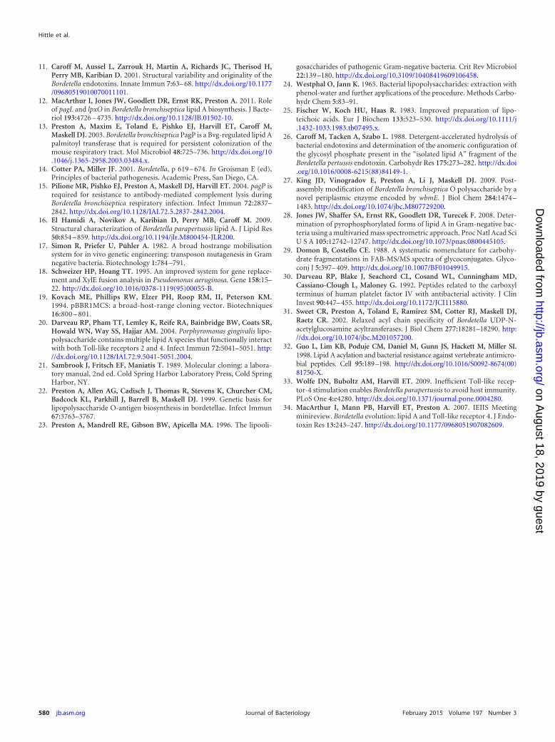
TLR4 stimulation assays. A key activity of lipid A during in-fection is stimulation of TLR4-mediated signaling. The acylationpattern of lipid A is a key determinant of this activity. The relativepotencies for stimulation of TLR4-mediated signaling of lipid Aisolated from the WT and pagP mutant strains were measured. Arecombinant reporter system was used (20), expressing either hu-man or murine TLR4 signaling complexes, as these two systemsrecognize and respond to different lipid A structures differently.Unstimulated cells acted as a negative control. Stimulation with aknown agonist of NF-B-mediated signaling, interleukin-1 (IL-1), resulted in relative light unit (RLU) values of 0.54 � 0.03 and
FIG 4 Mass spectra of the lipid A’s of B. parapertussis pagP complementedwith either pagPBPa (A) or pagPBB (B). Ions corresponding to lipid A with asingle palmitate are indicated by * (m/z 1,570), and those with two palmitatesare indicated by ** (m/z 1,808). Both strains produced identical lipid A’s,demonstrating that both B. parapertussis PagP and B. bronchiseptica PagP havedual functionality.
B. parapertussis pagP
February 2015 Volume 197 Number 3 jb.asm.org 577Journal of Bacteriology
on August 18, 2019 by guest
http://jb.asm.org/
Dow
nloaded from
0.59 � 0.05 for murine and human TLR4-mediated signaling,respectively, demonstrating the responsiveness of the system.Stimulation with an E. coli lipid A (1,000 ng/ml) biosynthetic in-termediate, lipid IVA, which is a strong agonist of murine but notof human TLR4 signaling, resulted in RLU values of 0.36 � 0.03and 0.05 � 0.00 for murine and human TLR4-mediated signaling,respectively, and demonstrated that the experimental systemcould detect species-specific stimulation.
B. parapertussis WT lipid A was a more potent stimulant ofmurine than of human TLR4-mediated signaling (Fig. 6). pagPmutant lipid A was significantly less potent than the WT forstimulation of both murine (Fig. 6A) and human (Fig. 6B) TLR4-mediated signaling but was particularly attenuated for the hu-man-derived system. These data demonstrate that PagPBPa-medi-ated modification of B. parapertussis lipid A significantly alters the
endotoxin activity of lipid A toward the human TLR4 signalingcomplex.
DISCUSSION
Previously, we characterized B. bronchiseptica PagP as a Bvg-reg-ulated lipid A palmitoyl transferase and determined that PagP-mediated palmitoylation was required for resistance to antibody-dependent complement-mediated killing (13, 15). Here we showthat B. parapertussis PagP is also a Bvg-regulated lipid A palmitoyltransferase but one that is able to transfer two palmitates to lipidA, a novel finding. When expressed in B. parapertussis, PagPBB alsomediates the addition of two palmitates to lipid A. We proposethat the lipid A acceptor substrate of B. parapertussis directs thisdual functionality.
Interestingly, the MS analyses presented here demonstrated
FIG 5 B. parapertussis pagP is more susceptible to killing by the synthetic antimicrobial peptide C18G than the WT. A significantly different level of killing of theWT compared to the pagP mutant was observed. This was observed in three independent experiments; data from a representative experiment are shown here.
FIG 6 Stimulation of murine or human TLR4-mediated signaling by WT and B. parapertussis lipid A. Both lipid A’s were more potent stimulators of murine thanof human TLR4-mediated signaling; however, B. parapertussis pagP lipid A was significantly less potent than that of the WT, particularly for the activation ofhuman TLR4. �, P � 0.05; ��, P � 0.001.
Hittle et al.
578 jb.asm.org February 2015 Volume 197 Number 3Journal of Bacteriology
on August 18, 2019 by guest
http://jb.asm.org/
Dow
nloaded from

considerable lipid A heterogeneity in addition to palmitoylation.Variability in the presence of C10(3-OH) was observed (Fig. 2).Ions at m/z 1,162 and 1,400 corresponded to derivatives of themajor tetra- and penta-acylated molecules lacking C10(3-OH),whereas the ion at m/z 1,502 corresponded to a derivative of thetetra-acylated molecule that contained an additional C10(3-OH),presumably at the C-3 position as a result of incomplete deacyla-tion by PagL (12). Furthermore, each of the peaks correspondingto the tetra-, penta-, and hexa-acylated species was surrounded bya series of peaks, each differing from the next by 14 mass units (Fig.2), corresponding to a series of structures, each differing by onemethylene (CH2) unit. This suggests that B. parapertussis incor-porates a range of different acyl chains into its lipid A, includingthose containing an odd number of carbon atoms. BordetellaLpxA has relaxed acyl chain specificity that may contribute to thisheterogeneity (31). In turn, this suggests that B. parapertussis lipidA contains not only three major molecular species but also nu-merous acylation variants of these major species. A full structuralcharacterization of this heterogeneity is under way.
PagPBPa mediates the conversion of tetra-acylated to penta-and hexa-acylated lipid A and thus alters the properties of theouter membrane. A pagP mutant was more susceptible to directkilling by an -helical antimicrobial peptide (C18G), suggestingthat a loss of palmitoylation affects the integrity or fluidity of theouter membrane. This has also been observed for Salmonella en-terica serovar Typhimurium, where a pagP mutant displayed in-creased outer membrane permeability in response to C18G (32).
PagP-mediated palmitoylation results in an altered potency ofB. parapertussis lipid A for stimulating signaling via TLR4. WTlipid A was more proinflammatory than pagP mutant lipid A,particularly via the human-derived signaling complex. When con-sidering the basis for the effect of palmitoylation on lipid A po-tency, it is important to consider that the individual lipid A prep-arations are comprised of a mixture of lipid A molecules and thatthe overall potency is the sum of their individual potencies, whichmay include some antagonistic activities. Either way, the Bvg-reg-ulated expression of pagPBPa appears to confer on B. parapertussisa mechanism by which to regulate the potency of its lipid A forTLR4 stimulation. The Bvg� phase, in which PagPBPa appears tobe maximally expressed, is thought to be that adopted by the bac-teria during infection. In this case, our data suggest that bothsingly and doubly palmitoylated lipid A will be synthesized andthat lipid A is at its most potent, compared to the Bvg-minusphase. However, B. parapertussis has low-potency lipid A com-pared to that of B. bronchiseptica, and it has been demonstrated inmice that B. parapertussis avoids invoking host immune responsesthrough the lack of stimulation of TLR4-mediated signaling (33).This suggests that Bvg-mediated regulation of lipid A potencydoes not play a role during infection.
Clearly, the in vitro system used here does not reveal the preciseimmune response stimulated by either palmitoylated or non-palmitoylated lipid A during infection, and thus, the conse-quences of regulating the level of lipid A palmitoylation are notclear. In addition, our data demonstrate that PagP activity regu-lates outer membrane function in other ways, such as resistance tohost antimicrobial peptides, and that the bacterium may have tobalance increased lipid A potency for TLR4 stimulation versusinherent resistance to immune effector molecules. In support ofthis, B. bronchiseptica pagP is required for persistent colonizationof the mouse respiratory tract (13). However, a pagPBB mutant
appears to colonize similarly to the WT during the early course ofinfection, with attenuation manifesting only between days 3 and 7postinoculation, perhaps suggesting that there is little differencein early events when innate immunity resulting from TLR4 stim-ulation is operational. Furthermore, we demonstrated that pagPBB
is required for resistance to antibody-dependent complement-mediated lysis (15), suggesting that the major role of pagP in B.bronchiseptica is to contribute to the integrity of the outer mem-brane.
It is also of interest to consider the difference in lipid A biosyn-thetic enzymes between Bordetella subspecies. B. pertussis, B. bron-chiseptica, and B. parapertussis share a very similar set of lipid Abiosynthesis and modification genes, but they each synthesize spe-cies-specific lipid A’s (34). Some genes are mutated in one species(e.g., pagP in B. pertussis [13]), while subtle allelic differences re-sult in differences in activity among species, e.g., lpxA (31). Hereour data suggest that slight alterations in the lipid A structure candirect different activities of enzymes common to more than onespecies. Presumably, the evolution of these three species in theirdifferent niches and different infection biologies has selected forthese subtly different combinations of lipid A biosynthesis activi-ties. Thus, while the regulated expression of Bordetella pagP sug-gests that B. parapertussis may modulate its lipid A structure dur-ing infection, the extent to which this happens and theconsequences of it require analyses in vivo, with consideration ofthe different potencies of stimulation of B. parapertussis lipid Atoward TLR4 from different hosts.
ACKNOWLEDGMENT
This work was supported in part by a National Institutes of Health grant(1U54 AI57141) (R.K.E.).
REFERENCES1. Cherry JD. 1992. Pertussis—the trials and tribulations of old and new
pertussis vaccines. Vaccine 10:1033–1038. http://dx.doi.org/10.1016/0264-410X(92)90113-X.
2. Cherry JD. 1996. Historical review of pertussis and the classical vaccine. JInfect Dis 174(Suppl 3):S259 –S263.
3. Heininger U, Stehr K, Schmittgrohe S, Lorenz C, Rost R, ChristensonPD, Uberall M, Cherry JD. 1994. Clinical characteristics of illness causedby Bordetella parapertussis compared with illness caused by Bordetellapertussis. Pediatr Infect Dis J 13:306 –309. http://dx.doi.org/10.1097/00006454-199404000-00011.
4. Nennig ME, Shinefield HR, Edwards KM, Black SB, Fireman BH. 1996.Prevalence and incidence of adult pertussis in an urban population. JAMA275:1672–1674. http://dx.doi.org/10.1001/jama.1996.03530450062034.
5. Pichichero ME, Treanor J. 1997. Economic impact of pertussis. ArchPediatr Adolesc Med 151:35– 40. http://dx.doi.org/10.1001/archpedi.1997.02170380039006.
6. Watanabe M, Nagai M. 2004. Whooping cough due to Bordetella parap-ertussis: an unresolved problem. Expert Rev Anti Infect Ther 2:447– 454.http://dx.doi.org/10.1586/14787210.2.3.447.
7. Mattoo S, Cherry JD. 2005. Molecular pathogenesis, epidemiology, andclinical manifestations of respiratory infections due to Bordetella pertussisand other Bordetella subspecies. Clin Microbiol Rev 18:326 –382. http://dx.doi.org/10.1128/CMR.18.2.326-382.2005.
8. Zhang X, Rodriguez ME, Harvill ET. 2009. O antigen allows B. parap-ertussis to evade B. pertussis vaccine-induced immunity by blocking bind-ing and functions of cross-reactive antibodies. PLoS One 4:e6989. http://dx.doi.org/10.1371/journal.pone.0006989.
9. Luderitz O, Galanos C, Lehmann V, Nurminen J, Rietschel ET, Rosen-felder G, Simon M, Westphal O. 1973. Lipid A: chemical structure andbiological activity. J Infect Dis 128:S17–S29. http://dx.doi.org/10.1093/infdis/128.Supplement_1.S17.
10. Akira S, Takeda K. 2004. Toll-like receptor signalling. Nat Rev Immunol4:499 –511. http://dx.doi.org/10.1038/nri1391.
B. parapertussis pagP
February 2015 Volume 197 Number 3 jb.asm.org 579Journal of Bacteriology
on August 18, 2019 by guest
http://jb.asm.org/
Dow
nloaded from
11. Caroff M, Aussel L, Zarrouk H, Martin A, Richards JC, Therisod H,Perry MB, Karibian D. 2001. Structural variability and originality of theBordetella endotoxins. Innate Immun 7:63– 68. http://dx.doi.org/10.1177/09680519010070011101.
12. MacArthur I, Jones JW, Goodlett DR, Ernst RK, Preston A. 2011. Roleof pagL and lpxO in Bordetella bronchiseptica lipid A biosynthesis. J Bacte-riol 193:4726 – 4735. http://dx.doi.org/10.1128/JB.01502-10.
13. Preston A, Maxim E, Toland E, Pishko EJ, Harvill ET, Caroff M,Maskell DJ. 2003. Bordetella bronchiseptica PagP is a Bvg-regulated lipid Apalmitoyl transferase that is required for persistent colonization of themouse respiratory tract. Mol Microbiol 48:725–736. http://dx.doi.org/10.1046/j.1365-2958.2003.03484.x.
14. Cotter PA, Miller JF. 2001. Bordetella, p 619 – 674. In Groisman E (ed),Principles of bacterial pathogenesis. Academic Press, San Diego, CA.
15. Pilione MR, Pishko EJ, Preston A, Maskell DJ, Harvill ET. 2004. pagP isrequired for resistance to antibody-mediated complement lysis duringBordetella bronchiseptica respiratory infection. Infect Immun 72:2837–2842. http://dx.doi.org/10.1128/IAI.72.5.2837-2842.2004.
16. El Hamidi A, Novikov A, Karibian D, Perry MB, Caroff M. 2009.Structural characterization of Bordetella parapertussis lipid A. J Lipid Res50:854 – 859. http://dx.doi.org/10.1194/jlr.M800454-JLR200.
17. Simon R, Priefer U, Pühler A. 1982. A broad hostrange mobilisationsystem for in vivo genetic engineering: transposon mutagenesis in Gramnegative bacteria. Biotechnology 1:784 –791.
18. Schweizer HP, Hoang TT. 1995. An improved system for gene replace-ment and XylE fusion analysis in Pseudomonas aeruginosa. Gene 158:15–22. http://dx.doi.org/10.1016/0378-1119(95)00055-B.
19. Kovach ME, Phillips RW, Elzer PH, Roop RM, II, Peterson KM.1994. pBBR1MCS: a broad-host-range cloning vector. Biotechniques16:800 – 801.
20. Darveau RP, Pham TT, Lemley K, Reife RA, Bainbridge BW, Coats SR,Howald WN, Way SS, Hajjar AM. 2004. Porphyromonas gingivalis lipo-polysaccharide contains multiple lipid A species that functionally interactwith both Toll-like receptors 2 and 4. Infect Immun 72:5041–5051. http://dx.doi.org/10.1128/IAI.72.9.5041-5051.2004.
21. Sambrook J, Fritsch EF, Maniatis T. 1989. Molecular cloning: a labora-tory manual, 2nd ed. Cold Spring Harbor Laboratory Press, Cold SpringHarbor, NY.
22. Preston A, Allen AG, Cadisch J, Thomas R, Stevens K, Churcher CM,Badcock KL, Parkhill J, Barrell B, Maskell DJ. 1999. Genetic basis forlipopolysaccharide O-antigen biosynthesis in bordetellae. Infect Immun67:3763–3767.
23. Preston A, Mandrell RE, Gibson BW, Apicella MA. 1996. The lipooli-
gosaccharides of pathogenic Gram-negative bacteria. Crit Rev Microbiol22:139 –180. http://dx.doi.org/10.3109/10408419609106458.
24. Westphal O, Jann K. 1965. Bacterial lipopolysaccharides: extraction withphenol-water and further applications of the procedure. Methods Carbo-hydr Chem 5:83–91.
25. Fischer W, Koch HU, Haas R. 1983. Improved preparation of lipo-teichoic acids. Eur J Biochem 133:523–530. http://dx.doi.org/10.1111/j.1432-1033.1983.tb07495.x.
26. Caroff M, Tacken A, Szabo L. 1988. Detergent-accelerated hydrolysis ofbacterial endotoxins and determination of the anomeric configuration ofthe glycosyl phosphate present in the “isolated lipid A” fragment of theBordetella pertussis endotoxin. Carbohydr Res 175:273–282. http://dx.doi.org/10.1016/0008-6215(88)84149-1.
27. King JD, Vinogradov E, Preston A, Li J, Maskell DJ. 2009. Post-assembly modification of Bordetella bronchiseptica O polysaccharide by anovel periplasmic enzyme encoded by wbmE. J Biol Chem 284:1474 –1483. http://dx.doi.org/10.1074/jbc.M807729200.
28. Jones JW, Shaffer SA, Ernst RK, Goodlett DR, Turecek F. 2008. Deter-mination of pyrophosphorylated forms of lipid A in Gram-negative bac-teria using a multivaried mass spectrometric approach. Proc Natl Acad SciU S A 105:12742–12747. http://dx.doi.org/10.1073/pnas.0800445105.
29. Domon B, Costello CE. 1988. A systematic nomenclature for carbohy-drate fragmentations in FAB-MS/MS spectra of glycoconjugates. Glyco-conj J 5:397– 409. http://dx.doi.org/10.1007/BF01049915.
30. Darveau RP, Blake J, Seachord CL, Cosand WL, Cunningham MD,Cassiano-Clough L, Maloney G. 1992. Peptides related to the carboxylterminus of human platelet factor IV with antibacterial activity. J ClinInvest 90:447– 455. http://dx.doi.org/10.1172/JCI115880.
31. Sweet CR, Preston A, Toland E, Ramirez SM, Cotter RJ, Maskell DJ,Raetz CR. 2002. Relaxed acyl chain specificity of Bordetella UDP-N-acetylglucosamine acyltransferases. J Biol Chem 277:18281–18290. http://dx.doi.org/10.1074/jbc.M201057200.
32. Guo L, Lim KB, Poduje CM, Daniel M, Gunn JS, Hackett M, Miller SI.1998. Lipid A acylation and bacterial resistance against vertebrate antimicro-bial peptides. Cell 95:189–198. http://dx.doi.org/10.1016/S0092-8674(00)81750-X.
33. Wolfe DN, Buboltz AM, Harvill ET. 2009. Inefficient Toll-like recep-tor-4 stimulation enables Bordetella parapertussis to avoid host immunity.PLoS One 4:e4280. http://dx.doi.org/10.1371/journal.pone.0004280.
34. MacArthur I, Mann PB, Harvill ET, Preston A. 2007. IEIIS Meetingminireview. Bordetella evolution: lipid A and Toll-like receptor 4. J Endo-toxin Res 13:243–247. http://dx.doi.org/10.1177/0968051907082609.
Hittle et al.
580 jb.asm.org February 2015 Volume 197 Number 3Journal of Bacteriology
on August 18, 2019 by guest
http://jb.asm.org/
Dow
nloaded from