
Boosting Self-Assembly Diversity in the Solid-State by ...
13
Boosting Self-Assembly Diversity in the Solid-State by Chiral/non- Chiral Zn II -Porphyrin Crystallization Wenjie Qian a , Arántzazu González-Campo a , Ana Pérez a , Sabina Rodríguez-Hermida b , Inhaz Imaz b , Klaus Wurst c , Daniel Maspoch b,d , E. Ruiz e,f , C. Ocal a , E. Barrena a , David B. Amabilino g* and Núria Aliaga-Alcalde a,d * a Institut de Ciència de Materials de Barcelona (ICMAB–CSIC), Campus Universitari, 08193 Bellaterra, Spain. b Catalan Institute of Nanoscience and Nanotechnology (ICN2), CSIC and The Barcelona Institute of Science and Technology, Campus UAB, Bellaterra, 08193 Barcelona, Spain. c Institut für Allgemeine Anorganische und Theoretische Chemie, Universität Innsbruck, A-6020, Innrain 52a, Austria. d ICREA (Institució Catalana de Recerca i Estudis Avançats), Passeig Lluís Com- panys 23, 08010 Barcelona, Spain. e Departament de Química Inorgànica i Orgànica, Universitat de Barcelona, Barcelona 08007, Spain. f Institut de Química Teórica i Computacional de la Universitat de Barcelona (IQTCUB), Barcelona 08007, Spain. g School of Chemistry, The University of Nottingham, University Park, Nottingham, NG7 2RD, UK. KEYWORDS. Zinc-porphyrin, chirality, supramolecular architectures, X-ray diffraction, solid 13 C NMR, circular dichroism, vibronic circular dichroism, mica and HOPG surfaces, SEM, AFM Supporting Information Placeholder ABSTRACT: This work bases on the solid-state study of a chiral Zn II -porphyrin derivative (5,10,15,20-tetra[(4-R,R,R,R)-methyl-2- phenoxy-propanoate, 1) building block and its achiral analogous (2). Here, foreseen the rich molecular recognition of the designed metallo-porphyrins (1 and 2) and taking advantage of their tendency to crystallize, we recrystallized both following identical meth- odologies with two sets of solvents (CH 2 Cl 2 /CH 3 OH and CH 2 Cl 2 /hexane). As a result, four different crystalline arrangements (1a-b, 2a-b, from 0D to 2D) were successfully analyzed by single crystal X-ray diffraction. We performed solid state studies for all the species, analyzing the role played by chirality, solvent mixtures and surfaces (mica and HOPG), on the supramolecular ar- rangements. From those, we highlight ATR-FTIR and solid-state 13 C NMR, CD and VCD as the most useful techniques, corrobo- rating the observations made by diffraction, where the groups involved in the self-assembly processes were identified. As for the combination of solvents and different substrates we obtained a variety of micro-sized species, from vesicles to flower-shaped ar- rays, including geometrical microcrystals, describing the fullness presented in our choice. Our results emphasize the relevance of porphyrin crystallization toward their future applications and face the present challenges, as the lack of anticipation of the final architectures INTRODUCTION Structural diversity and advanced activity based on supramolecular self-assembly macrocycles are challeng- ing research topics in the field of materials science. 1,2 In the design of non-covalent intermolecular interactions that resembles nature, 3-5 the synthetic methodologies existing are mostly limited by the difficulties of anticipat- ing the final arrangements and the lack of reversibility, where the thermodynamic products are the ending tract, hampering both, control and functioning. Owing to the premature stage of the subject basic studies are required, where a key-point is the use of molecular units that can be easily tailored. Regarding this, porphyrin derivatives are excellent mo- lecular prototypes due to the possibility of direct infor- mation transfer from bench-experiments into biological facts. 6 We can ensure self-assembly because of the sum of its manifold features: (i) the conjugated core that has given copious studies with plenty of data regarding the formation of H/J aggregates; 7 (ii) the use of a metal cen- ter that provides additional interactive site in the final metallo-porphyrins; 8 and (iii) the addition of organic groups in the meso- positions of the tetrapyrrolic units that afford extra interactions depending on their number and nature. 1,9 Self-assembled aggregates of meso-substituted metallo- porphyrins are therefore quite difficult to analyze and specially to anticipate. An approach, adopted by us 10,11 and others 12,13 relies on chirality as the driving force in the achievement of highly ordered structures and the analyses in solution of their noncovalent interactions (hydrogen bonds, - stacking or coordination of through the metal center) and self-assembly mechanisms. Howev- er, much less it is known about the genuine effect of chirality on the final architectures, with no strict compari- son of chiral/non-chiral analogous porphyrin types, as well as there is scare information on the projection of the knowledge gathered in solution to the solid state. Here, we unify these two ideas toward the analysis of two novel Zn-porphyrins containing phenoxy propanoate and methyl 2-phenoxy acetate groups (1 and 2, respectively) in the four meso-positions of porphyrins (Scheme 1). The choice of such final entities relate to previous experiences with related systems 10,14-17 foreseen their rich molecular recognition capacity and tendency to crystallize. Restricting our recrystallization methods, identical oth- erwise, for our chiral and non-chiral Zn-porphyrin sys- tems allow new findings in the solid state primarily by the use of X-ray diffraction. In addition, our study inte- grates the effect of solvent polarity and highlights the
Transcript of Boosting Self-Assembly Diversity in the Solid-State by ...

Boosting Self-Assembly Diversity in the Solid-State by Chiral/non-
Chiral ZnII-Porphyrin Crystallization
Wenjie Qiana, Arántzazu González-Campo
a, Ana Pérez
a, Sabina Rodríguez-Hermida
b, Inhaz Imaz
b,
Klaus Wurstc, Daniel Maspoch
b,d, E. Ruiz
e,f, C. Ocal
a, E. Barrena
a, David B. Amabilino
g* and Núria
Aliaga-Alcaldea,d
*
aInstitut de Ciència de Materials de Barcelona (ICMAB–CSIC), Campus Universitari, 08193 Bellaterra, Spain. bCatalan
Institute of Nanoscience and Nanotechnology (ICN2), CSIC and The Barcelona Institute of Science and Technology, Campus
UAB, Bellaterra, 08193 Barcelona, Spain. cInstitut für Allgemeine Anorganische und Theoretische Chemie, Universität
Innsbruck, A-6020, Innrain 52a, Austria. dICREA (Institució Catalana de Recerca i Estudis Avançats), Passeig Lluís Com-
panys 23, 08010 Barcelona, Spain. e Departament de Química Inorgànica i Orgànica, Universitat de Barcelona, Barcelona
08007, Spain. f Institut de Química Teórica i Computacional de la Universitat de Barcelona (IQTCUB), Barcelona
08007, Spain. gSchool of Chemistry, The University of Nottingham, University Park, Nottingham, NG7 2RD, UK.
KEYWORDS. Zinc-porphyrin, chirality, supramolecular architectures, X-ray diffraction, solid 13C NMR, circular dichroism,
vibronic circular dichroism, mica and HOPG surfaces, SEM, AFM
Supporting Information Placeholder
ABSTRACT: This work bases on the solid-state study of a chiral ZnII-porphyrin derivative (5,10,15,20-tetra[(4-R,R,R,R)-methyl-2-
phenoxy-propanoate, 1) building block and its achiral analogous (2). Here, foreseen the rich molecular recognition of the designed metallo-porphyrins (1 and 2) and taking advantage of their tendency to crystallize, we recrystallized both following identical meth-
odologies with two sets of solvents (CH2Cl2/CH3OH and CH2Cl2/hexane). As a result, four different crystalline arrangements (1a-b,
2a-b, from 0D to 2D) were successfully analyzed by single crystal X-ray diffraction. We performed solid state studies for all the
species, analyzing the role played by chirality, solvent mixtures and surfaces (mica and HOPG), on the supramolecular ar-
rangements. From those, we highlight ATR-FTIR and solid-state 13
C NMR, CD and VCD as the most useful techniques, corrobo-
rating the observations made by diffraction, where the groups involved in the self-assembly processes were identified. As for the
combination of solvents and different substrates we obtained a variety of micro-sized species, from vesicles to flower-shaped ar-rays, including geometrical microcrystals, describing the fullness presented in our choice. Our results emphasize the relevance of
porphyrin crystallization toward their future applications and face the present challenges, as the lack of anticipation of the final architectures
INTRODUCTION
Structural diversity and advanced activity based on supramolecular self-assembly macrocycles are challeng-ing research topics in the field of materials science.1,2 In
the design of non-covalent intermolecular interactions that resembles nature,3-5 the synthetic methodologies existing are mostly limited by the difficulties of anticipat-ing the final arrangements and the lack of reversibility, where the thermodynamic products are the ending tract, hampering both, control and functioning. Owing to the premature stage of the subject basic studies are required, where a key-point is the use of molecular units that can be easily tailored.
Regarding this, porphyrin derivatives are excellent mo-lecular prototypes due to the possibility of direct infor-
mation transfer from bench-experiments into biological facts.6 We can ensure self-assembly because of the sum of its manifold features: (i) the conjugated core that has given copious studies with plenty of data regarding the formation of H/J aggregates;7 (ii) the use of a metal cen-ter that provides additional interactive site in the final metallo-porphyrins;8 and (iii) the addition of organic groups in the meso- positions of the tetrapyrrolic units
that afford extra interactions depending on their number and nature.1,9
Self-assembled aggregates of meso-substituted metallo-
porphyrins are therefore quite difficult to analyze and specially to anticipate. An approach, adopted by us10,11
and others12,13 relies on chirality as the driving force in the achievement of highly ordered structures and the analyses in solution of their noncovalent interactions
(hydrogen bonds, - stacking or coordination of through
the metal center) and self-assembly mechanisms. Howev-er, much less it is known about the genuine effect of chirality on the final architectures, with no strict compari-
son of chiral/non-chiral analogous porphyrin types, as well as there is scare information on the projection of the knowledge gathered in solution to the solid state.
Here, we unify these two ideas toward the analysis of two
novel Zn-porphyrins containing phenoxy propanoate and methyl 2-phenoxy acetate groups (1 and 2, respectively) in the four meso-positions of porphyrins (Scheme 1). The choice of such final entities relate to previous experiences with related systems10,14-17 foreseen their rich molecular recognition capacity and tendency to crystallize.
Restricting our recrystallization methods, identical oth-
erwise, for our chiral and non-chiral Zn-porphyrin sys-tems allow new findings in the solid state primarily by
the use of X-ray diffraction. In addition, our study inte-grates the effect of solvent polarity and highlights the

3
polyvalent coordination of the ZnII ions together with the
application of solid-state techniques to describe in a great manner the final self-assembled architectures. Here, we do not focus on the dynamics of the supramolecular arrangements but on the outcomes, portraying how small changes can make a great difference in the organization of the porphyrin entities.
Scheme 1. General scheme of the disposition of the arms in the meso-porphyrins, chiral (1) and non-chiral versions (2).
EXPERIMENTAL DETAILS
Materials and Methods. Experiments were carried out in
aerobic conditions or under N2 atmosphere when re-quired, using commercial grade solvents for the synthesis of the four crystallographic species. Solvents were dried and distilled for some of the synthetic steps and for the
absorption UV-Vis studies. Methyl (4-formylphenoxy) acetate was purchased in Activate Scientific. (R)-methyl-2-(4-formyl phenoxy)-propionate was achieved by modi-fying prior procedure improving the yield.14 5,10,15,20-tetra[(4-R,R,R,R)-methyl-2-phenoxy-propanoate]-porphyrin, here described as 4R-H2PPP, was synthesized according to the procedure described elsewhere.10
Synthesis. Synthesis of 5,10,15,20-tetra[(4-R,R,R,R)-
methyl-2-phenoxy-propanoate]-porphyrin, (4R-H2PPP). Freshly distilled pyrrole (810 μL, 11.53 mmol) was mixed with 4-formylphenoxy propanoate (2.4 g, 11.53
mmol) and refluxed during 2 h using propionic acid as solvent (42 mL). After the vacuum distillation of propi-onic acid, the remaining dark viscous solid was washed with a saturated sodium carbonate solution to remove residual acid. The free-base porphyrin (4R-H2PPP) was then isolated as a purple solid after purification by col-umn chromatography (SiO2, CH2Cl2/CH3OH 100:0.5)15. Yield: 790 mg (27 %). Anal. calcd for C60H54N4O12
(1023.09 g·mol−1): C 70.44; H 5.32; N 5.48. Found: C 70.57; H 5.27; N 5.39.1H NMR (300 MHz, CDCl3, 25 ℃): 8.84 (s, 8H, pyrroleH), 8.10 (d, J= 8.09, 8H, ArH),
7.25(d, J= 7.25, 8H, ArH ), 5.09(q, J= 5.08, 4H, ArOCH2COOMe), 3.93 (s, 12H, OCH3), 1.83 (d, J= 1.82, 12H, ArOCH2COOMe), 2.79 (s, 2H, pyrroleNH). ATR-FTIR date (cm−1): 3319(w), 2925(w), 1755(s), 1739(s), 1605(m), 1504(m), 1133(s), 967(m), 736(w). Maldi-TOF/MS m/z (%): 1022.68 ([4R-H2PPP]+).
Synthesis of [Zn(OH2)(4R-PPP)] (1a). 4R-H2PPP (300
mg, 293 μmol) was dissolved and refluxed in 40 mL of CH2Cl2 under N2 atmosphere. A solution of
Zn(CH3COO)2·2H2O (220 mg, 1 mmol) in a mixture of
CH3OH/CH2Cl2 (10 ml:10 ml) was added drop wise. The reaction was monitored by absorption UV-Vis spectros-copy (~ 3 h). Afterward, the final solution was washed with NaHCO3 saturated aqueous solution, brine and distilled water. The organic phase was extracted using CH2Cl2. The removal of the solvent gave the desired product as a shining purple solid.17 Yield: 278 mg (86 %). Suitable crystals for X-ray diffraction analyses of 1a were
achieved after few days by dissolving the solid in a 1:1 mixture of CH2Cl2 and CH3OH, leaving the final solution open to air. Anal. calcd for C60H52N4O12Zn·1.45H2O: C 64.77; H 4.97; N 5.04. Found: C 64.64; H 4.80; N 4.94. ATR-FTIR date (cm−1): 3642(w), 3478(w), 2989(w), 1751(s), 1733(s), 1605(m), 1505(s), 1488(s), 1445(m), 1338(m), 1277(m), 1203(s), 1172(s), 1129(s), 995(s), 846(m), 795(s). Maldi-TOF/MS m/z (%): 1084.62 ([Zn(4R-PPP)]+).
Synthesis of [Zn(4R-PPP)]n (1b). 1b was achieved by following previous procedure but using dehydrated
Zn(CH3COO)2. Yield: 277 mg (86 %). Crystals of 1b were achieved by dissolving the final solid in CH2Cl2 and layering the solution with C6H14. Anal. calcd for C60H52ZnN4O12 (1086.45 g·mol−1): C 66.33; H 4.82; N 5.16. Found: C 66.61; H 5.07; N 5.02. ATR-FTIR date cm−1): 2988(w), 1757(s), 1739(s), 1721(s), 1605(m), 1506(s), 1445(m), 1340(m), 1201(s), 1176(s), 1131(s), 996(s), 799(s). Maldi-TOF/MS m/z (%): 1084.63 ([Zn(4R-PPP)]+).
Synthesis of H2PPP. Freshly distilled pyrrole (800 μL, 11.53 mmol) was mixed with methyl (4-formylphenoxy)
acetate (2.239 g, 11.53 mmol) in refluxing propionic acid (42 mL). After 2 h, propionic acid was removed by vacu-um distillation. The dark viscous material that remained was washed with saturated sodium carbonate solution. The free-base porphyrin (H2PPP) was isolated as a purple solid (723 mg, 26 %) after purification by column chro-matography (SiO2, CH2Cl2/ CH3OH 100:0.5). Anal. calcd for C56H46N4O12 (966.98 g·mol−1): C 69.56; H 4.79; N 5.79. Found: C 69.45; H 4.67; N 5.64. ATR-FTIR date
(cm−1): 3650(w), 2954(m), 1753(s), 1604(m), 1507(m), 1219(s), 1170(s), 1084(m), 997(w), 966(w), 802(m) Maldi-TOF/MS m/z (%): 966.22 ([H2PPP]+).
Synthesis of [Zn(PPP)] (2a). The procedure was identical
to that described in 1a but using H2PPP. Yield: 249 mg (78 %). Crystals of 2a were achieved as for 1a. Anal. calcd for C56H44N4O12Zn·1.85H2O: C 63.23; H 4.52; N 5.27. Found: C 63.01; H 4.29; N 5.07. ATR-FTIR date (cm−1): 3347(w), 2851(w), 1752(s), 1606(m), 1509(s), 1434(m), 1284(w), 1217(s), 1204(s), 1171(s), 1084(s), 997(s), 799(s), 717(m). Maldi-TOF/MS m/z (%): 1028.15
([Zn(PPP)]+).
Synthesis of [Zn(PPP)]n (2b). The procedure was as
described for 2a but using H2PPP. Crystals of 2b were achieved by following the procedure described in 1b. Crystallograhpic data was pursued and shown below. Further analyses were unfeasible due to the scarce amount of the crystalline samples.

4
Physical Measurements. C, H and N analyses were
performed with a Perkin-Elmer 2400 series II analyzer in London Metropolitan University and Thermo Fisher Scientific Flash EA 2000 CHNS with accessory Microbalança MX5 Mettler Toledo in Servei d’Anàlisi Química in UAB. MALDI-TOF/TOF MS: Mass spectra were recorded using matrix assisted laser desorption ionization with time of flight (MALDI-TOF) mass spec-trometer ULTRAFLEXTREME (Bruker) at Servei de
Proteòmica i Biologia Estructural (SePBioEs) from UAB. FTIR: Infrared spectra (4000-450 cm-1) were recorded from Spectrometer Perkin-Elmer Spectrum One. The measurement was performed in universal attenuated NMR spectra: 1H-NMR spectra were obtained on Bruker
Advanced Ⅱat 300 MHz and 298 K. The 13C-NMR
spectra were recorded using frequency 100 MHz in the Servei de Ressonància Magnètica of the Universitat Autònoma de Barcelona, performed on a Bruker AVANCE-III 400 MHz spectrometer. UV-Visible ab-sorption measurements: All UV-visible spectra for liquid samples were obtained on a Varian Cary 5000 using quartz cells. For solid samples it is available a Diffuse
Reflectance Sphere DRA-2500 accessory in the UV-Vis-NIR Varian Cary 5000 spectrophotometer, with opera-tional range of 190-3300 nm, and be measured mainly in reflectance or transmittance mode. Powder X-ray diffrac-tion (PXRD): Dates were collected on Panalytical X'PERT PRO MPD diffractometer using Cu K α radia-tion at 295K. CD: KBr discs were used as the solid solu-tion for the study of circular dichroism spectra. The solid-
state circular dichroism spectra were recorded on a JASCO-715 spectrometer fitted with a sample holder. For vibrational circular dichroism (VCD), the equipment is a PMA50 module coupled to a Bruker Tensor 27 FT-IR spectrometer. Investigation of morphology and surface self-organization phenomena were performed on Lumi-nescence spectrometer Perkin Elmer LS4 and Scanning Electron Microscope (SEM) QUANTA FEI 200 FEG-
ESEM. Atomic force microscopy (AFM) contact mode measurements were carried out at room temperature using a commercial head and control unit from Nanotec
Electr nica and Si tips mounted in soft (k 0.01-0.5 Nm-
1) Veeco cantilevers. All data were analysed with the WSxM freeware.18
Single crystal X-ray diffraction. Crystallographic data for 1a were measured on a Nonius Kappa CCD diffractometer with graphite monochromatic Mo-radiation (λ = 0.71073 Å) at 233 K and used in the struc-ture solution and refinement with the SHELXTL 5.10 package19 without absorption correction. Hydrogen atoms at the coordinated water molecule O(13) were
found and refined with bond restraints (d = 0.83Å) and fixed isotropic thermal parameter (1.2 times higher than Ueq of O(13). The absolute structure of the compound could be confirmed by a Flack parameter of 0.003(10). Crystallographic data for 1b,2a and 2b were collected at
100K at XALOC beamline at ALBA synchrotron ( =
0.97472 Å for 1b and 0.79472 Å for 2a and 2b). Data were indexed, integrated and scaled using the XDS20 and IMOSFLM21 programs. Absorption corrections were not
applied. The structures were solved by direct methods
and subsequently refined by correction of F2 against all
reflections, using SHELXS201322 and SHELXL201323 within the WinGX package.24 All non-hydrogen atoms were refined with anisotropic thermal parameters by full-matrix least-squares calculations on F2 using the program SHELXL2013. Hydrogen atoms were inserted at calcu-lated positions and constrained with isotropic thermal parameters. Some reflections (2 in 2a and 6 in 2b) were omitted from the refinement due to the Iobs and Icalc
differ more than 10 times SigmaW. In 1b and 2a, the low data completeness is attributed to the data collection having been performed with only omega scan. This fea-ture is also reflected in the low value of the refined Flack parameter in 1b (-0.264(5)) but, although this value does not permit unambiguous determination of the absolute structure; its absolute configuration is established by reference to an unchanging chiral centre in the synthetic procedure.
RESULTS AND DISCUSSION
Metallo-porphyrins recrystallization method. The free
porphyrins 4R-H2PPP and H2PPP (Figures S1-S2) were reacted with ZnII salts and the resulting coordination compounds (Zn(4R-PPP), 1, and Zn(PPP), 2) were re-crystallized by slow liquid-liquid diffusion method using CH2Cl2/CH3OH and CH2Cl2/hexane mixtures, respective-ly (Scheme S1), when in a final step the samples were left open to air and most of the solvent evaporated. As a result, we found out crystals for all the combinations. 1a and 2a for the former and 1b and 2b for the second,
respectively. It has been postulated that solvent mixtures containing water rich solvents can guide self-aggregation processes.25,26 However, here additional factors require our attention as (i) the non-innocent action of the side groups of the porphyrin cores (noncovalent coordination), (ii) their chiral and non-chiral nature, and (iii) the differ-ent coordination possibilities of the ZnII ions; altogether they trigger the final structures (1a-b, 2a-b).
Structural Descriptions. Table S1 shows general crystal data information of the four ZnII-porphyrin species (1a-b, 2a-b). Selected bond lengths and angles for each system
are listed in Tables S2-S5, respectively, as well as addi-tional Figures in S3-S6 displaying different projections. Overall, the core, for all the porphyrin units shown in 1a-b and 2a-b is identical, described as a ZnII ion coordinat-ed to the N atoms of a porphyrin ring. On the other hand, the main difference between single molecules resides in the chiral center on the peripheral groups in the formers, as Scheme 1 shows. Here we focus on the major differ-
ences between the four species and provide basic molecu-lar descriptions but concentrate in the supramolecular arrangements of all the systems.
[Zn(OH2)(4R-PPP)]. 1a crystallizes in the orthorhombic
P212121 space group. The mononuclear species contain one penta-coordinated ZnII center bonded to four N atoms from the porphyrin core and one O from a H2O molecule (Figure 1). The ZnII site is above the porphyrin plane (by 0.207 Å) toward the axially bonded H2O. Such binding is nearly perpendicular to the plane of the Zn-porphyrin core (O-Zn-N angles between 89.85 – 101.43 °) with a

5
Zn-O bond length of 2.247 Å. This meso-ZnII-porphyrin
displays four identical substituents containing chiral ester groups (C45, C49, C53, C57 in Figure 1), all of them with an absolute R configuration. These peripheral moie-ties, from the 4R-PPP2- system, spread in different orien-tations from the porphyrin plane with a variety of dihe-dral angles (-122.04, -109.56, 77.88 and-62.42 °).
Figure 1. (Top) View of 1a with thermal ellipsoids fixed at 50 %. Protons are omitted for the sake of simplifica-tion. (Bottom) View of the supramolecular arrangement of molecules of 1a, hydrogen bonding between coordi-nated H2O molecules and one specific branch of the porphyrin moieties from neighbor molecules. Hydrogen bonds (except for the ones of the H2O molecule) and side branches not involved in the intermolecular interactions
are omitted for clarity. Color legend: Zn in purple, O in red, N in blue, C in gray, and H in yellow.
The supramolecular arrangement of 1a molecules con-sists on linear arrays of the porphyrin species, held to-gether by O-H···O hydrogen bonds (Figure 1 bottom, O···O 2.901 Å) between the O from the H2O molecule coordinated to the ZnII center and the O from the C=O of one of the peripheral arms of a neighboring porphyrin unit. Thus, every porphyrin is hydrogen bonded to one adjacent moiety. 1D arrays are formed by the linear
coordination of one of the four branches of a porphyrin with the nearest neighbor. The Zn-porphyrin molecules in the chain are facing opposite to each other in an alternat-ing fashion, where the hydrogen bonding makes the porphyrin moieties displaced. The chains are aligned with closes distances superiors to 3.3 Å. Further interactions of 1a molecules between chains lead to the 2D and 3D organizations shown in the SI (Figures S7-S8); in particu-
lar, the lattice parameters of the 3D orthorhombic unit cell being a = 9.579Å, b = 17.304 Å and c = 32.657 Å.
The large size of the crystals obtained for 1a (see inset in
Figure 2 and Figure S10) permitted positioning them in the stage of an AFM equipment (see physical measure-
ments) to check the quality of the crystal surface. The
surface of this particular 3D crystal as measured by AFM is shown in Figure 2a. It consists of large and atomically flat terraces (several micrometers long and hundreds of nm wide in average) separated by well-defined steps. As it can be extracted from the line profile in Figure 2b, the
steps heights are 1.8 nm, which fully agrees with the
crystallographic parameter b and multiples of it (see details of the 3D structure of 1a in Table S1 in the SI).
Figure 2. (a) Topographic AFM image of the surface of the crystal of 1a. (b) Line profile corresponding to the
segment signaled in (a). A cartoon with molecules at scale and with the appropriate orientation has been in-cluded. Inset: optical image of the particular crystal ana-lyzed. Note the sharp angles defining the macroscopic shape of the crystal.
[Zn(4R-PPP)n]. 1b crystallizes in the orthorhombic P212121 space group. 1b is a coordination polymer made by the self-assembly of the same porphyrin molecule described in 1a (same porphyrinic ring and chiral branch-es, Figure 1). The difference now resides in the fact that the ZnII center of each porphyrin unit is not coordinated to a molecule of H2O but to the C=O group of an ester branch from the adjacent molecule. Such coordinative
bond provides the final polymeric structure depicted in Figure 3. Therefore, each monomeric unit shows a penta-coordinated ZnII center, with the metal slightly shifted up from the plane of the porphyrinic core (by 0.188 Å). Here, the organic moiety, 4R-PPP2-, presents sprains, where one of the phenyl rings is bended up with respect to the others. The Zn-O bond length distance is now of 2.210 Å, shorter than the Zn-O distance shown in 1a (Zn-
OH2), with a noticeable deviation from previous perpen-dicular disposition (O-Zn-N angles of 88.60 °, 88.72 ° and reciprocal). As it happened in 1a, only one C=O from a chiral ester group interacts with the neighbor molecule,

6
leaving free the other three. The final arrangement of a
single 1D coordination system is shown in Figures 3 bottom and 4.
From our best knowledge, system 1b is the first chiral
coordination polymer of porphyrin nature that is formed by the coordination of one branch of the porphyrin sys-tem and one metal of the nearby molecule and the first one displaying chiral properties. Other two coordination polymers have been described in the literature where the metallo-porphyrin units are connected by means of coor-dinative bounding of the metallic center and groups from the neighbors, providing also 1D systems.27,28 However,
the ones described in the past, present always the coordi-nation of two branches of the same monomeric porphyrin unit with two others.29,30
Figure 3. (Top) View of 1b with thermal ellipsoids fixed
at 50 %. Hydrogen atoms are omitted for clarity. Color legend as the previous. (Bottom) Arrangement of the coordination polymer (1D system). Side branches not involved in the intermolecular interactions and hydrogen
atoms are omitted for clarity.
Figure 4. Side-view of the 1D supramolecular structure of 1a (left) and the polymeric 1b system (right) empha-sizing the distances among porphyrin units. Side branches not involved in the intermolecular interactions and hy-
drogen atoms are omitted for clarity. Figures enlarged in SI.
[Zn(PPP)]. Compound 2a crystallizes in the C2/c mono-clinic space group and the discrete unit consists exclu-sively in one molecule of the ZnII-porphyrin moiety. Here, the ZnII center is tetra-coordinated and adopts a
square planar geometry, forming a perfect plane with the
four porphyrin nitrogen atoms of the core. The planarity of the PPP2- ligand does not present any deformation, in contrast with what it happens in the units of 1b. As Fig-ure 5 (Top) shows, the structure has some resemblance to that describe in 1a, due to the similarities of the peripher-al groups, but now the replacement of the CH3 group in all four branches by a proton lets to achiral molecules. Although the compacted packing of the molecules shows
proximity among neighbors, the ZnII center in 2a is fur-ther away from the C=O moiety (3.333 and 3.908 Å) and no molecules of solvent/H2O appear in the final supramolecular architecture (Figure 5, Bottom).
Figure 5. (Top) View of 2a with thermal ellipsoids fixed
at 50 %. Hydrogen atoms are omitted for clarity. Color legend: Zn in purple, O in red, N in blue and C in gray. (Bottom) Side views of the disposition of 2a molecules and intermolecular interactions among them. Some of the side branches not involved in the intermolecular interac-tions and hydrogen atoms are omitted for clarity.
[Zn(PPP)n]. 2b crystallizes, as the previous, in the mono-clinic space group C2/c. The asymmetric unit is described by a molecule of “Zn(PPP)O2”, where the oxygen atoms
relate to the C=O groups of two neighboring molecules (Figure 6, top). The ZnII center is therefore hexa-coordinated, with a pseudo-octahedral symmetry due to its coordination to four nitrogen atoms from the PPP2- organic moiety and to the two oxygen atoms already mentioned. As it happens in 2a, the ZnII center forms a perfect plane within the chromophore core. In the rear-rangement, the oxygen atoms are tilted from 90 ° (84.27 °, 85.64 ° and reciprocal). Each ZnPPP is attached to four
other units creating 2D networks with sql topology, where the ZnII-porphyrin molecules present alternating orientations and one single Zn···Zn distance of 13.663 Å (Figure 6, bottom). The structure grows layer by layer, where the adjacent 2D networks are parallel to each other

7
with small interactions between them through the branch-
es that are not involved in coordination (Figure S9).
Altogether, we found two Zn-porphyrin setups (1 and 2) with great ability toward crystallization, and confined the
combination of solvents for such task (CH2Cl2/CH3OH and CH2Cl2/hexane) to investigate chirality and polarity. By doing so, we created a map with a rich variety of supramolecular arrangements.
Figure 6. (Top) View of 2b with thermal ellipsoids fixed
at 50 %. Hydrogen atoms are omitted for clarity. Color legend: Color legend: Zn in purple, O in red, N in blue and C in gray. (Bottom) General view of nine molecules of 2b forming the 2D structure. ZnII···ZnII distances are
in all cases 13.663 Å.
Having a ZnII center and four terminal benzylic ester moieties at the meso- positions, both electron-acceptor and donor parts in that order, we expected self-assemble through coordinative bonds, fact that we see in two of the systems under study, 1b and 2b. However, the final picture is more complex, where the coordination number
of the ZnII metal center varies from four to six, unlikely to anticipate. Single crystal X-ray diffraction shows that in the case of the chiral systems, 1a and 1b, the Zn is always pentacoordinated, choosing as the fifth ligand a molecule of H2O or the terminal benzylic branch of one neighboring species, respectively. The same methodology applied to the non-chiral versions provides tetra- and hexacoordinated ZnII centers, 2a and 2b, respectively. In
all cases, the size and geometry of the crystals for all the four samples differ (Figure S10). We could argue that the changes in solubility regarding the existence of a –CH3 vs. a –H (1 vs. 2) in the winds of the porphyrins, makes
unfeasible a rigorous comparison but the two methodolo-
gies used work in a similar manner in the four systems, having crystals in all the cases after few days. Therefore, chirality may be involved in the results; where the –CH3
of the coordinated branches are always outside, improv-ing the disposition of the C=O group toward the ZnII
centers (Figure S11).
On the other hand, the polarity of the solvent provided us
with additional features, where the use of CH3OH as a precipitating agent gave always isolated molecules (0D). Here, the intermolecular associations within discrete porphyrin coordination compounds rely on H-bonds
involving coordinated water molecules (1a), or C-H…π interactions (2a). Instead, hexane always provided com-pact supramolecular moiety, although compounds 1b and 2b present different arrangements. This seems to indicate that the most apolar solvent forces aggregation of the molecules that re-organize through the metallic center and moieties on the arms. The differences in the self-assemble however, must be related to the affinity among
the molecules and therefore to the nature of such branch-es.
In addition, studies in solution by 1H NMR and UV-Vis
absorption spectroscopy in CD2Cl2 (Figures S12-S14) of all ZnII-porphyrins showed isolated porphyrin units, chiral (ZnII (4R-PPP), 1) and achiral (ZnII(PPP), 2), with no additional info regarding aggregation even at different concentrations (Figures S12, S13). In the case of 1, the absence of CD signals using the same solvent was ex-pected, due to the distance between the chromophore core and the chiral centers and the freedom of rotation of the
latest.31 MALDI experiments of 1a and 1b in CH2Cl2 provided almost identical spectra, showing one m/z signal with maximum at 1084.29 and isotropic patterns that matched well with the existence of [ZnII(4R-PPP)] units. Additional experiments were performed with samples 1a and 1b to contemplate the origin of the coordinated H2O in the case of 1a, missed in 1b. For that, we used dehy-drated Zn acetate salts to achieve both systems following the same methodology explained above. Our analyses
showed that the final compounds, 1a and 1b, were totally reproducible, being the H2O molecules from crystal processing, therefore contained in the solvents used to recrystallize the samples (Figures S15-S17).
In a further step to characterize in detail the systems and
chiral nature in the case of 1a and 1b, we gathered infor-mation with the use of current and specialized solid-state techniques, having the advantage of correlating the changes to the structures described above. The following paragraphs describe the outcome encountered and our impressions regarding their practical use.
Solid state studies. In a first step, we performed XRD analyses on different crystalline batches for each com-
pound (1a-b, 2a-b) for phase identification and con-sistency of the final structures. Figures S18-S21 depict the diagrams found experimentally for the four species and the comparison with their simulated spectra, respec-tively. 1a-b and 2a presented reproducible patterns that

8
allow us to use crystalline samples toward additional
characterizations in the bulk shown below. However, crystals of 2b were scarce. Attempts to recrystallize such solid showed PXRD patterns that differed from the ex-pected from the powder pattern of 2b; this was a draw-back for the rest of the analyses in the solid-state where high amounts of samples were required. Hence, the com-parison of 2b with the rest of systems was limited to the crystallographic information.
Following with our analysis, ATR-FTIR provides us clear evidences of the changes shown in the crystal structures. In the case of the chiral systems, 1a and 1b, the two
molecules present almost identical ATR-FTIR spectra but differing in two areas between 3450-3470 cm-1 and 1720-1760 cm-1 (Figures S22). The former area shows the appearance of a broad peak in the case of 1a missing in 1b that may correspond to the inserted H2O molecule as the structure shows. On the other hand, the former area relates to the C=O stretching vibrational modes from the terminal ester groups of the four branches. In the case of
1a, appear two peaks at 1732 and 1750 cm-1 but 1b shows three peaks, at 1719, 1739 and 1757 cm-1, agreeing well with the fact of having one ester group coordinated, different from the rest. Regarding the same area, com-pound 2a displays a single peak with a shoulder at 1752 cm-1 (Figure S23) and in agreement with the absence of different coordination on the branches with ZnII centers.
Our studies show that the existence of different
supramolecular formations has scarce effect on the solid-state UV-Vis absorbance of the final systems, remaining all, chiral and achiral, very similar as it happens in solu-
tion (Figure S25).
On the other hand, high resolution solid-state 13C NMR spectroscopy was one of the most sensitive techniques for
our studies; Figure 7 shows the spectra of 1a, 1b and 2a (4R-H2PPP is shown in Figure S24). The general assign-ments of the chemical shifts were made by comparison with the free achiral porphyrin and the contrast among the coordination compounds (1a-b and 2a) and reported literature.32 From the latest, similarities of our porphyrins to the 5,10,15,20-tetraphenyl porphyrin ZnII compound, studied by Grant et al.33 allowed us to interpret the nature
of most of the chemical shifts by their range of appear-ance although specifics about individual shift assigna-tions were not possible to elucidate.
We found out that multiple signals appeared in the range
of 180 to 0 ppm for compounds 1a and 1b and from 180 to 30 ppm for 2a. The shifts could be grouped in four areas: signals appearing between 180-165 ppm (A), 165-125 ppm (B), 125-100 ppm (C) and from 80-20/0 (D), from down fields to high ones (Figure 7). “A” encloses C3 (Figure 7), related to the C=O part of the ester. “B” is
the sum of Cpara, C, Cortho, Ci and probably part of the
C shifts. “C” would include C, Cmeso and Cmeta and
finally “D” will rely to the non-conjugated part of the molecule (branches) therefore to C1, C4 and C2, from
down to high fields, in that order. The absence of C2 in
2a (Figure 7) corroborates our assignation in the other
two systems (1a and 1b).
Figure 7. Comparison between the solid-state 13C NMR spectra of 1a (top), 1b (central) and 2a (bottom) between 180 – 0 ppm.
The variation in the number and shift of the chemical signals differ, from one sample to another, in a complex manner. It is already established that such variations can
relate to overlap of the signals, small structural differ-ences inside the molecules (e.g.: bendings provide differ-
ent number and shifts for C, C, etc.) and to the proximi-
ty of neighboring molecules (e.g.: electron cloud of the
core) providing different environments to most of the groups.34 Comparing 1a and 1b, the multiple chemical shift corresponding to C3 of 1b suffers (at least one over four per molecule) such alterations in a strong manner than the others. The final shape of these signals is differ-ent in both and shifted to higher fields for 1b (172.5 ppm (1a) and 170.7 ppm (1b)). The shift and shape of C1 also
clearly differs in all three systems showing the dramatic effect that different environment has on the chiral/achiral carbons.
Chirality. Taking advantage of the chiral nature of some
of the samples, we performed solid-state circular dichroism studies of 1a and 1b using a KBr matrix due to the absence of chiral response in solution. The key as-pects for finding optimum conditions in the achievement of the spectra were described elsewhere35,36 and detailed information regarding our procedure here is described in the SI.
In the past, some of us studied the correlation of experi-mental Cotton effects (CE) with the conformational stereoisomerism of chiral systems in the solid state.37 Our
objective here was to establish the relationship between the CD spectra of the compounds and their supramolecular conformations, comparing systems with identical chiral species. Figure 8 top shows the compari-son between the two species (1a-b) under such conditions and Figure 8 bottom emphasizes the crystallographic differences between the two compounds.
Taking advantage of the collected X-ray diffraction data,
we now could match structural differences between the

9
molecules with changes in the CD spectra. This way, the
R-nature of the ligand, for 1a shows a bisignate signal displaying a negative CE at the lowest wavelength and a positive Cotton effect of similar intensity at higher wave-lengths. This matches well with the position of the Soret absorption band of the porphyrinic chromophor in solid and solution (Figure S29), where the presence of bisignate CD may also relate to the orientation of the porphyrin cores in a sliding face-to-face creating a 1D
supramolecular structure through the H2O interactions (Figure 1 bottom and 8 bottom).
Figure 8. (Top) Solid state CD spectra of 1a and 1b. (Bottom) Scheme of 1a and 1b emphasizing the general orientation of the interactions.
In the solid state, compound 1a displays two intense and
proximate CD bands of opposite sign (positive-negative; positive chirality)38 between 375 and 500 nm (black solid line) centered at 431 and 444 nm, respectively. In the case of 1b (dotted line), CE are observed in the same range, although now three CD signals appear (positive-negative-positive) being the one in the middle the most intense.
The above features are present in the CD spectrum of 1b
too, however, this system differs from the previous by displaying a third positive CD signal, absent in 1a, head-ed by the already mentioned strong positive-negative
bisignate CD sign. The negative Cotton effect is here more pronounced than for 1a. Such results could be associated to the expanded 1D coordination network of 1b, by the coordination of the ZnII centers with CH3CO2-R moieties (instead of molecules of H2O (1a)) and differ-ences in the tridimensional packing between the two systems (Figure 8 bottom).39 The first positive CD signals (highest wavelengths) for the two systems, 1a and 1b,
with maxima at 444 nm and 448 nm, respectively, follow
similar trends, and intensity, having again close relation to the Soret band and therefore intrinsic structure of the otherwise identical porphyrinic units.
Finally, we analyzed the chiral systems using solid-state
vibrational circular dichroism (VCD). VCD has great advantages toward the detailed analysis of molecular conformations because the number of molecular vibra-tions, in the IR region, and the sensitivity of the technique are larger than the electronic transitions in the UV-Vis region.40 Nevertheless, working in the solid state one must be extremely careful minimizing the spectral arti-
facts. For that, we took into account the noise in each experiment, the signals found in the non-chiral system 2a
(Figure S30) and performed theoretical calculations on 1a
(Figure S31).
Figure 9. Solid state VCD spectra (top) and solid-state absorbance IR spectra (bottom) of 1a and 1b.
The workout of the material and procedure is described in the SI. Figure 9 shows the comparison between the IR and the VCD spectra of both systems. As expected, each absorbance band in the IR spectrum has a correspondence
with a VCD band. DFT calculations taking into account the single molecule 1a (the length of the chain of 1b difficult the study) displayed a number of motifs in the window under study in a similar manner although few shifted from the experimental values displaying, at times, opposite sign from the observed. Albeit contradiction, such results point out that the chiral response of the sys-tem involves the surroundings, where the supramolecular
network is the responsible for the chiral phenomenon.41
The measured and calculated VCD spectra of each sys-tem match well with those found in the IR experiment
corresponding to 4R-systems (intensities are not compa-rable). The bands described between 1800-1600 cm-1 vary between the two species, as anticipated due to the
10
difference upon coordination of the ZnII centers of the
porphyrinic moieties with H2O (1a) or some C=O groups (1b). In this region, the number of VCD bands found in this region for 1b is higher than 1a, with some of them shifted to lower energies, probably due to the difference between the C=O groups bounded with respect to the non-bounded ones and additional intermolecular interac-tions in the tridimensional map of each system.
Overall, all the techniques used in the solid-state studies
agree with the structures but emphasize their restricted outcome individually.
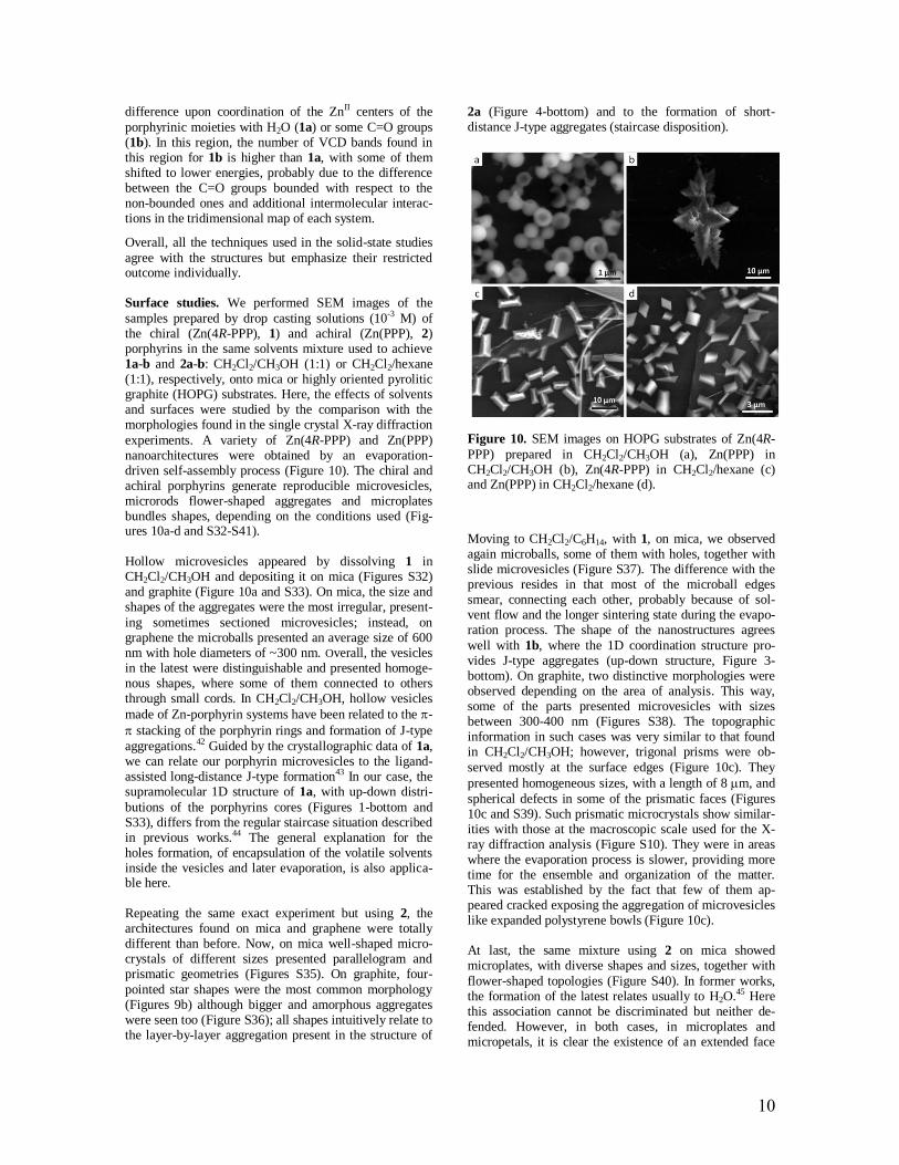
Surface studies. We performed SEM images of the
samples prepared by drop casting solutions (10-3 M) of the chiral (Zn(4R-PPP), 1) and achiral (Zn(PPP), 2) porphyrins in the same solvents mixture used to achieve 1a-b and 2a-b: CH2Cl2/CH3OH (1:1) or CH2Cl2/hexane (1:1), respectively, onto mica or highly oriented pyrolitic graphite (HOPG) substrates. Here, the effects of solvents and surfaces were studied by the comparison with the morphologies found in the single crystal X-ray diffraction
experiments. A variety of Zn(4R-PPP) and Zn(PPP) nanoarchitectures were obtained by an evaporation-driven self-assembly process (Figure 10). The chiral and achiral porphyrins generate reproducible microvesicles, microrods flower-shaped aggregates and microplates bundles shapes, depending on the conditions used (Fig-ures 10a-d and S32-S41).
Hollow microvesicles appeared by dissolving 1 in CH2Cl2/CH3OH and depositing it on mica (Figures S32) and graphite (Figure 10a and S33). On mica, the size and shapes of the aggregates were the most irregular, present-
ing sometimes sectioned microvesicles; instead, on graphene the microballs presented an average size of 600 nm with hole diameters of ~300 nm. Overall, the vesicles in the latest were distinguishable and presented homoge-nous shapes, where some of them connected to others through small cords. In CH2Cl2/CH3OH, hollow vesicles
made of Zn-porphyrin systems have been related to the -
stacking of the porphyrin rings and formation of J-type
aggregations.42 Guided by the crystallographic data of 1a, we can relate our porphyrin microvesicles to the ligand-assisted long-distance J-type formation43 In our case, the supramolecular 1D structure of 1a, with up-down distri-
butions of the porphyrins cores (Figures 1-bottom and S33), differs from the regular staircase situation described in previous works.44 The general explanation for the holes formation, of encapsulation of the volatile solvents inside the vesicles and later evaporation, is also applica-ble here.
Repeating the same exact experiment but using 2, the architectures found on mica and graphene were totally different than before. Now, on mica well-shaped micro-crystals of different sizes presented parallelogram and prismatic geometries (Figures S35). On graphite, four-
pointed star shapes were the most common morphology (Figures 9b) although bigger and amorphous aggregates were seen too (Figure S36); all shapes intuitively relate to the layer-by-layer aggregation present in the structure of
2a (Figure 4-bottom) and to the formation of short-distance J-type aggregates (staircase disposition).
Figure 10. SEM images on HOPG substrates of Zn(4R-
PPP) prepared in CH2Cl2/CH3OH (a), Zn(PPP) in CH2Cl2/CH3OH (b), Zn(4R-PPP) in CH2Cl2/hexane (c) and Zn(PPP) in CH2Cl2/hexane (d).
Moving to CH2Cl2/C6H14, with 1, on mica, we observed again microballs, some of them with holes, together with slide microvesicles (Figure S37). The difference with the previous resides in that most of the microball edges smear, connecting each other, probably because of sol-vent flow and the longer sintering state during the evapo-ration process. The shape of the nanostructures agrees
well with 1b, where the 1D coordination structure pro-vides J-type aggregates (up-down structure, Figure 3-bottom). On graphite, two distinctive morphologies were observed depending on the area of analysis. This way, some of the parts presented microvesicles with sizes between 300-400 nm (Figures S38). The topographic information in such cases was very similar to that found in CH2Cl2/CH3OH; however, trigonal prisms were ob-
served mostly at the surface edges (Figure 10c). They
presented homogeneous sizes, with a length of 8 m, and
spherical defects in some of the prismatic faces (Figures 10c and S39). Such prismatic microcrystals show similar-ities with those at the macroscopic scale used for the X-ray diffraction analysis (Figure S10). They were in areas where the evaporation process is slower, providing more time for the ensemble and organization of the matter. This was established by the fact that few of them ap-peared cracked exposing the aggregation of microvesicles like expanded polystyrene bowls (Figure 10c).
At last, the same mixture using 2 on mica showed microplates, with diverse shapes and sizes, together with
flower-shaped topologies (Figure S40). In former works, the formation of the latest relates usually to H2O.45 Here this association cannot be discriminated but neither de-fended. However, in both cases, in microplates and micropetals, it is clear the existence of an extended face

11
versus the others, connecting with the 2D layers observed
in the 2b system typical in other layered materials as well.46 Regarding the experiment in graphite, now multi-ple microblocks were present again with a variety of sizes and prismatic shapes (Figure 10d and S41). The size may depend on the deposition and evaporation processes, where the concentration of the material can also rely on the different terraces of the graphite surface. Either way, this result differs with the macroscopic scale in solution,
where the crystals were scarce and difficult to achieve, pointing out the tendency of graphite to provide crystal-line material under these conditions.
CONCLUSIONS
Using metallo-porphyrin derivatives as unit models, we described how through a unique molecular design, we created well-defined and isolable nanostructures; where, the tuning of solvent conditions directly affected the self-assembly process, and therefore the morphologies of aggregates. Our results emphasize the relevance of porphyrin crystallization toward their future applications
and face the present challenges, as the lack of anticipation of the final architectures. The addition of a metal center increases the range of diversity in the coordinative way
meanwhile blocks strong - stacking interactions among
the porphyrin cores giving priority to the nature of the arms, now key parts in the creation of the supramolecular arrays. The four arms are flexible enough and the carbon-yl moieties, from the ester groups at the edges, present the freedom to attach the ZnII centers in different manners where the final coordination number of the metallic
center can go from four to six, depending on the overall conditions. The comparison between chiral metallo-porphyrin (1) and non-chiral (2) one, repeating exact conditions, provides different crystallographic species, pointing out the complexity of adding chirality among the other factors already mentioned (coordination and solu-bility). The nature of the precipitant solvents, from polar (CH3OH) to apolar (C6H14), can promote the stabilization
of mononuclear entities, like in the case of former, in both, chiral and non-chiral metallo-porphyrins (1a and 2a, respectively). In addition, 1a shows a quite distinctive 1D supramolecular structure. Hexane promotes aggrega-tion in the chiral and non-chiral systems, having at the end coordination 1D structures (1b) or 2D systems (2b), respectively.
Regarding characterization in the solid state, 13C NMR showed highly sensitivity to identify small but critical changes in the final conformations. On the topic of chiral-ity, CD and VCD solid-state experiments presented dif-
ferences between 1a and 1b, stressing their sensitivity as well, although the variations in the former were not intui-tive and difficult to understand without the assistance of the crystallographic data. In the case of the second, it presented relevant changes in the area related to the C=O stretching, in agreement with the coordination to Zn atoms. Yet, our results emphasize that the proper analysis of the structures by the exclusive use of such techniques requires the creation of extended libraries and corrobora-tion from theoretical calculations.
Finally, the studies of our systems on two different sub-
strates show the enormous effect of the surfaces on the final structures, providing a variety of vesicles, flower-shapes and well defined geometrical architectures, in-creasing at times the capacity of achieving microcrystals (e.g.: 2b in graphite).
ASSOCIATED CONTENT
General description of the methodology followed to re-
crystallize 1a, 1b, 2a and 2b (Scheme S1). Crystal data and structure refinement for compounds 1a-2b (Table S1). Selected interatomic distances [Å] and angles for compound 1a-2b (Table S2-S5). Crystallographic data files (cif format) for compounds 1a-b and 2a-b have been deposited at the Cambridge Crystallographic Data Centre and allocated the deposition numbers CCDC 1831559, 1831558, 1831556 and 1831557, respectively. IR and 1H
NMR OF 4R-H2PPP (1) and H2PPP (2) (Figure S1, S2). Different crystallographic projections of 1a-2b (Figure S3-S6). 2D and 3D supramolecular organizations of 1a (Figure S7, S8). 3D supramolecular organizations of 2b
(Figure S9). Microscope images of crystals (Figure S10). Coordinative 1D system of 1b (Figure S11). NMR of 1a
and 1b in different concentration in CDCl3 (Figure S12, S13). UV-Vis absorption spectroscopy of Zn-porphyrins (Figure S14). XRD of crystal collected in different con-
ditions to analyze the origin of the coordinated H2O molecule in 1a (Figure S15-S17.) PXRD profiles of 1a
1b 2a and 2b (Figure S18-S21.)
IR of 1a 1b and 2a (Figure S22-S23). The solid-state 13C
NMR spectra of 1(Figure S24). The solid-state UV-Vis
and CD spectra of 1a 1b and 2a crystals (Figure S25-
S29). Solid state VCD spectra of 2a (Figure S30). SEM
images of 1 and 2 on surface (Figure S31-S40).
AUTHOR INFORMATION
Corresponding Authors
[email protected] [email protected]
ACKNOWLEDGMENT
This work has been supported by the Spanish Govern-ment under projects MAT2013-47869-C4-1-P, MAT2015-65354-C2-1-R and MAT2016-77852-C2-1-R (AEI/FEDER, UE) and the Generalitat de Catalunya 2014 (FI-DGR). We acknowledge the MINECO project MAT2015-68994-REDC and the ‘‘Severo Ochoa’’ Pro-gram for Centers of Excellence in R&D (SEV-2015-0496 and SEV-2013-0295).
ABBREVIATIONS
4R-H2PPP, 5,10,15,20-Tetra[(4-R,R,R,R)-methyl-2-phenoxy-propanoate]-porphyrin; H2PPP, 5,10,15,20-Tetra (2-aryloxypropionic ether)-porphyrin; 1 and 2, Zn-porphyrins containing (4-R,R,R,R)-methyl-2-phenoxy-propanoate and 2-aryloxypropionic ether groups; MALDI-TOF MS, matrix assisted laser desorption ioni-
zation with time of flight mass spectrometer; FTIR, Fou-rier-transform Infrared spectra; NMR, Nuclear Magnetic Resonance; UV, Ultraviolet–visible; PXRD, Powder X-

12
ray diffraction; VCD, vibrational circular dichroism;
SEM, Scanning Electron Microscope; HOPG, highly oriented pyrolitic graphite.
REFERENCES
(1) Fujita, M. Chemical Society
Reviews 1998, 27, 417.
(2) Raymond, J. E.; Bhaskar, A.;
Goodson III, T.; Makiuchi, N.; Ogawa, K.;
Kobuke, Y. Journal of the American
Chemical Society 2008, 130, 17212.
(3) Whitesides, G. M.; Grzybowski, B.
Science 2002, 295, 2418.
(4) McClements, D. J. Biotechnology
advances 2006, 24, 621.
(5) Johnson, E. R.; Keinan, S.; Mori-
Sanchez, P.; Contreras-Garcia, J.; Cohen,
A. J.; Yang, W. Journal of the American
Chemical Society 2010, 132, 6498.
(6) Shelnutt, J.; Medforth, C. Chemical
Society Reviews 1998, 27, 31.
(7) Hoeben, F. J.; Wolffs, M.; Zhang,
J.; De Feyter, S.; Leclère, P.; Schenning,
A. P.; Meijer, E. Journal of the American
Chemical Society 2007, 129, 9819.
(8) Rousseaux, S. A.; Gong, J. Q.;
Haver, R. e.; Odell, B.; Claridge, T. D.;
Herz, L. M.; Anderson, H. L. Journal of
the American Chemical Society 2015, 137,
12713.
(9) Shoji, O.; Okada, S.; Satake, A.;
Kobuke, Y. Journal of the American
Chemical Society 2005, 127, 2201.
(10) Oliveras- on le , C. Di Meo, F.
on le -Campo, A. n. Bel onne, D.
orman, P. Sim n-Sorbed, M.; Linares,
M.; Amabilino, D. B. J. Am. Chem. Soc
2015, 137, 15795.
(11) Iavicoli, P.; Xu, H.; Feldborg, L.
N.; Linares, M.; Paradinas, M.; Stafstr m,
S.; Ocal, C.; Nieto-Ortega, B.; Casado, J.;
Lopez Navarrete, J. T. Journal of the
American Chemical Society 2010, 132,
9350.
(12) Helmich, F.; Lee, C. C.; Schenning,
A. P.; Meijer, E. Journal of the American
Chemical Society 2010, 132, 16753.
(13) Bellacchio, E.; Lauceri, R.;
Gurrieri, S.; Scolaro, L. M.; Romeo, A.;
Purrello, R. Journal of the American
Chemical Society 1998, 120, 12353.
(14) Minguet, M.; Amabilino, D. B.;
Vidal-Gancedo, J.; Wurst, K.; Veciana, J.
Journal of Materials Chemistry 2002, 12,
570.
(15) Linares, M.; Iavicoli, P.;
Psychogyiopoulou, K.; Beljonne, D.; De
Feyter, S.; Amabilino, D. B.; Lazzaroni, R.
Langmuir 2008, 24, 9566.
(16) Iavicoli, P.; Linares, M.; del Pino,
A. P.; Lazzaroni, R.; Amabilino, D. B.
Superlattices and Microstructures 2008,
44, 556.
(17) Feldborg, L. N.; Saletra, W. J.;
Iavicoli, P.; Amabilino, D. B. Journal of
Porphyrins and Phthalocyanines 2011, 15,
995.
(18) Horcas, I.; Fernández, R.; Gomez-
Rodriguez, J.; Colchero, J.; Gómez-
Herrero, J.; Baro, A. Review of Scientific
Instruments 2007, 78, 013705.
(19) Bruker, S. Bruker Analytical X-ray
Instruments Inc., Madison, WI, USA 1997.
(20) Kabsch, W. Acta Crystallographica
Section D: Biological Crystallography
2010, 66, 133.
(21) Leslie, A. G. Acta
Crystallographica Section D: Biological
Crystallography 2006, 62, 48.
(22) Sheldrick, G. M.; Dauter, Z.;
Wilson, K.; Hope, H.; Sieker, L. Acta
Crystallographica Section D: Biological
Crystallography 1993, 49, 18.
(23) Sheldrick, G. M. Acta
Crystallographica Section C: Structural
Chemistry 2015, 71, 3.
(24) Farrugia, L. J. Journal of Applied
Crystallography 2012, 45, 849.
(25) Wakisaka, A.; Komatsu, S.; Usui,
Y. Journal of Molecular Liquids 2001, 90,
175.
(26) Monti, D.; De Rossi, M.; Sorrenti,
A.; Laguzzi, G.; Gatto, E.; Stefanelli, M.;
Venanzi, M.; Luvidi, L.; Mancini, G.;

13
Paolesse, R. Chemistry-A European
Journal 2010, 16, 860.
(27) Rui, X.; Zha, Q.-Z.; Wei, T.-T.;
Xie, Y.-S. Inorganic Chemistry
Communications 2014, 48, 111.
(28) Fleischer, E. B.; Shachter, A. M.
Inorganic Chemistry 1991, 30, 3763.
(29) Huijser, A.; Suijkerbuijk, B. M.;
Klein Gebbink, R. J.; Savenije, T. J.;
Siebbeles, L. D. Journal of the American
Chemical Society 2008, 130, 2485.
(30) Teo, T.-L.; Vetrichelvan, M.; Lai,
Y.-H. Organic letters 2003, 5, 4207.
(31) Zsila, F.; Bikádi, Z.; Fitos, I.;
Simonyi, M. Current drug discovery
technologies 2004, 1, 133.
(32) Dugar, S.; Fu, R.; Dalal, N. S. The
Journal of Physical Chemistry B 2012,
116, 9215.
(33) Strohmeier, M.; Orendt, A. M.;
Facelli, J. C.; Solum, M. S.; Pugmire, R. J.;
Parry, R. W.; Grant, D. M. Journal of the
American Chemical Society 1997, 119,
7114.
(34) Bampos, N.; Prinsep, M. R.; He,
H.; Vidal-Ferran, A.; Bashall, A.;
McPartlin, M.; Powell, H.; Sanders, J. K.
Journal of the Chemical Society, Perkin
Transactions 2 1998, 715.
(35) Minguet, M.; Amabilino, D. B.;
Wurst, K.; Veciana, J. Journal of the
Chemical Society, Perkin Transactions 2
2001, 670.
(36) Castiglioni, E.; Biscarini, P.;
Abbate, S. Chirality 2009, 21.
(37) Avula, S. K.; Hussain, H.; Csuk,
R.; Sommerwerk, S.; Liebing, P.; Górecki,
M.; Pescitelli, G.; Al-Rawahi, A.; Rehman,
N. U.; Green, I. R. Tetrahedron:
Asymmetry 2016, 27, 829.
(38) Hembury, G. A.; Borovkov, V. V.;
Inoue, Y. Chemical reviews 2008, 108, 1.
(39) Zheng, C.-Y.; Li, H. Inorganic
Chemistry Communications 2013, 34, 30.
(40) Frelek, J.; Górecki, M.; Łaszcz, M.;
Suszczyńska, A.; Vass, E.; Szczepek, W. J.
Chemical Communications 2012, 48, 5295.
(41) Jiang, N.; Tan, R. X.; Ma, J. The
Journal of Physical Chemistry B 2011,
115, 2801.
(42) Huang, C.; Li, Y.; Yang, J. e.;
Cheng, N.; Liu, H.; Li, Y. Chemical
Communications 2010, 46, 3161.
(43) Morisue, M.; Morita, T.; Kuroda,
Y. Organic & biomolecular chemistry
2010, 8, 3457.
(44) Tamaru, S.-i.; Nakamura, M.;
Takeuchi, M.; Shinkai, S. Organic letters
2001, 3, 3631.
(45) Wang, T.; Chen, S.; Jin, F.; Cai, J.;
Cui, L.; Zheng, Y.; Wang, J.; Song, Y.;
Jiang, L. Chemical Communications 2015,
51, 1367.
(46) Lu, J.; Zhang, D.; Wang, H.; Jiang,
J.; Zhang, X. Inorganic Chemistry
Communications 2010, 13, 1144.
Insert Table of Contents artwork here

14
Prospective porphyrin crystallization is revised by means of two Zn-porphyrin derivatives, chiral
(Zn(5,10,15,20-Tetra[(4-R,R,R,R)-methyl-2-phenoxy-propanoato]-porphyrin), named Zn(4R-PPP)) and achi-
ral (Zn(5,10,15,20-Tetra (2-aryloxypropionic ether)-porphyrin, called Zn(PPP)), using slow liquid-liquid
diffusion methods with two sets of solvents, CH2Cl2/MeOH and CH2Cl2/Hexanes. Here, we highlight the
creation of different crystalline architectures for all the possible combinations. This paper shows the analysis
performed in the solid state with all the species and the relevance that chirality, solvent mixtures and surfaces
(mica and HOPG) have on the supramolecular arrangement.


![Delta Boosting Machine and its Application in Actuarial ... · (MARS), regression trees [22] and boosting. 1.1. The Boosting Algorithms. Boosting methods are based on an idea of com-bining](https://static.fdocuments.us/doc/165x107/5f39fd86e92ad51969114a8c/delta-boosting-machine-and-its-application-in-actuarial-mars-regression-trees.jpg)


![Boosting the performance of lithium batteries with solid ... · electrolytes can be achieved. However, they normally operate at an elevated temperature [13–16]; (iii) solid-liquid](https://static.fdocuments.us/doc/165x107/5f738e0695797216be0c6ad4/boosting-the-performance-of-lithium-batteries-with-solid-electrolytes-can-be.jpg)
