![Aspergillosis in AIDS David W. Denning Director, National Aspergillosis Centre University Hospital South Manchester [Wythenshawe Hospital] The University.](https://static.fdocuments.us/doc/165x107/5515c395550346a3758b476e/aspergillosis-in-aids-david-w-denning-director-national-aspergillosis-centre-university-hospital-south-manchester-wythenshawe-hospital-the-university.jpg)
Boomeritis: The Shoulder › wp-content › ... · Connecticut Center for Orthopedic Surgery...
171
Boomeritis: The Shoulder James T. Mazzara, M.D. Connecticut Center for Orthopedic Surgery Manchester / Wethersfield Manchester Memorial Hospital / Hartford Hospital
Transcript of Boomeritis: The Shoulder › wp-content › ... · Connecticut Center for Orthopedic Surgery...

Boomeritis: The Shoulder
James T. Mazzara, M.D.
Connecticut Center for Orthopedic Surgery
Manchester / WethersfieldManchester Memorial Hospital / Hartford Hospital

Patient Education Disclaimer
This presentation provides information to educate consumers on varioushealth topics. Its is NOT intended to provide instruction on medicaldiagnosis or treatment. The information contained in this presentation is compiled from a variety ofsources. It may not be complete or timely. It does not cover alldiseases, physical conditions, ailments or treatments. You should NOTrely on this information to determine a diagnosis or course oftreatment. The information should NOT be used in place of anindividual consultation, examination, visit or call with your physician or otherqualified health care provider. You should never disregard the advice of yourphysician or other qualified health care provider because of any informationyou read in this handout or on any websites you visit as a result of thispresentation. If you have any health care questions, please consult your physician or otherqualified health care provider promptly. Always consult your physician orother qualified health provider before you begin any new treatment, diet orfitness program.

My Background
• 16 years in practice• Over 7000 surgical procedures• Board Certified, Recertified Adult
reconstructive surgery• Hartford Hospital• Manchester Memorial Hospital• Shoulder, Upper Extremity and Knee
Surgery

A second of shameless self promotion
• Only one ECHN orthopedic surgeon made the Top Docs List
• Can you guess who?
April, 2007
What is Boomeritis?
• Describes wear and tear changes, vulnerabilities and injuries that most of us have or will develop with our musculoskeletal system
• Nicholas DiNubile, MD
• Baby boomers have a desire to remain active despite age-related changes
• Boomers are born between 1946 and 1964
Life expectancy increases
• 1900 46 years• 2007 80 years
• Life expectancy increases while the durability on our frame and joints remains unchanged
• Wear and tear is predictable with age

Orthopedic Ailments• 2001: Orthopedic complaints are number 1
reason for doctors’ visits• 14% healthcare spending• Affects 1 in 7 Americans• 2.5% of GNP

When are you “old”?• When you back goes out more that you do• When you sing along with the elevator music• When your ears are hairier than your head• When you watch cable for the weather channel• When your address book starts with names
that start with Dr.• When you and your teeth don’t sleep together• When you try to straighten out the wrinkles in
your socks and discover that you are not wearing any

Age-related Cartilage Changes
• Older cartilage is less cellular
• Cartilage cells do not reproduce after growth plates close
• Chodrocytes only in lower layers
• Water content decreases

Age-related Cartilage Changes
• Proteoglycans change– Chondriotin decreases– Keratin increases– PG chains become shorted and retain less water– Decreased PG levels leads to decreased cartilage
function

Grading Cartilage Wear
• Outerbridge Classification– Stage I Soft discolored superficial
fibrillation– Stage II Fragmentation < 1.3 cm2
– Stage III Fragmentation > 1.3 cm2
– Stage IV Erosion to subchondral bone (eburnation)

Grading Cartilage Wear
I II
III IV

Osteoarthritis
• 43 million American adults have doctor-diagnosed arthritis
• 100 different types• Osteoarthritis – “wear-and-tear arthritis” – is
the most common• Exact causes are unknown• Researchers say genetics may play a role in
40-65 percent of knee osteoarthritis cases

Age-related Muscle Changes
• “Senile Sarcopenia”– Muscle mass decreases by 1/3 between 50
and 85• Loss of strength is a major cause of falls• Sarcopenia is not diffuse atrophy
– Loss of muscle mass, fiber number, and specific force reduced
– Shift toward slow twitch fibers

Age-related Ligament Changes
• Ligaments become weaker and are more likely to tear in midsubstance rather than pull away from the bone as in younger stronger ligaments

Age-related Tendon Changes
• Older tendon contains fewer fibroblasts, decreased tendon fibril diameter, decreased stiffness
• Tendon can be partially restored to strength (65%) and elasticity (69%) with eccentric exercises
Managing These Inevitable Changes
• Nutrition– Weight control
• Exercise– Properly designed and performed program
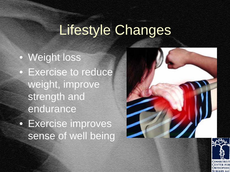
Lifestyle Changes
• Weight loss• Exercise to reduce
weight, improve strength and endurance
• Exercise improves sense of well being
Exercise as a Prescription
• Too little exercise can have negative effects• Incorrect exercise can result in injuries• Good nutrition combined with the right dose
of balanced well designed exercises can lead to a healthy frame and joints

Glucosamine• Involved in maintenance and repair of joint
cartilage• Stimulates production of synovial fluid,
proteoglycans, and glycosaminoglycans• Anti-inflammatory• 1200 – 2000 mg/ day• Higher doses
• Obesity• GERD• Diuretic use

Glucosamine
• Proteoglycans form the ground substance of the extracellular matrix in cartilage
• Of these, glycosaminoglycan hyaluronic acid is vital for the structure and function of cartilage
• Decrease incidence of severe joint space narrowing by 60%
• Treatment for >12 months reduces risk for TKR by 73% at 5 years
Pavelka, Am Coll Rheum2004

Chondroitin Sulfate
• Influences synthesis and metabolism of glycosaminoglycans
• Increases total proteoglycan production• Inhibits collagen breakdown by chondrocytes• Increased production of synovial fluid• Anti-inflammatory• Chondroprotective• 600-1500 mg per day

Glucosamine / Chondroitin• Multiple conflicting studies• No problems with side effects on liver or kidney• No affect on diabetes• Mild infrequent GI upset• Seems to help moderate to severe OA• Must be taken for 1-3 months to see effects
Nonsteroidal Anti-inflammatories
• Affect the inflammatory mechanism• NSAIDS may cause
– Gastric ulceration– Renal insufficiency– Prolonged bleeding time
• Patients >60 may have 4-5 X risk of– GI ulceration and bleeding– Renal failure requiring hospitalization
NSAIDs
• High risk individuals– >60 – h/o peptic ulcer disease– Anticipated duration of treatment over 3
months– Moderate to higher doses– Concurrent oral steroid use

NSAIDs Efficacy
• Mild OA– NSAID = Tylenol– COX-2 > Nonselective NSAID > Tylenol
• COX-2– Faster onset for OA– Efficacy within first 6 days correlates with
efficacy at 6 weeks

NSAIDs Adverse Effects
• Nonselective NSAID > COX-2> Tylenol– Edema– Hypertension– Cardiovascular disease is a concern in
COX-2 use

Corticosteroids
• Very effective in acute flairs
• Most effective in first 1-3 weeks
• Less effective than viscosupplements from 6 weeks - 6 months
• No more than 3 times per year

Common Shoulder Problems
• Rotator Cuff– Tendinitis, Bursitis, Partial and complete tears
• Arthritis– Wearing out of the joint cartilage
• Instability– Loose and Dislocating joints

The Shoulder ComplexDeltoid Muscle
Pectoralis
Clavicle (Collar Bone)
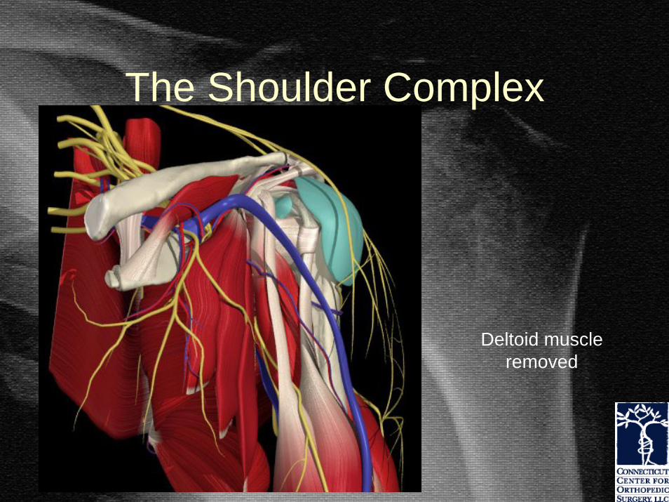
The Shoulder Complex
Deltoid muscle removed

The Shoulder Complex
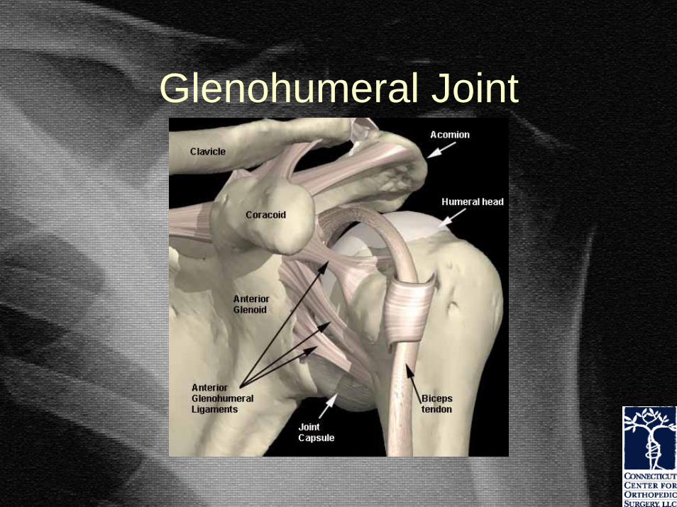
Glenohumeral Joint

Coracoacromial Arch
• Acromion & CA ligament– Protective arch
over the GH joint– Secondary
restraint for the humeral head
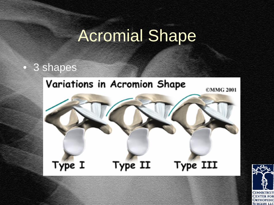
Acromial Shape
• 3 shapes

Rotator Cuff
• Supraspinatus– Active in any
elevation of the arm
– Stabilizes the shoulder joint

Rotator Cuff
• Infraspinatus– Depressor of the
humeral head
– Stabilizer of the back of the shoulder

Rotator Cuff
• Teres Minor– Rotates the shoulder
out from the side

Rotator Cuff
• Subscapularis– Stabilizes the front of
the shoulder – Rotates the arm
inward

Bursa
• Subacromial and subdeltoid bursa– Thin sac-like
structure– Lubricate motion
between rotator cuff and overlying CA arch

Rotator Cuff Balance• Proper function depends
upon balance between all muscle and ligament forces around the shoulder

Rotator Cuff
A weak or torn rotator cuff results in abnormal shoulder mechanics and abnormal motion that results in pain and further damage.

Why Tears Occur
• Tendon connective tissue weakens with age and disuse– Weakened tendons require less force to
disrupt• Repetitive and / or substantial loads

Tendon Degeneration
• Age-related changes– Decreased
vascularity at the tendon attachment to the bone
– Leads to weak tendon that tears easily

Rotator Cuff Tears
• Tears begin where the stresses are the greatest– Tendon fibers fail a
few at a time or all at once
– Arm may be at rest– Torn fibers retract
when torn Humeral Head
Partial SupraspinatusTear

Consequences of rupture
• Increasing loads applied to the intact fibers
• Muscle fibers become detached from the bone resulting in weakness

Consequences of rupture
• Retracted cuff fibers place additional tension on remaining microcirculation compromising cuff viability
• Increasing amounts of tendon are exposed to joint fluid which prevents tendon healing

Full Thickness Tears• Loads are
concentrated at the margins of the tear
• Further tearing occurs with smaller loads
• Partial tears become complete
• Smaller tears become large
• Large tears eventually become unfixable

Progressive Tearing
• Spacer effect of the cuff is lost• Humeral head displaces
superiorly• Biceps tendon eventually
ruptures

Early Cuff Failure
• Compression of the humeral head is less effective– Deltoid pulls head
upward– Upward pull of the
deltoid results in cuff abrasion & further cuff damage
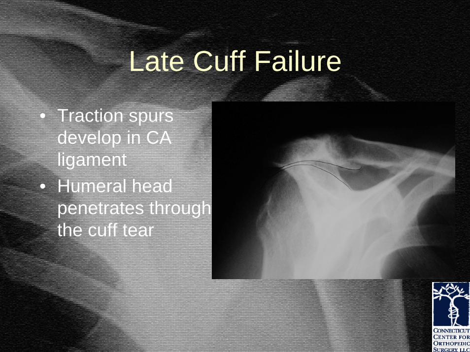
Late Cuff Failure
• Traction spurs develop in CA ligament
• Humeral head penetrates through the cuff tear

Chronic Cuff Failure
• Humeral head forms a joint with the arch above
• Secondary joint disease occurs called cuff tear arthropathy

Chronic Cuff Tears
• Muscle atrophy• Fatty infiltration of
muscle belly• Tendon retraction• Bone osteoporosis• Loss of muscle and
tendon excursion• Irreversible• Progressively worse
Fatty infiltration with muscle wasting
Healthy muscle, no fat stripes

Prevalence of Rotator Cuff Tears
• Cadaver studies 7-40%• MRI & Ultrasound studies
– 34% of asymptomatic individuals– 54% of asymptomatic individuals over 60y
• Ultrasound study– 13% of asymptomatic individuals: 50-59y– 51% of asymptomatic individuals: over 80y
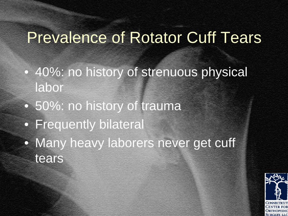
Prevalence of Rotator Cuff Tears
• 40%: no history of strenuous physical labor
• 50%: no history of trauma• Frequently bilateral• Many heavy laborers never get cuff
tears
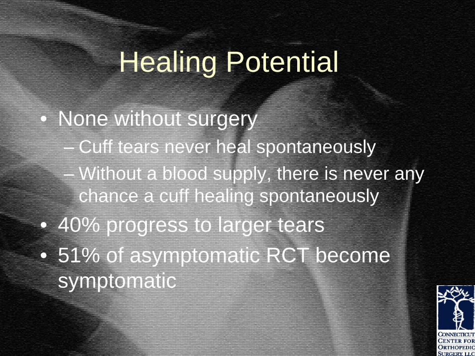
Healing Potential
• None without surgery– Cuff tears never heal spontaneously– Without a blood supply, there is never any
chance a cuff healing spontaneously• 40% progress to larger tears• 51% of asymptomatic RCT become
symptomatic

Patient History
• Important things to know– Chronic symptoms or
acute exacerbation– Stiffness, loss of motion– Weakness (when)
– Functional impairment– Catching, crepitus,
grinding– Treatments and response

Shoulder Pain with Cuff Tears
• Rotator cuff pain– Constant ache – Varies with activity– Night pain– Wake up with position
change– May be severe– Constant or intermittent

Rotator Cuff Shoulder Pain
• Deep, dull, diffuse ache
C5
DeltoidAxillary nerve
The pain from rotator cuff pathology is often referred to the outer part of the arm. Sometimes as far as the elbow.

Non Rotator Cuff Shoulder Pain
• Pain to the back of shoulder upper back or neck
• Pain to top of shoulder– Think arthritis of the neck
• Pain beyond the elbow– Think pinched nerve in
the neck

Timing of Pain• Rest Pain (constant)
– Synovitis (Inflammation of the joint)
Calcification
–Calcific tendinitis or bursitis (constant and intense)

Timing of Pain
• Pain in mid range of motion– Arthritis - Damaged
joint surface– Inflamed irregular
joint surface– Inflamed tissues
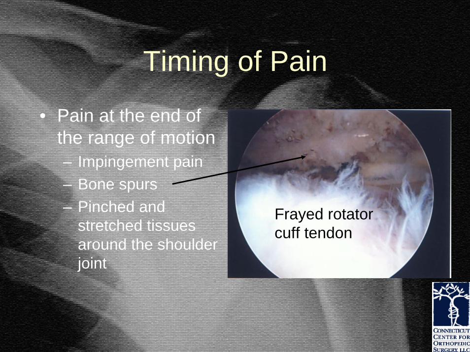
Timing of Pain
• Pain at the end of the range of motion– Impingement pain– Bone spurs– Pinched and
stretched tissues around the shoulder joint
Frayed rotator cuff tendon

Physical Examination

Physical Examination
• Inspection– Symmetry– Atrophy– AC prominence– Biceps rupture
This patient cannot lift his arm due to a nerve injury not a rotator cuff tear.

Range of Motion
• Range of Motion– Active and passive– Forward elevation– ER @ side– ER in abduction– Internal rotation

Rotator Cuff Weakness• May be due splinting from
pain• Significant only if present
after a xylocaine block• Weakness after an injury
– Rotator cuff tear– Nerve injury
• Suprascapular• Axillary• Brachial plexus
– Cervical disk herniation

Strength Testing
• Assess elevation, abduction, ER, IR strength
• Compare to opposite side
• If weak, check reflexes

External Rotation Weakness
• Supraspinatus &/or infraspinatus• Small tears
– Minimal if any weakness noted• Large tears
– Weakness notable– May result in inability to maintain external
rotation• Fall off into internal rotation
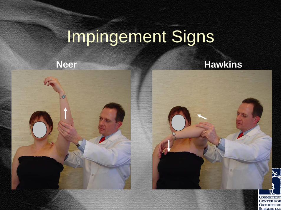
Impingement SignsNeer Hawkins

Impingement Test
• Subacromial injection of 10cc 1% xylocaine (often with a corticosteroid)
• Rotator cuff tear– Pain relieved, weakness persists
• Impingement (tendinitis), bursitis– Pain relieved, strength improves
• Adhesive capsulitis / arthritis– Pain persists, motion unchanged

Impingement Test
• Subacromial injection of 1% xylocaine– At least 50% pain relief

AC Joint Exam
• Inspect for prominence• Palpate for tenderness• Provoke pain with
“cross-body adduction”• Relieve pain with 1cc
xylocaine injection• Consider arthritis and
osteolysis

Best Tests for Diagnosing Cuff Tears
1. Weak supraspinatus testing– Arm in 90O forward elevation in scapular
plane2. Weakness in External Rotation
– Arm at the side3. Positive Impingement Sign
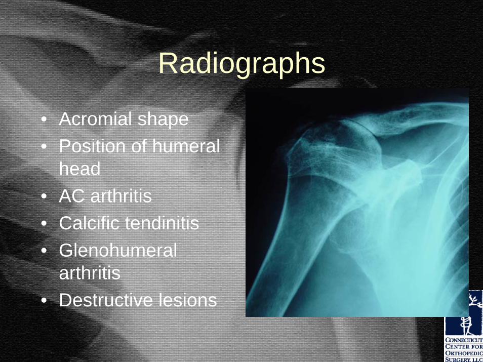
Radiographs
• Acromial shape• Position of humeral
head• AC arthritis• Calcific tendinitis• Glenohumeral
arthritis• Destructive lesions

1 & 2: AP in Scapular Plane
• 2 Views: IR, ER• Calcium deposits• Greater tuberosities:
excrescences, cysts

1 & 2: AP in Scapular Plane
• 2 Views: IR, ER• Calcium deposits• Greater tuberosities:
excrescences, cysts
Moderate osteoarthritis
Severe osteoarthritis

3: Axillary View
• Evaluate GH joint & tuberosities
• Glenoid version• Joint space
narrowing • Os acromiale
– This is an anatomic variation best seen on this special view

4: Outlet View
• Evaluate subacromial space
• Acromial shape and thickness

5: 30O Caudal Tilt View
• AP view with a 30O caudal tilt
• Demonstrates anterior acromial projection
spur

Tendon Imaging
• MRI– 90% accurate in
diagnosing complete RC tears
– 70% accurate in diagnosing partialRC tears
– These data may vary. It depends on who is reading the MRI. This spur is pushing on the rotator
cuff causing “impingement”.

Best Studies for Diagnosing Cuff Tears
Full Thickness Tears AccuracyClinical Exam 0.4Ultrasound 0.7MRI 0.7Arthroscopy 0.9
Partial Thickness Tears <0.2 for all studies
Murrell, AAOS Instr. Course Lect. March, 2004

Overall Detection Accuracy
Full thickness Partial Thickness
Ultrasound 98% 68%MRI 100% 63%
JBJS, 86-A, April, 2004

Full thickness Partial ThicknessUltrasound 73% (retraction) 85% (length)
87% (width) 54% (width)
MRI 63% (retraction) 75% (length)85% (width) 75% (width)
JBJS, 86-A, April, 2004

Nonoperative Treatment
• Helpful in ~50% (33-92%)• Acute rupture
– 75% may have reduced pain with therapy– But the tendon tear will never heal without
surgery.• Chronic pain (>6 months)
– poor response with therapy
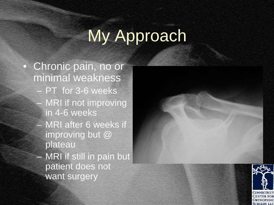
My Approach
• Chronic pain, no or minimal weakness– PT for 3-6 weeks– MRI if not improving
in 4-6 weeks– MRI after 6 weeks if
improving but @ plateau
– MRI if still in pain but patient does not want surgery

My Approach
• Acute pain, weakness– Office evaluation– X-rays– Injection– MRI
• May be age dependent

Analyzing the Data
• If the weakness and pain are inconsistent with MRI findings– Look for other causes
• C spine, nerve injuries– Consider multiple causes
• Older patients with dislocations• Concurrent cuff tears, brachial plexus injuries,
or axillary nerve injuries

Surgical Indications
• Patient dependent• Impingement syndrome & Partial tears
– Pain with functional impairments– Failure to respond to nonoperative treatment
• Chronic tears– Consider 3-4 months of nonsurgical treatment
• Acute tears– Best results if repaired within 3 weeks

Arthroscopic Acromioplasty
• Relieves impingement between the CA arch & the cuff
• Performed with arthroscopic or mini-open cuff repair

Technique of Arthroscopic Acromioplasty
• Bone spurs can be removed through small arthroscopic incisions by using a motorized burr.

Arthroscopic v. Open Acromioplasty
• Arthroscopic group do better in first 3 months
• After 3 months, both methods give equal results
• Long-term: no difference• 90% excellent results

Post-op Arthroscopic Acromioplasty
• Sling for 1-2 days• Begin active motion immediately• Advance as tolerated
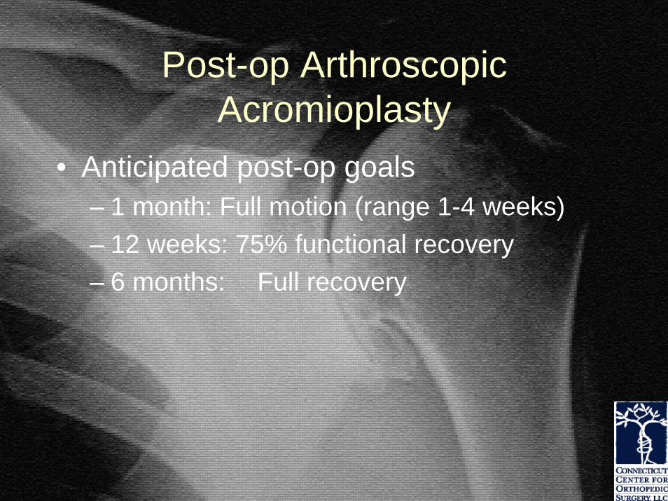
Post-op Arthroscopic Acromioplasty
• Anticipated post-op goals– 1 month: Full motion (range 1-4 weeks)– 12 weeks: 75% functional recovery– 6 months: Full recovery

Surgery for Partial Thickness Tears
• Debridement alone
• Debridement and acromioplasty
• Acromioplasty, excision of damaged tendon with primary repair

Partial RCT: Debridement Alone
• Young athletes and workers
• Failed nonoperative therapy
• Tears related to overuse not impingement
• 80-85% success
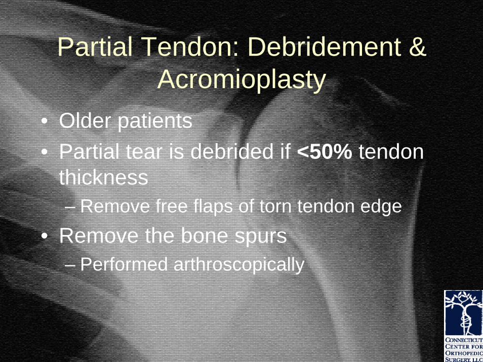
Partial Tendon: Debridement & Acromioplasty
• Older patients• Partial tear is debrided if <50% tendon
thickness– Remove free flaps of torn tendon edge
• Remove the bone spurs– Performed arthroscopically
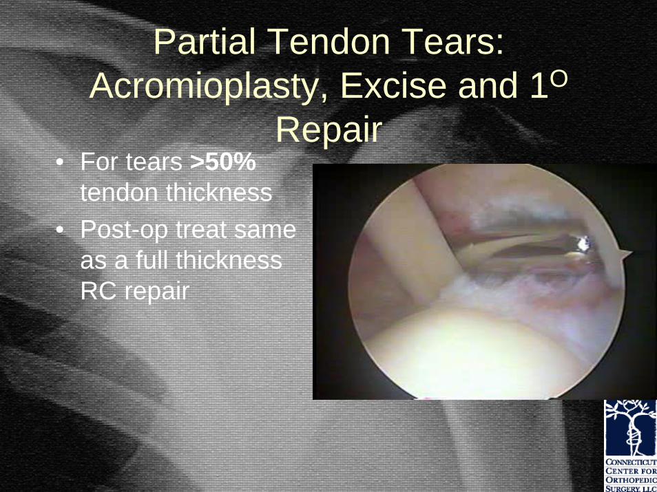
Partial Tendon Tears: Acromioplasty, Excise and 1O
Repair• For tears >50%
tendon thickness• Post-op treat same
as a full thickness RC repair

Full Thickness Cuff Tears
• Arthroscopic repairs
• Mini-open repairs
• Open repairs

The Outcome and Repair Integrity of Completely Arthroscopically Repaired Large
and Massive Rotator Cuff Tears• Massive defined as > 2 cm• 17 of 18 retears @ 12 months post op (94%)• 2/3 improved after surgery• Not doing as well @ 24 mo.
• “An arthroscopic repair arguably may not be the most appropriate procedure for a younger person with a massive tear in whom long-term strength is more important…”
Yamaguchi, et.al., JBJS February, 2004

Cuff Integrity Following Arthroscopic v. Open Rotator
Cuff Repair• American Shoulder and Elbow Surgeons Meeting• Intact Cuffs are associated with better strength and
outcome scoresIntact by MRI after 1 year
Open Repair Arthroscopic Repair
Tears < 3cm 74% 84%* 82%
Tears > 3cm 62% 24%* 21%
•*Bishop, Flatow, et.al., ASES Meeting, March 2004•Bishop, Flatow, et.al., ASES Meeting, October 2004

• 65 patients, 2 1/2 year follow up• All arthroscopic repair• CT-arthrogram evaluation post-op• 71% healed, 95% patient satisfaction• Healed tendons were stronger w/ better function• Factors negatively impacting healing rate
– Age over 65y– Larger tears (Delamination of subscap and infraspinatus
tendons)
JBJS June 2005

• Studied 84 patients with tears smaller than 5 cm (=massive tears)
• Found equivalent results regarding patient satisfaction between mini-open and arthroscopic repair at 2 years
• Results determined by subjective scoring system

• Issues with this study– Healing time was the same in the 2 groups– MRI s were not performed to evaluate cuff integrity
which impacts long term success– 2 years may no be a long enough follow up on young
active patients or those who perform physical labor

Outcome and Patient Satisfaction of Arthroscopic Rotator Cuff Repair v.
Mini-open Cuff RepairCompared 2 groups of patients with tears
of various sizes• 24-70 month follow up: equivalent
functional and patient satisfaction scores (UCLA and ASES scores)
• Despite less post-operative morbidity in ARCR, mid-term results are equivalent
Youm, Rokito, et.al., ASES Meeting , March, 2004
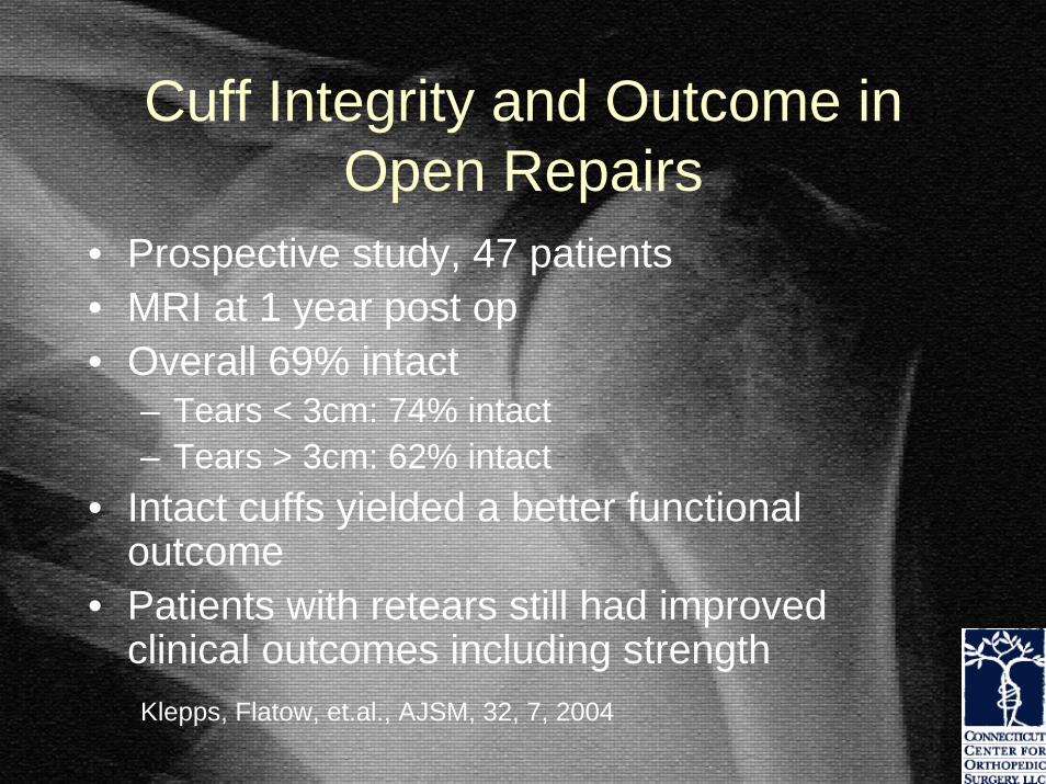
Cuff Integrity and Outcome in Open Repairs
• Prospective study, 47 patients• MRI at 1 year post op• Overall 69% intact
– Tears < 3cm: 74% intact– Tears > 3cm: 62% intact
• Intact cuffs yielded a better functional outcome
• Patients with retears still had improved clinical outcomes including strengthKlepps, Flatow, et.al., AJSM, 32, 7, 2004

Other Literature
• Harryman, JBJS, 1991
– 80% 1 tendon tears intact at follow up– 57% 2 tendon tears– 32% 3 tendon tears– Better results with an intact repair
• Liu & Baker, Arthroscopy, 1994
– 66% intact with mini-open repair– Tear size correlated with cuff integrity at follow up– Functional outcome did not correlate with cuff
integrity

Other Literature
• Thomazeau, Clinics in Orthopedics, 1997
– 73% intact– Better outcome correlate with intact repair
• Gerber, JBJS, 2000
– 66% intact, massive 2 tendon tears– Better results with intact repairs

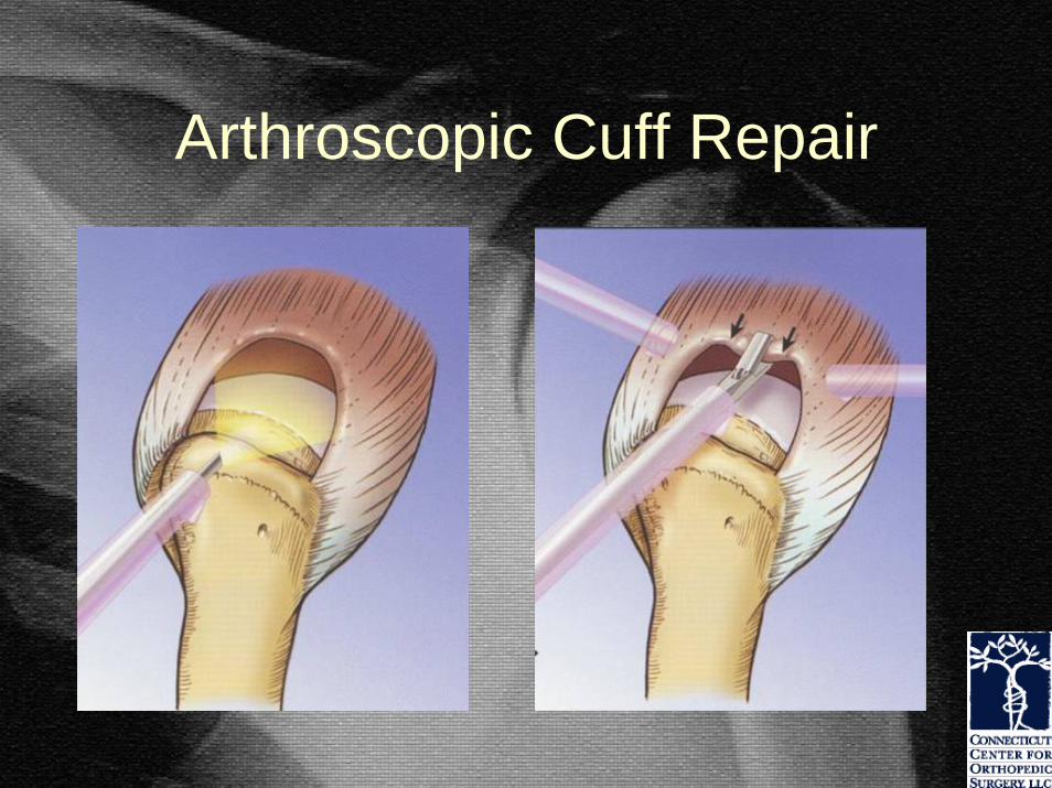
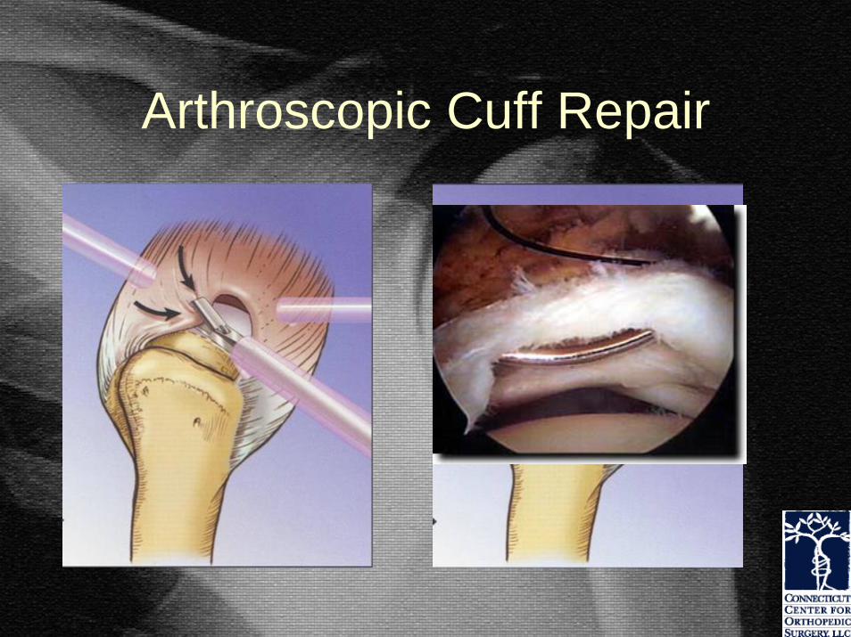
Arthroscopic Cuff Repair
• Arthroscopy allows for a more complete evaluation of the joint and tendon
• Removal of bone spurs
• Rotator cuff repair using anchors
Arthroscopic Cuff Repair
• Advantages– Improved joint assessment, incl. biceps– Improved tendon mobilization– Decreased surgical trauma to deltoid– Faster rehabilitation (in first 3 months)

Arthroscopic Cuff Repair
• Advantages– Earlier return to function
• 6 weeks to heal, 6 months for overhead work– Less Pain
• No evidence of this– Shorter hospitalization
• Every cuff repair goes home the day of surgery– Cosmetic
• Multiple smaller incisions vs. one incision

Arthroscopic Cuff Repair
• Disadvantages– Longer operative time– Cannot place tendon
gripping sutures– Anchors less secure in
weak bone– Anchors are costly– No studies have
proven the long term results to be superior to mini-open repairs
Arthroscopic Cuff Repair
Arthroscopic Cuff Repair

Arthroscopic Cuff Repair

Arthroscopic Cuff Repair

Arthroscopic Cuff Repair

Arthroscopic Repair of Partial Tears
• For selected partial tears
• Repairs only the torn portion of the tendon
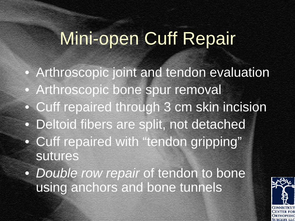
Mini-open Cuff Repair
• Arthroscopic joint and tendon evaluation• Arthroscopic bone spur removal• Cuff repaired through 3 cm skin incision• Deltoid fibers are split, not detached• Cuff repaired with “tendon gripping”
sutures• Double row repair of tendon to bone
using anchors and bone tunnels

Mini-open Cuff Repair
• Gold standard• Allows double row
repair• Suture anchors with
bone tunnels provide strongest repair with best restoration of RC footprint (Andrews AJSM, 2003)

Suture Fixation Techniques
• Holding strength with open suture placement techniques was superior– JBJS, 2002

Suture Anchor Fixation
• Dependent on the quality of bone
• Anchors have a limited pull-out strength from bone

Suture Anchor Fixation
This osteoporosis is common in older patients, larger chronic tears and may not provide strong tendon repairs

Double Row Cuff Repair
This type of repair through a small incision remains the “gold standard” for rotator cuff repair surgery.

Open Cuff Repair
Open cuff repairs may be appropriate for larger tears and complete rotator cuff avulsions.

Open Cuff Repair
12 weeks post massive cuff repair

Open Cuff Repair
• 5cm incision• Deltoid is taken
down from acromion– Must be securely
repaired • For larger tears

Post-op RC Repair
• Usually 6 weeks of limited arm use regardless of repair method
• Often require 2-4 months of formal physical therapy followed by home exercises
• Can take 12-18 months to reach maximum improvement

Post-op RC Repair
• Same restriction regardless of repair method
• At 1-6 weeks Passive motion– Passive forward elevation, ER with stick
supine, pendulums– Avoid internal rotation and AROM until
healed• AROM of elbow/wrist and hand

Post-op RC Repair
• At 6 weeks begin AROM and advanced stretching
• At 8-12 weeks begin Theraband PREs depending on tear size
• At 4-6 months begin progressive resistance and dynamic strengthening

Rotator Cuff Repair Results
• Good to excellent– 85% - 95%
• Good-excellent pain relief– 78%
• Risk of rerupture– Large (2+ tendon tears)
• 40% – Smaller tears
• 10-20%– Severely retracted tears
• 66%
This man is 7 weeks following and arthroscopic cuff repair.

Partial Repairs• Massive retracted cuff tears• Insufficient tendon to repair to
bone• Repair as much as possible• Margin convergence restores
some function• Provides good pain relief• Unpredictable functional
recovery

Unsatisfactory Results
• Associated with retears
• Loss of function• Often have good
pain relief– This patient has
retorn her repair but is happy since she no longer has pain. Her motion before and after surgery are the same.

Factors Affecting Outcomes• Tear size (most
important)– Affects recovery of
strength (85-90% recovery)
• Age (>65)• Pre-op function
(inability to abduct > 100O)
• Larger tears and chronic retracted tears are more likely to rerupture

Recurrent Tears• Number of tendons
– 1 tendon 33%– 2 tendons 30-56%– 3 tendons 50%
• Muscle Atrophy– Increasing degrees of
atrophy lead to increasing rates of rerupture
• Cuffs with no noticeable atrophy– 20% rerupture

Complications of Cuff Repair
• Rerupture• Stiffness• Infection• Deltoid detachment• Nerve injury
– Weakness, numbness

Arthroscopy Without Repair
• Arthroscopic cuff debridement & limited acromioplasty
• Smaller tears get better pain relief• No improvement with overhead activity
and strength• Beneficial in older low demand patients

Open Surgery Without Repair
• Open cuff debridement• Better results with intact
biceps, deltoid and no prior surgery
• 50-80% Improved comfort and function
• Preserve the CA arch– Avoids humeral head
escape

Why Preserve the CA Arch?
If the CA arch is disrupted, the head of the humerus escapes up through the defect and pain and limited motion result.

Biceps Tenotomy
• Indicated in older low demand patients with irreparable cuff tears
• Unconcerned about biceps bulge
• Relieves pain from the impinged or dislocated biceps
• Minimally invasive, palliative, minimal rehab

The Stiff Shoulder• Not associated with
cuff tears alone• Consider
– Adhesive capsulitis / Frozen shoulder
– Shoulder arthritis– Missed shoulder
dislocation– Fracture or post
traumatic deformity

The Stiff Shoulder
• Frozen Shoulder = Adhesive Capsulitis
• Cause is Unknown– May be autoimmune– May occur after injury, fracture
or surgery– Related to intense inflammation
causing pain and decreased use of the shoulder leading to stiffness

The Stiff Shoulder
• Reduced motion even with help lifting the arm– As if the motion is
“blocked”• Pain at night and
with daily activities• X-rays and MRI
usually normal

Shoulder Stiffness• Not associated with cuff tears alone• Full active and/or passive motion is
present even if painful• Consider
– Adhesive capsulitis / Frozen shoulder– Glenohumeral arthritis– Missed posterior dislocation– Fracture or post traumatic deformity

Frozen Shoulder
• Recovery is slow – May take many months
• Anti-inflammatory meds and stretching exercises
• May benefit from cortisone injections• Surgical treatment may help and
involves arthroscopy to remove the scarred joint capsule

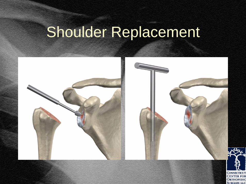
Shoulder Replacement• Arthritis
– Wear and tear– Multiple dislocations– Rheumatoid arthritis

Shoulder Replacement
• Osteonecrosis / Avascular Necrosis– Post Trauma– Steroid or Alcohol
Use

Shoulder Replacement
• Fractures

Shoulder Replacement
Shoulder Replacement

Shoulder Replacement

Shoulder Replacement
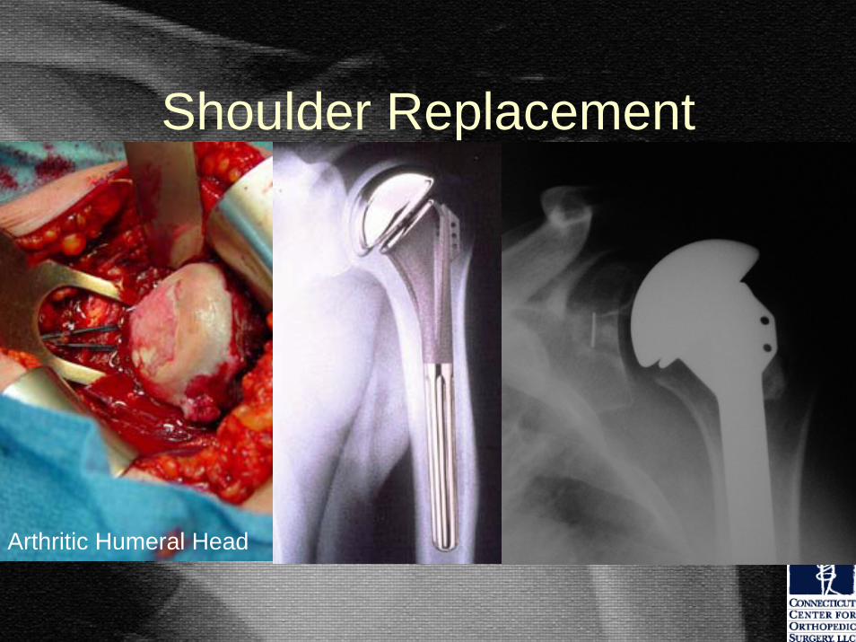
Shoulder Replacement
Arthritic Humeral Head

Partial Shoulder Replacements
• Chronic arthritis with an irreparable rotator cuff tear
• Must be able to elevate the arm
• Must have intact and functioning deltoid
• Hemiarthroplasty is useful in relieving pain

Reverse Shoulder Replacements
Reverse shoulder replacements are helpful when treating arthritis associated with irreparable rotator cuff tears in patients unable to lift the arm due to tendon tears.

Reverse Shoulder Replacements
Standard Shoulder Replacement

Reverse Shoulder Replacement

Shoulder Instability• Shoulder joint is too loose
and is able to slide around too much in the socket
• Dislocation– Head of humerus slips
completely out of the socket• Instability without dislocation
– Causes pain and apprehension due to excessive motion

Shoulder Dislocation
• Head of humerus slips completely out of the socket

Shoulder Dislocation
• May be due to a lax shoulder capsule and ligaments
• May be due to significant trauma

Shoulder Dislocation
• As the result of a shoulder dislocation, the labrum and the ligaments are torn and stretched.

Shoulder Dislocation
• Nonsurgical treatment for initial dislocation– 3-4 weeks immobilization– Arm in External Rotation– Followed by therapy– Study
• 0% recurrence in ER group v. 30 % recurrence in IR group
(JSES 2003)

Shoulder Dislocation
• Can lead to fractures and joint damage
• Repeated dislocations can lead to eventual arthritis
Bony Bankart Lesion
Hill-Sachs Lesion
CT Scan of Chronic Dislocation

Shoulder Dislocations
• Cause tearing of the ligaments and cartilage that normally stabilize the shoulder
• Shoulder may need to be manually put back into socket

Shoulder Dislocations
Torn labrum undergoing arthroscopic repair

Shoulder Dislocations
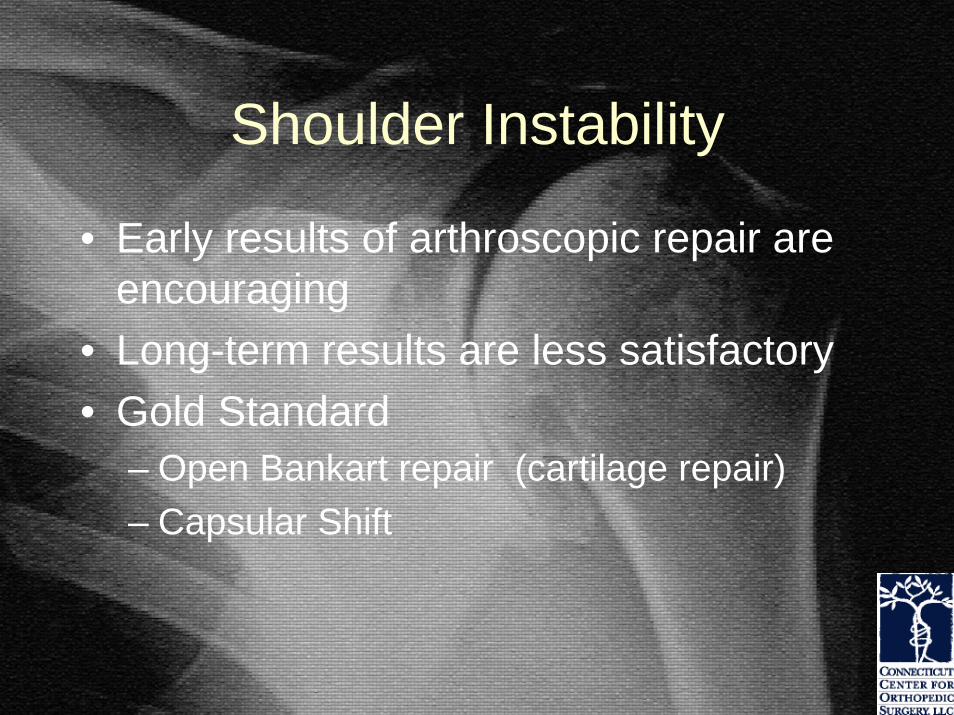
Shoulder Instability
• Early results of arthroscopic repair are encouraging
• Long-term results are less satisfactory• Gold Standard
– Open Bankart repair (cartilage repair)– Capsular Shift

Shoulder InstabilityA capsular shift performed for multidirectional instability: the lax shoulder capsule is cut and sutured in such a way so as to reduce the volume of the shoulder joint to increase stability.
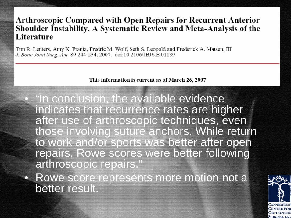
• “In conclusion, the available evidence indicates that recurrence rates are higher after use of arthroscopic techniques, even those involving suture anchors. While return to work and/or sports was better after open repairs, Rowe scores were better following arthroscopic repairs.”
• Rowe score represents more motion not a better result.
Complications of Surgery
• Always part of pre-op discussion• Nerve damage
– Weakness, numbness • Bleeding• Infection• Tendon rupture• Stiffness• Continued pain and impairment• Stretched repair and recurrent instability

Thank You
Be careful out there.


![Clinical and radiological presentation and diagnosis David W. Denning National Aspergillosis Centre University Hospital South Manchester [Wythenshawe Hospital]](https://static.fdocuments.us/doc/165x107/5515c3be55034693758b476d/clinical-and-radiological-presentation-and-diagnosis-david-w-denning-national-aspergillosis-centre-university-hospital-south-manchester-wythenshawe-hospital.jpg)