BONY LANDMARKS - Visible Body
13
There are more than 200 bones in the human body. Each one is different and unique. Bone landmarks help us describe individual bone with all of its bumps, ridges, and holes. In this eBook, we will look at common bone landmark terminology. BONY LANDMARKS
Transcript of BONY LANDMARKS - Visible Body

There are more than 200 bones in the human body. Each one is different and unique. Bone landmarks help us describe individual bone with all of its bumps, ridges, and holes. In this
eBook, we will look at common bone landmark terminology.
BONY LANDMARKS
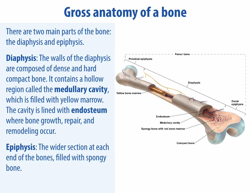
Gross anatomy of a boneThere are two main parts of the bone: the diaphysis and epiphysis.
Diaphysis: The walls of the diaphysis are composed of dense and hard compact bone. It contains a hollow region called the medullary cavity, which is filled with yellow marrow. The cavity is lined with endosteum where bone growth, repair, and remodeling occur.
Epiphysis: The wider section at each end of the bones, filled with spongy bone.

Gross anatomy of a boneBetween the diaphysis and the epiphysis is the epiphyseal growth plate, which contains hyaline cartilage in a growing bone. Once the bone stops growing, the cartilage is replaced by osseous tissue and the plate becomes a line.
The outside of the bone is covered in a fibrous membrane called the periosteum. This contains blood vessels, nerves, and lymphatic vessels. Tendons and ligaments attach to the bone here. When bones meet to form a joint, the epiphysis of each bone is covered with articular cartilage, which reduces friction and acts as a shock absorber.
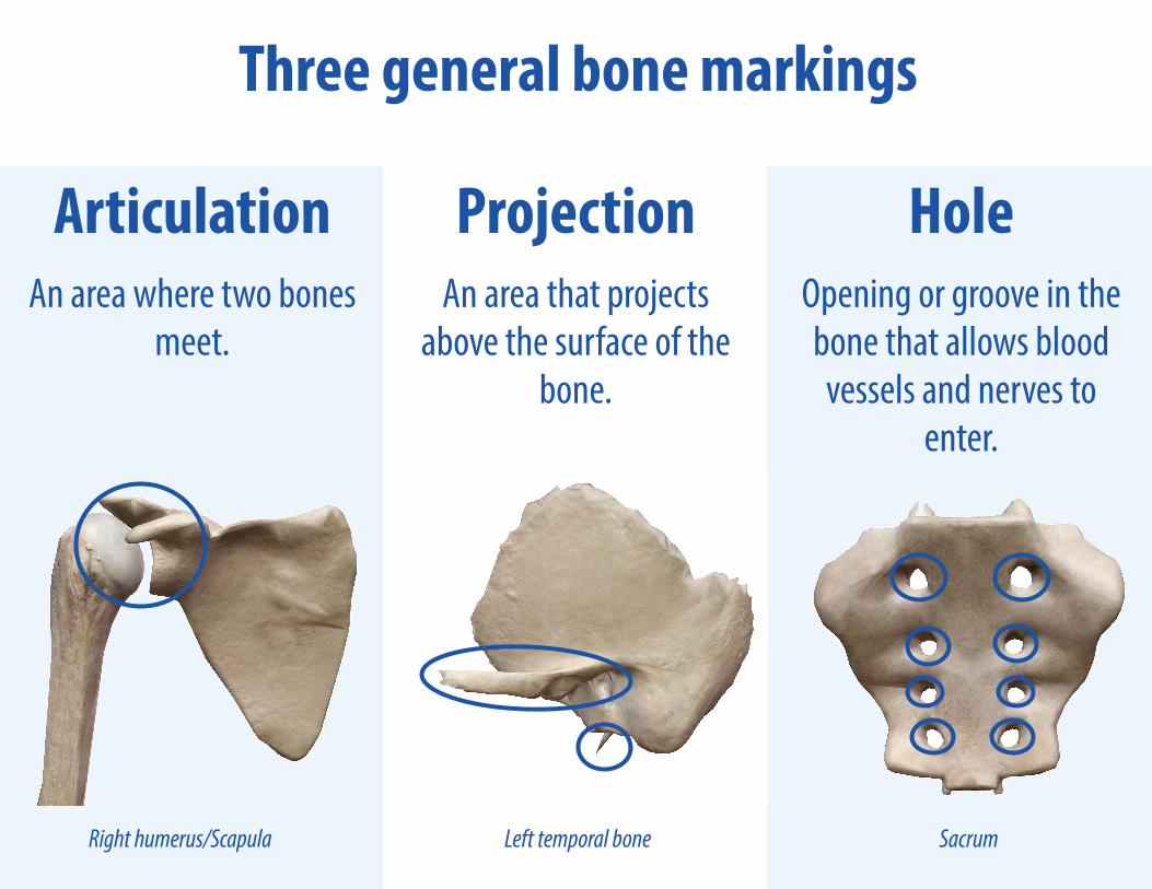
Three general bone markings
Articulation An area where two bones
meet.
Projection An area that projects
above the surface of the bone.
HoleOpening or groove in the bone that allows blood vessels and nerves to
enter.
SacrumLeft temporal boneRight humerus/Scapula
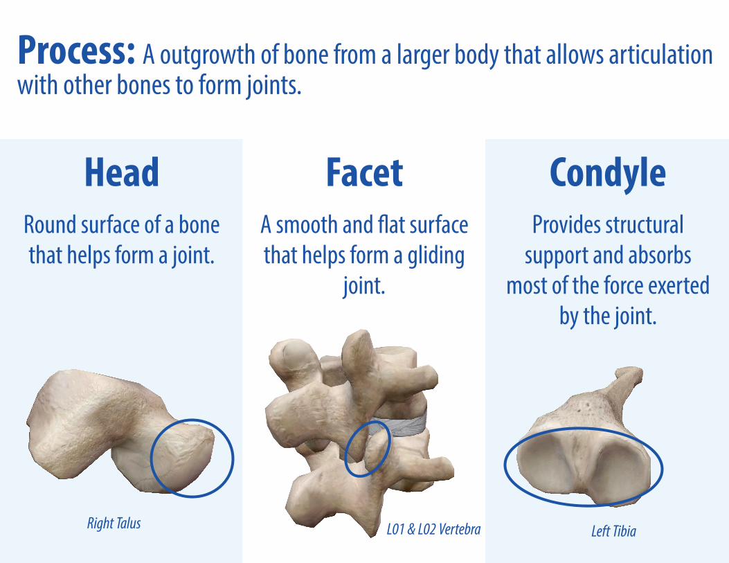
HeadRound surface of a bone that helps form a joint.
Facet A smooth and flat surface that helps form a gliding
joint.
CondyleProvides structural
support and absorbs most of the force exerted
by the joint.
Process: A outgrowth of bone from a larger body that allows articulation with other bones to form joints.
Left TibiaL01 & L02 VertebraRight Talus

Body: The largest and main segment of the bone.
Neck: Narrowing of the bone where the shaft of the long bone (diaphysis) meets the end of the long bone.
Left Radius
Body of SternumLeft CalcaneusLeft Scaphoid
Right 1st Rib

Crest: A raised ridge projection that is part of the edge of a bone and allows connective tissues to connect to the bone.
Epicondyle: A rounded projection that sits on top of the condyle and allows connective tissues to connect to the bone.
Left Ilium Left Pubis Frontal bone
Left FemurLeft humerus

Tuberosity: A large projection on the side of the bone that allows connective tissues to connect to the bone.
Spine (spinous process): A more pronounced raised, sharp elevation of bone that allows connective tissues and muscles to connect to the bone.
Left Femur
Right ScapulaT01 Vertebra L03 Vertebra

C03 Vertebra Mandible Left Fifth Rib
Tubercle: A small round projection that allows connective tissues to connect to the bone.
Right IschiumRight 5th Metatarsal
Tuberosity: A moderate projection that allows connective tissues to connect to the bone.
Left Radius

Foramen: A round hole where blood vessels, nerves, or ligaments pass through.
Meatus: A tube-like channel that provides passage and protection to nerves and vessels within the bone.
Spheniod OccipitalRight zygomatic bone
Right temporal bone

Fissure: A slit in the bone that usually houses nerves and blood vessels.
Sulcus: A groove that accommodates a nerve, blood vessel, or tendon.
Right Maxilla
Right Talus

Fossa: A shallow depression in a bone’s surface that allows other bones to articulate with it.
Sinus: A cavity within an organ or tissue.
Ethmoid bone
Left Humerus
A universe of anatomical and physiological visuals and reference texts at your fingertips!
www.visiblebody.com
View a 3D Tour of all the images featured in this eBook!
If you have a mobile version of Human Anatomy Atlas 2021.1 or later: 1. Click here to view the tour.
If you have a web version of Atlas:1. Copy this link:
https://apps.visiblebody.com/share/?p=vbhaa&t=4_31278_637546902370634390_49950
2. Use the share link button in the app.3. Paste the link to view the tour.