Bones of the Appendicular Skeleton - A Visual Guide
33
Appendicular Skeleton Visual Atlas Anatomy & Physiology I BIOL 121 Rob Swatski Assistant Professor of Biology HACC – York Campus Mariana Ruiz Villarreal, May 2009, http://commons.wikimedia.org/wiki/File:Appendicular_skeleton_diagram_blank.svg 1
-
Upload
isabella-suescum-baquerizo -
Category
Documents
-
view
30 -
download
0
description
Bones of the appendicular skeleton.Learn the anatomy f the bones of the appendicular skeleton on this visual pdf guide.MEDICIINEANATOMYSCIENCE
Transcript of Bones of the Appendicular Skeleton - A Visual Guide

AppendicularSkeleton
Visual Atlas
Anatomy & Physiology I BIOL 121
Rob Swatski
Assistant Professor of Biology
HACC – York Campus
Mar
ian
a R
uiz
Vill
arre
al, M
ay 2
00
9, h
ttp
://c
om
mo
ns.
wik
imed
ia.o
rg/w
iki/
File
:Ap
pen
dic
ula
r_sk
elet
on
_dia
gram
_bla
nk.
svg
1
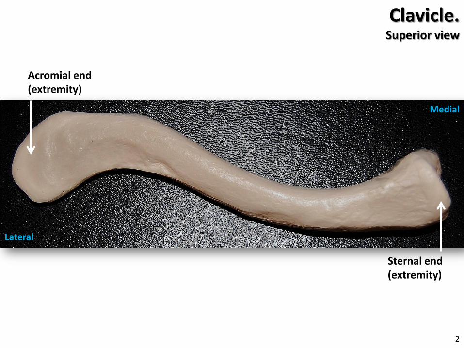
Sternal end (extremity)
Acromial end (extremity)
Clavicle.Superior view
Medial
Lateral
2

Manubrium
Body of Sternum
Sternal ends (extremities)
Acromial end (extremity)
Coracoidprocess
Acromion
Pectoral girdleLeft anterior view
Conoid tubercle (Coracoid tuberosity)
Clavicle
Humerus
3

Acromion
Coracoidprocess
Glenoidcavity
Axillary (Lateral) border
Inferior angle
Superior angle
Scapular notch
Vertebral (Medial) border
Subscapularfossa
Scapula.Right anterior view 4
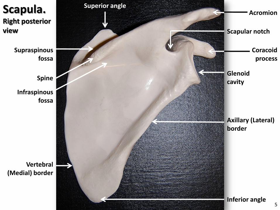
Acromion
Coracoidprocess
Glenoidcavity
Axillary (Lateral) border
Inferior angle
Spine
Superior angle
Supraspinousfossa
Infraspinousfossa
Vertebral (Medial) border
Scapula.Right posterior view Scapular notch
5

Humerus.Left anterior
viewAnatomical neck
Head
Lesser tubercle
Greater tubercle
Intertubercularsulcus
Surgical neck
Deltoid tuberosity
6

TrochleaCapitulum
Medial epicondyle
Lateral epicondyle
Coronoid fossa
Head of radius
Ulna
Coronoid process
Radius
Styloid process of radius
Humerus
Ulnar tuberosity
Radial tuberosity
Neck of radius
Elbow & Forearm.Right anterior view7

Trochlea
Capitulum
Medial epicondyle
Lateral epicondyle
Coronoid fossa
Head of radius
Coronoidprocess
UlnartuberosityNeck of
radius
Elbow. Right anterior view
Humerus
8

Lateral epicondyle
Medial epicondyle
Olecranonfossa
Olecranon
Head of radius
Neck of radius
Elbow.Left posterior view
Ulna
Humerus
9

Head of radius
Styloid process of radius
Neck of radius
Radial tuberosity
Radius. Right anterior view
10

Ulna.
Coronoidprocess
Trochlearnotch
Radial notch
Anterior view
Olecranon
Styloid process of ulna
Posterior view
Ulnartuberosity
11

Coronoidprocess
Ulnartuberosity
Trochlearnotch
Radial notch
Ulna. Right anterior view
Olecranon
12

Coronoid process
Olecranon Trochlear notch
Radial notch Head of radius
Neck of radius
Radial tuberosity
Elbow.Right lateral view
RadiusUlna13

Carpals
Phalanges(Digits)
Metacarpals
IVII III
IV
Proximal phalanx
Distal phalanx
Middle phalanx
Distal phalanx
Proximal phalanx
Hand.Right posterior
view
Lateral Medial 14

Scaphoid
Lunate
Triquetrum
Pisiform
Trapezium
Trapezoid Capitate Hamate
I
II IIIIV
V
Carpals. Right posterior view
Lateral Medial
15

Trapezium
Trapezoid
Capitate
HamatePisiform
Scaphoid
Lunate
I
IIIIIIV
V
Triquetrum
Carpals. Right anterior view
LateralMedial
16

Posterior superior iliac spine
Posterior inferior iliac spine
Anterior superior iliac
spine
Anterior inferior iliac
spine
Iliac crest
Greater sciatic notch
Pubis
Ischium
Ischial spine
Ilium
Iliac fossa
Obturator foramen Lesser sciatic notch
Coxal (Pelvic) bone.Right anterior view
17

Posterior superior
iliac spine
Posterior inferior iliac
spine Anterior superior iliac spine
Anterior inferior iliac spine
Iliac crest
Greater sciatic notch
Pubis
Ischium
Ischial spine
Ilium
Obturatorforamen
Lesser sciatic notch
Acetabulum
Ischialtuberosity
Coxal (Pelvic) bone.
Right posterior view18

Posterior superior iliac
spine
Anterior superior iliac spine
Anterior inferior iliac spine
Iliac crest
Pubis
Ischium
Ilium
Obturatorforamen
Acetabulum
Ischialtuberosity
Coxal (Pelvic) bone.Right lateral view
19

Pelvic girdle. Anterior view
Sacrum
Sacroiliac joint
Coccyx
Pubic symphysis
20

Pelvic girdle.Posterior view
Sacrum
Coccyx
Sacroiliac joint
21

Head
Neck
Glutealtuberosity
Greater trochanter
Lesser trochanter
Linea aspera
Medialcondyle
Medial condyle
Intercondylarfossa
Femur.
Lesser trochanter
Lateral condyles
Left Anterior
view
Left Posterior
view
22

Femur.
Head
Neck
Greater trochanters
Lesser trochanter
Glutealtuberosity
Lesser trochanter
Left anterior view
Left posterior view
23

Head
Lesser trochanter
Fovea capitisfemoris
Femur.Medial view
Greater trochanter
24

Femur.
Lateral condyle
Medial condyle
Intercondylar fossa
Lateral condyle
Medial condyle
Left anterior view Left posterior view
25

Patella.
Base
Articularsurface (facet)
Apex
Anterior view Posterior view
26

Lateral condyles
Medial condyle
Medial condyle
Medial malleolus
Tibialtuberosity
Anterior crest
(border)
Tibia.
Left posterior viewLeft anterior view
Medial malleolus
27

Intercondylareminence
Lateral condyle
Medial condyle
Tibialtuberosity
Anterior crest (border)
Tibia.Proximal end, anterior view
28

Lateral malleolus
HeadFibula.Right anterior view
29

Foot & ankle.Right superior view
Tarsals
Calcaneus
Talus
Navicular
Cuboid
1st (Medial) cuneiform
2nd (Intermediate) cuneiform
Metatarsals
Phalanges (Digits)
I II III IV V
Middle phalanxDistal phalanx
Proximal phalanx
3rd (Lateral) cuneiform
30

Cal
Cub
Tal
Nav
M I L
Tarsals schematic by Rob Swatski31

Foot & ankle.Right medial view
Metatarsals
Calcaneus
Phalanges (Digits)
Talus
Cuboid
Navicular3rd
cuneiform2nd
cuneiform
Great (Big) toe
Tarsals
V
IV
III
II
I
32

c Credits cPhotographs and labels by Rob Swatski, 2009-2010
http://robswatskibiology.wetpaint.com
This work bears an Attribution-Noncommercial Share Alike Creative Commons license.
Visit my website for more Anatomy study resources!
http://www.flickr.com/photos/rswatski
33
Please send your comments and feedback to: [email protected]