Bone tumor alrafdain dina patho
106
-
Upload
dina-alshayaa -
Category
Documents
-
view
67 -
download
2
Transcript of Bone tumor alrafdain dina patho


Osteoma. The radiog raph shows a pedunculatedOsteoma. The radiog raph shows a pedunculatedcancellousosteomaarising from the l ingualsurface of the cancellousosteomaarising from the l ingualsurface of the mandib le near t he crest of t he alveolar ridge.mandib le near t he crest of t he alveolar ridge.

Osteoma. This compact osteo ma is composed Osteoma. This compact osteo ma is composed ofofdense bone. with only minimal marrow dense bone. with only minimal marrow elements.elements.


Gardner syndrome. Panoramic radiograph Gardner syndrome. Panoramic radiograph showing mult ip le osteomas of the mandible.showing mult ip le osteomas of the mandible.

Osteomas of Gardner's syndrome.Osteomas of Gardner's syndrome.

Gardner syndrome . A segment of resected largeGardner syndrome . A segment of resected largebowel showing polyp formation bowel showing polyp formation (arrow) .(arrow) .

Gardner syndrome. This patient has multiple,Gardner syndrome. This patient has multiple,large epidermoid cysts. (Courtesy of Dr. large epidermoid cysts. (Courtesy of Dr. Will iam Welton.)Will iam Welton.)


Radiograph illustrating mandibular benign osteoblastoma or cementoblastoma without calcification that was associated with periapical regions of permanent premolars (arrows).


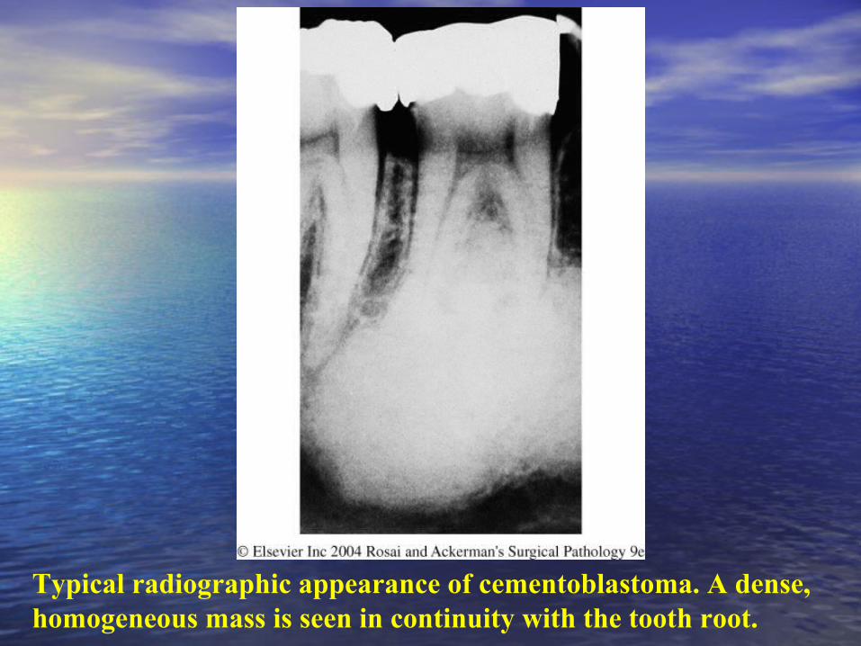
Typical radiographic appearance of cementoblastoma. A dense, homogeneous mass is seen in continuity with the tooth root.


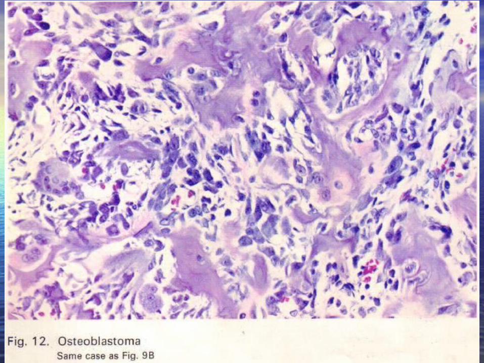
Osteoblastoma. Numerous neoformed osteoid trabeculae are lined by osteoblasts and separated by a highly vascularized matrix.


Desmoplastic f ibromaDesmoplastic f ibroma . Microscopic differential diagnosis . Microscopic differential diagnosis included fibrosarcoma.included fibrosarcoma.

Desmoplastic f ibroma. Desmoplastic f ibroma. Note evenly distributed and benign-Note evenly distributed and benign-appearing fibroblasts in collagenousappearing fibroblasts in collagenousstroma.stroma.

Desmoplastic f ibroma. Desmoplastic f ibroma. Note evenly distributed and benign-Note evenly distributed and benign-appearing fibroblasts in collagenousappearing fibroblasts in collagenousstroma.stroma.

Hemangioma of bone Hemangioma of bone showing honeycomb radiographic pattern with showing honeycomb radiographic pattern with associated root resorption.associated root resorption.

HemangiomaHemangioma

Hemangioma of bone. Note numerous vascular channels Hemangioma of bone. Note numerous vascular channels surrounded by trabeculae of bone.surrounded by trabeculae of bone.


Osteosarcoma Osteosarcoma surrounding the roots of first molar tooth. surrounding the roots of first molar tooth. Note widened periodontal ligament.Note widened periodontal ligament.

A, Central low-grade osteosarcoma A, Central low-grade osteosarcoma of the mandible.of the mandible.


Osteosarcoma Osteosarcoma between a mandibular lateral incisor and a canine. Note slight between a mandibular lateral incisor and a canine. Note slight widening of periodontalwidening of periodontalligaments of both teeth. ligaments of both teeth. B B and and C, C, Surgical specimen shows a malignant bone-Surgical specimen shows a malignant bone-producing neoplasm occupying theproducing neoplasm occupying theperiodontal ligament space. The tooth is to the right, and alveolar bone is to the periodontal ligament space. The tooth is to the right, and alveolar bone is to the left.left.
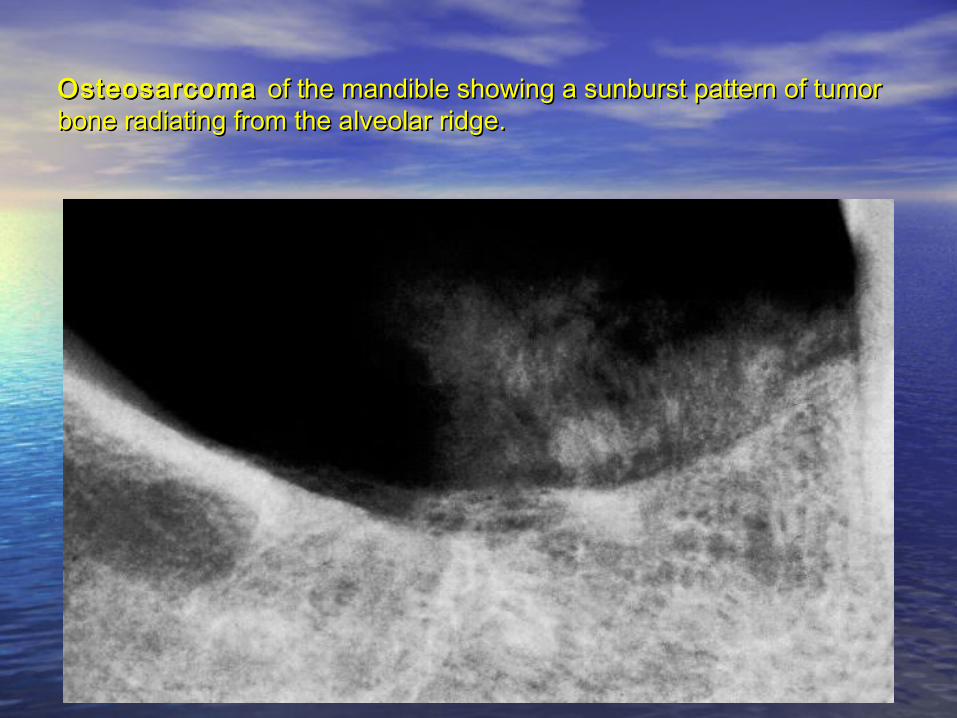
Osteosarcoma Osteosarcoma of the mandible showing a sunburst pattern of tumor of the mandible showing a sunburst pattern of tumor bone radiating from the alveolar ridge.bone radiating from the alveolar ridge.

Osteosarcoma of the mandible Osteosarcoma of the mandible exhibiting sunburst pattern.exhibiting sunburst pattern.

C, CT scan of persistent tumor 15 years later, now a high-grade C, CT scan of persistent tumor 15 years later, now a high-grade tumor.tumor.

Gross appearance of osteosarcoma of mandible.

D, Surgical specimen of the high-grade tumor (chondroblastic D, Surgical specimen of the high-grade tumor (chondroblastic osteosarcoma).osteosarcoma).

Chondroblastic osteosarcoma. Chondroblastic osteosarcoma. Note cartilage and bone at Note cartilage and bone at lower left.lower left.

Fibroblastic osteosarcoma Fibroblastic osteosarcoma composed of spindled tumor cells and composed of spindled tumor cells and small islands of tumor bone.small islands of tumor bone.


Osteosarcoma of jaw. The neoplastic bone (left) is clearly distinguishable from the residual normal bone (right)).


Osteosarcoma Osteosarcoma between a mandibular lateral incisor and a canine. Note slight between a mandibular lateral incisor and a canine. Note slight widening of periodontal ligaments of both teeth. widening of periodontal ligaments of both teeth. B B and and C, C, Surgical specimen Surgical specimen shows a malignant bone-producing neoplasm occupying the periodontal shows a malignant bone-producing neoplasm occupying the periodontal ligament space. The tooth is to the right, and alveolar bone is to the left.ligament space. The tooth is to the right, and alveolar bone is to the left.


A A and and B, Osteosarcoma B, Osteosarcoma composed of atypical cells in association with composed of atypical cells in association with tumor bone.tumor bone.

Osteosarcoma Osteosarcoma exhibiting a partially myxoid microscopic appearance.exhibiting a partially myxoid microscopic appearance.


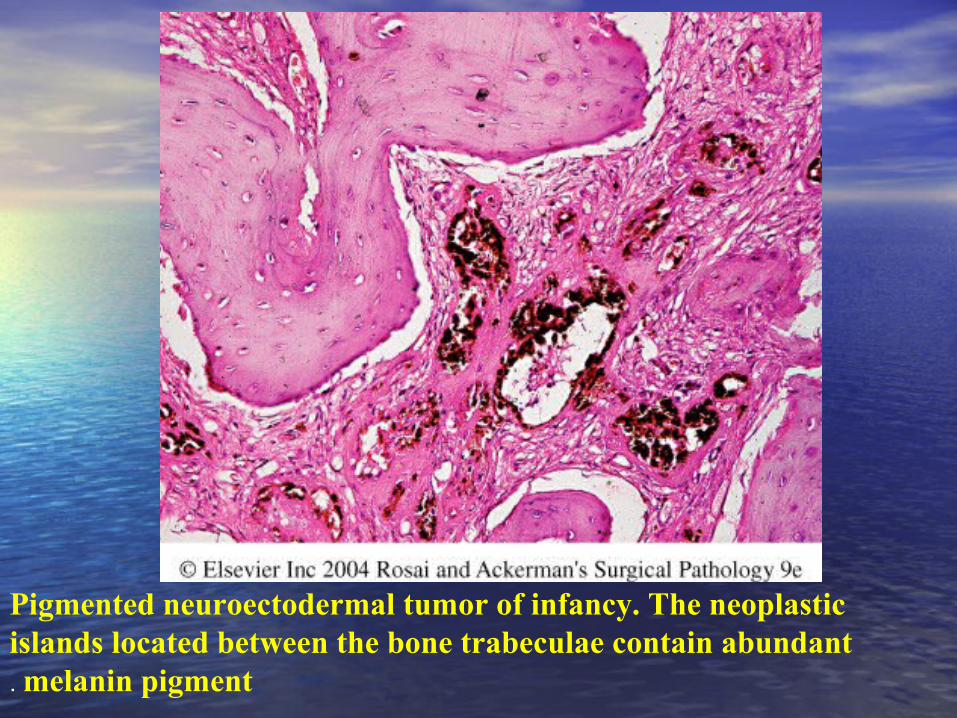
Pigmented neuroectodermal tumor of infancy. The neoplastic islands located between the bone trabeculae contain abundant melanin pigment.

Pigmented neuroectodermal tumor of infancy. This example shows the classic pattern of neuroblast-like cells surrounded by larger melanin-containing cells.


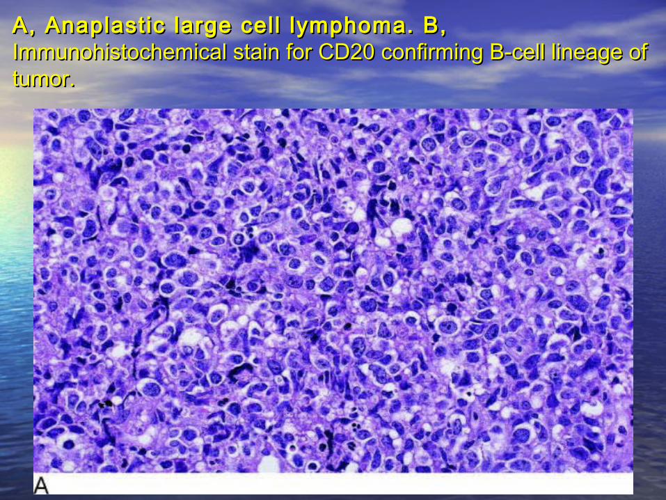
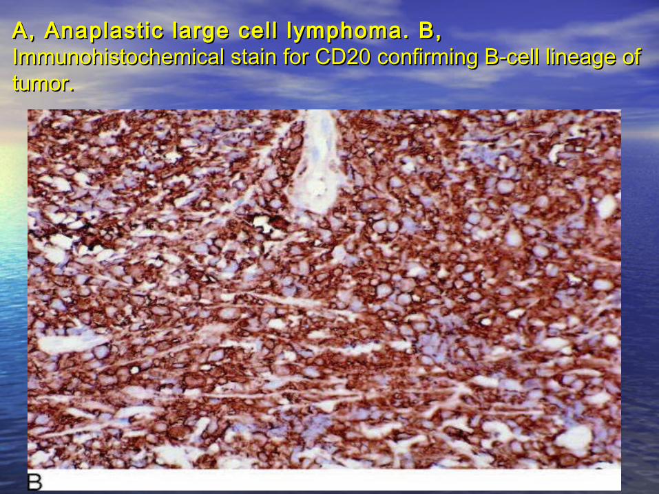
A, Anaplastic large cell lymphoma. B, A, Anaplastic large cell lymphoma. B, Immunohistochemical stain for CD20 confirming B-cell lineage ofImmunohistochemical stain for CD20 confirming B-cell lineage oftumor.tumor.
A, Anaplastic large cell lymphoma. B, A, Anaplastic large cell lymphoma. B, Immunohistochemical stain for CD20 confirming B-cell lineage ofImmunohistochemical stain for CD20 confirming B-cell lineage oftumor.tumor.

Burkitt 's lymphoma Burkitt 's lymphoma of the left maxilla.of the left maxilla.

Burkit t 's lymphoma Burkit t 's lymphoma presenting as a periapical presenting as a periapical radiolucency (mandibular left first molar). The patient alsoradiolucency (mandibular left first molar). The patient alsohad a numb lip.had a numb lip.


Burkit t 's lymphoma Burkit t 's lymphoma exhibiting starry sky effect. Pale cells are exhibiting starry sky effect. Pale cells are tingible body macrophages.tingible body macrophages.

Angiocentric T-cell lymphoma. A. This 62 ~ ye a r- o l d man had a destructive palatal lesion that proved to be a "l-celllymphoma. and evaluation showed cervical lymph node involvement aswell. B. Resolution of the lesion 1 month later.after multiagent chemotherapy.

Midline granuloma Midline granuloma presenting as oropharyngeal ulcers.presenting as oropharyngeal ulcers.

Angiocentric T-cell lymphoma. Thismediumpower photomicrograph shows atypical lymphoid cells infi l trating the wall and fi l l ing the lumen of a blood vessel. Such a pattern is termedangiocentric (meaning "around blood vessels").

Multiple myeloma. A Mult iple myeloma. A and and B, B, Right mandibular mass.Right mandibular mass.

Multiple myeloma Mult iple myeloma involving the left maxillary tuberosity.involving the left maxillary tuberosity.

Multiple myeloma Mult iple myeloma presenting orally as an ulcerated gingival presenting orally as an ulcerated gingival mass.mass.

Multiple myeloma. A Multiple myeloma. A and and B, B, Right mandibular mass.Right mandibular mass.


Multiple myeloma Mult iple myeloma showing multiple punched-out lesions of the showing multiple punched-out lesions of the skull.skull.

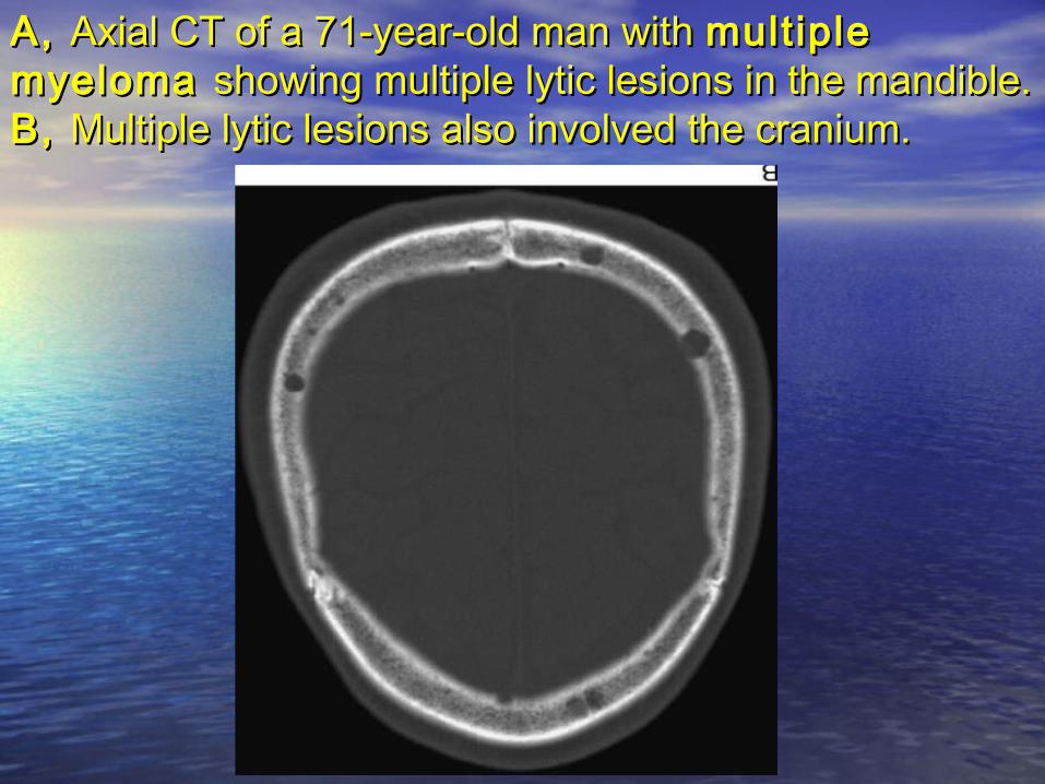
A, A, Axial CT of a 71-year-old man with Axial CT of a 71-year-old man with multiple multiple myeloma myeloma showing multiple lytic lesions in the mandible. showing multiple lytic lesions in the mandible. B, B, Multiple lytic lesions also involved the cranium.Multiple lytic lesions also involved the cranium.
A, A, Axial CT of a 71-year-old man with Axial CT of a 71-year-old man with multiple multiple myeloma myeloma showing multiple lytic lesions in the mandible. showing multiple lytic lesions in the mandible. B, B, Multiple lytic lesions also involved the cranium.Multiple lytic lesions also involved the cranium.


Multiple myeloma Mult iple myeloma composed of neoplastic plasma cells. composed of neoplastic plasma cells. B B and and C, C, Immunohistochemical stains for kappa (Immunohistochemical stains for kappa (BB) and lambda () and lambda (CC ) ) light chains demonstrating monoclonality of the plasma cells.light chains demonstrating monoclonality of the plasma cells.

Multiple myeloma Mult iple myeloma composed of neoplastic plasma cells. composed of neoplastic plasma cells. B B and and C, C, Immunohistochemical stains for kappa (Immunohistochemical stains for kappa (BB) and lambda () and lambda (CC ) ) light chains demonstrating monoclonality of the plasma cells.light chains demonstrating monoclonality of the plasma cells.


langerhans cell histiocytosis. There is a diffuse infi l trate of pale-staining langerhans cells intermixed with numerous red granular eosinophils.