Preparation and Characterization of Liposomal Everolimus ...
BONE MARROW TARGETED LIPOSOMAL DRUG DELIVERY SYSTEMS
A THESIS SUBMITTED TO
THE GRADUATE SCHOOL OF NATURAL AND APPLIED SCIENCES
OF
MIDDLE EAST TECHNICAL UNIVERSITY
BY
MERT BAKĠ
IN PARTIAL FULLFILLMENT OF THE REQUIREMENTS
FOR
THE DEGREE OF MASTER OF SCIENCE
IN
BIOMEDICAL ENGINEERING
MAY 2011

Approval of Thesis
BONE MARROW TARGETED LIPOSOMAL DRUG DELIVERY SYSTEMS
submitted by MERT BAKİ in partial fulfillment of the requirements for the degree of
Master of Science in Biomedical Engineering Department, Middle East Technical
University by,
Prof. Dr. Canan Özgen
Dean, Graduate School of Natural and Applied Sciences
Prof. Dr. Semra Kocabıyık
Head of Department, Biomedical Engineering
Assoc. Prof. Dr. Ayşen Tezcaner
Supervisor, Engineering Sciences Dept., METU
Prof. Dr. Duygu Çetinkaya,
Co-Supervisor, Pediatric Hematology Dept., Hacettepe University
Examining Committee Members
Assist. Prof. Dr. Sreeparna Banerjee
Biology Dept., METU
Assoc. Prof. Dr. Ayşen Tezcaner
Engineering Sciences Dept., METU
Prof. Dr. Duygu Çetinkaya,
Pediatric Hematology Dept., Hacettepe University
Assist. Prof. Dr. Dilek Keskin
Engineering Sciences Dept., METU
Assist. Prof. Dr. Can Özen
Biotechnology Dept., METU
Date: _____________

iii
I hereby declare that all information in this document has been obtained and
presented in accordance with academic rules and ethical conduct. I also declare
that, as required by these rules and conduct, I have fully cited and referenced all
material and results that are not original to this document.
Name, Last name: Mert Baki
Signature:

iv
ABSTRACT
BONE MARROW TARGETED LIPOSOMAL DRUG DELIVERY SYSTEMS
Baki, Mert
M. Sc., Department of Biomedical Engineering
Supervisor: Assoc. Prof. Dr. Ayşen Tezcaner
Co-Supervisor: Prof. Dr. Duygu Çetinkaya
May 2011, 80 pages
Homing is the process that stem cells move to their own stem cell niches under
the influence of chemokines like stromal-derived factor-1α (SDF-1α) upon bone marrow
transplantation (BMT). There is a need for increasing homing efficiency after BMT
since only 10-15% of the transplanted cells can home to their own niches and a limited
amount of donor marrow can be transplanted. In this study, we aimed to develop and
characterize bone marrow targeted liposomal SDF-1α delivery system prepared by
extrusion method. Alendronate conjugation was chosen to target the liposomes to bone
marrow microenvironment, particularly the endosteal niche. Optimization studies were
conducted with the model protein (-lactoglobulin). 200 nm sized 5% pegylated
DPPC:Cho (2:0.5) liposomes were chosen for targeted SDF-1α loaded large unilamellar
liposomes (LUVs). DSPE-PEG2000-Carboxylic Acid was conjugated with alendronate
via carbodiimide chemistry for preparing targeted liposomes. Alendronate (ALE)
conjugation was shown by FT-IR and the conjugation efficiency was found 34.5±4.6 %.
5%ALE-PEG/LUV200 encapsulated 48.3 ± 0.3% of SDF-1α and released 44.10.9%
after 24h, with a similar profile as 5%PEG/LUV200 and 2.5%ALE-PEG/LUV200.
5%ALE-PEG/LUV200 had more negative potential (-21.9 mV) and significantly higher
v
affinity to hydroxyapatite than 5%PEG/LUV200 and 2.5%ALE-PEG/LUV200. Migration
assays conducted with human mesenchymal stem cells showed that SDF-1α released
(24.4 ng/ml) from the liposomes in 24 hours increased the chemotactic activity of these
cells. SDF-1 loaded 5%ALE-PEG/LUV200, reported for the first time in literature, has
potential as an effective vehicle for improving homing efficiency and thereby permitting
successful BMT from young donors. Additionally, this system could also be considered
for treating large and difficult bone fractures with recruitment of host stem cells.
However, further studies including migration assays with human hematopoietic stem
cells and in-vivo distribution of the liposomal system are suggested.
Keywords: Targetted liposome, bone marrow, homing, SDF-1α, alendronate.
vi
ÖZ
KEMĠK ĠLĠĞĠ HEDEFLĠ LĠPOZOMAL ĠLAÇ SALIM SĠSTEMLERĠ
Baki, Mert
Yüksek Lisans, Biyomedikal Mühendisliği Bölümü
Tez Yöneticisi: Doç. Dr. Ayşen Tezcaner
Ortak Tez Yöneticisi: Prof. Dr. Duygu Çetinkaya
Mayıs 2011, 80 sayfa
Kemik iliği transplantasyonu (KĠT) sonrasında Stromal Kaynaklı Faktör-1α
(SDF-1α) gibi kemokinlerin etkisi altına giren kök hücrelerin nişlerine (mikroçevre)
dönme sürecine yuvaya dönüş adı verilir. Nakil sonrasında yuvalanma verimini artırmak
gerekmektedir çünkü nakil sırasında hastaya verilen hücrelerin yalnızca %10-15‘lik bir
bölümü yuvalanma sürecini tamamlayabilmekte ve nakil edilen donor ilik miktarının
sınırlı olmaktadır. Bu çalışmada, ekstrüzyon yöntemi ile hazırlanan kemik iliği hedefli
SDF-1α yüklü lipozomal salım sistemleri geliştirilmesi ve karakterize edilmesi
amaçlanmaktadır. Kemik iliği mikroçevresi ve özellikle endosteal mikroçevresine salım
hedeflenmiş ve bunun için alendronat ile konjügasyonu yapılmıştır. Optimizasyon
çalışmaları için model protein (-lactoglobulin) kullanılmıştır. 200 nm boyutunda %5
pegile DPPC:Cho (2:0.5) kompozisyonundaki tek katmanlı lipozomlara seçilmiş ve
hedefleme için DSPE-PEG2000-Karboksilik Asit ile alendronat karbodiimid bağı ile
konjüge edilmiştir. Alendronat konjügasyonu FT-IR ile gösterilmiş ve etkinliği
%34.5±4.6 olarak bulunmuştur. 5%ALE-PEG/LUV200 lipozomlar SDF-1α‘nın %48.3 ±
0.3‘ünü hapsetmiş ve 24 saatin sonunda %44.1 0.9‘unu salmıştır. Toplam %5 pegile
lipid içeren lipozomlarda, hedeflenmiş %5 ve %2.5 alendronatlı lipozomlar ile
vii
hedeflenmemiş lipozomların benzer salım profillerine sahip olduğu gözlenmiştir.
5%ALE-PEG/LUV200 lipozomlar daha negatif bir elektrik potansiyele (-21.9 mV) sahip
olup, 5%PEG/LUV200 ve 2.5%ALE-PEG/LUV200 lipozomlarla karşılaştırıldığında
hidroksiapatite (HA) karşı istatiksel olarak daha yüksek afinite göstermiştir. Ġnsan
kaynaklı mezenkimal kök hücreler ile yapılan migrasyon deneylerinde 24 saat sonunda
salınan 24.4 ng/ml SDF-1α‘nın hücreler üzerindeki kemotaktik etkiyi artırdığı
gözlenmiştir. Literatürde ilk kez çalışılan SDF-1 yüklü 5%ALE-PEG/LUV200
lipozomlar KĠT sonrasında yuvalanma verimini artırma ve böylece genç yaştaki
donörlerden ilik naklinin başarıyla yapılmasını sağlama potansiyeline sahiptir. Ancak,
önerilen sisteminin insan kaynaklı hematopoietik kök hücreler ile yapılacak migrasyon
deneyleri ve in-vivo dağılım deneyleri ile daha detaylı olarak araştırılması
önerilmektedir. Ayrıca bu sistem hasta kök hücreleri çağırması ile büyük ve zor kemik
kırıkların tedavisi için değerlendirelebileceği düşünülmektedir
Anahtar Kelimeler: Hedeflenmiş lipozom, kemik iliği, yuvalanma, SDF-1α,
alendronat.

viii
ACKNOWLEDGEMENTS
This project would not have been possible without the support of many people. I
would like to thank to my advisor Assoc. Prof. Dr. Ayşen Tezcaner for her enlightening
wisdom, guidance and patience throughout this study. We have been through a rough
road but she never gave up on me and never lost her understanding. If I believe that I
can make a difference for humanity and science, this is her success.
I would like to express my gratitude to my co-advisor Prof. Dr. Duygu Uçkan
Çetinkaya. This study would not have been complete without her genius and help. I will
always appreciate her will for me to learn more about stem cells. Because of her, I
gained a whole different vision and this improved me.
I would like to express my special thanks to Assist. Prof. Dr. Dilek Keskin for
always being helpful and a problem-solver. She was with me in this study from the
beginning and her contributions are priceless. Thanks for believing in me and never
losing your smile.
I owe my thanks to Assoc. Prof. Dr. Zafer Evis and Ġdil Uysal for supplying
material for my HA affinity studies, Tuğba Endoğan for her hard-work during TEM
imaging of liposomes, Ġbrahim Çam for helping me on zeta potential analysis, Ceren
Bora for her academic informational support, Handan Acar from Bilkent University for
staining my liposomes for TEM and Sevil Arslan from Hacettepe University for her
support on cell culture studies.
I must thank to TÜBĠTAK for the financial support by Project 109S104 both on
me and this study. I also want to acknowledge METU Graduate School of Natural and
Applied Sciences for supporting my thesis with LTP project fund
I have to thank my extraordinary lab friends. Aslı Erdog (my liposome queen)
for sharing her genius, Özge Erdemli for always being there for me, Ayşegül Kavas for
being the other Leo and laughing at my jokes, Özlem Aydın for being tough and loving

ix
at the same time, Mine Toker for making me laugh so much, Yiğit Öcal for being a
supportive brother, Ömer Aktürk for a different point of view, Ġdil Uysal for her
wonderfully funny personality, Serap Geridönmez for the early morning chats and
Aydın Tahmasebifar for sharing my frustration when I really need. We all deserve the
best of things!
Also thanks to my dearest friends Can, Çağlar, Harun, Erkan, Egemen, Okan,
Liya, Merve and many more, you are the reason I keep my sanity (and lose sometimes).
I love you all very much!
My lovely grandmothers, my dearest aunt, uncle and cousins have always given
their support to me no matter what. They are very special. I owe them so much.
Finally, the real big ‗thank you‘ is for my king, my queen and my prince. You
mean the world to me and I am nothing without you. My love is endless.

x
TABLE OF CONTENTS
ABSTRACT ............................................................................................................ iv
ÖZ ............................................................................................................................ vi
ACKNOWLEDGMENTS....................................................................................... viii
TABLE OF CONTENTS ........................................................................................ x
LIST OF TABLES .................................................................................................. xiii
LIST OF FIGURES.................................................................................................. xiv
LIST OF ABBREVIATIONS.................................................................................. xvii
CHAPTERS
1.INTRODUCTION………………………………………………………………. 1
1.1 Liposomes……………………..…………………………………………… 1
1.2 Liposomes as delivery systems…………………………………………….. 4
1.2.1 In-vivo Fate of Liposomes…………………………………………… 6
1.2.1.1 Factors Affecting In-vivo Fate of Liposomes………………... 6
1.2.1.2 Prolonging Half-Life and Efficacy of Liposomes…………… 9
1.3 Bone Marrow and Stem Cells……………………………………………… 11
1.3.1 Stem Cell Niche……………………………………………………… 12
1.3.2 Homing……………………………………………………………….. 14
1.3.3 Stromal Cell-Derived Factor-1α…………………………………………………… 15
1.3.4 Bone Marrow Transplantation………………………………………... 17
xi
1.4 Bone and Bone Marrow Targeting Strategies……………………………… 19
1.5 Aim of the Study…………………………………………………………… 22
2. MATERIALS AND METHODS……................................................................ 24
2.1 Materials…………………………………………………………………..... 24
2.2 Methods.......................................................................................................... 25
2.2.1 Preparation of liposomes................................................................. 25
2.2.2 Conjugation of ALE to DSPE-PEG-Carboxylic Acid……………… 25
2.2.2.1 Determination of ALE Conjugation Efficiency………….. 27
2.2.3 Characterization of Liposomes…………………………………... 28
2.2.3.1 Particle Size Measurement……………………………….. 28
2.2.3.2 Surface Charge Measurement……………………………. 28
2.2.3.3 Transmission Electron Microscopy………………………… 28
2.2.3.4 Entrapment Efficiency & Lipid Recovery After Extrusion… 29
2.2.3.5 -Lactoglobulin and SDF-1 Release Studies……………… 29
2.2.3.6 Bone (HA) Affinity of Liposomal Preparations……………… 30
2.2.4 Cell Culture Studies…………………………………………………... 31
2.2.4.1 Isolation and Expansion of hBMMSCs ……………………… 31
2.2.4.2 Cell Migration Assay………………………………………… 32
2.2.5 Statistical Analysis…………………………………………………… 33
3. RESULTS & DISCUSSION…………………………………………………… 34
3.1 Optimization Studies with β-Lactoglobulin………………………………... 34
3.1.1 Effects of Liposome Composition and Size………………………….. 34
xii
3.1.2 Effects of Pegylation of Liposomes………………………………… 39
3.2 Characterization of SDF-1 Loaded Pegylated Liposomes ………….......... 42
3.2.1 Size of SDF-1α Loaded and Pegylated Liposomes ………… ……… 42
3.2.2 Encapsulation and Release of SDF-1α Loaded Liposomes …………. 43
3.3 Conjugation of Alendronate to DSPE-PEG(2000)-Carboxylic Acid…… 46
3.3.1 FTIR Analysis………………………………………………………... 46
3.3.2 Encapsulation and Release of Targeted Liposomes………………….. 50
3.3.3 Surface Charge and Size of Targeted Liposomes …………………… 52
3.3.4 Targetting Efficiency of the ALE Conjugated Liposomes…………… 55
3.3.5 Morphology of Liposomes by TEM…………………………………. 58
3.4 In-Vitro Cell Culture Studies………………………………………………. 59
3.4.1 Migration Assay……………………………………………………… 59
4. CONCLUSION………………………………………………………………… 66
REFERENCES……………………………………………………………………. 68

xiii
LIST OF TABLES
TABLES
Table 1.1 Different methods for preparation of liposomes…………………………. 3
Table 1.2 Liposomal formulations of different drugs in the market………………... 4
Table 1.3 Roles of SDF-1α in health and disease states……………………………. 16
Table 1.4 Characteristics of human stem cells from different sources……………... 19
Table 2.1 Compositions of liposomes expressed in mole ratios and pore sizes of
filters used for these different liposomal formulations……………………………...
26
Table 3.1 Comparison of encapsulation efficiencies (EE%) and release profiles of
100 nm sized LUVs for -lactoglobulin…………………………………………….
37
Table 3.2 The effect of degree of pegylation on the encapsulation efficiency and
release properties of DPPC: Cho (2:0.5) LUVs of 200 nm size…………………….
40

xiv
LIST OF FIGURES
FIGURES
Figure 1.1 Schematic illustration of three dimensional structure of a liposome… 1
Figure 1.2 Liposomes of different sizes and lamellarity………………………… 2
Figure 1.3 Stem cell niche and cellular components in the bone marrow………… 13
Figure 1.4 Illustration of the pathway taken by HSCs during their homing to their
bone marrow niche and subsequent transendothelial migration out of the niche…
15
Figure 1.5 Structure of alendronate, DSPE-PEG(2000)-Carboxylic Acid
and the alendronate conjugated DSPE-PEG(2000)………………………………
21
Figure 1.6 Model of alendronate conjugated pegylated liposomes designed for
the delivery of SDF-1 in this study………………………………………………
23
Figure 2.1 Basic structure of the cell migration assay…………………………… 32
Figure 3.1 A representative chromatogram showing turbidity readings of -
lactoglobulin loaded DPPC:Cho (2:0.5) liposomes and protein amounts in the
unentrapped fractions determined by BCA protein assay…………………………
35
Figure 3.2 A representative DLS result of size distribution for -lactoglobulin
loaded LUVs prepared by extrusion through 100 nm filter………………………
36
Figure 3.3 Effect of liposome size on the release of -lactoglobulin from the
DPPC:Cho (1:0.5) liposomes (n=3)……………………………………………….
39
Figure 3.4 Effect of PEGylation on the release profiles of DPPC:Cho (2:0.5)

xv
LUVs of 200 nm size (n=3)……………………………………………………… 41
Figure 3.5 Representative size distribution result for SDF1-5%PEG/LUV200
obtained by DLS analysis ……………………………………………………….
43
Figure 3.6 Release profiles as (a) cumulative % releases and (b) total release
amounts of SDF1-5%PEG/LUV200 prepared with 4 freeze-thaw cycles and
without any freeze-thaw cycle……………………………………………………
45
Figure 3.7 FTIR spectrums of alendronate, DSPE-PEG(2000)-Carboxylic Acid,
alendronate conjugated DSPE-PEG(2000)-Carboxylic Acid and alendronate
DSPE-PEG(2000)-Carboxylic Acid mixture ………………………………………
47
Figure 3.8 Release profiles and total release amounts of SDF-1α loaded a)
2.5%ALE-PEG/LUV200, b) 5%ALE-PEG/LUV200 liposomes (n=3)1.3.3 Stromal
Cell-Derived Factor-1α……………………………………………………………
41
Figure 3.9 Representative size distribution DLS result for 2.5%ALE-
PEG/LUV200 liposomes…………………………………………………………..
52
Figure 3.10 Representative size distribution DLS result for 5%ALE-PEG/LUV200
liposomes…………………………………………………………………………
53
Figure 3.11 Zeta potential distribution of non-targeted LUVs (5%PEG/LUV200).. 54
Figure 3.12 Zeta potential distribution of 2.5%ALE-PEG/LUV200……………………….. 54
Figure 3.13 Zeta potential distribution of 5%ALE-PEG/LUV200………………………….. 55
Figure 3.14 HA affinity (%) of targeted (2.5%ALE-PEG/LUV200 and 5%ALE-
PEG/LUV200) and non-targeted (5%PEG/LUV200) liposomes determined by
turbidity measurements (n=3)……………………………………………………..
56
Figure 3.15 HA affinity (%) of targeted (2.5%ALE-PEG/LUV200 and 5%ALE-
PEG/LUV200) and non-targeted (5%PEG/LUV200) liposomes calculated from the

xvi
phospholipid amounts (n=3)……………………………………………………… 57
Figure 3.16 TEM images of a) a single empty non-targeted liposome
(5%PEG/LUV200), b) single empty targeted liposome (ALE-5%PEG/LUV200)…
58
Figure 3.17 TEM images showing the general view of a) empty targeted
liposomes (ALE-5%PEG/LUV200), b) aggregated empty non-targeted liposomes
(5%PEG/LUV200)…………………………………………………………………
59
Figure 3.18 Phase contrast micrograph of a) first passage human mesenchymal
stem cells b) human mesenchymal stem cells in confluency (20X)………………
60
Figure 3.19 Representative images of transmigrated MSCs in response to a)
SDF-1α released from 5%ALE-PEG/LUV200 liposomes b) DMEM-low glucose
c) empty liposomes (5%ALE-PEG/LUV200) after 16 hours hours………………...
61
Figure 3.20 Representative images of transmigrated MSCs in response to a)
SDF-1α released from 5%ALE-PEG/LUV200 liposomes b) DMEM-low
glucose c) empty liposomes (5%ALE-PEG/LUV200) after 24 hours……………..
63
Figure 3.21 Average number of transmigrated MSCs………………………... 65
xvii
LIST OF ABBREVIATIONS
ALE: Alendronate
AUC: Area Under the Concentration-Time Curve
BCA: Bicinchoninic Acid
BM: Bone Marrow
BMT: Bone Marrow Transplantation
CB: Cord Blood
Cho: Cholesterol
CMC: Critical Micelle Concentration
DCC: N,N-Dicyclohexylcarbodiimide
DDAB: Dimethyldioctadecyl ammonium bromide
DLS: Dynamic Light Scattering
DMEM: Dulbecco‘s modified Eagle‘s medium
DMPC: Dimyristoylphosphatidylcholine
DMSO: Dimethyl sulphoxide
DODAB: Dioleoyldimethylammoniumpropane
DOTAP: Dioleoyltrimethylammoniumpropane
DPPC: Dipalmitoylphosphatidylcholine
DSPC: Distearoylphosphatidylcholine
DSPE: Distearoylglycerophosphoethanolamine
EE: Encapsulation Efficiency
EPC: Egg Phosphatidylcholine
FBS: Fetal Bovine Serum
FTIR/ATR: Fourier Transform Infrared Spectrometer/Attenuated Total Reflectance
xviii
GVHD: Graft Versus Host Disease
GUV: Giant Unilamellar Vesicles
HA: Hydroxyappatite
HIV: Acquired Immune Deficiency Syndrome
HSC: Hematopoietic Stem Cells
LG: Low Glucose
LUV: Large Unilamellar Vesicles
MLV: Multilamellar Vesicles
mPEG: Methoxy Polyethylene Glycol
MSC: Mesenchymal Stem Cells
MW: Molecular Weight
MWCO: Molecular Weight Cut-Off
NHS: N-Hydroxysuccinimide
NOD/SCID: Non-obese Diabetic/Severe Combined Immune Deficiency
PB: Peripheral Blood
PBS: Phosphate Buffer Saline
PEG: Polyethylene glycol
PEG-LD: Pegylated Liposomal Doxorubicin
PHPMA: Poly[N-(2-hydroxypropyl)methacrylamide]
PL: Phospholipid
PLA: Polylactic Acid
PLGA: Poly(lactic-co-glycolic acid)
RES: Reticuloendothelial System
RPM: Revolutions Per Minute
SDF: Stromal Derived Factor
SDF1-5%PEG/LUV200: SDF-1 encapsulated 200 nm sized 5% PEGylated DPPC:Cho
(2:0.5) liposomes
SDS: Sodium Dodecylsulfate
xix
SUV: Small Unilamellar Vesicles
Tm: Transition Temperature
TBI: Total Body Irradiation
TEM: Transmission Electron Microscopy
TFL: Trifluralin
5%PEG/LUV200: 200 nm sized 5% PEGylated DPPC:Cho (2:0.5) liposomes(untargeted)
2.5%ALE-PEG/LUV200: 200 nm sized DPPC: Cho (2:0.5) liposomes with 2.5%
alendronate conjugated lipid + 2.5% DSPE-mPEG(2000) (targeted)
5%ALE-PEG/LUV200: 200 nm sized DPPC: Cho (2:0.5) liposomes with 5% alendronate
conjugated lipid (targeted)
1
CHAPTER 1
INTRODUCTION
1.1 Liposomes
Liposomes were first described by Bangham in 1960s as self-assembled lipid
vesicles composed of one or more lipid bilayers [1]. They are microscopic closed
vesicles consisting of mainly phospholipid bilayers surrounding an aqueous medium
(Fig. 1.1) [2]. Phospholipids, when dispersed in an aqueous environment at a
concentration above their critical micelle concentration (CMC), tend to form these
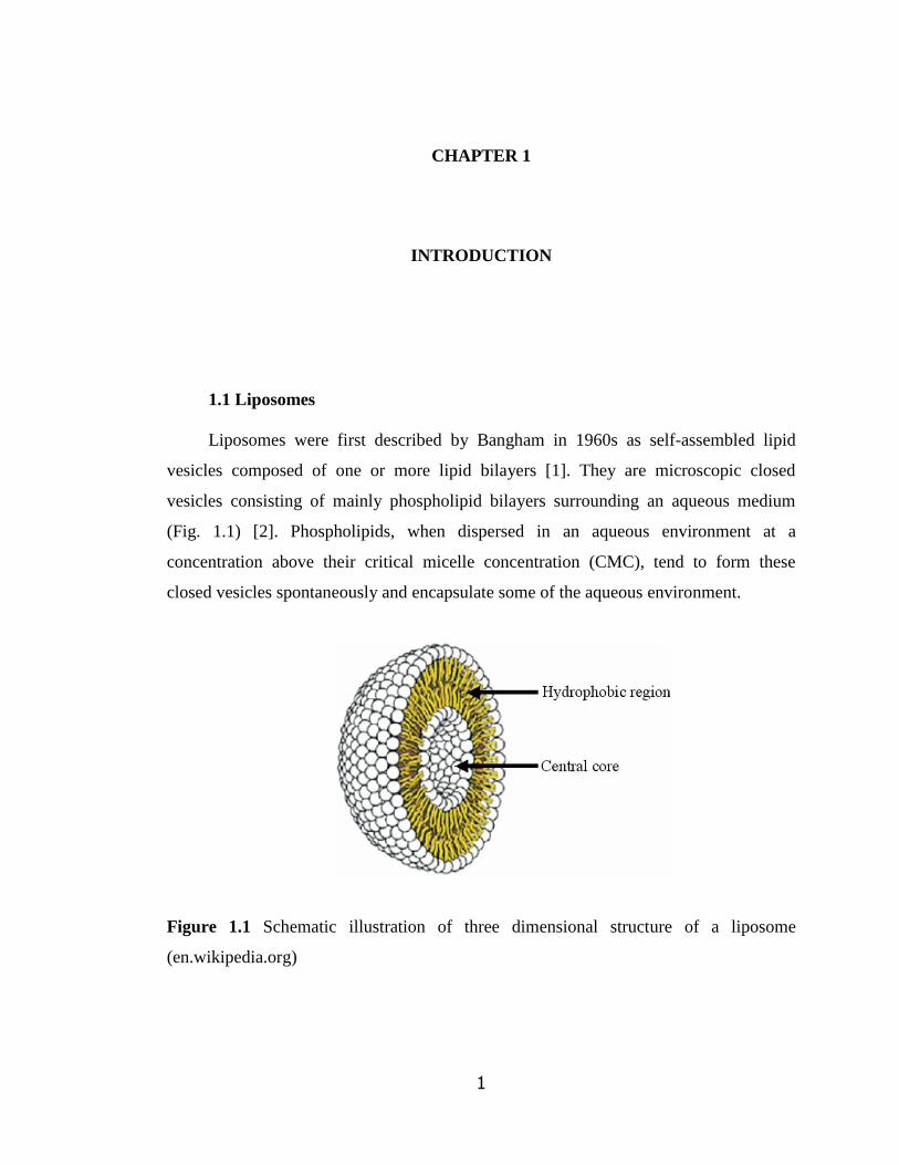
closed vesicles spontaneously and encapsulate some of the aqueous environment.
Figure 1.1 Schematic illustration of three dimensional structure of a liposome
(en.wikipedia.org)

2
Most widely used lipids are phospholipids (PLs), especially phosphatidylcoline,
phosphatidic acid, phosphatidylglycerol, phosphatidylserine and phosphatidyl-
ethanolamine. PLs have different combinations of fatty acid chains in the hydrophobic
region of the molecule with different chain length and degree of unsaturation [3].
Liposomes vary in size ranging from 30 nm to several micrometers, phospholipid
composition, and surface characteristics (Fig 1.2). These properties can be modified for
specific applications. Liposomes composed of single lipid bilayer structures are referred
as unilamellar liposomes. Unilamellar liposomes vary in size. Small unilamellar vesicles
(SUVs) range in size from 20 to 100 nm whereas liposomes larger than 100 nm are
referred as large unilamellar vesicles (LUVs). The diameters of LUVs are in a very
broad range; from 100 nm up to cell size and they are called the giant vesicles (GUV).
Figure 1.2 Liposomes of different sizes and lamellarity (modified from
www.avantilipids.com)
They contain a large aqueous core. Therefore, they are preferred to entrap water
soluble drugs [4]. Multilamellar vesicles (MLVs) have two or more lipid bilayers and
3
their sizes differ from a few hundred nanometers to several microns [2]. Their layers are
separated from each other by a layer of aqueous phase (Fig. 1.2)
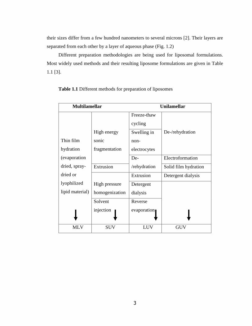
Different preparation methodologies are being used for liposomal formulations.
Most widely used methods and their resulting liposome formulations are given in Table
1.1 [3].
Table 1.1 Different methods for preparation of liposomes
Multilamellar Unilamellar
Thin film
hydration
(evaporation
dried, spray-
dried or
lyophilized
lipid material)
High energy
sonic
fragmentation
Freeze-thaw
cycling
De-/rehydration Swelling in
non-
electrocytes
De-
/rehydration
Electroformation
Extrusion Solid film hydration
High pressure
homogenization
Extrusion Detergent dialysis
Detergent
dialysis
Solvent
injection
Reverse
evaporation
MLV SUV LUV GUV

4
1.2 Liposomes as Delivery Systems
The application areas of liposomes range from medicine (for developing vaccines
[5, 6], delivery systems for diagnostic agents [7, 8], chemotherapeutic drugs [9, 10], and
DNA [11], to textile (i.e., delivery of dyes) [12] and food industry (i.e., as delivery
systems for pesticides, enzymes besides nutritional liposomal formulations used in food
supplementation [13].
Liposomes have been widely used as delivery systems for different bioactive
agents like therapeutic drugs (i.e., paclitaxel, topotecan, doxorubicin) [14-16], hormones
(parathyroid hormone, growth hormone) [17, 18] and enzymes (i.e., elastase, beta
glucorinedase, etc) [19, 20] because of their ease and convenient preparation, low
toxicity, biocompatibility, biodegradability. There are several liposomal formulations in
the market that are designed for immuno-compromised patients [21-23]. Some of these
are presented in Table 1.2.
Table 1.2 Liposomal formulations of different drugs in the market (modified from
en.wikipedia.org)
Bioactive Agent Trade Name Company Name Indication
Amphotericin B Ambisome Gilead Sciences Fungal & Protozoal
infections Cytarabine Depocyte Pacira Meningitis Daunorubicin DaunoXome Gilead Sciences HIVrelated Kaposi‘s
sarcoma Doxorubicin Myocet Zeneus Metastatic breast cancer IRIV Vaccine Epaxal,
Inflexal V Berna Biotech Hepatitis A,
Influenza Morphine Depodur Skye Pharma,Endo Postsurgical analgesia Verteporfin Visudyne QLT,Novartis Age-related macular
degeneration, Pathologic
myopia, Ocular histoplasmosis
Doxorubicin Doxil/Caelyx Ortho Biotech,
Schering-Plough HIV-related Kaposi‘s
sarcoma, Metastatic breast
cancer, Metastatic ovarian cancer
5
There is still high interest among researchers for developing and/improving
liposomal delivery systems targeted to different cancer types such as Kaposi‘s sarcoma,
leukemia and breast cancer [24-27]. Doxorubicin loaded liposomes (under the trade
name: Doxil) is used to treat Kaposi's sarcoma and metastatic ovarian cancer. It is also
sometimes used for other types of cancer, such as multiple myeloma. Preclinical studies
showed that pegylated liposomal doxorubicin was as effective as free doxorubicin
(Adriamycin) in a variety of tumor models [28, 29]. Pharmacokinetic studies revealed
differences between pegylated liposomal doxorubicin (PEG-LD) and doxorubicin, with
PEG-LD having a higher area under the concentration-time curve (AUC), lower
clearance rate, and smaller volume of distribution [30]. The ability of PEG-LD
liposomes to remain intact while in circulation and retain most of the doxorubicin in
encapsulated formulation were believed to be responsible for the reduced toxicity seen
with this agent without sacrificing efficacy.
It has been recently shown that liposomal cisplatin used for the cure of pancreatic
cancer in mice was less toxic than free cisplatin and had similar response rate in mice.
These results pointed out that especially in cancer treatment; these liposomal systems do
not have strong side effects as observed for therapeutic drugs. Thus, they are highly
important on improving patients‘ life quality and empowering the treatment effect.
Liposomal Cisplatin (Lipoplatin) has received Orphan Drug designation (a
pharmaceutical especially developed to cure a rare medical condition) for Pancreatic
Cancer from European Medicines Agency [31].
With the possibility of targeting, liposomes have become good and promising
vehicles for cancer treatment due to enhanced biological response and low systemic
toxicity. There are many research groups actively working on the development of
immunoliposomes targeted to cancer cells. However, there is no targeted liposomal
delivery system for cancer or any tissue site in the body in clinical use up to this date.
6
1.2.1 In Vivo Fate of Liposomes
Liposomal formulations are administered through nasal aspirations, skin and
intravenous routes. Following systemic administration, reticuloendothelial system (RES)
is the site for highest liposome accumulation. The main organs of RES are liver, spleen
and lungs [32]. Among them, liver has the largest capacity to uptake the liposomes,
shortening their half-lives.
Liposomes interact with cells in different ways: endocytosis by macrophages and
neutrophils, fusion with the plasma membrane of body cells and releasing their contents
into the cytoplasm, and transfer of lipids to cellular membranes without any release
and/or adsorption to cell surface with weak hydrophobic forces or specific interactions.
Macrophages do not recognize the liposomes themselves, they recognize the opsonins
(serum proteins) bound onto the surface of the liposomes. Some of these opsonizing
proteins are immunoglobulins, fibronectin and -2-macroglobulin [4].
1.2.1.1 Factors Affecting In Vivo Fate of Liposomes
The bio-distribution, structural stability, and circulation time of liposomes can be
influenced by particle size, lipid composition, surface charge, hydration, and sensitivity
to pH changes, bilayer rigidity/fluidity, the binding kinetics of opsonins to liposomes
and the presence of targeting moieties on the liposome surface. Many different
molecules from basic structures like monosaccharides to complex structures like
antibodies can be used for this purpose [33].
Particle size is also important to avoid from the RES. In general, larger liposomes
are eliminated from the circulation more rapidly than the smaller ones. Nanoparticles
(size under 200 nm) are preferred for less RES uptake. SUVs have a longer half-lives
than the multilamellar liposomes. Studies showed that liposome uptake was serum and
vesicle size dependent. It was reported that the degree of opsonization decreased with a
decrease in size from 800 nm to 200 nm [34]. It was shown that smaller liposomes could
not support opsonization but the larger ones did [32].

7
Liposome composition is another important parameter that affects structural
stability, in vivo half life and release characteristics of liposomal formulations of
bioactive agents. Optimizing the lipid composition is the very first step for developing of
liposomal systems. Most popular natural and synthetic phospholipid derivatives used in
liposomal formulations are egg phosphatidylcholine (EPC),
dimyristoylphosphatidylcholine (DMPC), dipalmitoylphosphatidylcholine (DPPC) and
distearoylphosphatidylcholine (DSPC). There are several issues to consider when
selecting the lipids for liposomal composition. One is the phase transition temperature of
lipids. The phase transition temperature of lipids is the temperature at which the lipid‘s
physical state changes from the ordered gel phase (hydrocarbon chains are closely
packed) to the disordered liquid crystalline phase (hydrocarbon chains are randomly
oriented) [35]. Hydrocarbon length, degree of unsaturation, charge, and head group
species affect the phase transition temperature. As the hydrocarbon length is increased,
Van der Waals interactions become stronger; thus the phase transition temperature
increases. Different lipids have different transition temperatures. DSPC has the highest
transition temperature of all phospholipids mentioned above; therefore, it exhibits high
stability and leaktightness in a wider temperature range [36].
While encapsulating proteins and growth factors into liposomes, it is especially
important to select the proper lipid or lipid mixture for the formulation to achieve good
loading and release behaviour as well as to prevent the loss of biological activity of these
bioactive molecules during preparation. These molecules are highly sensitive to
temperature and they easily decompose above the body temperature. While using a lipid
composition with a high transition temperature (Tm), it is inevitable for proteins to
denature and lose functionality during hydration and other processing steps like
extrusion. For this reason, the composition for protein liposomal delivery systems
usually involves phospholipids with lower Tm together with cholesterol for further
structural stabilization [37, 38].
The use of liposomes as bioactive delivery system is also highly related to the
water solubility of the compound. Liposomes are predominantly used as carriers for
hydrophilic molecules [39]. These molecules do not interact with the lipid moiety of the
8
vesicle. The size and volume of the inner aqueous region are the two important
parameters for encapsulating water-soluble bioactive agents. However, these two
parameters are not considered critical for hydrophobic molecules that will be
incorporated in the hydrophobic lipid bilayer region.
Liposomal suspensions are destabilized after intravenous injection because of the
adipose exchange of phospholipids under plasma lipoprotein effect [40]. Liposomes
adsorb the blood plasma components, which lead to their clearance from the blood
circulation [41]. Cholesterol is a membrane constituent widely found in biological
systems. It is used for modifying membrane fluidity, elasticity, and permeability. It
literally fills in the gaps created by imperfect packing of other lipid species when
proteins are embedded in the membrane. Cholesterol serves much the same purpose in
model membranes. Cholesterol incorporated into lipid bilayer blocks this lipid exchange
and creates a stabilizing effect [42]. Also it was shown that adding cholesterol to the
bilayer structure of liposome causes an increase in phospholipids packing and reduces
the transfer of phospholipids to lipoproteins [43].
Most commercially available cholesterol sources are derived from egg or wool
grease (sheep derived) [42]. These animal sources are potentially not suitable as human
pharmaceuticals due to the potential viral contamination. The surface charge of liposome
affects their in vivo fate. Researchers add either negatively or positively charged
phosholipids into the composition to create a charged surface. Anionic liposomes can
generally be formulated by using acidic phospholipids such as phosphoglycerol,
phosphoserine, phosphatidic acid and PEGylated phosphoethanolamine [44-46]. It was
also reported that negatively charged liposomes have shorter half-lives than neutral ones
[47].
Cationic liposomes are made of positively charged lipids such as 1,2-dioleoyl-3-
trimethylammoniumpropane (DOTAP), 1,2-dioleoyl-3-dimethylammoniumpropane
(DODAP) and dimethyldioctadecyl ammonium bromide (DDAB), which are generally
used for gene transfer as non-viral vectors [48-50]. They can entrap and condense large
amount of negatively charged DNA. Apart from DNA, researchers also encapsulated
9
low molecular weight heparin in pegylated cationic liposomes and reported that these
cationic liposomes could be a trustable carrier for inhalable formulation of the drug [51].
The complement system evolved as an immediate host defense against invading
pathogens. The complement system can be a major dominant factor in the clearance of
liposomes from the circulation since it plays a critical role in the removal of particle
materials, such as pathogens [52]. It has been reported that both positively and
negatively charged liposomal surfaces are activating the complement system in different
ways [53]. Highly cationic regions of the polypeptide chains (first complement protein
C1) in complement system reacts with the negatively charged surface of liposomes and
this mechanism initiates the activation of classical complement cascade [46]. This
activation is followed by immune activation and anaphylaxis shock [54]. Cationic
liposomes tend to activate the human complement system via the alternative pathway
[55].
Conventional liposomes are quickly coated with plasma proteins after injection
intravenously. This adsorption increases their phagocytosis by RES so that they are
rapidly removed from the systemic circulation. This response was used in treating liver
and spleen parasites using liposomes. It was shown that liposomal formulation of
antiparasitic drug trifluralin (TFL) reduced the number of parasites by up to one third or
one half as compared to negative control and to free TFL, respectively [56].
1.2.1.2 Strategies for Prolonging Half-life and Efficacy of Liposomal Delivery
Systems
When a site other than RES is targeted, liposome uptake and removal by
macrophages become a main challenge. Using saturated phospholipids and cholesterol in
liposome composition cannot fully overcome the opsonization problem and consequent
uptake of the vesicles by RES. Different strategies have been used to overcome these
obstacles by coating the liposome surface with an inert molecule to create a barrier.
Modification of liposomal surfaces with compounds like peptides, antibodies, and
polymers can lead to prolonged circulation time [57].

10
One of the most important developments in liposomal delivery systems is the
surface modification of liposomes with polyethylene glycol (PEG) and eventual
development of long circulating (stealth) liposomes [58-60]. Other than pegylated lipids,
other polymers like polyacrylamide, polyvinyl alcohol, and polyvinylpyrrolidone
(referred as steric protectors) are also used for preparing stealth liposomes [61, 62]. One
of the most important features of stealth liposomes is their ability to extravasate at sites
where there is high permeability at the vascular walls.
PEG is a linear polytetherdiol that bears properties like biocompatibility, solubility
in aqueous environment, non-toxicity, low immunogenicity and also good excretion
behaviour. Surface modification with PEG can be done in different ways: by including
PEG-lipid conjugates during preparation of liposome, by covalently attaching PEG onto
the surface of liposome or physically adsorbing PEG to vesicle surface [63].
Pegylation of liposomes serves many important functions. As described above, this
modification increases the bioavailability of drugs. It has been also shown that it slows
down the release of bioactive agent content of the liposomes. PEG chains increase the
hydrophilicity of the liposome, thereby improving their biocompatibility. However, its
main effect is in reducing the interactions of liposomes with plasma proteins. A PEG
chain possesses a flexible chain that occupies the space adjacent to the liposome surface,
which reduces interactions with plasma proteins. By reducing the uptake by
macrophages, long-circulating liposomes can be passively accumulated inside the tissues
and organs. Such strategy is called passive targeting [4]. This results in minimal side
effects and toxicity. Additionally, PEG chains avoid the vesicle aggregation, thereby
improving the stability.
Apart from prolonging the clearance time of liposomes, efforts have been put to
target them to a given site in the body. Targeting moieties are monoclonal antibodies or
their fragments, peptides involved in cell to cell interactions, growth factors,
glycoproteins, carbohydrates or receptor ligands [4]. Grafting specific ligands to the
liposome surface facilitates a fusion of the liposome with target cells by endocytosis,
thus releasing material to be delivered inside the cells [32].

11
Immunoliposomes are antibody targeted liposomal systems that can actively target
and recognize specific cells and organs of the body. This recognition is achieved by the
antibodies or antibody fragments conjugated onto the surface of liposomes [64, 65].
Immunoliposomes must be long circulating and non-immunogenic. For this end, the
surfaces of these liposomes are modified with hydrophilic components like PEG [66].
This process makes the liposomes unrecognizable by the RES and guides it to the target
region.
Immunoliposomes provide higher and more selective therapeutic activity than any
other liposomes can have, owing to highly increased drug amount delivered to the target
site. Also, number of the ligands per liposome can be modified by which the uptake by
the cells can be increased more. It is the most promising way of lowering the side effects
of the specific drug.
The main application of immunoliposomes is for treatment of cancer [67-70].
However, studies on their use in different diseases like cerebral ischemia and collagen-
induced arthritis were also documented [71, 72].
1.3 Bone Marrow and Stem Cells
In adult human, bone marrow is the place for production of hematopoietic stem
cells (HSCs) from which all blood cells are derived. It is the only permanent
hematopoietic organ in human [73]. It lies within the trabecular bone. Bone marrow
stroma and trabecula support and maintain the hematopoietic tissue. Stroma has
osteocytes, adipocytes, reticular cells, vascular endothelium and extracellular matrix.
Extracellular matrix is composed of collagen, proteoglycans, glycosaminoglycans and
adhesive proteins. The adult human bone marrow normally makes 2.5 billion red blood
cells, 2.5 billion platelets and 1 billion granulocytes per kilogram of body weight per day
[74].
All stem cells have two important properties, namely self-renewal and potency.
Self-renewal is the ability of the cell to divide while maintaining the undifferentiated
state and potency is the capacity to differentiate into specialized cell types [75]. HSCs

12
are defined by their ability to differentiate into all blood cell types (multipotency) and
their ability to self-renew. A small number of HSCs can expand to generate a very large
number of daughter HSCs. When they proliferate, at least some of their daughter cells
remain as HSCs, so the pool of stem cells does not become depleted. The other
daughters of HSCs (myeloid and lymphoid progenitor cells), however, can each commit
to any of the alternative differentiation pathways that lead to the production of one or
more specific types of blood cells, but cannot self-renew [76]. This phenomenon is used
in bone marrow transplantation, when a small number of HSCs reconstitute the
hematopoietic system [75, 76]. HSCs have been used in the form of bone marrow or
stem cell transplantation for the treatment of patients with blood and bone marrow
diseases for over 30 years [75].
The bone marrow stroma also contains mesenchymal stem cells (MSC). These
cells are multipotent adult stem cells that can differentiate into a variety of cell types like
osteoblasts, chondrocytes, myocytes, adipocytes. They can also transdifferentiate into
neuronal cells. They support the survival and the proliferation of hematopoietic stem
cells. Clinically, MSCs may be used to enhance HSCs engraftment after transplantation,
to correct inherited disorders of bone and cartilage or as vehicles for gene therapy [77,
78].
1.3.1 Stem Cell Niche
Stem cell self-renewal is thought to occur in the ―stem cell niche‖ in the bone
marrow, and it is logical to think that the signaling pathway necessary for the self-
renewal process occurs in the particular stem cell niche. When the niche is filled with
stem cells, the excess cells are pushed out into another microenvironment/niche. By this
way, stem cells can mature and consequently pass to the blood circulation through the
sinusoids [79].
Figure 1.3 is an illustration representing the microenvironment and its cellular
components present in the bone marrow of a trabecular bone. A stem cell niche can be
defined as a spatial structure in which stem cells are housed and are maintained by
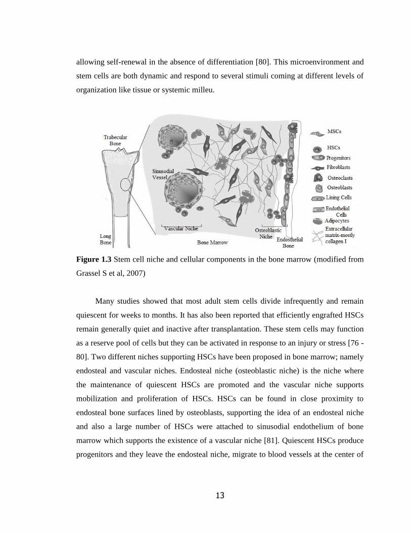
13
allowing self-renewal in the absence of differentiation [80]. This microenvironment and
stem cells are both dynamic and respond to several stimuli coming at different levels of
organization like tissue or systemic milleu.
Figure 1.3 Stem cell niche and cellular components in the bone marrow (modified from
Grassel S et al, 2007)
Many studies showed that most adult stem cells divide infrequently and remain
quiescent for weeks to months. It has also been reported that efficiently engrafted HSCs
remain generally quiet and inactive after transplantation. These stem cells may function
as a reserve pool of cells but they can be activated in response to an injury or stress [76 -
80]. Two different niches supporting HSCs have been proposed in bone marrow; namely
endosteal and vascular niches. Endosteal niche (osteoblastic niche) is the niche where
the maintenance of quiescent HSCs are promoted and the vascular niche supports
mobilization and proliferation of HSCs. HSCs can be found in close proximity to
endosteal bone surfaces lined by osteoblasts, supporting the idea of an endosteal niche
and also a large number of HSCs were attached to sinusodial endothelium of bone
marrow which supports the existence of a vascular niche [81]. Quiescent HSCs produce
progenitors and they leave the endosteal niche, migrate to blood vessels at the center of
14
the bone marrow (vascular niche) where they mature and differentiate. Both niches have
important roles in HSC mobilization and in its reverse process called homing.
Microenvironment regulates stem cells with the presence of some specific
chemical substances called chemokines. Chemokines are like growth factors and their
gradient is the key factor to instruct stem cells to differentiate or remain quiescent in the
niche. Stem cell factor, interleukin, transforming growth factor, granulocyte colony
stimulating factor, stromal derived factor and bone morphogenic proteins are examples
for these chemokines. In homing process, endosteal niche expresses high levels of a
chemokine called Stromal Derived Factor-1α (SDF-1α) and this chemokine attracts
HSCs expressing CXCR4 receptors. Migration to the endosteal niche plays a crucial role
for the engraftment and anchoring of HSCs [82]. It is known that HSCs are significantly
enriched within the endosteal region after bone marrow transplantation [83].
Hematopoietic stem cells need bone marrow microenvironment (niches) which
regulates their migration, proliferation and differentiation for carrying out the successful
hematopoiesis throughout life. [84]
1.3.2 Homing
Homing is the first and fairly rapid process following transplantation in which
circulating hematopoietic cells actively cross the blood bone marrow barrier and lodge at
least transiently in the bone marrow by activation of adhesion interactions prior to their
proliferation [84]. This event can, in general, be defined as recruitment of circulating
HSCs to the bone marrow microvasculature and subsequent transendothelial migration
into the extravascular hematopoietic cords of the bone marrow [85]. Bone marrow
endothelium is the first region for homing cells to anchor with the help of adhesion
molecules and stimulating chemokines present in the bone marrow niche as shown in
Figure 1.4. Several adhesion molecules are necessary for homing of HSCs to the bone
marrow niche. A very important factor for migration, retention and mobilization of
HSCs during homeostasis and after injury or transplantation is CXCL12 / SDF-1α.
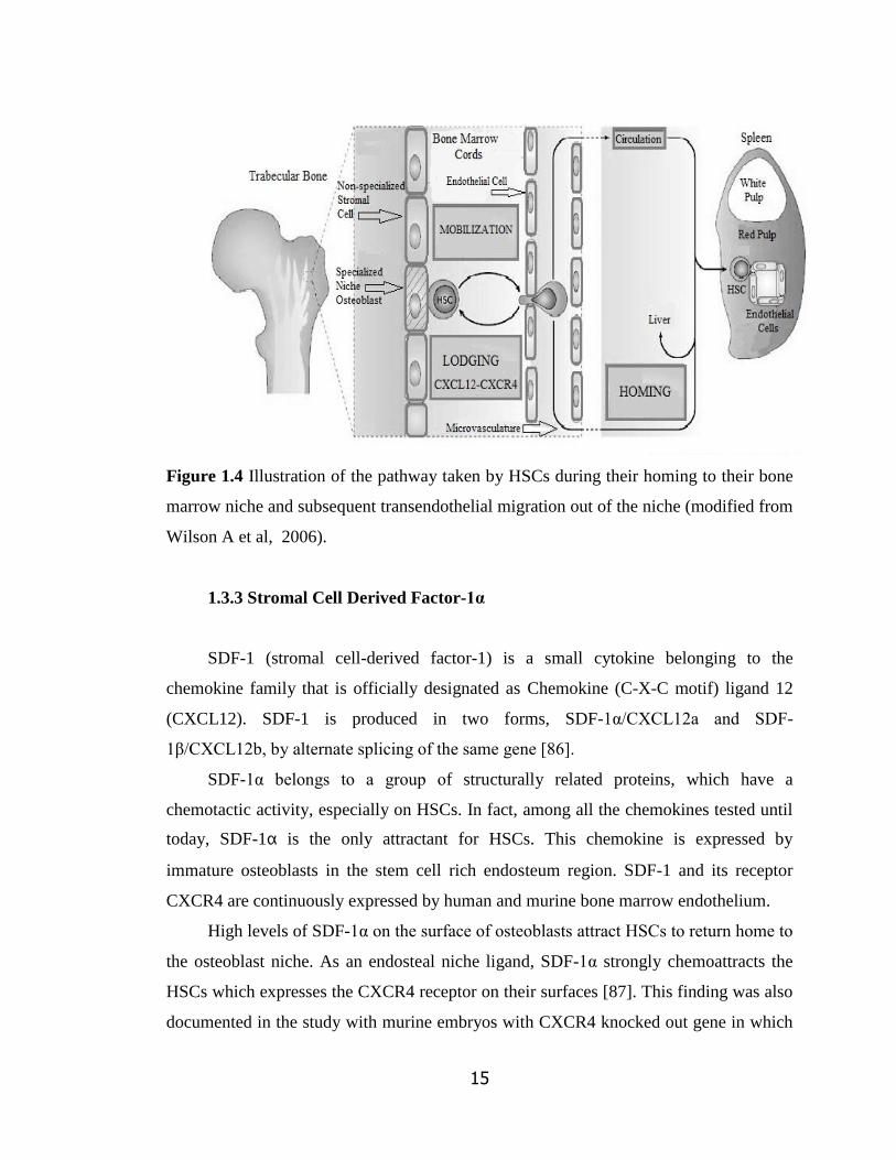
15
Figure 1.4 Illustration of the pathway taken by HSCs during their homing to their bone
marrow niche and subsequent transendothelial migration out of the niche (modified from
Wilson A et al, 2006).
1.3.3 Stromal Cell Derived Factor-1α
SDF-1 (stromal cell-derived factor-1) is a small cytokine belonging to the
chemokine family that is officially designated as Chemokine (C-X-C motif) ligand 12
(CXCL12). SDF-1 is produced in two forms, SDF-1α/CXCL12a and SDF-
1β/CXCL12b, by alternate splicing of the same gene [86].
SDF-1α belongs to a group of structurally related proteins, which have a
chemotactic activity, especially on HSCs. In fact, among all the chemokines tested until
today, SDF-1α is the only attractant for HSCs. This chemokine is expressed by
immature osteoblasts in the stem cell rich endosteum region. SDF-1 and its receptor
CXCR4 are continuously expressed by human and murine bone marrow endothelium.
High levels of SDF-1α on the surface of osteoblasts attract HSCs to return home to
the osteoblast niche. As an endosteal niche ligand, SDF-1α strongly chemoattracts the
HSCs which expresses the CXCR4 receptor on their surfaces [87]. This finding was also
documented in the study with murine embryos with CXCR4 knocked out gene in which

16
a significant decrease in HSCs was observed in their niches [88]. The overall effects of
SDF-1α in health and disease states are summarized in Table 1.3.
Table 1.3 Roles of SDF-1α in health and disease states
Embryonic development Cardiogenesis
Arteriogenesis
Colonization of the bone marrow with HSCs
Hematopoiesis Retention of hematopoietic progenitor cells in the bone
marrow
Supporting megakaryocyte maturation
Migration of HSCs into proliferative niches
Bone marrow
transplantation
Engraftment of HSCs
Angiogenesis Endothelial cell chemotaxis and tube formation
Stem-cell based tissue
repair
Liver disease
Renal ischemia
Myocardial infarction
Ischemic neovascularization
Vascular pathologies Neointimal hyperplasia (restenosis, transplant
asteriosclerosis)
SDF-1α has a pivotal role in the regulation of the CD34+ progenitor cell adhesion
during their homing from the peripheral blood to the bone marrow. It also works with
other molecules (i.e., VLA-4, VLA-5, LFA-1) to potentiate CD34+ cell adhesion and
motility [66, 84, 89]. It was shown that SDF-1α-CXCR4 coupling plays an important
role in homing and engraftment of hematopoietic stem/progenitor cells and on
colonization of bone and bone marrow by metastatic breast and prostate cancer cells [90,
91]. Accordingly, the injection of SDF-1α into the bone marrow upregulates the
17
repopulation of stem cells after total body irradiation [92]. There are studies showing
that the response of HSCs to SDF-1α can be positively affected by small molecules like
complement cleavage fragments [84] and platelet derived microparticles [84]. On the
other hand, treatment of isolated HSCs with a CXCR4 blocking antibody resulted in
inhibition of engraftment in NOD/SCID mice [92].
1.3.4 Bone Marrow Transplantation
Over the past 40 years, bone marrow transplantation and hematopoietic stem
cell transplantation have been used with increasing frequency to treat numerous
malignant (ie., acute lymphoblastic leukemia, acute and chronic myelogenous leukemia,
plasma cell disorders and Hodgkin and non-Hodgkin lymphoma) and nonmalignant
diseases (i.e., inherited metabolic, immune disorders, and red cell disorders (e.g pure red
cell aplasia), marrow failure states (e.g., severe aplastic anemia), autoimmune diseases
(e.g systemic sclerosis, Crohn disease) and acquired immune deficiency syndrome
(HIV) [93, 94].
Hematopoietic stem cells are crucial and most needed for successful
transplantation. Currently, the major sources of stem cells for transplantation include
bone marrow, peripheral blood, and cord blood. These cells have 3 main sources:
1) the patient (an autologous transplant)
2) someone other than the patient (an allogeneic transplant)
3) donated umbilical cord blood (a cord blood or umbilical cord blood transplant)
[95].
Early studies with animals quickly revealed that bone marrow was the organ most
sensitive to the damaging effects of radiation [96]. The reinfusion of marrow cells was
subsequently used to rescue lethally irradiated animals. In the 1950s, patients were given
lethal doses of radiation to treat leukemia. Although many had hematologic recovery
following this treatment, all patients eventually succumbed to relapse of their
malignancies or to infections. Transplants for nonmalignant diseases generally have
more favorable outcomes, with survival rate of 70-90% if the donor is a matched sibling

18
and 36-65% if the donor is unrelated. Transplants for acute leukemias in remission at the
time of transplant have survival rates of 55-68% if the donor is related and 26-50% if the
donor is unrelated. Many failures are due to 2 main reasons: nonengraftment and Graft
versus Host Disease (GVHD) [97].
Graft-versus-host disease (GVHD) is a common complication of allogeneic bone
marrow transplantation. Immune cells in the transplanted marrow recognize the recipient
as "foreign" and starts an immunologic attack. When an immunocompetent graft with
many functional cells are administered, GHVD can be developed if the recipient is
histoincompatible or immunocompromised. This disease has acute and chronic forms.
Acute GVHD is observed within the first 100 days after the transplant. Chronic GVHD
is usually observed after 100 days of the bone marow transplantation. Both of them are
major challenges against the transplant success and effects the long-term survival of
patient [98].
There are some conditioning cures applied for the success of transplantation.
These are classified as myeloablative, nonmyeloablative, and reduced intensity.
Myeloablative cures are for killing all residual cancer cells in transplantation and to
cause immunosuppression for engraftment. Total-body irradiation (TBI) and drugs such
as cyclophosphamide, busulfan and cyclophosphamide are the commonly used
myeloablative therapies. Nonmyeloablative regimens are the use of chemotherapy drugs
and radiation in a lower dose than that of myeloablative regimens. They rely on graft‘s
effect on killing cancer cells with donor T lymphocytes. Reduced-intensity regimens can
range in intensity from myeloablative to nonmyeloablative, and involve drugs such as
fludarabine, melphalan, antithymocyte globulin, and busulfan. These cures have lower
toxicity. The onset of GVHD is delayed with this regime compared with the other
regimens [99, 100].
19
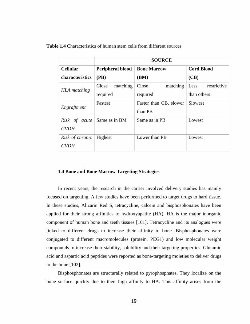
Table 1.4 Characteristics of human stem cells from different sources
1.4 Bone and Bone Marrow Targeting Strategies
In recent years, the research in the carrier involved delivery studies has mainly
focused on targetting. A few studies have been performed to target drugs to hard tissue.
In these studies, Alizarin Red S, tetracycline, calcein and bisphosphonates have been
applied for their strong affinities to hydroxyapatite (HA). HA is the major inorganic
component of human bone and teeth tissues [101]. Tetracycline and its analogues were
linked to different drugs to increase their affinity to bone. Bisphosphonates were
conjugated to different macromolecules (protein, PEG1) and low molecular weight
compounds to increase their stability, solubility and their targeting properties. Glutamic
acid and aspartic acid peptides were reported as bone-targeting moieties to deliver drugs
to the bone [102].
Bisphosphonates are structurally related to pyrophosphates. They localize on the
bone surface quickly due to their high affinity to HA. This affinity arises from the
SOURCE
Cellular
characteristics
Peripheral blood
(PB)
Bone Marrow
(BM)
Cord Blood
(CB)
HLA matching Close matching
required
Close matching
required
Less restrictive
than others
Engraftment Fastest Faster than CB, slower
than PB
Slowest
Risk of acute
GVDH
Same as in BM Same as in PB Lowest
Risk of chronic
GVDH
Highest Lower than PB Lowest

20
attraction of the diphosphonate moiety to calcium ions present in HA crystals. In recent
years, their uses for treatment of osteoporosis and osteogenesis imperfecta have been
studied because of their ability to inhibit bone resorption [103]. Bisphosphonates have
been conjugated to drugs, proteins and other molecules such as radiopharmaceuticals to
obtain novel agents for bone scintigraphy [89]. Also, several strategies using
bisphosphonate-conjugated drugs have been investigated at a preclinical level to
optimize treatments for osteoporosis, osteoarthritis, and bone cancer. However, targeted
drug delivery systems are preferable over drug conjugates alone due to several factors
including drug protection from biodegradation in bloodstream, transport efficiency, and
drug-payload [104]. Prostaglandin E2 (PGE2) and alendronate conjugates were studied in
rats for osteoporosis treatment and it was found that their new conjugates bind bone
more effectively than free PGE2 [105]. In a study performed with the conjugates of PEG
and poly[N-(2-hydroxypropyl)methacrylamide] (PHPMA) with alendronate and aspartic
acid peptide as bone targeting moieties showed high accumulation in bone tissue. Both
in vitro and in vivo trials with rats indicated that these novel polymeric carriers were
useful for targeting drugs to bone [106].
It was shown that the vasculature in bone structure have pores of approximately
80-100 nm in diameter. Sizes of liposomes should be less than at least 80 nm to
extravasate and be localized in bone after i.v. administration. 30 minutes after i.v.
administration of liposomes, only 15% of them remain in the blood and the rest are
found mainly in liver, spleen and bone marrow as the parts of RES. This leads to the
idea of passively targeting liposomes to the bone marrow [107]. In a study conducted
with dogs, the effect of size of the antimony encapsulated liposomes was studied for
passive targeting and 410 nm liposomes showed an improved drug targeting to the bone
marrow [108]. There is only one study on liposomal delivery to the bone marrow with
active targetting of macrophages by Sou et al (2010). Using l-glutamic acid, N-(3-
carboxy-1-oxopropyl)-, 1,5-dihexadecyl ester as targeting moiety, liposomes were
targeted to bone marrow phagocytic cells (macrophages).
Alendronate, a type of bisphosphonate, was chosen for targeting SDF-1 loaded
pegylated liposomes to bone sites because of its high affinity to bone and easy

21
conjugation with Carboxylic Acid-PEG-DSPE (2000) (one of the PLs used in liposome
preparation) via carbodiimide chemistry [66]. Carbodiimide chemistry between
alendronate and different polymers such as PLGA and PLA was used for targeted drug
delivery (i.e. estrogen) to bone in previous studies [66, 89]. It is an amide bond reaction
between a carboxyl group and a primary or secondary amine group. The bonding
chemistry between DSPE2000-Carboxylic Acid and alendronate is shown in Figure 1.5
[66]. This amide linkage is not cleavable, it shows high resistance to enzymatic
hydrolysis in plasma compared to an ester bond. Therefore, alendronate is stable on the
surface and is not being released when linked covalently. In a previous study, it was
shown that both PEG and alendronate existed on the surface of PLGA nanoparticles
after conjugation due to their hydrophilicity [107].
Figure 1.5 Structure of alendronate, DSPE-PEG(2000)-Carboxylic Acid and the
alendronate conjugated DSPE-PEG(2000)
22
1.5 Aim of the Study
Homing is the process by which stem cells move to their own niches upon BMT
under the influence of chemokines released by the cells present in the particular
microenvironment. This movement is crucial for hematopoiesis. Bone marrow
microenvironment is drained after total body irradiation in cancer patients. Thus, the
cells producing these chemotactic chemokines are damaged. There is a need for these
chemokines to improve the engrafment after bone marrow transplantation for curing
leukemia, multiple myeloma like diseases. Stromal-derived factor-1 (SDF-1) is the key
chemokine which regulates homing.
The hypothesis of this thesis is that targeting of SDF-1α loaded, pegylated
liposomes to damaged endosteal niche of bone marrow and obtaining local release of
SDF-1 in this environment will attract HSCs and MSCs for the homing process, thereby,
increasing homing efficiency. In this study, we aimed to develop and characterize
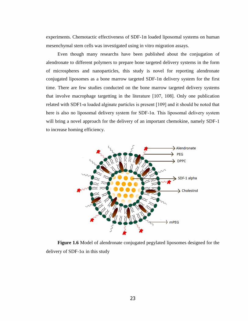
alendronate conjugated and pegylated SDF-1α loaded liposomal delivery system (Figure
1.6) for providing local release of SDF-1α at the border of endosteum and bone marrow
as a new strategy to increase homing efficiency after BMT.
Liposomes were chosen as the delivery system because of their biocompatibility
and controlled release profiles. Large unilamellar vesicles were prepared to provide
more homogenous systems compared to multilamellar ones in terms size and loading.
Alendronate, an osteotropic molecule with a hydroxyapatite affinity was conjugated to
pegylated phosphatidylethanolamine. Both passive targetting of liposomes with its size
and affinity of the system towards osteoblasts in the endosteal niche with alendronate on
the surface were used for this end. With alendronate conjugation to liposomes, it was
also aimed to impose a more negative surface on liposomes for prolonging their half-
lives. The effects of composition and size of unilamellar liposomes, degree of pegylation
on encapsulation efficiency and release profiles of -lactoglobulin (model protein) were
investigated in the optimization studies. Alendronate conjugated and pegylated
liposomes loaded with SDF-1α were evaluated in terms of protein encapsulation
efficiency, release profiles, morphology, surface charge, HA affinity with in situ
23
experiments. Chemotactic effectiveness of SDF-1α loaded liposomal systems on human
mesenchymal stem cells was investigated using in vitro migration assays.
Even though many researchs have been published about the conjugation of
alendronate to different polymers to prepare bone targeted delivery systems in the form
of microspheres and nanoparticles, this study is novel for reporting alendronate
conjugated liposomes as a bone marrow targeted SDF-1α delivery system for the first
time. There are few studies conducted on the bone marrow targeted delivery systems
that involve macrophage targetting in the literature [107, 108]. Only one publication
related with SDF1-α loaded alginate particles is present [109] and it should be noted that
here is also no liposomal delivery system for SDF-1α. This liposomal delivery system
will bring a novel approach for the delivery of an important chemokine, namely SDF-1
to increase homing efficiency.
Figure 1.6 Model of alendronate conjugated pegylated liposomes designed for the
delivery of SDF-1 in this study
24
CHAPTER 2
MATERIALS AND METHODS
2.1 Materials
1,2-Dimyristoyl-sn-glycero-3-phosphocoline (DMPC) was a product of Fluka
(USA). 1,2-Distearoyl-sn-glycero-3-Phosphoethanolamine-N-[Methoxy (Polyethylene
Glycol)2000] (Ammonium Salt) (MPEG(2000)-DSPE) was provided by Lipoid
(Germany). Mini Extruder set, Nucleopore Track-Etch membranes (800, 400, 200, 100
nm), filter supports, 1,2-Distearoyl-sn-glycero-3-Phosphoethanolamine-N-
[Carboxy(Polyethylene Glycol)2000] (Ammonium Salt) (DSPE-PEG(2000)-Carboxylic
acid) was obtained from Avanti Polar Lipids, Inc. (USA).
1,2-Dipalmitoyl-sn-glycero-3-phosphocoline (DPPC), 1,2-Dimystroyl-sn-glycero-
3-phosphocoline (DMPC), cholesterol, alendronate sodium trihydrate, dialysis sacks,
benzoylated dialysis tubing, bicinchonicic acid solution, uranyl acetate dihydrate,
chloroform (HPLC grade), N,N-dicyclohexylcarbodiimide (DCC), N-
hydroxysuccinimide (NHS), stromal derived factor-1 human (SDF-1), β-
lactoglobulin, Giemsa staining solution were obtained Sigma-Aldrich Chem. Co.
(USA). Human SDF-1 ELISA kit was a product of RayBiotech, Inc. (USA).
Transwell permeable support 8.0 nm polycarbonate membrane 6.5mm insert, 24-
well plate tissue culture polystrene plates were purchased from Corning Life Sciences
Inc. (USA). Polyethersulfone syringe membrane (0.45 μm pore size) was obtained from
Whatman Co. (UK). Dulbecco‘s modified Eagle‘s medium (DMEM) low glucose (4.5
g/l) with L-glutamine and fetal bovine serum (FBS) were purchased from Biochrom AG

25
(Germany). Penicillin/Streptavidin and trypsin EDTA were purchased from PAA
Laboratories (Germany). Dimethyl sulphoxide (molecular biology grade) (DMSO) was
the product of AppliChem Co. (Germany). PD-10 columns and Sephadex G-75 were
purchased from GE Healthcare (UK).
2.2 Methods
2.2.1 Liposome preparation
Large unilamellar liposomes (LUVs) were prepared from MLVs by extrusion
method [110]. Initially, phospholipids and cholesterol were dissolved in chloroform at
different ratios (Table 2.1) and organic solvent was evaporated under nitrogen stream to
form a lipid film. This process was followed by removal of residual chloroform under
vacuum overnight.
For encapsulation studies either model protein (-Lactoglobulin) or stromal derived
factor-1α (SDF-1α) was dissolved in 1 ml 0.1 M PBS solution (pH 7.2) and the lipid
films were hydrated with the protein solution by heating and vortexing at 38-40°C in 2
minute cycles for 50 minutes. MLVs were subjected to cycles of freeze-thaw using
liquid nitrogen and 35°C water bath. -lactoglobulin loaded MLVs were applied 10
cycles and SDF-1α loaded ones were applied 4 or no cycles of freeze-thaw during
preparation of protein loaded liposomes.
MLVs were then extruded sequentially through 800, 400 and 200 and/or 100 nm
track etched polycarbonate filters to form LUVs. Extrusion was performed at 38-40°C
by passing liposome suspension 10 times through 800 nm membranes, 10 times through
400 nm and 6 times through 200 nm membranes. For 100 nm sized liposomes, the last
step was done with 6 times extrusion from 100 nm membranes instead of 200 nm.
Unincorporated micellar lipids and unentrapped protein molecules were separated by
Sephadex G-75 size exclusion chromatography using disposable PD-10 columns (GE
Healthcare). Elution buffer was 0.1 M PBS (pH 7.2). Collected fractions of LUVs were
pooled for further studies. Turbidity analysis was performed to determine which

26
fractions will be pooled for liposomes. Each fraction sample was analysed for optical
density at 410 nm by UV-Spectrophotometer (Hitachi U-2800A, Japan) and those with
the highest absorbance/turbidity values were pooled.
Table 2.1 Compositions of liposomes expressed in mole ratios and pore sizes of
filters used for these different liposomal formulations.
Liposome composition Filter Pore Size
DPPC:DMPC (1:1) 100 nm
DPPC:DMPC:Cho (2:0.2:0.3) 100 nm
DPPC:Cho (2:0.5) 100 nm
DPPC:DMPC:Cho (1:1:0.5) 100 nm
DPPC:Cho (2:0.5) 200 nm
DPPC:Cho (2:0.5)+ DSPE-mPEG2000 (2% of total lipid
content) (2%PEG/LUV200)
200 nm
DPPC:Cho (2:0.5)+ DSPE-mPEG2000 (5% of total lipid
content) (5%PEG/LUV200)
200 nm
DPPC:Cho (2:0.5)+ ALE-DSPE-PEG2000 (2.5% of total lipid
content) + DSPE-mPEG2000 (2.5% of total lipid content)
(2.5%ALE-PEG/LUV200)
200 nm
DPPC:Cho (2:0.5)+ ALE-DSPE-PEG2000 (5% of total lipid
content) (5%ALE-PEG/LUV200)
200 nm
2.2.2 Conjugation of Alendronate to DSPE-PEG(2000)-Carboxylic Acid
DSPE-PEG(2000)-carboxylic acid (23 mg) was dissolved in acetone (5 ml) and
activated by 4.2 mg N,N-Dicyclohexylcarbodiimide (DCC) and 2.4 mg N-
hydroxysuccinimide (NHS) overnight at room temperature. Dicyclohexylurea, the
insoluble by-product of the activation, was removed using a polyethersulfone syringe
27
filter with 0.45 µm pore size. NHS activated lipid was dried under nitrogen for 2 hours.
Activated lipid and 2 mg alendronate sodium trihydrate were dissolved in 5 ml of a
mixture of 4.5 ml DMSO and 0.5 ml water and then stirred for 24h at room temperature.
In order to get rid of the cross-linkers, dialysis was done. Conjugated lipid was placed
inside the benzoylated cellulose dialysis bag (MWCO 2000, Sigma-Aldrich, USA) and it
was dialysed against water for 24 hours at room temperature. The water was changed
every 6 hours. The milky suspension inside the dialysis bag was then centrifuged at
14.000 rpm for 10 minutes at 4°C (Eppendorf 5804R, Germany). The conjugated lipid
pellet was then dried under nitrogen. The supernatant obtained was also dried for 24
hours in a vacuum oven. All of the activated lipid was pooled together in acetone, dried
and stored at 4C in a dessicator after flushing with nitrogen.
2.2.2.1 Determination of Alendronate Conjugation Efficiency
Fourier Transform Infrared Spectrometer/Attenuated Total Reflectance (FTIR/ATR)
spectrums of conjugated lipid, PEG-DSPE-Carboxylic Acid, alendronate and the
mixture of alendronate and lipid were performed using Fourier Transform Infrared and
Raman Spectrometer and Microscope (Bruker IFS 66/S, USA).
Conjugation efficiency (Con. Eff) was calculated by the ratio of area differences
under the amide bond formation peak for conjugated and unconjugated DSPE-PEG200
to the corresponding area in the spectrum of unconjugated DSPE-PEG2000. Areas were
found using Excel 2010 (Microsoft Co, USA) and the formula below was used to
determine the conjugation efficiency.
Con. Eff.=[(Area conj.lipid-Area mixture)/ Area conj.lipid] x 100
28
2.2.3 Characterization of Liposomes
2.2.3.1 Partical Size by Dynamic Light Scattering
Freshly made liposome suspensions were diluted to 1:10 for the analysis. The
particle size distributions of LUVs were determined by dynamic light scattering method
(Malvern Nano ZS90; Malvern Instruments, METU Central Laboratory).
2.2.3.2 Surface Charge by Zeta Potential and Mobility Measurement System
Freshly made liposome suspensions were diluted to 1:2 for the analysis. The surface
charge of LUVs were determined by zeta potential method (Malvern Nano ZS90;
Malvern Instruments, METU Central Laboratory).
2.2.3.3 Transmisson Electron Microscopy (TEM)
Transmission electron microscopy was used to observe the size, morphology and
lamellarity of liposomes after size reduction by extrusion. A drop of liposomal
suspension was placed on the copper grid and the excess liposomal suspension was
removed with filter paper. It was then let dry at room temperature. 2% uranyl acetate
(Sigma-Aldrich Co., USA) solution was dropped onto the grid and the excess of staining
solution was removed with filter paper. The liposomes were examined under the
transition electron microscope (Philips, JEM-100CX) at 80 kV.
2.3.3.4 Determination of Entrapment Efficiency of Liposomes and Lipid
Recovery After Extrusion
The encapsulation efficiency of the liposomes was determined from the unentrapped
protein using fractions 9 through 18. Total amount of unentrapped protein was

29
subtracted from the total amount of protein used in liposome to obtain the amount of
encapsulated protein.
The encapsulation efficiency was calculated according to the following equation
Encapsulation efficiency (%) = [ (Atotal – Aunetrapped) / Atotal ] x 100
where,
Atotal is the total amount of β-Lactoglobulin or SDF-1α used in liposome fraction
Auntrapped is the total amount of β-Lactoglobulin or SDF-1α calculated from the
unentrapped protein fractions in size exclusion choromatography by BCA Assay or
ELISA, respectively.
For -Lactoglobulin loaded liposomes, the unentrapped protein was determined
using a modified colorimetric protein assay (BCA Assay) [106]. Sodium dodecylsulfate
(SDS) was added to each protein sample at a final concentration of 2% to minimize the
interference of lipids to the protein determination. Briefly, 100-μL of sample and 100-μL
BCA working solution was incubated in 96 well plates at 60°C for 30 min and then
cooled to the room temperature. Absorbances were measured at 562 nm using
microplate spectrophotometer (GMI Biotech 3550, USA). Protein calibration curve was
constructed in the range of 1-250 ug/ml using -Lactoglobulin.
For SDF-1 loaded liposomes, the unentrapped SDF-1 was determined with
Human SDF-1α ELISA kit according to protocol given by the manufacturer. SDF-1α
calibration curve was constructed in the range of 6.14-15000 pg/ml using the standards
of the kit.
The amount of DPPC in LUVs after extrusion was determined by the Stewart
method. Aliquots from liposomal fractions were dried with nitrogen flush and dissolved
in chloroform. After appropriate dilution with chloroform, they were mixed with
ammonium ferrothiocyanate solution (1:1, v/v) and the absorbance was measured at 485
nm by UV-visible spectrophotometer (Hitachi U-2800A, Japan). DPPC was quantified
by calibration curve constructed with DPPC (5–50 μg/ml).

30
2.3.3.5 -Lactoglobulin and SDF-1 Release Studies
Liposome suspension (1 ml) was placed in cellulose dialysis bags (12.000 Da
MWCO, Sigma-Aldrich, USA) and was transferred into vial containing 10 ml 0.1 M
PBS (pH 7.2). The PBS release media were stirred on magnetic stirrer and incubated at
37°C. Release studies were carried out in triplicates. 1 ml PBS samples were taken from
each vial at different incubation periods. BCA assay was used to determine amount of
the released -Lactoglobulin at each incubation period as described in Section 2.3.3.3.
Human SDF-1α ELISA kit was used to quantitate SDF-1 released at each time period
according to the kit protocol.
2.3.3.6 Bone (HA) Affinity of Liposomal Preparations
Nanosized HA powders were kindly given by the lab of Assoc. Prof. Dr. Zafer Evis.
The method used to produce pure HA samples was precipitation method as described in
Burçin et al [111]. The amount of liposome associated with HA was evaluated with the
change in the turbidity of PBS [112] and decrease in lipid amount in PBS with time. HA
powder was added to PBS (pH 7.2) at a concentration of 10 mg/ml . Liposomes were
added to the HA suspension (at a final lipid concentration = 100 µM in 2 ml). Two
different liposomal preparations (alendronate conjugated and alendronate free-pegylated
empty liposomes) were prepared. For determining the degree of HA affinity of the
liposomes, the suspensions were centrifuged at 5000 rpm for 5 minutes after 0, 2, 4, 6
and 24 hours of incubation at room temperature. The turbidity of the suspension was
then measured by UV/VIS spectrophotometer (Hitachi U-2800A, Japan) at 410 nm. At
each incubation period, aliquots (50 µl) were also taken for further lipid analysis by
Stewart assay as described in Section 2.3.3.4. The suspensions were gently shaken at
room temperature between the time periods.
For two methods %HA affinity was calculated as follows
Turbidity measurement: [(initial absorbance-sample absorbance)/initial absorbance]x100
Lipid measurement: [(initial lipid amount-sample lipid amount)/initial lipid amnt.]x100
31
2.2.4 Cell culture studies
2.2.4.1 Isolation and Expansion of Human Bone Marrow Derived Mesenchymal
Stem Cells (hBMMCs)
Human bone marrow stromal cells were obtained from Hacettepe University Bone
Marrow Transplantation (BMT) Unit with an approval Ethical Committee of Hacettepe
University (Certification Number: LUT10/17) and isolated from healthy donors with
their consent. BMT Unit isolated the MSCs from the bone marrow aspirates (1–3 ml) of
healthy donors sent for routine analysis before transplantation The mesenchymal stem
cells used were positive for certain MSC markers, namely CD105, CD44, CD90 and
CD106. Shortly, the bone marrow aspirate samples were diluted in equal volumes with
PBS, and mononuclear cells were isolated from the marrow by density centrifugation
using Ficoll gradient (density, 1.077 g/l). The cells were then washed twice with PBS
and cultured in a medium consisting of DMEM-low glucose (LG), 10% FBS, L-
glutamine (0.584g/l), penicillin (100 units/ml), streptomycin (100 g/ml), and
amphotericin-B (2.5 g/ml) at 37°C in a 5% CO2 environment. Culture medium was
replaced twice a week. Upon reaching confluence in 2 weeks, cell were splitted in a ratio
of 1:3 with 0.1 % trypsin-EDTA by incubating at 37°C for 5 minutes. Hematopoietic
cells were excluded by sorting for CD45, CD34, CD14, CD33, and CD3 conjugated with
phycoerythrin (Becton,Dickinson and Company, USA).
2.2.4.2 Cell Migration Assay
The migration behaviour of human mesenchymal cells treated with alendronate
conjugated SDF1 loaded liposomes for 24 h was studied in a 24-well transwell using
polycarbonate membranes with 8 μm pores (Corning Costar, USA). Empty liposomes
and DMEM-low glucose were used as controls. MSCs at a density of 5 × 10⁵ cells/ml in
100 μl of medium (DMEM + 0.5% FBS) were placed in the upper chamber of the
transwell assembly. The lower chamber contained 600 μl of liposome solution [300 μl of

32
liposome solution (empty or SDF-1α loaded) + 300 μl DMEM-low glucose] or 600 µl of
DMEM-low glucose. Cells were allowed to migrate or invade for a total either 16 or 24
hours of incubation at 37°C and 5% CO2 (Figure 2.1). After each incubation period, the
upper surface of the membrane was scraped gently to remove non-migrating cells and
washed with phosphate-buffered saline. The membrane was then fixed in 4%
formaldehyde for 15 minutes and stained with 4% Giemsa staining solution for 10
minutes. The number of migrating cells was determined by counting five random fields
per well under the microscope with 20X magnification and taking the mean of all 5
random field counts. Experiments were performed in sets of four. The amounts of SDF-
1α released from liposomal formulation during migration assay were determined with
Human SDF-1 ELISA kit for 16 and 24 hours. Shortly, at each incubation period, the
media were collected and centrifuged at 14000 rpm at 4°C for precipitating the
liposomes using high-speed centrifuge (Eppendorf 5804R, Germany). SDF-1α released
was quantitated in the supernatant of liposomal formulation using the ELIZA kit
Figure 2.1 Schematic presentation of the cell migration assay
33
2.2.5 Statistical Analyis
In comparing the groups for a single parameter, one-way ANOVA test was used
with Tukey‘s Multiple Comparison Test for the post-hoc pairwise comparisons (SPSS-9
Software Programme, SPPS Inc., USA). Differences were considered significant for p <
0.05.

34
CHAPTER 3
RESULTS AND DISCUSSIONS
3.1 Optimization Studies with -Lactoglobulin
3.1.1 Effects of Liposome Composition and Size
Liposomal formulations have been approved and are being used in the clinics for
many years. The in vivo stability, fate, drug loading, and release profile of liposomes
depend on several parameters. To this end, the effects of liposome composition and size
on the encapsulation efficiency and release profile were investigated.
As a model protein for stromal derived factor-1 (SDF-1) which has a molecular
weight (MW) of 8 kDa, -lactoglobulin with 18.4 kDa MW) was used in the
optimization studies. Large unilamellar liposomes with the model protein were prepared
by film hydration followed by the extrusion method using different combinations of
lipids. Dipalmitoyl phosphatidylcholine (DPPC; C16:0) and dimyristoyl
phosphatidylcholine (DMPC; C14:0) were chosen as phospholipids for protein loading
in liposomes due to their low transition temperatures (41°C and 23°C, respectively).
During both hydration of lipid films and extrusion steps the temperature was set to 38˚C.
This was crucial due to biological activity loss of proteins above body temperature.
Large unilamellar vesicles (LUVs) of 100 nm were prepared with extrusion using
different molar ratios of DPPC, DMPC and cholesterol (Cho) (Table 3.1).
After extrusion process liposomes were pooled from the fractions 3 through 7,
which had highest liposome amounts according to turbidity results (Figure 3.1). The
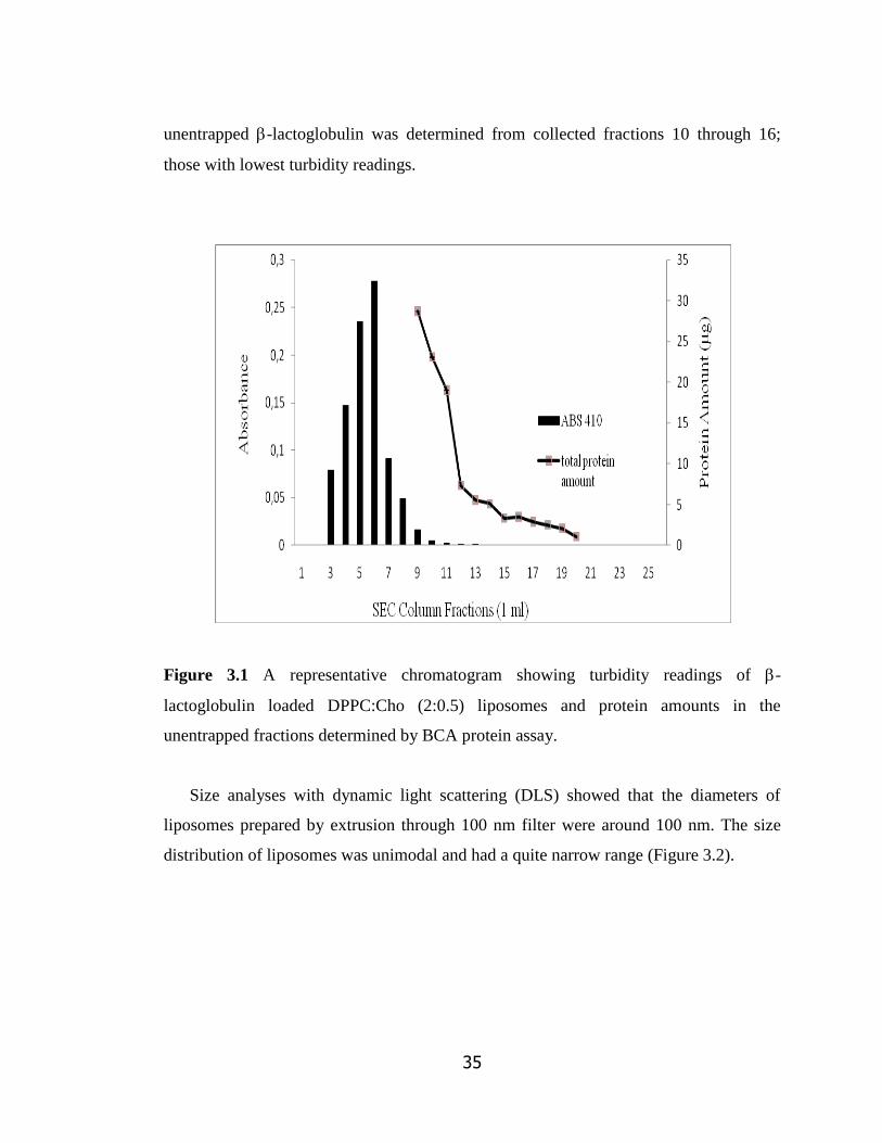
35
unentrapped -lactoglobulin was determined from collected fractions 10 through 16;
those with lowest turbidity readings.
Figure 3.1 A representative chromatogram showing turbidity readings of -
lactoglobulin loaded DPPC:Cho (2:0.5) liposomes and protein amounts in the
unentrapped fractions determined by BCA protein assay.
Size analyses with dynamic light scattering (DLS) showed that the diameters of
liposomes prepared by extrusion through 100 nm filter were around 100 nm. The size
distribution of liposomes was unimodal and had a quite narrow range (Figure 3.2).

36
Figure 3.2 A representative DLS result of size distribution for -lactoglobulin loaded
LUVs prepared by extrusion through 100 nm filter.
BCA protein assay or ELISA was used to obtain the unencapsulated protein
amounts of liposomes. Initial studies showed insufficiency of BCA assay for
quantitating the entrapped protein in pooled liposome fractions due to high interference
by phospholipids. Therefore, SDS was added (as 2% final SDS concentration) to size
exclusion chromatograpy (SEC) fractions with low or no liposomes to determine
unentrapped proteins by this method without lipid interference.
-lactoglobulin encapsulation efficiency of LUVs ranged between 68% and 71%.
As seen in Table 3.1, the addition of cholesterol to the liposome composition slightly
increased the encapsulation efficiency of LUVs in agreement with the literature. It was
indicated that the proportion of cholesterol is an important factor in liposome
formulation as it improves the maintenance of liposome integrity and stability as well as
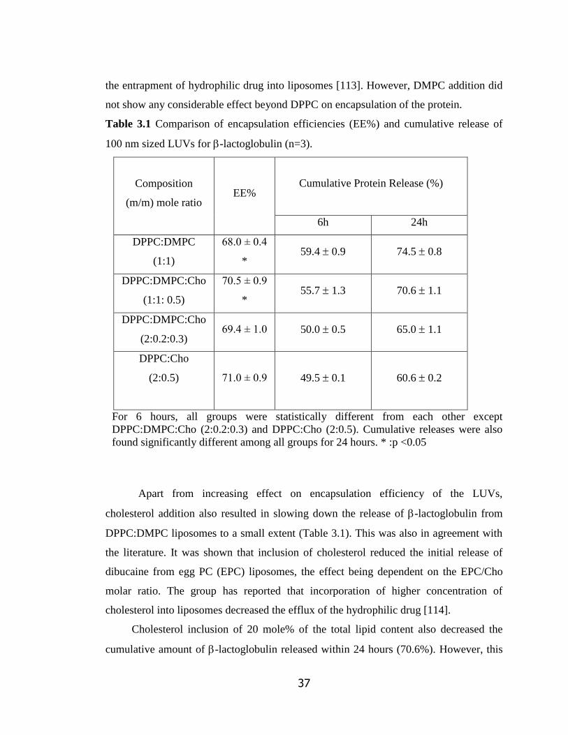
37
the entrapment of hydrophilic drug into liposomes [113]. However, DMPC addition did
not show any considerable effect beyond DPPC on encapsulation of the protein.
Table 3.1 Comparison of encapsulation efficiencies (EE%) and cumulative release of
100 nm sized LUVs for -lactoglobulin (n=3).
Composition
(m/m) mole ratio EE%
Cumulative Protein Release (%)
6h 24h
DPPC:DMPC
(1:1)
68.0 ± 0.4
* 59.4 0.9 74.5 0.8
DPPC:DMPC:Cho
(1:1: 0.5)
70.5 ± 0.9
* 55.7 1.3 70.6 1.1
DPPC:DMPC:Cho
(2:0.2:0.3) 69.4 ± 1.0 50.0 0.5 65.0 1.1
DPPC:Cho
(2:0.5)
71.0 ± 0.9 49.5 0.1 60.6 0.2
For 6 hours, all groups were statistically different from each other except
DPPC:DMPC:Cho (2:0.2:0.3) and DPPC:Cho (2:0.5). Cumulative releases were also
found significantly different among all groups for 24 hours. * :p <0.05
Apart from increasing effect on encapsulation efficiency of the LUVs,
cholesterol addition also resulted in slowing down the release of -lactoglobulin from
DPPC:DMPC liposomes to a small extent (Table 3.1). This was also in agreement with
the literature. It was shown that inclusion of cholesterol reduced the initial release of
dibucaine from egg PC (EPC) liposomes, the effect being dependent on the EPC/Cho
molar ratio. The group has reported that incorporation of higher concentration of
cholesterol into liposomes decreased the efflux of the hydrophilic drug [114].
Cholesterol inclusion of 20 mole% of the total lipid content also decreased the
cumulative amount of -lactoglobulin released within 24 hours (70.6%). However, this

38
was still constituting a large amount of the protein making this formulation not suitable
for the aim of the study (Table 3.1). To further modify release behavior, the mole ratio
of DMPC was lowered in the liposome composition. Lowering the molar ratio of DMPC
relative to DPPC in the composition (DPPC:DMPC:Cho; 2:0.2:0.3) resulted with a
slower release without significant change in the encapsulation efficiency as compared
with the other groups (Table 3.1). DPPC has a higher transition temperature compared to
DMPC which results in higher stability and leaktightness in a wide temperature range.
Therefore, liposomes containing higher DPPC mole% relative to DMPC tend to have a
considerably more rigid membrane bilayer [60]. This property was thought to cause
lower leakage of the model protein in the present study. Based on these results, DPCC:
Cho (2:0.5) was studied as the last liposome composition. Liposomes prepared by this
composition had the highest -lactoglobulin EE% (71.0±0.9%) and lowest 24 hour
cumulative protein release amounts (60.6 0.2%).
The amount of the lipid recovered in liposomes after size exclusion
chromatography was almost the same for all formulations and ranged from 73% to 79%
of the total lipid amount used (data not shown).
In order to obtain slower release apart from lipid composition, size of liposomes
was considered. The average liposome size was increased from 100 nm to 200 nm using
200 nm filters and similar encapsulation efficiencies of the model protein were obtained
(71% versus 68.5%). The release profiles of DPPC:Cho (2:0.5) liposomes of the two
sizes were compared in Figure 3.3. Doubling the liposome size resulted with a decrease
in both initial burst and cumulative % release after 24 hours which was also in good
agreement with the study of Manosnoi et al. [115]. They compared the surface charge
and size properties of tranexamic acid loaded liposomes and observed a higher release
rate constant and release amount (almost 80% of the entrapped material) after 24 hours
incubation time for the smaller liposomes.
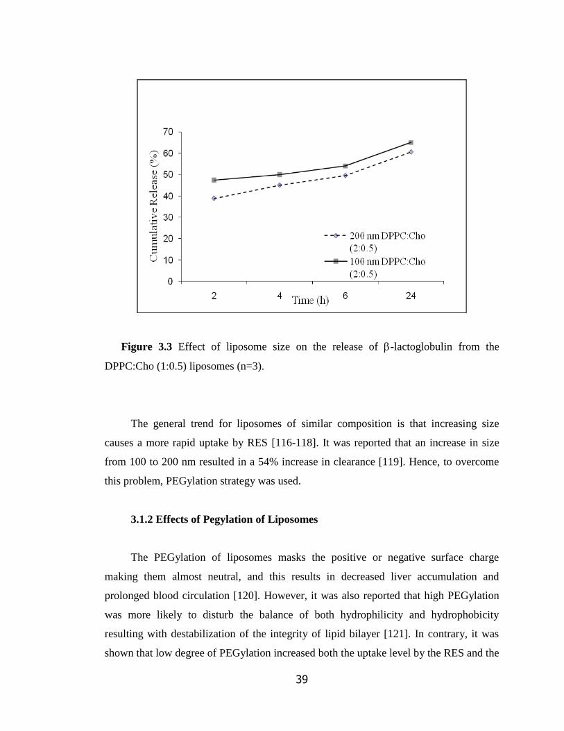
39
Figure 3.3 Effect of liposome size on the release of -lactoglobulin from the
DPPC:Cho (1:0.5) liposomes (n=3).
The general trend for liposomes of similar composition is that increasing size
causes a more rapid uptake by RES [116-118]. It was reported that an increase in size
from 100 to 200 nm resulted in a 54% increase in clearance [119]. Hence, to overcome
this problem, PEGylation strategy was used.
3.1.2 Effects of Pegylation of Liposomes
The PEGylation of liposomes masks the positive or negative surface charge
making them almost neutral, and this results in decreased liver accumulation and
prolonged blood circulation [120]. However, it was also reported that high PEGylation
was more likely to disturb the balance of both hydrophilicity and hydrophobicity
resulting with destabilization of the integrity of lipid bilayer [121]. In contrary, it was
shown that low degree of PEGylation increased both the uptake level by the RES and the
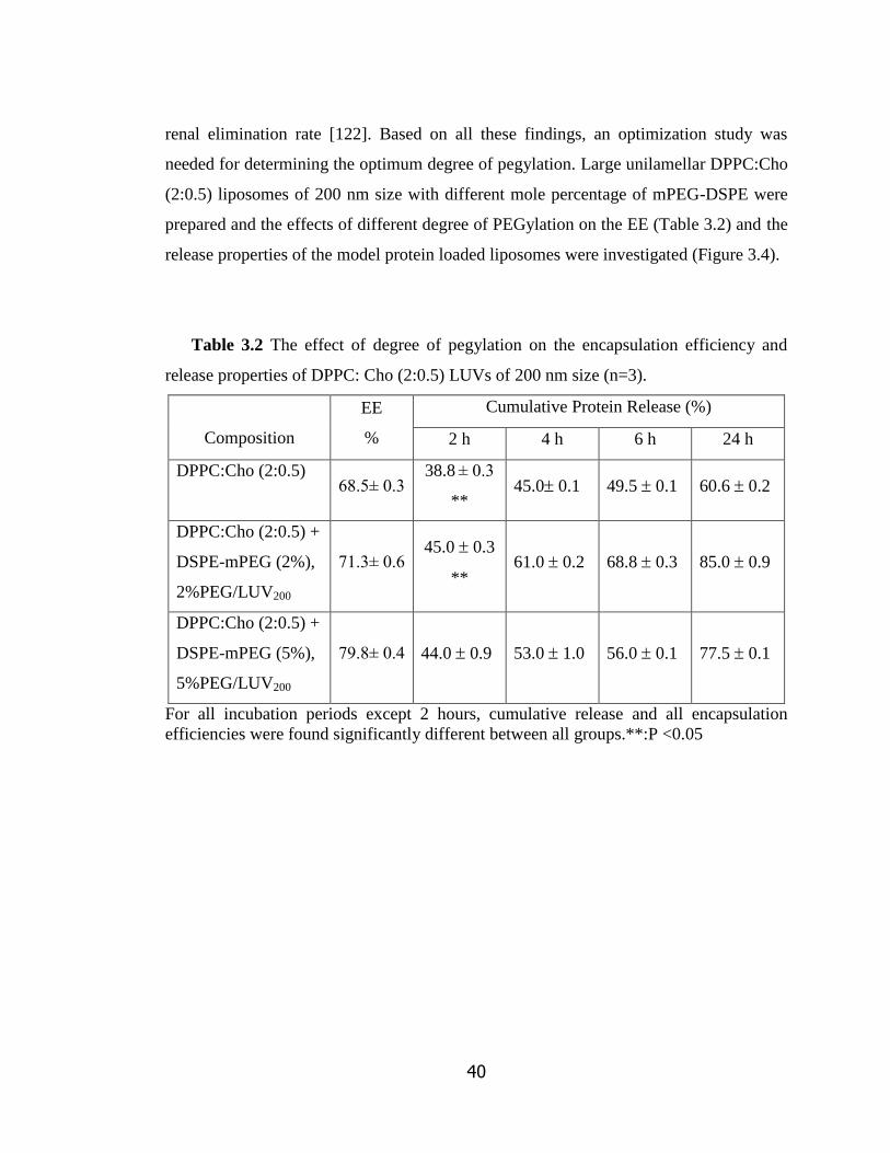
40
renal elimination rate [122]. Based on all these findings, an optimization study was
needed for determining the optimum degree of pegylation. Large unilamellar DPPC:Cho
(2:0.5) liposomes of 200 nm size with different mole percentage of mPEG-DSPE were
prepared and the effects of different degree of PEGylation on the EE (Table 3.2) and the
release properties of the model protein loaded liposomes were investigated (Figure 3.4).
Table 3.2 The effect of degree of pegylation on the encapsulation efficiency and
release properties of DPPC: Cho (2:0.5) LUVs of 200 nm size (n=3).
Composition
EE
%
Cumulative Protein Release (%)
2 h 4 h 6 h 24 h
DPPC:Cho (2:0.5)
68.5± 0.3
38.8 ± 0.3
** 45.0 0.1 49.5 0.1 60.6 0.2
DPPC:Cho (2:0.5) +
DSPE-mPEG (2%),
2%PEG/LUV200
71.3± 0.6 45.0 0.3
** 61.0 0.2 68.8 0.3 85.0 0.9
DPPC:Cho (2:0.5) +
DSPE-mPEG (5%),
5%PEG/LUV200
79.8± 0.4 44.0 0.9 53.0 1.0 56.0 0.1 77.5 0.1
For all incubation periods except 2 hours, cumulative release and all encapsulation
efficiencies were found significantly different between all groups.**:P <0.05
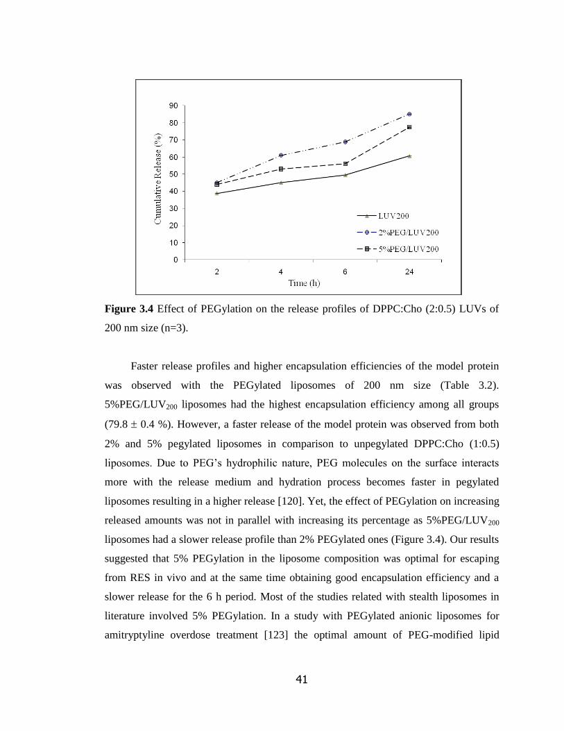
41
Figure 3.4 Effect of PEGylation on the release profiles of DPPC:Cho (2:0.5) LUVs of
200 nm size (n=3).
Faster release profiles and higher encapsulation efficiencies of the model protein
was observed with the PEGylated liposomes of 200 nm size (Table 3.2).
5%PEG/LUV200 liposomes had the highest encapsulation efficiency among all groups
(79.8 0.4 %). However, a faster release of the model protein was observed from both
2% and 5% pegylated liposomes in comparison to unpegylated DPPC:Cho (1:0.5)
liposomes. Due to PEG‘s hydrophilic nature, PEG molecules on the surface interacts
more with the release medium and hydration process becomes faster in pegylated
liposomes resulting in a higher release [120]. Yet, the effect of PEGylation on increasing
released amounts was not in parallel with increasing its percentage as 5%PEG/LUV200
liposomes had a slower release profile than 2% PEGylated ones (Figure 3.4). Our results
suggested that 5% PEGylation in the liposome composition was optimal for escaping
from RES in vivo and at the same time obtaining good encapsulation efficiency and a
slower release for the 6 h period. Most of the studies related with stealth liposomes in
literature involved 5% PEGylation. In a study with PEGylated anionic liposomes for
amitryptyline overdose treatment [123] the optimal amount of PEG-modified lipid

42
incorporated into liposomes was found to be 5% as in our study. Also, it was reported
that 5 mol% PEGylation of liposome was optimal for a nanocarrier, judged by the
reduction of RES uptake level and the maintenance of the stability of the liposomal
structure [124].
3.2 Preparation and Characterization of SDF-1 Loaded Pegylated
Liposomes
In the light of all optimization results, DPPC:Cho (2:05) liposomes with 5%
PEGylation and 200 nm average size were selected to be used for preparing alendronate
conjugated bone marrow targetted SDF-1 delivery system. MLVs were treated with 4
cycles of freeze-thawing before extrusion for a more homogenous distribution. SDF-1α,
was shown to be potent at a concentration of 30 ng/ml on hematopoietic progenitor cells
in various in-vitro studies [66, 89]. It was also indicated by other researchers that 10 - 25
ng/ml SDF-1α loaded PLGA microspheres (loading efficiency 75 ± 1%) were sufficient
to migrate the endothelial progenitor cells [109]. It was also reported that higher
chemotactic index values were reached for a higher dose of SDF-1α (300 ng/ml) on the
migration assays conducted with CD34+ mononuclear cells [87].
3.2.1 Size of SDF-1α Loaded and Pegylated Liposomes
A representative size distribution result of SDF-1α loaded, 5% PEGylated
liposomes (SDF1-5%PEG/LUV200) prepared using 200 nm filter was shown in Figure
3.5. Size distributions were unimodal and had an average diameter value of 208.6 nm.

43
Figure 3.5 Representative size distribution result for SDF1-5%PEG/LUV200 obtained
by DLS analysis.
3.2.2 Encapsulation Efficiency and Release Profiles of SDF-1α Loaded and
Pegylated Liposomes
ELISA was used to determine the encapsulation efficiency and release profile of
SDF1-5%PEG/LUV200. Encapsulation efficiency of these liposomes was found as 39.5
± 0.5%. In order to minimize the loss of biological activity of SDF-1 during liposome
preparation, another set of liposomes was prepared without any freeze-thaw cycles. This
modification significantly improved the encapsulation efficiency to 50.1 ± 0.6%, as
expected.
The encapsulation efficiencies of SDF1-5%PEG/LUV200 was signifiantly lower
than their -lactoglobulin loaded counterparts. This result could be due to different
methods used for protein amount determination. ELISA method relies on the specific
interaction between protein and antibody. Thus, only biologically active SDF-1α
molecules can be detected with ELISA. However, the colorimetric BCA assay does not
require maintenance of three dimensional protein structure since it relies on the complex

44
formation between peptide bonds of the protein and Cu+2
ions of BCA solution. There
are many studies related with the development of liposomal delivery systems for protein
drugs and growth factors. While encapsulating hydrophilic molecules, it is known that
with the higher lipid concentration and/or with the larger liposomes, higher
encapsulation efficiency results are obtained [118]. Our 200 nm sized liposomes had
significantly higher encapsulation efficiency than the other growth factor loaded
liposome studied (i.e., nerve growth factor -NGF-, hepatocyte growth factor -HGF-,
etc.), either due to difference in their sizes or structure. For a 100 nm sized PEGylated
liposomal delivery system for encapsulating NGF was shown to have 34% encapsulation
efficiency [125]. In another study, the encapsulation efficiency of 1,2-dioleoyl-sn-
glycero-3-phosphoethanolamine (DOPE) liposomes for hepatocyte growth factor was
reported as 32.38% and the size of these liposomes was given as 91.56 nm [126].
Similarly, a different study group was able to encapsulate only 35% of recombinant
epidermal growth factor into giant DPPC:Chol liposomes (700-2000 nm) [127].
The release profiles of SDF1-5%PEG/LUV200 liposomes are given in Figure 3.6.
In Figure 3.6a, it is clear that liposomes prepared with or without freeze-thaw cycles
showed similar cumulative release profiles. Initial (6h) release percentages were 18.4
0.6% and 16.6 1.0% of the total encapsulated amount, respectively which were
significantly lower than those observed for -lactoglobulin loaded liposomes (Table
3.2). This decrease could be due to the lowering of the initial total protein amount from
500 ug (for model protein) to 500 ng (for SDF-1α) during hydration step which resulted
with a higher concentration gradient causing an increase in the diffusion of the protein.
For the total release amount (Figure 3.6b), liposomes prepared without freeze-thaw
cycles released higher amount of SDF-1 at the end of each time point due to their
lesser activity loss during preparation. After 6 hours, 1 ml of liposome (w/o freeze-thaw)
suspension released 10.3 0.2 ng SDF-1 while freeze thawed ones released only 8.7 ±
0.1 ng indicating loss of bioactive structure due to protein denaturation with this process.

45
Figure 3.6 Release profiles as (a) cumulative % release and (b) total release amounts of
SDF1-5%PEG/LUV200 prepared with 4 freeze-thaw cycles and without any freeze-
thaw cycle (n=3).

46
3.3 Conjugation of Alendronate to DSPE-PEG(2000)
Alendronate was conjugated to DSPE-PEG(2000)-Carboxylic Acid using
carbodiimide chemistry. An amide bond was formed between carboxylic group of
DSPE-PEG and amino group of alendronate. After final dialysis of conjugated lipid
against water for 24 hours, 60.0 ± 4.5% of the total lipids were recovered.
3.3.1 FTIR Analysis
For verifying the alendronate conjugation to lipid, FTIR analysis was performed.
In Figure 3.7, spectrums of DSPE-PEG, alendronate, alendronate conjugated DSPE-
PEG, and alendronate- DSPE-PEG mixture without conjugation are shown. For FTIR
analyses, same amounts of lipids were used in the samples.
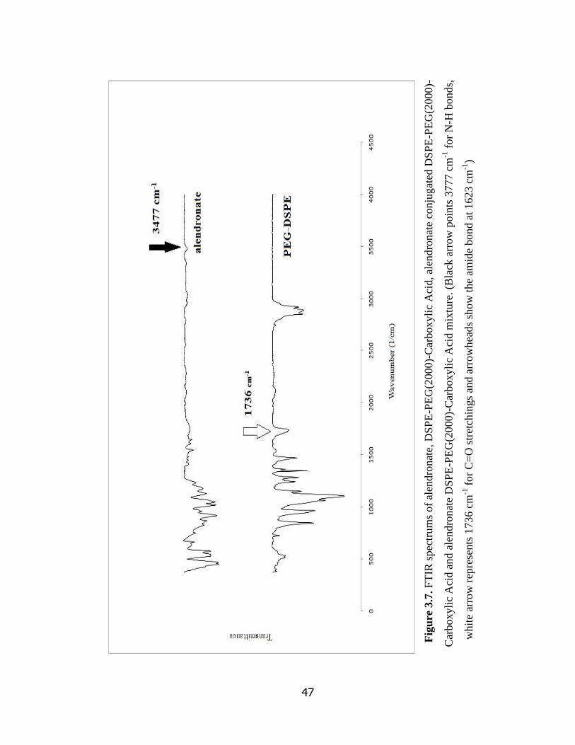
47
Fig
ure
3.7
. F
TIR
spec
tru
ms
of
alen
dro
nat
e, D
SP
E-P
EG
(2000)-
Car
boxyli
c A
cid, al
endro
nat
e co
nju
gat
ed D
SP
E-P
EG
(2000)-
Car
boxyli
c A
cid a
nd a
lendro
nat
e D
SP
E-P
EG
(20
00)-
Car
boxyli
c A
cid m
ixtu
re. (B
lack
arr
ow
poin
ts 3
777 c
m-1
for
N-H
bond
s,
whit
e ar
row
rep
rese
nts
1736 c
m-1
for
C=
O s
tret
chin
gs
and a
rrow
hea
ds
sho
w t
he
amid
e bond a
t 162
3 c
m-1
)

48
Fig
ure
3.7
. F
TIR
spec
tru
ms
of
alen
dro
nat
e, D
SP
E-P
EG
(2000)-
Car
boxyli
c A
cid, al
endro
nat
e co
nju
gat
ed D
SP
E-P
EG
(2000)-
Car
boxyli
c A
cid a
nd a
lendro
nat
e D
SP
E-P
EG
(20
00)-
Car
boxyli
c A
cid m
ixtu
re. (B
lack
arr
ow
poin
ts 3
777 c
m-1
for
N-H
bond
s,
whit
e ar
row
rep
rese
nts
1736 c
m-1
for
C=
O s
tret
chin
gs
and a
rrow
hea
ds
sho
w t
he
amid
e bo
nd a
t 162
3 c
m-1
) (c
onti
nued
)
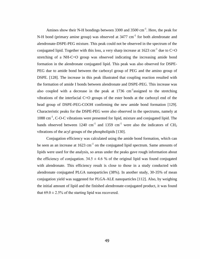
49
Amines show their N-H bondings between 3300 and 3500 cm-1
. Here, the peak for
N-H bond (primary amine group) was observed at 3477 cm-1
for both alendronate and
alendronate-DSPE-PEG mixture. This peak could not be observed in the spectrum of the
conjugated lipid. Together with this loss, a very sharp increase at 1623 cm-1
due to C=O
stretching of a NH-C=O group was observed indicating the increasing amide bond
formation in the alendronate conjugated lipid. This peak was also observed for DSPE-
PEG due to amide bond between the carboxyl group of PEG and the amino group of
DSPE. [128]. The increase in this peak illustrated that coupling reaction resulted with
the formation of amide I bonds between alendronate and DSPE-PEG. This increase was
also coupled with a decrease in the peak at 1736 cm-1
assigned to the stretching
vibrations of the interfacial C=O groups of the ester bonds at the carboxyl end of the
head group of DSPE-PEG-COOH confirming the new amide bond formation [129].
Characteristic peaks for the DSPE-PEG were also observed in the spectrums, namely at
1088 cm-1
, C-O-C vibrations were presented for lipid, mixture and conjugated lipid. The
bands observed between 1240 cm-1
and 1359 cm-1
were also the indicators of CH2
vibrations of the acyl groups of the phospholipids [130].
Conjugation efficiency was calculated using the amide bond formation, which can
be seen as an increase at 1623 cm-1
on the conjugated lipid spectrum. Same amounts of
lipids were used for the analysis, so areas under the peaks gave rough information about
the efficiency of conjugation. 34.5 ± 4.6 % of the original lipid was found conjugated
with alendronate. This efficiency result is close to those in a study conducted with
alendronate conjugated PLGA nanoparticles (38%). In another study, 30-35% of mean
conjugation yield was suggested for PLGA-ALE nanoparticles [112]. Also, by weighing
the initial amount of lipid and the finished alendronate-conjugated product, it was found
that 69.0 ± 2.5% of the starting lipid was recovered.

50
3.3.2 Encapsulation Efficiency and Release Profiles of SDF-1 Loaded and
Targetted (Alendronate Conjugated and Pegylated) Liposomes
After conjugation of alendronate into DSPE-PEG(2000)-Carboxylic Acid were
successfully performed, targetted liposomes with different mole% of alendronate
conjugated DSPE were prepared, DPPC: Cho (2:0.5) liposomes with either 2.5%
alendronate conjugated lipid + 2.5% DSPE-mPEG(2000) or with 5% alendronate
conjugated lipid were prepared. These targetted liposomes were named 2.5%ALE-
PEG/LUV200 and 5%ALE-PEG/LUV200, accordingly. Encapsulation efficiencies of
2.5%ALE-PEG/LUV200 and 5%ALE-PEG/LUV200 liposomes, which were found as 51.2
0.8% and 48.3 ± 0.3% for SDF-1α, respectively, had similar values with those of
untargetted liposomes (50.1 0.6%). Release profiles of targetted SDF-1 loaded
liposomes are presented in Figure 3.8.
At the end of 24 hours, the total amount of released SDF-1 was 24.4 0.7 ng
and 21.8 ± 0.4 ng for 2.5%ALE-PEG/LUV200 and 5%ALE-PEG/LUV200 liposomes
which were efficient values for stimulating stem cells to migrate to endosteal niche for
the homing process. It was observed that non-targeted (Figure 3.7) and targeted
(alendronate conjugated) (Figure 3.8) SDF-1α loaded liposomes had similiar cumulative
release profiles and total amount release versus time graphs with model protein.

51
a)
b)
Figure 3.8 Release profiles and total release amounts of SDF-1α loaded a) 2.5%ALE-
PEG/LUV200, b) 5%ALE-PEG/LUV200 liposomes (n=3).
0
5
10
15
20
25
30
35
40
0
10
20
30
40
50
60
70
80
3 6 24 48 72
Total R
elease Am
ount (n
g)C
um
ula
tive
Rel
ease
(%
)
Time (h)
cumulative
releasereleased
amount

52
3.3.3 Surface Charge and Size of Targetted Liposomes
Representative results for size distribution of targeted SDF-1α loaded PEGylated
liposomes are shown in Figures 3.9 and 3.10. Size distrubutions were unimodal with
average size of 219.6 nm and 224.3 nm for 2.5%ALE-PEG/LUV200 liposomes and
5%ALE-PEG/LUV200 liposomes, respectively. These values were very close to the value
measured for the non-targeted liposomes (Figure 3.5). Alendronate conjugation to the
pegylated lipid may be the reason for the increase observed in the diameter of the
liposomes [58].
Figure 3.9 Representative size distribution DLS result for 2.5%ALE-PEG/LUV200
liposomes .
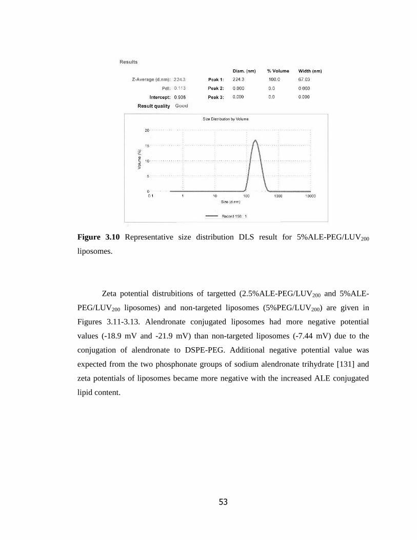
53
Figure 3.10 Representative size distribution DLS result for 5%ALE-PEG/LUV200
liposomes.
Zeta potential distrubitions of targetted (2.5%ALE-PEG/LUV200 and 5%ALE-
PEG/LUV200 liposomes) and non-targeted liposomes (5%PEG/LUV200) are given in
Figures 3.11-3.13. Alendronate conjugated liposomes had more negative potential
values (-18.9 mV and -21.9 mV) than non-targeted liposomes (-7.44 mV) due to the
conjugation of alendronate to DSPE-PEG. Additional negative potential value was
expected from the two phosphonate groups of sodium alendronate trihydrate [131] and
zeta potentials of liposomes became more negative with the increased ALE conjugated
lipid content.
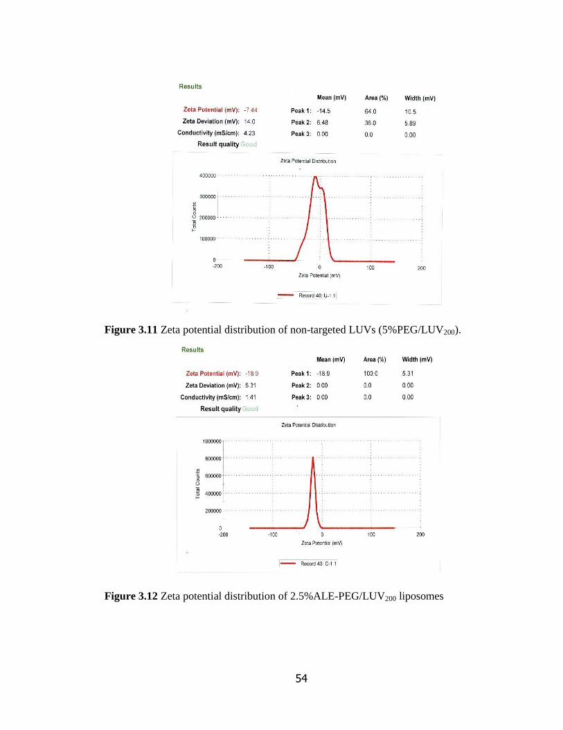
54
Figure 3.11 Zeta potential distribution of non-targeted LUVs (5%PEG/LUV200).
Figure 3.12 Zeta potential distribution of 2.5%ALE-PEG/LUV200 liposomes

55
Figure 3.13 Zeta potential distribution of 5%ALE-PEG/LUV200 liposomes .
3.3.4 Targetting Efficiency of the Alendronate Conjugated Liposomes
HA affinity of the targeted and non-targeted liposomes was compared to evaluate
targetting potency of the liposomes. At the end of 2 hours of incubation with HA
particles, HA affinities of non targeted (6.0 1.0 %) and targeted liposomes (10.9
0.2% for 2.5%ALE-PEG/LUV200 liposomes, 18.8 ± 0.9% for 5%ALE-PEG/LUV200
liposomes) were significantly different (Figure 3.14). After 24 hours, the difference
between targetted and nontargetted liposomes became more significant. Nontargeted
liposomes showed 30.0 0.3% affinity while alendronate conjugated-targeted liposomes
using either 2.5% or 5% alendronate conjugated DSPE-PEG in the composition had 61.9
0.8% and 79.9 ±0.7% affinity after 24 hours, respectively (Figure 3.14). By the end of
the 6 hours, 5%ALE-PEG/LUV200 showed 67.7 ± 0.6% affinity, which was higher than
the affinity, observed for 2.5%ALE-PEG/LUV200 after 24 hours (61.9 ± 0.8%). The HA
affinity observed for the targetted liposomes were significantly higher than the HA
affinity observed for alendronate conjugated PLGA-PEG copolymer nanospheres (50%)
in the study of Choi et al [112]. The nanospheres were prepared from alendronate

56
conjugated (80% in preparation mixture) and unconjugated PLGA-PEG (20% in
preparation mixture).
In order to verify the data observed by changes in turbidity with time; aliquots
were collected from the supernatants after centrifugation of the liposomes at the end of
each time point. These aliquots were dried under nitrogen and Stewart Assay was
performed to determine the lipid amount of each supernatant sample. Total lipid
concentration was 100 µM at the beginning of this experiment. HA affinities of targeted
and non-targeted liposomes calculated from phospholipid amounts are shown in Figure
3.15.
.
Figure 3.14 HA affinity (%) of targeted (2.5%ALE-PEG/LUV200 and 5%ALE-
PEG/LUV200) and non-targeted (5%PEG/LUV200) liposomes determined by turbidity
measurements (n=3). *P <0.05

57
Figure 3.15 HA affinity (%) of targeted (2.5%ALE-PEG/LUV200 and 5%ALE-
PEG/LUV200) and non-targeted (5%PEG/LUV200) liposomes calculated from the
phospholipid amounts (n=3). *P <0.05
Even though an increase in the HA affinity was observed for both types of
liposomes, significantly higher HA affinities of the targeted liposomes were evident as
shown in Figures 3.14 and 3.15. Especially, at the end of 24 hours, the percent affinity
was more than twice of the value calculated for the non-targeted liposomes. After 6
hours of incubation, HA affinity percentages of 2.5%ALE-PEG/LUV200 liposomes were
found 50.0 ± 0.2% versus 52.2 ± 0.6% for targeted liposomes and 24.0 ± 0.8% versus
25.3 ± 0.4% for non-targeted liposomes by the two different methods (Figures 3.14 and
3.15) as mentioned above. The results were consistent for these methods
Thus, it might be suggested that with the succesful conjugation of alendronate to
the phospholipids a liposomal delivery sytem with good bone targeting potential was
developed.
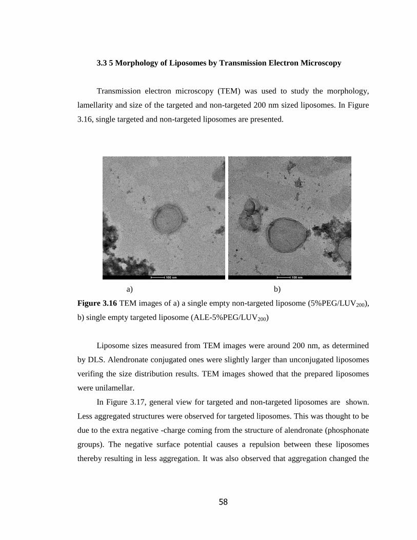
58
3.3 5 Morphology of Liposomes by Transmission Electron Microscopy
Transmission electron microscopy (TEM) was used to study the morphology,
lamellarity and size of the targeted and non-targeted 200 nm sized liposomes. In Figure
3.16, single targeted and non-targeted liposomes are presented.
a) b)
Figure 3.16 TEM images of a) a single empty non-targeted liposome (5%PEG/LUV200),
b) single empty targeted liposome (ALE-5%PEG/LUV200)
Liposome sizes measured from TEM images were around 200 nm, as determined
by DLS. Alendronate conjugated ones were slightly larger than unconjugated liposomes
verifing the size distribution results. TEM images showed that the prepared liposomes
were unilamellar.
In Figure 3.17, general view for targeted and non-targeted liposomes are shown.
Less aggregated structures were observed for targeted liposomes. This was thought to be
due to the extra negative -charge coming from the structure of alendronate (phosphonate
groups). The negative surface potential causes a repulsion between these liposomes
thereby resulting in less aggregation. It was also observed that aggregation changed the
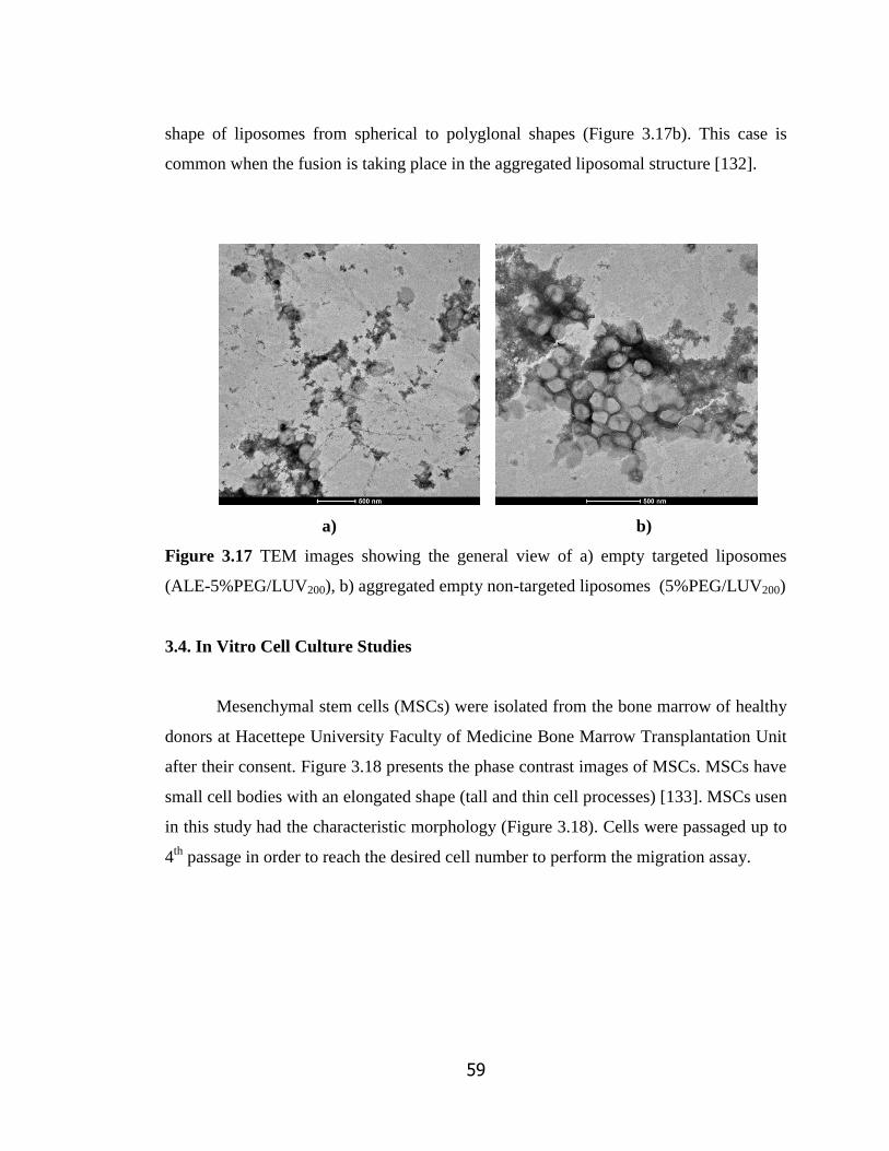
59
shape of liposomes from spherical to polyglonal shapes (Figure 3.17b). This case is
common when the fusion is taking place in the aggregated liposomal structure [132].
a) b)
Figure 3.17 TEM images showing the general view of a) empty targeted liposomes
(ALE-5%PEG/LUV200), b) aggregated empty non-targeted liposomes (5%PEG/LUV200)
3.4. In Vitro Cell Culture Studies
Mesenchymal stem cells (MSCs) were isolated from the bone marrow of healthy
donors at Hacettepe University Faculty of Medicine Bone Marrow Transplantation Unit
after their consent. Figure 3.18 presents the phase contrast images of MSCs. MSCs have
small cell bodies with an elongated shape (tall and thin cell processes) [133]. MSCs usen
in this study had the characteristic morphology (Figure 3.18). Cells were passaged up to
4th
passage in order to reach the desired cell number to perform the migration assay.
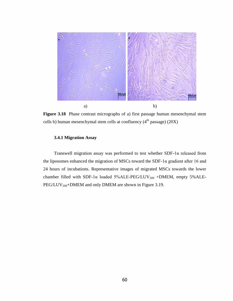
60
a) b)
Figure 3.18 Phase contrast micrographs of a) first passage human mesenchymal stem
cells b) human mesenchymal stem cells at confluency (4th
passage) (20X)
3.4.1 Migration Assay
Transwell migration assay was performed to test whether SDF-1α released from
the liposomes enhanced the migration of MSCs toward the SDF-1α gradient after 16 and
24 hours of incubations. Representative images of migrated MSCs towards the lower
chamber filled with SDF-1α loaded 5%ALE-PEG/LUV200 +DMEM, empty 5%ALE-
PEG/LUV200+DMEM and only DMEM are shown in Figure 3.19.

61
a)
b)
Figure 3.19 Representative images of transmigrated MSCs in response to a) SDF-1α
released from 5%ALE-PEG/LUV200 liposomes b) DMEM-low glucose c) empty
liposomes (5%ALE-PEG/LUV200) after 16 hours (Star marks show transmigrated
MSCs)
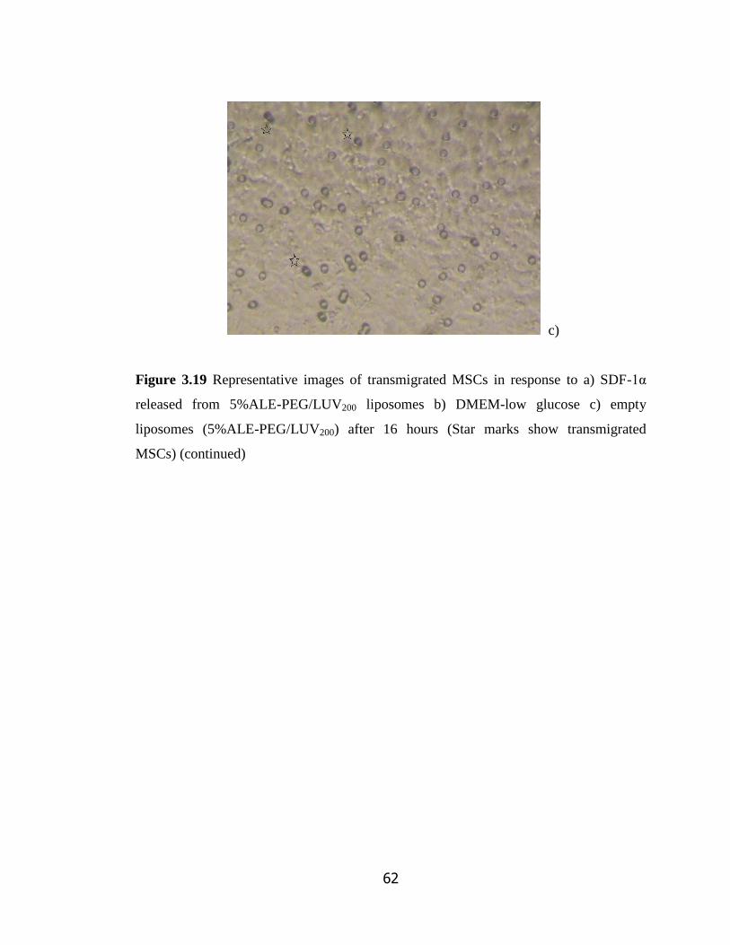
62
c)
Figure 3.19 Representative images of transmigrated MSCs in response to a) SDF-1α
released from 5%ALE-PEG/LUV200 liposomes b) DMEM-low glucose c) empty
liposomes (5%ALE-PEG/LUV200) after 16 hours (Star marks show transmigrated
MSCs) (continued)
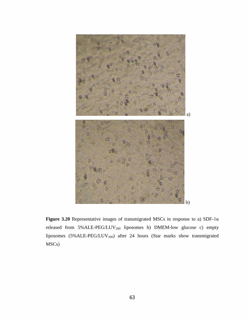
63
a)
b)
Figure 3.20 Representative images of transmigrated MSCs in response to a) SDF-1α
released from 5%ALE-PEG/LUV200 liposomes b) DMEM-low glucose c) empty
liposomes (5%ALE-PEG/LUV200) after 24 hours (Star marks show transmigrated
MSCs)

64
c)
Figure 3.20 (cont) Representative images of transmigrated MSCs in response to a) SDF-
1α released from 5%ALE-PEG/LUV200 liposomes b) DMEM-low glucose c) empty
liposomes (5%ALE-PEG/LUV200) after 24 hours (Star marks show transmigrated
MSCs) (continued)
It can be observed from Figures 3.19 and 3.20 that the number of cells migrated
toward SDF-1α released from liposomes was remarkably higher than those in other
wells. The average number of transmigrated MSCs toward the SDF-1α releasing
liposomes was more than twice higher than the number of migrating MSCs toward tboth
empty liposomes and medium only. The transmigration assay results suggested that
liposomes released biologically active SDF-1α and this resulted with enhancement of the
ability of MSCs to transmigrate across 8 m pores.

65
Figure 3.21 Average number of transmigrated MSCs. Results are mean values of five
different fields from four independent experiments. *P <0.05
Cumulative SDF-1α (%) release from liposomes was 44.9 ± 1.4% and 50.3 ±
0.8% at the end of 16 and 24 hours, respectively. 24 hour cumulative percent release was
found higher than the findings for the release in PBS medium (Figure 3.8b). In PBS,
liposomes released 44.1 ± 0.5 % of the encapsulated while in the presence of serum
(almost 0.1% FBS for the total volume of upper and lower chambers) release increased
by a 6%. It was also reported that serum addition mimicks the in-vivo conditions for in
vitro release studies and in result with fastening the release from liposomal delivery
systems [135,134].
66
CHAPTER 4
CONCLUSION
ALE-5%PEG/LUV200 was found as a promising bone-marrow targeted drug
delivery system. The use of alendronate conjugated DSPE-PEG2000 in the liposomal
formulation resulted with high affinity towards HA (i.e., 67.7% after 6h and 77.9% after
24h). In vitro release and cell migration studies showed that the amount of SDF-1α
released from the liposomes was sufficient to increase the transmigration of human bone
marrow mesenchymal stem cells. SDF-1 loaded and alendronate conjugated liposomal
delivery system has been reported for the first time in literature. Our liposomal system
can be considered as an effective vehicle to improve homing efficiency after
transplantation by providing local SDF1- release in the endosteal niche of the bone
marrow. This gives the opportunity of using limited amount of transplanted marrow in a
better efficiency and young donors would become more available for BMT. Also, the
use of cord blood, where the stem cell amount is limited, for BMT can be applicable.
However, further studies including migration assays with hematopoietic stem cells and
in-vivo distribution analysis for the liposomal system are suggested and are being
planned to be tested the efficacy of the system in detail.
This alendronate conjugated liposomal delivery can also be considered as a bone
targetted delivery system for providing efficient treatment for bone-related diseases such
as osteoporosis or for treating large and difficult fractures by recruiting host
mesenchymal stem cells towards defect site SDF-1α encapsulated liposomes can serve
as an efficient tool for curing osteogenesis imperfecta by attracting MSCs to the

67
endosteum region in order to heal the bone fractures. Also, it can be used as an
additional treatment for diseases like osteopetrosis which uses BMT for the cure.
68
REFERENCES
[1] Shailesh S, Neelam S, Sandeep K, Gupta GD. Liposomes: A review. Journal of
Pharmacy Research. 2009; 2(7): 1163-1167
[2] Riaz M. Liposome Preparation Methods. Pakistan Journal of Pharmaceutical
Sciences. 1996; 19(1): 65-77
[3] Gomez-Hens, A., Fernandez-Romero, J.M. Analytical methods for the control of
liposomal delivery systems. Trends in Analytical Chemistry. 2006; 25: 167–178
[4] Jesorka A, Orwar O. Liposomes: Technologies and analytical applications. Annual
Reviews of Analytical Chemistry. 2008; 1: 801-832
[5] Betz G, Nowbakth P, Imboden R, Imanidis G. Heparin penetration into and
permeation through human skin from aqueous and liposomal formulations in vitro.
International Journal of Pharmaceutics. 2001; 228: 147-159
[6] Cevc G, Blume G. New, highly efficient formulation of diclofenac for the topical,
transdermal administration in ultradeformable drug carriers, Transfersomes. Biochimica
et Biophysica Acta - Biomembranes. 2001; 1514: 191-205
[7] Erdogan S, Medarova ZO, Roby A, Torchilin VP. Enhanced tumor MR imaging
with gadolinium-loaded polychelating polymer-containing tumor-targeted
liposomes. Journal of Magnetic Resonance Imaging. 2008; 27(3): 574-580
[8] Torchilin VP. Surface-modified liposomes in gamma- and MR-imaging.
Advanced Drug Delivery Reviews. 1997; 24(2-3): 301-313
[9] Lukyanov AN, Elbayoumi T, Chakilam AR, Torchilin VP. Tumor-targeted
liposomes: doxorubicin-loaded long-circulating liposomes modified with anti-cancer
antibody. Journal of Cont. Release. 2004; 100(1): 135-144
[10] Lee MJ, Cho SS, You JR, Choi JS, Park JW, Suh YW, Kim JA, Park JS.
Intraperitoneal gene delivery mediated by a novel cationic liposome in a peritoneal
disseminated ovarian cancer model. Gene Theraphy. 2002; 9: 859-866
69
[11] Torchilin VP, Levchenko TS, Rammohan R, Volodina N, Papahadjopoulos-
Sternberg B, GGM D'Souza. Cell transfection in vitro and in vivo with nontoxic TAT
peptide-liposome–DNA complexes. Proceedings of National Academy of Sciences of
USA. 2003; 100(4): 1972-1977
[12] Barani H, Montazer M. A review on applications of liposomes in textile processing.
Journal of Liposome Research. 2008; 18(3): 249-262
[13] Meure LA, Knott R, Foster NR, Dehghani F. The depressurization of an expanded
solution into aqueous media for the bulk production of liposomes. The Acs Journal of
Surfaces And Colloids. 2009; 25(1): 326-337
[14] Strieth S, Eichhorn ME, Werner A, Sauer B, Berghaus A, Dellian M. Paclitaxel
encapsulated in cationic liposomes increases tumor microvessel leakiness and improves
therapeutic efficacy in combination with cisplatin. Clinical Cancer Research. 2008; 14:
4603-4611
[15] O‘Brien MER, Wigler N, Inbar M, Rosso R, Santoro A, Tomczak P, Orlandi F,
Mellars L, Tendler C. Reduced cardiotoxicity and comparable efficacy in a phase III
trial of pegylated liposomal doxorubicin HCl (CAELYX™/Doxil®) versus conventional
doxorubicin for first-line treatment of metastatic breast cancer. Annals of Oncology.
2004; 15(3): 440-449
[16] Zou Y, Ling YH, Van NT, Perez-Soler R. Antitumor activity of free and liposome-
entrapped annamycin, a lipophilic anthracycline antibiotic with non-cross-resistance
properties. Cancer Research. 1994; 54: 1479-1485
[17]Fleisher D, Oh CK, Hu Z, Ramachandran C, Weine N. Topical delivery of growth
hormone releasing peptide using liposomal systems: An in vitro study using hairless
mouse skin. Life Sciences. 1995; 57(13): 1293-1297
[18] Fukunaga M, Miller MM, Deftos LJ. Liposome-entrapped calcitonin and
parathyroid hormone are orally effective in rats. Hormone and Metabolism Research.
1991; 23(4): 166-167
[19] Meers P. Enzyme activated targeting of liposomes. Advanced Drug Delivery
Reviews. 2001; 53(3): 265-272
[20] Fonseca MJ, Jagtenberg JC, Haisma HJ, Storm G. Liposome-Mediated targeting of
enzymes to cancer cells for site-specific activation of prodrugs: comparison with the
corresponding antibody-enzyme conjugate. Pharmaceutical Research. 2002; 20(3): 423-
428

70
[21] Harrington KJ, Lewanski CR, Whittaker J, Vile RG, Peters AM, Stewart JSW.
Phase I-II study of pegylated liposomal cisplatin (SPI-077 TM
) in patients with
inoperable head and neck cancer. Annals of Oncology. 2000; 12(4): 493-496
[22] Tollemar J, Klingspor L, Ringden O. Liposomal amphotericin B (AmBisome)
for fungal infections in immunocompromised adults and children. Clinical
Microbiology and Infection. 2001; 7: 68-79
[23] Singhal S, Ellis RW, Jones SG, Miller SJ, Fisher NC, Mutimer DJ. Targeted
prophylaxis with amphotericin B lipid complex in liver transplantation. Liver
Transplantation. 2000; 6(5): 588-595
[24] Lorusso D, Naldini
A, Testa
A, D‘Agostino
G, Scambia
G, Ferrandina G. Phase II
study of pegylated liposomal doxorubicin in heavily pretreated epithelial ovarian cancer
patients. Oncology. 2004; 67: 243-249
[25] Martin-Carbonero L, Valencia R, Sirera G, Santos I, Alegre M, Pedreira J, Ocampo
A, Alsina M, Santos S, Podzamczer D, Gonzalez-Lahoz J. Long-term prognosis of HIV-
infected patients with kaposi sarcoma treated with pegylated liposomal doxorubicin.
Clinical Infectious Diseases. 2008; 47(3): 410-417
[26] Keller AM, Mennel RG, Georgoulias VA, Nabholtz JM, Erazo A, Lluch A,
Vogel CL, Kaufmann M, von Minckwitz G, Henderson IC, Mellars L, Alland L,
Tendler C. Randomized phase III trial of pegylated liposomal doxorubicin versus
vinorelbine or mitomycin c plus vinblastine in women with taxane-refractory advanced
breast cancer. Journal of Clinical Oncology. 2004; 22(19): 3893-3901
[27] Ralf-Dietera H; Gnad-Vogt SU, Beyer U, Hochhaus .A Liposomal encapsulated
anti-cancer drugs. Anti-Cancer Drugs. 2005; 16(7): 691-707
[28] Siegal T, Horowitz A, Gabizon A. Doxorubicin encapsulated in sterically stabilized
liposomes for the treatment of a brain tumor model: biodistribution and therapeutic
efficacy. Journal of Neurosurgery. 1995; 83(6)
[29] Williams SS, Alosco TR, Lasic DD, Martin FJ, Bankert RB. Arrest of human lung
tumor xenograft growth in severe combined immunodeficient mice using Doxorubicin
encapsulated in sterically stabilized liposomes. Cancer Research. 1993; 53: 3964-3974
[30] Woking PK, Newman MS, Huang SK. Pharmacokinetics, biodistribution and
therapeutic efficacy of doxorubicin encapsulated in STEALTH liposomes (Doxil).
Liposome Research. 1994; 4:667-687

71
[31] Stathopoulos GP, Boulikas T, Rigatos S, Eleftheria D, Vasiliki V, Stathopoulos JG.
Pharmacokinetics and adverse reactions of a new liposomal cisplatin (Lipoplatin): Phase
I study. Oncology Reports. 2005; 13(4): 589-595
[32] Chrai S, Murari R, Ahmad I. Liposomes (a review) Part Two: Drug Delivery
Systems. Pharmaceutical Technology. 2003; 44: 178-182
[33] Shehata T, Ogawara K, Higaki K, Kimura T. Prolongation of residence time of
liposome by surface modification with mixture of hydrophilic polymers. International
Journal of Pharmaceutics. 2008; 359: 272-279
[34] Wang JC, Goh BC, Lu WL, Qiang Zhang Q, Chang A, Liu XY, Tan TMC, Lee
HS. In-vitro cytotoxicity of stealth liposomes co-encapsulating doxorubicin and
verapamil on doxorubicin resistant tumor cells. Biological and Pharmacology Bulletin.
2005; 28(5): 822-829
[35] Small, DM. The Physical Chemistry of Lipids from Alkanes to Phospholipids,
Handbook of Lipid Research Series,1984; Vol. 4, ed. D. Hanahan, Plenum Press, N.Y.
pp. 1-672
[36] Sulkowski WW, Pentak D, Nowak K, Sulkowska A. The influence of temperature,
cholesterol content and pH on liposome stability. Journal of Molecular Structure. 2005;
744-747: 737-747
[37] Cryan SA, Devocelle M, Moran P, Hickey AJ, Kelly JG. Increased intracellular
targeting to airway cells using octa-arginine coated liposomes - in vitro assessment of
their suitability for inhalation. Molecular Pharmaceutics. 2005; 3(2): 104-112
[38] Bot AI, Tarara TE, Smith DJ, Bot SR, Woods CM, Weers JG. Novel lipid based
hollow porous microparticles as a platform for immunoglobulin delivery to respiratory
tract. Pharmaceutical Research. 2000; 17: 275-283
[39] Schwendener RA. Liposomes in Biology and Medicine. Advances in Experimental
Medicine and Biology. 2007; 620: 117-128
[40] Adrian JE, Poelstrab K, Scherphof GL, Meijer DKF, Reker-Smit C, Morselt HWM,
Kamps JAAM. Interaction of targeted liposomes with primary cultures hepatic state
cells: Involvement of multiple receptor systems. Journal of Hepatology. 2006; 44(3):
560-567

72
[41] Senior J, Delgado C, Fisher D, Gregoryadis G. Influence of surface hydrophilicity
of liposomes on their interaction with plasma protein and clearance from the circulation:
Studies with poly(ethylene glycol)-coated vesicles. Biochimica et Biophysica Acta-
Biomembranes. 1991; 1062(1): 77-82
[42] Socaciu C, Jessel R, Diehl RA. Competitive carotenoid and cholesterol
incorporation into liposomes: effects on membrane phase transition, fluidity, polarity
and anisotropy. Chemistry and Physics of Lipids. 2000; 106(1): 79-88
[43] Damen J. Transfer and exchange of phospholipid between small unilamellar
liposomes and rat plasma high-density lipoproteins: dependence on cholesterol and
phospholipid composition. Biochimica et Biophyica Acta, 2005; 665: 538–45.
[44] Loughrey HC, Bally MB, Reinish LW, Cullis PR.The binding of
phosphatidylglycerol liposomes to rat platelets is mediated by complement. Journal of
Thrombosis and Hematopoiesis. 1990; 64(1): 172-176
[45] Moghimi SM, Szebeni J. Stealth liposomes and long circulating nanoparticles:
critical issues in pharmacokinetics, opsonization and protein-binding properties.
Progressive Lipid Research, 2003; 42: 463–78.
[46] Chonn A, Cullis PR, Devine DV. The role of surface charge in the activation of the
classical and alternative pathways of complement by liposomes. Journal of Immunology.
1991; 146(12): 4234-4241
[47] Sou K, Tsuchida E. Electrostatic interactions and complement activation on the
surface of phospholipid vesicle containing acidic lipids: Effect of structure of acidic
groups. Biochimica et Biophysica Acta. 2008; 1778: 1035-1041
[48] Gao X, Huang L. Cationic liposome-mediated gene transfer. Gene Theraphy. 1995;
2(10): 710-722
[49] Banerjee R, Mahidhar YV, Chaudhuri A. Design, synthesis and transfection biology
of novel cationic glycolipids for use in liposomal gene delivery. Journal of Medicinal
Chemistry. 2001; 44(24): 4176–4185
[50] Lv H, Zhang S, Wang B, Cui S, Yan J. Toxicity of cationic lipids and cationic
polymers in gene delivery. Journal of Controlled Release. 2006; 114(1): 1100-1109
[51] Bai S, Gupta V, Ahsan F. Cationic liposomes as carriers for aerosolized
formulations of an anionic drug: Safety and efficacy study. European Journal of
Pharmaceutical Sciences. 2009; 38(2): 2165-2171
73
[52] Ishida T, Kirchmeiner MJ, Moase EH, Zalipsky S, Allen TM. Targeted delivery and
triggered release of liposomal doxorubicin enhances cytotoxicity against human
Blymphoma cells. Biochimica et Biophysica Acta (BBA) - Biomembranes. 2001;
1515(2): 144-158
[53] Sou K, Goins B, Takeoka S, Tsuchida E, Phillips WT. Selective uptake of surface-
modified phospholipid vesicles by bone marrow macrophages in vivo. Biomaterials.
2007; 28(16): 2655-2666
[54] Bradley A J., Devine DV, Ansell SM, Janzen J, Brooks DE. Inhibition of liposome
induced complement activation by incorporated poly(ethylene glycol) lipids. Archieves
of Biochemistry and Biophysics. 1998; 357: 185-194
[55] De Villiers MM, Pornanong A, Kwon GS. Nanotechnology in Drug Delivery
Series: Biotechnology: Pharmaceutical Aspects. 2009, XIV, 662 p. 180 illus., 29 in
color.
[56] Marques C, Carvalheiro M, Pereira MA, Jorge J, Cruz MEM, Santos-Gomes GM.
Efficacy of the liposome trifluralin in the treatment of experimental canine
leishmaniosis. The Veterinary Journal 2008; 178(1): 133-137
[57] Shetata T, Ogawara K, Higaki K, Kimura K. Prolongation of residence time of
liposome by surface-modification with mixture of hydrophilic polymers. International
Journal of Pharmaceutics. 2008; 359(1-2): 272-279
[58] Garbuzenko O, Barenholz Y, Priev A. Effect of grafted PEG on liposome size and
compressibilty and packing of lipid bilayer. Chemistry and Physics of Lipids. 2005; 135:
117-129
[59] Celano M, Calvagno MG, Bulotta S, Fresta M, Arturi F, Rotiroti D, Filetti S, Russo
D. Cytotoxic effects of Gemcitabine-loaded liposomes in human anaplastic thyroid
carcinoma cells. BMC Cancer. 2004; 4: 1471-1479
[60] Dadashzadeh S, Mirahmadi N, Babaei MH, Vali AM. Periotenal retention of
liposomes : Effects of lipid composition, PEG coating and liposome charge. Journal of
Controlled Release. 2010; 33(16): 99-106
[61] Takeuchi H, Kojima H, Yamamoto H, Kawashima Y. Evaluation of circulation
profiles of liposomes coated with hydrophilic polymers having different molecular
weights in rats. Journal of Controlled Release. 2001; 75(1-2): 83-91

74
[62] Torchilin VP, Levchenko TS, Whiteman KR, Yaroslavov AA, Tsatsakis AM, Rizos
AM, Michailova EV, Shtilman MI. Amphiphilic poly-N-vinylpyrrolidones: : synthesis,
properties and liposome surface modification. Biomaterials. 2001; 22(22): 3035-3044
[63] Immordino ML, Dosio F, Cattel L. Stealth liposomes: review of the basic science,
rationale, and clinical applications, existing and potential. International Journal of
Nanomedicine. 2006; 1(3): 297–315
[64]Park JW, Benz CB, Martin FJ. Future directions of liposome- and immunoliposome-
based cancer therapeutics. Seminars in Oncology. 2004; 31(13): 196-205
[65]Maruyama K, Takizawa T, Yuda T, Kennel SJ, Huang L, Iwatsuru M. Targetability
of novel immunoliposomes modified with amphipathic poly(ethylene glycol)s
conjugated at their distal terminals to monoclonal antibodies. Biochimica et Biophysica
Acta (BBA) - Biomembranes. 1995; 1234(1): 74-80
[66] Yatuv R, Dayan I, Robinson M, Baru M. Binding of proteins to PEGylated
liposomes and improvement of G-CSF efficacy in mobilization of HSCs. Journal of
Controlled Release. 2009; 135: 44-50
[67] Iyer AK, Khaled G, Fang J, Maeda H. Exploiting the enhanced permeability and
retention effect for tumor targeting. Drug Discovery Today. 2006; 11(17-18): 812-818
[68] Wang X, Deng L, Chen X, Pei H, Cai L, Wei Y, Chen L. Truncated bFGF-mediated
cationic liposomal paclitaxel for tumor-targeted drug delivery: Improved
pharmacokinetics and biodistribution in tumor-bearing mice. Journal of Pharmaceutical
Sciences. 2011; 100(3): 1196-1205
[69] Mamot C, Drummond DC, Hong K, Kirpotin DB, Park JW. Liposome-based
approaches to overcome anticancer drug resistance. Drug Resistance Updates. 2003;
6(5): 271-279
[70] Wu HC, Chang DK. Peptide-mediated liposomal drug delivery system targeting
tumor blood vessels in anticancer therapy. Journal of Oncology. 2010; 10: 1155-1165
[71] Zhao H, Bao X, Wang R, Li G, Gao J, Ma S, Wei J, Feng M, Zhao Y, Ma W, Yang
Y, Li Y, Kong Y. Postacute ischemia vascular endothelial growth factor transfer by
ransferrin-targeted liposomes attenuates ischemic brain injury after experimental stroke
in rats. Human Gene Therapy. 2011; 22(2): 207-215

75
[72] Konigsberg PJ, Debrick JE, Pawlowski TJ, Staerz UD. Liposome encapsulated
aurothiomalate reduces collagen induced arthritis in DBA/1J mice. Biochimica et
Biophysica Acta (BBA) - Biomembranes. 1999; 1421(1): 149-162
[73] Panoskaltsis N, Mantalaris A, Wu JHD. Engineering a mimicry of bone marrow
tissue ex-vivo. Journal of Bioscience and Bioengineering. 2005; 100: 28-35
[74] Tuch BE. Stem cells—a clinical update. Australian Family Physician. 2006; 35(9):
719–721
[75] Aiuti A, Webb IJ, Bleul C, Springer T, Gutierrez Ramos JC. The chemokine SDF-1
is a chemoattractant for human CD34+ HPC and provides a new mechanism to explain
the mobilization of CD34+ progenitors to peripheral blood. Journal of Experimental
Medicine. 1997; 185(1): 111-120
[76] Sacchetti B, Funari A, Piersanti S, Saggio I, Ferrari S, Robey PG, Riminucci M,
Bianco P. Self-renewing osteoprogenitors in bone marrow sinusoids can organize a
hematopoietic microenvironement. Cell. 2007; 131: 324-336
[77] Avecilla ST, Hattori K, Heissig B, Liao F, Jin DK, Dias S, Zhang F, Hackett NR,
Witte L, Hicklin DJ, Eaton D, Lyden D, Rafii S. Chemokine-mediated interaction of
hematopoietic progenitors with the bone marrow vascular niche is required for
thrombopoiesis. Nature Medicine. 2004; 10: 64-73
[78] Ferrer SM, Michurina TV, Mazloom AR, Lira SA, Scadden DT, Enikolopov GN,
Frenette PS. Mesenchymal and haematopoietic stem cells form a unique bone marrow
niche. Nature. 2010; 466: 829-834
[79] Parmar K, Mauch P, Sackstein R, Down JD. Distribution of HSCs in the bone
marrow according to regional hypoxia. Proceedings of the National Academy of
Science. 2007; 104(13): 5431-5436
[80] Adams GB, Alley IR, Olson DP, Poznansky MC, Kos CH, Pollak MR, Brown EM,
Scadden DT. Stem cell engraftment at the endosteal niche is specified by the calcium-
sensing receptor. Nature. 2006; 439: 599-603
[81] Wilson A, Trumpp A. Bone marrow HSC niches. Nature. 2006; 6: 93-106
[82] Yin T, Li L. Stem cell niches in bone. Journal of Clinical Investigation. 2006;
116(5): 1195-1201
76
[83] Arai F, Suda T. Maintenance of quiescent HSCs in the osteoblastic niche. Annals of
the New York Academy of Sciences. 2007; 1106: 41-53
[84] Lapidot T, Dar A, Kollet O. How do stem cells find their way home? Blood. 2005;
106: 1901-1910
[85] Spradling A, Barbosa DD, Kai T. Stem cells find their niche. Nature. 2001; 414:
98-104
[86] De La Luz Sierra M, Yang F, Narazaki M, Salvucci O, Davis D, Yarchoan R,
Zhang HH, Fales H, Tosato G. Differential processing of stromal-derived factor-1α and
stromal-derived factor-1β explains functional diversity. Blood. 2004; 103(7): 2452-2459
[87] Wysoczynski M, Reca R, Ratajczak J, Shirvaikar N, Honczarenko M, Mills M.
Incorporation of CXCR4 into membrane lipid rafts primes homing related responses of
HS/PCs to an SDF-1 gradient. Blood. 2005; 105: 40-48
[88] Ratajczak MZ, Reca R, Yan J, Ratajczak J. Modulation of the SDF-1-CXCR4 axis
by the third complement component (C3) – Implications for trafficking of CXCR4+ stem
cells. International Society of Experimental Hematology. 2006; 34: 986-995
[89] Voermans C, Anthony EC, Mul E, Hordijk P. SDF-1 induced actin polymerization
and migration in human HSCs. International Society of Experimental Hematology.
2001; 29: 1456-1464
[90] Wynn RF, Hart CA, O‘Neill L, Evans CA, Fairbairn LJ, Bellantuono I. A small
proportion of MSCs strongly expresses functionally active CXCR4 receptor capable of
promoting migration to bone marrow. Blood. 2009; 104:2643-2645
[91] Mori T, Doi R, Koizumi M, Toyoda E, Ito D, Kami K, Fujimoto K, Tamamura H,
Hiramatsu K, Fujii N, Imamura M. CXCR4 antagonist inhibits SDF-1 induced migration
and invasion of human pancreatic cancer. Molecular Cancer Theraphy. 2004; 3(1): 29-
37
[92] Schober A, Karshovska E, Zernecke A, Weber C. SDF-1 mediated tissue repair by
stem cells: a promising tool in cardiovascular medicine? Trends in Cardiovascular
Medicine. 2006; 16: 103-108
[93] Gratwohl A, Baldomero H, Aljurf M. Hematopoietic stem cell transplantation: a
global perspective. Journal of the American Medical Association. 2010; 30 (16): 1617-
24.
77
[94] Nivison-Smith I, Bradstock KF, Dodds AJ, Hawkins PA, Szer J. Haemopoietic
stem cell transplantation in Australia and New Zealand, 1992-2001: progress report from
the Australasian Bone Marrow Transplant Recipient Registry. International Medicine
Journal. 2005; 35 (1): 18–27
[95] Bishop MR, Pavletic SZ. Hematopoietic stem cell transplantation. In: Clinical
Oncology. Editors Abeloff MD, Armitage JO, Niederhuber JE, Kastan MB, McKena
WG, 4th
ed. Philadelphia, Pa: Elsevier Churchill Livingstone; 2008
[96] Barrett A. Total body irradiation before bone marrow transplantation: A review.
Clinical Radiology. 1982; 33(2): 131-135
[97] Thomas ED. Bone marrow transplantation: a review. Seminars in
Hematology. 1999; 36(4): 95-103
[98] Horwitz ME, Sullivan KM. Chronic GVHD. Blood Reviews. 2006; 20: 15-27
[99] Doney K, Loken M, Bryant E, Smith A, Appelbaum F. Lack of utility of chimerism
studies obtained 2-3 months after myeloablative hematopoietic cell transplantation for
ALL. Bone Marrow Transplant 2008; 42: 271–274
[100] Baron F, Sandmaier BM. Chimerism and outcomes after allogeneic hematopoietic
cell transplantation following nonmyeloablative conditioning. Leukemia. 2006; 20:
1690–1700
[101] Fanord F, Fairbarn K, Kim H, Garces A, Gupta AK. Bisphophonate-modified gold
nanoparticles: a useful vehicle to study the treatment of osteonecrosis of the femoral
head. Nanotechnology. 2010; 22: 1-12
[102] Wang D, Miller SC, Kopeckova P, Kopecek J. Bone targeting macromolecular
therapeutics. Advanced Drug Delivery Reviews. 2005; 57: 1049-1076
[103] Coxon FP, Thompson K, Rogers MJ. Recent advances in understanding the
mechanism of action of bisphosphonates. Current Opinion in Pharmacology. 2006; 6:
307-312
[104] Uludað H, Gao T, Kantoci D. Bisphosphonate conjugation to proteins as a means
to impart bone affinity. Biotechnology Progress. 2000; 16: 258-267
[105] Gil Y, Han Y, Opas EE, Rodan GA, Ruel R, Tyler PC, Young RN. Prostaglandin
E2-Bisphosphonate conjugates: potential agents for treatment of osteoporosis.
Bioorganic & Medicinal Chemistry. 1999; 7: 901-919
78
[106] Wang D, Miller S, Sima M, Kopecková P, Kopecek J. Synthesis and evaluation of
water-soluble polymeric bone-targeted drug delivery systems. Bioconjugate
Chemistry. 2003; 14(5): 853-9.
[107] Papahadjopoulos D, Allen TM, Gabizon A, Mayhew E, Matthay K, Huang SK,
Lee KD, Woodle MC, Lasic DD, Redemann C, Martin FJ. Sterically stabilized
liposomes: improvements in pharmacokinetics and antitumor therapeutic efficacy..
1991; 88: 11460-11464.
[108] Schettini DA, Ribeiro RR, Demicheli C, Rocha OGF, Melo MN, Frezard F.
Improved targeting of antimony to the bone marrow of dogs using liposomes of reduced
size. International Journal of Pharmaceutics. 2006; 315: 140-147
[109] McKee D, Ruel M, Zhang J, Li F, Harden J, Suuronen EJ. Controlled release of
SDF-1 to enhance progenitor cell recruitment. Journal of Thoracic Cardiovascular
Surgery. 2010; 140: 1174-1180
[110] Armengol X, Estelrich J. Physical stability of different liposome compositions
obtained by extrusion method. Journal of Microencapsulation. 1995; 12(5): 525-35.
[111] Basar B, Tezcaner A, Keskin D, Evis Z. Improvements in microstructural,
mechanical, and biocompatibility properties of nano-sized hydroxyapatites doped with
yttrium and fluoride. Ceramics International. 2010; 36(5): 1633-1643
[112] Choi SW, Kim JH.Design of surface-modified poly(D,L-lactide-co-glycolide)
nanoparticles for targeted drug delivery to bone. Journal of Controlled Release. 2007;
122(1): 24-30
[113] Suresh S, Wang C, Nanekar R, Kursula P,Edwardson MJ. Myelin Basic Protein
and Myelin Protein 2 Act Synergistically to Cause Stacking of Lipid Bilayers.
Biochemistry. 2010; 49(16): 3456–3463
[114] Nounou MM, El-Khordagui LB, Khalafallah N. Effect of various formulation
variables on the encapsulation and stability of dibucaine base in MLVs. Acta Poloniae
Pharmaceutica and Drug Research. 2005; 62(5): 369-379
[115] Manosroi A, Podjanasoonthon K, Manosroi J. Development of novel
topical tranexamic acid liposome formulations. International Journal of Pharmaceutics.
2002; 235(1-2): 61-70

79
[116] Abra RM, Hunt CA. Liposome disposition in vivo: III. Dose and vesicle-size
effects. Biochimica et Biophysica Acta (BBA) - Lipids and Lipid Metabolism. 1981;
666(3): 493-503
[117] Hwang KJ, Padki MM, Chow DD, Essien HE, Lai JY, Beaumier PL. Uptake of
small liposomes by non-reticuloendothelial tissues. Biochimica et Biophysica Acta
(BBA) - Biomembranes. 1987; 901(1): 88-96
[118] Senior JH. Fate and behavior of liposomes in vivo: a review of controlling factors.
Critical Reviews of Therapeutic Drug Carrier Systems. 1987; 3(2): 123-93.
[119] Woodle MC, Lasic DD. Sterically stabilized liposomes. Biochimica et
Biophysica Acta. 1992;1113(2): 171-99.
[120] Levchenko TS, Rammohan R, Lukyanov AN, Whiteman KR, Torchilin
VP., Liposome clearance in mice: the effect of a separate and combined presence of
surface charge and polymer coating. International Journal of Pharmaceutics. 2002;
240(1-2): 95-102
[121] Dos Santos N, Allen C, Doppen AM, Anantha M, Cox KAK, Gallagher
RC, Karlsson G, Edwards K, Kenner G, Samuels L, Webb MS, Mally MB. Influence of
poly(ethylene glycol) grafting density and polymer length on liposomes:
Relating plasma circulation lifetimes to protein binding. Biochimica et Biophysica Acta
- Biomembranes. 2007; 1768(6): 1367-1377
[122]Chow TH, Lin YY, Hwang JJ,Wang HE, Tseng YL, Wang SJ,Liu RS, Lin
WJ, Yang CS, Ting G. Improvement of biodistribution and therapeutic index
via increase of polyethylene glycol on drug-carrying liposomes in an HT-
29/luc xenografted mouse model. Anticancer Research. 2009; 29(6): 2111-2120
[123] Howell B, Chauhan A. Amitriptyline overdose treatment by pegylated anionic
liposomes. Journal of Colloid and Interface Science. 2008; 324(1-2): 61-70
[124] Li SD, Huang L. Pharmacokinetics and biodistribution of nanoparticles. Molecular
Pharmaceutics. 2008; 5(4): 496-504
[125] Xie Y, Ye L, Zhang X, Cui W, Lou J, Nagai T, Hou X. Transport of nerve growth
factor encapsulated into liposomes across the blood–brain barrier: In vitro and in vivo
studies. Journal of Controlled Release. 2005; 105(1-2): 106-119

80
[126] Li F, Sun J, Wang J, Du S, Lu W, Liu M,Xie C, Shi J.Effect of hepatocyte growth
factor encapsulated in targeted liposomes on liver cirrhosis. Journal of Controlled
Release. 2008; 131(1): 77-82
[127] Lanio ME, Luzardo MC, Alvarez C, Martínez Y, Calderón Y, Alonso ME, Zadi
B, Gregoriadis G, Craig DQM, Disalvo A. Humoral immune response against
epidermal growth factor encapsulated in dehydration rehydration vesicles of different
phospholipid composition. Journal of Liposome Research. 2008; 18(1): 1-19
[128] Xu H, Deng Y ,Chen D, Hong W, Lu Y, Dong X. Esterase-catalyzed
dePEGylation of pH-sensitive vesicles modified with cleavable PEG-lipid derivatives.
Journal of Controlled Release. 2008; 130(3): 238-245
[129] Infrared Spectroscopy, http://www2.chemistry.msu.edu/faculty/reusch/VirtTxtJml/
Spectrpy/InfraRed/infrared. htm, last visited on 01.05.2011
[130] Vernooij EAAM, Kettenes-van den Bosch JJ, Crommelin DJA. Fourier transform
infrared spectroscopic determination of the hydrolysis of poly(ethylene glycol)-
phosphatidylethanolamine-containing liposomes. Langmuir. 2002; 18: 3466-347
[131] Shinkai I, Ohta Y. New drugs--reports of new drugs recently approved by the
FDA. Alendronate. Bioorganic & medicinal chemistry. 1996; 4(1): 3–[132] Basanez G,
Alonso A, Goni FM, Karlsson G, Edwards K. Morphological changes induced by
phospholipase c and by sphingomyelinase on large unilamellar vesicles: A cryo-
transmission electron microscopy study of liposome fusion. Biophysical Journal. 1997;
72: 2630-2637
[133] Brighton, Carl T. Brighton; Hunt, Robert M. Early histological and ultrastructural
changes in medullary fracture callus. The Journal of Bone and Joint Surgery.
1991; 73(6): 832–847
[134] Hao YL, Deng YJ, Chen Y, Wang XM, Suo XB. In vitro and in vivo studies of
different liposomes containing topotecan. Archieves of Pharmaceutical Research. 2005;
28(5): 626-635
[135] Pradhan P, Giri J, Rieken F, Koch C, Mykhaylyk O, Döblinger M, Banerjee R,
Bahadur D, Plank C. Targeted temperature sensitive magnetic liposomes for thermo-
chemotheraphy. Journal of Controlled Release. 2010; 142: 108-121