Bone Function Structure Mr Lee Van Rensburg Mr Staton Phillips 2014.
42
Bone Function Structure Mr Lee Van Rensburg Mr Staton Phillips 2014
-
Upload
nya-uttley -
Category
Documents
-
view
222 -
download
2
Transcript of Bone Function Structure Mr Lee Van Rensburg Mr Staton Phillips 2014.

BoneFunctionStructure
Mr Lee Van Rensburg
Mr Staton Phillips
2014

Function
1 Mechanical Role
2 Ionic Reservoir
3 Haemopoietic Marrow


Structure
10% Cells(functional)
90% Matrix(structural)

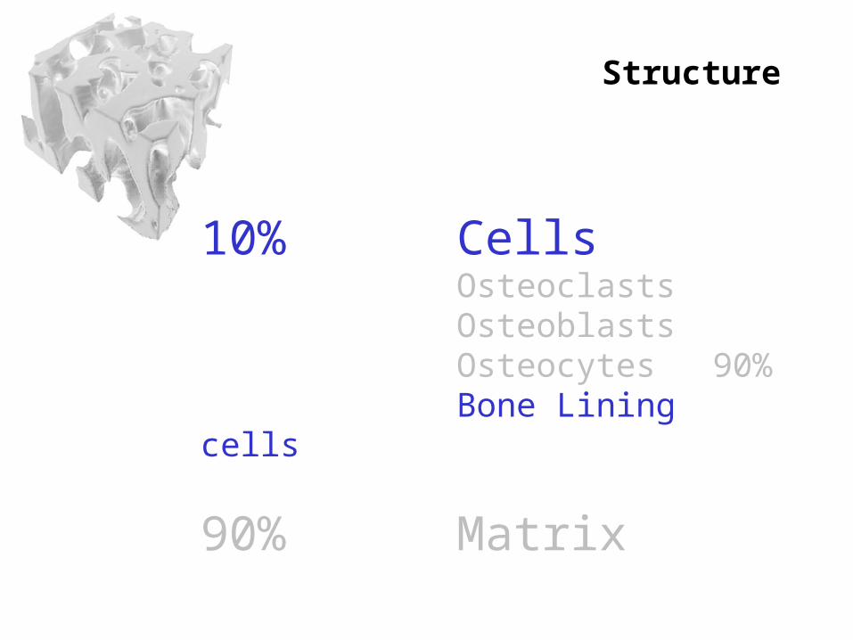
Structure
10% CellsOsteoclastsOsteoblastsOsteocytesBone Lining cells
90% Matrix

Multinucleated giant cellsHaemopoetic origin (monocyte progenitors)Resorb bone
Osteoclasts

Osteoclasts
Resorb bone by forming:Howships lacunae

Integrins – attach to bone sealing spaceProduce H+ via carbonic anhydraseLower PH increases solubility of Hydroxyapatite Organic matrix resorbed by proteolysis
Osteoclasts

Structure
10% CellsOsteoclastsOsteoblastsOsteocytesBone Lining cells
90% Matrix
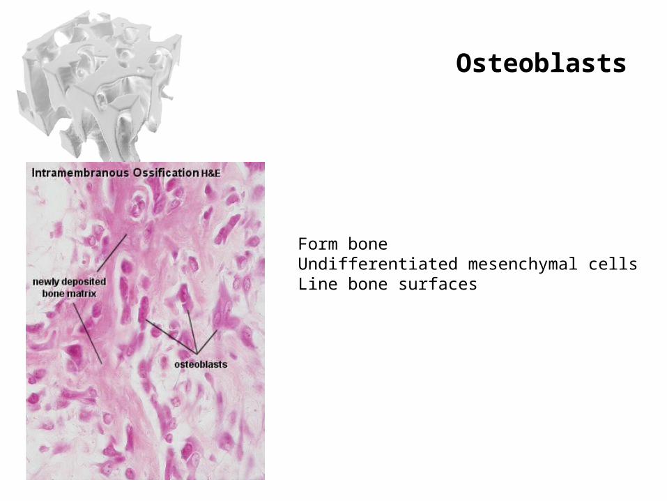
Osteoblasts
Form boneUndifferentiated mesenchymal cellsLine bone surfaces

Osteoblasts
Osteoblasts affected by:
ILPDGFIDGFPTH1,25 Dihydroxy vitamin DGlucocorticoidsProstaglandinsOestrogen

Structure
10% CellsOsteoclastsOsteoblastsOsteocytesBone Lining cells
90% Matrix

Osteocytes
90% of CellsOsteoblasts trapped in matrix
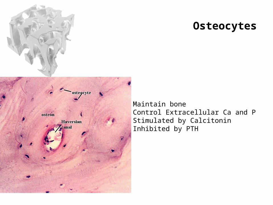
Maintain boneControl Extracellular Ca and PStimulated by CalcitoninInhibited by PTH
Osteocytes
Structure
10% CellsOsteoclastsOsteoblastsOsteocytes 90%Bone Lining cells
90% Matrix

Structure
10% CellsOsteoclastsOsteoblastsOsteocytes 90%Bone Lining cells
90% Matrix

Structure
10% CellsOsteoclastsOsteoblastsOsteocytes 90%Bone Lining cells
90% MatrixOrganic 40%Inorganic 60%

Organic (40%)
Collagen (90%)ProteoglycansNon collagenous matrix proteins
GlycoproteinsPhospholipidsPhosphoproteins
Growth factorsCytokines

Organic (40%)Collagen (90%)
Type - BONEPolypeptide triple helix Tropocolagen bond togetherForming fibrils


Most Hydroxyapatite
Fills in holes in Collagen
Inorganic (60%)
Ca10 (PO4)6 (OH)2
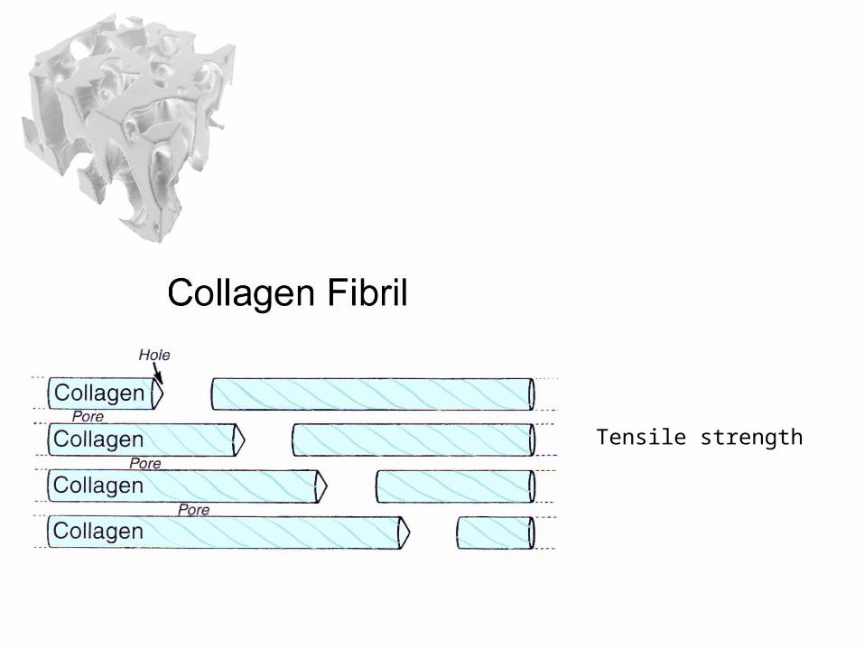
Tensile strength

Compressive strength

Microscopic
PrimaryImmatureWoven
SecondaryMatureLamellar

Woven Bone
LOCATION
Embryonic SkeletonNeonatal Skeleton Growing Metaphysis in under 4 yr olds
Near sutures of skullIn tooth socketsSome Tendon insertions
Callus
PROPERTIES
ISOTROPIC
SOFT
FLEXIBLE
RAPID DEPOSITION/TURNOVER
HIGH No. OF CELLS
uniform physical properties in all directions

Microscopic
PrimaryImmatureWoven
SecondaryMatureLamellar

Lamellar Bone
LOCATION
Throughout the adult skeleton
PROPERTIES
ANISOTROPIC
HARD
RIGID
SLOW DEPOSITION/TURNOVER
LOW No. OF CELLS
Properties differ based on the direction that is measured
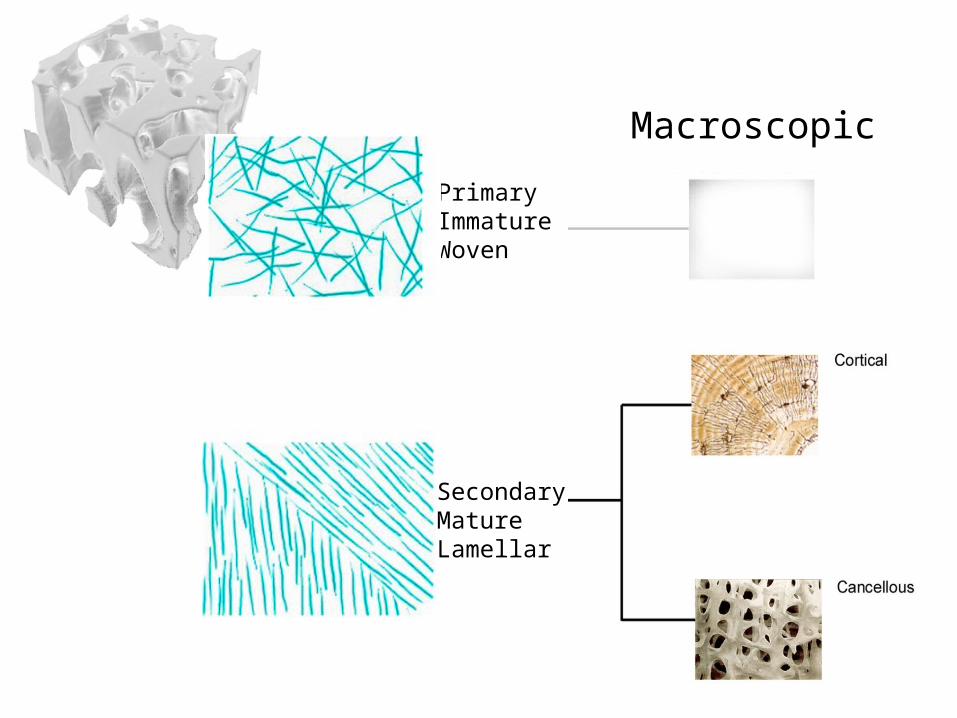
Macroscopic
PrimaryImmatureWoven
SecondaryMatureLamellar

Cortical BoneCompact
80% of the adult skeleton
20 times stiffer than cancellous bone
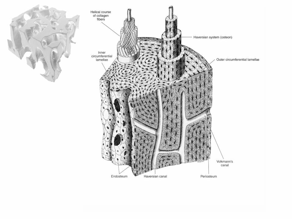
Lamellae in concentric rings aligned with lines of force
Complex arrangement of canals serving the lamellae(Haversian System)


Cancellous Bonetrabecular
20% of the adult skeleton
20 times less stiff than cortical bone
Lamellae also present aligned with lines of force
No Haversian System


Bone circulation
McCarthy I. J Bone Joint Surg 2006:88:4-9
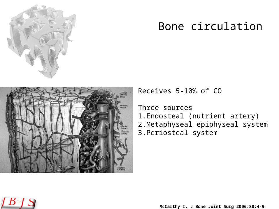
Bone circulation
Receives 5-10% of CO
Three sources1.Endosteal (nutrient artery)2.Metaphyseal epiphyseal system3.Periosteal system

McCarthy I. J Bone Joint Surg 2006:88:4-9
Bone circulation
1. Nutrient arteryEnters diaphysis to medullary cavityAscending and descending arteriolesCentrifugal high pressureInner 2/3rds of cortex

McCarthy I. J Bone Joint Surg 2006:88:4-9
Bone circulation
2. 2. Metaphyseal epiphyseal systemPeriarticular vascular plexus
eg. geniculate arteries

McCarthy I. J Bone Joint Surg 2006:88:4-9
Bone circulation
3. Periosteal systemlow pressure on periosteum Outer 1/3rd of cortex

Questions ?

Biomechanics