Bone destructive mechanisms of cholesteatoma - ORL-HNS · destructive mechanisms of cholesteatoma....
1
Bone destructive mechanisms of cholesteatoma Shin-ichi Kanemaru, M.D., Ph.D. 1) Yayoi Kikkawa, M.D., Ph.D. 2) , Koichi Omori, M.D., Ph.D. 3) , Juichi Ito, M.D., Ph.D. 4) Abstract Hypothesis: Our objective for the current studies was to observe the expression of Ki 67, P53, MMP-8,13 L26, UCHLI, CD68 and MIF in cholesteatoma and to determine their roles in the destruction of bone and extra cellar matrix. Background: Cholesteatoma is characterized by the highly invasive epithelium into the tympanic cavity. Though the mechanisms of the destructive properties of cholesteatoma have not yet been known, they may be caused by enzymes and cytokines released by cholesteatomas. Materials and Methods:Specimens: 11 samples from cholesteatoma and 4 normal external auditory tissues, were analysed by immunohistochemistry using monoclonal antibodies to Ki67, p53, MMP-8, MMP-13, L26, UCHLI, CD68 and MIF. The rates of positive expression of these cytokins were estimated. The percentage of positive expression of these cytokines were determined by the ratio of average number of the positive cells in the total cells in the 3 microscopic fields of cholesteatoma minus that of normal control tissue. Results: The overexpression of Ki67, MMP8, MMP13 and MIF were observed simultaneously in cholesteatoma with/without inflammatory granulation. Especially, MMP8 and MIF were strongly expressed in all specimens of cholesteatoma. Conclusions: 1. This study indicated that the over expression of cytokins; Ki67, MMP-8, 13 and MIF was observed in cholesteatoma. 2. From the results of this study and the former reports, we established bone destructive mechanism model of cholesteatoma that MIF induced mechanical stress as a trigger played a possible key role of the bone destructive mechanisms of cholesteatoma. 1) Department of Otolaryngology-HNS ,Medical Research Institute, Kitano Hospital, Osaka, Japan 2) Department of Otolaryngology-HNS, Graduate School of Medicine, Tokyo University, Tokyo, Japan 3) Department of Otolaryngology, Fukushima Medical University, School of Medicine, Fukushima, Japan 4) Department of Otolaryngology-HNS, Graduate School of Medicine Kyoto University, Kyoto, Japan Aim of our study To observe the expression of cytokins; Ki67, p53, L26, MMP-8,13 , UCHLI, CD68, and MIF in cholesteatoma and to determine their roles in the destruction of bone and ECM(extracellular matrix) . To explain the destructive mechanism of cholesteatomas Enzymes and cytokines released by cholesteatomas Matrix-metalloproteinases (MMPs) , Transforming growth factor (TGF-alpha/beta) Interleukin-1 alpha (IL-1 alpha) , Receptor activator of nuclear factor-kappa B (RANK) Nuclear factor-kappa B ligand (RANKL) , Tumor necrosis factor-alpha (TNF-alpha) Material and Method 11 samples from cholesteatoma and 4 normal tissues of external auditory meatus were analyzed. Specimens were analysed by immunohistochemistry using monoclonal antibodies to Ki67, p53, MMP-8, MMP-13, L26, UCHLI, CD68 and MIF. Cp: The average number of the positive cells in the 3 microscopic fields of cholesteatoma Ct : The average number of the total cells in the 3 microscopic fields of cholesteatoma Np: The average number of the positive cells in the 3 microscopic fields of normal control tisuue Nt : The average number of the total cells in the 3 microscopic fields of normal control tisuue The rate of positive expression Cp Np Ct Nt x 100 % = Background Middle ear cholesteatoma is a disease of inflammatory bone resorption leading to hearing loss, vestibular dysfunction and facial palsy. It follows that cholesteatoma has not benign property but malignant one though it dose not metastasize. In some cases, it may cause intratemporal and intracranial complications,which have high morbidity and mortality rates. Summary Results Histopathological findings Ki 67 P 53 MMP-8 MMP-13 L 26 UCHLI CD68 MIF Group I Control Overall Results Group I + - ++ + - - - +++ Group II ++ - +++ ++ ++ +++ - ++ Group I : cholesteatoma without inflammatory granulation Group II: cholesteatoma with inflammatory granulation Positive cells or regions: +>10%, ++>30%, +++>50% K i 6 7 K i 6 7 P 5 3 P 5 3 M M P -8 M M P -8 M M P - 1 3 M M P - 1 3 L 2 6 L 2 6 U C H L I U C H L I C D6 8 C D6 8 M I F M I F Bone destructive mechanism model of cholesteatoma Ki67 Autocrine Paracrine MMPs 1,2,3,8,9,13 Collagenase Bone and ECM destruction Bone and ECM destruction IL-1, 6, TNF-alpha Inflammation LPS Osteoblast Osteoclast Cholesteatoma Cholesteatoma RANKL Discussion Macrophage T cell MIF Angiogenesis Cell proliferation Angiogenesis Angiogenesis Cell proliferation Cell proliferation Mechanical stress Pressure due to accumulated keratin and other waste produces mechanical stress. Mechanical stress induces the production of MIF. MIF induces the MMPs. MMPs work for agiogenesis and cell proliferation. Degradation of the extracellular matrix is essential for the development of cholesteatomas and that this is induced by activation of MMPs. MMPs play a destructive role of bone and ECM degradation. At the same time, MIF also increases the production of pro-inflammatory cytokines and chemokines by macrophages. Especially, IL1, 6, and TNF-alpha may play a role of bone destruction. Moreover, osteoclast and osteoblast are activated by these cytokines and chemokines through MIF. Therefore, MIF is the possible key factor for bone destructive mechanisms of cholesteatoma. 1. This study indicated that the over expression of cytokins; Ki67, MMP-8, 13 and MIF was observed in cholesteatoma. 2. From the results of this study and the former reports, we established bone destructive mechanism model of cholesteatoma that MIF induced mechanical stress as a trigger played a possible key role of the bone destructive mechanisms of cholesteatoma.
Transcript of Bone destructive mechanisms of cholesteatoma - ORL-HNS · destructive mechanisms of cholesteatoma....
Bone destructive mechanisms of cholesteatomaShin-ichi Kanemaru, M.D., Ph.D.1)
Yayoi Kikkawa, M.D., Ph.D. 2), Koichi Omori, M.D., Ph.D.3), Juichi Ito, M.D., Ph.D. 4)
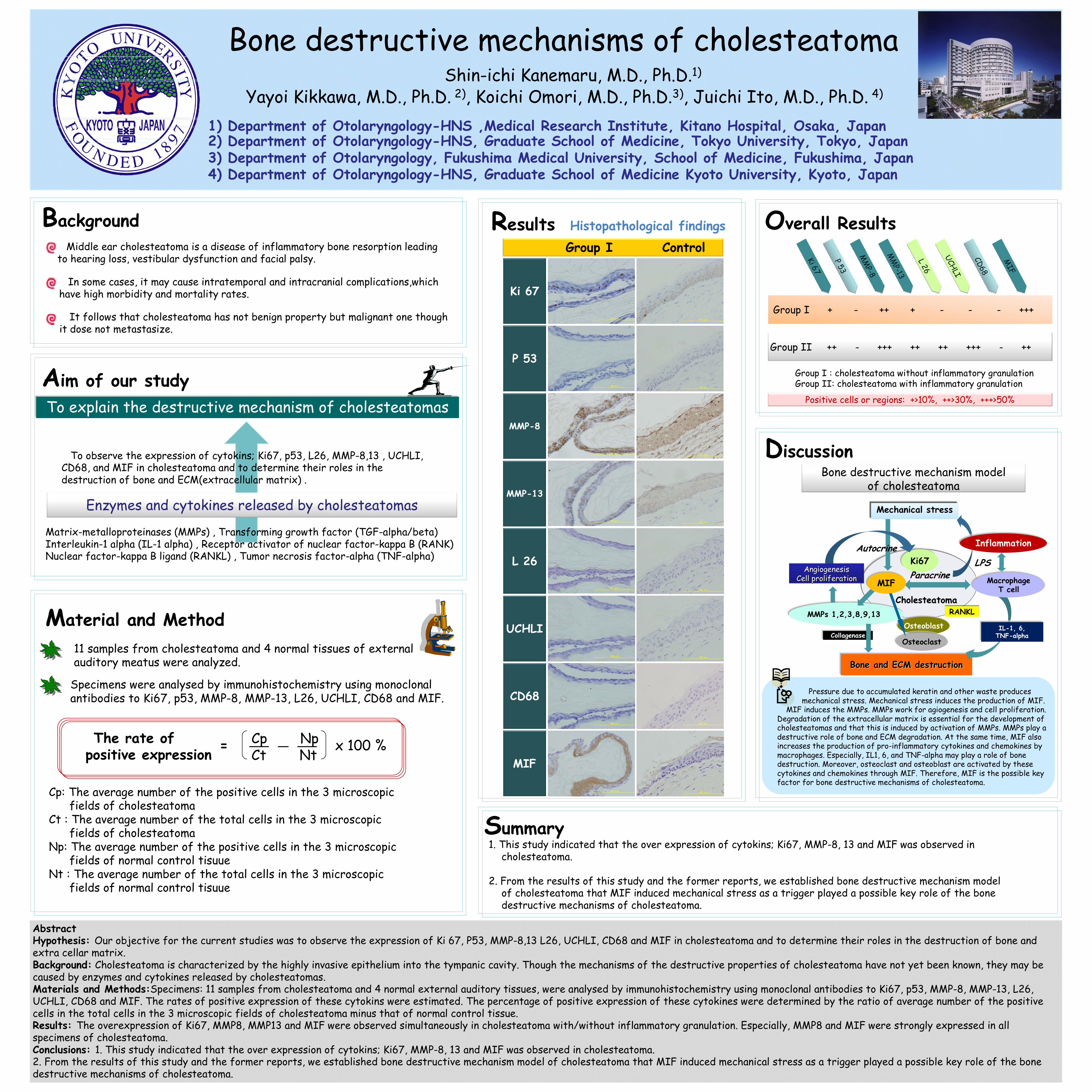
AbstractHypothesis: Our objective for the current studies was to observe the expression of Ki 67, P53, MMP-8,13 L26, UCHLI, CD68 and MIF in cholesteatoma and to determine their roles in the destruction of bone and extra cellar matrix.Background: Cholesteatoma is characterized by the highly invasive epithelium into the tympanic cavity. Though the mechanisms of the destructive properties of cholesteatoma have not yet been known, they may be caused by enzymes and cytokines released by cholesteatomas.Materials and Methods:Specimens: 11 samples from cholesteatoma and 4 normal external auditory tissues, were analysed by immunohistochemistry using monoclonal antibodies to Ki67, p53, MMP-8, MMP-13, L26, UCHLI, CD68 and MIF. The rates of positive expression of these cytokins were estimated. The percentage of positive expression of these cytokines were determined by the ratio of average number of the positive cells in the total cells in the 3 microscopic fields of cholesteatoma minus that of normal control tissue. Results: The overexpression of Ki67, MMP8, MMP13 and MIF were observed simultaneously in cholesteatoma with/without inflammatory granulation. Especially, MMP8 and MIF were strongly expressed in all specimens of cholesteatoma.Conclusions: 1. This study indicated that the over expression of cytokins; Ki67, MMP-8, 13 and MIF was observed in cholesteatoma.2. From the results of this study and the former reports, we established bone destructive mechanism model of cholesteatoma that MIF induced mechanical stress as a trigger played a possible key role of the bone destructive mechanisms of cholesteatoma.
1) Department of Otolaryngology-HNS ,Medical Research Institute, Kitano Hospital, Osaka, Japan2) Department of Otolaryngology-HNS, Graduate School of Medicine, Tokyo University, Tokyo, Japan3) Department of Otolaryngology, Fukushima Medical University, School of Medicine, Fukushima, Japan4) Department of Otolaryngology-HNS, Graduate School of Medicine Kyoto University, Kyoto, Japan
Aim of our study
To observe the expression of cytokins; Ki67, p53, L26, MMP-8,13 , UCHLI, CD68, and MIF in cholesteatoma and to determine their roles in the destruction of bone and ECM(extracellular matrix) .
To explain the destructive mechanism of cholesteatomas
Enzymes and cytokines released by cholesteatomas
Matrix-metalloproteinases (MMPs) , Transforming growth factor (TGF-alpha/beta)Interleukin-1 alpha (IL-1 alpha) , Receptor activator of nuclear factor-kappa B (RANK) Nuclear factor-kappa B ligand (RANKL) , Tumor necrosis factor-alpha (TNF-alpha)
Material and Method 11 samples from cholesteatoma and 4 normal tissues of external auditory meatus were analyzed.
Specimens were analysed by immunohistochemistry using monoclonalantibodies to Ki67, p53, MMP-8, MMP-13, L26, UCHLI, CD68 and MIF.
Cp: The average number of the positive cells in the 3 microscopic fields of cholesteatoma
Ct : The average number of the total cells in the 3 microscopic fields of cholesteatoma
Np: The average number of the positive cells in the 3 microscopic fields of normal control tisuue
Nt : The average number of the total cells in the 3 microscopic fields of normal control tisuue
The rate of positive expression
Cp NpCt Nt x 100 %=
BackgroundMiddle ear cholesteatoma is a disease of inflammatory bone resorption leading
to hearing loss, vestibular dysfunction and facial palsy.
It follows that cholesteatoma has not benign property but malignant one though it dose not metastasize.
In some cases, it may cause intratemporal and intracranial complications,which have high morbidity and mortality rates.
Summary
Results Histopathological findings
Ki 67
P 53
MMP-8
MMP-13
L 26
UCHLI
CD68
MIF
Group I Control
Overall Results
Group I + - ++ + - - - +++
Group II ++ - +++ ++ ++ +++ - ++
Group I : cholesteatoma without inflammatory granulationGroup II: cholesteatoma with inflammatory granulation
Positive cells or regions: +>10%, ++>30%, +++>50%
Ki 67Ki 67
P53
P53
MM
P-8M
MP-8
MM
P-13M
MP-13
L26
L26
UCHLIUCHLI
CD68CD68
MIF
MIF
Bone destructive mechanism modelof cholesteatoma
Ki67Autocrine
Paracrine
MMPs 1,2,3,8,9,13
Collagenase
Bone and ECM destructionBone and ECM destruction
IL-1, 6, TNF-alpha
Inflammation
LPS
Osteoblast
Osteoclast
CholesteatomaCholesteatomaRANKLRANKL
Discussion
MacrophageT cell
MIFAngiogenesis
Cell proliferationAngiogenesis Angiogenesis
Cell proliferationCell proliferation
Mechanical stress
Pressure due to accumulated keratin and other waste produces mechanical stress. Mechanical stress induces the production of MIF.
MIF induces the MMPs. MMPs work for agiogenesis and cell proliferation. Degradation of the extracellular matrix is essential for the development of cholesteatomas and that this is induced by activation of MMPs. MMPs play a destructive role of bone and ECM degradation. At the same time, MIF also increases the production of pro-inflammatory cytokines and chemokines by macrophages. Especially, IL1, 6, and TNF-alpha may play a role of bone destruction. Moreover, osteoclast and osteoblast are activated by these cytokines and chemokines through MIF. Therefore, MIF is the possible key factor for bone destructive mechanisms of cholesteatoma.
1. This study indicated that the over expression of cytokins; Ki67, MMP-8, 13 and MIF was observed in cholesteatoma.
2. From the results of this study and the former reports, we established bone destructive mechanism model of cholesteatoma that MIF induced mechanical stress as a trigger played a possible key role of the bone destructive mechanisms of cholesteatoma.