Hierarchy of organization Cells → Tissues → Organs → Organ Systems → Organism.
Upload
nguyendiepCategory
view
217download
2
© 2014 Pearson Education, Inc.
Lecture Presentation
Anne Gasc
Hawaii Pacific University and
University of Hawaii–Honolulu Community College
BIOLOGY OF HUMANSConcepts, Applications, and Issues
Fifth Edition
Judith Goodenough Betty McGuire
4Body Organization
and Homeostasis
© 2014 Pearson Education, Inc.
Body Organization and Homeostasis
OUTLINE:
From Cells to Organ Systems
Skin: An Organ System
Homeostasis
© 2014 Pearson Education, Inc.
From Cells to Organ Systems
Tissue
A group of cells of similar type that work together
to serve a common function
Humans have four primary tissue types
Epithelial
Connective
Muscle
Nervous
© 2014 Pearson Education, Inc.
Tissues
Humans have four types of tissues:
Epithelial tissue (Epi: on, upon)
Covers the body surfaces
Lines cavities and organs
Forms glands
Connective tissue
Provides support and protection for organs
Serves as a storage site for fat
Participates in our immunity
© 2014 Pearson Education, Inc.
Tissues
Humans have four types of tissues (cont’d):
Muscle tissue, responsible for:
Body movement
Movement of fluids through the body
Nervous tissue
Conducts nerve impulses through the body
© 2014 Pearson Education, Inc.
Epithelial Tissue
All epithelial tissues share two structural
characteristics
A free surface that may be specialized for
protection, secretion, or absorption
A basement membrane that binds the epithelial
cells to underlying connective tissue and helps
the epithelial tissue resist stretching
© 2014 Pearson Education, Inc.
Epithelial Tissue
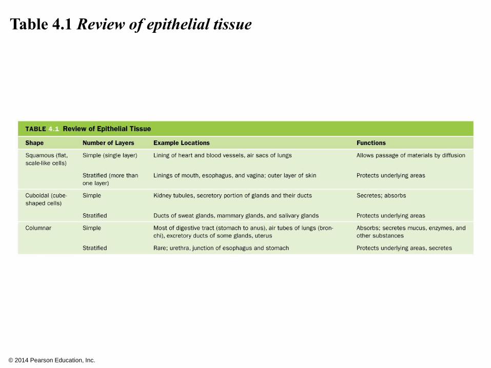
The three basic shapes of epithelial cells are
suited to their functions
1. Squamous epithelium
2. Cuboidal epithelium
3. Columnar epithelium
These cells can be either simple (a single layer
of cells) or stratified (multiple layers of cells)
© 2014 Pearson Education, Inc.
Epithelial Tissue
Squamous epithelium
Has flattened cells
Shape allows for diffusion of materials and can
provide a slick surface to reduce friction
Cuboidal epithelium
Has cube-shaped cells
Specialized for secretion and absorption
© 2014 Pearson Education, Inc.
Epithelial Tissue
Columnar epithelium
Has tall, column-shaped cells
Specialized for secretion and absorption
Lines the small intestine

© 2014 Pearson Education, Inc.
Figure 4.1 Types of epithelial tissue. These are named for the shape
of the cell and the number of cell layers.
© 2014 Pearson Education, Inc.
Table 4.1 Review of epithelial tissue
© 2014 Pearson Education, Inc.
Epithelial Glands
A gland is composed of epithelial tissue that
secretes a product
Exocrine glands secrete into ducts leading to
body surfaces, cavities or organs (exo: out)
Endocrine glands lack ducts and secrete their
products, hormones into spaces just outside the
cells but in the body (endo: inside)
© 2014 Pearson Education, Inc.
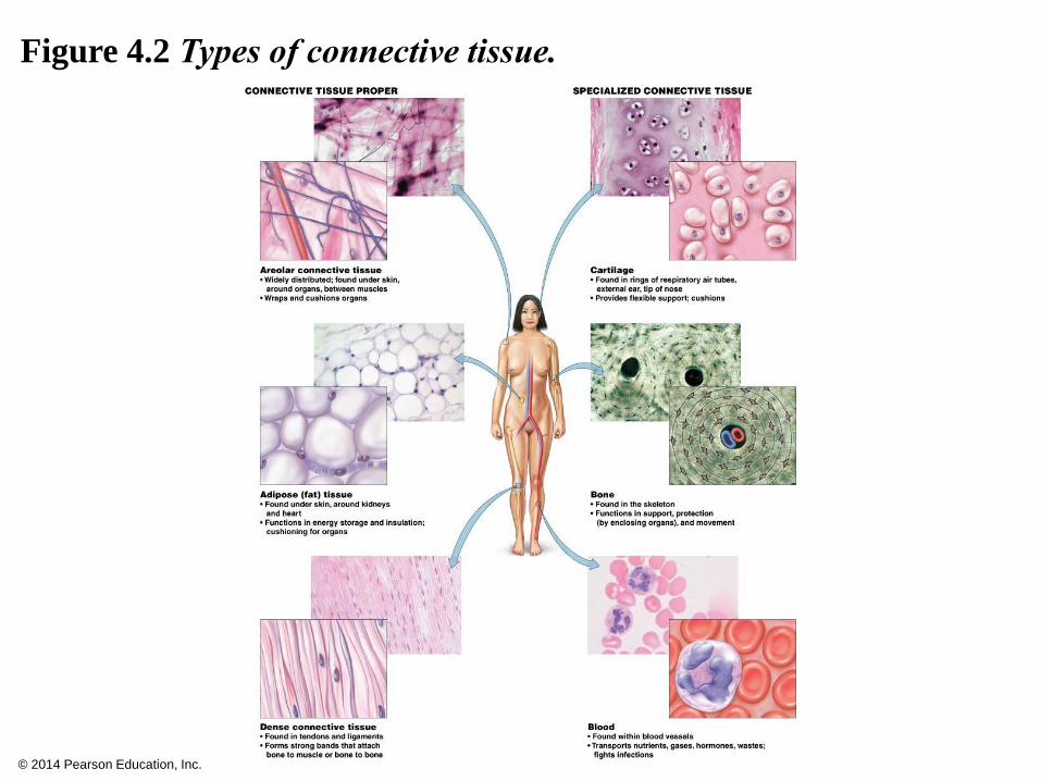
Connective Tissue
The most abundant and widely distributed tissue in the
body
Cells are contained in an extracellular matrix of protein
fibers and ground substance
Protein fibers
Collagen, elastic, and reticular fibers
Produced by fibroblasts, which are also responsible for tissue
repair
Ground substance
Noncellular material
May be solid (bone), fluid (blood), or gelatinous (cartilage)
© 2014 Pearson Education, Inc.
Connective Tissue
Two categories
Connective tissue proper
Specialized connective tissue
© 2014 Pearson Education, Inc.
Connective Tissue
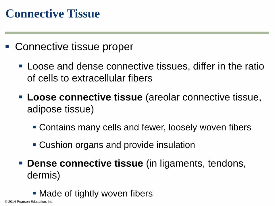
Connective tissue proper
Loose and dense connective tissues, differ in the ratio
of cells to extracellular fibers
Loose connective tissue (areolar connective tissue,
adipose tissue)
Contains many cells and fewer, loosely woven fibers
Cushion organs and provide insulation
Dense connective tissue (in ligaments, tendons,
dermis)
Made of tightly woven fibers
© 2014 Pearson Education, Inc.
Connective Tissue
Specialized connective tissue
Cartilage
Bone
Blood
© 2014 Pearson Education, Inc.
Connective Tissue
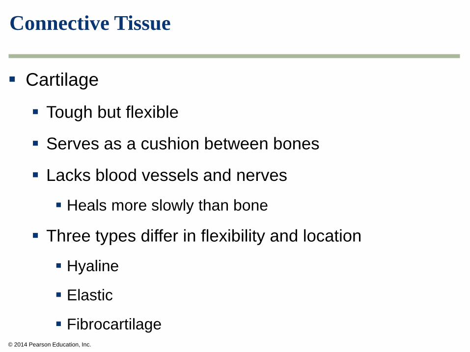
Cartilage
Tough but flexible
Serves as a cushion between bones
Lacks blood vessels and nerves
Heals more slowly than bone
Three types differ in flexibility and location
Hyaline
Elastic
Fibrocartilage
© 2014 Pearson Education, Inc.
Connective Tissue
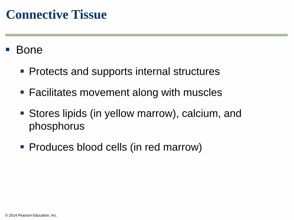
Bone
Protects and supports internal structures
Facilitates movement along with muscles
Stores lipids (in yellow marrow), calcium, and
phosphorus
Produces blood cells (in red marrow)
© 2014 Pearson Education, Inc.
Figure 4.2 Types of connective tissue.
© 2014 Pearson Education, Inc.
Muscle Tissue
Three types vary in structure, location, and whether
voluntary or involuntary
Skeletal
Cardiac
Smooth
© 2014 Pearson Education, Inc.
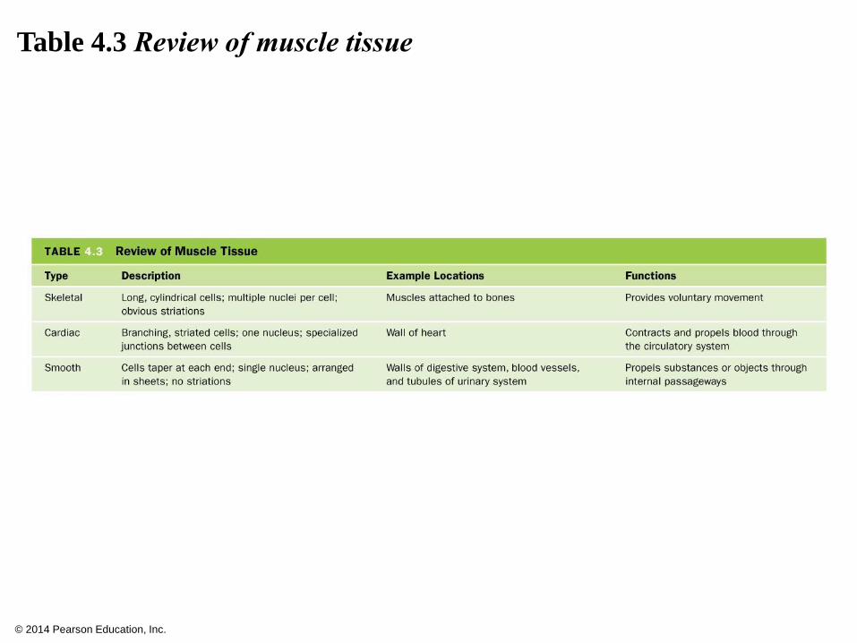
Table 4.3 Review of muscle tissue
© 2014 Pearson Education, Inc.
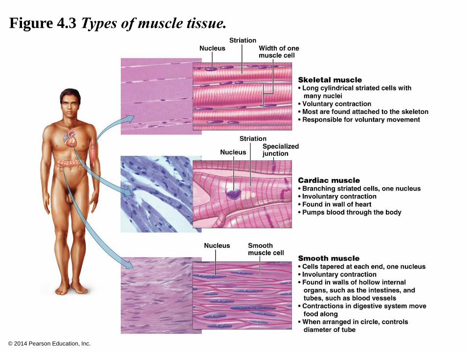
Figure 4.3 Types of muscle tissue.
© 2014 Pearson Education, Inc.
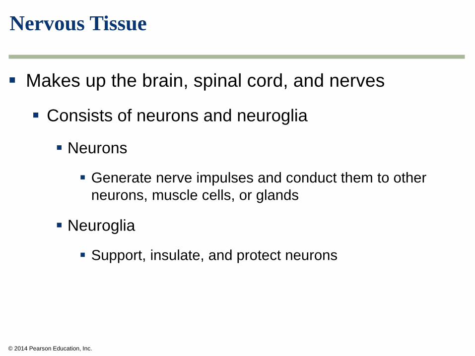
Nervous Tissue
Makes up the brain, spinal cord, and nerves
Consists of neurons and neuroglia
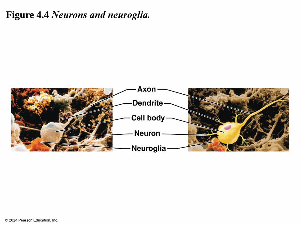
Neurons
Generate nerve impulses and conduct them to other
neurons, muscle cells, or glands
Neuroglia
Support, insulate, and protect neurons
© 2014 Pearson Education, Inc.
Figure 4.4 Neurons and neuroglia.
© 2014 Pearson Education, Inc.
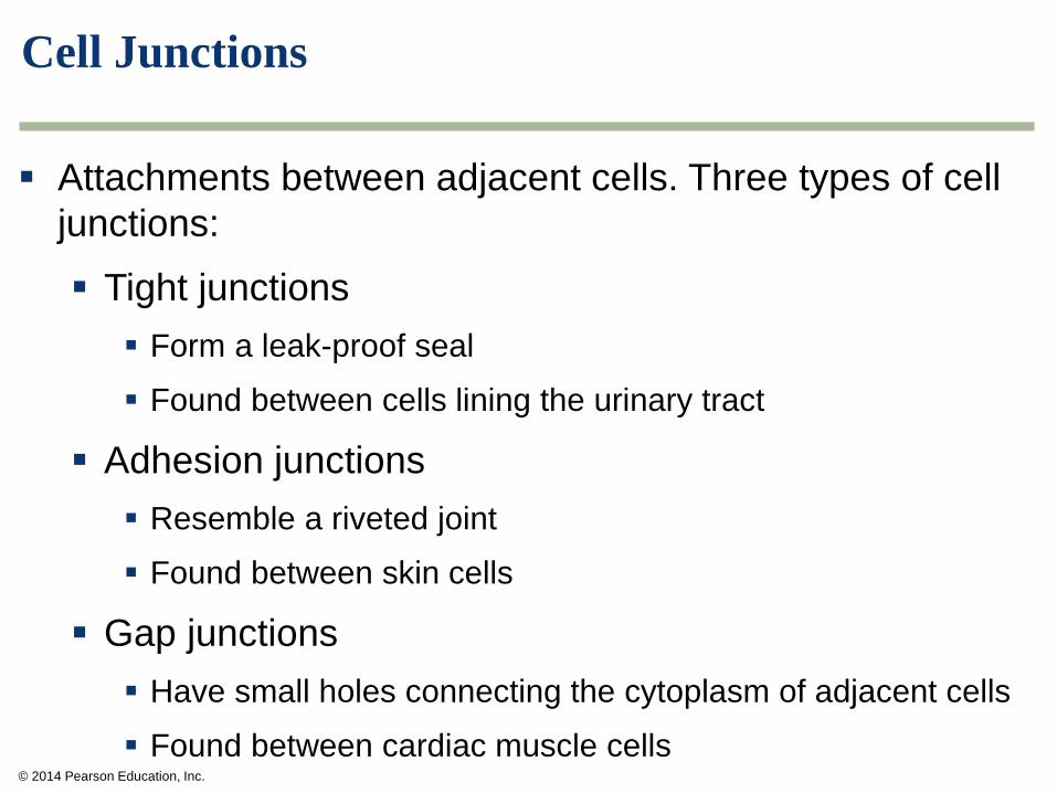
Cell Junctions
Attachments between adjacent cells. Three types of cell
junctions:
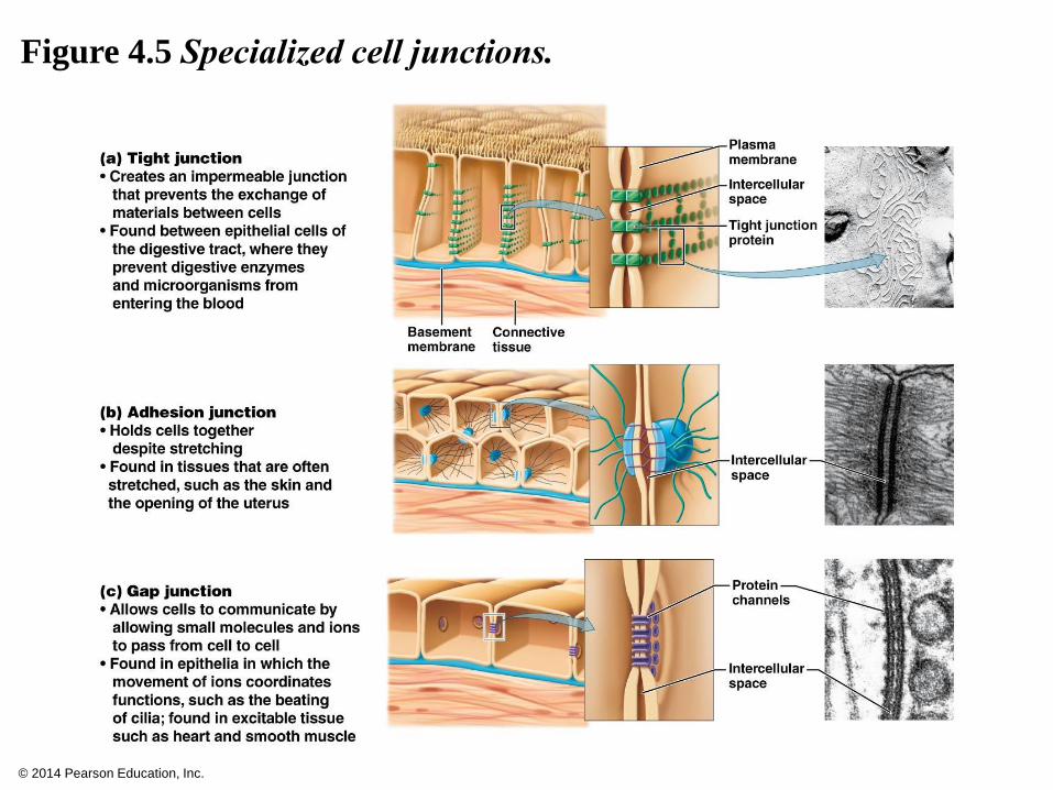
Tight junctions
Form a leak-proof seal
Found between cells lining the urinary tract
Adhesion junctions
Resemble a riveted joint
Found between skin cells
Gap junctions
Have small holes connecting the cytoplasm of adjacent cells
Found between cardiac muscle cells
© 2014 Pearson Education, Inc.
Figure 4.5 Specialized cell junctions.
© 2014 Pearson Education, Inc.
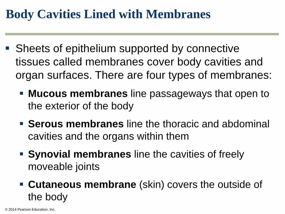
Body Cavities Lined with Membranes
Sheets of epithelium supported by connective
tissues called membranes cover body cavities and
organ surfaces. There are four types of membranes:
Mucous membranes line passageways that open to
the exterior of the body
Serous membranes line the thoracic and abdominal
cavities and the organs within them
Synovial membranes line the cavities of freely
moveable joints
Cutaneous membrane (skin) covers the outside of
the body
© 2014 Pearson Education, Inc.
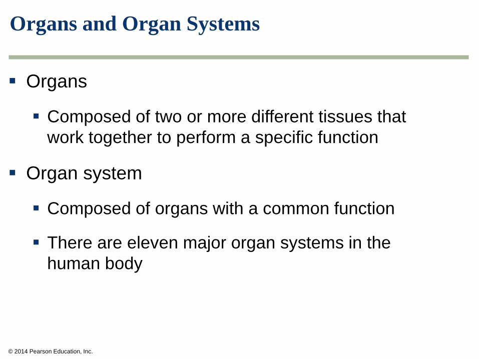
Organs and Organ Systems
Organs
Composed of two or more different tissues that
work together to perform a specific function
Organ system
Composed of organs with a common function
There are eleven major organ systems in the
human body
© 2014 Pearson Education, Inc.
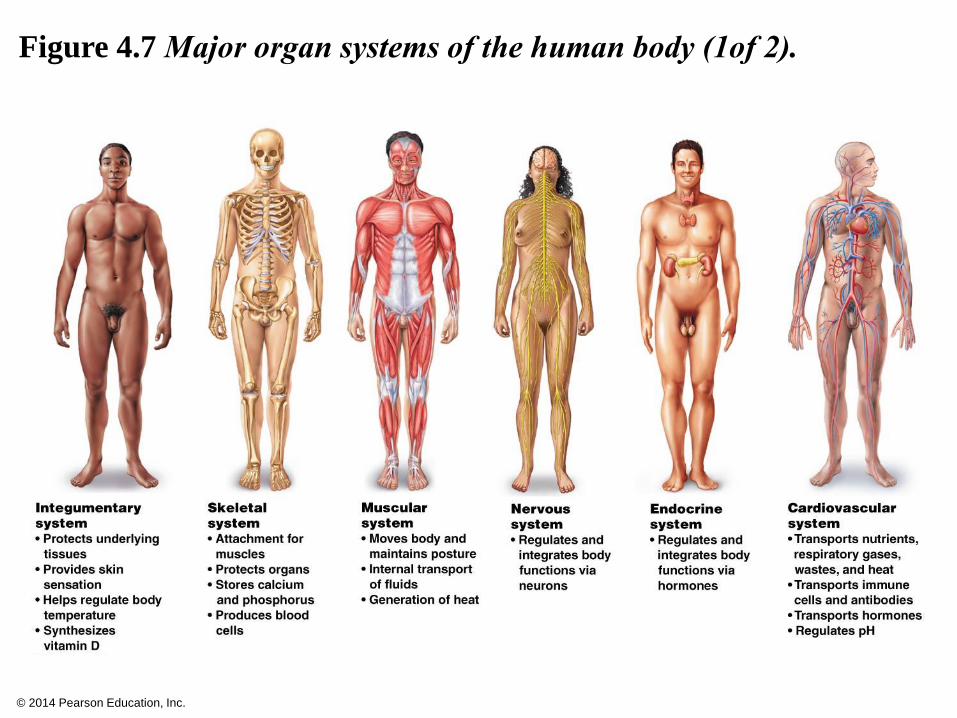
Figure 4.7 Major organ systems of the human body (1of 2).
© 2014 Pearson Education, Inc.
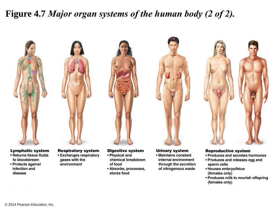
Figure 4.7 Major organ systems of the human body (2 of 2).

© 2014 Pearson Education, Inc.
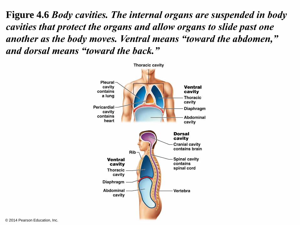
Body Cavities Lined with Membranes
Most of our organs are suspended in internal body
cavities that protect vital organs and allow them to slide
past one another and change shape. There are two main
body cavities:
Ventral cavity
Thoracic cavity
Abdominal cavity
The diaphragm separates them
Dorsal cavity
Cranial cavity (encloses the brain)
Spinal cavity (houses the spinal cord)
© 2014 Pearson Education, Inc.
Figure 4.6 Body cavities. The internal organs are suspended in body
cavities that protect the organs and allow organs to slide past one
another as the body moves. Ventral means “toward the abdomen,”
and dorsal means “toward the back.”
© 2014 Pearson Education, Inc.
Skin: An Organ System
The integumentary system is composed of
Skin
Derivatives of the skin
Hair
Nails
Sweat glands
Oil glands
Wax glands
© 2014 Pearson Education, Inc.
Skin Functions
The skin is our largest organ
Functions of the skin
Protects against bacterial invasion, UV radiation,
and physical and chemical stress
Prevents water loss
Regulates body temperature
Synthesizes vitamin D
Receives stimuli
© 2014 Pearson Education, Inc.
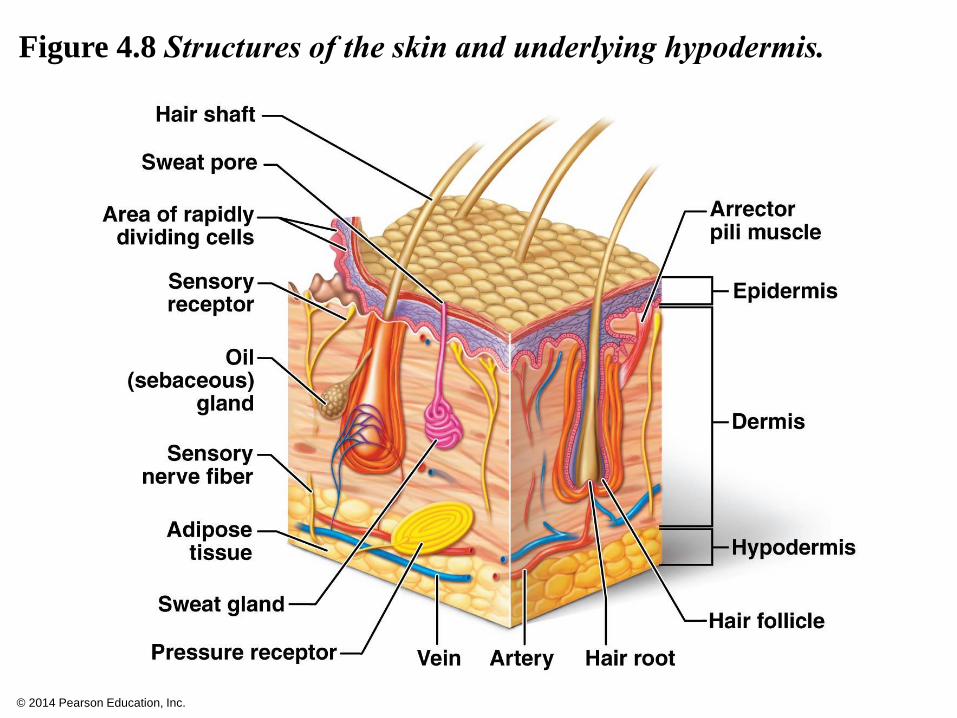
Skin Layers
The skin has two major layers
Epidermis (epi: on, over)
Thin outer layer
Dermis
Thicker inner layer containing nerves, blood vessels,
and glands
Hypodermis or subcutaneous layer
Layer of loose connective tissue just below the
epidermis and dermis
© 2014 Pearson Education, Inc.
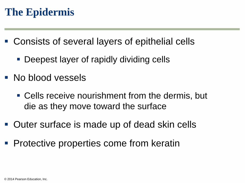
The Epidermis
Consists of several layers of epithelial cells
Deepest layer of rapidly dividing cells
No blood vessels
Cells receive nourishment from the dermis, but
die as they move toward the surface
Outer surface is made up of dead skin cells
Protective properties come from keratin
© 2014 Pearson Education, Inc.
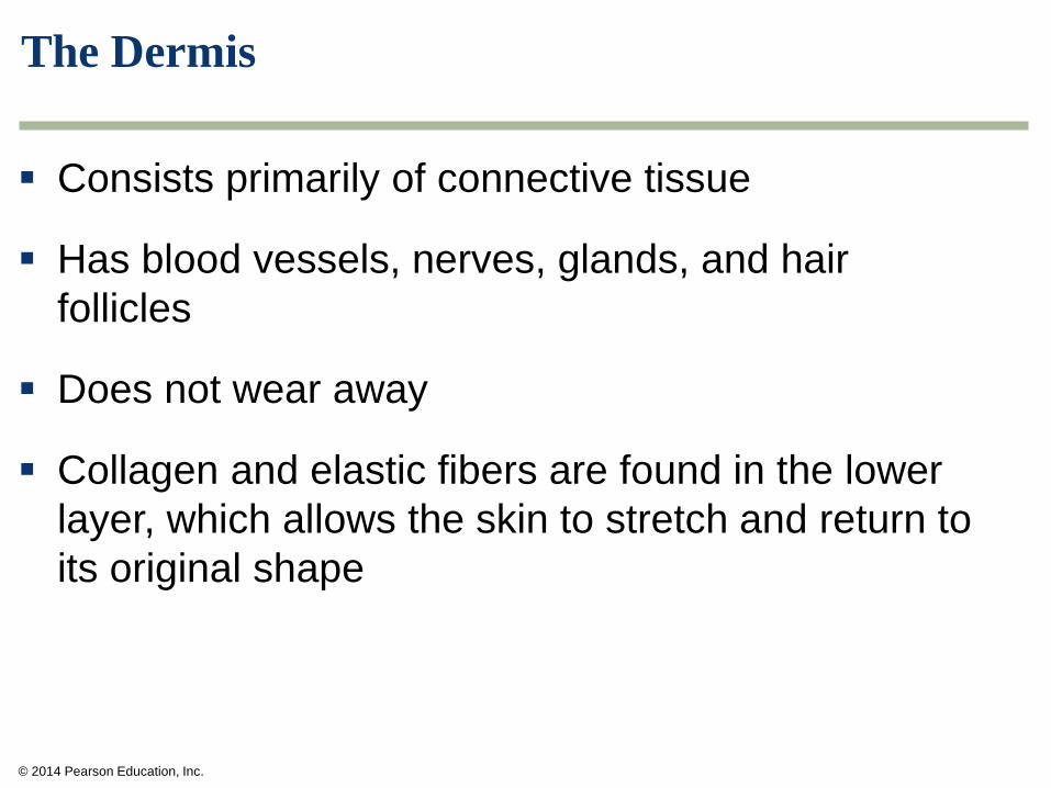
The Dermis
Consists primarily of connective tissue
Has blood vessels, nerves, glands, and hair
follicles
Does not wear away
Collagen and elastic fibers are found in the lower
layer, which allows the skin to stretch and return to
its original shape
© 2014 Pearson Education, Inc.
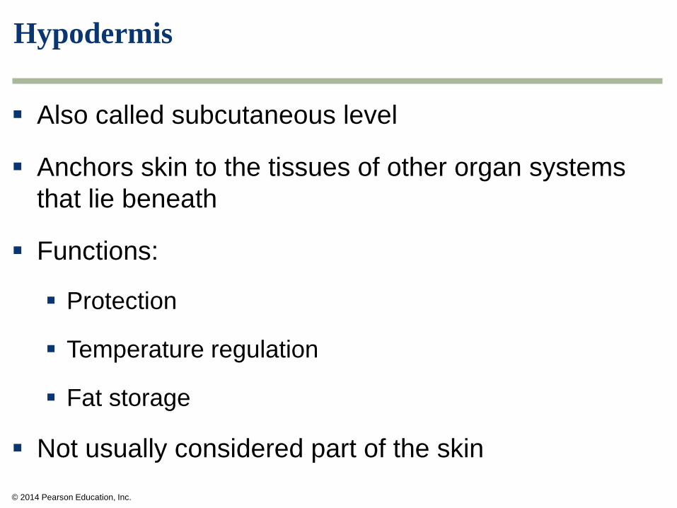
Hypodermis
Also called subcutaneous level
Anchors skin to the tissues of other organ systems
that lie beneath
Functions:
Protection
Temperature regulation
Fat storage
Not usually considered part of the skin
© 2014 Pearson Education, Inc.
Figure 4.8 Structures of the skin and underlying hypodermis.
© 2014 Pearson Education, Inc.
Skin Color
Skin color is determined by
Blood flow
Distribution and quantity of the pigment melanin
Melanin
Produced by melanocytes at the base of the epidermis
Comes in two forms
Yellow-to-red
Black-to-brown
In tanning, the melanocytes respond to UV radiation by
increasing production of melanin
© 2014 Pearson Education, Inc.
Hair, Nails, and Glands
The epidermis gives rise to diverse structures
Hair
Nails
Oil glands
Sweat glands
Wax glands
Teeth (will be covered with the digestive system)
© 2014 Pearson Education, Inc.
Hair, Nails, and Glands
Hair
Primary function is protection
Grows over most of the body
Components
Shaft—extends above the skin surface
Root—extends into the dermis or hypodermis where it
is embedded in a follicle
© 2014 Pearson Education, Inc.
Hair, Nails, and Glands
Nails
Protect the tips of our toes and fingers
Because nails are embedded in very sensitive
tissue, they also function as sensory antennas
© 2014 Pearson Education, Inc.
Hair, Nails, and Glands
Oil glands
Found all over the body except the palms of the hands and
soles of the feet
Oil lubricates hair and skin and contains substances that inhibit
bacteria
Sweat glands
Produce sweat that helps in the regulation of body temperature
Some metabolic wastes are excreted in sweat
Wax glands
Modified sweat glands found in external ear canal
Wax protects the ear by trapping small particles
© 2014 Pearson Education, Inc.
Health Issue: Fun in the Sun?
The ultraviolet (UV) radiation of sunlight causes the
melanocytes of the skin to increase their production
of the pigment melanin, which absorbs UV radiation
before it can damage the genetic information of
deeper layers of cells. Unfortunately, this protective
buildup of melanin is not instantaneous. In skin
cancer, UV radiation alters the genetic material in
skin cells so that the cells grow and divide
uncontrollably, forming a tumor.
© 2014 Pearson Education, Inc.
Health Issue: Fun in the Sun?
Three types of skin cancer are caused by over
exposure to the sun:
Basal cell carcinoma, the most common arises
in the rapidly dividing cells of the deepest layer of
epidermis
Squamous cell carcinoma, the second most
common, arises in the newly formed skin cells as
they flatten and move toward the skin surface
Melanoma is the least common and most
dangerous type of skin cancer
© 2014 Pearson Education, Inc.
Health Issue: Fun in the Sun?
Limiting the risks
Avoid prolonged exposure to the sun
If you must be out in the sun, wear a hat, long
sleeves, and sunglasses
Use a sunscreen with a sun protection factor
(SPF) of at least 15 and apply about 45 minutes
before going out into the sun
Use sunscreen even when it is overcast
Avoid tanning salons
© 2014 Pearson Education, Inc.
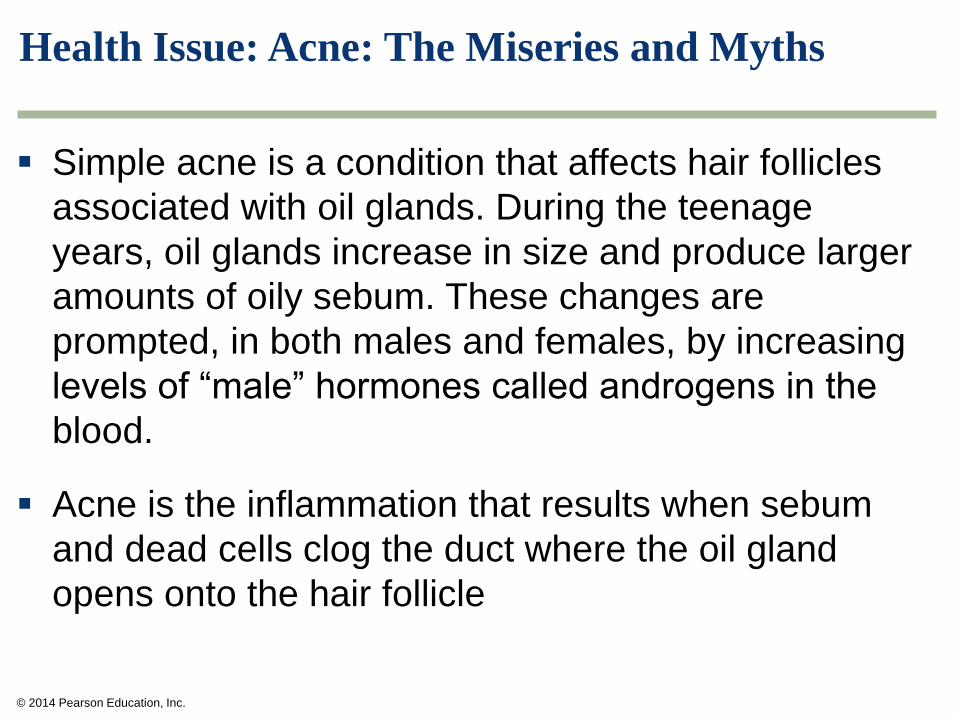
Health Issue: Acne: The Miseries and Myths
Simple acne is a condition that affects hair follicles
associated with oil glands. During the teenage
years, oil glands increase in size and produce larger
amounts of oily sebum. These changes are
prompted, in both males and females, by increasing
levels of “male” hormones called androgens in the
blood.
Acne is the inflammation that results when sebum
and dead cells clog the duct where the oil gland
opens onto the hair follicle
© 2014 Pearson Education, Inc.
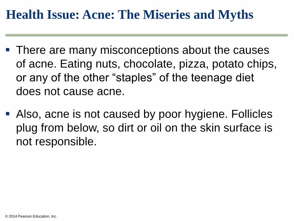
Health Issue: Acne: The Miseries and Myths
There are many misconceptions about the causes
of acne. Eating nuts, chocolate, pizza, potato chips,
or any of the other “staples” of the teenage diet
does not cause acne.
Also, acne is not caused by poor hygiene. Follicles
plug from below, so dirt or oil on the skin surface is
not responsible.
© 2014 Pearson Education, Inc.
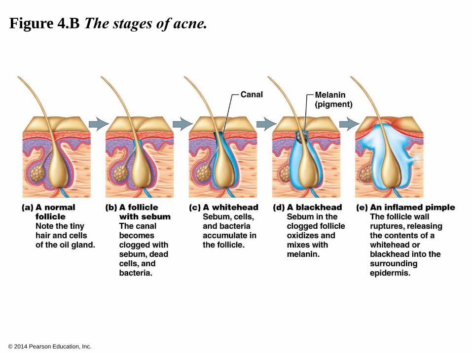
Figure 4.B The stages of acne.
© 2014 Pearson Education, Inc.
Homeostasis
Homeostasis is the constant adjustment made
by the organ systems to respond to changes in
the internal and external environments while
limiting too large variations of the internal
condition required for life
Depends on the nervous and endocrine systems,
which are responsible for internal communication
Maintained primarily through negative feedback
mechanisms
© 2014 Pearson Education, Inc.
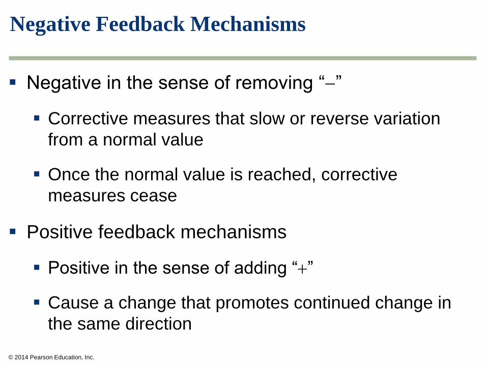
Negative Feedback Mechanisms
Negative in the sense of removing “”
Corrective measures that slow or reverse variation
from a normal value
Once the normal value is reached, corrective
measures cease
Positive feedback mechanisms
Positive in the sense of adding “”
Cause a change that promotes continued change in
the same direction
© 2014 Pearson Education, Inc.
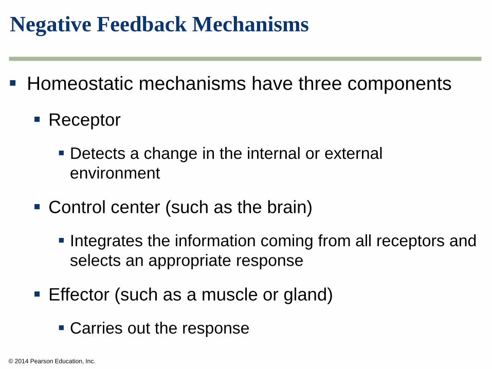
Negative Feedback Mechanisms
Homeostatic mechanisms have three components
Receptor
Detects a change in the internal or external
environment
Control center (such as the brain)
Integrates the information coming from all receptors and
selects an appropriate response
Effector (such as a muscle or gland)
Carries out the response
© 2014 Pearson Education, Inc.
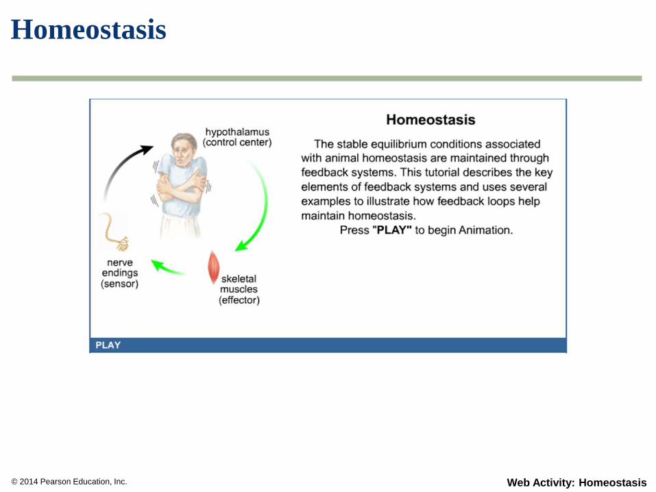
Homeostasis
Web Activity: Homeostasis

© 2014 Pearson Education, Inc.
Figure 4.12 The components of a homeostatic control system
maintained by negative feedback mechanisms.
© 2014 Pearson Education, Inc.
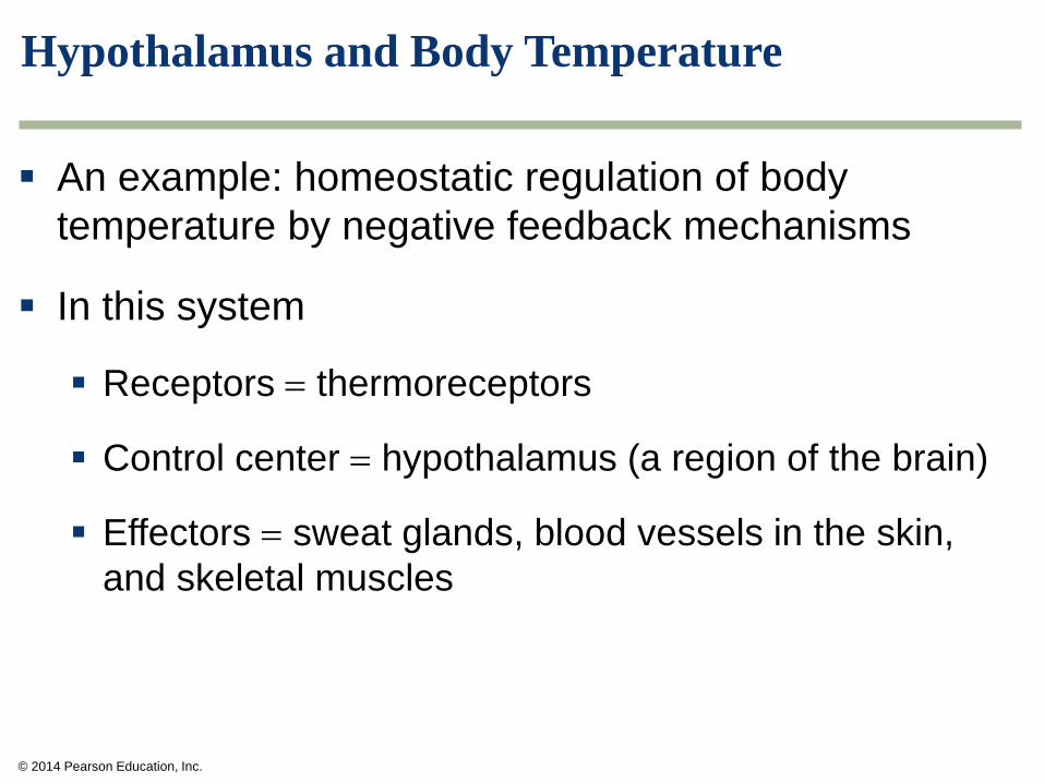
Hypothalamus and Body Temperature
An example: homeostatic regulation of body
temperature by negative feedback mechanisms
In this system
Receptors thermoreceptors
Control center hypothalamus (a region of the brain)
Effectors sweat glands, blood vessels in the skin,
and skeletal muscles
© 2014 Pearson Education, Inc.
Hypothalamus and Body Temperature
Hyperthermia and hypothermia are life-threatening
conditions
Hyperthermia: Abnormally elevated body temperature
Hypothermia: Abnormally low body temperature
© 2014 Pearson Education, Inc.
You Should Now Be Able To:
Know the four types of tissues in the human body and their
structure and functions
Know the three types of cell junctions
Understand how organs are combined into organ systems
Know the human body cavities
Know the structure and function of the skin and list its
derivative
Understand the risks of excessive sun exposure
Understand the mechanisms involved in homeostasis and
compare negative and positive feedback loops