BLOOD plasma proteins. Plasma Proteins Plasma contains a large variety of proteins including...
21
BLOOD plasma proteins
-
Upload
elisabeth-patterson -
Category
Documents
-
view
248 -
download
5
Transcript of BLOOD plasma proteins. Plasma Proteins Plasma contains a large variety of proteins including...
BLOOD plasma
proteins
Plasma Proteins
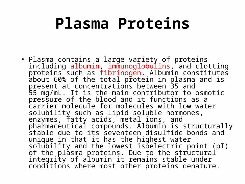
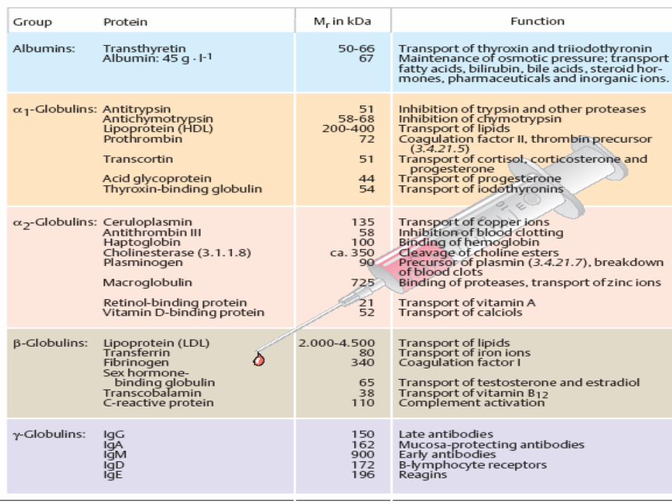
• Plasma contains a large variety of proteins including albumin, immunoglobulins, and clotting proteins such as fibrinogen. Albumin constitutes about 60% of the total protein in plasma and is present at concentrations between 35 and 55 mg/mL. It is the main contributor to osmotic pressure of the blood and it functions as a carrier molecule for molecules with low water solubility such as lipid soluble hormones, enzymes, fatty acids, metal ions, and pharmaceutical compounds. Albumin is structurally stable due to its seventeen disulfide bonds and unique in that it has the highest water solubility and the lowest isoelectric point (pI) of the plasma proteins. Due to the structural integrity of albumin it remains stable under conditions where most other proteins denature.
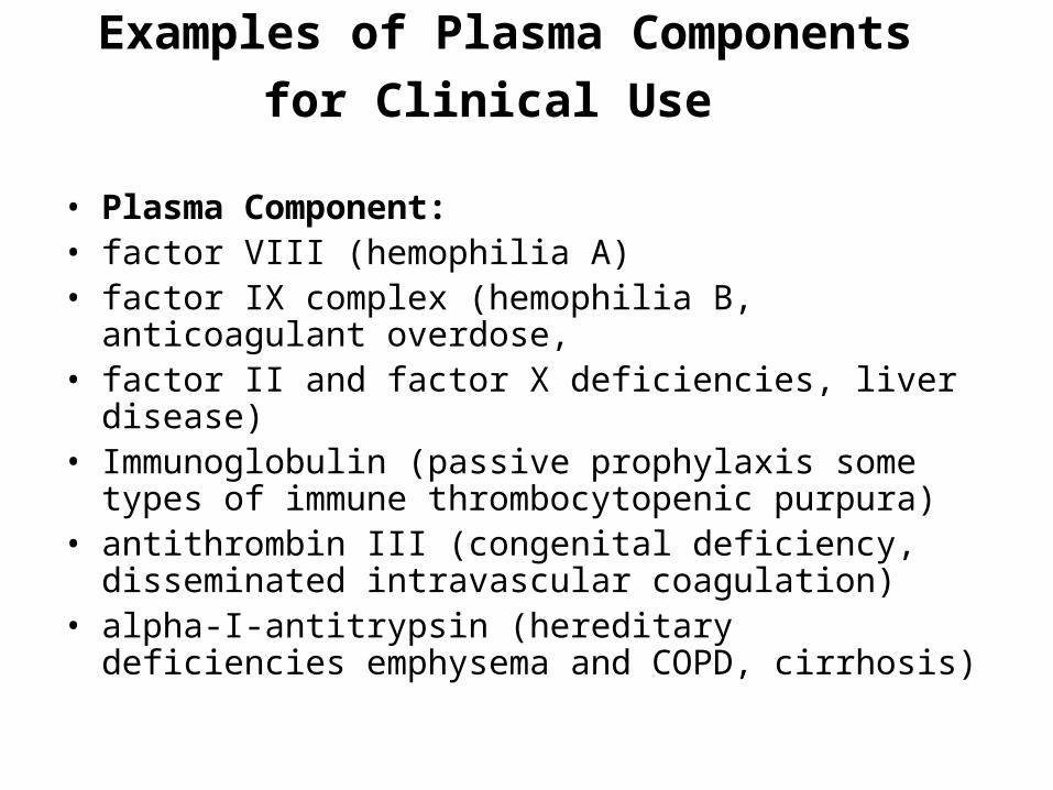
Examples of Plasma Components for Clinical Use
• Plasma Component:• factor VIII (hemophilia A)• factor IX complex (hemophilia B, anticoagulant overdose,• factor II and factor X deficiencies, liver disease)• Immunoglobulin (passive prophylaxis some types of immune
thrombocytopenic purpura)• antithrombin III (congenital deficiency, disseminated
intravascular coagulation)• alpha-I-antitrypsin (hereditary deficiencies emphysema and
COPD, cirrhosis)
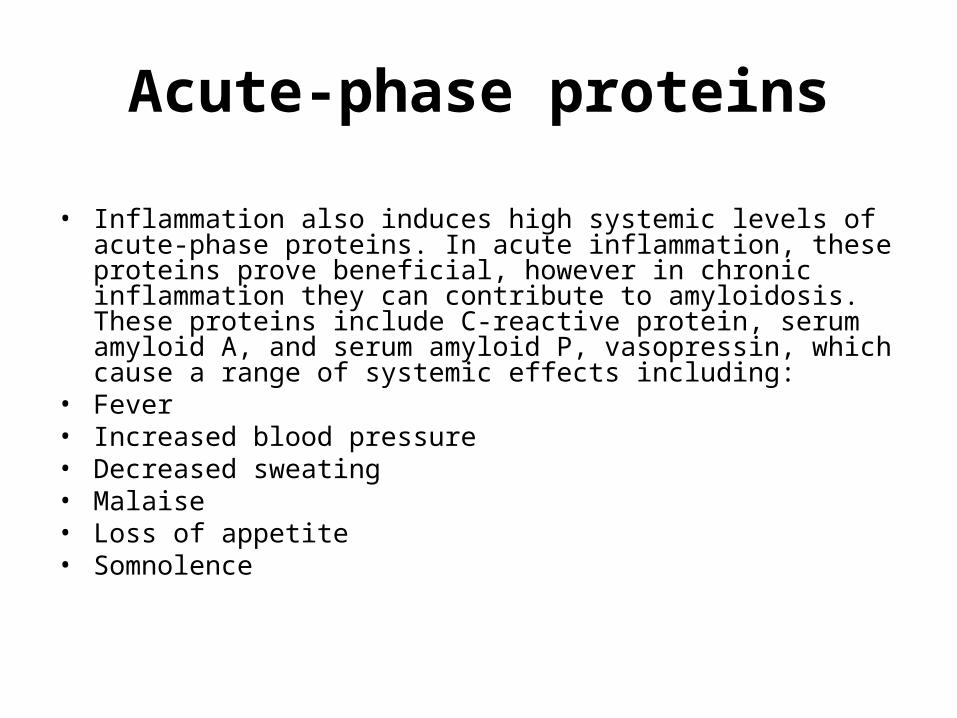
Acute-phase proteins
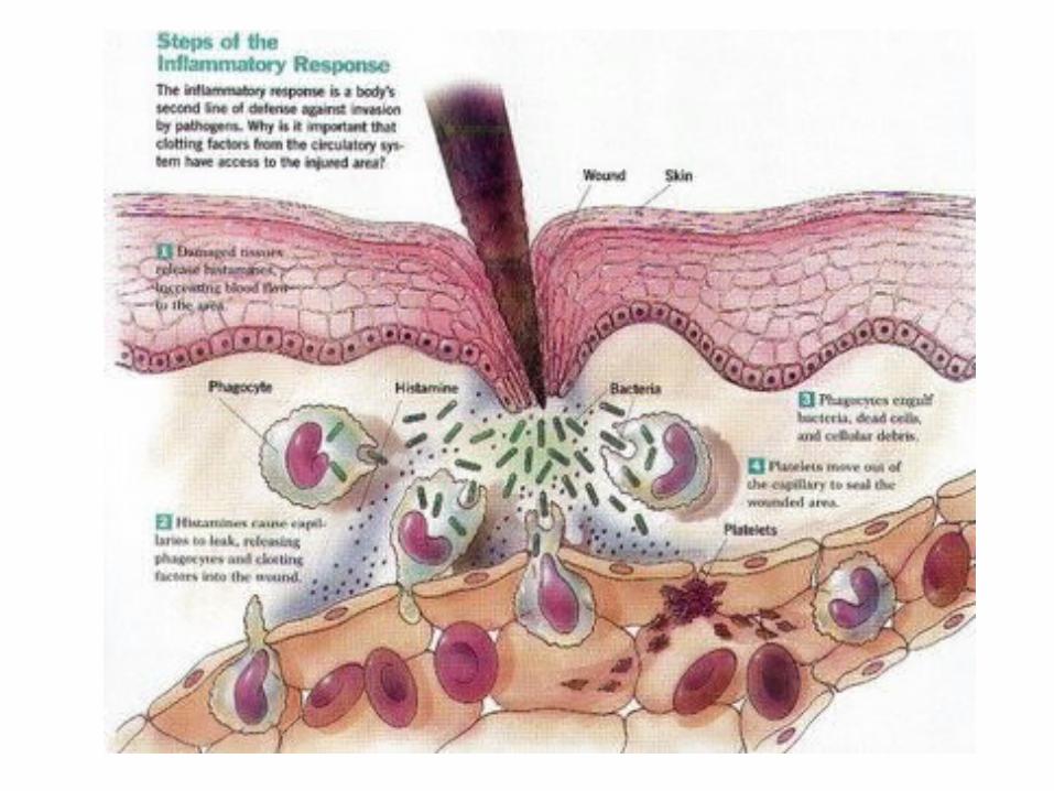
• Inflammation also induces high systemic levels of acute-phase proteins. In acute inflammation, these proteins prove beneficial, however in chronic inflammation they can contribute to amyloidosis. These proteins include C-reactive protein, serum amyloid A, and serum amyloid P, vasopressin, which cause a range of systemic effects including:
• Fever• Increased blood pressure• Decreased sweating• Malaise • Loss of appetite• Somnolence
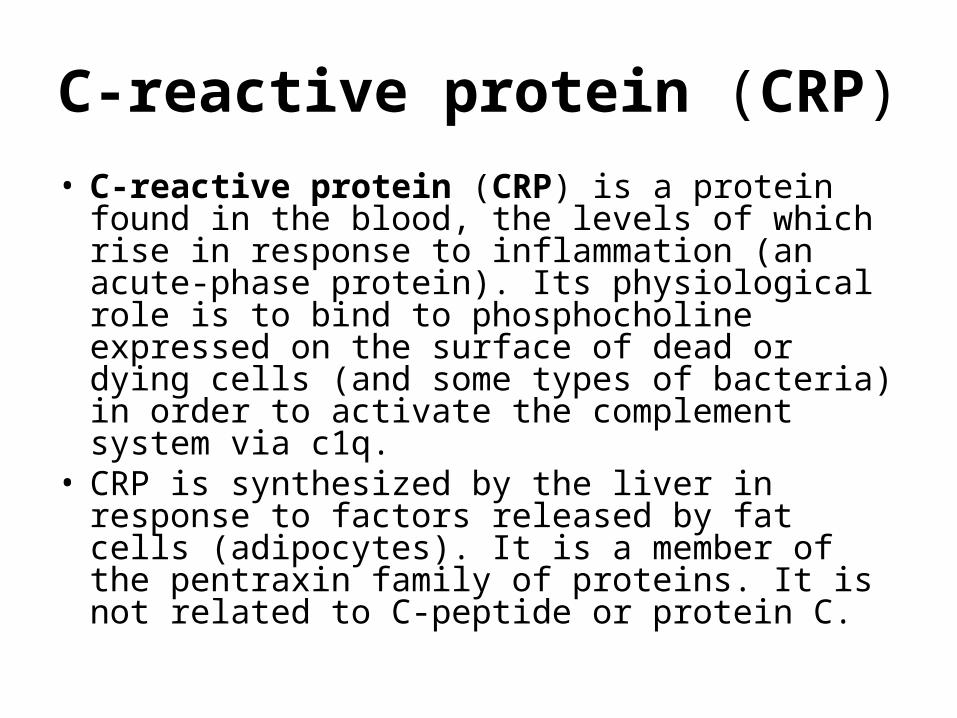
C-reactive protein (CRP)
• C-reactive protein (CRP) is a protein found in the blood, the levels of which rise in response to inflammation (an acute-phase protein). Its physiological role is to bind to phosphocholine expressed on the surface of dead or dying cells (and some types of bacteria) in order to activate the complement system via c1q.
• CRP is synthesized by the liver in response to factors released by fat cells (adipocytes). It is a member of the pentraxin family of proteins. It is not related to C-peptide or protein C.
Serum amyloid A (SAA)
• Serum amyloid A (SAA) proteins are a family of apolipoproteins associated with high-density lipoprotein(HDL) in plasma. Different isoforms of SAA are expressed constitutively (constitutive SAAs) at different levels or in response to inflammatory stimuli. These proteins are produced predominantly by the liver. The conservation of these proteins throughout invertebrates and vertebrates suggests that SAAs play a highly essential role in all animals.
• Acute-phase serum amyloid A proteins (A-SAAs) are secreted during the acute phase of inflammation. These proteins have several roles, including the transport of cholesterol to the liver for secretion into the bile, the recruitment of immune cells to inflammatory sites, and the induction of enzymes that degrade extracellular matrix. A-SAAs are implicated in several chronic inflammatory diseases, such as amyloidosis, atherosclerosis, and rheumatoid arthritis. Three acute-phase SAA isoforms have been reported in mice, called SAA1, SAA2, and SAA3. During inflammation, SAA1 and SAA2 are expressed and induced principally in the liver, whereas SAA3 is induced in many distinct tissues. SAA1 and SAA2 genes are regulated in liver cells by the proinflammatory cytokines IL-1, IL-6, and TNF-α. Both SAA1 and SAA2 are induced up to a 1000-fold in mice under acute inflammatory conditions following exposure to bacterial lipopolysaccharide (LPS). Three A-SAA genes have also been identified in humans[4], although the third gene, SAA3, is believed to represent a pseudogene that does not generate messenger RNA or protein

The main classes of lipoproteins
1.Chylomicrons.
2.Very low density lipoproteins (VLDL).
3.Intermediate density lipoproteins (IDL).
4.Low density lipoproteins (LDL).
5.High density lipoproteins (HDL).
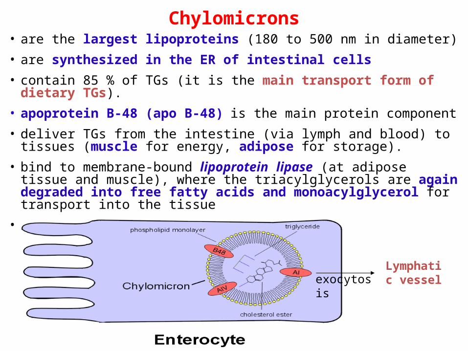
• are the largest lipoproteins (180 to 500 nm in diameter)
• are synthesized in the ER of intestinal cells
• contain 85 % of TGs (it is the main transport form of dietary TGs).
• apoprotein B-48 (apo B-48) is the main protein component
• deliver TGs from the intestine (via lymph and blood) to tissues (muscle for energy, adipose for storage).
• bind to membrane-bound lipoprotein lipase (at adipose tissue and muscle), where the triacylglycerols are again degraded into free fatty acids and monoacylglycerol for transport into the tissue
• are present in blood only after feeding
Chylomicrons
exocytosisLymphatic
vessel
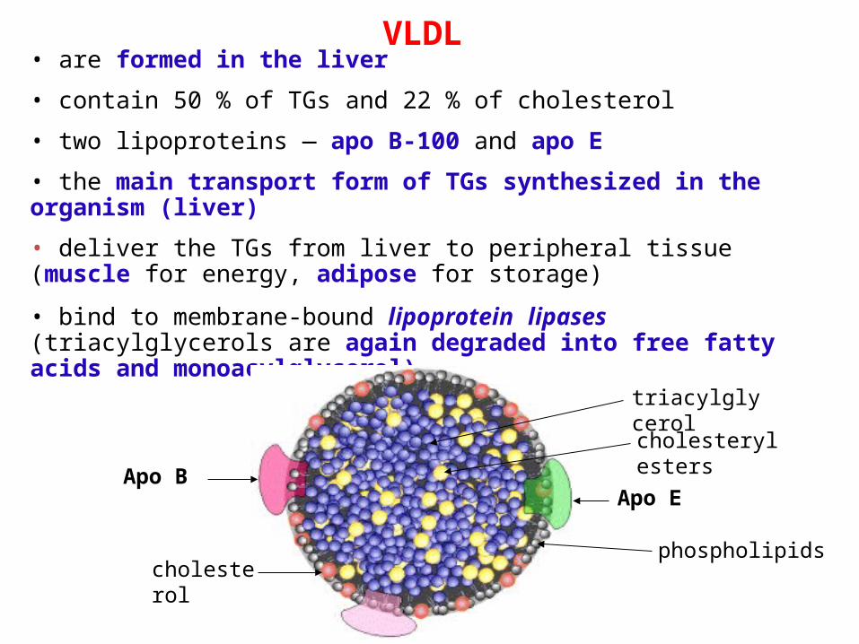
VLDL• are formed in the liver
• contain 50 % of TGs and 22 % of cholesterol
• two lipoproteins — apo B-100 and apo E
• the main transport form of TGs synthesized in the organism (liver)
• deliver the TGs from liver to peripheral tissue (muscle for energy, adipose for storage)
• bind to membrane-bound lipoprotein lipases (triacylglycerols are again degraded into free fatty acids and monoacylglycerol)
Apo BApo E
triacylglycerol
cholesteryl esters
phospholipidscholesterol
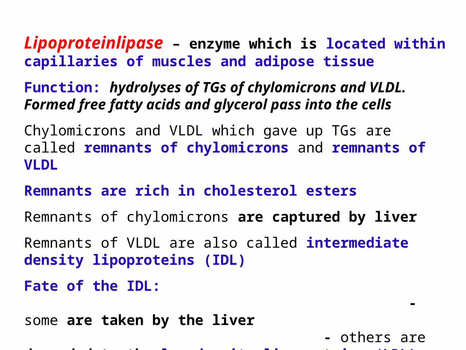
Lipoproteinlipase – enzyme which is located within capillaries of muscles and adipose tissue
Function: hydrolyses of TGs of chylomicrons and VLDL. Formed free fatty acids and glycerol pass into the cells
Chylomicrons and VLDL which gave up TGs are called remnants of chylomicrons and remnants of VLDL
Remnants are rich in cholesterol esters
Remnants of chylomicrons are captured by liver
Remnants of VLDL are also called intermediate density lipoproteins (IDL)
Fate of the IDL: - some are taken by the liver - others are degraded to the low density lipoproteins (LDL) (by the removal of more triacylglycerol)

LDL
LDL are formed in the blood from IDL and in liver from IDL (enzyme – liver lipase)
LDL are enriched in cholesterol and cholesteryl esters (contain about 50 % of cholesterol)
Protein component - apo B-100
LDL is the major carrier of cholesterol (transport cholesterol to peripheral tissue)

Cells of all organs have LDL receptors
Receptors for LDL are localized in specialized regions called coated pits, which contain a specialized protein called clathrin
Apo B-100 on the surface of an LDL binds to the receptor
Receptor-LDL complex enters the cell by endocytosis.
Endocytic vesicle is formed

Vesicle fuse with lysosomes
Lysosomal lipases and proteases degrade LDL
LDL receptor itself returns to the plasma membrane
Apo B-100 is hydrolyzed to amino acids
Cholesteryl esters are hydrolyzed to free cholesterol and fatty acids
Released free cholesterol: - is incorporated into the membranes or - is reesterified for storage inside the cell by the enzyme acyl CoA:cholesterol acyltransferase (ACAT)
Feedback regulation: abundance of intracellular cholesterol suppresses the synthesis of LDL receptors and so the uptake of additional cholesterol from plasma LDL is blocked
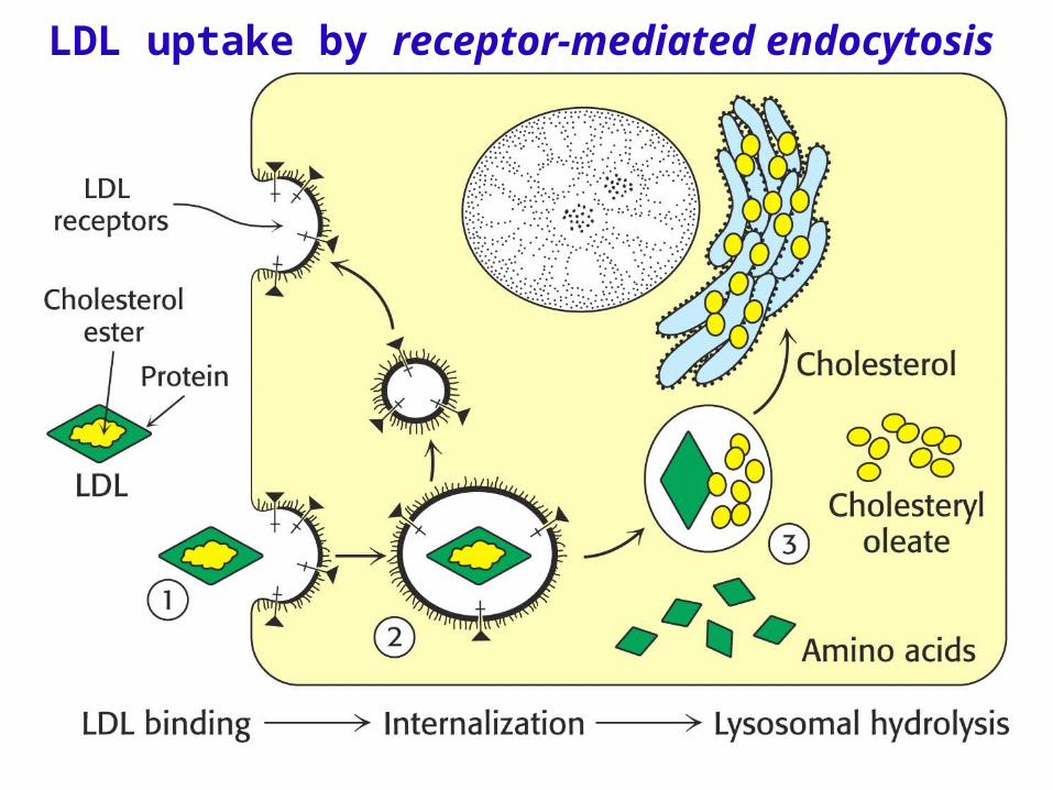
LDL uptake by receptor-mediated endocytosis

congenital disease when LDL receptor are not synthesized (mutation at a single autosomal locus)
the concentration of cholesterol in blood markedly increases
severe atherosclerosis is developed (deposition of cholesterol in arteries)
nodules of cholesterol called xanthomas are prominent in skin and tendons
most homozygotes die of coronary artery disease in childhood
the disease in heterozygotes (1 in 500 people) has a milder and more variable clinical course
Familial hypercholesterolemia
atherosclerosis
xanthomas
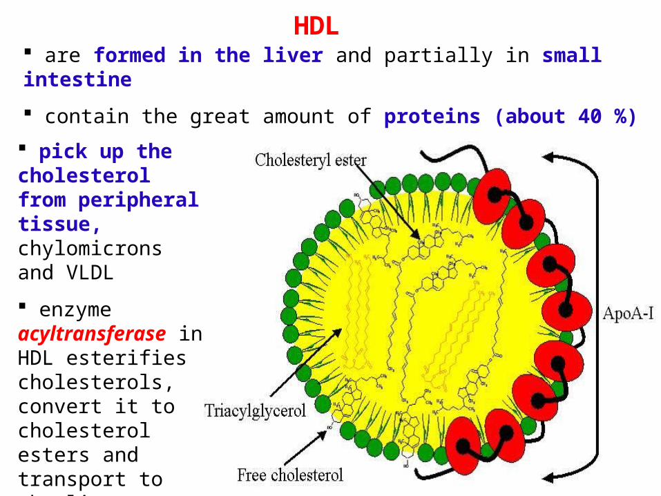
HDL are formed in the liver and partially in small intestine
contain the great amount of proteins (about 40 %)
pick up the cholesterol from peripheral tissue, chylomicrons and VLDL
enzyme acyltransferase in HDL esterifies cholesterols, convert it to cholesterol esters and transport to the liver

High serum levels of cholesterol cause disease and death by contributing to development of atherosclerosis
Cholesterol which is present in the form of the LDL is so-called "bad cholesterol."
Cholesterol in the form of HDL is referred to as "good cholesterol”
HDL functions as a shuttle that moves cholesterol throughout the body
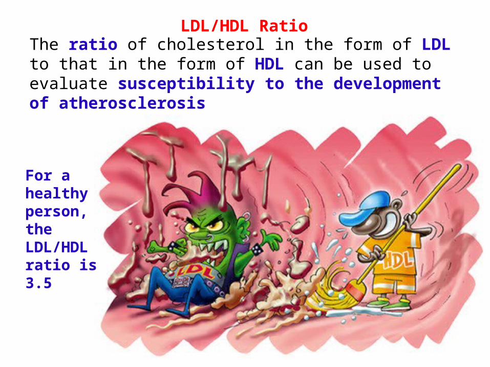
The ratio of cholesterol in the form of LDL to that in the form of HDL can be used to evaluate susceptibility to the development of atherosclerosis
LDL/HDL Ratio
For a healthy person, the LDL/HDL ratio is 3.5
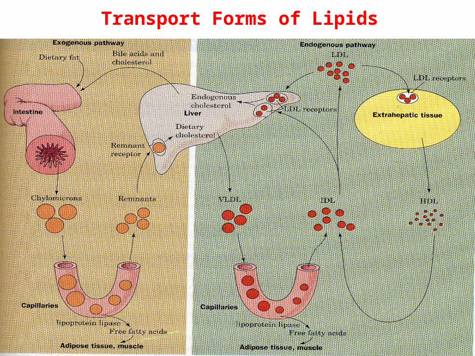
Transport Forms of Lipids