
Blood cicrulation disturbanses 1
51
Disturbances in Disturbances in interstitial fluid interstitial fluid amount. amount. Disturbances of blood Disturbances of blood circulation circulation : : venous venous hyperemia, bleeding hyperemia, bleeding , , hemorrhage, stasis hemorrhage, stasis . . Shock Shock . .
-
Upload
niklzlesnik -
Category
Education
-
view
80 -
download
0
description
Blood cicrulation disturbanses 1
Transcript of Blood cicrulation disturbanses 1
Disturbances in interstitial Disturbances in interstitial fluid amount. fluid amount.
Disturbances of blood Disturbances of blood circulationcirculation:: venous venous hyperemia, bleedinghyperemia, bleeding, , hemorrhage, stasishemorrhage, stasis..
ShockShock..

DISTURBANCES IN INTERSTITIAL FLUID DISTURBANCES IN INTERSTITIAL FLUID AMOUNT MAY BEAMOUNT MAY BE::
increased containment of interstitial increased containment of interstitial fluidfluid ( (edemaedema))
decreased decreased ((reductionreduction)) containment containment of interstitial fluid of interstitial fluid ((dehydratationdehydratation))

PATHOGENETIC CLASSIFICATION OF PATHOGENETIC CLASSIFICATION OF EDEMASEDEMAS::
hydrostatichydrostatic ооncoticncotic ( (colloid-osmoticcolloid-osmotic)) membranogenicmembranogenic from retention of water and from retention of water and
electrolyteselectrolytes lymphogeniclymphogenic
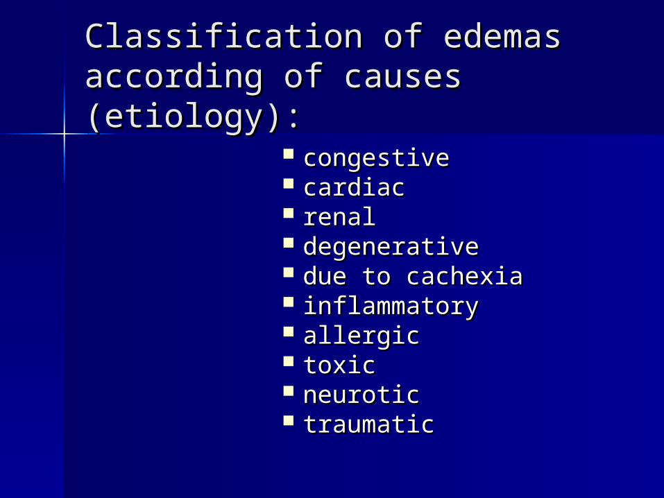
Classification of edemas Classification of edemas according of causes (etiology)according of causes (etiology)::
congestive congestive cardiaccardiac renalrenal degenerativedegenerative due to cachexiadue to cachexia inflammatoryinflammatory allergicallergic toxictoxic neuroticneurotic traumatictraumatic
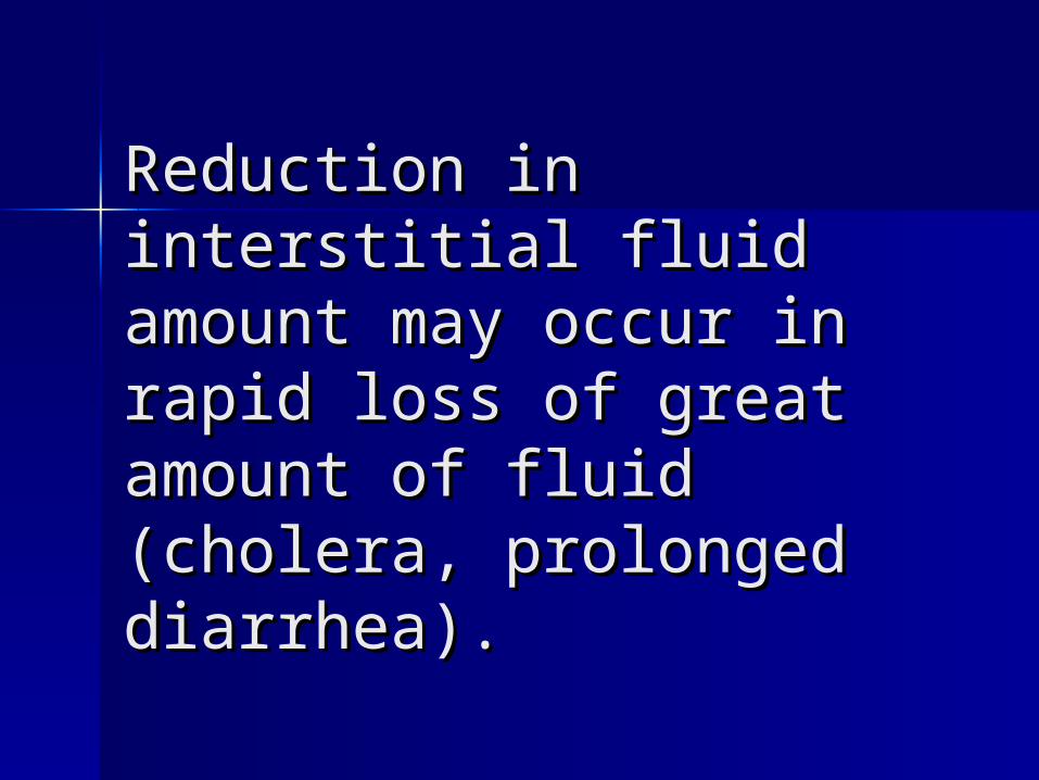
Reduction in interstitial Reduction in interstitial fluid amount may occur in fluid amount may occur in rapid loss of great amount rapid loss of great amount of fluid (cholera, of fluid (cholera, prolonged diarrhea).prolonged diarrhea).
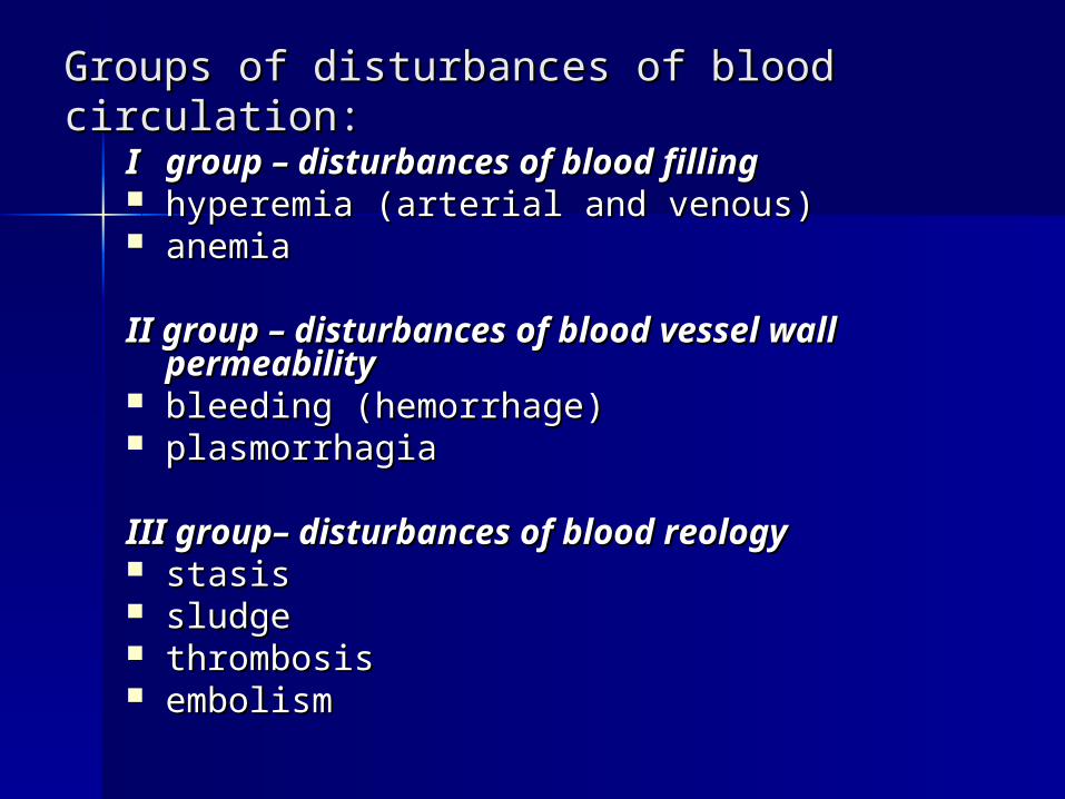
Groups of disturbances of blood circulationGroups of disturbances of blood circulation::
II groupgroup – – disturbances of blood fillingdisturbances of blood filling hyperemiahyperemia ( (arterial and venousarterial and venous)) anemiaanemia
II groupII group – – disturbances of blood vessel disturbances of blood vessel wall permeabilitywall permeability
bleedingbleeding ( (hemorrhagehemorrhage)) plasmorrhagiaplasmorrhagia
IIIIII groupgroup– – disturbances of blood reologydisturbances of blood reology stasisstasis sludgesludge thrombosisthrombosis embolismembolism

Arterial hyperemiaArterial hyperemia – – is is increased blood filling of the organ or increased blood filling of the organ or tissue due to increased flow of the tissue due to increased flow of the arterial bloodarterial blood (may be physiological)(may be physiological)
angioneuroticangioneurotic ( (neuroparalyticneuroparalytic)) collateralcollateral postanemicpostanemic vacantvacant inflammatoryinflammatory hyperemia caused by angiovenous hyperemia caused by angiovenous
fistulafistula
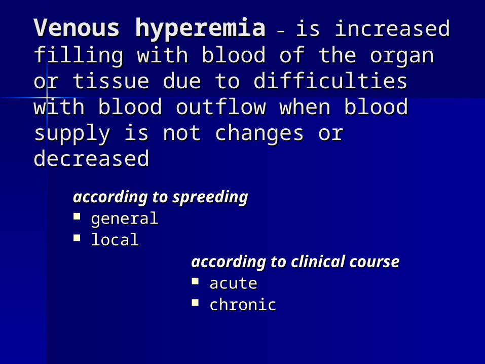
Venous hyperemiaVenous hyperemia – – is increased is increased filling with blood of the organ or tissue filling with blood of the organ or tissue due to difficulties with blood outflow due to difficulties with blood outflow when blood supply is not changes or when blood supply is not changes or decreaseddecreased
according to according to spreedingspreeding
generalgeneral locallocal according to clinical courseaccording to clinical course
acuteacute chronicchronic

Diseases which accompany Diseases which accompany formation of general venouse blood formation of general venouse blood congestioncongestion::
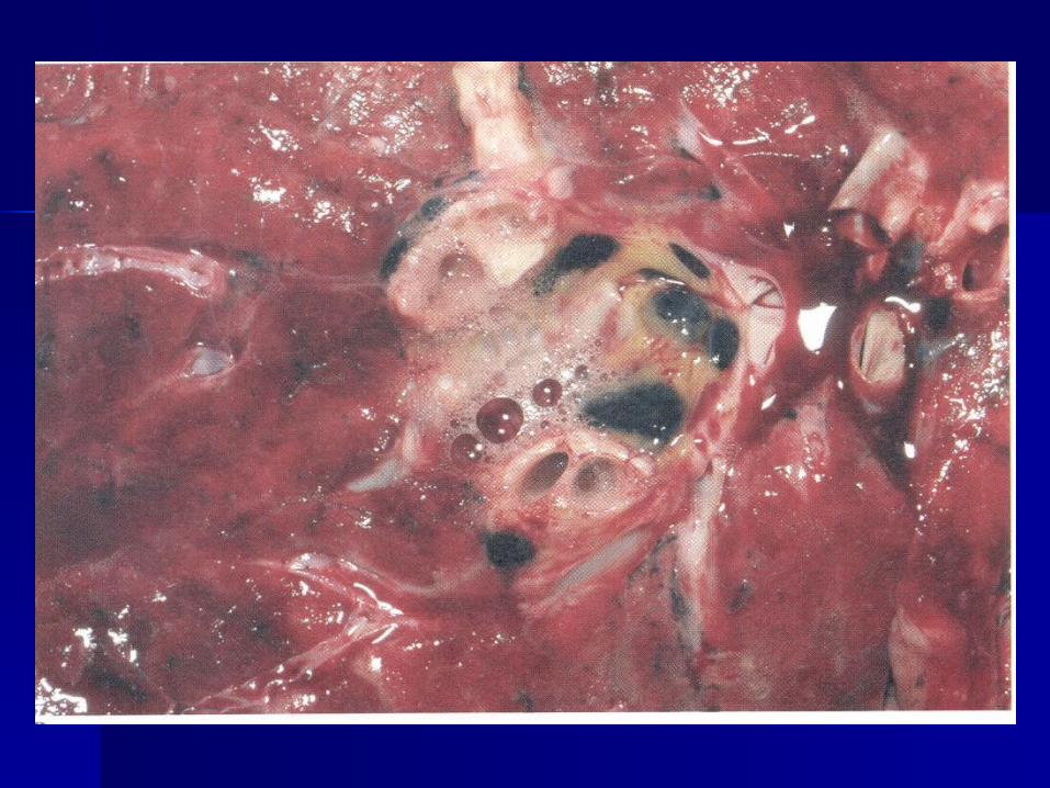
Acute general venous congestion Acute general venous congestion infarction myocardial infarction myocardial acute myocarditis acute myocarditis myocardiodysthrophiesmyocardiodysthrophies Chronic general venous congestionChronic general venous congestion decompensation of muocardial hypertrophy in decompensation of muocardial hypertrophy in
cases of primary essential hypertension, cases of primary essential hypertension, mitral stenosis at other, mitral stenosis at other,
cardiosclerosis of different genesis (chronic cardiosclerosis of different genesis (chronic ischemic disease, chronic myocarditis)ischemic disease, chronic myocarditis)
chronic diseases of lung with chronic diseases of lung with pneumosclerosispneumosclerosis

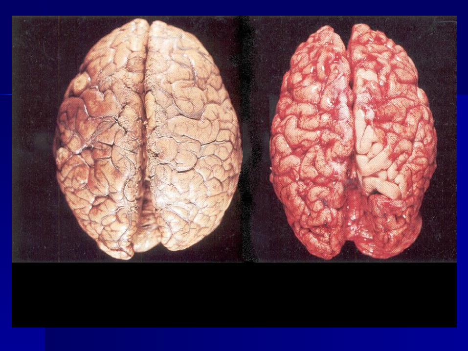
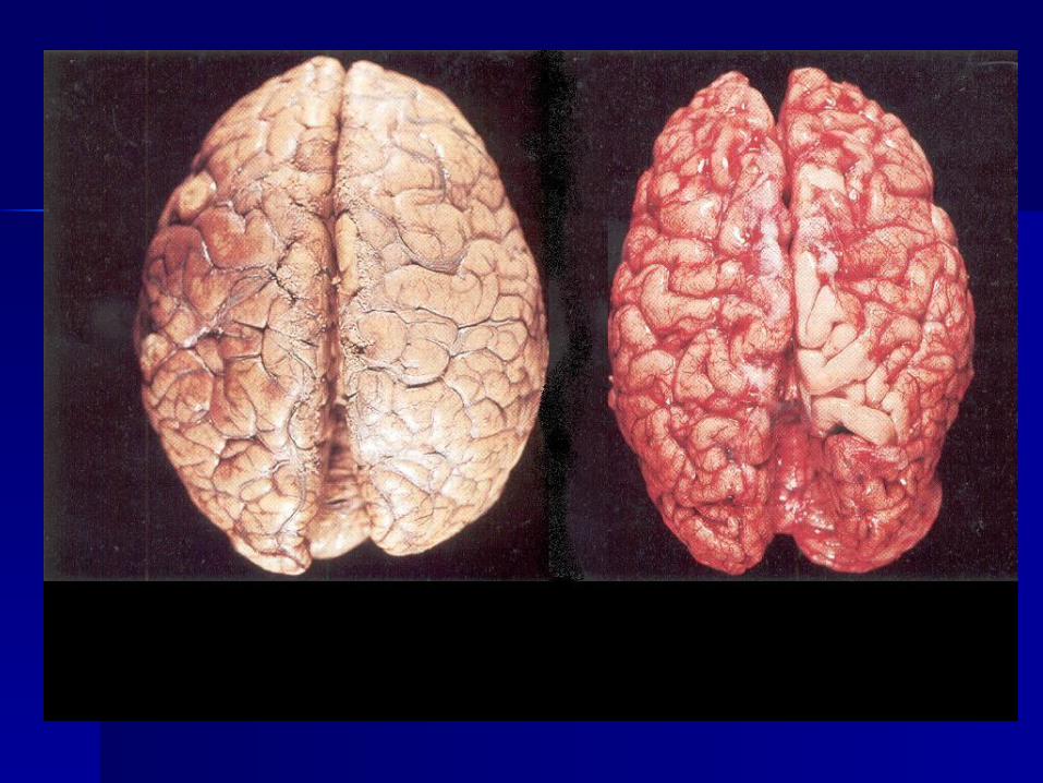
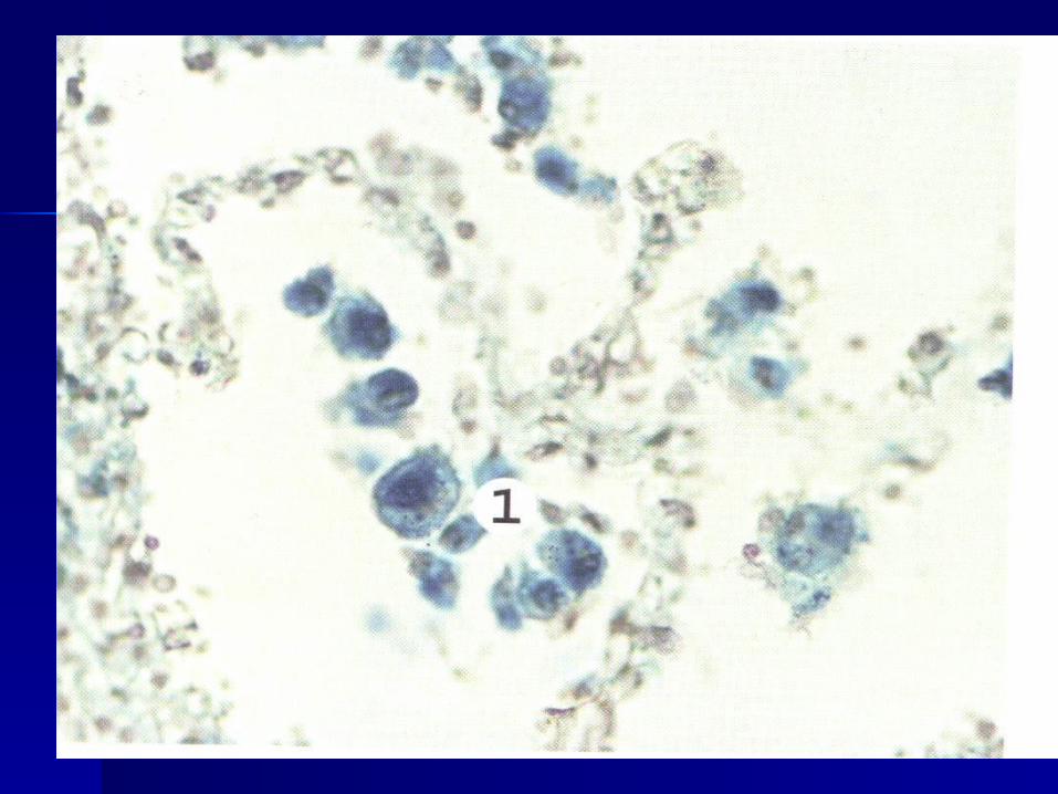
Microscopical changes Microscopical changes of organs and tissues in of organs and tissues in acute venous congestionacute venous congestion::
stretched and plethoric veinsstretched and plethoric veins and venulesand venules stasis and sludge in capillariesstasis and sludge in capillaries due to due to
disturbances of flow of blood,disturbances of flow of blood, exit of fluid part of plasma with proteins exit of fluid part of plasma with proteins
from vessels walls (plasmorrhagia)from vessels walls (plasmorrhagia) with development of oedema as with development of oedema as
consequence,consequence, exit of erithrocytes through vessels wall with exit of erithrocytes through vessels wall with
development of multiple diapedesis development of multiple diapedesis hemorrhageshemorrhages ,,
development of degeneration or necrosis of development of degeneration or necrosis of parenchymatous cells.parenchymatous cells.


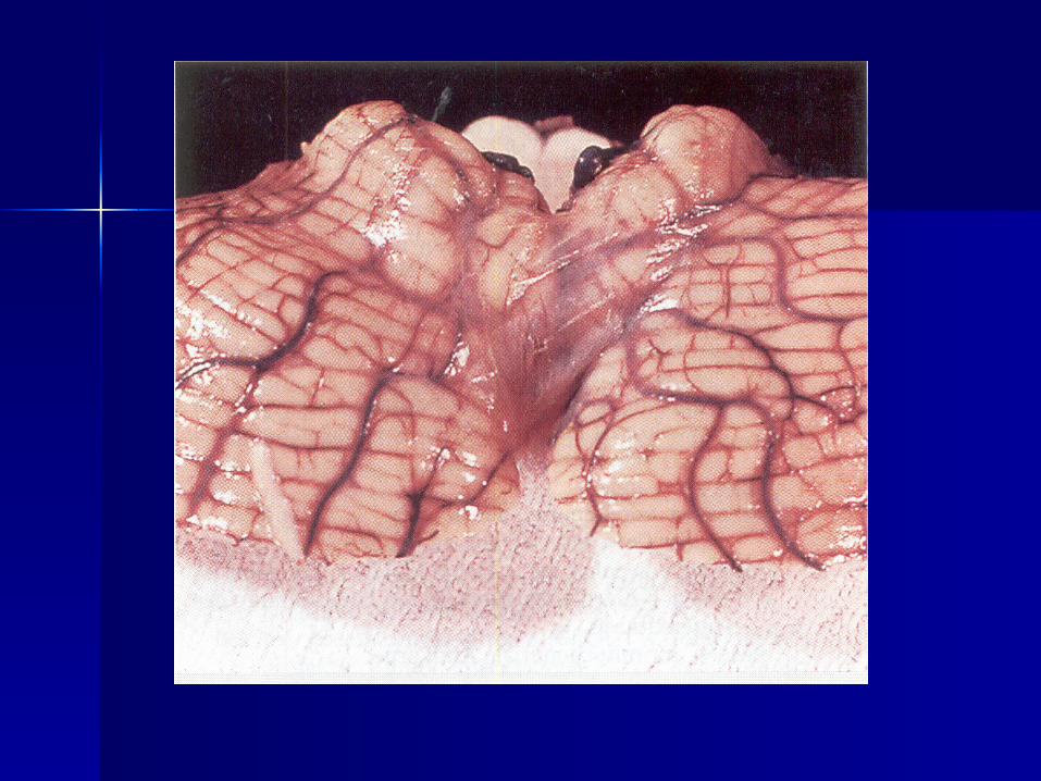
Microscopical changes in Microscopical changes in chronic venous congestion chronic venous congestion ::
the same as in acute general the same as in acute general venous hyperemiavenous hyperemia
plusplus atrophy of parenchymatous cells, atrophy of parenchymatous cells, sclerosis, because it's known that sclerosis, because it's known that
collagen fibroblast production collagen fibroblast production increases due to chronic hypoxiaincreases due to chronic hypoxia
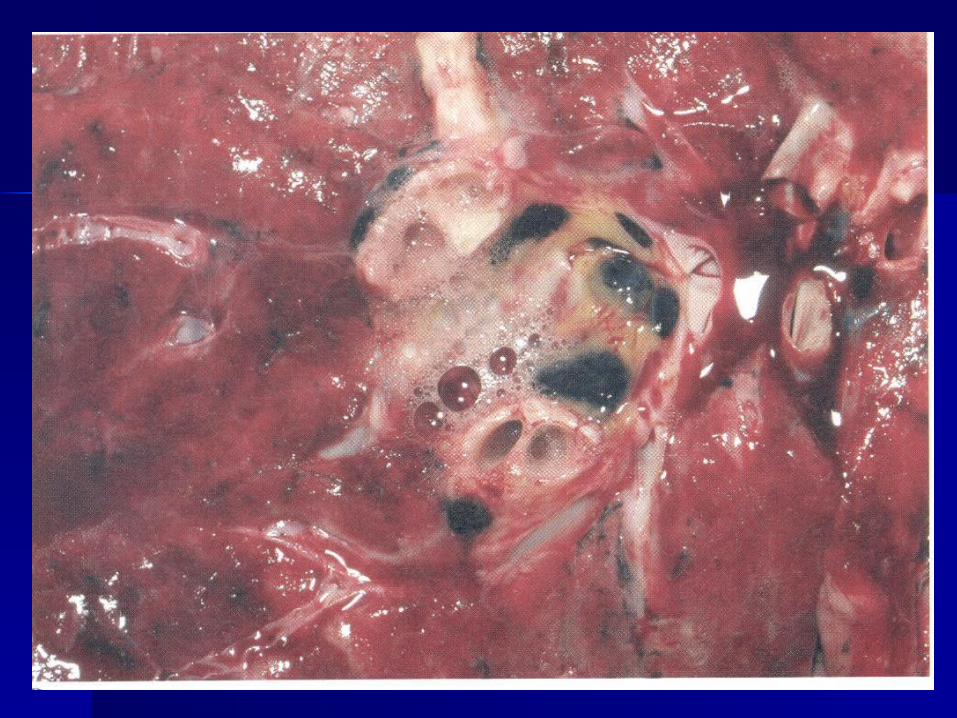
Morphological changes in organs and Morphological changes in organs and tissues which are occurred in corpses at tissues which are occurred in corpses at autopsy in cases of chronic cardiovascular autopsy in cases of chronic cardiovascular insufficiencyinsufficiency::
cyanosis of skin.cyanosis of skin. Oedema of subcutaneous fatty tissueOedema of subcutaneous fatty tissue ascitis (hydroperitoneum), ascitis (hydroperitoneum),
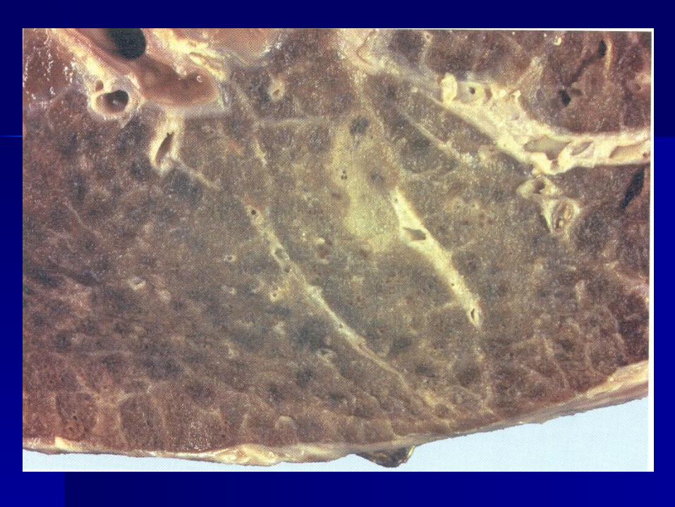
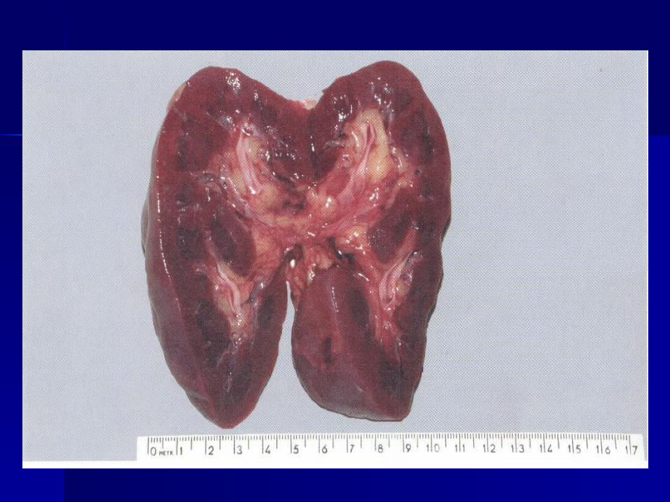
hydrothorax, hydropericardiumhydrothorax, hydropericardium "nutmeg" liver"nutmeg" liver "brown induration" of the lungs"brown induration" of the lungs cyanotic induration of kidneys and cyanotic induration of kidneys and
spleenspleen


IIII groupgroup – – disturbances of disturbances of blood vessel wall blood vessel wall permeabilitypermeability
bleedingbleeding ((hemorrhagehemorrhage))
plasmorrhagiaplasmorrhagia
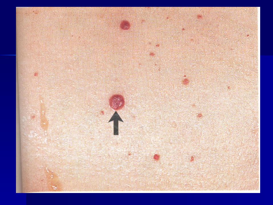
epistaxis - bleeding from the nose, epistaxis - bleeding from the nose, hemotemesis - vomiting of blood, hemotemesis - vomiting of blood, melena – faeces like tar in case of bleeding from melena – faeces like tar in case of bleeding from
the intestine,the intestine, hemoperitoneum,hemoperitoneum, hemothorax, hemothorax, hemopericardium hemopericardium hemartrosishemartrosis hematometrahematometra
Bleeding Bleeding (hemorrhage(hemorrhage)) is exit of the blood is exit of the blood from the lumen of the vessel or from the from the lumen of the vessel or from the heart cavityheart cavity which may accompany which may accompany
accumulation ofaccumulation of the blood in tissues the blood in tissues
(hemorrhage(hemorrhage))

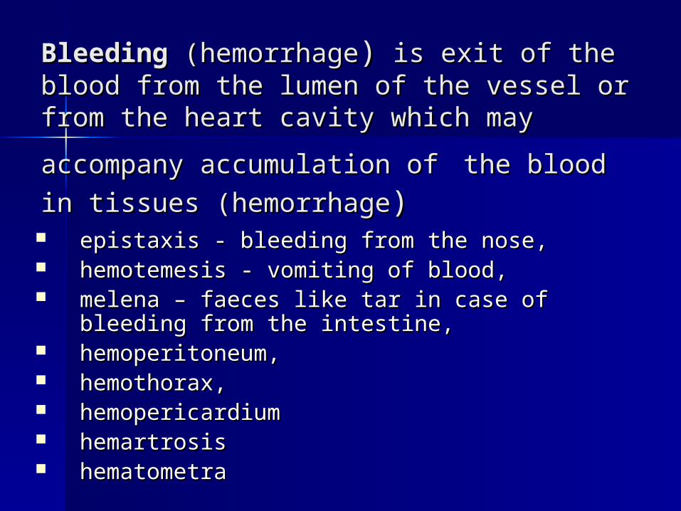
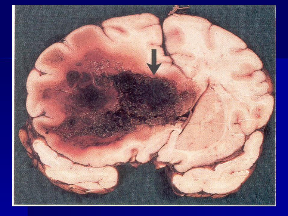
Types of hemorrhagesTypes of hemorrhages hematomahematoma - accumulation of blood in - accumulation of blood in
tissues with their destruction (which is tissues with their destruction (which is associated with tissue necrosis)associated with tissue necrosis)
hemorrhagic saturationhemorrhagic saturation – – accumulation of blood in tissues accumulation of blood in tissues without their destructionwithout their destruction
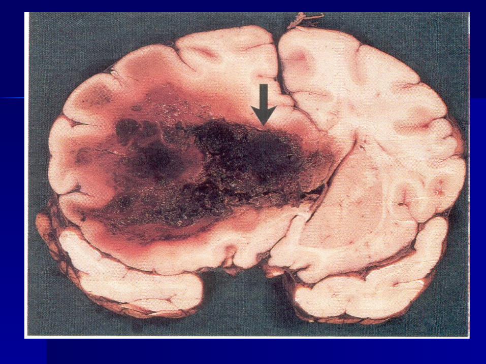
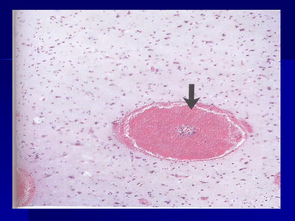
ecchymosesecchymoses – – multiple small multiple small petechias (point-like hemorrhage) per petechias (point-like hemorrhage) per diapedesisdiapedesis
bruisebruise – – large extravasations of blood large extravasations of blood into the skin and mucous membranesinto the skin and mucous membranes


Outcomes of hemorrhagesOutcomes of hemorrhages::
blood resorptionblood resorption local hemosiderosislocal hemosiderosis organizationorganization encapsulationencapsulation petrifaction (calcification)petrifaction (calcification) cyst formation (brain)cyst formation (brain) suppurationsuppuration
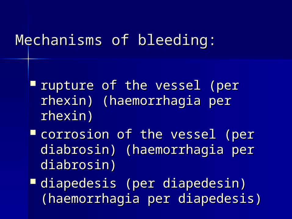
Mechanisms of bleedingMechanisms of bleeding::
rupture of the vessel (per rhexin)rupture of the vessel (per rhexin) ((haemorrhagia per rhexinhaemorrhagia per rhexin))
corrosion of the vessel (per corrosion of the vessel (per diabrosin) diabrosin) ((haemorrhagia per haemorrhagia per diabrosindiabrosin))
diapedesis (per diapedesin)diapedesis (per diapedesin) ((haemorrhagia per diapedesishaemorrhagia per diapedesis))

According to the origin According to the origin bleedings includebleedings include
arterialarterial venousvenous
capillariscapillaris parenchymatousparenchymatous
from heartfrom heart,,s cavitiess cavities
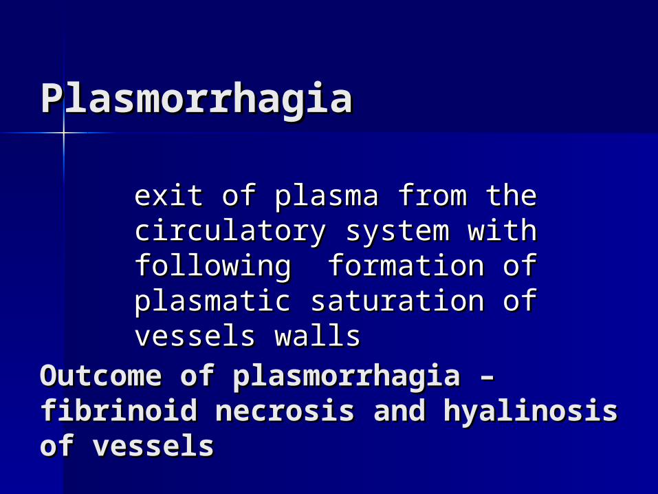
PlasmorrhagiaPlasmorrhagia
exit of plasma from the exit of plasma from the circulatory systemcirculatory system with with following following formation of formation of plasmatic saturation of plasmatic saturation of vessels wallsvessels walls
Outcome of plasmorrhagiaOutcome of plasmorrhagia – – fibrinoid necrosis and hyalinosisfibrinoid necrosis and hyalinosis of of vesselsvessels

StasisStasis is arrest of blood flow in the vessels of is arrest of blood flow in the vessels of microcirculatory system (capillaries). microcirculatory system (capillaries).
Causes of stasisCauses of stasis::- physical factors (temperature physical factors (temperature
elevation, cold); elevation, cold); - chemical factors; chemical factors; - infection; infection; - infectious-allergic factors; infectious-allergic factors; - autoimmune factors.autoimmune factors.
Sludge Sludge syndrome syndrome (phenomenon) is regarded (phenomenon) is regarded as a type of stasisas a type of stasis
sticking of erythrocytes, sticking of erythrocytes, leukocytes and thrombocytes leukocytes and thrombocytes
to each other, to each other,
which is accompanied by blood which is accompanied by blood viscosity increaseviscosity increase

ShockShock is acute pathologic process is acute pathologic process
due to development of due to development of extrapowerful stimuli and extrapowerful stimuli and
characterized by characterized by disturbances in CNS disturbances in CNS
function metabolism and function metabolism and microcirculatory system microcirculatory system
autoregulalion resulting in autoregulalion resulting in destructive changes in the destructive changes in the
organs and tissues.organs and tissues.
Classification of shock Classification of shock according to etiology and according to etiology and pathogenesispathogenesis Hypovolumic (blood loss, trauma, Hypovolumic (blood loss, trauma,
peritonitis, cholera).peritonitis, cholera). Cardiogenic (caused by the reduction Cardiogenic (caused by the reduction
in cardiac output in myocardial in cardiac output in myocardial infarction, vascular insufficiency).infarction, vascular insufficiency).
Bacterial (caused by endotoxins).Bacterial (caused by endotoxins). Neurogenic (in intoxication with Neurogenic (in intoxication with
hypnotic preparations, hypnotic preparations, ganglioblockers, narcotics).ganglioblockers, narcotics).
Anaphylactic (immediate reaction of Anaphylactic (immediate reaction of hypersensitivity).hypersensitivity).

Stages and morphology of Stages and morphology of shockshock 1st stage – the stage of 1st stage – the stage of hemodynamic changeshemodynamic changes- decrease of blood circulation volume- decrease of blood circulation volume- blood depot in skin, intestine, liver, spleen- blood depot in skin, intestine, liver, spleen- blood shunt in kidneys, liver, lungs- blood shunt in kidneys, liver, lungs 2st stage – the stage of 2st stage – the stage of reologic changesreologic changes- stasises, sludges and microthrombosis in - stasises, sludges and microthrombosis in
microcirculatory systemmicrocirculatory system- vascular permeability increase (plasmorrhagia, - vascular permeability increase (plasmorrhagia,
edema)edema)- hemorrhage syndrome- hemorrhage syndrome 3d stage – the stage of 3d stage – the stage of DIC (disseminated DIC (disseminated
intravascular coagulation) syndromeintravascular coagulation) syndrome- hemorrhage syndrome, edema and plasmorrhagia- hemorrhage syndrome, edema and plasmorrhagia- multiple thrombosis in microcirculatory system- multiple thrombosis in microcirculatory system- degeneration and necrosis in organs and tissue from - degeneration and necrosis in organs and tissue from
ishemiaishemia 4st stage – 4st stage – outcomesoutcomes
The most prominent changes The most prominent changes develop in parenchymatous develop in parenchymatous organs.organs.
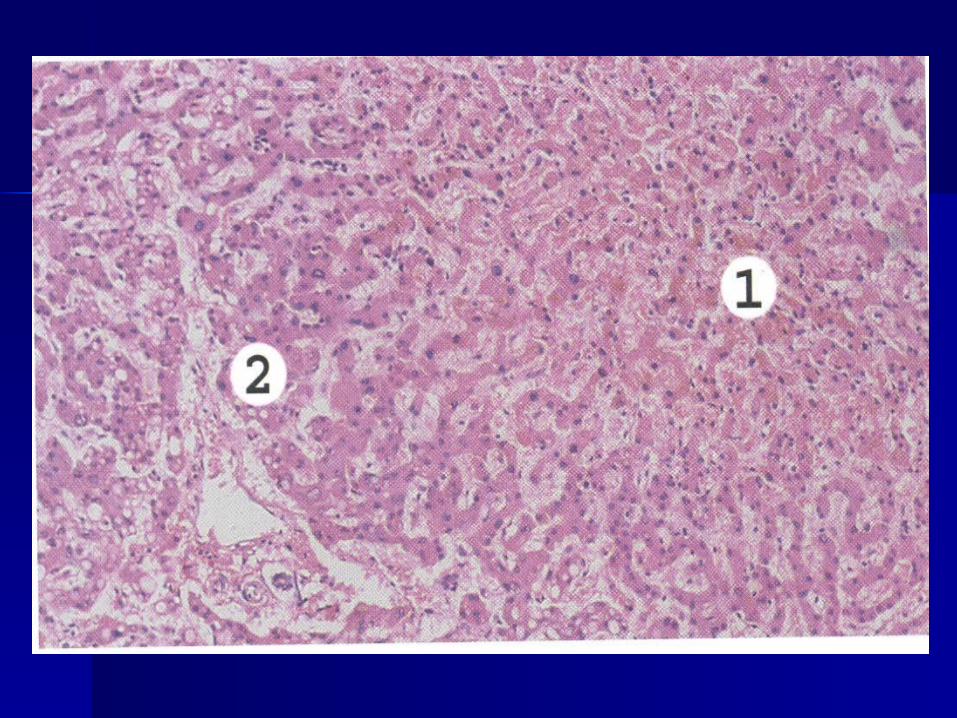
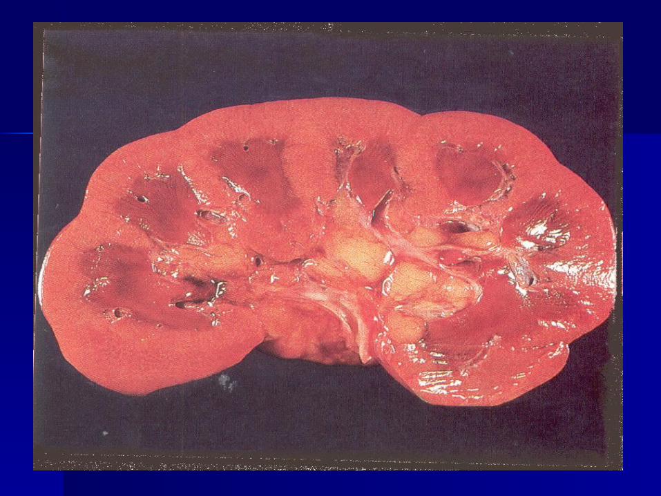
- - shock kidneyshock kidney, degeneration and necrosis in , degeneration and necrosis in proximal canals with development of necrotic proximal canals with development of necrotic nephrosis (or symmetrical cortical necroses) which nephrosis (or symmetrical cortical necroses) which results in acute renal insufficiency;results in acute renal insufficiency;
- - shock livershock liver, glycogen amount in the hepatocytes , glycogen amount in the hepatocytes decreases, hydropic degeneration and decreases, hydropic degeneration and centrolobular necroses resulting in acute hepatic centrolobular necroses resulting in acute hepatic insufficiency. insufficiency.
Combination of renal and hepatic insufficiency is Combination of renal and hepatic insufficiency is called hepatorenal syndrome.called hepatorenal syndrome.
- - shock lungshock lung, atelectasis foci, hemorrhages ,edema, , atelectasis foci, hemorrhages ,edema, hyperemia and thromboses in the microcirculatory hyperemia and thromboses in the microcirculatory resulting in acute respiratory insufficiency.resulting in acute respiratory insufficiency.
- - shock heartshock heart, degeneration and necrosis in , degeneration and necrosis in cardiomyocytes, reduction in glycogen amount, fat cardiomyocytes, reduction in glycogen amount, fat degeneration, necrosis foci.degeneration, necrosis foci.


![Pomalidomide Activity Blood 2012 Dispenzieri Blood 2012-02-413161[1][1]](https://static.fdocuments.us/doc/165x107/577d1d8f1a28ab4e1e8c86af/pomalidomide-activity-blood-2012-dispenzieri-blood-2012-02-41316111.jpg)