
Blood Cardiovascular System. Blood Functions 1. Transports Dissolved gasses Nutrients Waste products...
75
Blood Cardiovascular System
-
Upload
triston-cockayne -
Category
Documents
-
view
218 -
download
0
Transcript of Blood Cardiovascular System. Blood Functions 1. Transports Dissolved gasses Nutrients Waste products...
Blood
Cardiovascular System

Blood Functions
1. Transports • Dissolved gasses• Nutrients• Waste products to lungs and kidneys• Enzymes • Hormones from endocrine organs2. Regulates
• pH• Electrolyte concentration of body fluids• Body temperature
3. Restricts fluid loss4. Defends pathogens and toxins


Blood Composition
1. Formed elements• Erythrocytes• Leukocytes• Platelets
2. Plasma


Overview of Blood Circulation• Blood leaves the heart via arteries that branch repeatedly
until they become capillaries
• Oxygen (O2) and nutrients diffuse across capillary walls and enter tissues
• Carbon dioxide (CO2) and wastes move from tissues into the blood
• Oxygen-deficient blood leaves the capillaries and flows in veins to the heart
• This blood flows to the lungs where it releases CO2 and picks up O2
• The oxygen-rich blood returns to the heart

Blood Physical Characteristics and Volume
Sticky, opaque fluid with a metallic taste
• Color varies from scarlet (oxygen-rich) to dark red (oxygen-poor)
• pH of blood is 7.35–7.45
• Temperature is 38C, slightly higher than “normal” body temperature
• Blood accounts for approximately 8% of body weight
• Average volume of blood is 5–6 L for males, and 4–5 L for females
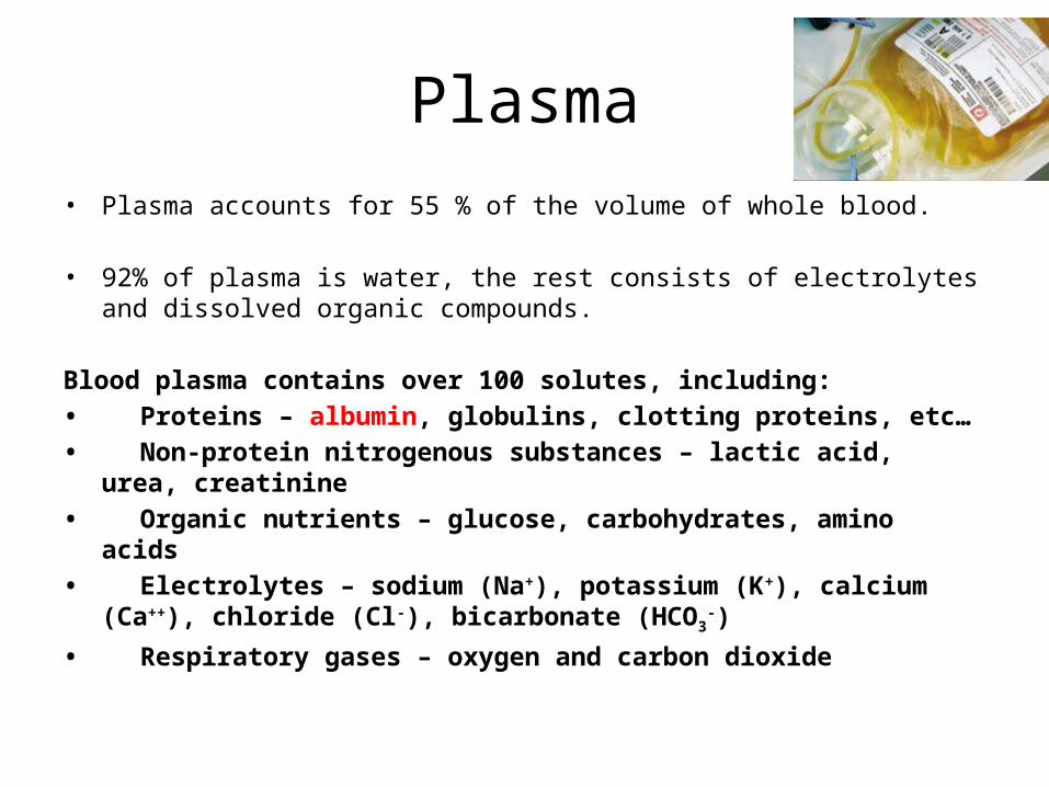
Plasma• Plasma accounts for 55 % of the volume of whole blood. • 92% of plasma is water, the rest consists of electrolytes and
dissolved organic compounds.
Blood plasma contains over 100 solutes, including:• Proteins – albumin, globulins, clotting proteins, etc…• Non-protein nitrogenous substances – lactic acid, urea,
creatinine• Organic nutrients – glucose, carbohydrates, amino acids• Electrolytes – sodium (Na+), potassium (K+), calcium (Ca+
+), chloride (Cl-), bicarbonate (HCO3-)
• Respiratory gases – oxygen and carbon dioxide
Plasma Proteins
• Albumin 1. Contributes to the osmotic pressure of the blood 2. Provides a transport mechanism for specific insoluble or
valuable materials in the blood.
• Globular proteins1. Binding and transporting hormones, lipids (lipoproteins),
and metal ions. 2. The immunoglobulins (antibodies) are proteins that
attack foreign proteins and pathogens. • Fibrinogen molecules aggregate to form large insoluble
strands of fibrin that establish the basis for a blood clot.

Erythrocytes
• Biconcave discs
• RBCs have no nuclei or organelles (anucleate) allow for a huge surface area to volume ratio
• Hematocrit – % of RBCs out of the total blood volume. (Ave) 46 adult men & 42 adult women.
• There are roughly 5 million RBCs in each microliter of blood
• Erythrocytes are unable to perform normal maintenance operations and usually degenerate after about 120 days in the circulation.


Hemoglobin
• Hemoglobin (Hgb), a globular protein formed from four subunits.
• Heme molecules bind to oxygen when plasma concentrations are high; the oxygen is released when plasma concentrations decline.
• Carbon dioxide molecules can be bound to
the globin portion of the hemoglobin molecule.
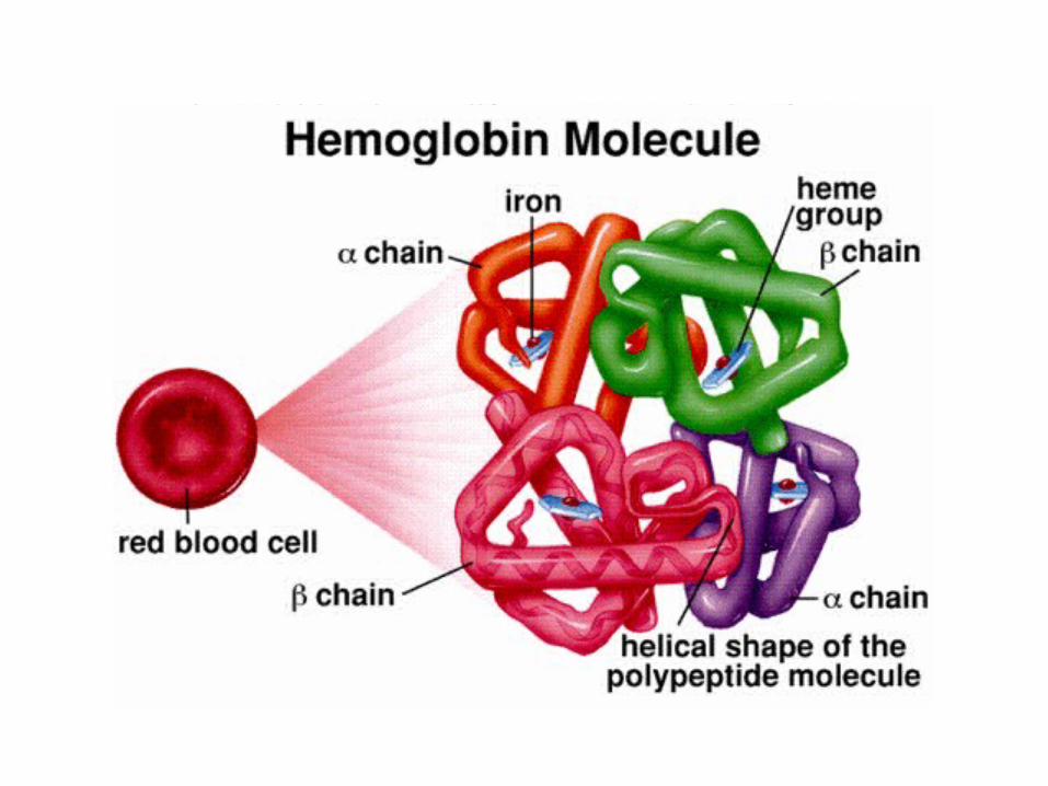
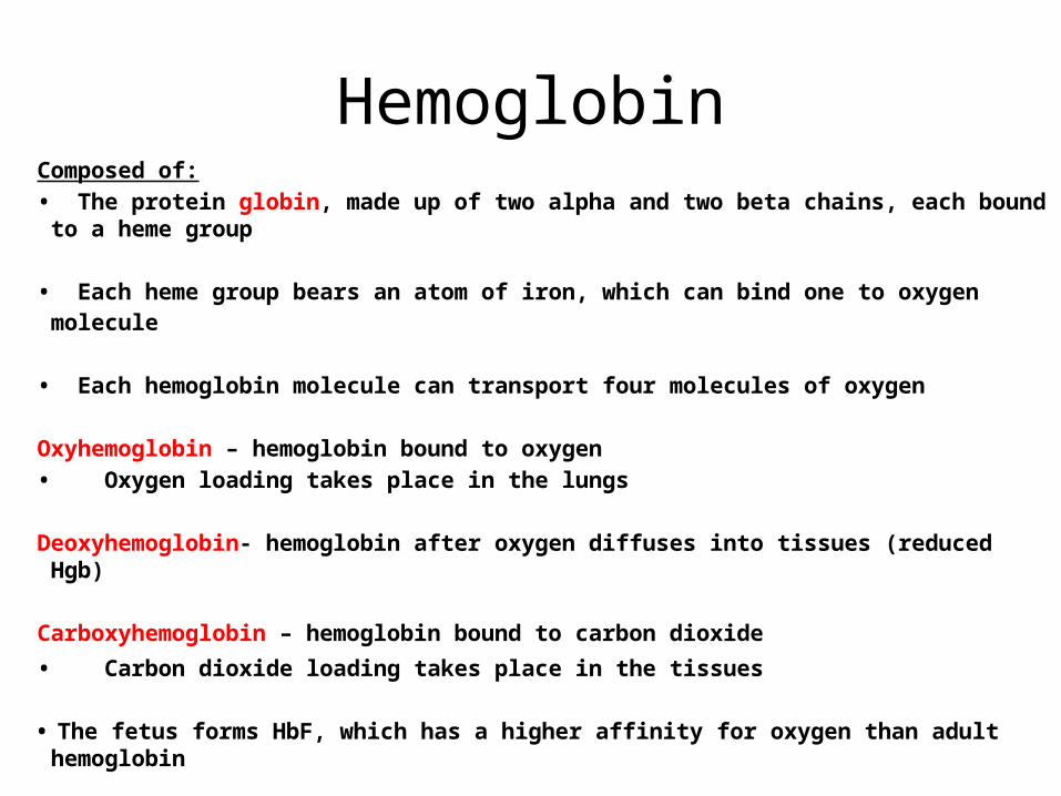
HemoglobinComposed of:• The protein globin, made up of two alpha and two beta chains, each
bound to a heme group
• Each heme group bears an atom of iron, which can bind one to oxygen
molecule
• Each hemoglobin molecule can transport four molecules of oxygen
Oxyhemoglobin – hemoglobin bound to oxygen• Oxygen loading takes place in the lungs
Deoxyhemoglobin- hemoglobin after oxygen diffuses into tissues (reduced Hgb)
Carboxyhemoglobin – hemoglobin bound to carbon dioxide
• Carbon dioxide loading takes place in the tissues
• The fetus forms HbF, which has a higher affinity for oxygen than adult hemoglobin

What happens to old RBC’S?
• Damaged or expired red blood cells are recycled by phagocytes.
• Proteins are disassembled into amino acids
• Iron gets bound to transferrin for transport to the bone marrow and liver
• Heme units are not recycled, but removed from the circulation by the liver

Fate and Destruction of Erythrocytes
• The life span of an erythrocyte is 100–120 days• Dying erythrocytes are engulfed by macrophages• Heme & globin are separated and the iron is salvaged
for reuse
Fate of Hemoglobin• Heme is degraded to a yellow pigment called bilirubin• The liver secretes bilirubin into the intestines as bile• The intestines metabolize it into urobilinogen • This degraded pigment leaves the body in feces, in a
pigment called stercobilin or as urobilinogen in urine• Globin is metabolized into amino acids and is
released into the circulation
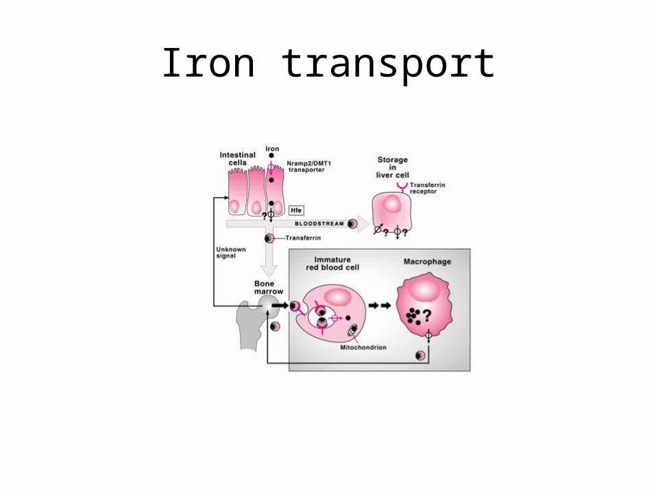
Iron transport
Blood Types
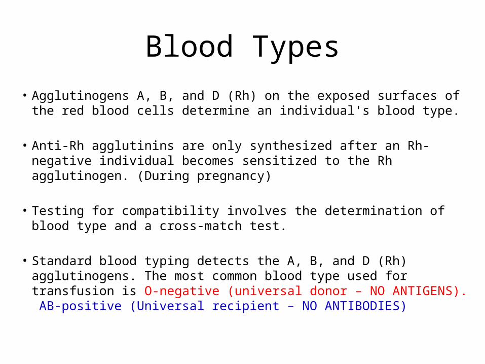
• Agglutinogens A, B, and D (Rh) on the exposed surfaces of the red blood cells determine an individual's blood type.
• Anti-Rh agglutinins are only synthesized after an Rh-negative individual becomes sensitized to the Rh agglutinogen. (During pregnancy)
• Testing for compatibility involves the determination of blood type and a cross-match test.
• Standard blood typing detects the A, B, and D (Rh) agglutinogens. The most common blood type used for transfusion is O-negative (universal donor – NO ANTIGENS). AB-positive (Universal recipient – NO ANTIBODIES)

Blood Type
Rh Type
Percent
A+ 34 %
- 6 %
B+ 9 %
- 2 %
AB+ 3 %
- 1 %
O+ 38 %
- 7 %


Erythroblastosis Fetalis

Hemolytic Disease of the Newborn (Erythroblastosis fetalis)
• Rh+ antibodies of a sensitized Rh– mother cross the placenta and attack and destroy the RBCs of an Rh+ baby
• Rh– mother become sensitized when Rh+ blood (from a previous pregnancy of an Rh+ baby or a Rh+ transfusion) causes her body to synthesis Rh+ antibodies
• The drug RhoGAM can prevent the Rh– mother from becoming sensitized

• Production of Blood Cells• Hematopoiesis – blood cell formation
• Hematopoiesis occurs in the red bone marrow of the:
- Axial skeleton and girdles - Epiphyses of the humerus and femur Hemocytoblasts give rise to all formed elements• Circulating stem cells give rise to embryonic blood
cells which migrate into the liver, spleen, thymus, and bone marrow.


Erythropoiesis
• Occurs within red marrow of the sternum, vertebrae, skull, scapulae, pelvis, and proximal limb bones.
• Red blood cell formation increases under erythropoietin stimulation. This hormone is released from the kidneys when they are not receiving adequate supplies of oxygen.
• Erythropoiesis is hormonally controlled and depends on adequate supplies of iron, amino acids, and B vitamins
• Reticulocytes (immature RBC’s) usually account for 0.8 percent of circulating red blood cells.


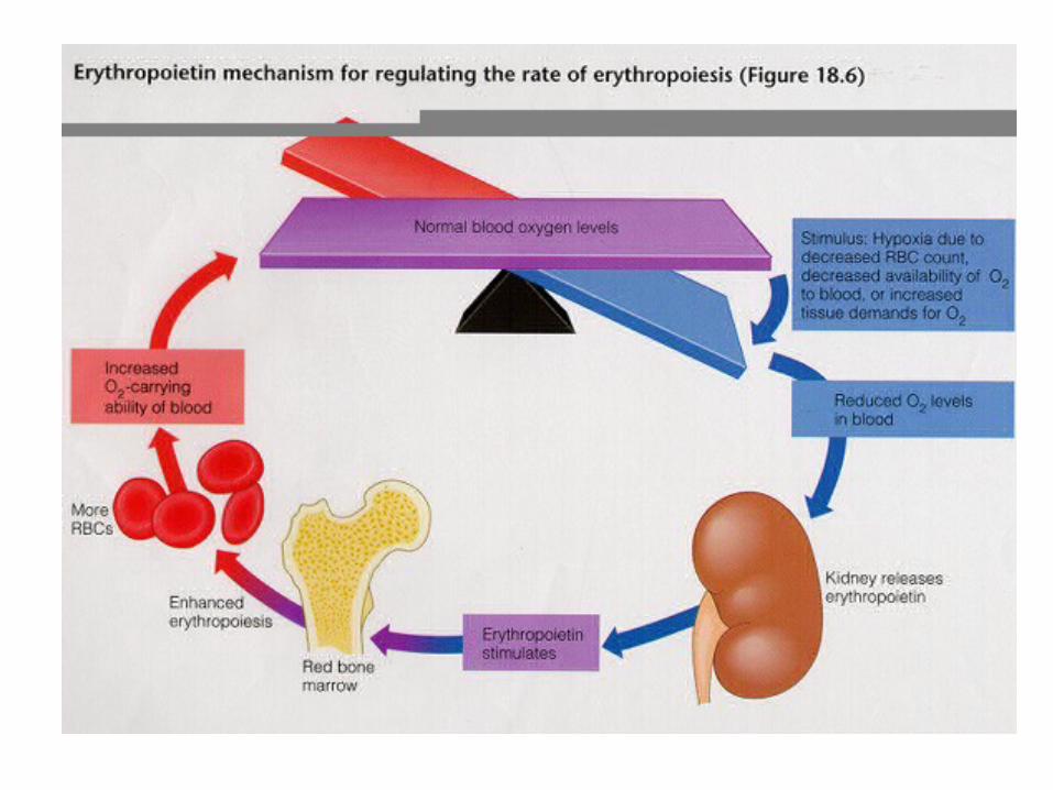
Erythropoietin
Erythropoietin (EPO) release by the kidneys is triggered by:
• Hypoxia due to decreased RBCs• Decreased oxygen availability• Increased tissue demand for oxygen
Erythropoiesis increases the: • RBC count in circulating blood• Oxygen carrying ability of the blood increases
Erythropoiesis requirement
• Proteins, lipids, and carbohydrates• Iron, vitamin B12, and folic acid
The body stores iron in Hgb (65%), the liver, spleen, and bone marrow
Intracellular iron is stored in protein-iron complexes such as ferritin and hemosiderin
Circulating iron is loosely bound to the transport protein transferrin

Erythrocyte pathophysiology
Anemia – blood has abnormally low oxygen-carrying capacity
• Blood oxygen levels cannot support normal metabolism
• Signs/symptoms include fatigue, paleness, shortness of breath, increased heartrate, low blood pressure, and chills

Anemia: Insufficient Erythrocytes
• Hemorrhagic anemia – result of acute or chronic loss of blood
(e.g.Trauma & Menstruation)
• Hemolytic anemia – prematurely ruptured erythrocytes
• Aplastic anemia – destruction or inhibition of red bone
marrow• Sickle cell anemia


Anemia: Decreased Hemoglobin Content
Iron-deficiency anemia results from:• A secondary result of hemorrhagic anemia • Inadequate intake of iron-containing foods• Impaired iron absorption
Pernicious anemia results from:• Deficiency of vitamin B12
• Often caused by lack of intrinsic factor needed for absorption of B12
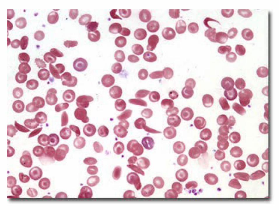
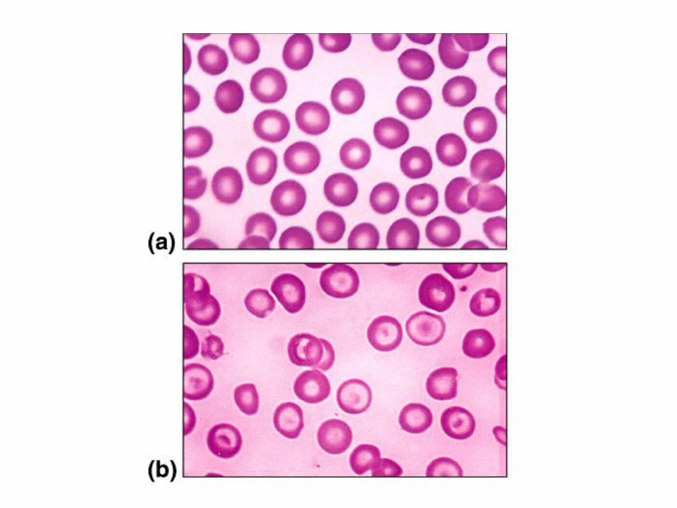
Anemia: Abnormal Hemoglobin
Thalassemias – absent or faulty globin chain in hemoglobin
• Erythrocytes are thin, delicate, & deficient in hemoglobin
Sickle-cell anemia – results from a defective gene coding for an abnormal hemoglobin called hemoglobin S (HbS)


Polycythemia
• Excess RBCs that increase blood viscosity
• Blood doping in athletics

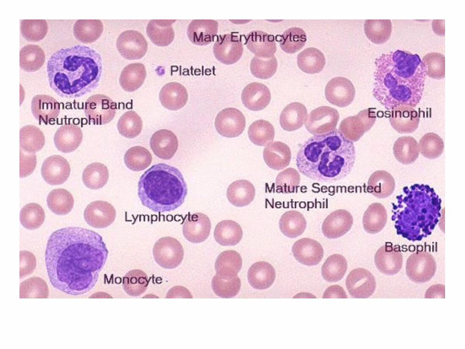
Leukocytes
• White blood cells are components of the immune system that defends the body against pathogens, toxins, wastes, and abnormal or damaged cells and tissues.
• There are 6,000-9,000 white blood
cells in each microliter of whole blood

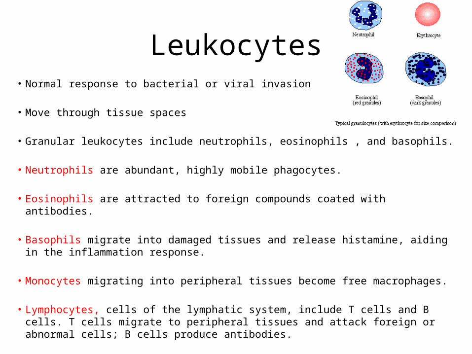
Leukocytes• Normal response to bacterial or viral invasion
• Move through tissue spaces
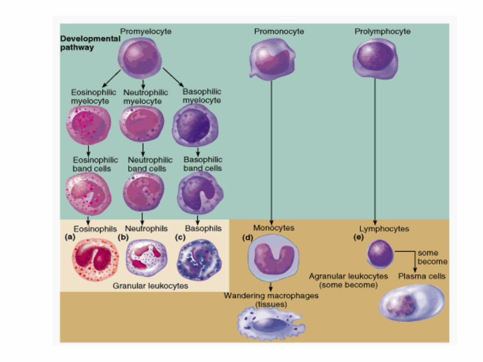
• Granular leukocytes include neutrophils, eosinophils , and basophils.
• Neutrophils are abundant, highly mobile phagocytes.
• Eosinophils are attracted to foreign compounds coated with antibodies.
• Basophils migrate into damaged tissues and release histamine, aiding in the inflammation response.
• Monocytes migrating into peripheral tissues become free macrophages.
• Lymphocytes, cells of the lymphatic system, include T cells and B cells. T cells migrate to peripheral tissues and attack foreign or abnormal cells; B cells produce antibodies.

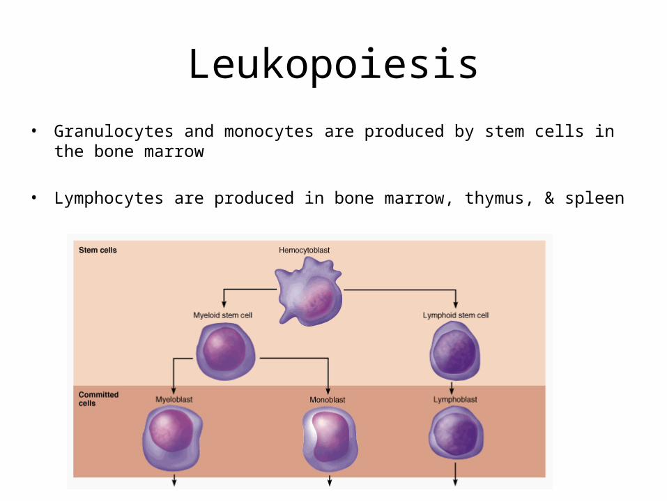
Leukopoiesis• Granulocytes and monocytes are produced by stem cells in the bone
marrow
• Lymphocytes are produced in bone marrow, thymus, & spleen
Granulocytes
Which WBC’s are granulocytes?

Neutrophils (Polymorphonuclear)
• 60-70% of WBC’s
Neutrophils have two types of granules that:• Take up both acidic and basic dyes• Give the cytoplasm a lilac color• Contain peroxidases, hydrolytic enzymes, and
defensins (antibiotic-like proteins)
Neutrophils are our body’s bacterial slayersLifespan : 1 day in blood; 1-2 days in tissue

Eosinophils
• 1-4% of WBC’s
• Have red-staining, bi-lobed nuclei connected via a broad band of nuclear material
• Lead the body’s counterattack against parasitic worms
• Lessen the severity of allergies by phagocytizing immune complexes
• Lifespan: 1 day in blood; weeks in tissue

Basophils
• 0.5-1% of WBC’s
• Have large, purplish-black (basophilic) granules that contain histamine
• Histamine – inflammatory chemical that acts as a vasodilator & attracts other WBCs
• Lifespan: 1 day in blood; hours in tissue

Agranulocytes
Which WBC’s are agranulocytes?

Lymphocytes• 20-25% of WBC’s
• Have large, dark-purple, circular nuclei with a thin rim of blue cytoplasm
• Found mostly enmeshed in lymphoid tissue (some circulate in the blood)
• There are two types of lymphocytes: T cells and B cells
• T cells function in the immune response
• B cells give rise to plasma cells, which produce antibodies
• Lifespan: Years
Monocytes
• 3-8% of WBC’s
• They are the largest leukocytes
• They have abundant pale-blue cytoplasms
• They have purple staining, U- or kidney-shaped nuclei
• They leave the circulation, enter tissue, and differentiate into macrophages
• Lifespan: Days in blood; years in tissue

Leukocyte pathophysiology• Leukemia refer to cancerous conditions involving white blood cells
• Immature white blood cells are found in the bloodstream in all leukemias
• Bone marrow becomes totally occupied with cancerous leukocytes
• The white blood cells produced, though numerous, are not functional
• Death is caused by internal hemorrhage and overwhelming infections
• Acute leukemia involves blast-type cells and primarily affects children
• Chronic leukemia is more prevalent in older people
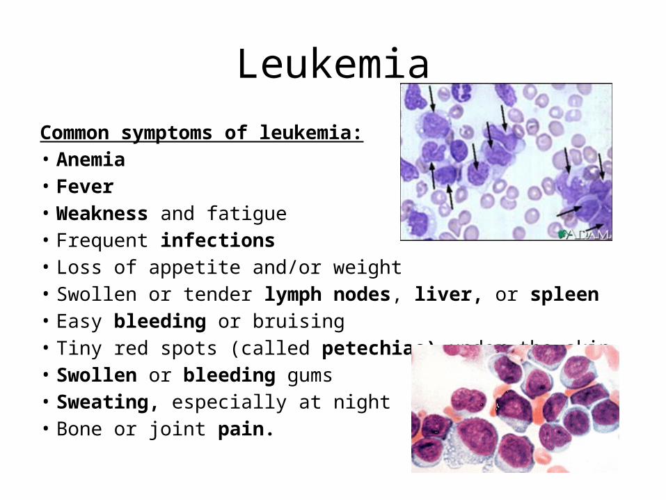
Leukemia
Common symptoms of leukemia: • Anemia • Fever • Weakness and fatigue • Frequent infections • Loss of appetite and/or weight • Swollen or tender lymph nodes, liver, or spleen • Easy bleeding or bruising • Tiny red spots (called petechiae) under the skin • Swollen or bleeding gums • Sweating, especially at night• Bone or joint pain.

Thrombocytes
• Platelets
• Megakaryocytes in the bone marrow release packets of cytoplasm, called platelets, into the circulating blood. There are 150,000-500,000 platelets in each microliter of whole blood.
• Platelet granules contain serotonin, Ca2+, enzymes, ADP, and platelet-derived growth factor (PDGF)
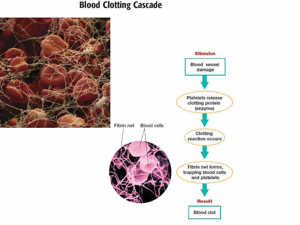
• Platelets function in the clotting mechanism by forming a temporary plug that helps seal breaks in blood vessels


Blood clottingCascade

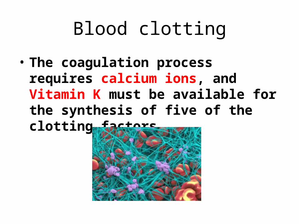
Blood clotting
• The coagulation process requires calcium ions, and Vitamin K must be available for the synthesis of five of the clotting factors.

Hemostasis Pathophysiology
• Thromboembolytic Disorders
Thrombus – a clot that develops and persist in an unbroken blood vessel
Embolus – a thrombus freely floating in the blood stream
Thrombocytopenia – condition where the number of circulating platelets is deficient
Hemophilias – hereditary bleeding disorders caused by lack of clotting factors


Hemophilia
Hemophilia A – most common type (83% of all cases) due to a deficiency of factor VIII
• Hemophilia B – results from a deficiency of factor IX
• Hemophilia C – mild type, caused by a deficiency of factor XI
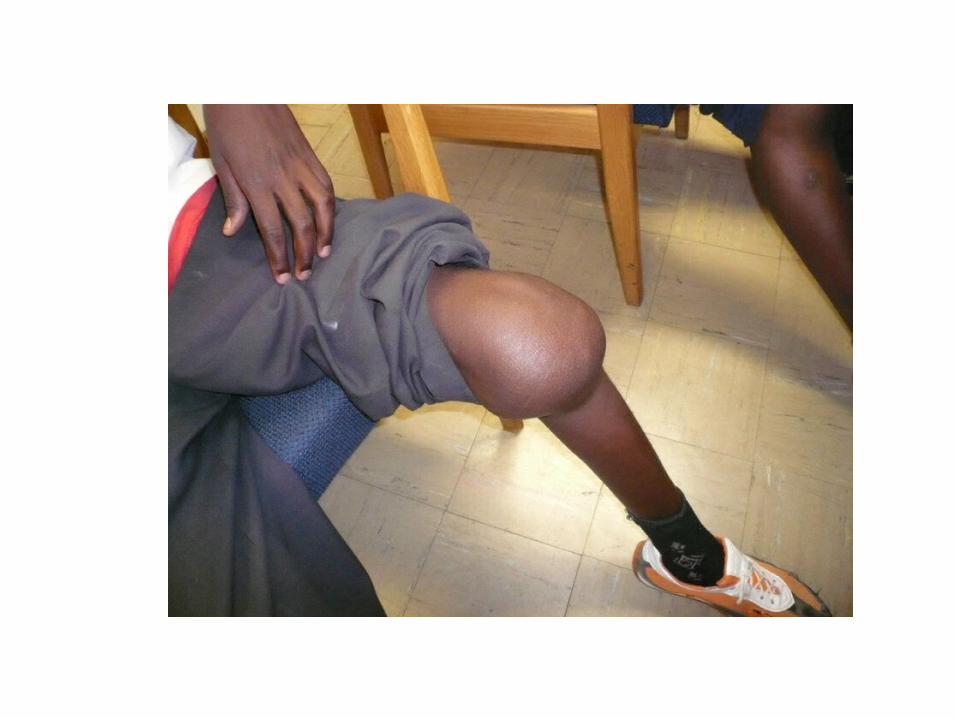
• Symptoms include prolonged bleeding and painful and disabled joints
• Treatment is with blood transfusions and the injection of missing factors
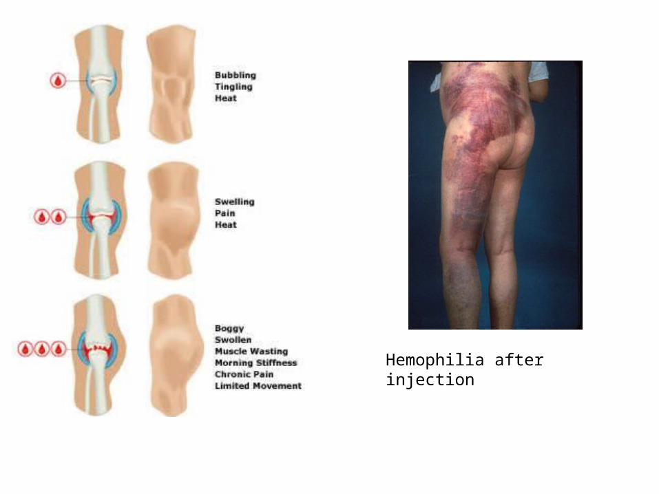
Hemophilia after injection
Prevention of undesirable clots
Substances used to prevent undesirable clots include:
• Aspirin
• Heparin
• Warfarin (Coumadin)
• Flavonoids – substances found in tea, red wine, and grape juice that have natural anticoagulant activity

Leukocyte
DifferentialPractice


Are we done yet?