Blood-based lung cancer biomarkers identified through ...
12
RESEARCH Open Access Blood-based lung cancer biomarkers identified through proteomic discovery in cancer tissues, cell lines and conditioned medium Charles E. Birse 1* , Robert J. Lagier 1 , William FitzHugh 1 , Harvey I. Pass 2 , William N. Rom 3 , Eric S. Edell 4 , Aaron O. Bungum 4 , Fabien Maldonado 4 , James R. Jett 5 , Mehdi Mesri 1 , Erin Sult 1 , Elizabeth Joseloff 1 , Aiqun Li 1 , Jenny Heidbrink 1 , Gulshan Dhariwal 1 , Chad Danis 1 , Jennifer L. Tomic 1 , Robert J. Bruce 1 , Paul A. Moore 1 , Tao He 1 , Marcia E. Lewis 1 and Steve M. Ruben 1 Abstract Background: Support for early detection of lung cancer has emerged from the National Lung Screening Trial (NLST), in which low-dose computed tomography (LDCT) screening reduced lung cancer mortality by 20 % relative to chest x-ray. The US Preventive Services Task Force (USPSTF) recently recommended annual screening for the high-risk population, concluding that the benefits (life years gained) outweighed harms (false positive findings, abortive biopsy/surgery, radiation exposure). In making their recommendation, the USPSTF noted that the moderate net benefit of screening was dependent on the resolution of most false-positive results without invasive procedures. Circulating biomarkers may serve as a valuable adjunctive tool to imaging. Results: We developed a broad-based proteomics discovery program, integrating liquid chromatography/mass spectrometry (LC/MS) analyses of freshly resected lung tumor specimens (n = 13), lung cancer cell lines (n = 17), and conditioned media collected from tumor cell lines (n = 7). To enrich for biomarkers likely to be found at elevated levels in the peripheral circulation of lung cancer patients, proteins were prioritized based on predicted subcellular localization (secreted, cell-membrane associated) and differential expression in disease samples. 179 candidate biomarkers were identified. Several markers selected for further validation showed elevated levels in serum collected from subjects with stage I NSCLC (n = 94), relative to healthy smoker controls (n = 189). An 8-marker model was developed (TFPI, MDK, OPN, MMP2, TIMP1, CEA, CYFRA 21–1, SCC) which accurately distinguished subjects with lung cancer (n = 50) from high risk smokers (n = 50) in an independent validation study (AUC = 0.775). Conclusions: Integrating biomarker discovery from multiple sample types (fresh tissue, cell lines and conditioned medium) has resulted in a diverse repertoire of candidate biomarkers. This unique collection of biomarkers may have clinical utility in lung cancer detection and diagnoses. Keywords: Lung cancer, Early detection, Biomarker, Mass spectrometry, Proteomics, Discovery * Correspondence: [email protected] 1 Celera employees during the course of these studies, Celera, 1311 Harbor Bay Parkway, Alameda, CA 94502, USA Full list of author information is available at the end of the article CLINICAL PROTEOMICS © 2015 Birse et al. Open Access This article is distributed under the terms of the Creative Commons Attribution 4.0 International License (http://creativecommons.org/licenses/by/4.0/), which permits unrestricted use, distribution, and reproduction in any medium, provided you give appropriate credit to the original author(s) and the source, provide a link to the Creative Commons license, and indicate if changes were made. The Creative Commons Public Domain Dedication waiver (http://creativecommons.org/publicdomain/zero/1.0/) applies to the data made available in this article, unless otherwise stated. Birse et al. Clinical Proteomics (2015) 12:18 DOI 10.1186/s12014-015-9090-9
Transcript of Blood-based lung cancer biomarkers identified through ...
CLINICALPROTEOMICS
Birse et al. Clinical Proteomics (2015) 12:18 DOI 10.1186/s12014-015-9090-9
RESEARCH Open Access
Blood-based lung cancer biomarkersidentified through proteomic discovery incancer tissues, cell lines and conditionedmedium
Charles E. Birse1*, Robert J. Lagier1, William FitzHugh1, Harvey I. Pass2, William N. Rom3, Eric S. Edell4,Aaron O. Bungum4, Fabien Maldonado4, James R. Jett5, Mehdi Mesri1, Erin Sult1, Elizabeth Joseloff1, Aiqun Li1,Jenny Heidbrink1, Gulshan Dhariwal1, Chad Danis1, Jennifer L. Tomic1, Robert J. Bruce1, Paul A. Moore1, Tao He1,Marcia E. Lewis1 and Steve M. Ruben1Abstract
Background: Support for early detection of lung cancer has emerged from the National Lung Screening Trial(NLST), in which low-dose computed tomography (LDCT) screening reduced lung cancer mortality by 20 % relativeto chest x-ray. The US Preventive Services Task Force (USPSTF) recently recommended annual screening for thehigh-risk population, concluding that the benefits (life years gained) outweighed harms (false positive findings,abortive biopsy/surgery, radiation exposure). In making their recommendation, the USPSTF noted that the moderatenet benefit of screening was dependent on the resolution of most false-positive results without invasiveprocedures. Circulating biomarkers may serve as a valuable adjunctive tool to imaging.
Results: We developed a broad-based proteomics discovery program, integrating liquid chromatography/massspectrometry (LC/MS) analyses of freshly resected lung tumor specimens (n = 13), lung cancer cell lines (n = 17), andconditioned media collected from tumor cell lines (n = 7). To enrich for biomarkers likely to be found at elevatedlevels in the peripheral circulation of lung cancer patients, proteins were prioritized based on predicted subcellularlocalization (secreted, cell-membrane associated) and differential expression in disease samples. 179 candidatebiomarkers were identified. Several markers selected for further validation showed elevated levels in serumcollected from subjects with stage I NSCLC (n = 94), relative to healthy smoker controls (n = 189). An 8-markermodel was developed (TFPI, MDK, OPN, MMP2, TIMP1, CEA, CYFRA 21–1, SCC) which accurately distinguishedsubjects with lung cancer (n = 50) from high risk smokers (n = 50) in an independent validation study (AUC = 0.775).
Conclusions: Integrating biomarker discovery from multiple sample types (fresh tissue, cell lines and conditionedmedium) has resulted in a diverse repertoire of candidate biomarkers. This unique collection of biomarkers mayhave clinical utility in lung cancer detection and diagnoses.
Keywords: Lung cancer, Early detection, Biomarker, Mass spectrometry, Proteomics, Discovery
* Correspondence: [email protected] employees during the course of these studies, Celera, 1311 HarborBay Parkway, Alameda, CA 94502, USAFull list of author information is available at the end of the article
© 2015 Birse et al. Open Access This article is distributed under the terms of the Creative Commons Attribution 4.0International License (http://creativecommons.org/licenses/by/4.0/), which permits unrestricted use, distribution, andreproduction in any medium, provided you give appropriate credit to the original author(s) and the source, provide alink to the Creative Commons license, and indicate if changes were made. The Creative Commons Public DomainDedication waiver (http://creativecommons.org/publicdomain/zero/1.0/) applies to the data made available in thisarticle, unless otherwise stated.

Birse et al. Clinical Proteomics (2015) 12:18 Page 2 of 12
BackgroundLung cancer is the leading cause of cancer mortality in theUnited States. Estimates for 2014 indicate that 224,210 in-dividuals will be diagnosed with lung cancer and 159,260will die from the disease [1]. The average 5-year survival isabout 17 %, with 79 % of cases being diagnosed as regionalor distant disease. If lung cancer is detected when local-ized, survival increases to over 50 % [1].Support for early lung cancer detection has emerged
from the landmark NLST, where LDCT screening wasshown to confer a 20 % reduction in lung cancer mortal-ity in a high risk population [2]. Despite concerns associ-ated with the low specificity (73.4 %) of CT screening [3]and the resulting large number of false-positive findingsfor lung cancer (96.4 %), the USPSTF recently recom-mended annual LDCT-screening for lung cancer in high-risk individuals [4, 5]. In their recommendation statement,the USPSTF stressed the need for more research into theuse of biomarkers to complement LDCT screening. Twokey clinical opportunities exist. First, the use of bio-markers for early detection of lung cancer could define anew high-risk population or refine the screening criteriarecommended by USPSTF (age: 55 to 80 years, smokinghistory: >30 pack-year). Such biomarkers would serve as apre-imaging filter, reducing the overall cost of screeningand lowering the number of false-positive findings andunnecessary follow-up procedures. The second opportun-ity lies in improving the accuracy of lung cancer diagnosis.Given the high frequency of positive findings (pulmonarynodules) with CT screening [2], new means of accuratelydetermining malignant risk are urgently required. In theNLST, 24 % of surgically resected nodules were found tobe benign [2]. By improving the accuracy with which ma-lignant risk is determined, biomarkers could potentiallyenhance diagnostic management by reducing unnecessarysurgical intervention, minimizing the use of costly PET-CT and lowering radiation exposure associated with CTmonitoring, while enabling detection of lung cancer at anearly, more curable, stage.A wide variety of approaches have been utilized to dis-
cover new blood-based lung cancer protein biomarkers[6]. These range from splice variant analysis and the isola-tion of tumor-enriched transcripts [7], to the developmentof novel proteomic platforms with the capacity to resolvecandidate markers in a highly multiplexed fashion [8]. Ad-vances in mass spectrometry (MS)-based technologieshave also enabled discovery of new lung cancer biomarkercandidates directly in serum or plasma [9–13]. While theidentification of biomarkers directly in blood-based matri-ces can be problematic due to their complexity and thepresence of multiple highly abundant factors [14], some ofthese challenges can be minimized through extensive frac-tionation [15]. Differentially expressed candidate markershave also been successfully identified through comparison
of blood draining from the tumor vascular bed matchedwith systemic arterial blood from the same patient [16].Alternative, “indirect” MS approaches have also been
successfully employed, wherein candidate markers ini-tially identified in lung cancer tissue specimens, cell linesor conditioned medium, have subsequently been shownto be differentially expressed in the peripheral circula-tion using immunoassay-based methodologies. Pioneer-ing discovery studies employed conditioned mediumderived from the lung cancer cell line A549 or cell andorgan cultures, followed by confirmation of expressionprofiles in serum and plasma [17, 18]. Thereafter, de-tailed analysis of conditioned medium collected frommultiple lung cancer lines revealed a novel collection ofcandidate biomarkers [19]. More recently, subcellularfractionation and organelle isolation from freshly col-lected tissue specimens has enabled further candidatediscovery, with verification achieved through multiplereaction monitoring (MRM) [20].We have expanded on these approaches, broadening
the scope of biomarker discovery by performing prote-omic analyses across multiple types of specimens: freshlyresected lung tissues, cancer cell lines and conditionedmedium, enabling the discovery of a diverse collection ofcandidate markers. We have confirmed disease-enrichedprofiles for several of these candidates in sera collectedfrom patients with early-stage disease. Moreover, amulti-marker model has been assembled which accur-ately distinguishing patients with NSCLC from smokerswith no known malignancies. These studies suggest thatthe integration of multiple indirect discovery approachesmay serve as a valuable means of identifying novelblood-based biomarkers that may be employed in theearly detection and diagnosis of lung cancer.
ResultsTissue/cell-line based biomarker discoveryThree distinct LC/MS-based discovery programs wereestablished to identify a diverse spectrum of candidatebiomarkers which could serve as the basis for a blood-based immunoassay for detection or diagnosis of lungcancer. To enrich for markers destined to be found in theperipheral circulation of lung cancer patients, discoveryfocused on glycoproteins predicted to be located either atthe cell membrane or secreted/shed from lung cancercells. Cell-membrane discovery was performed in twodistinct sample types: freshly resected tissue specimens(n = 13), and a collection of lung cancer cell lines (n = 17).The clinical specimens and cell lines studied providebroad coverage of tumor stage and prevalent histologicalcell types (Additional file 1: Table S1 and Additional file 2:Table S2). To focus discovery on differentially expressedcandidate markers, peptide levels measured in surgicallyresected malignant samples were compared with adjacent
Birse et al. Clinical Proteomics (2015) 12:18 Page 3 of 12
normal tissue. Expression in lung cancer cell lines was an-alyzed relative to the non-cancerous immortalized lungepithelial cell line Beas-2B [21]. A third discovery pro-gram, which served to complement the cell-membraneanalyses, resolved proteins secreted or shed from lungcancer cells into conditioned medium, in a subset of linesamenable to growth in serum-free conditions: A549,H1299, H358, H522, H2291, H520 and Calu-1.Candidate lung cancer markers were prioritized based
on MS data and predicted subcellular localization. To beselected, proteins had to: (i) be represented by multiple dif-ferentially expressed peptides (n > 1); (ii) be identified inmultiple malignant samples (n > 1) and (iii) exhibit elevatedexpression in lung cancer specimens, with a cancer: controlexpression ratio of > 4.0. Candidate biomarkers were alsoprioritized based on secondary structure, with proteinspredicted to be associated with either the cell membraneor secreted from the cell being selected; these two com-partments are enriched with markers destined for the per-ipheral circulation. 179 candidate markers were identifiedwhich met these criteria (Additional file 3: Table S3).Each of the cellular systems employed in these studies
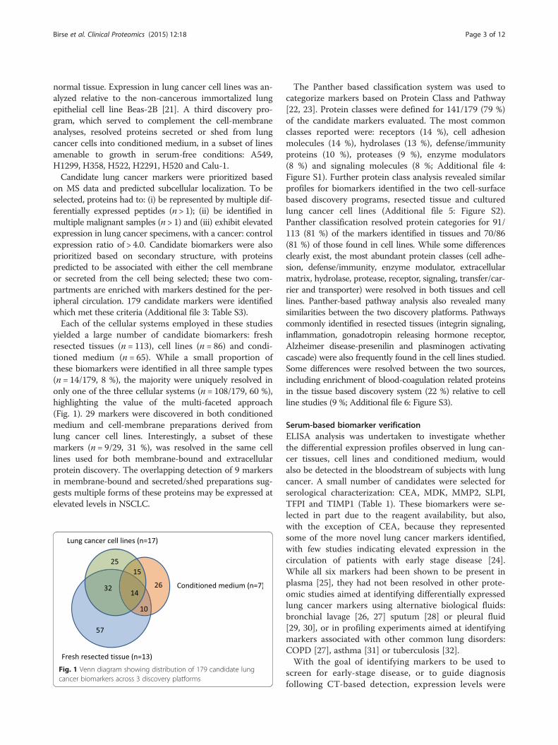
yielded a large number of candidate biomarkers: freshresected tissues (n = 113), cell lines (n = 86) and condi-tioned medium (n = 65). While a small proportion ofthese biomarkers were identified in all three sample types(n = 14/179, 8 %), the majority were uniquely resolved inonly one of the three cellular systems (n = 108/179, 60 %),highlighting the value of the multi-faceted approach(Fig. 1). 29 markers were discovered in both conditionedmedium and cell-membrane preparations derived fromlung cancer cell lines. Interestingly, a subset of thesemarkers (n = 9/29, 31 %), was resolved in the same celllines used for both membrane-bound and extracellularprotein discovery. The overlapping detection of 9 markersin membrane-bound and secreted/shed preparations sug-gests multiple forms of these proteins may be expressed atelevated levels in NSCLC.
Fig. 1 Venn diagram showing distribution of 179 candidate lungcancer biomarkers across 3 discovery platforms
The Panther based classification system was used tocategorize markers based on Protein Class and Pathway[22, 23]. Protein classes were defined for 141/179 (79 %)of the candidate markers evaluated. The most commonclasses reported were: receptors (14 %), cell adhesionmolecules (14 %), hydrolases (13 %), defense/immunityproteins (10 %), proteases (9 %), enzyme modulators(8 %) and signaling molecules (8 %; Additional file 4:Figure S1). Further protein class analysis revealed similarprofiles for biomarkers identified in the two cell-surfacebased discovery programs, resected tissue and culturedlung cancer cell lines (Additional file 5: Figure S2).Panther classification resolved protein categories for 91/113 (81 %) of the markers identified in tissues and 70/86(81 %) of those found in cell lines. While some differencesclearly exist, the most abundant protein classes (cell adhe-sion, defense/immunity, enzyme modulator, extracellularmatrix, hydrolase, protease, receptor, signaling, transfer/car-rier and transporter) were resolved in both tissues and celllines. Panther-based pathway analysis also revealed manysimilarities between the two discovery platforms. Pathwayscommonly identified in resected tissues (integrin signaling,inflammation, gonadotropin releasing hormone receptor,Alzheimer disease-presenilin and plasminogen activatingcascade) were also frequently found in the cell lines studied.Some differences were resolved between the two sources,including enrichment of blood-coagulation related proteinsin the tissue based discovery system (22 %) relative to cellline studies (9 %; Additional file 6: Figure S3).
Serum-based biomarker verificationELISA analysis was undertaken to investigate whetherthe differential expression profiles observed in lung can-cer tissues, cell lines and conditioned medium, wouldalso be detected in the bloodstream of subjects with lungcancer. A small number of candidates were selected forserological characterization: CEA, MDK, MMP2, SLPI,TFPI and TIMP1 (Table 1). These biomarkers were se-lected in part due to the reagent availability, but also,with the exception of CEA, because they representedsome of the more novel lung cancer markers identified,with few studies indicating elevated expression in thecirculation of patients with early stage disease [24].While all six markers had been shown to be present inplasma [25], they had not been resolved in other prote-omic studies aimed at identifying differentially expressedlung cancer markers using alternative biological fluids:bronchial lavage [26, 27] sputum [28] or pleural fluid[29, 30], or in profiling experiments aimed at identifyingmarkers associated with other common lung disorders:COPD [27], asthma [31] or tuberculosis [32].With the goal of identifying markers to be used to
screen for early-stage disease, or to guide diagnosisfollowing CT-based detection, expression levels were

Table 1 Candidate lung cancer biomarkers identified through MS discovery that were selected for serological characterization
Protein Differentially expressed peptides Number of samples where differentially expressedpeptides observed
Differential expression(Lung cancer/control)
Predicted subcellularlocalization
Tissues(n = 13)
Cell lines(n = 17)
Conditionedmedium (n = 7)
CEA (CEACAM5) CETQNPVSAR 9 1 20.4 Cell Membrane
TLTLFNVTRNDTASYK
MDK
YNAQCQETIR
7 4.9 Secreted
EGTCGAQTQR
FENWGACDGGTGTK
VTKPCTPK
YNAQCQETIRVTKPCTPK
MMP2
ESCNLFVLK
1 3 21.8 Secreted
TDKELAVQYLNTFYGCPK
ELAVQYLNTFYGCPK
CGNPDVANYNFFPR
YGFCPHEALFTMGGNAEGQPCK
SLPI
SCVSPVKA
3 5.5 SecretedAGVCPPK
SCVSPVK
AGVCPPKK
TFPI
ADDGPCK
1 3 10.0 SecretedQCEEFIYGGCEGNQNR
TTLQQEKPDFCFLEEDPGICR
YFYNNQTK
TIMP1
FVYTPAMESVCGYFHR
11 1 4 34.6 Secreted
HLACLPR
LQSGTHCLWTDQLLQGSEK
LQDGLLHITTCSFVAPWNSLSLAQR
FVGTPEVNQTTLYQRYEIK
AKFVGTPEVNQTTLYQR
FVGTPEVNQTTLYQR
SHNRSEEFLIAGK
EPGLCTWQSLR
Differentially expressed peptides resolved for each marker are listed together with the number of samples (tissues, cell lines or conditioned medium) wherepeptides with elevated expression in lung cancer were identified. Median ratio represents the overall level of elevated expression combining levels observed foreach differentially expressed peptide in disease samples relative to appropriate controls
Birse et al. Clinical Proteomics (2015) 12:18 Page 4 of 12
determined in subjects with stage I NSCLC (n = 94),relative to normal smoker controls (n = 189; Table 2).In an effort to minimize selection of markers associ-ated with pre-analytical variability, where differentialexpression profiles may be derived from serum samplecollection procedures specific to any single clinicalstudy site, subjects from two independent clinicalstudies were combined into a single testing set. Thefirst study collected at CRCCC (Clinical ResearchCenter of Cape Cod; West Yarmouth, MA), comprised pa-tients with stage I NSCLC (n = 30) and healthy smokercontrols (n = 99). The second cohort, collected at New
York University (NYU) School of Medicine/LangoneMedical Center, was selected from a high-risk populationwith a history of heavy tobacco usage. Serum sampleswere collected from patients with stage I NSCLC (n = 64)and healthy controls (n = 90).Levels of five of the six candidate biomarkers tested
(CEA, MDK, MMP2, SLPI, TIMP1, TFPI) were signifi-cantly higher in serum from subjects with NSCLC thanin controls (Table 3, Additional file 7: Figure S4), servingto support this indirect discovery approach. Three ex-tensively characterized markers: CYFRA 21–1, SCC andOPN were also evaluated. These markers served as a

Table 2 Demographic and clinical profiles of subjects tested with lung cancer biomarker candidates
Serum verification/model training Model testing
Controls Cases Controls Cases
(N = 189) (N = 94) (N = 50) (N = 50)
Gender
Male 109 33 28 28
Female 80 61 22 22
Age
Mean (SD) 62.1 (11.8) 66.6 (9.6) 63.0 (6.4) 65.6 (6.7)
Smoking Pack Years
Mean (SD) 37.6 (21.7) 43.9 (20.6) 54.3 (22.4) 54.2 (22.2)
Benign Nodules (n) 22
Lesion Size (cm)
Mean (range) 0.5 (0.2-1.2) 3.3 (0.8-12)
Stage
I (%) 94 (100) 18 (36)
II (%) 6 (12)
III (%) 16 (32)
IV (%) 5 (10)
NA (%) 5 (10)
Histology
Adenocarcinoma (%) 63 (67.0) 23 (46)
BAC (%) 4 (4.3)
Large Cell (%) 6 (6.4)
NSCLC (%) 4 (4.3) 6 (12)
Neuroendocrine (%) 2 (2.1)
Small Cell (%) 4 (8)
Squamous Cell (%) 15 (16.0) 17 (34)
Table 3 Expression levels of biomarker candidates in serum collected from patients with NSCLC (n = 94) and healthy volunteercontrols (n = 189)
Control Case KS test(p-val)
AUC
Median: ng/mL Median: ng/mL
(Interquartile range) (Interquartile range)
CEA (CEACAM5) 1.65 (0.85-2.92) 2.68 (1.85-4.90) <0.0001 0.706
MDK 0.15 (0.04-0.35) 0.43 (0.20-0.66) <0.0001 0.714
MMP2 207 (184-234) 207 (171-254) 0.1472 0.492
SLPI 39.6 (34.8-46.2) 43.3 (35.5-54.5) 0.0036 0.595
TFPI 39.7 (25.6-55.1) 54.1 (29.1-70.3) <0.0001 0.617
TIMP1 302 (269-346) 361 (306-440) <0.0001 0.692
CYFRA 21-1 0.58 (0.00-1.05) 1.60 (0.91-3.00) <0.0001 0.816
OPN 19.3 (10.0-31.0) 31.4 (16.7-52.4) <0.0001 0.666
SCC 0.58 (0.34-0.93) 1.21 (0.55-1.70) <0.0001 0.696
Median levels (ng/mL) and interquartile range are shown. The Kolmogorov–Smirnov test (KS test) was used to compare patient groups. Area under the curve(AUC) calculated from receiver operator characteristic (ROC) curve analysis. Markers in the upper section of the table (n = 6) represent proteins resolved throughMS analysis. The lower section (n = 3) represents well-characterized lung cancer biomarkers
Birse et al. Clinical Proteomics (2015) 12:18 Page 5 of 12

Birse et al. Clinical Proteomics (2015) 12:18 Page 6 of 12
reference in evaluating clinical accuracy of the MS-identified markers.
Multi-marker model development and testingThe identification of multiple differentially expressedmarkers prompted the development of a multi-markerpanel. Elastic net modeling [33] started with all 9 candidatemarkers (Table 3). The optimal value of the regularizationparameter, as determined by bootstrap resampling, re-duced the parameter estimate for SLPI to zero, whilethe remaining 8 markers: TFPI, MDK, OPN, MMP2,TIMP1, CEA, CYFRA 21–1 and SCC, which retainednon-zero coefficients, were selected in the final model.In the training dataset (Table 2), this 8-marker modelresolved lung cancer patients from smoker controlswith 75 % sensitivity at 90 % specificity (AUC = 0.913).A bootstrap validation procedure confirmed clinicalperformance of the model, AUC = 0.903.The accuracy of the 8-marker model was tested in an
independent study (Mayo Clinic). Controls (n = 50) wereselected from the high risk control population evaluatedin the Mayo CT-Screening Trial [34] and included sub-jects with pulmonary nodules (n = 22). Lung cancercases were pre-operative surgical referrals (n = 50). Ma-lignant lesions were significantly larger than screen de-tected benign nodules. Cases and controls were matchedon age, gender and smoking history (Table 2). EDTAplasma samples were utilized in this study. Levels of allmarkers included in the model had been shown to behighly correlated in serum and EDTA plasma (Additionalfile 8: Table S4). The 8-marker model distinguished pa-tients with malignant lesions from all smoker controlswith an AUC = 0.775 (Fig. 2), accurately classifying controlsubjects with (AUC= 0.745) or without pulmonary nod-ules (AUC= 0.799).While the 8-marker model was found to be substantially
correlated with nodule size (r = 0.739; p < 0.0001), it wasnot associated with any of the other clinicopathologicalvariables tested: age, sex, smoking history (unpublisheddata). Elevated expression of the multi-marker model wasobserved in tumors with a squamous cell histology, relativeto adenocarcinoma cases (p = 0.019), driven in part byhigher levels of CYFRA 21-1 (p < 0.0001) and OPN (p =0.013) in squamous cell carcinomas (unpublished data).
DiscussionLDCT screening of high-risk smokers has been shownto reduce lung cancer mortality by 20 %, relative to chestradiography. However, of the 24.2 % of participants withan abnormal screening test, the vast majority (96.4 %)were false positives for lung cancer. The low positivepredictive value of LDCT results in (i) higher screeningcosts and (ii) unnecessary invasive procedures for benigndisease. Non-invasive biomarkers are urgently needed to
improve LDCT-based screening. Biomarkers could beused to refine the high-risk population, thus limiting thenumber of individuals being screened by LDCT. Alterna-tively, biomarkers could be employed following screening,to distinguish relatively rare malignant nodules from com-monly found benign nodules. A number of novel blood-based markers have recently been characterized [7, 35],some of which have been evaluated in the form of multi-marker panels [15, 36–38]. However, to date, very fewhave been shown to add value to clinical variables alreadybeing employed in evaluating malignant risk [39].Marker discovery in blood-based systems (serum and
plasma) has been hampered by the complexity of thesematrices and presence of multiple highly abundant pro-teins. Alternative “indirect” approaches have successfullybeen applied to: freshly resected clinical specimens, pri-mary cultures, cell lines cultured in vitro and in vivo andconditioned medium, with collections of candidate bio-markers identified in each. However, as each of these stud-ies has been performed in isolation, it has been difficult toevaluate the relative merits of each of these approaches.We report, for the first time, a discovery approach thatcombines multiple cellular systems: resected tissues, cul-tured cell lines and conditioned medium. In so doing, wehave identified a number of markers commonly resolvedacross the platforms (Fig. 1). It is noteworthy that whilesignificant overlap across the systems clearly exists, withsimilar signaling pathways apparently activated across thedifferent discovery systems (Additional file 6: Figure S3),the majority of candidate markers were identified in onlyone of the three programs. Integrating discoveries fromthe three systems has not only served as a starting pointto understand the relative merits of these distinct ap-proaches, but has also produced a diverse pool of candi-date markers for future validation.All 179 candidate markers selected exhibited at least a
four-fold increased level of expression in lung cancersamples relative to appropriate controls. While this 4.0Xcut point provided a simple means of identifying themost differentially expressed biomarkers, additional ap-proaches using different cut-points and possibly integrat-ing key clinical variables such as histology and stage, willlikely reveal a more extensive collection of candidatesfor future studies.Analyses of the glycoproteins residing at the cell surface,
in both tissues and cell lines, enabled discovery of cell-membrane markers that may be shed and released intothe peripheral circulation. CEA (CEACAM5) provides anexample of this type of cell-surface marker, as it is shedinto the bloodstream and detected at elevated levels in awide variety of malignant disorders [40]. While the mo-lecular mechanism responsible for shedding remains un-clear [41], CEA is a widely employed serum biomarkerused in prognosis, staging and monitoring of colorectal

Fig. 2 Multi-marker model resolves lung cancer cases from smoker controls. Receiver Operator Curves are plotted for all controls, nodule controlsand no-nodule controls
Birse et al. Clinical Proteomics (2015) 12:18 Page 7 of 12
cancer. In addition to CEA, several other cell-surfacemarkers known to be shed into the circulation wereidentified in these studies including: MET (c-Metproto-oncogene product, hepatocyte growth factorreceptor) [42], mesothelin [43], EPCAM [44], andICAM-1 [45]. It is noteworthy that many additionalcell-membrane markers, with similar secondary struc-tures, were also resolved (Additional file 3: Table S3)and may serve as a valuable pool of candidate markersfor future studies.While CEA (CEACAM5) represents a well-characterized
tumor biomarker, the association of other biomarkers withlung cancer varies considerably, with limited evidence ofdifferential expression in early-stage disease. Increased ac-tivity of TFPI, a Kunitz-type serine protease inhibitor, hasbeen reported in the circulation of patients with late-stageNSCLC [46]. MDK appears to play a role in both angio-genesis [47] and lung cancer metastasis [48]. Elevatedlevels of MDK, a heparin-binding growth factor, have beenobserved in serum collected from patients with a broadrange of solid tumors, including lung cancer [49]. A num-ber of tumor-stimulating functions have been demon-strated for TIMP1 [50, 51]. Elevated levels of thismetallopeptidase have been observed in serum collectedfrom subjects with late-stage NSCLC [52], with the highestlevels reported in squamous cell carcinoma [53]. SLPI, a
member of the Kazal superfamily of serine-proteinases,appears to play a role in tumor growth and metastasis[54–57]. Elevated levels of SLPI protein have been ob-served in the bloodstream of patients with NSCLC [24, 58].While the matrix metalloproteinase MMP2 appears to playa role in lung cancer growth and migration [59–61], studiesinvestigating levels of MMP2 in the bloodstream have re-ported inconsistent findings [62–64]. The diverse range ofbiological functions observed for markers identified in thesestudies are summarized in Table 4.Our ELISA-based serological studies evaluated six can-
didate markers identified in these LC/MS analyses, alongwith three additional markers (CYFRA 21-1, SCC andOPN) that served as a benchmark of clinical accuracy.Two of these markers, CYFRA 21–1 and SCC, wouldnot have been predicted to be resolved in these LC/MSstudies as they lack an N-linked glycosylation site re-quired for selection in the glycoprotein enrichment pro-cedure. In contrast, two N-linked glycosylation sites arepresent in the mature form of the secreted protein OPN;as such, we would have expected peptides derived fromthis marker to be identified in these studies. It is unclearwhy OPN was not resolved; however it is possible thatthis marker may not have been differentially expressedin the samples analyzed, or that OPN-derived peptidesmay have been masked in the LC/MS separation.

Table 4 Biological function of markers identified through mass spectroscopy that were selected for further validation
Gene name Protein name Protein ID (UniProtKB) Function
CEA(CEACAM5)
Carcinoembryonic antigen-relatedcell adhesion molecule 5
P06731 Oncofetal glycoprotein not typically detected in adults. Playsrole in cell adhesion and intracellular signaling [70, 71].
MDK Midkine P21741 Heparin binding cytokine promotes cellular transformation,angiogenesis and metastasis [47, 48].
MMP2 72 kDa type IV collagenase(Matrix metalloproteinase-2)
P08253 Degrades extracellular matrix, associated with tissue invasion, cell-inducedangiogenesis and tumor growth and metastasis [59–61].
SLPI Antileukoproteinase (Secretoryleukocyte protease inhibitor)
P03973 Protects epithelial cells from serine proteases, promotestumorigenic and metastatic pathways [54–57].
TFPI Tissue factor pathway inhibitor P10646 Serine protease inhibitor involved in clotting homeostasis [72].
TIMP1 Tissue inhibitor of metalloproteinase 1 P01033 Inactivates metalloproteinases by binding to zinc cofactor.Promotes proliferation and inhibits apoptosis [50, 51].
Birse et al. Clinical Proteomics (2015) 12:18 Page 8 of 12
The ability of the multi-marker model to distinguishlung cancer cases from control subjects with or withoutnodules indicates potential roles for the test, either as anadjunct to CT- screening, in determining risk of malig-nancy of pulmonary nodules, or in early lung cancerscreening. Clearly additional studies are required to bet-ter characterize clinical performance of the currentmodel and to evaluate of larger numbers of candidatebiomarkers revealed in this study.
ConclusionsGiven the low PPV (4 %) of LDCT in screening the highrisk population, there is a pressing need to discover non-invasive biomarkers to complement radiographic im-aging in lung cancer screening and diagnosis.We describe a broad-based discovery platform that has
enabled the identification of a large, diverse collection ofcandidate lung cancer biomarkers. A subset of thesemarkers identified “indirectly” in freshly resected tissue,cell lines and conditioned medium retained elevatedcancer-associated expression profiles in the circulationof patients with early-stage disease. A multi-markermodel was developed which accurately distinguishedlung cancer cases from high risk smokers. This uniquecollection of markers should serve as a valuable resourcefor future clinical validation studies.
MethodsTissue specimensFreshly resected lung specimens (malignant lesions andnormal adjacent tissue) were collected from 4 clinical sitesusing IRB-approved protocols: 1. Department of Pathologyand Laboratory Medicine, University of Pennsylvania,Philadelphia, PA; 2. Division of Thoracic Surgery,University of Maryland Medical Center, Baltimore, MD; 3.Department of Cardiothoracic Surgery, George WashingtonUniversity, Washington DC; 4. Asterand, Detroit, MI. Toenrich for samples likely to produce strong MS signal, tis-sue specimens with a mass of at least 1 g were selected forthis study. Single cell suspensions were prepared from each
resected sample using a standard methodology before re-moval of red blood cells through addition of ACK lysis buf-fer [65]. Epithelial (EpCAM), leukocyte (CD45) content andcellular viability (PI exclusion) were determined by flowcytometry analysis (LSR I, BD Biosciences, San Jose, CA).Epithelial enrichment was undertaken using flow cytometrybased cell sorting (EpCAM, Clone EBA-1, BD Biosciences).Samples yielding a minimum of 1x10⁶ viable epithelial cellswere submitted for MS analysis.
Cell lines and tissue cultureLung cancer cell lines obtained from American TypeCulture Collection (ATCC, Manassas, VA) or EuropeanCollection of Cell Cultures (ECACC, Salisbury, UK) werecultured in the appropriate media as recommended.
Conditioned mediumCell-lines were cultured to 70 % confluence, transferredto protein free media (293 SFM II, Invitrogen, Carlsbad,CA) for 72 h, after which cell debris was removed bycentrifugation.
Blood-based studiesSerum: A total of 283 subjects were evaluated in theverification/model training study, healthy smoker con-trols n = 189 and early stage NSCLC cases n = 94(Table 2). Samples were collected during the period2003–2008. Histological classification followed WHOguidelines recommended at the time of diagnosis.Plasma: EDTA plasma was collected from 100 sub-
jects in the model testing study, controls n = 50 andcases n = 50 (Table 2).Serum: plasma correlations: Blood was drawn from
subjects (n = 10) and collected into serum (red-cap) andEDTA plasma tubes on the same visit to the clinic(CRCCC). Concentrations levels were determined for allcandidate biomarkers (n = 9). Marker levels were highlycorrelated (Additional file 8: Table S4).

Birse et al. Clinical Proteomics (2015) 12:18 Page 9 of 12
For all studies, written informed consent was obtainedfrom each subject. Samples were obtained prior to anytreatment and were stored at −80 °C until use.
Mass spectrometryThe discovery approach combined the enrichment ofcell surface glycoproteins and secreted proteins with adecoupled (label-free), quantitative proteomics method.These programs focused discovery on markers contain-ing short, tryptically-cleaved 5–25 amino acid peptidesencoding either a cysteine residue or an N-linked glyco-sylation site, providing broad coverage of the proteome.A quantitative liquid LC/MS analysis of normal andtumor samples was used to identify peptide ions thatwere expressed at >4x levels in the cancer cells relativeto the adjacent normal tissue. In cell line studies tumorlines were compared to Beas-2B. Subsequent MS/MSidentification focused exclusively on peptides that had arelative change in abundance. To ensure data quality,manual inspection of each differentially expressed pep-tide ion was performed.
Cell surface protein enrichmentViable cells were incubated with 1 mM sodium period-ate for 10 min to oxidize glycoproteins [66]. Oxidizedglycoproteins were conjugated to hydrazide resin (Bio-Rad, Hercules, CA) at 4 °C overnight [67]. After wash-ing sequentially with: 2 M NaCl, 2 % SDS, 200 mMpropanolamine (0.1 M NaAcetate, pH 5.5), 40 % etha-nol and 80 % ethanol; bound proteins were reducedwith dithiothreitol, and alkylated with ICAT™ reagent(Life Technologies/Thermo Fisher Scientific, Applied Bio-systems, Framingham, MA). Alkylated proteins weredigested with trypsin and cysteine-containing peptideswere captured using an avidin column (Life Technologies/Thermo Fisher Scientific). In addition to the cysteine-containing peptide fraction, peptides bound to the resinwere also collected and analyzed. Release of peptides wasachieved through PNGase-F digestion (New EnglandBioLabs, Ipswich, MA.). While we found some overlap be-tween the proteins identified in the two fractions, analysisof both the cysteine -containing fraction and the resin-bound fraction resulted in complementary coverage of thecell surface protein population.
Conditioned medium preparationSamples were lyophilized, reconstituted with deionizedH2O, and dialyzed against 0.6 M Guanidine HCl,10 mM Tris buffer, pH 8. Proteins were reduced withTris (2-carboxyethyl) phosphine and alkylated with ICAT™reagent (Life Technologies/Thermo Fisher Scientific). Fol-lowing dialysis (0.1 M NH4Acetate), alkylated proteins weredigested with trypsin. Cysteine-containing peptides were
purified using an avidin column (Life Technologies/Thermo Fisher Scientific).
LC/MS analysisPeptides, including standards used for mass calibrationand retention time normalization, were separated andanalyzed using methods of Kim et al. [68].
Data alignment and expression analysisPeptide ion peaks of LC/MS maps were aligned based onmass to charge ratio (m/z), retention time (Rt), and chargestate (z). Retention time normalization was accomplishedin two steps: a primary alignment using the internal stand-ard peptides and a secondary fine tuning using all of thecommon features. Ion intensities were normalized acrossnormal and tumor samples by minimizing the sum of thedifferences between the intensities of each of the ions andthe mean intensity for that ion across all maps. Differen-tially expressed peptide ions were manually verified beforeLC-MS/MS-based peptide sequencing. Subcellular predic-tions determined by UniProt, release 2014_07 [69].
Serum/plasma analysesEnzyme-linked immunosorbent assay (ELISA) kits wereobtained from a variety of commercial sources: Bio-Techne/R&D Systems, Minneapolis, MN (MMP2, OPN,SLPI, TFPI); Siemens Healthcare Diagnostics, Cambridge,MA (TIMP1); and IBL-America, Minneapolis, MN (CEA,CYFRA 21–1, MDK, SCC). Assays were performed follow-ing the manufacturers’ instructions. Plates were read on aSpectra Max M2 Microplate Reader (Molecular Devices,Sunnyvale, CA.).
Model developmentLogistic regression of lung cancer status on the 9 candidatemarkers (ng/mL) via elastic net regularization wasemployed to select a final set of markers and their associ-ated parameter estimates. Elastic net regularization penal-izes the parameter estimates (shrinks them toward zero)and performs variable subset selection by allowing suffi-ciently small parameter estimates to be reduced entirelyto zero. Regularization of the parameter estimates tendsto produce stable regression models with smaller pre-diction error than those that are not regularized. Boot-strap resampling (10,000 iterations), and maximizationof the mean area under the ROC curve (AUC) for theout-of-bag samples, was used to select the optimalweight of the shrinkage penalty.
Additional files
Additional file 1: Table S1. Demographics and clinical profiles forsubjects selected for fresh tissue discovery study.
Birse et al. Clinical Proteomics (2015) 12:18 Page 10 of 12
Additional file 2: Table S2. Histology of lung cancer cell lines used forMS discovery study.
Additional file 3: Table S3. Candidate lung cancer biomarkers (n = 179)identified through LC/MS analysis.
Additional file 4: Figure S1. Panther-based classification of proteinclass for candidate lung cancer markers (n = 179).
Additional file 5: Figure S2. Panther-based classification of proteinclass comparing lung cancer markers identified in tissues and cell lines.
Additional file 6: Figure S3. Panther-based classification of proteinpathways comparing lung cancer markers identified in tissues and celllines.
Additional file 7: Figure S4. Expression levels of biomarker candidatesin serum collected from patients with NSCLC (n = 94) and healthyvolunteer controls (n = 189).
Additional file 8: Table S4. Correlation of marker levels in serum andplasma.
AbbreviationsACK: Ammonium-Chloride-Potassium; AUC: Area under curve;CEA: Carcinoembryonic antigen-related cell adhesion molecule 5;CYFRA 21–1: Cytokeratin-19 fragment; EDTA: Ethylenediaminetetraacetic acid;ELISA: Enzyme-linked immunosorbent assay; EpCAM: Epithelial cell adhesionmolecule; ICAM: Intercellular Adhesion Molecule; IRB: Institutional ReviewBoard; LC/MS: Liquid chromatography/mass spectrometry; LDCT: Low-dosecomputed tomography; MDK: Midkine; MET: c-Met proto-oncogene product;MRM: Multiple reaction monitoring; MMP2: Matrix metalloproteinase-2;NLST: National Lung Screening Trial; NSCLC: Non-small cell lung cancer;PI: Propidium Iodide; OPN: Osteopontin; PET-CT: Positron emissiontomography–computed tomography; PPV: Positive predictive value;ROC: Receiver Operating Characteristic; SCC: Squamous cell carcinomaantigen; SLPI: Secretory Leukocyte Peptidase Inhibitor; TFPI: Tissue factorpathway inhibitor; TIMP1: Tissue inhibitor of metalloproteinase 1; USPSTF: USPreventive Services Task Force.
Competing interestsCEB and RJL are currently employed by Celera, a wholly-owned subsidiary ofQuest Diagnostics. CEB, MM, MEL and SMR are inventors of lung cancermarkers patents issued to Celera. The other authors have nothing to declare.
Authors’ contributionsCEB, RJL, WF, MM, PAM, TH, MEL, SMR designed the study. HIP, WNR, ESE,AOB, FM, JRJ provided clinical samples and contributed in study design. RJLand WF provided statistical analyses. MM, ES, EJ, AL, JH, GD, CD, JLT, RJBperformed mass spectrometry analyses and provided immunoassay data.CEB wrote the manuscript with assistance from others. All authors reviewedthe manuscript and provided their approval for publication.
AcknowledgementsWe thank Sam Broder for his enthusiastic support during the course of thesestudies and for assistance in reviewing manuscript. We also thank Carl H.June and Steven M. Albelda, Department of Pathology and LaboratoryMedicine and Department of Medicine, University of Pennsylvania,Philadelphia, PA; King Kwong, Division of Thoracic Surgery, University ofMaryland Medical Center, Baltimore, MD; The Department of CardiothoracicSurgery, George Washington University, Washington DC; and colleagues atAsterand, Detroit, MI, and Clinical Research Center of Cape Cod (WestYarmouth, MA) for help in providing clinical specimens employed in thesestudies. Support for specimen collection at New York University School ofMedicine was provided by grants from the NCI Early Detection ResearchNetwork: U01CA086137 (WNR) and 2U01CA111295-04 (HIP).
Author details1Celera employees during the course of these studies, Celera, 1311 HarborBay Parkway, Alameda, CA 94502, USA. 2Department of CardiothoracicSurgery, NYU Langone Medical Center, 530 First Avenue, New York, NY, USA.3Division of Pulmonary, Critical Care, and Sleep Medicine, NYU School ofMedicine, New York, NY, USA. 4Division of Pulmonary and Critical Care
Medicine, Mayo Clinic, Rochester, MN, USA. 5Division of Oncology, NationalJewish Health, Denver, CO, USA.
Received: 1 December 2014 Accepted: 7 July 2015
References1. Siegel R, Ma J, Zou Z, Jemal A. Cancer statistics, 2014. CA Cancer J Clin.
2014;64:9–29.2. Aberle DR, Adams AM, Berg CD, Black WC, Clapp JD, Fagerstrom RM, et al.
Reduced lung-cancer mortality with low-dose computed tomographicscreening. N Engl J Med. 2011;365:395–409.
3. Church TR, Black WC, Aberle DR, Berg CD, Clingan KL, Duan F, et al. Resultsof initial low-dose computed tomographic screening for lung cancer. NEngl J Med. 2013;368:1980–91.
4. Humphrey LL, Deffebach M, Pappas M, Baumann C, Artis K, Mitchell JP, et al.Screening for lung cancer with low-dose computed tomography: asystematic review to update the US Preventive services task forcerecommendation. Ann Intern Med. 2013;159:411–20.
5. Moyer VA, USPSTF. Screening for lung cancer: U.S. Preventive Services TaskForce recommendation statement. Ann Intern Med. 2014;160:330–8.
6. Hassanein M, Callison JC, Callaway-Lane C, Aldrich MC, Grogan EL, MassionPP. The state of molecular biomarkers for the early detection of lung cancer.Cancer Prev Res (Phila). 2012;5:992–1006.
7. Higgins G, Roper KM, Watson IJ, Blackhall FH, Rom WN, Pass HI, et al. VariantCiz1 is a circulating biomarker for early-stage lung cancer. Proc Natl AcadSci U S A. 2012;109:E3128–35.
8. Ostroff RM, Bigbee WL, Franklin W, Gold L, Mehan M, Miller YE, et al.Unlocking biomarker discovery: large scale application of aptamerproteomic technology for early detection of lung cancer. PLoS One.2010;5:e15003.
9. Howard BA, Wang MZ, Campa MJ, Corro C, Fitzgerald MC, Patz Jr EF.Identification and validation of a potential lung cancer serum biomarkerdetected by matrix-assisted laser desorption/ionization-time of flight spectraanalysis. Proteomics. 2003;3:1720–4.
10. Patz Jr EF, Campa MJ, Gottlin EB, Kusmartseva I, Guan XR, Herndon 2nd JE.Panel of serum biomarkers for the diagnosis of lung cancer. J Clin Oncol.2007;25:5578–83.
11. Yildiz PB, Shyr Y, Rahman JS, Wardwell NR, Zimmerman LJ, Shakhtour B,et al. Diagnostic accuracy of MALDI mass spectrometric analysis ofunfractionated serum in lung cancer. Food Funct. 2007;2:893–901.
12. Zeng X, Hood BL, Sun M, Conrads TP, Day RS, Weissfeld JL, et al. Lungcancer serum biomarker discovery using glycoprotein capture and liquidchromatography mass spectrometry. J Proteome Res. 2010;9:6440–9.
13. Zeng X, Hood BL, Zhao T, Conrads TP, Sun M, Gopalakrishnan V, et al. Lungcancer serum biomarker discovery using label-free liquid chromatography-tandem mass spectrometry. J Thorac Oncol. 2011;6:725–34.
14. Anderson NL, Anderson NG. The human plasma proteome: history,character, and diagnostic prospects. Mol Cell Proteomics. 2002;1:845–67.
15. Taguchi A, Politi K, Pitteri SJ, Lockwood WW, Faca VM, Kelly-Spratt K, et al.Lung cancer signatures in plasma based on proteome profiling of mousetumor models. Cancer Cell. 2011;20:289–99.
16. Yee J, Sadar MD, Sin DD, Kuzyk M, Xing L, Kondra J, et al. Connectivetissue-activating peptide III: a novel blood biomarker for early lung cancerdetection. J Clin Oncol. 2009;27:2787–92.
17. Huang LJ, Chen SX, Huang Y, Luo WJ, Jiang HH, Hu QH, et al. Proteomics-based identification of secreted protein dihydrodiol dehydrogenase as anovel serum markers of non-small cell lung cancer. Lung Cancer.2006;54:87–94.
18. Xiao T, Ying W, Li L, Hu Z, Ma Y, Jiao L, et al. An approach to studying lungcancer-related proteins in human blood. Mol Cell Proteomics. 2005;4:1480–6.
19. Planque C, Kulasingam V, Smith CR, Reckamp K, Goodglick L, Diamandis EP.Identification of five candidate lung cancer biomarkers by proteomicsanalysis of conditioned media of four lung cancer cell lines. Mol CellProteomics. 2009;8:2746–58.
20. Li XJ, Hayward C, Fong PY, Dominguez M, Hunsucker SW, Lee LW, et al. Ablood-based proteomic classifier for the molecular characterization ofpulmonary nodules. Sci Transl Med. 2013;5:207ra142.
21. Reddel RR, Ke Y, Gerwin BI, McMenamin MG, Lechner JF, Su RT, et al.Transformation of human bronchial epithelial cells by infection with SV40 oradenovirus-12 SV40 hybrid virus, or transfection via strontium phosphate
Birse et al. Clinical Proteomics (2015) 12:18 Page 11 of 12
coprecipitation with a plasmid containing SV40 early region genes. CancerRes. 1988;48:1904–9.
22. Mi H, Muruganujan A, Thomas PD. PANTHER in 2013: modeling theevolution of gene function, and other gene attributes, in the context ofphylogenetic trees. Nucleic Acids Res. 2013;41:D377–86.
23. Mi H, Muruganujan A, Casagrande JT, Thomas PD. Large-scale genefunction analysis with the PANTHER classification system. Nat Protoc.2013;8:1551–66.
24. Ameshima S, Ishizaki T, Demura Y, Imamura Y, Miyamori I, Mitsuhashi H.Increased secretory leukoprotease inhibitor in patients with nonsmall celllung carcinoma. Cancer. 2000;89:1448–56.
25. Nanjappa V, Thomas JK, Marimuthu A, Muthusamy B, Radhakrishnan A,Sharma R, et al. Plasma Proteome Database as a resource for proteomicsresearch: 2014 update. Nucleic Acids Res. 2014;42:D959–65.
26. Almatroodi SA, McDonald CF, Collins AL, Darby IA, Pouniotis DS.Quantitative proteomics of bronchoalveolar lavage fluid in lungadenocarcinoma. Cancer Genomics Proteomics. 2015;12:39–48.
27. Pastor MD, Nogal A, Molina-Pinelo S, Melendez R, Salinas A, Gonzalez De laPena M, et al. Identification of proteomic signatures associated with lungcancer and COPD. J Proteomics. 2013;89:227–37.
28. Yu L, Shen J, Mannoor K, Guarnera M, Jiang F. Identification of ENO1 as apotential sputum biomarker for early-stage lung cancer by shotgunproteomics. Clin Lung Cancer. 2014;15:372–8. e371.
29. Li Y, Lian H, Jia Q, Wan Y. Proteome screening of pleural effusions identifiesIL1A as a diagnostic biomarker for non-small cell lung cancer. BiochemBiophys Res Commun. 2015;457:177–82.
30. Tyan YC, Wu HY, Lai WW, Su WC, Liao PC. Proteomic profiling of humanpleural effusion using two-dimensional nano liquid chromatographytandem mass spectrometry. J Proteome Res. 2005;4:1274–86.
31. Verrills NM, Irwin JA, He XY, Wood LG, Powell H, Simpson JL, et al. Identificationof novel diagnostic biomarkers for asthma and chronic obstructive pulmonarydisease. Am J Respir Crit Care Med. 2011;183:1633–43.
32. Xu D, Li Y, Li X, Wei LL, Pan Z, Jiang TT, et al. Serum protein S100A9, SOD3,and MMP9 as new diagnostic biomarkers for pulmonary tuberculosis byiTRAQ-coupled two-dimensional LC-MS/MS. Proteomics. 2015;15:58–67.
33. Hastie T, Zou H. Regularization and variable selection via the elastic net. JRStatist Soc. 2005;67:301–20.
34. Swensen SJ, Jett JR, Hartman TE, Midthun DE, Sloan JA, Sykes AM, et al. Lungcancer screening with CT: Mayo Clinic experience. Radiology. 2003;226:756–61.
35. Ajona D, Pajares MJ, Corrales L, Perez-Gracia JL, Agorreta J, Lozano MD, et al.Investigation of complement activation product c4d as a diagnostic andprognostic biomarker for lung cancer. J Natl Cancer Inst. 2013;105:1385–93.
36. Bigbee WL, Gopalakrishnan V, Weissfeld JL, Wilson DO, Dacic S, Lokshin AE, et al.A multiplexed serum biomarker immunoassay panel discriminates clinical lungcancer patients from high-risk individuals found to be cancer-free by CTscreening. J Thorac Oncol. 2012;7:698–708.
37. Daly S, Rinewalt D, Fhied C, Basu S, Mahon B, Liptay MJ, et al. Developmentand validation of a plasma biomarker panel for discerning clinicalsignificance of indeterminate pulmonary nodules. J Thorac Oncol.2013;8:31–6.
38. Mehan MR, Williams SA, Siegfried JM, Bigbee WL, Weissfeld JL, Wilson DO,et al. Validation of a blood protein signature for non-small cell lung cancer.Clinical proteomics. 2014;11:32.
39. Pecot CV, Li M, Zhang XJ, Rajanbabu R, Calitri C, Bungum A, et al. Addedvalue of a serum proteomic signature in the diagnostic evaluation of lungnodules. Cancer Epidemiol Biomarkers Prev. 2012;21:786–92.
40. Zamcheck N. Carcinoembryonic antigen. Quantitative variations incirculating levels in benign and malignant digestive tract diseases.Adv Intern Med. 1974;19:413–33.
41. Hakim AA, Siraki CM, Joseph CE. Carcinoembryonic antigen from humanmalignant melanoma cells. I. Production and shedding characteristics. AnnImmunol. 1983;134D:319–31.
42. Galvani AP, Cristiani C, Carpinelli P, Landonio A, Bertolero F. Suraminmodulates cellular levels of hepatocyte growth factor receptor by inducingshedding of a soluble form. Biochem Pharmacol. 1995;50:959–66.
43. Ho M, Onda M, Wang QC, Hassan R, Pastan I, Lively MO. Mesothelin is shedfrom tumor cells. Cancer Epidemiol Biomarkers Prev. 2006;15:1751.
44. Abe H, Kuroki M, Imakiire T, Yamauchi Y, Yamada H, Arakawa F, et al.Preparation of recombinant MK-1/Ep-CAM and establishment of an ELISAsystem for determining soluble MK-1/Ep-CAM levels in sera of cancerpatients. J Immunol Methods. 2002;270:227–33.
45. Harning R, Mainolfi E, Bystryn JC, Henn M, Merluzzi VJ, Rothlein R. Serumlevels of circulating intercellular adhesion molecule 1 in human malignantmelanoma. Cancer Res. 1991;51:5003–5.
46. Koldas M, Gummus M, Seker M, Seval H, Hulya K, Dane F, et al.Thrombin-activatable fibrinolysis inhibitor levels in patients withnon-small-cell lung cancer. Clin Lung Cancer. 2008;9:112–5.
47. Choudhuri R, Zhang HT, Donnini S, Ziche M, Bicknell R. An angiogenic rolefor the neurokines midkine and pleiotrophin in tumorigenesis. Cancer Res.1997;57:1814–9.
48. Salama RH, Muramatsu H, Zou P, Okayama M, Muramatsu T. Midkine, aheparin-binding growth factor, produced by the host enhances metastasisof Lewis lung carcinoma cells. Cancer Lett. 2006;233:16–20.
49. Ikematsu S, Yano A, Aridome K, Kikuchi M, Kumai H, Nagano H, et al. Serummidkine levels are increased in patients with various types of carcinomas. BrJ Cancer. 2000;83:701–6.
50. Hayakawa T, Yamashita K, Tanzawa K, Uchijima E, Iwata K. Growth-promotingactivity of tissue inhibitor of metalloproteinases-1 (TIMP-1) for a wide range ofcells. A possible new growth factor in serum. FEBS Lett. 1992;298:29–32.
51. Liu XW, Bernardo MM, Fridman R, Kim HR. Tissue inhibitor ofmetalloproteinase-1 protects human breast epithelial cells against intrinsicapoptotic cell death via the focal adhesion kinase/phosphatidylinositol3-kinase and MAPK signaling pathway. J Biol Chem. 2003;278:40364–72.
52. Jumper C, Cobos E, Lox C. Determination of the serum matrixmetalloproteinase-9 (MMP-9) and tissue inhibitor of matrixmetalloproteinase-1 (TIMP-1) in patients with either advanced small-celllung cancer or non-small-cell lung cancer prior to treatment. Respir Med.2004;98:173–7.
53. Pesta M, Kulda V, Kucera R, Pesek M, Vrzalova J, Liska V, et al. Prognosticsignificance of TIMP-1 in non-small cell lung cancer. Anticancer Res.2011;31:4031–8.
54. Devoogdt N, Hassanzadeh Ghassabeh G, Zhang J, Brys L, De Baetselier P,Revets H. Secretory leukocyte protease inhibitor promotes the tumorigenicand metastatic potential of cancer cells. Proc Natl Acad Sci U S A.2003;100:5778–82.
55. Devoogdt N, Revets H, Ghassabeh GH, De Baetselier P. Secretory leukocyteprotease inhibitor in cancer development. Ann N Y Acad Sci.2004;1028:380–9.
56. Nukiwa T, Suzuki T, Fukuhara T, Kikuchi T. Secretory leukocyte peptidaseinhibitor and lung cancer. Cancer Sci. 2008;99:849–55.
57. Sugino T, Yamaguchi T, Ogura G, Kusakabe T, Goodison S, Homma Y, et al.The secretory leukocyte protease inhibitor (SLPI) suppresses cancer cellinvasion but promotes blood-borne metastasis via an invasion-independentpathway. J Pathol. 2007;212:152–60.
58. Zelvyte I, Wallmark A, Piitulainen E, Westin U, Janciauskiene S. Increasedplasma levels of serine proteinase inhibitors in lung cancer patients.Anticancer Res. 2004;24:241–7.
59. Ku MJ, Park JW, Ryu BJ, Son YJ, Kim SH, Lee SY. CK2 inhibitor CX4945induces sequential inactivation of proteins in the signaling pathways relatedwith cell migration and suppresses metastasis of A549 human lung cancercells. Bioorg Med Chem Lett. 2013;23:5609–13.
60. Sun Q, Yao X, Ning Y, Zhang W, Zhou G, Dong Y. Overexpression ofresponse gene to complement 32 (RGC32) promotes cell invasion andinduces epithelial-mesenchymal transition in lung cancer cells via theNF-kappaB signaling pathway. Tumour Biol. 2013;34:2995–3002.
61. Zucker S, Cao J, Chen WT. Critical appraisal of the use of matrixmetalloproteinase inhibitors in cancer treatment. Oncogene.2000;19:6642–50.
62. Ali-Labib R, Louka ML, Galal IH, Tarek M. Evaluation of matrixmetalloproteinase-2 in lung cancer. Proteomics Clin Appl. 2014;8:251–7.
63. Hida Y, Hamada J. Differential expressions of matrix metalloproteinases, adisintegrin and metalloproteinases, and a disintegrin andmetalloproteinases with thrombospondin motifs and their endogenousinhibitors among histologic subtypes of lung cancers. Anticancer AgentsMed Chem. 2012;12:744–52.
64. Kanoh Y, Abe T, Masuda N, Akahoshi T. Progression of non-small cell lungcancer: diagnostic and prognostic utility of matrix metalloproteinase-2,C-reactive protein and serum amyloid A. Oncol Rep. 2013;29:469–73.
65. Mesri M, Birse C, Heidbrink J, McKinnon K, Brand E, Bermingham CL, et al.Identification and characterization of angiogenesis targets throughproteomic profiling of endothelial cells in human cancer tissues. PLoS One.2013;8, e78885.
Birse et al. Clinical Proteomics (2015) 12:18 Page 12 of 12
66. Bobbitt JM. Periodate oxidation of carbohydrates. Adv Carbohydr Chem.1956;48:1–41.
67. Zhang H, Li XJ, Martin DB, Aebersold R. Identification and quantification ofN-linked glycoproteins using hydrazide chemistry, stable isotope labelingand mass spectrometry. Nat Biotechnol. 2003;21:660–6.
68. Kim YJ, Zhan P, Feild B, Ruben SM, He T. Reproducibility assessment ofrelative quantitation strategies for LC-MS based proteomics. Anal Chem.2007;79:5651–8.
69. UniProt Consortium. Activities at the Universal Protein Resource (UniProt).Nucleic Acids Res. 2014;42:D191–8.
70. Benchimol S, Fuks A, Jothy S, Beauchemin N, Shirota K, Stanners CP.Carcinoembryonic antigen, a human tumor marker, functions as anintercellular adhesion molecule. Cell. 1989;57:327–34.
71. Nittka S, Bohm C, Zentgraf H, Neumaier M. The CEACAM1-mediatedapoptosis pathway is activated by CEA and triggers dual cleavage ofCEACAM1. Oncogene. 2008;27:3721–8.
72. Lwaleed BA, Bass PS. Tissue factor pathway inhibitor: structure, biology andinvolvement in disease. J Pathol. 2006;208:327–39.
Submit your next manuscript to BioMed Centraland take full advantage of:
• Convenient online submission
• Thorough peer review
• No space constraints or color figure charges
• Immediate publication on acceptance
• Inclusion in PubMed, CAS, Scopus and Google Scholar
• Research which is freely available for redistribution
Submit your manuscript at www.biomedcentral.com/submit


![Department of Translational Molecular Pathology, The ......Lung Cancer Biomarkers Pamela Villalobos, MD [Post-doctoral Fellow] and Department of Translational Molecular Pathology,](https://static.fdocuments.us/doc/165x107/5ea8a4647ff3b73b0c12a6a0/department-of-translational-molecular-pathology-the-lung-cancer-biomarkers.jpg)