Blood bacterial wilt disease of banana: the … layu bakteri darah pada pisang: distribusi patogen...
6
ISSN: 2087-3948 (print) Vol. 3, No. 3, Pp. 112-117 ISSN: 2087-3956 (electronic) November 2011 DOI: 10.13057/nusbiosci/n030302 Blood bacterial wilt disease of banana: the distribution of pathogen in infected plant, symptoms, and potentiality of diseased tissues as source of infective inoculums HADIWIYONO ♥ Faculty of Agriculture, Sebelas Maret University. Jl. Ir. Sutami 36a, Surakarta 57126, Central Java, Indonesia. Tel. +62- 271-646994 Fax. +62-271- 646655., email: [email protected] Manuscript received: 1 July 2011. Revision accepted: 6 November 2011 ABSTRACT Abstract. Hadiwiyono. 2011. Blood bacterial wilt disease of banana: the distribution of pathogen in infected plant, symptoms, and potentiality of diseased tissues as source of infective inoculums. Nusantara Bioscience 3: 112-117. Bacterial wilt caused by blood disease bacterium (BDB) is the most important disease of banana in Indonesia. The disease was difficult to control due to by poorly understood of ecology and epidemiology of the disease. This paper reports the distribution of pathogen infected plant, symptoms, and potentiality of diseased tissues as source of inoculums. For studying the distribution of BDB in diseased banana, a number of 14 points of plant organ tissue was sampled and the pathogen was detected by PCR using a couple of specific primer for BDB, 121F and 121R. In addition to the detection of BDB using PCR, both external and internal symptoms were observed. All the points of detection were also used as source of inoculums to search the potentiality of the tissues as source of infective inoculums. The results showed that BDB was distributed systemically in infected banana. The pathogen infection caused systemic symptom and all part of infected banana were potential as source of infective inoculums. Key words: blood disease bacterium, banana, distribution, inoculums, PCR. Abstrak. Hadiwiyino. 2011. Penyakit layu bakteri darah pada pisang: distribusi patogen pada tanaman yang terinfeksi, gejala, dan potensi jaringan yang sakit sebagai sumber inokulum infektif. Nusantara Bioscience 3: 112-117. Layu bakteri yang disebabkan oleh penyakit darah (BDB) adalah penyakit paling penting yang menyerang tanaman pisang di Indonesia. Penyakit ini sulit dikontrol karena ekologi dan epidemiologinya kurang dipahami. Penelitian ini melaporkan distribusi patogen pada tanaman yang terinfeksi, gejala, dan potensi jaringan yang sakit sebagai sumber inokulum. Untuk mempelajari distribusi BDB pada pisang yang sakit, sejumlah 14 titik jaringan dari berbagai organ tanaman dicuplik dan patogen dideteksi dengan PCR menggunakan sepasang primer spesifik untuk BDB, yaitu 121F dan 121R. Selain deteksi BDB menggunakan PCR, baik gejala eksternal maupun internal diamati. Semua titik deteksi juga digunakan sebagai sumber inokulum untuk mencari potensi jaringan sebagai sumber inokulum infektif. Hasil penelitian menunjukkan bahwa BDB terdistribusi sistemik pada pisang yang terinfeksi. Infeksi patogen menyebabkan gejala sistemik dan semua bagian pisang yang terinfeksi berpotensi sebagai sumber inokulum infektif. Kata kunci: penyakit darah bakteri, pisang, distribusi, inokulum, PCR. INTRODUCTION Banana and plantain (Musa spp.), hereafter referred to as bananas are important horticultural commodities. In the latest years, export growth of banana fruits from Indonesia is less conducive. The significant growth was occurred during 1984-1994 with growth rate in the volume and the value 57.7% and 46.7% (Setiajie 1997). After the years however, the production tend to stagnant or decline. In the period of 2001-2005 Indonesian banana production was 4.30, 4.38, 4.21, 4.20, and 4,28 million tons respectively (General Directorate of Horticulture 2006). It was seem that blood bacterial wilt disease caused by blood disease bacterium (BDB) have involved in the case of low production of bananas (Supriadi 2005). The national loss of banana production due to blood bacterial wilt disease was estimated around 36% in 1991 (Muharam and Subijanto 1991). The damage of banana mats was extremely serious in certain districts in where ABB genomic group were planted such as Bondowoso and Lombok, the disease incidence could reach over 80 % (Mulyadi and Hernusa 2002; Supeno 2002; Supriadi 2005). Now, the pathogen has been distributed in 90 % of provinces in Indonesia with various disease incidences from 10 thousands to millions of banana clusters (Subandiyah et al. 2006). Blood bacterial wilt disease is remain difficult to control due to poor fundamental knowledge about the ecology and epidemiology of the disease. How long does the pathogen survive in soil? Does the pathogen associate
Transcript of Blood bacterial wilt disease of banana: the … layu bakteri darah pada pisang: distribusi patogen...
ISSN: 2087-3948 (print)Vol. 3, No. 3, Pp. 112-117 ISSN: 2087-3956 (electronic)November 2011 DOI: 10.13057/nusbiosci/n030302
Blood bacterial wilt disease of banana: the distribution of pathogen ininfected plant, symptoms, and potentiality of diseased tissues as source
of infective inoculums
HADIWIYONO♥
Faculty of Agriculture, Sebelas Maret University. Jl. Ir. Sutami 36a, Surakarta 57126, Central Java, Indonesia. Tel. +62- 271-646994 Fax. +62-271-646655., email: [email protected]
Manuscript received: 1 July 2011. Revision accepted: 6 November 2011
ABSTRACT
Abstract. Hadiwiyono. 2011. Blood bacterial wilt disease of banana: the distribution of pathogen in infected plant, symptoms, andpotentiality of diseased tissues as source of infective inoculums. Nusantara Bioscience 3: 112-117. Bacterial wilt caused by blooddisease bacterium (BDB) is the most important disease of banana in Indonesia. The disease was difficult to control due to by poorlyunderstood of ecology and epidemiology of the disease. This paper reports the distribution of pathogen infected plant, symptoms, andpotentiality of diseased tissues as source of inoculums. For studying the distribution of BDB in diseased banana, a number of 14 pointsof plant organ tissue was sampled and the pathogen was detected by PCR using a couple of specific primer for BDB, 121F and 121R. Inaddition to the detection of BDB using PCR, both external and internal symptoms were observed. All the points of detection were alsoused as source of inoculums to search the potentiality of the tissues as source of infective inoculums. The results showed that BDB wasdistributed systemically in infected banana. The pathogen infection caused systemic symptom and all part of infected banana werepotential as source of infective inoculums.
Key words: blood disease bacterium, banana, distribution, inoculums, PCR.
Abstrak. Hadiwiyino. 2011. Penyakit layu bakteri darah pada pisang: distribusi patogen pada tanaman yang terinfeksi, gejala, danpotensi jaringan yang sakit sebagai sumber inokulum infektif. Nusantara Bioscience 3: 112-117. Layu bakteri yang disebabkan olehpenyakit darah (BDB) adalah penyakit paling penting yang menyerang tanaman pisang di Indonesia. Penyakit ini sulit dikontrol karenaekologi dan epidemiologinya kurang dipahami. Penelitian ini melaporkan distribusi patogen pada tanaman yang terinfeksi, gejala, danpotensi jaringan yang sakit sebagai sumber inokulum. Untuk mempelajari distribusi BDB pada pisang yang sakit, sejumlah 14 titikjaringan dari berbagai organ tanaman dicuplik dan patogen dideteksi dengan PCR menggunakan sepasang primer spesifik untuk BDB,yaitu 121F dan 121R. Selain deteksi BDB menggunakan PCR, baik gejala eksternal maupun internal diamati. Semua titik deteksi jugadigunakan sebagai sumber inokulum untuk mencari potensi jaringan sebagai sumber inokulum infektif. Hasil penelitian menunjukkanbahwa BDB terdistribusi sistemik pada pisang yang terinfeksi. Infeksi patogen menyebabkan gejala sistemik dan semua bagian pisangyang terinfeksi berpotensi sebagai sumber inokulum infektif.
Kata kunci: penyakit darah bakteri, pisang, distribusi, inokulum, PCR.
INTRODUCTION
Banana and plantain (Musa spp.), hereafter referred toas bananas are important horticultural commodities. In thelatest years, export growth of banana fruits from Indonesiais less conducive. The significant growth was occurredduring 1984-1994 with growth rate in the volume and thevalue 57.7% and 46.7% (Setiajie 1997). After the yearshowever, the production tend to stagnant or decline. In theperiod of 2001-2005 Indonesian banana production was4.30, 4.38, 4.21, 4.20, and 4,28 million tons respectively(General Directorate of Horticulture 2006). It was seemthat blood bacterial wilt disease caused by blood diseasebacterium (BDB) have involved in the case of lowproduction of bananas (Supriadi 2005).
The national loss of banana production due to bloodbacterial wilt disease was estimated around 36% in 1991(Muharam and Subijanto 1991). The damage of bananamats was extremely serious in certain districts in whereABB genomic group were planted such as Bondowoso andLombok, the disease incidence could reach over 80 %(Mulyadi and Hernusa 2002; Supeno 2002; Supriadi 2005).Now, the pathogen has been distributed in 90 % ofprovinces in Indonesia with various disease incidencesfrom 10 thousands to millions of banana clusters(Subandiyah et al. 2006).
Blood bacterial wilt disease is remain difficult tocontrol due to poor fundamental knowledge about theecology and epidemiology of the disease. How long doesthe pathogen survive in soil? Does the pathogen associate

HADIWIYONO – Blood bacterial wilt disease of banana 113
with root systems of non-host plants? How widespread isthe problem in the naturally occurring of Helliconia andMusa spp? It is obvious that in-depth studies on theecology and epidemiology of blood disease bacterium isurgently required (Fegan 2005). This paper reports thedistribution of pathogen, symptoms, and potentiality ofdiseased tissues as source of inoculums.
MATERIALS AND METHODS
Sampling methodPlant materials were used in this study determined with
purposive sampling method using the criteria: fromendemic area of BDB, early symptom of BDB, generativestage, no symptom caused by other diseases or pests. Anumber of 14 points of plant organ tissue was sampledfrom infected plants. The main parts of infected plants thatwere detected for the present of BDB cell were flower,brack, fruit pulp, fruit shelter, fruit stalk, bunch peduncle,middle peduncle, basal peduncle, leaf lamina, midrib,petiole, pseudodstem, corm, and root.
BDB-DNA extractionBacterial cells of BDB were gained through the
following technique. Five thin pieces of the tissue approx.0.2x0.5x1.5 cm3 obtained from particular tissue point ofdiseased bananas were immersed in 5 ml sterile water intest tube and left over night for oozing. One ml of bacterialooze was transferred into Eppendorf tube and severalsamples were centrifuged using microcentrifuge at 13000rpm for 10 minutes. The supernatants were discarded andthe pellets were re-suspended each with 1 ml sterile waterfor washing potential inhibitors of the PCR. TheSupernatants were discarded again and the left pellets wereused for DNA extraction. The extraction was done using“MicroLYSIS PLUS” Kit, Microzone TM. The DNAextraction Kit was containing Taq-polymerase, Anti-tag-polymerase, 2x reaction buffer (6 mM MgCl2), 400μMdNTPs, stabilizer, and blue loading dye.
Each of the clean pellets was added with 20 l solutionof Microlysis Plus. The extraction was run in AutomaticThermocycler Machine (Bio RadTM) with the program asfollowing. Seven steps of heating were programmed for theextraction, that were step 1: 65 oC for 15 minutes, step 2:96 oC for 2 minutes, step 3: 65 oC for 4 minutes, step 4: 96oC for 1 minutes, step 5: 65 oC for 1 minutes, step 6: 96 oCfor 30 seconds, and step 7: 20 oC for hold. After cycling,the DNA mixture was stored at -20 oC before using as atemplate of PCR. Before using, the DNA was centrifuged10000 rpm for 3 minutes and clean supernatant was used asPCR template.
BDB DetectionThe existence of BDB in the tissue points was
employed through DNA finger printing of PCR(Polymerase Chain Reaction)-based method. PCR wasdone using ”Mega Mix Royal” Kit, MicrozoneTM(Appendix2) added 0.1% BSA in PCR mix. A couple of BDB specificprimers 121F and 121R was used in the DNA amplification
(Hadiwiyono 2010).The PCR amplification program of DNA was
conducted using Automatic Thermocycler Machine(BioRadTM). Thermal cycle of PCR program was arrangedas described by Fegan (Unpublished) one cycle of initialdenaturizing at 96 oC for 5 minutes, followed by 30 cyclesof 94 oC for 15 seconds, 59 oC for 30 seconds, and 72 oCfor 30 seconds, ended with one cycle of 72 oC for 10minutes, then hold at 11 oC. Amplified DNA fragmentswere visualized by electrophoresis using agarose gel 2 %(weigh/volume) in 0.5XTBE buffer for 30 minutes at 100volt current. A volume of 1 g/ml ethidium bromide wasadded in the melted agarose gel to stain the DNA,subsequently, the gel was poured in a mold to form gelwells by cooling in room temperature for 20 minutes.The agarose gel was removed to be soaked in TBE runningbuffer in the electrophoresis tank. The PCR products at 5L volume was loaded into the well on the gel. The DNAfragments were visualized under UV Tranilluminator anddocumented by taking the photograph.
BDB Detection using plantlets-indicatorA volume of one ml of washed bacterial ooze collected
as described above was injected in a plantlet, KepokKuning having been acclimated for 3 months. If theplantlets were showing wilt symptom of blood disease, thebacterial ooze samples were considered that the tissuepositively contain BDB and the tissues were potential assource of infective inoculums.
Redetection of BDB from inoculated plantletsTo make sure that wilting on the plantlets were caused
by BDB, re-detection of the pathogen was done using PCR.When the PCR gives a positive result, it means that BDB isdistributed in the tissue.
RESULTS AND DISCUSSION
Three cultivated varieties of banana have been achievedfrom different location in this study, that are Kepok Arab,Kepok Kuning, and Raja Bandung sampled from Sragen,Karanganyar, and Klaten respectively (Figure 1).
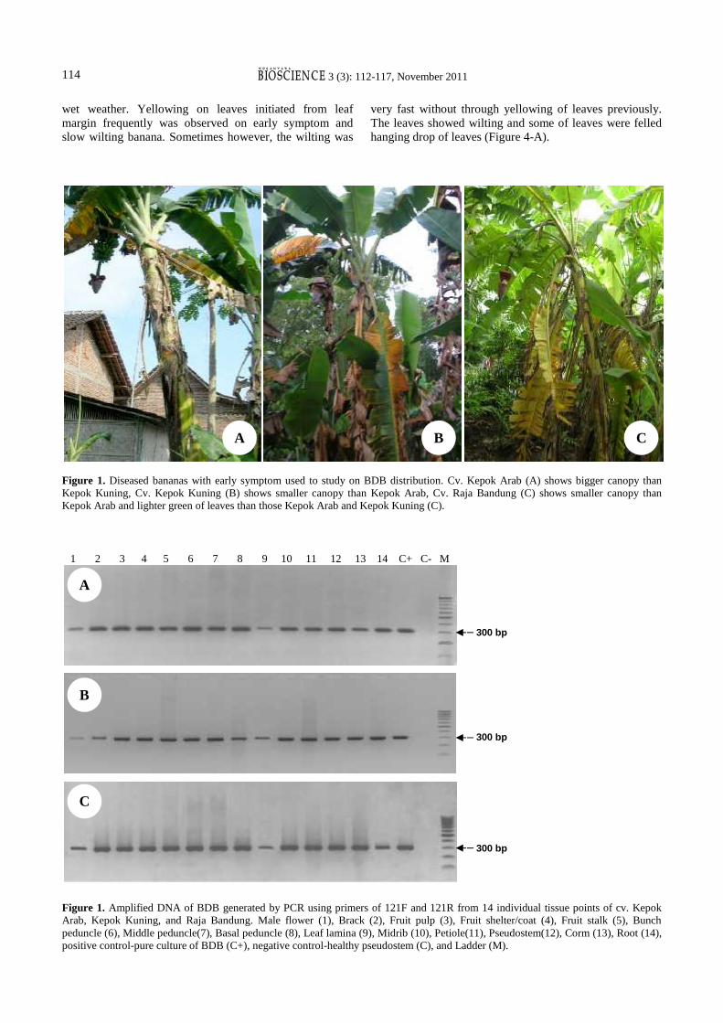
For purposing detection of BDB existence in diseasedplant by PCR using primers of 121F and 121R, at least 14tissue points of individual infected plant for each cultivarwere sampled. The detection using PCR showed that BDBcould be detected from all of the plant tissue points. Theseresults were supported by the established symptoms onindicator susceptible plantlets and re-detection BDB fromthe seedlings. It was occurred on all of cultivars, KepokArab, Kepok Kuning, and Raja Bandung (Figure 1, 2, 3).This means that BDB is existent in all of the sample tissuepoints. Thus, these observations give evidences that BDB issystemically distributed in all parts of infected plants.
For supporting the detection results, the sign (ooze),external and internal symptom on the inoculated seedlingwere observed. The observations of natural infection gotthat the bacterial ooze was usually exuded by brack andmale flower of inflorescence in the early morning and in
3 (3): 112-117, November 2011114
wet weather. Yellowing on leaves initiated from leafmargin frequently was observed on early symptom andslow wilting banana. Sometimes however, the wilting was
very fast without through yellowing of leaves previously.The leaves showed wilting and some of leaves were felledhanging drop of leaves (Figure 4-A).
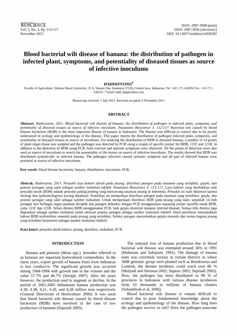
Figure 1. Diseased bananas with early symptom used to study on BDB distribution. Cv. Kepok Arab (A) shows bigger canopy thanKepok Kuning, Cv. Kepok Kuning (B) shows smaller canopy than Kepok Arab, Cv. Raja Bandung (C) shows smaller canopy thanKepok Arab and lighter green of leaves than those Kepok Arab and Kepok Kuning (C).
1 2 3 4 5 6 7 8 9 10 11 12 13 14 C+ C- M
Figure 1. Amplified DNA of BDB generated by PCR using primers of 121F and 121R from 14 individual tissue points of cv. KepokArab, Kepok Kuning, and Raja Bandung. Male flower (1), Brack (2), Fruit pulp (3), Fruit shelter/coat (4), Fruit stalk (5), Bunchpeduncle (6), Middle peduncle(7), Basal peduncle (8), Leaf lamina (9), Midrib (10), Petiole(11), Pseudostem(12), Corm (13), Root (14),positive control-pure culture of BDB (C+), negative control-healthy pseudostem (C), and Ladder (M).
300 bp
A B C
A
B
C
300 bp
300 bp
3 (3): 112-117, November 2011114
wet weather. Yellowing on leaves initiated from leafmargin frequently was observed on early symptom andslow wilting banana. Sometimes however, the wilting was
very fast without through yellowing of leaves previously.The leaves showed wilting and some of leaves were felledhanging drop of leaves (Figure 4-A).
Figure 1. Diseased bananas with early symptom used to study on BDB distribution. Cv. Kepok Arab (A) shows bigger canopy thanKepok Kuning, Cv. Kepok Kuning (B) shows smaller canopy than Kepok Arab, Cv. Raja Bandung (C) shows smaller canopy thanKepok Arab and lighter green of leaves than those Kepok Arab and Kepok Kuning (C).
1 2 3 4 5 6 7 8 9 10 11 12 13 14 C+ C- M
Figure 1. Amplified DNA of BDB generated by PCR using primers of 121F and 121R from 14 individual tissue points of cv. KepokArab, Kepok Kuning, and Raja Bandung. Male flower (1), Brack (2), Fruit pulp (3), Fruit shelter/coat (4), Fruit stalk (5), Bunchpeduncle (6), Middle peduncle(7), Basal peduncle (8), Leaf lamina (9), Midrib (10), Petiole(11), Pseudostem(12), Corm (13), Root (14),positive control-pure culture of BDB (C+), negative control-healthy pseudostem (C), and Ladder (M).
300 bp
A B C
A
B
C
300 bp
300 bp

HADIWIYONO – Blood bacterial wilt disease of banana 115
Figure 2. Wilting symptom on seedlings of cv. Kepok Kuning inoculated by washed ooze of tissue points of individual diseased plantsof cv. Kepok Arab 5 weeks after inoculation. Male flower (A), Brack (B), Fruit pulp (C), Fruit shelter/coat (D), Fruit stalk (E), Bunchpeduncle (F), middle peduncle(G), Basal peduncle(H), Pseudostem(I), Leaf lamina(J), Midrib(K), Petiole(L), Corm (M), Root (N),Control-positive (C+).
C- 1 2 3 4 5 6 7 8 9 10 11 12 13 14 15 M
Figure 3. Amplified DNA of BDB generated by PCR using primer 121F and 121R from corm of seedlings of cv. Kepok Kuninginoculated with washed ooze from 14 tissue points of cv. Kepok Arab on 5 weeks after inoculation, Male flower (1), Brack (2), Fruitpulp (3), Fruit shelter/coat (4), Fruit stalk (5), Bunch peduncle (6), Middle peduncle(7), Basal peduncle(8), Leaf lamina(9), Midrib(10),Petiole(11), Pseudostem(12), Corm(13), Root(14), positive control-pure culture of BDB (C+), negative control (C-), and Ladder (M).
Figure 4. Specific symptoms on bacterial wilt banana caused by BDB. A. Vegetative stage of Kepok Kuning with hanging drop ofleaves. B. Generative stage of Kepok Abu with dried inflorescence of flower. C. Bunch of Kepok Kuning with dried inflorescence offlower extending to upper parts or fruits.
A B C D
300 bp317 bp
A B C

3 (3): 112-117, November 2011116
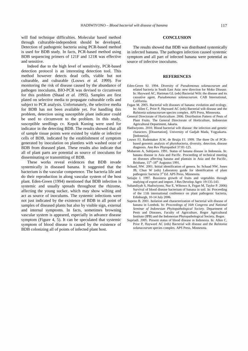
Figure 5. Internal/external symptoms of blood disease on organs of infected plant cv. Kepok Kuning, inflorescence with bacterial ooze(A) browning pulp at along section of the fruit and its stalk (B), browning vessel at the sliced fruit shelter (C), browning vessel at thepseudostem (D), browning vessel concentrated at the peduncle (E), browning vessel concentrated at margin of the middle peduncle (F),browning vessel concentrated at margin of the basal peduncle (G) browning vessel at the midrib (I), browning vessel at the petiole (J),browning vessel at the corm (H), yellowing leaf lamina at the margin (K), and browning diseased root (L).
Wilting of inflorescence flower on generative stage ofbananas was observed frequently (Figure 4-B). The wiltinginflorescence developed to upper parts of bunch includingfruits (Figure 4-C). If peduncle was cut in some pointsfrom upper to lower part would be observed a gradualbrowning in vessel tissues which was observed most severeat the upper part. Discoloration vascular tissues representedby brown dots/points were gradually less frequent on thefurther lower parts of pseudostem or peduncle. Suchsymptom can be speculated that the infection is startedfrom the inflorescence. Some diseased plants were incontrary, the symptom was with no or light browning at theupper parts and gradual more severe to the lower part withthe most severe in the corm. The latest symptom might be
started from the mother plant the growing sucker. Globally,the browning in vessel cells usually can be occurred in themost part of plants, pulp, stalk, fruit shelter, pedundle,middle peduncle and psedustem, basal peduncle, midrib,petiole, corm, and root (Figure 5).
BDB is difficult to isolate from almost point of diseasedplant tissues except from the upper peduncle and the bunchparticularly from the fruits. From the fruit shelter is themost frequent to be able to isolate BDB on CPG mediumwhereas from lower tissue points it is very difficult due tothe existence of high population of saprophytic that aresuppressive the growth of slow growing BDB. Selectivemedium for BDB has not been developed yet. Therefore,detection of BDB using culturable-dependent approaches
A D
E F G H
I J K L
B CA D
E F G H
I J K L
B CA D
E F G H
I J K L
B CA D
E F G H
I J K L
B CA D
E F G H
I J K L
B CA D
E F G H
I J K L
B CA D
E F G H
I J K L
B CA D
E F G H
I J K L
B CA D
E F G H
I J K L
B CA D
E F G H
I J K L
B CA D
E F G H
I J K L
B CA D
E F G H
I J K L
B CA D
E F G H
I J K L
B CA D
E F G H
I J K L
B CA D
E F G H
I J K L
B CA D
E F G H
I J K L
B CA D
E F G H
I J K L
B CA D
E F G H
I J K L
B CA D
E F G H
I J K L
B CA D
E F G H
I J K L
B CA D
E F G H
I J K L
B CA D
E F G H
I J K L
B CA D
E F G H
I J K L
B CA D
E F G H
I J K L
B CA B C D
E FB
GC
H
I JB
KC
L
HADIWIYONO – Blood bacterial wilt disease of banana 117
will find technique difficulties. Molecular based methodthrough culturable-independent should be developed.Detection of pathogenic bacteria using PCR-based methodis used for BDB study. In facts, PCR-based method usingBDB sequencing primers of 121F and 121R was effectiveand sensitive.
Indeed due to the high level of sensitivity, PCR-baseddetection protocol is an interesting detection tool. Thismethod however detects dead cells, viable but notculturable, and culturable (Louws et al. 1999). Formonitoring the risk of disease caused by the abundance ofpathogen inoculums, BIO-PCR was devised to circumventfor this problem (Shaad et al. 1995). Samples are firstplated on selective media to propagate culturable cells andsubject to PCR analysis. Unfortunately, the selective mediafor BDB has not been available yet. For handling thisproblem, detection using susceptible plant indicator couldbe used to circumvent to the problem. In this study,susceptible seedlings cv. Kepok Kuning were used forindicator in the detecting BDB. The results showed that allof sample tissue points were existed by viable or infectivecells of BDB, indicated by the establishment of symptomgenerated by inoculation on plantlets with washed ooze ofBDB from diseased plant. These results also indicate thatall of plant parts are potential as source of inoculums fordisseminating or transmitting of BDB.
These works reveal evidences that BDB invadesystemically in diseased banana. It suggested that thebacterium is the vascular competence. The bacteria life anddo their reproduction in along vascular system of the hostplant. Eden-Green (1994) mentioned that BDB infection issystemic and usually spreads throughout the rhizome,affecting the young sucker, which may show wilting andact as source of inoculums. The systemic infections werenot just indicated by the existence of BDB in all point ofsamples of diseased plants but also by visible sign, externaland internal symptoms. In facts, sometimes browningvascular system is appeared, especially in advance diseasesymptom (Figure 4, 5). It can be speculated that systemicsymptom of blood disease is caused by the existence ofBDB colonizing all of points of infected plant host.
CONCLUSION
The results showed that BDB was distributed systemicallyin infected banana. The pathogen infection caused systemicsymptom and all part of infected banana were potential assource of infective inoculums.
REFERENCES
Eden-Green SJ. 1994. Diversity of Pseudomonas solanacearum andrelated bacteria in South East Asia: new direction for Moko Disease.In: Hayward AC, Hartman GL (eds) Bacterial Wilt: the disease and itscausative agent, Pseudomonas solanacearum. CAB International,California.
Fegan M. 2005. Bacterial wilt diseases of banana: evolution and ecology.In: Allen C, Prior P, Hayward AC (eds) Bacterial wilt disease and theRalstonia solanacearum species complex. APS Press, Minnesota.
General Directorate of Horticulture. 2006. Distribution Pattern of Pests ofPlant Fruits. The General Directorate of Horticulture, IndonesianAgricultural Department, Jakarta.
Hadiwiyono. 2010. Blood bacterial wilt disease: the infection and geneticcharacters. 〔Dissertation〕. University of Gadjah Mada, Yogyakarta.〔Indonesia〕.
Louws FJ, Rademaker JLW, de Bruijn FJ. 1999. The three Ds of PCR-based genomic analysis of phytobacteria, diversity, detection, diseasediagnosis. Ann Rev Phytopathol 37:81-125.
Muharom A, Subijanto. 1991. Status of banana disease in Indonesia. In:banana disease in Asia and Pacific. Proceeding of technical meetingon diseases affecting banana and plantain in Asia and the Pacific,Brisbane, 15th–18th Augustus 1991.
Schaad, NW. 2001. Initial identification of genera. In: Schaad NW, JonesJB, Chun W (eds) Laboratory guide for identification of plantpathogenic bacteria 3rd Ed. APS Press, Minnesota.
Setiajie I. 1997. Bussiness growth of fruits and vegetables throughstudying of export and import. J Res Develop Agric 19:135-143.
Subandiyah S, Hadiwiyono, Nur E, Wibowo A, Fegan M, Taylor P. 2006)Survival of blood disease bacterium of banana in soil. In: Proceedingof the 11th international conference on plant pathogenic bacteria,Edinburgh, 10-14 July 2006.
Supeno B. 2001. Isolation and characterization of bacterial wilt disease ofbanana in Lombok. In: Proceedings of 16th Congress and NationalSeminar of Indonesian Phyitopathological Society. Department ofPests and Diseases, Faculty of Agriculture, Bogor AgriculturalInstitute (IPB) and the Indonesian Phytopathological Society, Bogor.
Supriadi. 2005. Present status of blood disease in Indonesia. In: Allen C,Prior P, Hayward AC (eds) Bacterial wilt disease and the Ralstoniasolanacearum species complex. APS Press, Minnesota.