
Black Molds and Melanized Yeasts Pathogenic to Humans
22
Black Molds and Melanized Yeasts Pathogenic to Humans Anuradha Chowdhary 1 , John Perfect 2 , and G. Sybren de Hoog 3 1 Department of Medical Mycology, Vallabhbhai Patel Chest Institute, University of Delhi, Delhi 110 007, India 2 Division of Infectious Diseases, Department of Medicine, Duke University Medical Center Durham, North Carolina 27710 3 CBS-KNAW Fungal Biodiversity Centre, 3508 AD Utrecht, The Netherlands Correspondence: [email protected] A review is given of melanized fungi involved in human infection, including species forming budding cells and strictly filamentous representatives. Classically, they are known as “phaeoid” or “dematiaceous” fungi, and, today, agents are recognized to belong to seven orders of fungi, of which the Chaetothyriales and Pleosporales are the most important. Infections range from cutaneous or pulmonary colonization to systemic or disseminated invasion. Subcutaneous involvement, either primary or after dissemination, may lead to host tissue proliferation of dermis or epidermis. Particularly in the Chaetothyriales, subcuta- neous and systemic infections may occur in otherwise apparently healthy individuals. Infections are mostly chronic and require extended antifungal therapy and/or surgery. B ecause melanin is a factor-enhancing viru- lence, black fungi are overrepresented as etiologic agents of opportunistic infection. Tra- ditionally, they have been collectively indicated under umbrella terms, such as “dematiaceous” or “phaeoid” fungi, referring to the presence of brown hyphae or yeast cells. Today, the lead- ing principle of fungal classification is molecu- lar phylogeny. The melanized fungi appear to belong to distantly related orders of Ascomy- cota, and the descriptive terminology above has therefore become obsolete. Clinically, they are involved in infections ranging from mild, hardly noticeable cutaneous infections (Saunte et al. 2011) to fatal brain diseases in other- wise healthy individuals (Al-Tawfiq and Boukhamseen 2011). Tissue forms range from melanized hyphae, yeast cells, or muriform cell clumps. The term “phaeohyphomycosis” (or phaeomycosis, a better term not excluding yeast cells) is therefore useful by negation, that is, a mycosis not caused by a hyaline fungus, but oth- erwise its information content is minimal. The fungi listed in this article are in alphabetical or- der according to genus with their phylogenetic affiliation in parentheses. The characteristic common to all species treated in this article is the presence of melanin in cell walls, which is responsible for the dark color of hyphae, yeast cells, muriform cell clumps, and conidia, and is believed to be a major virulence factor– enhancing opportunism. The function of melanin in their natural habitat mostly is protection against solar irradiation Editors: Arturo Casadevall, Aaron P. Mitchell, Judith Berman, Kyung J. Kwon-Chung, John R. Perfect, and Joseph Heitman Additional Perspectives on Human Fungal Pathogens available at www.perspectivesinmedicine.org Copyright # 2015 Cold Spring Harbor Laboratory Press; all rights reserved; doi: 10.1101/cshperspect.a019570 Cite this article as Cold Spring Harb Perspect Med 2015;5:a019570 1 www.perspectivesinmedicine.org Press on November 30, 2021 - Published by Cold Spring Harbor Laboratory http://perspectivesinmedicine.cshlp.org/ Downloaded from
Transcript of Black Molds and Melanized Yeasts Pathogenic to Humans
Black Molds and Melanized Yeasts Pathogenicto Humans
Anuradha Chowdhary1, John Perfect2, and G. Sybren de Hoog3
1Department of Medical Mycology, Vallabhbhai Patel Chest Institute, University of Delhi, Delhi 110 007, India2Division of Infectious Diseases, Department of Medicine, Duke University Medical Center Durham, NorthCarolina 27710
3CBS-KNAW Fungal Biodiversity Centre, 3508 AD Utrecht, The Netherlands
Correspondence: [email protected]
A review is given of melanized fungi involved in human infection, including species formingbudding cells and strictly filamentous representatives. Classically, they are known as“phaeoid” or “dematiaceous” fungi, and, today, agents are recognized to belong to sevenorders of fungi, of which the Chaetothyriales and Pleosporales are the most important.Infections range from cutaneous or pulmonary colonization to systemic or disseminatedinvasion. Subcutaneous involvement, either primary or after dissemination, may lead tohost tissue proliferation of dermis or epidermis. Particularly in the Chaetothyriales, subcuta-neous and systemic infections may occur in otherwise apparently healthy individuals.Infections are mostly chronic and require extended antifungal therapy and/or surgery.
Because melanin is a factor-enhancing viru-lence, black fungi are overrepresented as
etiologic agents of opportunistic infection. Tra-ditionally, they have been collectively indicatedunder umbrella terms, such as “dematiaceous”or “phaeoid” fungi, referring to the presenceof brown hyphae or yeast cells. Today, the lead-ing principle of fungal classification is molecu-lar phylogeny. The melanized fungi appear tobelong to distantly related orders of Ascomy-cota, and the descriptive terminology abovehas therefore become obsolete. Clinically, theyare involved in infections ranging from mild,hardly noticeable cutaneous infections (Saunteet al. 2011) to fatal brain diseases in other-wise healthy individuals (Al-Tawfiq andBoukhamseen 2011). Tissue forms range from
melanized hyphae, yeast cells, or muriform cellclumps. The term “phaeohyphomycosis” (orphaeomycosis, a better term not excluding yeastcells) is therefore useful by negation, that is, amycosis not caused by a hyaline fungus, but oth-erwise its information content is minimal. Thefungi listed in this article are in alphabetical or-der according to genus with their phylogeneticaffiliation in parentheses.
The characteristic common to all speciestreated in this article is the presence of melaninin cell walls, which is responsible for the darkcolor of hyphae, yeast cells, muriform cellclumps, and conidia, and is believed to be amajor virulence factor–enhancing opportunism.The function of melanin in their natural habitatmostly is protection against solar irradiation
Editors: Arturo Casadevall, Aaron P. Mitchell, Judith Berman, Kyung J. Kwon-Chung, John R. Perfect, and Joseph Heitman
Additional Perspectives on Human Fungal Pathogens available at www.perspectivesinmedicine.org
Copyright # 2015 Cold Spring Harbor Laboratory Press; all rights reserved; doi: 10.1101/cshperspect.a019570
Cite this article as Cold Spring Harb Perspect Med 2015;5:a019570
1
ww
w.p
ersp
ecti
vesi
nm
edic
ine.
org
Press on November 30, 2021 - Published by Cold Spring Harbor Laboratoryhttp://perspectivesinmedicine.cshlp.org/Downloaded from

because of growth on exposed surfaces, such asnatural rock, or against factors prevalent underconditions of stress. Another main type of nat-ural ecology is in decomposing plant material;if the orders concerned preponderantly containplant pathogens, then the human opportunistsare found among the few saprobes on plant de-bris in that group. Clinical pathology mostlyemerges from traumatic introduction in or be-low the skin resulting in a suppurative foreignbody response. Inhalation mycoses are excep-tional and mostly confined to pulmonary colo-nization in patients with cystic fibrosis. Deep-seated infections can be, depending on the spe-cies, disseminated or cerebral; their portal ofentry is poorly understood. A special categoryis chromoblastomycosis, a disease exclusivelycaused by members of the Herpotrichiellaceae(black yeasts and relatives in the order Chaeto-thyriales), and is clinically exceptional by a hostresponse with hyperproliferation rather thannecrosis; the tissue form consists of muriform(sclerotic) cells. Although diseases by melanizedfungi are rare, they are significant because oftheir occurrence in otherwise healthy individu-als, and no notable increase in their frequency isnoticed with the emergence of immunocom-promised hospital populations. Decreased im-munity, as well as diabetes, nevertheless, are riskfactors for infection. Recently, some of the high-ly recalcitrant, disseminated infections appearedto be associated with mutations in the host’sdectin signaling pathway (Wang et al. 2014).
CLINICAL SPECTRUM OF DISEASESCAUSED BY BLACK FUNGI
Colonization. Asymptomatic growth of mela-nized fungi is known on human skin as well asin the lungs, particularly when patients sufferfrom cystic fibrosis. The systemic colonizershave an invasive potential when the host im-mune barriers are broken, whereas colonizersof skin and nails cause cutaneous infections atmost.
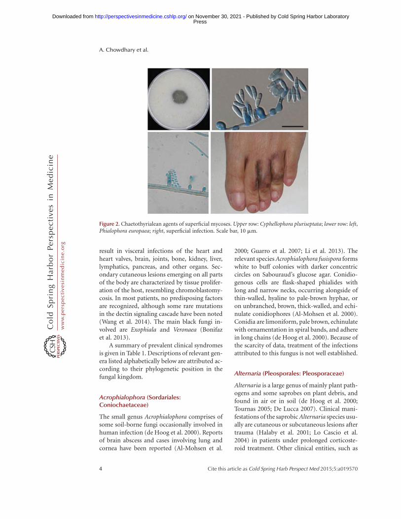
Superficial. Growth on human skin hasclassically been reported from Hortaea wer-neckii, which mainly colonizes the epidermisbut also from members of Cyphellophora and
Phialophora, which are known to be involved inmild skin infections and onychomycoses (Fig. 2).
Cutaneous. Cutaneous infections are un-common. Mostly cystic or papular lesions onexposed body areas are of concern in patientsunder prolonged corticosteroid therapy. Alter-naria (Fig. 1B, left) and Exophiala species arethe most common etiological agents.
Subcutaneous. Subcutaneous infections canbe necrotic phaeomycoses with hyphae or yeast-like cells in tissue, or are eumycetomata, lesionsbeing granulomatous abscesses with drainingsinuses from which granules of dense fungalmaterial may be recovered. Osteomyelitis mayoccur. Etiologic agents mainly are found inMadurella, Phaeoacremonium, in coelomyce-tous representatives of Pleosporales, and occa-sionally in Exophiala.
Chromoblastomycosis. This is a chronic sub-cutaneous infection caused by members of Her-potrichiellaceae (black yeasts and relatives) andcharacterized by the presence of muriform cells(sclerotic bodies) in tissue sections or wet prep-arations of pus or scrapings. Cladophialophoracarrionii, Fonsecaea monophora, Fonsecaea pe-drosoi, Rhinocladiella aquaspersa, and occasion-ally Phialophora verrucosa are the fungal speciesinvolved. C. carrionii is endemic to arid climatezones, whereas Fonsecaea species are prepon-derantly found under tropical conditions.
Systemic. Cerebral abscesses are rare but fatalif untreated and are mainly reported from im-munocompetent individuals (Carter and Bou-dreaux 2004; Delfino et al. 2006; Chang et al.2009; Al-Tawfiq and Boukhamseen 2011). Themost common neurotropic fungi, Rhinocla-diella mackenziei, Cladophialophora bantiana,Exophiala dermatitidis, and F. monophora, aremembers of Herpotrichiellaceae, but also Exser-ohilum, Bipolaris (Pleosporaceae), and Verruco-nis (Sympoventuriaceae) can be involved. Somespecies are endemic to the Middle East or EastAsia or show an increased prevalence in India(Delfino et al. 2006; Garg et al. 2007; Li and deHoog 2009; Al-Tawfiq and Boukhamseen 2011;Jabeen et al. 2011; Pedersen et al. 2011). Centralnervous system (CNS) infections by Herpotri-chiellaceae are hypothesized to be acquired viainhalation and then quickly disseminated to the
A. Chowdhary et al.
2 Cite this article as Cold Spring Harb Perspect Med 2015;5:a019570
ww
w.p
ersp
ecti
vesi
nm
edic
ine.
org
Press on November 30, 2021 - Published by Cold Spring Harbor Laboratoryhttp://perspectivesinmedicine.cshlp.org/Downloaded from

CNS via the hematogenous route; but because ofa long asymptomatic period of incubation, theactual route of infection may be difficult to estab-lish. Another better-known development of CNSinfection, mostly seen in Pleosporaceae, is second-ary to chronic fungal sinusitis (Fig. 1B, right).
Disseminated. This type of infection is al-most exclusively caused by members of Herpo-trichiellaceae. The infections are very chronic,with a long incubation period, and highly re-fractory to therapy. Hematogenous spread ofthe fungus to one or more distant sites may
Figure 1. Pleosporalean agents. (A) Top row, Curvularia lunata; middle row, Bipolaris hawaiiensis; bottom row,Alternaria alternata. (B) Left, Alternaria cutaneous infection; right, Bipolaris chronic sinusitis.
Black Molds and Yeasts Pathogenic to Humans
Cite this article as Cold Spring Harb Perspect Med 2015;5:a019570 3
ww
w.p
ersp
ecti
vesi
nm
edic
ine.
org
Press on November 30, 2021 - Published by Cold Spring Harbor Laboratoryhttp://perspectivesinmedicine.cshlp.org/Downloaded from
result in visceral infections of the heart andheart valves, brain, joints, bone, kidney, liver,lymphatics, pancreas, and other organs. Sec-ondary cutaneous lesions emerging on all partsof the body are characterized by tissue prolifer-ation of the host, resembling chromoblastomy-cosis. In most patients, no predisposing factorsare recognized, although some rare mutationsin the dectin signaling cascade have been noted(Wang et al. 2014). The main black fungi in-volved are Exophiala and Veronaea (Bonifazet al. 2013).
A summary of prevalent clinical syndromesis given in Table 1. Descriptions of relevant gen-era listed alphabetically below are attributed ac-cording to their phylogenetic position in thefungal kingdom.
Acrophialophora (Sordariales:Coniochaetaceae)
The small genus Acrophialophora comprises ofsome soil-borne fungi occasionally involved inhuman infection (de Hoog et al. 2000). Reportsof brain abscess and cases involving lung andcornea have been reported (Al-Mohsen et al.
2000; Guarro et al. 2007; Li et al. 2013). Therelevant species Acrophialophora fusispora formswhite to buff colonies with darker concentriccircles on Sabouraud’s glucose agar. Conidio-genous cells are flask-shaped phialides withlong and narrow necks, occurring alongside ofthin-walled, hyaline to pale-brown hyphae, oron unbranched, brown, thick-walled, and echi-nulate conidiophores (Al-Mohsen et al. 2000).Conidia are limoniform, pale brown, echinulatewith ornamentation in spiral bands, and adherein long chains (de Hoog et al. 2000). Because ofthe scarcity of data, treatment of the infectionsattributed to this fungus is not well established.
Alternaria (Pleosporales: Pleosporaceae)
Alternaria is a large genus of mainly plant path-ogens and some saprobes on plant debris, andfound in air or in soil (de Hoog et al. 2000;Tournas 2005; De Lucca 2007). Clinical mani-festations of the saprobic Alternaria species usu-ally are cutaneous or subcutaneous lesions aftertrauma (Halaby et al. 2001; Lo Cascio et al.2004) in patients under prolonged corticoste-roid treatment. Other clinical entities, such as
Figure 2. Chaetothyrialean agents of superficial mycoses. Upper row: Cyphellophora pluriseptata; lower row: left,Phialophora europaea; right, superficial infection. Scale bar, 10 mm.
A. Chowdhary et al.
4 Cite this article as Cold Spring Harb Perspect Med 2015;5:a019570
ww
w.p
ersp
ecti
vesi
nm
edic
ine.
org
Press on November 30, 2021 - Published by Cold Spring Harbor Laboratoryhttp://perspectivesinmedicine.cshlp.org/Downloaded from

cerebral infections, sinusitis, keratitis, and aller-gic bronchopulmonary mycosis, are very rare(Hipolito et al. 2009; Chowdhary et al. 2012,2014a). The most common etiologic agents areAlternaria alternata and Alternaria infectoriaand, occasionally, Alternaria chlamydospora. A.infectoria may be difficult to recognize because ofits often poor sporulation on routine media andloss of melanin in vitro and in tissue. The taxon-omyand identification of other reported species,
suchasAlternariadianthicola,Alternaria longipes,and Alternaria tenissima, has been insufficientlyclarified. Morphologically, colonies generally areexpanding, hairy, with gray to olivaceous blackcolors. Conidiophores are mostly erect, brown,and multicelled, producing conidia in sympo-dial order, leaving flat, dark-brown scars. Theconidia are usually brown, smooth walled, orverruculose with a round base and beaked tip,with muriform septation, and are produced in
Table 1. Overview of spectrum of prevalent diseases caused by black fungi
Order Etiologic genus Prevalent clinical manifestationa
Capnodiales Hortaea Cutaneous colonizationChaetothyriales Cladophialophora Brain abscess, (sub)cutaneous infection, chromoblastomycosis
Cyphellophora Cutaneous colonization, onychomycosisExophiala Brain abscess, chromoblastomycosis, (sub)cutaneous infection,
pneumonia, eumycetomaFonsecaea Chromoblastomycosis, brain abscessKnufia Skin colonizationPhialophora Subcutaneous infection, disseminated, chromoblastomycosis,
cutaneous colonizationRhinocladiella Brain abscess, chromoblastomycosisVeronaea (Sub)cutaneous infection, disseminated
Diaporthales Phaeoacremonium Subcutaneous infection, eumycetoma, fungemia, osteomyelitis,arthritis, endocarditis
Dothideales Aureobasidium (Sub)cutaneous infection, fungemia, meningitis, peritonitisNeoscytalidium Onychomycosis, cutaneous infection, fungemia
Pleosporales Alternaria (Sub)cutaneous infection, sinusitis, keratitis, onychomycosis,ABPM, disseminated
Biatriospora EumycetomaBipolaris (Sub)cutaneous infection, sinusitis, keratitis, ABPM,
pneumonia, disseminatedCurvularia (Sub)cutaneous infection, sinusitis, keratitis, ABPM,
eumycetoma, peritonitis, onychomycosis, brain abscess,disseminated
Exserohilum Brain abscess, chromoblastomycosis, (sub)cutaneous infection,pneumonia, eumycetoma
Falciformispora EumycetomaMedicopsis EumycetomaPhoma (Sub)cutaneous infection, keratitisPseudochaetosphaeronema EumycetomaPyrenochaeta Keratitis, onychomycosis, (sub)cutaneous infection, eumycetomaTrematosphaeria Subcutaneous infections
Sordiales Acrophialophora Brain abscess, keratitisChaetomium Brain abscess, (sub)cutaneous infection, pneumonia, eumycetomaMadurella Eumycetoma
Venturiales Ochronis Cutaneous infectionVerruconis Pneumonia, brain abscess, disseminated
aInfections possible in otherwise healthy-appearing patients. Infections exclusively occurring in immunocompromised
patients are shown in bold.
Black Molds and Yeasts Pathogenic to Humans
Cite this article as Cold Spring Harb Perspect Med 2015;5:a019570 5
ww
w.p
ersp
ecti
vesi
nm
edic
ine.
org
Press on November 30, 2021 - Published by Cold Spring Harbor Laboratoryhttp://perspectivesinmedicine.cshlp.org/Downloaded from

chains. Morphological characteristics distin-guishing A. alternata from A. infectoria are theconidia, which often become nearly tubular andoccur in strongly branched chains in the latterspecies. Conidia resemble multicelled chlamy-dospores in A. chlamydospora. Sequencing ofthe rDNA internal transcribed spacer (ITS) re-gion is sufficient for routine identification ofA. alternata and A. infectoria (Hipolito et al.2009; Cunha et al. 2012).
Aureobasidium (Dothideales:Dothiodeaceae)
Aureobasidium pullulans is a black yeast-likefungus that is ubiquitous on poor-nutrient sur-faces, such as on plant leaves, glass, and dampbathroom walls, and is commonly found as acontaminant in clinical laboratories. Recently,intraspecific molecular diversity has been found,which has led to the description of varieties andsibling species (Zalar et al. 1999). Opportunisticinfections are mostly caused by A. pullulans butalso other taxa have been reported, such as Au-reobasidium proteae (de Hoog et al. 2000;Kutlesa et al. 2012). Most infections occur bytraumatic inoculation, such as keratitis and cu-taneous lesions; disseminated mycoses are veryrare and occur only in severely immunocom-promised patients (Kaczmarski et al. 1986; Sal-kin et al. 1986; Arranz et al. 2006; Panda et al.2006; Mise et al. 2008; Pikazis et al. 2009; Chawlaet al. 2010; Joshi et al. 2010; Mershon-Shier et al.2011). A. pullulans has an affinity for syntheticmaterials and surgically implanted devices, asevidenced by the relatively frequent isolationof the organism from peritoneal dialysis cathe-ters and central venous lines (Clark et al. 1995;Caporale et al. 1996; Hawkes et al. 2005; Miseet al. 2008). The in vitro activity of antifun-gals against A. pullulans revealed resistance tofluconazole and high minimum inhibitoryconcentrations of voriconazole, isavuconazole,caspofungin, and micafungin. However, the iso-lates exhibit susceptibility to amphotericin B,posaconazole, and itraconazole (Najafzadeh etal. 2014). Colonies grow rapidly and are smooth,cream to pink, covered with slimy exudates, andbecome variably brown or black in a later stage.
On microscopic examination, large, hyaline hy-phae are seen, which variably convert into thick-walled, dark-blackish-brown chlamydospore-like cells. Conidia are produced synchronouslyin groups alongside undifferentiated hyphae.Species distinction is by ITS sequencing.
Biatriospora (Pleosporales:Trematosphaeriaceae)
This single species B. mackinonii was formerlyclassified in Pyrenochaeta or Nigrograna. Thespecies is an occasional agent of human myce-toma. The natural habitat of this fungus is un-known. Colonies are gray, velvety, becomingdark gray to black with age. Pycnidia are soli-tary, globose to pyriform, with papillate osti-oles. Conidiogenous cells are hyaline, phialidic,and discrete. Conidia are subhyaline, brown inmass, one celled, and ellipsoidal (Ahmed et al.2014).
Bipolaris (Pleosporales: Pleosporaceae)
Most species belonging to the genus Bipolarisare host-specific pathogens on grasses, whereassome saprobic species in soil with dead and de-caying plant material can be found as humanopportunists (Revankar et al. 2010). The preva-lent clinically significant saprobes are Bipolarisaustraliensis, Bipolaris hawaiiensis, and Bipolarisspicifera (Sivanesan 1987; da Cunha et al. 2012a).They are particularly associated with chronicpansinusitis (Toul et al. 2006). Other cases in-clude endophthalmitis and orbital cellulitis(Newell et al. 2006; Sheyman et al. 2013), necro-tizing pneumonia and allergic bronchopulmo-nary mycosis (Saenz et al. 2001; Chowdhary etal. 2011, 2014b), peritonitis (Bava et al. 2003),endarteritis (Ogden et al. 1992), and encephalitis(Morton et al. 1986; Pauzner et al. 1997). Colo-nies are black, hairy, and expanding. Conidio-phores are brown, erect, multicelled, producingellipsoidal, straight, orcurved conidiawith dark-brown, flat conidial scars. Molecular identifica-tion can be done using ITS sequencing. A spe-cific polymerase chain reaction (PCR) has beendeveloped for the direct detection of Bipolarisspecies (Shin et al. 2003; El-Morsy et al. 2010).
A. Chowdhary et al.
6 Cite this article as Cold Spring Harb Perspect Med 2015;5:a019570
ww
w.p
ersp
ecti
vesi
nm
edic
ine.
org
Press on November 30, 2021 - Published by Cold Spring Harbor Laboratoryhttp://perspectivesinmedicine.cshlp.org/Downloaded from

Chaetomium (Sordariales: Chaetomiaceae)
This genus comprises more than 180 speciesfrom straw, plant debris, and animal dung(Guarro 1998; de Hoog and Vitale 2007). Thesignificance of the genus has been underesti-mated because clinical strains are mostly sterilein culture, and could not be recognized by mor-phology (de Hoog et al. 2013). Clinically signif-icant species include Chaetomium globosum,followed by Chaetomium strumarium, Chaeto-mium atrobrunneum, Chaetomium funicola, andChaetomium perlucidum (Abbott et al. 1995;Guarro et al. 1995; Yeghen et al. 1996; Guppyet al. 1998; Lesire et al. 1999; Thomas et al.1999). Chaetomium species have mainly beenreported from onychomycosis and sinusitis(Aru et al. 1997; Stiller et al. 1992). Brain infec-tion is sometimes seen in individuals duringillicit intravenous drug use (Abbott et al.1995). A chromoblastomycosis-like infectionby C. funicola was reported by Piepenbringet al. (2007); this is an exceptional case of thisdisease caused by a fungus in the Herpotrichiel-laceae. The distinctive feature of Chaetomiumspecies is the presence of pronounced hairs, orsetae, on the spherical to pyriform fruit bodies.The hairs may be dichotomously branched orunbranched and are often undulate or spirallycoiled. Asci are usually eight-spored and deli-quescent so that brown, one-celled ascosporesare easily liberated in large masses. Opportunis-tic species are thermotolerant; C. globosumgrows at 35˚C but not at 40˚C, whereas the neu-rotropic species C. atrobrunneum and C. perlu-cidum grow at 40˚C (von Arx et al. 1986; Barronet al. 2003).
Cladophialophora (Chaetothyriales:Herpotrichiellaceae)
Cladophialophora is morphologically character-ized by one-celled, ellipsoidal to fusiform, anddry conidia, arising in long, branched, or un-branched chains. Cladophialophora species areinvolved in a wide diversity of infections, rang-ing from mild cutaneous to fatal encephalitis(Borelli 1980; Ho et al. 1999; de Hoog et al.2000). An important species is the neurotropicfungus, C. bantiana, of which more than 100
case reports in mostly healthy individuals havebeen published. Cladophialophora modesta wasresponsible for a case after head trauma (Mc-Ginnis et al. 1999). C. carrionii is the prevalentagent of chromoblastomycosis in dry climatesand desert zones (Mendoza et al. 1993; Jaya-keerthi et al. 2004; Ameen 2009). Cladophialo-phora devriesii and Cladophialophora arxii arerare agents of disseminated diseases, whereasCladophialophora boppii, Cladophialophora em-monsii, and Cladophialophora saturnica arecauses of mild cutaneous infections (Fig. 3)(Badali et al. 2008).
Curvularia (Pleosporales: Pleosporaceae)
This genus consists of �100 species, most ofwhich are either saprobes in soil or on plantdebris, or are plant pathogens mainly infectinggrasses (Sivanesan 1987). Human infectionshave been reported in Curvularia aeria, Curvu-laria geniculata, and Curvularia lunata; isolatedcases occurred in Curvularia brachyspora, Cur-vularia clavata, Curvularia inaequalis, Curvu-laria pallescens, and Curvularia verruculosa.Curvularia species may cause allergic sinusitis,which can disseminate to the brain of immuno-competent patients (Ebright et al. 1999). Othermanifestations include subcutaneous infectionsfollowing traumatic implantation, such as ker-atitis. Colonies of Curvularia are black in color,expanding, and hairy (Tanabe et al. 2010; Moo-dy et al. 2012). Conidiophores are brown anderect, producing ellipsoidal, brown, usuallycurved conidia with three or four transversesepta. Species can be differentiated on the basisof ITS and glyceraldehyde-3-phosphate dehy-drogenase gene sequences (de Hoog and Vitale2007).
Cyphellophora (Chaetothyriales:Herpotrichiellaceae–Cyphellophoraceae)
The natural habitat of members of the smallgenus Cyphellophora has not been establishedwith certainty. They are mainly known fromhuman skin and nails, where they can be symp-tomatic or subclinical, but detailed case reportsare as yet lacking (Feng et al. 2014). The species
Black Molds and Yeasts Pathogenic to Humans
Cite this article as Cold Spring Harb Perspect Med 2015;5:a019570 7
ww
w.p
ersp
ecti
vesi
nm
edic
ine.
org
Press on November 30, 2021 - Published by Cold Spring Harbor Laboratoryhttp://perspectivesinmedicine.cshlp.org/Downloaded from

grow very slowly with olivaceous black coloniesand produce thin-walled, sickle-shaped conidiawith one or several transverse septa. Conidia areproduced in packages from phialide openingson spherical conidiogenous cells or directlyfrom hyphae.
Exophiala (Chaetothyriales:Herpotrichiellaceae)
Exophiala is the major genus of black yeasts,characterized by annelidic conidiogenesis andthe presence of torulose hyphae, that is, hyphae
Figure 3. Chaetothyrialean agents of deep infections. Upper row: Fonsecaea pedrosoi causing chromoblastony-cosis with muriform cells; middle row: Ramichloridium mackenziei, agent of cerebral phaemycosis (case showncaused by Cladaphialophora bantiana); bottom row: disseminated infection caused by Veronaea botryosa.
A. Chowdhary et al.
8 Cite this article as Cold Spring Harb Perspect Med 2015;5:a019570
ww
w.p
ersp
ecti
vesi
nm
edic
ine.
org
Press on November 30, 2021 - Published by Cold Spring Harbor Laboratoryhttp://perspectivesinmedicine.cshlp.org/Downloaded from

composed of chains of more or less sphericalcells. They are typically associated with nutri-ent-poor or toxic environments, such as creo-soted wood (Dogen et al. 2013), toxic mines(Seyedmousavi et al. 2011), or steam baths (Sud-hadham et al. 2008). The common clinically rel-evant species include E. dermatitidis, Exophialaxenobiotica, Exophiala spinifera, and Exophialaoligosperma. Less common are Exophiala leca-nii-corni, Exophiala asiatica, Exophiala phaeo-muriformis, Exophiala jeanselmei, Exophialabergeri, and Exophiala mesophila (de Hooget al. 2003; Zeng et al. 2007; Badali et al. 2010).E. dermatitidis causes disseminated infectioneventually with neurotropism, whereas E. spini-fera is osteotropic (Horre and de Hoog 1999;Kantarcioglu and de Hoog 2004; Harris et al.2009; Badali et al. 2010). Exophiala coloniesare restricted, olivaceous black, and often initial-ly slimy at the center and then becoming velvetytoward the margin. Conidiogenous cells are in-tercalary or lateral, cylindrical, flask shaped oracicular, with relatively narrow, short, or veryshort annellated zones. Conidia are generallypresent in clumps. Molecular identification formost species is by ITS sequencing because theygenerally show very limited morphological dif-ferentiation (Zeng et al. 2007; Najafzadeh et al.2013).
Exserohilum (Pleosporales: Pleosporaceae)
The genus Exserohilum comprises �35 saprobicspecies mainly feeding on plant debris. In theclinical literature, three clinically significantspecies have been reported (Exserohilum rostra-tum, Exserohilum longirostratum, and Exserohi-lum mcginnisii), but molecular studies demon-strated that they belong to a single species,E. rostratum (da Cunha et al. 2012b). Exserohi-lum species are mainly involved in traumaticinfections, such as keratitis (Burges et al. 1987;Pauzner et al. 1997; Mathews and Maharajan1999; Joseph et al. 2012). Otherwise speciesmay cause invasive infections in immunocom-promised patients (Aquino et al. 1995), withrisk factors including aplastic anemia (Lasalaet al. 2005; Adler et al. 2006) and hematopoieticstem cell transplant (HSCT) (Togitani et al.
2007; Ritter et al. 2013). Recently, E. rostratumwas implicated in a large meningitis outbreakthat was traced back to contaminated steroidinjections (Lyons et al. 2012; Gade et al. 2013;Lockhart et al. 2013; Smith et al. 2013). Thespecies can be identified morphologically usingnutritionally deficient media in which its mor-phology is most pronounced. Colonies are usu-ally gray or black, hairy, and spreading. Conid-iophores are brown to olivaceous brown incolor, simple, thick, and smooth walled. Conid-ia are fusiform, cylindrical, or obclavate. Se-quencing the rDNA ITS region is sufficient forroutine identification, and a real-time assay forrapid identification of E. rostratum has beendeveloped (Guerra et al. 2013; Zhao et al. 2013).
Falciformispora (Pleosporales:Trematosphaeriaceae)
Two species producing ascigerous fruit bodiesformerly classified in Leptosphaeria belong tothis genus, namely, Falciformispora senegalensisand Falciformispora tompkinsii, both agents ofhuman mycetoma (Ahmed et al. 2014). Colo-nies are radially folded, grayish green, withbrown exudate on the colony surface; the reverseis dark brown, with dark-brown pigment diffus-ing into the agar, and conidia absent. Ascomataappear on the agar surface after months of in-cubation, black, solitary, (sub)spherical, andthick walled. Asci are clavate, rounded at theapex, bitunicate, and contain eight ascospores.Ascospores are ellipsoidal, four-septate, withconstrictions at the septa; the second cell fromthe top is the largest, leading to widening of thethin sheath that surrounds the ascospore.
Fonsecaea (Chaetothyriales:Herpotrichiellaceae)
The clinically relevant members of this genuscomprise three closely related species, F. pedro-soi, F. monophora, and Fonsecaea nubica, allcausing human chromoblastomycosis. Sapro-bic Fonsecaea species are found in the environ-ment as degraders of plant debris (Najafzadeh etal. 2010b). The disease may be acquired by ac-cidental inoculation of plant debris carrying
Black Molds and Yeasts Pathogenic to Humans
Cite this article as Cold Spring Harb Perspect Med 2015;5:a019570 9
ww
w.p
ersp
ecti
vesi
nm
edic
ine.
org
Press on November 30, 2021 - Published by Cold Spring Harbor Laboratoryhttp://perspectivesinmedicine.cshlp.org/Downloaded from
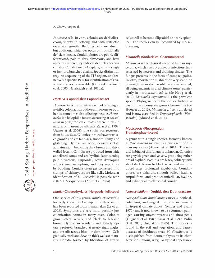
Fonsecaea cells. In vitro, colonies are dark oliva-ceous, velvety to cottony, and with restrictedexpansion growth. Budding cells are absent,but additional phialides occur on nutritionallydeficient media. Conidiophores are poorly dif-ferentiated, pale to dark olivaceous, and haveapically clustered, cylindrical denticles bearingconidia. Conidia are 0–1 septate, arising singlyor in short, branched chains. Species distinctionrequires sequencing of the ITS region, or alter-natively a specific PCR for identification of Fon-secaea species is available (Gunde-Cimermanet al. 2000; Najafzadeh et al. 2010a).
Hortaea (Capnodiales: Capnodiaceae)
H. werneckii is the causative agent of tinea nigra,a visible colonization of the palm on one or bothhands, sometimes also affecting the sole. H. wer-neckii is a halophilic fungus occurring at coastalareas in (sub)tropical climates, where it lives innatural or man-made saltpans (Zalar et al. 1999;Uezato et al. 2006); one strain was recoveredfrom house dust. Colonies in vitro have restrict-ed growth and are tar black, smooth, slimy, andglistening. Hyphae are wide, densely septateat maturation, becoming dark brown and thickwalled locally. Conidia are produced from wideannellated zones and are hyaline, later turningpale olivaceous, ellipsoidal, often developinga thick median septum; and they reproduceby budding. Conidia often get converted intoclumps of chlamydospore-like cells. Molecularidentification of H. werneckii is possible withrDNA ITS sequencing (Abliz et al. 2004).
Knufia (Chaetothyriales: Herpotrichiellaceae)
One species of this genus, Knufia epidermidis,formerly known as Coniosporium epidermidis,has been reported from human skin (Li et al.2008). Symptoms are very mild, possibly justcolonization occurs in many cases. Coloniesgrow slowly, velvety, and black to blackishbrown. Hyphae are regularly and densely sep-tate, profusely branched at nearly right angles,and are olivaceous black or dark brown. Cellsgradually swell and develop thick walls at matu-rity. Conidia formed by liberation of arthric
cells swell to become ellipsoidal or nearly spher-ical. The species can be recognized by ITS se-quencing.
Madurella (Sordariales: Chaetomiaceae)
Madurella is the classical agent of human my-cetoma, which is a subcutaneous infection char-acterized by necrosis and draining sinuses. Thefungus presents in the form of compact grains.In vitro, sporulation is absent or very scant. Atpresent, three molecular siblings are recognized,all being endemic in arid climate zones, partic-ularly in northeastern Africa (de Hoog et al.2012). Madurella mycetomatis is the prevalentspecies. Phylogenetically, the species cluster as apart of the ascomycete genus Chaetomium (deHoog et al. 2013). Madurella grisea is unrelatedand is now classified in Trematosphaeria (Pleo-sporales) (Ahmed et al. 2014).
Medicopsis (Pleosporales:Trematosphaeriaceae)
A genus with a single species, formerly knownas Pyrenochaeta romeroi, is a rare agent of hu-man mycetoma (Ahmed et al. 2014). The nat-ural habitat of this fungus is unknown. Coloniesare grayish green and velvety, producing ratherbroad hyphae. Pycnidia are black, solitary withshort dark brown to black setae, and are pro-duced after prolonged incubation. Conidio-phores are phialidic, smooth walled, hyaline,ampulliform, and produce unicellular, hyaline,and cylindrical to ellipsoidal conidia.
Neoscytalidium (Dothideales: Dothioraceae)
Neoscytalidium dimidiatum causes superficial,cutaneous, and ungual infections in humansin tropical climate zones (Gentles and Evans1970), and is now known to be a common path-ogen causing onychomycosis and tinea pedis(Gugnani et al. 1989; Lacaz et al. 1999; Padinet al. 2005; Ungpakorn 2005). The species isfound in the soil and vegetation, and causesdiseases of deciduous trees. N. dimidiatum isdistinguished from dermatophytes by its char-acteristic sinuous, irregular hyphal appearance
A. Chowdhary et al.
10 Cite this article as Cold Spring Harb Perspect Med 2015;5:a019570
ww
w.p
ersp
ecti
vesi
nm
edic
ine.
org
Press on November 30, 2021 - Published by Cold Spring Harbor Laboratoryhttp://perspectivesinmedicine.cshlp.org/Downloaded from

on direct microscopy of cutaneous specimens,its fast-growing, black, and hairy colonies, andits sensitivity to cycloheximide (Tan et al. 2008).Chains of arthroconidia with brown walls areproduced in abundance in the aerial mycelium;many have two cells separated by a thick septum.Smooth- and thick-walled pycnidia may beformed after 2 wk, showing typical 1–2 septateconidia, which develop a darkened central cellon liberation. ITS sequencing is sufficient foridentification (Tan et al. 2008; Madrid et al.2009).
Ochroconis (Venturiales:Sympoventuriaceae)
The species from this genus have been isolatedworldwide from soil, water, and nutritionallypoor environments. About 20 species are knownin the genus, many of which are rare and pres-ently are not available for sequencing (Boggildet al. 2006; Samerpitak et al. 2014). Human in-fections are mainly caused by Ochroconis musaeand usually remain superficial, because speciesof the genus do not grow above 35˚C (Samerpi-tak et al., in press). Colonies are brown olivewith a velvety texture, and the reverse is oftenrust brown. Microscopically, species are charac-terized by brown hyphae with small, un-branched conidiophores bearing apical collar-ette-like denticles arranged sympodially, andellipsoidal conidia with one to three transversesepta (de Hoog et al. 2000). For molecular iden-tification, sequencing of ITS and D1/D2 re-gions of LSU rDNA can be used.
Phaeoacremonium (Diaporthales:Togniniaceae)
The genus Phaeoacremonium (Crous et al. 1996)contains species that occur in plant debris andgenerally are known to cause plant diseases;some cause subcutaneous infections in humanswhen introduced traumatically. Opportunisticdiseases are particularly seen with Phaeoacre-monium parasiticum, whereas Phaeoacremon-ium alvesii, Phaeoacremonium amstelodamense,Phaeoacremonium griseorubrum, Phaeoacremo-nium krajdenii, Phaeoacremonium rubrigenum,Phaeoacremonium inflatipes, Phaeoacremonium
tardicrescens, and Phaeoacremonium venezue-lense (Padhye et al. 1998; Guarro et al. 2003;Mostert et al. 2005, 2006) are mainly observedas causes of mycetoma. Occasional cases of ony-chomycosis and arthritis have also been report-ed (Torstrick et al. 1979; Reyes and Buchman1986; Fincher et al. 1988; Guarro et al. 2003;Farina et al. 2007). Morphologically, the Phaeo-acremonium colonies are grayish olivaceousto grayish brown in color, expanding, wooly tocottony, and often produce bright pigmentsinto the agar. Conidiophores are generally erect,stiff, cylindrical, and irregularly branched. Thephialides are cylindrical, tapering at the apex,showing a wide diversity of conidial shapes in-cluding ellipsoidal, obovoidal, cylindrical, orallantoid. Sequencing of ITS regions of rDNAwas able to detect and identify species of Phaeo-acremonium (Aroca and Raposo 2007), al-though sequencing of theb-tubulin gene is gen-erally used in taxonomy (Mostert et al. 2005;Crous et al. 2006).
Phialophora (Chaetothyriales:Herpotrichiellaceae)
Members of the genus may cause subcutaneousor occasionally disseminated (Hofmann etal. 2005), chromoblastomycosis-like infectionswith hyperproliferation of host tissue. Infec-tions by P. verrucosa can be destructive and re-fractory to therapy (Saunte et al. 2011). Otherspecies, such as Phialophora europaea are colo-nizers of human skin and nail without causingmajor symptoms (Gao et al. 2013). The naturalhabitat of most species is unknown, althoughsome are regularly found in bathing facilitiesand similar humid, nutritionally poor envi-ronments. In vitro, the species have velvety, ol-ivaceous colonies and produce conidia fromsimple, flask-shaped, to cylindrical phialides.Conidia are produced in slimy balls throughmore or less pronounced collarettes. Speciesidentification is possible by ITS sequencing.
Phoma (Pleosporales: Pleosporaceae)
Phoma species are ubiquitous saprobes on plantmaterial (de Hoog et al. 2000) and have beenoccasionally associated with human infections
Black Molds and Yeasts Pathogenic to Humans
Cite this article as Cold Spring Harb Perspect Med 2015;5:a019570 11
ww
w.p
ersp
ecti
vesi
nm
edic
ine.
org
Press on November 30, 2021 - Published by Cold Spring Harbor Laboratoryhttp://perspectivesinmedicine.cshlp.org/Downloaded from

including subcutaneous infection, keratitis, oronychomycosis that are traumatically acquired(Arrese et al. 1997; Zaitz et al. 1997; Rishi andFont 2003; Errera et al. 2008; Tullio et al. 2010).Phoma species produce colonies with spherical,solitary dark pycnidia, usually each with a singleor sometimes with several ostioles. Phialides arearranged as the innermost wall of the fruit bodyand produce abundant unicellular, ellipsoidal,or cylindrical conidia, which are hyaline to palecolored and ooze out in large slimy masses. Foridentification of species, ITS sequencing is suf-ficient.
Pseudochaetosphaeronema (Pleosporales:Lentitheciaceae)
The single species, Pseudochaetosphaeronemalarense (formerly Chaetosphaeronema larense),is known in this genus. The fungus was de-scribed from a case of human mycetoma. Col-onies grow slowly, velvety, and gray. Pycnidia areproduced after several months of incubationand they are black, solitary, and obpyriformwith long necks. Conidia are hyaline, unicellu-lar, and subspherical to ellipsoidal. Conidio-phores are ampulliform, hyaline, and phialidic.For identification of species, ITS sequencing issufficient.
Pyrenochaeta (Pleosporales: Cucurbitaceae)
Pyrenochaeta is a genus of Coelomycetes withconidial fruit bodies covered by setae. Recently,the genus has been found to be phylogeneticallydiverse and most species have been reclassifiedinto other genera. Only Pyrenochaeta keratino-phila and Pyrenochaet unguis-hominis, agents ofsuperficial infections, have been retained in thegenus. The species are characterized by the pres-ence of rapidly growing flat, velvety, or floccosecolonies producing dark olive-gray aerial hy-phae with olivaceous black reverse. Pycnidiaare usually solitary, spherical to subspherical,ostiolate, setose, and brown in color, and areproduced after 2 to 3 wk. Abundant dark brownsetae are present all around the ostiole usuallytapering at the tip. Conidia are produced fromampulliform phialides lining the innermost
pycnidial wall and are hyaline, one celled, andellipsoidal to bacilliform. P. romeroi and Pyre-nochaet mackinnonii were accommodated in thenew genera Medicopsis and Nigrograna, respec-tively, based on the analysis of ITS, D1/D2, b-tubulin, and chitin synthase 1 gene sequences(de Gruyter et al. 2009).
Rhinocladiella (Chaetothyriales:Herpotrichiellaceae)
The genus presently contains four clinicallyrelevant species, namely, R. aquaspersa, R. mack-enziei, Rhinocladiella basitona, and Rhinocla-diella similis. Two of these, R. basitona andR. similis, are rare agents of skin infections.R. aquaspersa is one of the agents of chromo-blastomycosis. R. mackenziei is one of the mostnotorious causative agents of cerebral phaeo-hyphomycosis, often occurring in otherwisehealthy individuals (del Palacio-Hernanz et al.1989; Kanj et al. 2001; de Gruyter et al. 2009;Gonzalez et al. 2013); it is strictly endemic tothe Middle East, and its natural habitat is pres-ently unknown. Rhinocladiella species have darkolivaceous-brown colonies producing conidio-phores, which are pale to dark brown, some-what or clearly differentiated from the myceli-um, suberect, and mostly unbranched. Conidiaarise sympodially on denticles and are hyaline tobrown, one celled, and smooth walled. R. mack-enziei is characterized by its stout conidiophoresand brown, thick-walled conidia. Definitiveidentification of the species requires sequencingof ITS and/or D1/D2 regions of the LSU rDNAgene (Jabeen et al. 2011).
Trematosphaeria (Pleosporales:Trematosphaeriaceae)
This coelomycete, previously classified asMadurella grisea, occasionally has been report-ed to cause human subcutaneous infections(Ahmed et al. 2014), but is mostly found inwater. Colonies are dark gray at the center, be-coming faint toward the margin; the reverse isdark brown to black. Colonies on oatmeal agarare flat and olivaceous brown to black. Environ-mental isolates of Trematosphaeria grisea grow
A. Chowdhary et al.
12 Cite this article as Cold Spring Harb Perspect Med 2015;5:a019570
ww
w.p
ersp
ecti
vesi
nm
edic
ine.
org
Press on November 30, 2021 - Published by Cold Spring Harbor Laboratoryhttp://perspectivesinmedicine.cshlp.org/Downloaded from

rapidly with expanding, gray conidial fruit bod-ies (pycnidia) and are produced only after pro-longed incubation. Pycnidia are globose witha wide opening from which slimy conidialmasses emerge. Conidia are hyaline to palebrown, unicellular, and clavate to ellipsoidal.Conidiophores are hyaline and rostrate with avery short and hardly detectable collarette.
Veronaea (Chaetothyriales:Herpotrichiellaceae)
The genus Veronaea contains one clinical spe-cies, namely, Veronaea botryosa, which is readilyrecognizable by its morphology (de Hoog et al.2000). V. botryosa probably is an environmentalfungus but its ecological niche is still unknown.Most human infections are disseminated, lead-ing to secondary, dry eruptions with significanthypergrowth in the infected skin (Bonifaz et al.2013). Cutaneous lesions are nodular and sub-cutaneous, resembling those of chromoblasto-mycosis, with muriform cells in tissue; a strongtendency to disseminate is the clinical hallmarkof this fungus (Ayadi et al. 1995; Chen et al.2006; Sang et al. 2011; Bonifaz et al. 2013).The colonies are usually fast growing, velvetyto lanose, and grayish brown in color. The large,erect conidiophores with sympodial, one-sep-tate conidia on flat scars make this fungusunmistakable. Sequencing of the ITS region isapplicable for the precise identification of thisfungus (Badali et al. 2013).
Verruconis (Venturiales, Sympoventuriaceae)
This is a small group of fungi found in heatedenvironments, such as chicken coop litter and theeffluents of thermal nuclear reactors. In warm-blooded animals, including wild ones, brain in-fection is primarily noted; human patients arealmost invariably immunocompromised. Theexact route of infection is unclear, but inhalationof conidia has been hypothesized (Bravo andNgamy 2004). The fungus produces rust browncolonies and has short conidiophores, whichhave only 1–3 open-ended conidial scars. Theconidia are hyaline, clavate, and two celled. Ver-ruconis is remote from other fungi and can eas-
ily be recognized by ITS sequencing (Jenney etal. 1998; Malani et al. 2001; Fukushima et al.2005; Hollingsworth et al. 2007).
ROUTINE DIAGNOSIS OF BLACK FUNGI
Distinctive histopathologic features are ob-served depending on the clinical form of thedisease. However, different etiological agentsmay produce identical pathologic features. Clin-ical diagnosis of chromoblastomycosis is con-firmed by the presence of single or clusteredthick-walled brown cells intracellularly withinmacrophages and lying freely in the dermis asthe characteristic sclerotic cells (muriform cells,Medlar bodies) (de Hoog et al. 2000). Tissueshows hyperkeratosis with pseudoepithelioma-tous hyperplasia with a lichenoid granuloma-tous inflammatory pattern. In (sub)cutaneousphaeohyphomycosis, necrosis rather than hy-perproliferation is present, with brown hyphalelements or yeast-like cells in 20% KOH-digest-ed tissue (Kwon-Chung and Bennett 1992). Thebrown pigment in the walls of fungal elementsmay be visible in haemotoxylin and eosin-stained tissue sections. Skin infections by A. in-fectoria often yield hyaline yeast-like elementsrather than dark hyphae. In cerebral phaeohy-phomycosis, a KOH preparation of pus from thelesion may also show lightly pigmented hyphae.Fontana-Masson staining helps to identify themelanized nature of the fungus.
Isolation of the fungus is recommendedto confirm the diagnosis. Initial growth of blackfungi is on Sabouraud’s glucose agar. Often oth-er media are recommended to enhance mor-phology and sporulation: potato dextrose agarfor slow-growing Chaetothyriales, and nutri-tionally poor media for many Pleosporales,which show rapid expanding growth. In coelo-mycetes, fruit bodies are often produced afterprolonged incubation, whereas in others spor-ulation may remain absent. Rapid identificationby sequencing is therefore recommended; ITSmostly provides sufficient resolution down tothe species level.
No specific clinical or radiological featuresare available for the diagnosis of cerebral phae-ohyphomycosis. A computerized tomography
Black Molds and Yeasts Pathogenic to Humans
Cite this article as Cold Spring Harb Perspect Med 2015;5:a019570 13
ww
w.p
ersp
ecti
vesi
nm
edic
ine.
org
Press on November 30, 2021 - Published by Cold Spring Harbor Laboratoryhttp://perspectivesinmedicine.cshlp.org/Downloaded from

scan of the cranium often reveals a unilateral,well-circumscribed mass lesion in the frontallobe (Jayakeerthi et al. 2004; Garg et al. 2007).Abscesses may be single or multiple and local-ized within the cerebral cortex (Garg et al. 2007).Purulent meningitis, with or without brain ab-scess, may also be seen (Walz et al. 1997). Fordefinite diagnosis and distinction from otherbrain disorders, biopsy specimens are needed.
ANTIFUNGAL SUSCEPTIBILITY TESTINGAND THERAPY
The majority of clinical experiences with blackmolds represent isolated cases or small series ofinfections with different fungi. Therefore, evi-dence-based algorithms for specific treatmentsare not robust, but there are several importantsurgical and medical principles to consider.
Surgery can be an essential feature in man-agement of certain phaeomycosis. For a sub-cutaneous cyst, complete removal of the en-capsulated structure can be curative. However,care must be taken to not leak contents in thewound, and a single aspiration of a cyst or sys-temic antifungal agents alone for treatment arenot optimal. Furthermore, ulcerative lesions ofthe skin and soft tissue without a cyst can beeffectively managed with debridement, such asthe use of Mohs-type micrographic surgery,which can achieve low recurrence rate andmaximal preservation of host tissue. In brainabscesses, surgical debulking of abscess withadjunctive medical therapy is recommended forcure because complete debridement/removal ofabscess is generally not possible. Although oc-casionally medical therapy alone is successful, itis likely that a combined medical–surgical ther-apeutic approach is preferred, and medicaltreatment only should be reserved for patientswith multiple abscesses and for patients forwhom surgery is contraindicated. Eumyceto-mas in the extremities can be extremely difficultto manage. Because of their indolent nature,scarring, fistula formation, and bone involve-ment, the ability to obtain disease-free tissuemargins may be difficult via surgery, and med-ical therapy for these infections may be the bestoption.
Medical therapy with antifungal agents isbased on in vitro and in vivo evidence for anti-fungal drug activity (McGinnis et al. 1997; Es-pinel-Ingroff 1998a,b; McGinnis and Pasarell1998), but is generally not genus and speciesspecific. In fact, for these invasive fungal infec-tions, it is reasonable to check in vitro antifun-gal susceptibility results of the strain to helpwith antifungal drug management. In vitro an-tifungal susceptibility data for the melanizedfungi have been increasingly reported and gen-erally reveals that most species are susceptible totriazoles. However, it is important to note thatthere are no clinical break points or randomizedclinical trials available to evaluate efficacy ofantifungal agents in this group of fungi.
With regard to the present antifungal drugclasses, there are several general comments fortheir use in phaeohyphomycosis. First, polyenedrugs show modest antifungal activity in vitroand have been used successfully in some cases ofdisseminated disease. However, occasional re-sistance is found in some species, such as strainsof Curvularia, Exophiala, and R. mackenziei.Second, flucytosine has variable activity againstdematiaceous fungi, and its use should be guid-ed by in vitro susceptibility testing and shouldalways be used in combination with anotheragent(s) secondary to rapid development ofdrug resistance. Third, terbinafine use shouldalso be guided by in vitro antifungal suscepti-bility testing of the specific isolate (Clancy et al.2000; Queiroz-Telles et al. 2009). Finally, theechinocandins do have in vitro antifungal activ-ity against some dematiaceous fungi (Del Poetaet al. 1997; Espinel-Ingroff 1998b, 2003), butclinical experience with this class of antifungalagents remains minimal and their use will bemost commonly considered in disseminatedduring combination therapy.
The azoles have been the primary agents forphaeohyphomycosis because of excellent in vi-tro activity, safety in long-term use, and clinicalexperience. Itraconazole has been the best stud-ied with a reported success rate of 60% (Sharkeyet al. 1990). The European Society of ClinicalMicrobiology and Infectious Diseases and Eu-ropean Confederation of Medical MycologyJoint Guidelines for the diagnosis and manage-
A. Chowdhary et al.
14 Cite this article as Cold Spring Harb Perspect Med 2015;5:a019570
ww
w.p
ersp
ecti
vesi
nm
edic
ine.
org
Press on November 30, 2021 - Published by Cold Spring Harbor Laboratoryhttp://perspectivesinmedicine.cshlp.org/Downloaded from

ment of systemic phaeohyphomycosis suggestsoral itraconazole to be the drug of choice formost situations given the extensive clinical ex-perience with this agent (Chowdhary et al.2014b). However, voriconazole is preferred be-cause of better tolerability, safety, and the avail-ability of an intravenous formulation, and it isspecifically recommended for central nervoussystem infections as a result of its ability toachieve good cerebrospinal fluid and brain tis-sue levels, unlike itraconazole (Chowdhary et al.2014b). Both of the extended-spectrum triazoles,voriconazole and posaconazole, have also beenreported to achieve excellent outcomes (Mc-Ginnis et al. 1997; Espinel-Ingroff, 1998a,b;Perfect et al. 2003; Negroni et al. 2004, 2005;Fothergill et al. 2009), but, in disseminated in-fection with a serious underlying disease, fail-ures occur secondary to direct drug resistanceand biofilm formation on foreign bodies and/or progression of underlying diseases or cancer(Ben-Ami et al. 2009). The length of treatmentremains empirical for these infections and mustbe judged individually. However, these infec-tions generally need at least several months ofdrug exposure. In life-threatening central ner-vous system infections, there has been enthusi-asm for using combination antifungal therapyalong with surgery because there is some sup-portive in vitro additive or synergistic dataagainst dematiaceous fungi (McGinnis and Pa-sarell 1998; Clancy et al. 2000). Because ran-domized studies to prove efficacy are unlikely,it is reasonable to consult experts to help in thetherapeutic strategies against these relativelyrare and fatal brain abscesses and disseminatedinfections.
CONCLUDING REMARKS
Melanized fungi in general are underestimatedas etiologic agents of varied clinical entities pri-marily attributed to difficulties in classical id-entification owing to often slow growth andpoor morphology. However, this scenario haschanged considerably with the introduction ofmolecular diagnostics. Nearly all species canconfidently be recognized by the rDNA ITS bar-coding marker. Also, a majority of the mela-
nized fungi are associated with non-life-threat-ening infections in the clinical lab and aregenerally not reported. However, despite theirrarity, they are highly relevant because of theirpotential to infect and kill apparently healthyindividuals. Chronic CNS infections may re-main unnoticed for a long time, or are misdi-agnosed as tumors, and then take a fatal course.Disseminated and (sub)cutaneous infections,such as chromoblastomycosis, are recalcitrantto therapy and may relapse despite the fungus’in vitro susceptibility to the antifungals. Colo-nizers of skin and cystic fibrosis lungs are muchmore frequent than generally supposed. Thetreatment options for melanized fungi are gen-erally limited because of the efficacy of antifun-gal agents and limitations of surgical interven-tions. Novel and rapid diagnostic methods arebeing developed and are likely to change thelandscape of infectious molds considerably.
REFERENCES
Abbott SP, Sigler L, McAleer R, McGough DA, Rinaldi MG,Mizell G. 1995. Fatal cerebral mycoses caused by the as-comycete Chaetomium strumarium. J Clin Microbiol 33:2692–2698.
Abliz P, Fukushima K, Takizawa K, Nishimura K. 2004. Iden-tification of pathogenic dematiaceous fungi and relatedtaxa based on large subunit ribosomal DNA D1/D2 do-main sequence analysis. FEMS Immunol Med Microbiol40: 41–49.
Adler A, Yaniv I, Samra Z, Yacobovich J, Fisher S, Avra-hami G, Levy I. 2006. Exserohilum: An emerging hu-man pathogen. Eur J Clin Microbiol Infect Dis 25: 247–253.
Ahmed SA, van de Sande WWJ, Stevens DA, Fahal A, vanDiepeningen A, Menken SBJ, de Hoog GS. 2014. Revisionof agents of black-grain eumycetoma in the order Pleo-sporales. Persoonia (in press).
Al-Mohsen IZ, Sutton DA, Sigler L, Almodovar E, MahgoubN, Frayha H, Al-Hajjar S, Rinaldi MG, Walsh TJ. 2000.Acrophialophora fusispora brain abscess in a child withacute lymphoblastic leukemia: Review of cases and tax-onomy. J Clin Microbiol 38: 4569–4576.
Al-Tawfiq JA, Boukhamseen A. 2011. Cerebral phaeohypho-mycosisduetoRhinocladiellamackenziei (formerlyRami-chloridium mackenziei): Case presentation and literaturereview. J Infect Public Health 4: 96–102.
Ameen M. 2009. Chromoblastomycosis: Clinical presen-tation and management. Clin Exp Dermatol 34: 849–854.
Aquino VM, Norvell JM, Krisher K, Mustafa MM. 1995.Fatal disseminated infection due to Exserohilum rostra-tum in a patient with aplastic anemia: Case report andreview. Clin Infect Dis 20: 176–178.
Black Molds and Yeasts Pathogenic to Humans
Cite this article as Cold Spring Harb Perspect Med 2015;5:a019570 15
ww
w.p
ersp
ecti
vesi
nm
edic
ine.
org
Press on November 30, 2021 - Published by Cold Spring Harbor Laboratoryhttp://perspectivesinmedicine.cshlp.org/Downloaded from
Aroca A, Raposo R. 2007. PCR-based strategy to detect andidentify species of Phaeoacremonium causing grapevinediseases. Appl Environ Microbiol 73: 2911–2918.
Arranz Sanchez DM, de la Calle MC, Martın-Dıaz MA,Flores CR, Gonzalez-Beato MJ, Pinto PH, Dıaz DıazRM. 2006. Subcutaneous mycosis produced by Aureoba-sidium pullulans in a renal transplant recipient. J Eur AcadDermatol Venereol 20: 229–230.
Arrese JE, Pierard-Franchimont C, Pierard GE. 1997. Un-usual mould infection of the human stratum corneum.J Med Vet Mycol 35: 225–227.
Aru A, Munk-Nielsen L, Federspiel BH. 1997. The soil fun-gus Chaetomium in the human paranasal sinuses. EurArch Otorhinolaryngol 254: 350–352.
Ayadi A, Huerre MR, de Bievre C. 1995. Phaeohyphomyco-sis caused by Veronaea botryosa. Lancet 346: 1703–1704.
Badali H, Gueidan C, Najafzadeh MJ, Bonifaz A, van denEnde AH, de Hoog GS. 2008. Biodiversity of the genusCladophialophora. Stud Mycol 61: 175–191.
Badali H, Najafzadeh MJ, van Esbroeck M, van den Enden E,Tarazooie B, Meis JF, de Hoog GS. 2010. The clinicalspectrum of Exophiala jeanselmei, with a case reportand in vitro antifungal susceptibility of the species. MedMycol 48: 318–327.
Badali H, Yazdanparast SA, Bonifaz, Mousavi B, de HoogGS, Klaassen CH, Meis JF. 2013. Veronaea botryosa: Mo-lecular identification with amplified fragment lengthpolymorphism (AFLP) and in vitro antifungal suscepti-bility. Mycopathologia 175: 505–513.
Barron MA, Sutton DA, Veve R, Guarro J, Rinaldi M,Thompson E, Cagnoni PJ, Moultney K, Madinger NE.2003. Invasive mycotic infections caused by Chaetomiumperlucidum, a new agent of cerebral phaeohyphomycosis.J Clin Microbiol 41: 5302–5307.
Bava AJ, Fayad A, Cespedes C, Sandoval M. 2003. Fungalperitonitis caused by Bipolaris spicifera. Med Mycol 41:529–531.
Ben-Ami R, Lewis RE, Raad II, Kontoyiannis DP. 2009.Phaeohyphomycosis in a tertiary care cancer center.Clin Infect Dis 48: 1033–1041.
Boggild AK, Poutanen SM, Mohan S, Ostrowski MA. 2006.Disseminated phaeohyphomycosis due to Ochroconisgallopavum in the setting of advanced HIV infection.Med Mycol 44: 777–782.
Bonifaz A, Davoudi MM, de Hoog GS, Padilla-DesgarennesC, Vazquez-Gonzalez D, Navarrete G, Meis JF, Badali H.2013. Severe disseminated phaeohyphomycosis in an im-munocompetent patient caused by Veronaea botryosa.Mycopathologia 175: 497–503.
Borelli D. 1980. Causal agents of chromoblastomycosis(Chromomycetes). In Proceedings of the 5th InternationalConference on Mycoses, pp. 335–340. Pan AmericanHealth Organization, Venezuela.
Bravo LO, Ngamy V. 2004. Ochroconis gallopavum andMycobacterium avium intracellulare in an immunocom-petent patient. Chest 126: 975S.
Burges GE, Walls CT, Maize JC. 1987. Subcutaneous phaeo-hyphomycosis caused by Exserohilum rostratum in animmunocompetent host. Arch Dermatol 123: 1346–1350.
Caporale NE, Calegari L, Perez D, Gezuele E. 1996. Perito-neal catheter colonization and peritonitis with Aureoba-sidium pullulans. Perit Dial Int 16: 97–98.
Carter E, Boudreaux C. 2004. Fatal cerebral phaeohypho-mycosis due to Curvularia lunata in an immunocompe-tent patient. J Clin Microbiol 42: 5419–5423.
Chang X, Li R, Yu J, Bao X, Qin J. 2009. Phaeohyphomycosisof the central nervous system caused by Exophiala der-matitidis in a 3-year-old immunocompetent host. J ChildNeurol 24: 342–345.
Chawla B, Sharma N, Titiyal JS, Nayak N, Satpathy G. 2010.Aureobasidium pullulans keratitis following automatedlamellar therapeutic keratoplasty. Ophthalmic Surg LasersImaging 9: 1–3.
Chen YT, Lin HC, Huang CC, Lo YH. 2006. Cutaneousphaeohyphomycosis caused by an itraconazole and am-photericin B resistant strain of Veronaea botryosa. Int JDermatol 45: 429–432.
Chowdhary A, Randhawa HS, Singh V, Khan ZU, Ahmad S,Kathuria S, Roy P, Khanna G, Chandra J. 2011. Bipolarishawaiiensis as etiologic agent of allergic bronchopulmo-nary mycosis: First case in a paediatric patient. Med Mycol49: 760–765.
Chowdhary A, Agarwal K, Randhawa HS, Kathuria S, GaurSN, Najafzadeh MJ, Roy P, Arora N, Khanna G, Meis JF.2012. A rare case of allergic bronchopulmonary mycosiscaused by Alternaria alternata. Med Mycol 50: 890–896.
Chowdhary A, Agarwal K, Kathuria S, Gaur SN, RandhawaHS, Meis JF. 2014a. Allergic bronchopulmonary mycosisdue to fungi other than Aspergillus: A global overview.Crit Rev Microbiol 40: 30–48.
Chowdhary A, Meis JF, Guarro J, de Hoog GS, Kathuria S,Arendrup MC, Arikan-Akdagli S, Akova M, Boekhout T,Caira M, et al. 2014b. ESCMID and ECMM joint guide-lines for the diagnosis and management of systemicphaeohyphomycosis: Diseases caused by black fungi.Clin Microbiol Infect Dis 20: 47–75.
Clancy CJ, Wingard JR, Nguyen MH. 2000. Subcutaneousphaeohyphomycosis in transplant recipients: Review ofthe literature and demonstration of in vitro synergy be-tween antifungal agents. Med Mycol 38: 169–175.
Clark EC, Silver SM, Hollick GE, Rinaldi MG. 1995. Con-tinuous ambulatory peritoneal dialysis complicated byAureobasidium pullulans peritonitis. Am J Nephrol 15:353–355.
Crous PW, Gams W, Wingfield MJ, Van Wyk PS. 1996.Phaeoacremonium gen. nov. associated with wilt and de-cline diseases of woody hosts and human infections.Mycologia 88: 786–796.
Crous PW, Slippers B, Wingfield MJ, Rheeder J, Marasas WF,Philips AJ, Alves A, Burgess T, Barber P, Groenewald JZ.2006. Phylogenetic lineages in the Botryosphaeriaceae.Stud Mycol 55: 235–253.
Cunha D, Amaro C, Vieira MR, Martins Mda L, Maduro AP,Inacio J, Afonso A, Pinto GM, Cardoso J. 2012. Phaeo-hyphomycosis caused by Alternaria infectoria presentingas multiple vegetating lesions in a renal transplant pa-tient. Rev Iberoam Micol 29: 44–46.
da Cunha KC, Sutton DA, Fothergill AW, Cano J, Gene J,Madrid H, de Hoog GS, Crous PW, Guarro J. 2012a.Diversity of Bipolaris species in clinical samples in the
A. Chowdhary et al.
16 Cite this article as Cold Spring Harb Perspect Med 2015;5:a019570
ww
w.p
ersp
ecti
vesi
nm
edic
ine.
org
Press on November 30, 2021 - Published by Cold Spring Harbor Laboratoryhttp://perspectivesinmedicine.cshlp.org/Downloaded from

United States and their antifungal susceptibility profiles.J Clin Microbiol 50: 4061–4066.
da Cunha KC, Sutton DA, Gene J, Capilla J, Cano J, Guarro J.2012b. Molecular identification and in vitro response toantifungal drugs of clinical isolates of Exserohilum. Anti-microb Agents Chemother 56: 4951–4954.
De Gruyter J, Aveskamp MM, Woudenberg JHC, Verkley GJ,Groenewald JZ, Crous PW. 2009. Molecular phylogeny ofPhoma and allied anamorph genera: Towards a reclassi-fication of the Phoma complex. Mycol Res 113: 508–519.
de Hoog GS, Vitale RG. 2007. Bipolaris, Exophiala, Scedo-sporium, Sporothrix and other dematiaceous fungi. InManual of clinical microbiology, 9th ed. (ed. Murray PR,Baron EJ, Jorgenson JH, Landry ML, Pfaller MA), Vol. 2,pp. 1898–1917. ASM, Washington, DC.
de Hoog GS, Guarro J, Gene J, Figueras MJ. 2000. Atlas ofclinical fungi, 2nd ed. Centraalbureau voor Schimmelcul-tures, Amsterdam, 1126 pp.
de Hoog GS, Vicente V, Caligiorne RB, Kantarcioglu S, Tin-telnot K, Gerrits van den Ende AHG, Haase G. 2003.Species diversity and polymorphism in the Exophialaspinifera clade containing opportunistic black yeast-likefungi. J Clin Microbiol 41: 4767–4778.
de Hoog GS, van Diepeningen AD, Mahgoub ES, van deSande WWJ. 2012. New species in Madurella, causativeagents of black-grain mycetoma. J Clin Microbiol 50:988–994.
de Hoog GS, Ahmed SA, Najafzadeh MJ, Sutton DA, KeisariMS, Fahal AH, Eberhart U, Verkley GJ, Xin L, Stielow B, etal. 2013. Possible new routes of infection of human eu-mycetoma. PLoS Negl Trop Dis 7: e2229.
Delfino D, de Hoog S, Polonelli L, Benecchi M, Fanti F,Galatioto S, Manti G, Cusumano V. 2006. Survival of aneglected case of brain abscess caused by Cladophialo-phora bantiana. Med Mycol 44: 651–654.
Del Palacio-Hernanz A, Moore MK, Campbell CK, Del Pal-acio-Medel A, Del Castillo-Cantero R. 1989. Infection ofthe central nervous system by Rhinocladiella atrovirens ina patient with acquired immunodeficiency syndrome.J Med Vet Mycol 27: 127–130.
Del Poeta M, Schell WA, Perfect JR. 1997. In vitro antifungalactivity of pneumocandin L-743,872 against a variety ofclinically important moulds. Antimicrob Agents Chemo-ther 41: 1835–1836.
De Lucca AJ. 2007. Harmful fungi in both agriculture andmedicine. Rev Iberoam Micol 24: 3–13.
Dogen A, Ilkit M, de Hoog GS. 2013. Black yeast habitatchoices and species spectrum on high altitude creosote-treated railway ties. Fungal Biol 117: 692–696.
Ebright JR, Chandrasekar PH, Marks S, Fairfax MR, Ane-ziokoro A, McGinnis MR. 1999. Invasive sinusitis andcerebritis due to Curvularia clavata in an immunocom-petent adult. Clin Infect Dis 28: 687–689.
El-Morsy SM, Khafagy YW, El-Naggar MM, Beih AA. 2010.Allergic fungal rhinosinusitis: Detection of fungal DNAin sinus aspirate using polymerase chain reaction. J Lar-yngol Otol 124: 152–160.
Errera MH, Barale PO, Nourry H, Zamfir O, Guez A, WarnetJM, Sahel JA, Chaumeil C. 2008. Usefulness of voricona-zole in treatment of Phoma glomerata after penetratinginjury. J Fr Ophtalmol 31: 62–66.
Espinel-Ingroff A. 1998a. A comparison of in vitro activitiesof the new triazole SCH56592 and the echinocandins MK0991(L-743,872) and LY303366 against opportunistic fil-amentous and dimorphic fungi and yeasts. J Clin Micro-biol 36: 2950–2956.
Espinel-Ingroff A. 1998b. In vitro activity of the new triazolevoriconazole UK 109,496 against opportunistic filamen-tous and dimorphic fungi and common and emergingyeast pathogens. J Clin Microbiol 36: 198–202.
Espinel-Ingroff A. 2003. In vitro antifungal activities of ani-dulafungin and micafungin, licensed agents and the in-vestigational triazole posaconazole as determined byNCCLS methods for 12,052 fungal isolates: Review ofthe literature. Rev Iberoam Micol 20: 121–136.
Farina C, Gotti E, Mouniee D, Boiron P, Goglio A. 2007.Phaeoacremonium parasiticum subcutaneous infection ina kidney-transplanted patient successfully treated by sur-gery. Transpl Infect Dis 9: 253–255.
Feng P, Lu Q, Najafzadeh MJ, Gerrits van den Ende AHG,Sun J, Li RY, Xi LY, Vicente VA, Lai W, Lu C, et al. 2014.Cyphellophora and its relatives in Phialophora: Biodiver-sity and possible role in human infection. Fungal Div 65:17–45.
Fincher RM, Fisher JF, Padhye AA, Ajello L, Steele JC Jr.1988. Subcutaneous phaeohyphomycotic abscess causedby Phialophora parasitica in a renal allograft recipient.J Med Vet Mycol 26: 311–314.
Fothergill AW, Rinaldi MG, Sutton DA. 2009. Antifungalsusceptibility testing of Exophiala spp.: A head-to-headcomparison of amphotericin B, itraconazole, posacona-zole and voriconazole. Med Mycol 47: 41–43.
Fukushima N, Mannen K, Okamoto S, Shinogi T, Nishi-moto K, Sueoka E. 2005. Disseminated Ochroconis gallo-pavum infection in a chronic lymphocytic leukemia: Acase report and review of the literature on hematologicalmalignancies. Intern Med 44: 879–882.
Gade L, Scheel CM, Pham CD, Lindsley MD, Iqbal N, Cleve-land AA, Whitney AM, Lockhart SR, Brandt ME, Litvint-seva AP. 2013. Detection of fungal DNA in human bodyfluids and tissues during a multistate outbreak of fungalmeningitis and other infections. Eukaryot Cell 12: 677–683.
Gao LJ, Yu J, Wang DL, Li RY. 2013. Recalcitrant primarysubcutaneous phaeohyphomycosis due to Phialophoraverrucosa. Mycopathologia 175: 165–170
Garg N, Devi IB, Vajramani GV, Nagarathna S, Sampath S,Chandramouli BA, Chandramuki A, Shankar SK. 2007.Central nervous system cladosporiosis: An account of tenculture-proven cases. Neurol India 55: 282–288.
Gentles JC, Evans GV. 1970. Infection of the feet and nailswith Hendersonula toruloidea. Sabouraudia 8: 72–75.
Gonzalez GM, Rojas OC, Gonzalez JG, Kang YQ, de HoogGS. 2013. Chromoblastomycosis caused by Rhinocla-diella aquaspersa. Med Mycol Case Rep 2: 148–151.
Guarro J. 1998. Comments on recent human infectionscaused by ascomycetes. Med Mycol 36: 349–350.
Guarro J, Soler L, Rinaldi MG. 1995. Pathogenicity andantifungal susceptibility of Chaetomium species. Eur JClin Microbiol Infect Dis 14: 613–618.
Guarro J, Alves SH, Gene J, Grazziotin NA, Mazzuco R,Dalmagro C, Capilla J, Zaror L, Mayayo E. 2003. Two
Black Molds and Yeasts Pathogenic to Humans
Cite this article as Cold Spring Harb Perspect Med 2015;5:a019570 17
ww
w.p
ersp
ecti
vesi
nm
edic
ine.
org
Press on November 30, 2021 - Published by Cold Spring Harbor Laboratoryhttp://perspectivesinmedicine.cshlp.org/Downloaded from

cases of subcutaneous infection due to Phaeoacremoniumspp. J Clin Microbiol 41: 1332–1336.
Guarro J, Mendiratta DK, De Sequeira H, Rodrıguez V,Thamke D, Gomes AM, Shukla AK, Menezes F, NarangP, Roldao Vieira J, et al. 2007. Acrophialophora fusispora:An emerging agent of human mycoses. A report of 3 newclinical cases. Diagn Microbiol Infect Dis 59: 85–88.
Guerra RS, do Nascimento MM, Miesch S, Najafzadeh MJ,Ribeiro RO, Ostrensky A, de Hoog GS, Vicente VA,Boeger WA. 2013. Black yeast biota in the mangrove, insearch of the origin of the lethargic crab disease (LCD).Mycopathologia 175: 421–430.
Gugnani HC, Oyeka CA. 1989. Foot infections due to Hen-dersonula toruloidea and Scytalidium hyalinum in coalminers. J Med Vet Mycol 27: 167–79.
Gunde-Cimerman N, Zalar P, de Hoog GS, Plemenitas A.2000. Hypersaline waters in salterns—Natural ecologicalniches for halophilic black yeasts. FEMS Microbiol Ecol32: 235–240.
Guppy KH, Thomas C, Thomas K, Anderson D. 1998.Cerebral fungal infections in the immunocompromisedhost: A literature review and a new pathogen—Chaeto-mium atrobrunneum: Case report. Neurosurgery 43:1463–1469.
Halaby T, Boots H, Vermeulen A, van der Ven A, Beguin H,van Hooff H, Jacobs J. 2001. Phaeohyphomycosis causedby Alternaria infectoria in a renal transplant recipient. JClin Microbiol 39: 1952–1955.
Harris JE, Sutton DA, Rubin A, Wickes B, de Hoog GS,Kovarik C. 2009. Exophiala spinifera as a cause of cuta-neous phaeohyphomycosis: Case study and review of theliterature. Med Mycol 47: 87–93.
Hawkes M, Rennie R, Sand C, Vaudry W. 2005. Aureobasi-dium pullulans infection: Fungemia in an infant and areview of human cases. Diagn Microbiol Infect Dis 51:209–213.
Hipolito E, Faria E, Alves AF, de Hoog GS, Anjos J, Goncal-ves T. 2009. Alternaria infectoria brain abscess in a childwith chronic granulomatous disease. Eur J Clin MicrobiolInfect Dis 28: 377–380.
Ho MH-M, Castaneda RF, Dugan FM, Jong SC. 1999.Cladosporium and Cadophialophora in culture: Descrip-tions and an expanded key. Mycotaxon 72: 115–157.
Hofmann H, Choi S-M, Wilsmann-Theis D, Horre R,Bieber T, de Hoog GS. 2005. Phialophora verrucosa caus-ing invasive chromoblastomycosis and sinusitis in a childfrom northern Africa. Mycoses 48: 456–461.
Hollingsworth JW, Shofer S, Zaas A. 2007. Successful treat-ment of Ochroconis gallopavum infection in an immuno-competent host. Infection 35: 367–369.
Horre R, de Hoog GS. 1999. Primary cerebral infections bymelanized fungi: A review. Stud Mycol 43: 176–193.
Jabeen K, Farooqi J, Zafar A, Jamil B, Mahmood SF, Ali F,Saeed N, Barakzai A, Ahmed A, Khan E, et al. 2011.Rhinocladiella mackenziei as an emerging cause of cere-bral phaeohyphomycosis in Pakistan: A case series. ClinInfect Dis 52: 213–217.
Jayakeerthi SR, Dias M, Nagarathna S, Anandh B, Mahade-van A, Chandramuki A. 2004. Brain abscess due toCladophialophora bantiana. Indian J Med Microbiol 22:193–195.
Jenney A, Maslen M, Bergin P, Tang SK, Esmore D, Fuller A.1998. Pulmonary infection due to Ochroconis gallopavumtreated successfully after orthotopic heart transplanta-tion. Clin Infect Dis 26: 236–237.
Joseph NM, Kumar MA, Stephen S, Kumar S. 2012. Kera-tomycosis caused by Exserohilum rostratum. Indian JPathol Microbiol 55: 248–249.
Joshi A, Singh R, Shah MS, Umesh S, Khattry N. 2010.Subcutaneous mycosis and fungemia by Aureobasidiumpullulans: A rare pathogenic fungus in a post allogeneicBM transplant patient. Bone Marrow Transplant 45: 203–204.
Kaczmarski EB, Liu Yin JA, Tooth JA, Love EM, DelamoreIW. 1986. Systemic infection with Aureobasidium pullu-lans in a leukaemic patient. J Infect 13: 289–291.
Kanj SS, Amr SS, Roberts GD. 2001. Ramichloridium mack-enziei brain abscess: Report of two cases and review of theliterature. Med Mycol 39: 97–102.
Kantarcioglu AS, de Hoog GS. 2004. Infection of the centralnervous system by melanized fungi: A review of casespresented between 1999 and 2004. Mycoses 47: 4–13.
Kutlesa M, Mlinaric-Missoni E, Hatvani L, Voncina D, Si-mon S, Lepur D, Barsic B. 2012. Chronic fungal menin-gitis caused by Aureobasidium proteae. Diagn MicrobiolInfect Dis 73: 271–272.
Kwon-Chung KJ, Bennett JW. 1992. Medical mycology. Lea &Febiger, Philadelphia, 861 pp.
Lacaz CS, Pereira AD, Heins-Vaccari EM, Cuce LC, BenattiC, Nunes RS, de Melo NT, de Freitas-Leite RS, Hernan-dez-Arriagada GL. 1999. Onychomycosis caused by Scy-talidium dimidiatum: Report of two cases. Review of thetaxonomy of the synanamorph and anamorph forms ofthis coelomycete. Rev Inst Med Trop Sao Paulo 41: 319–323.
Lasala PR, Smith MB, McGinnis MR, Sackey K, Patel JA, QiuS. 2005. Invasive Exserohilum sinusitis in a patient withaplastic anemia. Pediatr Infect Dis J 24: 939–941.
Lesire V, Hazouard E, Dequin PF, Delain M, Therizol-FerlyM, Legras A. 1999. Possible role of Chaetomium globosumin infection after autologous bone marrow transplanta-tion. Intensive Care Med 25: 124–125.
Li DM, de Hoog GS. 2009. Cerebral phaeohyphomycosis—A cure at what lengths? Lancet Infect Dis 9: 376–383.
Li DM, de Hoog GS, Saunte DM, Gerrits van den EndeAHG, Chen XR. 2008. Coniosporium epidermidis sp.nov., a new species from human skin. Stud Mycol 61:131–136.
Li CW, Lee HC, Chang TC, Wan JY, Chen HM, Wu CJ, LeeNY, Chang CM, Lee CC, Ko WC. 2013. Acrophialophorafusispora brain abscess in a patient with acquired immu-nodeficiency syndrome: A case report and review of theliterature. Diagn Microbiol Infect Dis 76: 368–371.
LoCascio G, Ligozzi M, Maccacaro L, Fontana R. 2004. Util-ity of molecular identification in opportunistic mycoticinfections: A case of cutaneous Alternaria infectoria in-fection in a cardiac transplant recipient. J Clin Microbiol42: 5334–5336.
Lockhart SR, Pham CD, Gade L, Iqbal N, Scheel CM, Cleve-land AA, Whitney AM, Noble-Wang J, Chiller TM, ParkBJ, et al. 2013. Preliminary laboratory report of fungal
A. Chowdhary et al.
18 Cite this article as Cold Spring Harb Perspect Med 2015;5:a019570
ww
w.p
ersp
ecti
vesi
nm
edic
ine.
org
Press on November 30, 2021 - Published by Cold Spring Harbor Laboratoryhttp://perspectivesinmedicine.cshlp.org/Downloaded from

infections associated with contaminated methylpredni-solone injections. J Clin Microbiol 51: 2654–2661.
Lyons JL, Gireesh ED, Trivedi JB, Bell WR, Cettomai D,Smith BR, Karram S, Chang T, Tochen L, Zhang SX, etal. 2012. Fatal Exserohilum meningitis and central ner-vous system vasculitis after cervical epidural methyl pred-nisolone injection. Ann Intern Med 157: 835–836.
Madrid H, Ruız-Cendoya M, Cano J, Stchigel A, Orofino R,Guarro J. 2009. Genotyping and in vitro antifungal sus-ceptibility of Neoscytalidium dimidiatum isolates fromdifferent origins. Int J Antimicrob Agents 34: 351–354.
Malani PN, Bleicher JJ, Kauffman CA, Davenport DS. 2001.Disseminated Dactylaria constricta infection in a renaltransplant recipient. Transpl Infect Dis 3: 40–43.
Mathews MS, Maharajan SV. 1999. Exserohilum rostratumcausing keratitis in India. Med Mycol 37: 131–132.
McGinnis MR, Pasarell L. 1998. In vitro evaluation of terbi-nafine and itraconazole against dematiaceous fungi. MedMycol 36: 243–246.
McGinnis MR, Pasarell RL, Sutton DA. 1997. In vitro eval-uation of voriconazole against some clinically importantfungi. Antimicrob Agents Chemother 41: 1821–1834.
McGinnis MR, Lemon SM, Walker DH, de Hoog GS, HaaseG. 1999. Fatal cerebritis caused by a new species of Cla-dophialophora. Stud Mycol 43: 166–171.
Mendoza L, Karuppayil SM, Szaniszlo PJ. 1993. Calciumregulates in vitro dimorphism in chromoblastomycoticfungi. Mycoses 36: 157–164.
Mershon-Shier KL, Deville JG, Delair S, Fothergill AW,Wickes B, de Hoog GS, Sutton DA, Lewinski MA. 2011.Aureobasidium pullulans var. melanigenum fungemia in apediatric patient. Med Mycol 49: 80–83.
Mise N, Ono Y, Kurita N, Sai K, Nishi T, Tagawa H, Sugi-moto T. 2008. Aureobasidium pullulans peritonitis: Casereport and review of the literature. Perit Dial Int 28: 679–681.
Moody MN, Tschen J, Mesko M. 2012. Cutaneous Curvu-laria infection of the forearm. Cutis 89: 65–68.
Morton SJ, Midthun K, Merz WG. 1986. Granulomatousencephalitis caused by Bipolaris hawaiiensis. Arch PatholLab Med 110: 1183–1185.
Mostert L, Groenewald JZ, Summerbell RC, Robert V, Sut-ton DA, Padhye AA, Crous PW. 2005. Species of Phaeoa-cremonium associated with infections in humans andenvironmental reservoirs in infected woody plants. JClin Microbiol 43: 1752–1767.
Mostert L, Groenewald JZ, Summerbell RC, Gams W, CrousPW. 2006. Taxonomy and pathology of Togninia and itsPhaeocacremonium anamorphs. Stud Mycol 54: 1–115.
Najafzadeh MJ, Badali H, Illnait-Zaragozi MT, de Hoog GS,Meis JF. 2010a. In vitro activities of eight antifungal drugsagainst 55 clinical isolates of Fonsecaea spp. AntimicrobAgents Chemother 54: 1636–1638.
Najafzadeh MJ, Sun J, Vicente V, Xi L, Gerrits van den EndeAHG, de Hoog GS. 2010b. Fonsecaea nubica sp. nov., anew agent of human chromoblastomycosis revealed us-ing molecular data. Med Mycol 48: 800–806.
Najafzadeh MJ, Suh MK, Lee MH, Ha GY, Kim JR, Kim TH,Lee HJ, Choi JS, Meis JF, de Hoog GS. 2013. Sub-cutaneous phaeohyphomycosis caused by Exophiala
equina, with susceptibility to eight antifungal drugs.J Med Microbiol 62: 797–800.
Najafzadeh MJ, Sutton DA, Keisari MS, Zarrinfar H, deHoog GS, Chowdhary A, Meis JF. 2014. In vitro activityof 8 antifungal drugs against 104 environmental andclinical isolates of Aureobasidium pullulans. AntimicrobAgents Chemother pii: AAC/03095–14.
Negroni R, Helou SH, Petri N, Robles Am, Arechavala A,Bianchi MH. 2004. Case study: Posaconazole treatmentof disseminated phaeohyphomycosis due to Exophialaspinifera. Clin Infect Dis 38: e15–e20.
Negroni R, Tobon A, Bustamante B, Shikanai-Yasuda MA,Patino H, Restrepo A. 2005. Posaconazole treatment ofrefractory eumycetoma and chromoblastomycosis. RevInst Med Trop Sao Paulo 47: 339–346.
Newell CK, Steinmetz RL, Brooks HL Jr. 2006. Chronicpostoperative endophthalmitis caused by Bipolaris aus-traliensis. Retina 26: 109–110.
Ogden PE, Hurley DL, Cain PT. 1992. Fatal fungal endarter-itis caused by Bipolaris spicifera following replacement ofthe aortic valve. Clin Infect Dis 14: 596–598.
Padhye AA, Davis MS, Baer D, Reddick A, Sinha KK, Ott J.1998. Phaeohyphomycosis caused by Phaeoacremoniuminflatipes. J Clin Microbiol 36: 2763–2765.
Padin C, Fernandez-Zeppenfeldt G, Yegres F, Richard-YegresN. 2005. Scytalidium dimidiatum: Opportunistic fungusfor humans and Mangifera indica in Venezuela. RevtaIberoam Micol 22: 172–173.
Panda A, Das H, Deb M, Khanal B, Kumar S. 2006. Aureo-basidium pullulans keratitis. Clin Experiment Ophthalmol34: 260–264.
Pauzner R, Goldschmied-Reouven A, Hay I, Vered Z, Zis-kind Z, Hassin N, Farfel Z. 1997. Phaeohyphomycosisfollowing cardiac surgery: Case report and review of se-rious infection due to Bipolaris and Exserohilum species.Clin Infect Dis 25: 921–923.
Pedersen MB, Zhao Y, Arendrup MC, Bendix K, Bojsen-Møller M, Mølle I, D’Amore F. 2011. Co-existence ofcerebral infection with Rhinocladiella mackenziei and pri-mary central nervous system lymphoma in a HIV-nega-tive patient. APMIS 119: 221–223.
Perfect JR, Marr KA, Walsh TJ, Greenberg RN, DuPont B, dela Torre-Cisneros J, Just-Nubling G, Schlamm HT, LutsarI, Espinel-Ingroff A, et al. 2003. Voriconazole treatmentfor less common, emerging or refractory fungal infec-tions. Clin Infect Dis 36: 112–1131.
Piepenbring M, Caceres Mendez OA, Espino Espinoza AA,Kirschner R, Schofer H. 2007. Chromoblastomycosiscaused by Chaetomium funicola: A case report from West-ern Panama. Br J Dermatol 157: 1025–1029.
Pikazis D, Xynos ID, Xila V, Velegraki A, Aroni K. 2009.Extended fungal skin infection due to Aureobasidiumpullulans. Clin Exp Dermatol 34: e892–e894.
Queiroz-Telles F, Esterre P, Perez-Blanco M, Vital RG, Sal-gado CG, Bonifaz A. 2009. Chromoblastomycosis: Anoverview of clinical manifestations, diagnosis and treat-ment. Med Mycol 47: 3–15.
Revankar SG, Sutton DA. 2010. Melanized fungi in humandisease. Clin Microbiol Rev 23: 884–928.
Black Molds and Yeasts Pathogenic to Humans
Cite this article as Cold Spring Harb Perspect Med 2015;5:a019570 19
ww
w.p
ersp
ecti
vesi
nm
edic
ine.
org
Press on November 30, 2021 - Published by Cold Spring Harbor Laboratoryhttp://perspectivesinmedicine.cshlp.org/Downloaded from

Reyes FA, Buchman MT. 1986. Phialophora richardsiae in-fection mimicking a soft tissue mass of a finger. J HandSurg 11: 274.
Rishi K, Font RL. 2003. Keratitis caused by an unusual fun-gus, Phoma species. Cornea 22: 166–168.
Ritter JM, Muehlenbachs A, Blau DM, Paddock CD, ShiehWJ, Drew CP, Batten BC, Bartlett JH, Metcalfe MG, PhamCD, et al. 2013. Exserohilum infections associated withcontaminated steroid injections: A clinicopathologic re-view of 40 cases. Am J Pathol 183: 881–892.
Saenz RE, Brown WD, Sanders CV. 2001. Allergic broncho-pulmonary disease caused by Bipolaris hawaiiensis pre-senting as a necrotizing pneumonia: Case report andreview of literature. Am J Med Sci 321: 209–212.
Salkin IF, Martinez JA, Kemna ME. 1986. Opportunisticinfection of the spleen caused by Aureobasidium pullu-lans. J Clin Microbiol: 23: 828–831.
Samerpitak K, Van der Linde E, Choi HJ, Gerrits van denEnde AHG, Machouart M, Gueidan C, de Hoog GS.2014. Taxonomy of Ochroconis, genus including oppor-tunistic pathogens on humans and animals. Fung Divers65: 89–126.
Samerpitak K, Duarte APM, Attili-Angelis D, Pagnocca FC,Heinrichs G, Rijs AJMM, Alfjorden A, Gerrits van denEnde AHG, Menken SBJ, de Hoog GS. A new species ofthe oligotrophic genus Ochroconis (Sympoventuriaceae).Mycol Prog (in press).
Sang H, Zheng XE, Kong QT, Zhou WQ, He W, Lv GX, ShenYN, Liu WD. 2011. A rare complication of ear piercing: Acase of subcutaneous phaeohyphomycosis caused by Ver-onaea botryosa in China. Med Mycol 49: 296–302.
Saunte DM, Tarazooie B, Arendrup MC, de Hoog GS. 2011.Melanized fungi in skin and nail: It probably matters.Mycoses 55: 161–167.
Seyedmousavi S, Badali H, Chlebicki A, Zhao JJ, Prenafeta-Boldu FX, de Hoog GS. 2011. Exophiala sideris, a novelblack yeast obtained by enrichment with toxic alkyl ben-zenes and arsenic. Fungal Biol 115: 1030–1037.
Sharkey PK, Graybill JR, Rinaldi MG, Stevens DA, TuckerRM, Peterie JD, Hoeprich PD, Greer DL, Frenkel L,Counts GW. 1990. Itraconazole treatment of phaeohy-phomycosis. J Am Acad Dermatol 23: 577–586.
Sheyman AT, Cohen BZ, Friedman AH, Ackert JM. 2013. Anoutbreak of fungal endophthalmitis after intravitreal in-jection of compounded combined bevacizumab and tri-amcinolone. JAMA Ophthalmol 131: 864–869.
Shin EJ, Guertler N, Kim E, Lalwani AK. 2003. Screening ofmiddle ear effusion for the common sinus pathogen Bi-polaris. Eur Arch Otorhinolaryngol 260: 78–80.
Sivanesan A. 1987. Graminicolous species of Bipolaris, Cur-vularia, Drechslera, Exserohilum, and their teleomorphs(mycology papers), 261 pp. CABI, Wallingford, UK.
Smith RM, Tipple M, Chaudry MN, Schaefer MK, Park BJ.2013. Relapse of fungal meningitis associated with con-taminated methylprednisolone. N Engl J Med 368: 2535–2536.
Stiller MJ, Rosenthal S, Summerbell RC, Pollack J, ChanA. 1992. Onychomycosis of the toenails caused byChaetomium globosum. J Am Acad Dermatol 26: 775–776.
Sudhadham M, Prakitsin S, Sivichai S, Chaiyarat R, Dorre-stein GM, Menken SBJ, de Hoog GS. 2008. The neu-rotropic black yeast Exophiala dermatitidis has a possi-ble origin in the tropical rain forest. Stud Mycol 61: 145–155.
Tan DHS, Sigler L, Gibas CFC, Fong IW. 2008. Disseminatedfungal infection in a renal transplant recipient involvingMacrophomina phaseolina and Scytalidium dimidiatum:Case report and review of taxonomic changes amongmedically important members of the Botryosphaeria-ceae. Med Mycol 46: 285–292.
Tanabe K, Seino M, Senda S. 2010. Superficial mycoses ofthe breast caused by Curvularia inaequalis. Eur J Derma-tol 20: 658–659.
Thomas C, Mileusnic D, Carey RB, Kampert M, AndersonD. 1999. Fatal Chaetomium cerebritis in a bone marrowtransplant patient. Hum Pathol 30: 874–879.
Togitani K, Kobayashi M, Sakai M, Uemura Y, Taguchi H,Morita T, Sugihara S, Sano A, Nishimura K. 2007. Eth-moidal sinusitis caused by Exserohilum rostratum in apatient with malignant lymphoma after non-myeloabla-tive allogeneic peripheral blood stem cell transplantation.Transpl Infect Dis 9: 137–141.
Torstrick RF, Harrison K, Heckman JD, Johnson JE. 1979.Chronic bursitis caused by Phialophora richardsiae. JBone Joint Surg Am 61: 772–774.
Toul P, Castillo L, Hofman V, Bouchara JP, Chanalet S, Gari-Toussaint M. 2006. A pseudo tumoral sinusitis causedby Bipolaris sp. J Infect 53: e235–e237.
Tournas VH. 2005. Spoilage of vegetable crops by bacteriaand fungi and related health hazards. Crit Rev Microbiol31: 33–44.
Tullio V, Banche G, Allizond V, Roana J, Mandras N, ScalasD, Panzone M, Cervetti O, Valle S, Carlone N, et al. 2010.Non-dermatophyte moulds as skin and nail foot mycosisagents: Phoma herbarum, Chaetomium globosum andMicroascus cinereus. Fungal Biol 114: 345–349.
Uezato H, Gushi M, Hagiwara K, Kayo S, Hosokawa A,Nonaka S. 2006. A case of tinea nigra palmaris in Okina-wa, Japan. J Dermatol 33: 23–29.
Ungpakorn R. 2005. Mycoses in Thailand: Current con-cerns. Nippon Ishinkin Gakkai Zasshi 46: 81–86.
von Arx JA, Guarro J, Figueras MJ. 1986. The ascomycetegenus Chaetomium. Beih Nova Hedwigia 84: 1–162.
Walz R, Bianchin M, Chaves ML, Cerski MR, Severo LC,Londero AT. 1997. Cerebral phaeohyphomycosis causedby Cladophialophora bantiana in a Brazilian drug abuser.J Med Vet Mycol 35: 427–431.
Wang X, Wang W, Lin Z, Wang X, Li T, Yu J, Liu W, Tong Z,Xu Y, Zhang J, et al. 2014. CARD9 mutations linked tosubcutaneous phaeohyphomycosis and TH17 cell defi-ciencies. J Allergy Clin Immunol 11: 905–908.
Yeghen T, Fenelon L, Campbell CK, Warnock DW, Hoff-brand AV, Prentice HG, Kibbler CC. 1996. Chaetomiumpneumonia in a patient with acute myeloid leukaemia.J Clin Pathol 49: 184–186.
Zaitz C, Heins-Vaccari EM, de Freitas RS, Arriagada GL,Ruiz L, Totoli SA, Marques AC, Rezze GG, Muller H,Valente NS, et al. 1997. Subcutaneous phaeohypho-mycosis caused by Phoma cava. Report of a case and
A. Chowdhary et al.
20 Cite this article as Cold Spring Harb Perspect Med 2015;5:a019570
ww
w.p
ersp
ecti
vesi
nm
edic
ine.
org
Press on November 30, 2021 - Published by Cold Spring Harbor Laboratoryhttp://perspectivesinmedicine.cshlp.org/Downloaded from

review of the literature. Rev Inst Med Trop Sao Paulo 39:43–48.
Zalar P, de Hoog GS, Gunde-Cimerman N. 1999. Ecology ofhalotolerant dothideaceous black yeasts. Stud Mycol 43:38–48.
Zeng JS, Sutton DA, Fothergill AW, Rinaldi MG, Harrak MJ,de Hoog GS. 2007. Spectrum of clinically relevant Exo-
phiala species in the United States. J Clin Microbiol 45:3713–3720.
Zhao Y, Petraitiene R, Walsh TJ, Perlin DS. 2013. A real-timePCR assay for rapid detection and quantification ofExserohilum rostratum, a causative pathogen of fungalmeningitis associated with injection of contaminatedmethylprednisolone. J Clin Microbiol 51: 1034–1036.
Black Molds and Yeasts Pathogenic to Humans
Cite this article as Cold Spring Harb Perspect Med 2015;5:a019570 21
ww
w.p
ersp
ecti
vesi
nm
edic
ine.
org
Press on November 30, 2021 - Published by Cold Spring Harbor Laboratoryhttp://perspectivesinmedicine.cshlp.org/Downloaded from

November 10, 20142015; doi: 10.1101/cshperspect.a019570 originally published onlineCold Spring Harb Perspect Med
Anuradha Chowdhary, John Perfect and G. Sybren de Hoog Black Molds and Melanized Yeasts Pathogenic to Humans
Subject Collection Human Fungal Pathogens
PathogensEvolutionary Perspectives on Human Fungal
John W. Taylor Pathogenicity TraitPolyphyletic Pathogens with a Convergent
−−Thermally Dimorphic Human Fungal Pathogens
Anita Sil and Alex Andrianopoulos
HumansBlack Molds and Melanized Yeasts Pathogenic to
de HoogAnuradha Chowdhary, John Perfect and G. Sybren
Mechanisms of Antifungal Drug Resistance
Howard, et al.Leah E. Cowen, Dominique Sanglard, Susan J.
within MacrophagesFungal Pathogens: Survival and Replication
MayAndrew S. Gilbert, Robert T. Wheeler and Robin C.
Cryptococcus and CandidaTreatment Principles for
Laura C. Whitney and Tihana Bicanic
Innate Defense against Fungal Pathogens
Hise, et al.Rebecca A. Drummond, Sarah L. Gaffen, Amy G.
The Human MycobiomePatrick C. Seed
PharmacodynamicsAntifungal Pharmacokinetics and
Alexander J. Lepak and David R. AndesInfectionsTreatment Principles for the Management of Mold
Dimitrios P. Kontoyiannis and Russell E. Lewis
EntomophthoralesHuman Fungal Pathogens of Mucorales and
al.Leonel Mendoza, Raquel Vilela, Kerstin Voelz, et
Adaptive Immunity to Fungi
al.Akash Verma, Marcel Wüthrich, George Deepe, et
GenomesFunctional Profiling of Human Fungal Pathogen
Alexi I. Goranov and Hiten D. Madhani
Pathogenic Species ComplexCandidaThe Siobhán A. Turner and Geraldine Butler
and Related SpeciesAspergillus fumigatus
R. Juvvadi, et al.Janyce A. Sugui, Kyung J. Kwon-Chung, Praveen
Fungal Morphogenesis
al.Xiaorong Lin, J. Andrew Alspaugh, Haoping Liu, et
http://perspectivesinmedicine.cshlp.org/cgi/collection/ For additional articles in this collection, see
Copyright © 2015 Cold Spring Harbor Laboratory Press; all rights reserved
Press on November 30, 2021 - Published by Cold Spring Harbor Laboratoryhttp://perspectivesinmedicine.cshlp.org/Downloaded from


