
Biosynthesis of Silver Nanoparticles Using Agaricus...
155
Ministry of Higher Education and Scientific Research University of Al- Qadiysiah College of Medicine Department of Microbiology Biosynthesis of Silver Nanoparticles Using Agaricus bisporus Extract and Its Antibacterial Activity against Multi- Drug Resistant Bacteria A Thesis Submitted to the Council of College of Medicine, University of Al- Qadiysiah in Partial Fulfillment of the requirements for the degree of Doctor of Philosophy in Medical Microbiology By Aqeel Abbass Kareem AL-Khafaji B.Sc. Biology (2005) M.Sc. Medical Microbiology (2010) M Supervised by Prof. Dr. Adnan H. Al-Hamadani 2017 A.D. 1438 A.H.
Transcript of Biosynthesis of Silver Nanoparticles Using Agaricus...

Ministry of Higher Education
and Scientific Research
University of Al- Qadiysiah
College of Medicine
Department of Microbiology
Biosynthesis of Silver Nanoparticles Using
Agaricus bisporus Extract and Its
Antibacterial Activity against Multi- Drug
Resistant Bacteria
A Thesis
Submitted to the Council of College of Medicine,
University of Al- Qadiysiah in Partial
Fulfillment of the requirements for the
degree of Doctor of Philosophy
in
Medical Microbiology
By
Aqeel Abbass Kareem AL-Khafaji
B.Sc. Biology (2005)
M.Sc. Medical Microbiology (2010)
M Supervised by
Prof. Dr. Adnan H. Al-Hamadani
2017 A.D. 1438 A.H.

سورة طه -
من االية (114) -


List of Contents
No. Title Page
Summary A
List of contents I
List of figures VIII
List of tables I X
List of abbreviations XI
1 Chapter one
Introduction and Literatures Review
1.1. Introduction 1
1.2. Literature review 4
1.2.1. Nanotechnology 4
1.2.2 Nanobiotechnology 6
1.2.3. 1 Classification of nanoparticles(NPs) 7
1.2.4. . Silver nitrate (AgNO3) 8
1.2.5. Application of NPs and silver NPs 10
1.2.6 Methods of NPs synthesis 13
1.2.7. Biosynthesis of silver NPs by microorganisms 15
1.2.8 Characterization of NPs 16
1.2.8.1 Scanning Electron Microscopy (SEM) 17
1.2.8.2 Transmission Electron Microscope
18

1.2.8.3 Fourier Transform Infrared Spectrometry (FTIR)
19
1.2.9. Factors influencing the bactericidal effect of Ag-NPs 19
1.2.9.1 Size of nanoparticles
20
1.2.9.2 Shape of nanoparticles 20
1.2.9.3 Concentration of nanoparticles 20
1.2.10 Edible Mushroom )Agaricus bisporus ( 21
1.2.11 . Antibacterial properties
22
1.3 . Multidrug Resistance 26
1.3.1 Producing enzyme modification that breaks down
antibiotics
26
1.3.2 Changes in cell permeability 27
1.3.3 Alteration in efflux mechanisms 28
1.3.4 Alteration in the structure of target site
28
1.3.5 By pass of metabolic pathway 29
1.4 Virulence factors 29
1.4.1 Biofilm Formation 29
1.4.2 ATPase activity 32
1.4.3 Antibacterial activity with detergents and ATPase
inhibitors.
34

. Chapter Two
Materials and methods
2 Materials and Methods 35
2.1 Samples collection 35
2.2 Materials 35
2.2.1. Laboratory equipment’s and instruments 35
2.2.2. Biological and Chemical materials 37
2.2.3. Culture Media 38
2.2.4. Commercial diagnostic kits 39
2.2.5. Standard bacterial strains 39
2.2.6. Fungal isolate 40
2.2.7. Antibiotic discs 40
2.2.8. Antibiotic powders 40
2.3. Methods 42
2.3.1. Sterilization Methods(Benson,2002)- A- Wet-heat
sterilization B - Dry hot sterilization - C- Filtration
sterilization
42
2.3.2. Preparation of reagents, solutions and stains 43
2.3.2.1 Reagents 43
2.3.2.1.1 Catalase reagent 43
2.3.2.1.2 Oxidase Reagent 43
2.3.2.1.3 Kovacs Reagent 43

2.3.3. Preparation of Buffers and Solutions 43
2.3.3.1 Phosphate Buffer Solution (PBS) 43
2.3.3.2 McFarland 0.5 Turbidity Standard 43
2.3.3.3 Sodium Acetate Solutions 2% 44
2.3.3.4 Triton X-100 44
2.3.3.5 Sodium Azide NaN3 44
2.3.3.6 Tris buffer 44
2.3.4 Antibiotic Solutions 45
2.3.5. Stains 45
2.3.6 Biochemical Tests 45
2.3.6.1 Coagulase test (tube coagulase test) 45
2.3.6.2. Coagulase slide test 45
2.3.6.3 Oxidase test
46
2.3.6.4 Catalase Test
46
2.3.6.5 Indole Test
46
2.3.7 Ready-prepared culture Media 46
2.3.8. Maintenance medium 47
2.3.9. The identification of the isolates using Api staph.
47
2.3.9.1 Preparation of the inoculums.
48
2.3.9.2. Preparation of the strip 48
2.3.9.3. Addition of reagents 48

2.3.9.4. Reading the results 48
2.3.10. Antibiotics susptibilty test 48
2.3.11. Preparation of crude extract of edible mushroom( Agaricus
bisporus)
49
2.3.12. Preparation of biosynthesized silver nanoparticles by
using Edible Mushroom
50
2.3.13. Characterization of Ag-NPs
51
2.3.13.1. Color changing and UV-visible spectroscopy 51
2.3.13.2 . Scanning and transmission electron microscopes
(SEM,TEM)
51
2.3.13. 3. Fourier transmission infrared (FTIR) spectroscopy
measurements
51
2.3.14. Antibacterial activity of Ag-NPs 52
2.3.15. Combination of antibiotics and Ag-NPs 52
2.3.16. Detection of some virulence factors: 52
2.3.16.1. Biofilm formation 53
2.3.16.1.1. Tube method 53
2.3.16.1.2 Tissue culture plate ( TCP) method 54
2.3.16.2 ATPase inhibitors 54
2.3.15. Statistical analysis 55
3. Chapter Three
Results and Discussion
3.1. Bacterial isolation 56
3.1.1 Cultural characterization and identification 56
3.1.2 Primary diagnosis of bacterial isolates using biochemical
57

tests
3.1.3 The identification of bacterial isolates was confirmed using
API system strips
59
3.2 Antibiotic susceptibly testing of bacterial isolates 59
3.3 Characterization of Biosynthesized Silver Nanoparticles
(Ag-NPs)
62
3.3.1 Visual detection of Ag-NPs 62
3.3.2 UV/ visible spectrophotometer 62
3.3.3 Scanning &transmission electron microscopy analysis
(SEM,TEM)
64
3.3.4 Fourier Transmission Infrared Spectroscopy (FTIR)
65
3.4 Effect of optimum volumes and concentrations of Ag-NPs
65
3.5 Comparison between AgNO3 solution and Nanoparticles 71
3.6 Estimation of combination of Antibiotics and Ag-NPs 72
3.7. Detection of Virulence Factors 86
3.7.1 Detection of bacterial ability to produce biofilm by tissue
culture plate and tube methods
88
3.7.2. Evaluation of biofilm inhibition assay 91
3.7.3 Antibacterial Activity with Detergents and ATPase
inhibitors.
99
3.7.4 Antibacterial Activity of ATPase in TCP obtained by using
inhibitors in combination with Ag-NPs and drugs by
using standard strains
108
Conclusions and Recommendations 109
References 111
Appendices A
Arabic summary أ, ب

List of Figures
No. Title
Page
1.1 Size Comparisons. 5
1.2 Various modes of action of silver nanoparticles on bacteria 25
3.1 Color change results from adding AgNO3 to Mushroom( Agaricus bisporus)
63
3.2 A Peak(430) of silver nanoparticles synthesized by Agaricus bispor 64
3.3 Scanning and transmission electron microscopy analysis
(SEM,TEM
64
3.4 Image of spectrum of biosynthesized silver nanoparticles by Agaricus
bisporus .
65
3.5 Zone of growth inhibition of different concentration (Ag-NPs ) against
isolated MDR 70
3.6 growth of inhibition (mm) bacterial isolates MRSA, E. coli , P .aeruginosa
and P. mirabilis with 17mg /ml Ag-NPs with 20,30,40,50µl.
70
3.7 Zone of growth inhibition (mm) of E. coli with AgNO3 1.0 mg/ml and Ag-
NPs (A:. AgNO3 alone, B: 85mg/ml Ag-NPs, C: 10 mg/ml Ag-NPs, D:
17mg/ml
72
3.8 Zone of growth inhibition (mm) of E.coli with antibiotic alone and in
combination with Ag-NPs .(Ab= Antibiotic 83
3.9 Zone of growth inhibition (mm) of P. aeruginosa with antibiotic alone and
in combination with Ag-NPs .(Ab= Antibiotic 84
3.10 Zone of growth inhibition (mm) of P.mirabilis with antibiotic alone and in
combination with Ag-NPs .(Ab= Antibiotic 85
3.11 Zone of growth inhibition (mm) of MRSA with antibiotic alone and in
combination with Ag-NPs . .(Ab= Antibiotic)
86
3.12 Biofilm inhibition of E. coli determined using TCP method, the effect of
addition of Ag-NPs in combination with antibiotics
92
3.13 Biofilm inhibition of P. aeruginosa determined using TCP method, the 94

List of tables
effect of addition of Ag-NPs in combination antibiotics
3.14 Biofilm inhibition of P.mirabilis determined using TCP method. addition
of Ag-NPs in combination with antibiotics 95
3.15 Biofilm inhibition of MRSA determined using TCP method. The effect of
addition of Ag-NPs in combination with antibiotics
97
3.16 Effect of addition Ag-NPs in combination with ATPase inhibitors
determined using the TCP method, various conventional antibiotics.
(a)Control , (b)Tris,
100
3.17 Effects of membrane-permeabilizing agents on the susceptibility of bacterial
isolates. The presence of NaN3 alone and in combination with Ag-NPs 102
3.18 Effect of Tris alone and in combination with Ag-NPs on the susceptibility
of bacterial isolates 104
3.19 Effect of Triton X-100 alone and in combination with Ag-NPs on the
susceptibility of bacterial isolates 104
No. Title page
2-1 Equipment’s and instruments used in the study
35
2-2 Biological and Chemical materials
37
2-3 Culture media used in this study 38
2-4 Kits used in this study. 39
2-5 Standard bacterial strains used in the study 39
2-6 Antibiotics discs used in this study with their remarks.
40
2-7 Antibiotic powder used in this study 42
3.1 Number and percentage of bacterial isolates according to the
source of infection
56
3-2 Number and percentage of other bacterial isolates
57

3-3 The biochemical tests of bacterial isolates
58
3-4 Antibiotic resistant patterns of bacterial isolates against different
antibiotic
60
3-5 Antibiotic resistant patterns of bacterial isolates against different
61
3-6 MDR and non MDR Bacterial isolates 62
3-7 Zone of growth inhibition (mm) of standard strains tested with Ag-NPs .
67
3-8 Zone of growth inhibition (mm) of different concentration(Ag-NPs )
against MDR bacterial isolates 69
3-9 Demonstrated that bacterial isolates more sensitive to Ag-NPs 17mg/ml
compared with AgNO3 (X2= 7.603, p<0.05).
72
3-10 Zone of inhibition (mm) of different antibiotics against 10 isolates MDR ,
(in absence and in presence of Ag-NPs at content of 50 µl per disc) 75
3.11 Zone of inhibition (mm) of different antibiotics against 10 isolates MDR,
(in absence and in presence of Ag-NPs at content of 50 µl per disc
75
3-12 Zone of inhibition (mm) of different antibiotics against 10 isolates MDR,
(in absence and in presence of Ag-NPs at concentration of 50 µl per disc)
77
3-13 Zone of inhibition (mm) of different antibiotics against 10 isolates MDR,
(in absence and in presence of Ag-NPs at concentration of 50 µl per disc
77
3-14 Comparison between tube and tissue culture plate method for detection
biofilm
89
3-15 Positive and negative biofilm production with tissue culture plate method 89
3.16 Positive and negative biofilm production with Tube method 90
3.17 The optical density value of Biofilm activity in TCP obtained by using
antibiotics alone and in combination with Ag-NPs for E.coli
93
3.18 The optical density value of biofilm activity in TCP obtained by using
antibiotics alone and in combination with Ag-NPs for P. aeruginosa 94
3.19 The optical density value of biofilm activity in TCP obtained by using
antibiotics alone and in combination with Ag-NPs for P.mirabilis 96
3.20 The optical density value of biofilm activity in TCP obtained by using
antibiotics alone and in combination with Ag-NPs for MRSA 98
3.21 The optical density value of ATPase activity in TCP obtained by using
inhibitors alone and inhibitors -Ag-NPs for E. coli
105

List of Abbreviations
Abbreviations Key
Ag-NPs Silver nanoparticles
AgNo3 Silver nitrate
TCP , TB Tissue culture method - Tube method respectively
SEM,TEM Scanning and Transmission Electronic Microscope
FTIR Fourier transmission infrared spectroscopy
UV-Vis Ultraviolet Visible Spectroscopy
MDR,XDR,P
AN
Multi dsug resistance , extensively drug resistance , pan drug
résistance
SSI Surgical site infection
SMB Small multidrug resistance
MFS Major facilitator super family
RND Resistance nodulation division
HA-MRSA Healthcare acquired- Methysiline Resistant Staphylococcus aureus
SNPs- NPs Sliver nanoparticles – Nanoparticles
3.22 The optical density value of ATPase activity in TCP obtained by using
inhibitors alone and inhibitors -Ag-NPs for P. aeruginosa
106
3.23 The optical density value of ATPase activity in TCP obtained by using
inhibitors alone and in combination with Ag-NPs for P. mirabilis
106
3.24 The optical density value of ATPase activity in TCP obtained by using
inhibitors alone and in combination with Ag-NPs for MRSA 107

PBS Phosphate buffer solution
ATP, ADP Adenosine triple phosphate ; Adenosine di-phosphate
ESP Extra cellular substance

Summery
The present study conducted the evaluate silver nanoparticles (Ag-NPs)
biosynthesized by the edible mushroom (Agaricus bisporus), and there effect as
inhibitory agents alone and in combination with antibiotics against some multi
drug resistant bacteria.
A total of 250 clinical specimens were collected from patients (male and
female) at Al-Diwaniyah Teaching of Maternity and Pediatrics Hospitals in Al-
Diwaniyah city the period October 2016 to February 2017 in order to isolates
multi drug resistant bacteria . Out of 150(60%) have shown positive bacterial
cultures while 100(40%),were negative . According to specimens types ,the
results revealed that bacterial isolates were distributed as 60(40 %) from urine
,48(32%) catheter, 26(17.3%)wound and 16(10.6%) boils and ear swabs .
Culture and biochemical tests identified that the isolated bacteria were as
follows: 65(43%) Escherichia coli ,35(23%) Pseudomonas
aeruginosa,30(20%) Proteus mirabilis and 20(13%) Staphylococcus aureus ,
Edible mushroom (Agaricus bisporus)was used as a bio- reductant of
biosynthesis the silver nanoparticles Ag-NPs were visually detected by
changing color from yellow to dark brown suspension containing cell free
filtrate and silver nitrite.
The synthesized Ag-NPs have been characterized by UV/Vis spectroscopy,
Fourier Transform Infrared (FTIR) and Transmission Electron Microscopy
(TEM) and Scanning Electron Microscopy(SEM) which revealed that Ag-NPs
spherical or circular in shape, UV/Vis spectroscopy peak was in range of 430
nm and in rang of 5-35 nm by TEM and confirmed 5-50nm by SEM . FTIR
analysis showed that the spectra of Ag-NPs between 250 to 4250 cm-1 of Ag-
NPs that emphasized its contained: proteins, amino acids, aldehydes, alcohol
and carboxylic acids responsible for the reduction, stabilization and capping of
Ag-NPs.

Biosynthesis of Ag-NPs were found to possess remarkable antibacterial
activity against tested pathogenic bacteria, in comparison with effects of
antibiotics alone. 35 bacterial isolates of each bacterial species were selected
with high resistance to antibiotics, as follow: Methicillin- resistance S. aureus
(MRSA) P. aeruginosa, E. coli and P. mirabilis.
Antibacterial activity against tested pathogenic bacteria in comparison with
effects of antibiotics, Ag-NPs were also evaluated for their efficacy to enhance
the antibiotic activities of some important broad spectra commercial antibiotics ,
so the results revealed the increasing of growth inhibition of tested bacteria as
follow: lowest zone inhibition in E.coli , P. aeruginosa and P.mirabilis were
10 mm while the highest zone inhibition were 19mm but MRSA ranged from 10
to 18mm. and increase fold-area spectra between 0.2-4. E.coli, 0.2-4.4 P.
aeruginosa and,0.2-3.6 for MRSA and P. mirabilis .
On the other hand, the bacterial isolates were screened for biofilm production
as a virulence factors using two different methods (Tissue culture plate method
and Tube method). The results showed that all tested bacterial isolates were
biofilm producer and the tissue culture plate method was the most sensitive
method for MDR resistant bacteria . Additionally, the Ag-NPs in combination
with antibiotics showed a remarkable reduction of biofilm production of tested
bacteria in compared with the effect of antibiotic alone
Statistic analysis revealed a significant and non significant results P-value
between < 0.05 .
The ability of Ag-NPs to inhibit ATPase was investigated using Tris, Triton
X100 and NaN3 inhibitors. To elucidate the antibacterial activity of Ag-NPs was
associated with the altered membrane permeability ,the results showed that the
highest level of inhibition the inhibitor Tris with Ag-NPs. In conclusion ,Ag-
NPs may prove as a better candidate for drugs and can potentially eliminate the
problem of chemical agents .

Chapter One
Introduction and
literatures Review

Chapter One ………………………………………………. Introduction and Literatures Review
1
1.1Introduction
It is well-known that nanotechnology will be a standout amongst the greater
part quickly developing application to science so Nano biotechnology is a
multidisciplinary approach involves researches aid development of technology
in various areas of science as biotechnology, nanotechnology, physics,
chemistry, and material science this can be seen from the update literatures
refers to that. So sliver NPs have highly effective as result of its good
antimicrobial efficacy against bacteria, viruses and other eukaryotic micro-
organisms .They are undoubtedly the most widely used nanomaterials among
antimicrobial agents while some application of NPs in biomedicine include:
creating fluorescent biological labels for important biological markers and
molecules in research and diagnosis of diseases, Gene delivery systems in gene
therapy or biological detection of disease causing organisms and diagnosis,
detection of proteins(Ahmed et al ., 2010).
On the other hand , the advantages of using NPs as a drug delivery system
include: the size and surface characteristics of NPs can be easily manipulated.
This could be used for both passive and active drug targeting. The fungus
basidiomycete, Mushrooms have been part of the normal human diet for
thousands of years and in recent times, the amounts consumed have risen
greatly, involving a large number of species. So that Mushroom was
considered as a potent source of different antimicrobial agents to fighting such
as tomb infections in addition Mushroom have many active biological
components which causes health beneficial effects like immune system
modulation, biological response modification(Foulongne-Orio et al., 2013).
The problem with some of the chemical and physical methods of nanosilver
production is that they are extremely expensive and involve toxic, dangerous

Chapter One ………………………………………………. Introduction and Literatures Review
2
chemically, which may pose potential environmental and biological risks.
It is an unavoidable fact that the Ag-NPs synthesized have to be handled by
humans and must be available at cheaper rates for their effective utilization
there is a need for an eco-friendly and economically way to synthesize these
NPs .The growing need to develop environmentally friendly and economically
feasible technologies for material synthesis led to the search for biological
methods of synthesis. Biofilm may be one of the leading causes for a shift from
acute-phase diseases to chronic diseases. Most common diseases involving
bacteria able to form biofilm The most common biofilm-forming bacteria
associated with human infections are: Escherichia coli, Staphylococcus. aureus,
Proteus mirabilis and Pseudomonas aeruginosa (Vardanyan et al.,2015) .
These bacteria lead to increasing hospital and community-acquired
infections due to bacterial multidrug-resistant (MDR) pathogens for which
current antibiotic therapies are not effective represent a growing problem .
Antimicrobial resistance is one of the major threats to human health since it
determines an increase of morbidity and mortality as a consequence of the most
common bacterial diseases .Resistance genes have recently emerged favored by
improper use of antibiotics hence, the first step in combating resistance
envisions the reduction of antibiotic consumption (Cerceo et al.,2016).
ATPase is membrane-bound ion channels . triphosphate (ATP) synthase is
an anabolic enzyme that harnesses the energy of a
transmembrane proton gradient as an energy source for adding an inorganic
phosphate group to a molecule of adenosine diphosphate (ADP) to form a
molecule of adenosine ATP(Forrest et al., 2014).
.

Chapter One ………………………………………………. Introduction and Literatures Review
3
The Aim of the study
The aim of the present study was to evaluate the use of Silver NPs (Ag-
NPs) that bio-prepared by using edible mushroom (Agaricus bisporus ) as bio-
reductant of sliver ion and its effect against selected multi drug-resistant
pathogens .To achieve this aim, the following objectives were conducted:
1. Isolation and identification of bacterial isolates from different clinical
specimens( urine ,catheter ,wounds and ear swabs),using cultural and
biochemical characteristics.
2. Conducting the antibiotics susceptibility test for bacterial isolates in order
to determine of the multi drug resistant(MDR) by using disk diffusion
method.
3. Bio reductant of Ag-NPs from Mushroom(Agaricus bisporus) by using
standard methods and characterization of these NPs by using UV-visible
spectronic, SEM,TEM and FTIR measurements.
4. Study the effect Ag-NPs alone in on the growth of standard and MDR
isolate, with combination with conventional antibiotics using agar diffusion
method.
5- Study the effect of Ag-NPs in biofilm production and ATPase activity as a
virulence of the four tested bacteria .

Chapter One ………………………………………………. Introduction and Literatures Review
4
1.2. Review of Literature
1.2.1. Nanotechnology:
Nanotechnology is one of the most rapidly growing areas in science. ‘Nano’
originates from the Greek meaning ‘dwarf’ and refers to the microscopic size
that nanotechnology deals with Nanoparticles (NPs) structures usually range
from 1-100 nm in size and . exhibit different shapes like spherical, triangular,
rod ( Danquah - Amoah and Morya,2017).
‘Nano’ means small, very small, But why is this special? There are various
reasons why nanoscience and nanotechnologies are so promising in materials,
engineering and related sciences. First, at the nanometer scale, the properties of
matter, such as energy, change. This is a direct consequence of the small size of
nanomaterials, physically explained as quantum effects
Nanotechnology can be termed as the synthesis ,design, manipulation of
structure of particles with dimension smaller than 100nm , so dealing with
various aspects of research and technology (Ahmad et al., 2003).The
consequence is that a material(e.g. a metal) when in a Nano-sized form can
assume properties which are very different from those when the same material
is in a bulk form. For instance, bulk silver is non-toxic, whereas silver NPs are
capable of killing viruses upon contact. Properties like electrical conductivity,
color, strength and weight change when the nanoscale level is reached: the same
metal can become a semiconductor or an insulator at the nanoscale level. The
second exceptional property of nanomaterials is that they can be fabricated atom
by atom by a process called bottom up. The information for this fabrication
process is embedded in the material building blocks so that these can self-
assemble in the final product.Nanotechnology are particles between 1 and 100
nanometers in size (Filipponi and Sutherland, 2013).

Chapter One ………………………………………………. Introduction and Literatures Review
5
In nanotechnology, a particle is defined as a small object that behaves as a
whole unit with respect to its transport and properties. Particles are further
classified according to diameter, fine particles are sized between 100 and 250
nanometers , and coarse particles cover a range between 250 and
1000 nanometers. A nanometer is one thousandth of a micrometer = 10 9 meters
illustrated in Figure 1-1 (Stephenson and Hubler ,2015).. The unique properties
of metal NPs are determined by their size (1-100nm) and shape such a triangle,
hexagons, spheres, and rods (Gericke and Pinches ,2006).These particles have
been widely used in many fields such as electronics, photochemical,
biomedicine and chemistry (Di Guglielmo et al., 2010).
NPs have special and enhanced physical and chemical properties as
compared to their bulk materials .These physic-chemical properties of NPs
include size, different shape, composition and crystallinity, in addition to their
large surface area than small volume ratio, homogeneity, and other features will
provide valuable information of nanoscale systems ( Ramezani et al.,2006).
Figure (1-1) :Size Comparisons ( Clark and Nanette ,2009).
NPs may be solid or hollow and are composed of a variety of materials,
often in several discrete layers with separate functions. Typically there is a

Chapter One ………………………………………………. Introduction and Literatures Review
6
central functional layer, a protective layer, and an outer layer allowing
interaction with the biological world. The central functional layer usually
displays some useful optical or magnetic behavior. Most popular is
fluorescence. The protective layer shields the functional layer from chemical
damage by air, water, or cell components and conversely shields the cell from
any toxic properties of the chemicals composing the functional layer. The outer
layer(s) allow NPs to be specific recognition (Clark and Nanette ,2009).
1.2.2. Nanobiotechnology
Nanobiotechnology is a multidisciplinary field and involves researches aid
development of technology in different fields of science like biotechnology,
nanotechnology, physics, chemistry, and material science. Sliver nanoparticles
(Ag-NPs) have proved to be most effective because of its good antimicrobial
efficacy against bacteria (Hwang et al., 2012).
NPs exhibit new or improved properties based on specific characteristics
such as size, distribution and morphology. New applications of NPs and
nanomaterials are increasing rapidly. A new branch of nanotechnology is Nano
biotechnology which combines biological principles with physical and chemical
procedures to generate Nano-sized particles with specific functions and
represents an economic alternative for chemical and physical methods of
nanoparticles formation (Ahmad et al. 2003) Nanotechnology has provided the
possibility of delivering drugs to specific cells using NPs (Ranganathan et
al.,2012).Investigative methods of nanotechnology have made inroads into
uncovering fundamental biological processes, including self-assembly, cellular
processes, and systems biology (such as neural systems).
Key advances have been made in the ability to make measurements at the
subcellular level and in understanding the cell as a highly organized, self-
repairing, self-replicating, information-rich molecular machine

Chapter One ………………………………………………. Introduction and Literatures Review
7
( Ishijima and Yanagida et al 2001). Some uses of NPs in biology and medicine
include: Creating fluorescent biological labels for important biological markers
and molecules in research and diagnosis of diseases, Drug delivery systems.
Gene delivery systems in gene therapy or biological detection of disease
causing organisms and diagnosis and Detection of proteins. NPs are being
increasingly used in drug delivery systems. The advantages of using NPs as a
drug delivery system include: The size and surface characteristics of NPs can be
easily manipulated. This could be used for both passive and active drug
(Hwang et al., 2012).
1.2.3. Classification of nanoparticles (NPs)
NPs are broadly classified in to three classifications :
• One dimension NPs: One dimensional system (thin fi lm or manufactured
surfaces) has been used for decades. Thin films (sizes 1–100 nm) or monolayer
is now common place in the field of solar cells offering, different technological
applications, such as chemical and biological sensors, information storage
systems, magneto-optic and optical device, fiber-optic systems.
• Two dimension NPs :Carbon nanotubes
• Three dimension NPs: Dendrimers, Quantum Dots, Fullerenes (Carbon 60),
(QDs)( Hett ,2004).
Organic NPs include carbon NPs which defined as a solid particles consist
of organic compounds like (lipids and polymeric).previously this type of
organic NPs met a considerable expansion and a great investigation as result of
wide potentialities of this type of NPs ranging from photonic, electronic
,conducting materials and medicine (Hwang et al., 2012).
Inorganic NPs are more important in modern technology, readily and
inexpensive when synthesized and mass uses, so they can be easily integrated in
different applications (Singhal et al., 2011).They include magnetic NPs which
capable to be manipulated by using so many fields like ferrites (iron oxide NPs).

Chapter One ………………………………………………. Introduction and Literatures Review
8
Also they includes noble mate NPs as (gold and silver) and semiconductor NPs
as zinc oxide and titanium dioxide (Kim et al., 2010).These materials can be
synthesized with various chemical functional group that lead to conjugated with
different legends , drugs and antibodies. This development opened wide
applications in biotechnology, drug delivery magnetic separation and more
diagnostic imagining (Cheon and Horace,2009). The recent developments of the
biosynthesis of inorganic NPs including metallic NPs, oxide NPs, sulfide NPs,
and other typical NPs. Different formation mechanisms of these NPs will be
illustrated with the conditions to control the size/shape and stability of particles
(Singhal et al., 2011).
1.2.4. Silver nitrate (AgNO3)
Silver nitrate is an inorganic compound with chemical formula AgNO3
This compound is a versatile precursor to many other silver compounds, such as
those used in photography. It is far less sensitive to light than the halides. It was
once called lunar caustic because silver was called luna by the ancient
alchemists, who believed that silver was associated with the moon. colourless
rhombic crystals (molar mass): 169,870 g/mol , (melting point): 209,7 °C
(decomposing temperature): 300 °C ( Heyneman et al,. 2016). There are some
physical and chemical methods available for Ag-NPs synthesis but these are so
tedious, they consume lot of energy to maintain high pressure and temperature.
Involvement of toxic chemicals in the synthesis process may be harmful to
human beings (Chen et al., 2013).The use of silver for treating infections has
regained importance. However, the use of ionic silver has one major flaw: it is
easily inactivated by complexation and precipitation. As a result, the use of
silver ions has been limited. ( Heyneman et al,. 2016).
In 1881, silver nitrate eye drops was used by Carl S. F. Crede to cure
ophthalmic neonatorum; later, B. Crede designed silver-impregnated dressings

Chapter One ………………………………………………. Introduction and Literatures Review
9
for skin grafting (Sintubin et al., 2013).).In 1884, aqueous silver nitrate drops
were used to prevent the transmission of Neisseria gonorrhoeae from infected
mothers to children during childbirth .Early in the 19th century AgNO3(0.5%)
was used for the treatment and prevention of microbial infections such as
Ophthalmia neonatorum (by German obstetrician Carl Crede) (Chen et al.,
2013). The use of silver for creating infections has regained importance
with the progress of Nano production of the nanoparticle possesses more surface
atoms than micro particles, which greatly improves the particles physical and
chemical characteristics. silver ion (Ag+)is bioactive and in
sufficient concentration readily kills bacteria in vitro. Silver exhibits low toxicity
in the human body, and minimal risk is expected due to clinical exposure by
inhalation, ingestion, or dermal application.. Silver and silver NPs are used as an
antimicrobial in a variety of industrial, healthcare and domestic applications.
(Kim et al., 2010).
Silver NPs which are zero valent, can be a valuable alternative to ionic
silver. Silver nitrate is the solid compound of silver and known by different
names in different In the 19th century, silver nitrate was used to treat the burns,
and it was believed that silver nitrate allows epithelization and promotes crust
formation on the surface of wounds (Sintubin et al., 2013).
1.2.5. Application of NPs and silver NPs
Although the development and application of nanotechnology is primarily
still in the research phase , the different fields that find potential performance of
nanotechnology. Applying of nanotechnology for treatment , diagnosis,
monitoring, and control of diseases has been referred to as nanomedicine . The
most application of nanoparticle in medicine are drug delivery and cancer
therapy. Nanomedicine, Nano biotechnology, Green nanotechnology, Energy

Chapter One ………………………………………………. Introduction and Literatures Review
10
applications of nanotechnology , Industrial applications of nanotechnology
Potential applications of carbon nanotubes and Nanoar (Sintubin et al., 2013).
Nano particles are used for site specific drug delivery. the required drug dose
is used and side-effects are lowered significantly as the active agent is deposited
in the morbid region only. The highly selective approach can reduce costs and
pain to the patients (Lengke et al.,2007).
The strength of drug delivery systems is their ability to alter the
pharmacokinetics and biological distribution of the drug. Nano particles are
designed to avoid the body's defense mechanisms (Jha et al., 2009).
The development and optimization of drug delivery approaches based in
NPs concerns the early detection of cancer cells or specific tumor biomarkers,
and the enhancement of the efficacy of the treatments (Baptista, 2012).The
immobilization of biomolecule–NP conjugates on surfaces provide general
route for the development of optical or electronic biosensors .The advanced
techniques of nanotechnology can help storage of energy, its conversion into
other forms, ecofriendly manufacturing of materials and by better enhanced
renewable energy. Nanotechnology can help in developing new ecofriendly and
green technologies that can minimize undesirable pollution .
Biological methods can be used to synthesize AgNPs without the use of any
harsh, toxic and expensive chemical substances (Ahmad et al., 2003). The use
of microorganisms in the synthesis of NPs emerges as an ecofriendly and
exciting approach, for production of NPs due to its low toxic, environmental
permpatibility, reduced costs of manufacture, scalability, and nanoparticle
stabilization compared with the chemical synthesis. Both bacteria and fungi
make such an existing category of microorganisms having naturally bestowed
property of reducing/oxidizing metal ions into metallic/oxide nanoparticle
thereby functioning as mini nano factories (Jha et al., 2009). Both gram-positive

Chapter One ………………………………………………. Introduction and Literatures Review
11
and gram-negative bacteria have been used to synthesize AgNPs
(Sintubin et al., 2009).
The formation of extracellular and intracellular AgNPs by E.coli,
Pseudomonas aeruginosa, and Staphylococcus aureus has been investigated
(Lengke et al.,2007).The AgNPs were synthesized using a reduction of aqueous
Ag+ ion with the culture supernatants of the fungus (Li et al.,2012).NPs
biosynthesized when the microorganisms grab target ions from and then turn the
metal ions into the element metal through enzymes their environment generated
by the cell activities. It can be classified into intracellular and extracellular
synthesis according to the location where NPs are formed. The intracellular
method consists of transporting ions into the microbial cell to form NPs in the
presence of enzymes. The extracellular synthesis of NPs involves trapping the
metal ions on the surface of the cells and reducing ions in the presence of
enzymes (Zhang et al., 2011).
Microbial source to produce the AgNPs shows the great interest towards the
precipitation of NPs due to its metabolic activity. Nitrate reductase is an enzyme
in the nitrogen cycle responsible for the conversion of nitrate to nitrite ,the
reduction mediated by the presence of the enzyme in the organism has been
found to be responsible for the synthesis (Rai et al., 2014). Ag-NPs are one of
the most commonly used NPs both in everyday life, and in research
laboratories. Ag-NPs are of interest because of the unique properties (e.g. size
and shape depending optical, electrical, and magnetic properties) which can be
incorporated into antimicrobial applications, biosensor materials, composite
fibers, , cosmetic products, and electronic components (Jha et al., 2009).
Ag-NPs used as a selective coatings for solar energy absorption, as an
intercalation material for electrical batteries, and as optical receptors for
biolabeling (Bonsak et al,.2011). Ag-NPs are also commonly used in medical
practice as an integral part of both surgical and nonsurgical equipment such as

Chapter One ………………………………………………. Introduction and Literatures Review
12
wound dressings, bandages, catheters, etc.( Rai et al., 2014).AgNPs have been
well known for its strong inhibitory and bactericidal effects and can effectively
use for the treatment of various infectious diseases (Afreen et al., 2011).
The application of AgNps as antifungal agents has become more common
with advances of technology makes more economic ,one of the application is
management of plant diseases (kim et al., 2009). Ag-NPs dose-dependent
efficacy against S. aureus and P. aeruginosa biofilm was also demonstrated
(Jena et al., 2012). AgNPs with an average size up to 50 nm at concentrations of
100 nM were able to inhibit the formation of biofilm s by Gram-negative and
Gram-positive microorganisms almost equally (Kalishwaralal et al., 2010).
NPs can be use in biomedical applications to:Improve solubility - NPs can be
used as carriers for hydrophobic drugs (e.g., Abraxane)
Give multifunctional capability- NPs with dual functionality can be used for
diagnostic and therapeutic purposes (e.g., Fe2O3-Pt NPs)
Target tumors - NPs can be used to reduce toxicity of a therapeutic drug (e.g.,
Aurimune)
Gold NPs (AuNPs) are used in immunochemical studies for identification of
protein interactions. They are used as lab tracer in DNA fingerprinting to detect
presence of DNA in a sample. They are also used for detection of
aminoglycoside antibiotics like streptomycin, gentamycin and neomycin. Gold
nanorods are being used to detect cancer stem cells, beneficial for cancer
diagnosis and for identification of different classes of bacteria (Wei at
el.,2011) Alloy NPs exhibit structural properties that are different from their
bulk samples Since Ag has the highest electrical conductivity among metal
fillers and, unlike many other metals, their oxides have relatively better
conductivity Ag flakes are most widely used .Bimetallic alloy NPs properties

Chapter One ………………………………………………. Introduction and Literatures Review
13
are influenced by metals show more advantages over ordinary metallic NPs (
Narayanan and Sakthivel, 2010).
Magnetic: Magnetic NPs like Fe3O4 (magnetite) and Fe2O3 (maghemite)
are known to be biocompatible. They have been actively investigated for
targeted cancer treatment (magnetic hyperthermia), stem cell sorting and
manipulation, guided drug delivery and gene therapy (Narayanan and Sakthivel,
2010).
1.2.6. Methods of NPs synthesis
NPs can be synthesized by various methods such as physical ,chemical and
biological method. The NPs can be synthesized using the top-down in physical
approach and bottom-up in chemical and biological (Cao and Hu ,2009).The
physical method can be useful as a nanoparticle generator for long term
experiments for inhalation toxicity studies, and as a calibration device for
nanoparticle measurement equipment. Evaporation-condensation and laser
ablation are the most important physical approaches (Jung and Lee,2008).
Different types of physical and chemical methods are employed for the
synthesis of NPs but the use of these methods requires both strong and weak
chemical reducing agents and protective agents which are mostly toxic,
flammable, cannot be easily disposed due to environmental issues and , a low
production rate and elevated temperatures for synthesis process in addition these
are capital intensive and are inefficient in materials (Rai et al., 2008).The
biological method for the synthesis of NPs employs use of biological agents
are green algae, fungi and bacteria (Lengke et al.,2007). The biological method
provides a wide range of resources for the synthesis of NPs. The biological
agents secrete a large amount of enzymes, which are capable of hydrolyzing

Chapter One ………………………………………………. Introduction and Literatures Review
14
metals and thus bring about enzymatic reduction of metals ions (Rai et al.,
2014).
Despite the chemical and physical methods are able to produce large
quantities of NPs with a defined size and shape in a relatively short time, they
are complicated, outdated, costly, inefficient and produce hazardous toxic
wastes that are harmful not only to the environment but also to human health
The purpose to highlight on the biological synthesis of NPs, because of its
easiness of rapid synthesis, controlled, controlling on size characteristics,
reasonable, and ecofriendly approach (Danquah-Amoah and Morya,2017).
Biological method of NPs synthesis would help to remove harsh processing
conditions by enabling the synthesis at physiological, temperature, pressure, and
at the same time at lower cost. One of the options to achieve this goal is to use
microorganisms to synthesize NPs (Ahamed et al 2010).
1.2.7. Biosynthesis of silver NPs by microorganisms
Bacterial and fungal synthesis of NPs is practical because bacteria and fungi
are easy to handle and can be modified genetically with easiness. This provides
a means to develop biomolecules that can synthesize Ag-NPs of varying shapes
and sizes in high yield, which is at the forefront of current challenges in
nanoparticle synthesis . Fungal strains such as Mushroom and bacterial strains
such as E .coli can be used in the synthesis of silver NPs. When the
fungus/bacteria is added to solution, protein biomass is released into the
solution(Ahamed et al 2010).
Electron donating residues such as tryptophan and tyrosine reduce silver ions
in solution contributed by silver nitrate. These methods have been found to
effectively create stable monodisperse NPs without the use of harmful reducing
agents ( Gopinath et al., 2012). It was also found that this bacterium produced

Chapter One ………………………………………………. Introduction and Literatures Review
15
the NPs with the smallest size distribution and the NPs were found mostly on
the outside of the cells. It was also found that there was an increase in
the pH increased the rate of which the NPs were produced and the amount of
particles produced (Jha et al., 2009).
Biological methods can be used to synthesize Ag-NPs without the use of any
harsh, toxic and expensive chemical substances (Ahmad et al., 2010).The use of
microorganisms in the synthesis of NPs emerges as an ecofriendly and exciting
approach, for production of NPs due to its low toxic, environmental
compatibility, reduced costs of manufacture, scalability, and nanoparticle
stabilization compared with the chemical synthesis (Sintubin et al., 2009).
Fungi are ideal candidates in the synthesis of metal NPs with different sizes,
because of their ability to secrete large amount of enzymes This provides a
means to develop bimolecular that can synthesize Ag-NPs of varying shapes
and sizes in high yield, which is at the forefront of current challenges in
nanoparticle synthesis. When the fungus/bacteria is added to solution, protein
biomass is released into the solution. Electron donating residues such as
tryptophan and tyrosine reduce silver ions in solution contributed by silver
nitrate. These methods have been found to effectively create stable
monodisperse NPs without the use of harmful reducing agents. (Absar et al
.,2003) .
Both bacteria and fungi make such an existing category of microorganisms
having naturally give property of reducing/oxidizing metal ions into
metallic/oxide nanoparticle thereby functioning as mini Nano factories. Both
gram-positive and gram-negative bacteria have been used to synthesize Ag-NPs
(Sintubin et al., 2009). The formation of extracellular and intracellular Ag-NPs
by bacteria Escherichia coli, Pseudomonas aeruginosa, and Staphylococcus

Chapter One ………………………………………………. Introduction and Literatures Review
16
aureus (Lengke et al.,2007).The AgNps were synthesized using a reduction of
aqueous Ag+ ion with the culture supernatants of Aspergillus terreus
(Li et al.,2012).
1.2.8. Characterization of NPs
Nanoparticles have become an important branch of nanotechnology. A novel
biosynthesis route for Silver Nanoparticles was attempted by using Agaricus
bisporus in Iraq. Ag-NPs were spherical in shape and the average particle size
was about 1-50 nm . The efficiency of mushroom for synthesis of silver
Nanoparticles was found to be higher; also this method cost effective and easily
scaled up for large scale synthesis. (Lee, 2007 ).
electron microscopy including TEM and SEM
atomic force microscopy (AFM)
dynamic light scattering (DLS)
x-ray photoelectron spectroscopy (XPS)
powder X-ray diffraction (XRD)
Fourier transform infrared spectroscopy (FTIR)
matrix-assisted laser desorption/ionization time-of-flight mass
spectrometry (MALDI-TOF)
ultraviolet-visible spectroscopy
dual polarization interferometers
nuclear magnetic resonance (NMR)
Nanoparticle tracking analysis for tracking of the Brownian motion
The fastest and most popular techniques like photon-correlation
spectroscopy (PCS) or dynamic light scattering (DLS), widely used to
determine the size of Brownian nanoparticles in colloidal suspensions in the
nano and submicron ranges. In this technique solution of spherical particles in

Chapter One ………………………………………………. Introduction and Literatures Review
17
Brownian motion causes a Doppler shift when they are exposed against shining
monochromatic light (laser) (DeAssis et al 2008).
1.2.8.1. Scanning Electron Microscopy (SEM)
This electron microscopy based technique determines the size, shape and
surface
morphology with direct visualization of the nanoparticles. Therefore scanning
electron microscopy offer several advantages in morphological and sizing
analysis. However they provide limited information about the size distribution
and true population average (Hall et al., 2007).
Size distribution and shape of nanomaterials can be directly acquired from
SEM; however, the process of drying and contrasting samples may cause
shrinkage of the specimen and alter the characteristics of the
nanomaterials During the process of SEM characterization, solution of
nanoparticles. This dry powder is then further mounted on a sample holder
followed by coating with a conductive metal (e.g. gold) using a sputter coater.
Whole sample is then analyzed by scanning with a focused fine beam of
electrons (Jores et al., 2004).
1.2.8.2. Transmission Electron Microscope
Experimental difficulties in studying nanostructures stem from their small
size,which limits the use of traditional techniques for measuring their physical
properties. Transmission electron microscopy techniques can provide imaging,
diffraction and spectroscopic information, either simultaneously or in a serial
manner, of the specimen with an atomic or a sub-nanometer spatial resolution..
TEM imaging mode has certain benefits compared with the broad-beam
illumination mode(Wang, 2001).

Chapter One ………………………………………………. Introduction and Literatures Review
18
In the conventional TEM mode, an incident electron beam is transmitted
through a very thin foil specimen, during which the incident electrons
interacting with specimen are transformed to un scattered electrons, elastically
scattered electrons or in elastically scattered electrons (Williams and Carter,
2009). The magnification of TEM is mainly determined by the ratio of the
distance between objective lens and the specimen and the distance between
objective lens and its image plane Overall, both TEM and SEM can reveal the
size and shape heterogeneity of nanomaterials, as well as the degrees of
aggregation and dispersion. TEM has advantages over SEM in providing
(Williams and Carter, 2009).
1.2.8.3. Fourier Transform Infrared Spectrometry (FTIR)
Fourier Transform Infrared Spectrometry used in characterization of
complex and specific samples it is not a trivial task to be fulfilled by chemists.
The difficulty of FTIR characterization comes mainly from the high overlapping
degree of the infrared absorption bands, making difficult the truthful ascription
to certain functional groups, despite of the fact that up to date computer-
searchable databases of spectra .
FTIR spectroscopy is widely used to study the nature of surface adsorbents
in nanoparticles. Since the nanoparticles possess large surface area, the
modification of the surface by a suitable adsorbate can generate different
properties. The FTIR spectra of the nanoparticles, which contain some
adsorbates, possess additional peaks in comparison with the FTIR pattern of a
bare nanoparticle. So the property change with different adsorbates can easily
be detected with FTIR spectroscopy. Due to the high surface to volume ratio,
the activity at the surface of the nanoparticles would be significantly different
from that of the bulk. From FTIR data it is possible to study the oxidation levels

Chapter One ………………………………………………. Introduction and Literatures Review
19
of nanoparticles prepared at different partial oxygen pressures( Pakutinskiene et
al., 2007).
1.2.9. Factors influencing the bactericidal effect of Ag-NPs
1.2.9.1. Size of nanoparticles
Change in reactivity and properties of nanoparticles is attributable to their
small size, compared with bulk matter. The smaller size is the larger surface-
area-to volume ratio; hence, obviously the bactericidal activity of Inhibit cell
wall.Ag-NPs showing multiple bactericidal actions. Activity of Ag-NPs
against MDR The society for applied microbiology Ag-NPs is affected by the
size of the nanoparticles. Depending on the size of the nanoparticles, large
surface area comes in contact with the bacterial cells to provide a higher
percentage of interaction than bigger particles (Pakutinskiene et al., 2007).
Reactivity of nanoparticles is enhanced by the electronic effect produced by
the interaction of nanoparticles with bacterial surface, and nanoparticles smaller
than 10 nm have high percentage of interaction with bacteria. So, the
bactericidal effect of Ag-NPs is size dependent .While the size dependency of
bactericidal potential of nanoparticles 25 nm possessed highest antibacterial
activity(Gopinath et al.,2010).
1.2.9.2. Shape of nanoparticles
Those bactericidal possibility about nanoparticles will be additionally
impacted by their shapes, which may be indicated Eventually Tom's perusing
mulling over the bacterial development restraint by differentially formed
nanoparticles accounted for those impact for spherical, Pole furthermore
triangular nanoparticles synthesized Eventually Tom's perusing citrate
diminishment against E.coli at different focuses. It might have been found that
round nanoparticles would that's only the tip of the iceberg animated over Pole

Chapter One ………………………………………………. Introduction and Literatures Review
20
formed nanoparticles against E.coli something like that antibacterial exercises
from claiming Ag-NPs need aid impacted by shape (Mallikarjuna et al.,2014).
1.2.9.3. Concentration of nanoparticles
Performed those study for bactericidal impact of Ag-NPs for extent 1–100
nm on Gram negative bacterium . They broke down those association about Ag-
NPs with microscopic organisms toward developing those bacterial units
dependent upon mid-log phase, measuring 595 nm, examined. The impact
about distinctive focuses of silver ahead bacterial Growth What's more inferred
that focus up to 50 µg /ml) might have been addition to bacterial growth at over
that, might have no critical bacterial growth (Stephenson and Hubler et
al;,2015).
1.2.10. Edible Mushroom )Agaricus bisporus (
Mushroom(s) have been promising source of nutrients and part of human
diet. The carbohydrate content of mushrooms represents the bulk of fruiting
bodies accounting for 50 to 65% on dry weight basis. Generally have more
protein content than any other vegetable (Foulongne-Orio et al., 2013).
Agaricus bisporus (white button mushroom; WBM) contains high levels of
dietary fibers and antioxidants including vitamin C, D, and B12; folates; and
polyphenols that may provide beneficial effects on cardiovascular and diabetic
diseases (Muszynska et al.,2017 ) . Also Agaricus bisporus is an edible
mushroom known for its nutritional and bio-medicinal properties. Chitin and
chitosan are commonly used in the pharmaceutical industry Apart from their
antimicrobial activity, are also used in wound dressings. Their action involves
local pain relief (due to separating pain receptors from environmental exposure),
wound healing enhancement and prevention of scaring .An important property
of chitosan – its blood clotting ability – has been used in hemorrhaging wound

Chapter One ………………………………………………. Introduction and Literatures Review
21
dressings. Chitosan can work without setting up a normal blood clotting
cascade(De Castro et al., 2012).
A. bisporus is a rich source of dietary fiber (chitin), essential and semi-
essential amino acids and antioxidant substances (sterols, phenolic and indole
compounds, ergothioneine, vitamins) (Foulongne-Orio et al., 2013).
Indoles are important compounds due to their anti-cancer and anti-aging
activity(Muszyńska et al.,2013 ) .A. bisporus a source for supplementation. The
amino acids found in A. bisporus in the highest amounts are alanine, aspartic
acid, glutamic acid, arginine, leucine, lysine, phenylalanine, serine, proline,
tyrosine and threonine, They are antioxidant, antibacterial, antifungal, anti-infl
ammatory, and gastric-secretion stimulatory actions(Muszyńska et al., 2013 ).
The oxidant cavity of A. bisporus methanol extract was also due to these
bioactive compounds as most of them exhibited both antimicrobial and
antioxidant activity. They exhibit a wide spectrum of biological activities which
have been attributed to their strong antioxidant activity and ability to protect
vital cellular structures, such as cell membranes, and also structural proteins,
enzymes, membrane lipids or nucleic acids Flavonoid and phenolic compound
are potent water soluble and free radical scavenger which prevent oxidative cell
damage (Foulongne-Orio et al., 2013).
Arginine pres ent in the Agaricales taxon should be given special attention
because it is a component used in dietary supplements for patients with cancer.
Arginine delays tumor growth and metastasis, and also has a benefi cial infl
uence on the immunological system, body mass growth and the life-expectancy
of oncological patients (Novaes et al., 2011)
1.2.11. Antibacterial properties
SNP have a broad antibacterial effect on a range of Gram-negative and
Gram-positive bacteria Figure( 1-2 ) (Sadeghi et al ;2015). Antimicrobial
efficacy of depends on their size and concentration. Normally, a high

Chapter One ………………………………………………. Introduction and Literatures Review
22
concentration leads to more effective antimicrobial activity, while particles of
small sizes can kill bacteria at a lower concentration. Apart from size and
concentration, shape also influences the antimicrobial efficiency of SNP
(Sadeghi et al 2015), investigated the antimicrobial activity of different SNP
shapes, which included silver Nano plates, silver Nano rods, and silver NPs,
on S. aureus and E. coli. They found that silver Nano plates had the best
antimicrobial activity( Shin and Ye, 2012).
It has also been reported that SNP combined with various antibiotics have
better antimicrobial effects than SNP or antibiotics alone. ( Li et al; 2012).
for example, found a greater antibacterial effect on E. coli when amoxicillin and
silver NPs were combined than when they were applied separately .The overall
drug consumption and side-effects may be lowered significantly by depositing
the active agent in the morbid region only and in no higher dose than
needed(Chen et al .,2013). Targeted drug delivery is intended to reduce the side
effects of drugs with concomitant decreases in consumption and treatment
expenses. Drug delivery focuses on maximizing bioavailability both at specific
places in the body and over a period of time. This can potentially be achieved by
molecular targeting by Nano engineered devices. The NPs of metals like
platinum, silver, and gold are widely applicable in diagnostic sensors, as
antimicrobials, and as agents in drug and gene delivery (Bhowmik et al., 2010)
.Currently, there is a growing demand for the devising of environmentally
agreeable protocols for the synthesis of nanomaterials that would avoid the
hazardous byproducts associated with current physicochemical processes.
(Kumar et al., 2011) .
Although the antimicrobial effect of SNP has been widely studied, the
exact mechanism of NSPs is still elusive. It is widely accepted. The
formation of free radicals and subsequent free radical-induced membrane
damage is another potential mechanism, it has also been found that SNP

Chapter One ………………………………………………. Introduction and Literatures Review
23
can release silver ions and interact with the thiol groups of many vital
enzymes and phosphorus-containing bases, thus inhibiting some functions
in cells, such as preventing cell division and DNA replication. In addition,
SNP may modulate signal transduction through changing the
phosphotyrosine profile of bacterial peptides for the potential antibacterial
mechanism (Blanco -Andujar et al., 2014).AgNPs accumulation on the
membrane cell creates gaps in the integrity of the bilayer which predisposes it
to a permeability increase and finally bacterial cell death .
The antibacterial study of AgNPs was carried out on human pathogenic E
coli by ( Mahmood,2012). AgNPs have been shown to interact with bacterial
membrane proteins, intracellular proteins, phosphate residues in DNA, and to
interfere with cell division, leading to bacterial cell death (Sondi and Salopek-
Sondi, 2004).The application of AgNPs as antifungal agents has become more
common with advances of technology makes more economic ,one of the
application is management of plant diseases (kim et al., 2009).
The synergistic effect of synthesized AgNPs as antifungal was evaluated
and clearly revealed that AgNPs can be effectively used against various plant
pathogenic fungi. ( Mallmann et al.,2015).AgNPs with an average size and
concentrations of 50 nM were able to inhibit the formation of biofilms by
Gram-negative and Gram-positive microorganisms almost equally
(Kalishwaralal et al., 2010).The antiviral effects of Ag-NPs, most publications
have suggested that Ag-NPs could bind to outer proteins of viral particles,
resulting in inhibition of binding and the replication of viral particles in cultured
cells. Although the antiviral mechanism of Ag-NPs has not been fully known
yet, Ag-NPs are still suggested as potential antiviral agents in the future
(Galdiero et al., 2011).

Chapter One ………………………………………………. Introduction and Literatures Review
24
Silver NPs (AgNps) are one of the most commonly used NPs both in
everyday life, and in research laboratories. AgNps are of interest because of the
unique properties (e.g. size and shape depending optical, electrical, and
magnetic properties) which can be incorporated into antimicrobial applications,
biosensor materials, composite fibers, , cosmetic products, and electronic
components (Sondi and Salopek-Sondi, 2004).
AgNps used as a selective coatings for solar energy absorption, as an
intercalation material for electrical batteries, and as optical receptors for
biolabeling (Bonsak et al,.2011). AgNps are also commonly used in medical
practice as an integral part of both surgical and nonsurgical equipment such as
wound dressings, bandages, catheters, etc.( Singh et al., 2014).
The antibacterial study of AgNps was carried out on human pathogenic E coli
by (Mahmood, 2012). AgNps have been shown to interact with bacterial
membrane proteins, intracellular proteins, phosphate residues in DNA, and to
interfere with cell division, leading to bacterial cell death (Sondi and Salopek-
Sondi, 2004).

Chapter One ………………………………………………. Introduction and Literatures Review
25
Figure (1.2 )Various modes of action of silver nanoparticles on bacteria.
1.3. Multidrug Resistance
Multidrug Resistance MDR is antimicrobial resistance shown by a species
of microorganism to multiple antimicrobial drugs, other types include
MDR viruses, fungi, and parasites (resistant to multiple antifungal, antiviral,
and anti- parasitic drugs(of a wide chemical variety). Recognizing different
degrees of MDR, the terms extensively drug resistant (XDR) and pan drug-
resistant (PDR) have been introduced. Infection of bacteria that pose a physical
and rapid risk can cause patients in hospitals, especially patients in intensive
care units. Infection caused by dendritic strains is associated with increased
morbidity, mortality and prolonged hospitalization. Thus, these bacteria pose
not only a threat to global public health, but also create a significant burden on
health care systems. These bacteria pose a significant public health risk because
of the limited treatment options available, as well as the lack of newly
developed antimicrobial drugs(Cerceo et al.,2016).

Chapter One ………………………………………………. Introduction and Literatures Review
26
In fact, some strains have become virtually resistant to all the factors
normally available. A notorious condition is methicillin-resistant S. aureus
(MRSA), a resistance Not only for methicillin (which was developed to fight
against penicillinase-producing S.aureus, But also commonly to
aminoglycosides, macrolides, tetracycline and chloramphenicol. These strains
are also resistant to antiseptics, and MRSA can act as a key The source of
acquired infections in hospitals. The old antibiotic, Vancomycin, was revived
Treatment of urinary infections. However, the resistance to convert to
Vancomycin is now quite Common in the intestinal tract finally found its way
to MRSA in 2002, although such strains Still rare (de Lencastre et
al.,2007).There are many factors which could be contributed to the existence
and spread of MDR gram-negative bacteria such as the:
Overuse of existing antimicrobial agents, which has led to the development
of adaptive resistance mechanisms by bacteria; a lack of responsible
antimicrobial stewardship such that the use of multiple broad-spectrum agents
has helped perpetuate the cycle of increasing resistance and a lack of good
infection control practices Microorganisms can resist antibiotics through many
defense mechanisms. These mechanisms can be expressed in the following
(Cerceo et al.,2016):
1.3.1.Producing enzyme modification that breaks down antibiotics
Bacteria can produce an enzymes which most important clinically that
are inactive antibiotics are called beta-lactamase. This enzymes can
decompose the beta-lactam loop of β-lactamase. Other important enzymes can
destroy chloramphenicol by producing chloramphenicol acetyl transferase .
Beta-lactamases are enzymes that are major cause of bacterial resistance to the
beta-lactam family of antibiotics such as penicillins, cephalosporins,
cephamycins, and carbapenems. These enzymes catalyze the hydrolysis of the
amide bond of four-membered beta-lactam ring and render the antibiotic

Chapter One ………………………………………………. Introduction and Literatures Review
27
inactive against its original cellular target, the cell wall transpeptidase. On the
basis of their primary structure, beta-lactamases are grouped into four classes
A, B, C, and D enzymes (Eftekhar et al .,2012).
1.3.2.Changes in cell permeability
Gram-negative bacteria can resist antibiotic by developing a permeability
barrier. The outer membrane contains special protein holes known as porins, are
in the form of channels filled with water. They are non-specialized in exchange .
The reduction of the number of holes in the outer membrane of some intestinal
bacteria leads to a reduction in the flow of the antibody through these
membranes (Wilson, 2013).
One of the antibiotic to anti-betalactamase in the gram-negative bacilli
changes the proteins of the porine into the cell membrane causing reduced
permeability.(There are five families of bacterial drug efflux pumps: the ATP-
binding cassette (ABC) superfamily ,the major facilitator superfamily (MFS)
the multidrug and toxic compound extrusion (MATE) family , the small
multidrug resistance (SMR) family (a ) subgroup of the drug/metabolite
transporter superfamily(Kuroda et al., 2009) and the resistance-nodulation-
division (RND) superfamily (Jack et al.,2001).
1.3.3.Alteration in efflux mechanisms
Many organisms can show resistance to β-lactam, the aminoglycosides
group and macrolides by this tetracycline resistance mechanism are usually
mediated by Efflux. Efflux pumps naturally occur in bacterial cells. They are
primarily concerned with waste removal, but changes in morphology can enable
them to remove antimicrobial drugs. (Sun et al .,2014).

Chapter One ………………………………………………. Introduction and Literatures Review
28
1.3.4.Alteration in the structure of target site
The target site of the antibiotic to betalactamase is a group of penicillin-
binding protiens (PBPs) involved in the final phase of the peptidoglycan
process), and the change in target site is a mechanism of resistance to
Betelactam antibiotic. PBPs represent the site of association with anti-
Betelactam (Atlas, 1995). Bacteria are capable of not only altering the
enzyme targeted by antibiotics, but also by the use of enzymes to modify the
antibiotic itself and thus neutralize it. Example for the target-altering
pathogens are S. aureus, vancomycin – resistant , enterococci and macrolide-
resistant Streptococcus, while examples of antibiotic-modifying microbes
are P. aeruginosa and amino glycoside -resistant Acinetobacter baumannii.
(Hussain.,2015).
1.3.5.By pass of metabolic pathway
Some antibiotics work on enzymes in metabolic pathways. Microbial cells
can develop a novel metabolic pathway that bypasses the effect of the
antimicrobial, so rendering it ineffective. Resistance to sulphonamides and
trimethoprim is mediated by such metabolic bypass ,in this case bacteria tends
to synthesis altered dihydropteroate synthetase and dihydrofolate reductase,
then reduced susceptibility and affinity for sulphoamides and trimethoprim,
respectively (Floyd et al.,2010).
1.4. Virulence factors
1.4.1 Biofilm Formation
A biofilm is any group of microorganisms in which cells stick to each other
and often also to a surface. These adherent cells become embedded within a
slimy extracellular matrix that is composed of extracellular polymeric
substances (EPS). The EPS components are produced by the cells within the
biofilm and are typically a polymeric conglomeration of

Chapter One ………………………………………………. Introduction and Literatures Review
29
extracellular DNA, proteins, and polysaccharides. Because they have three-
dimensional structure and represent a community lifestyle for microorganisms,
biofilms are frequently described metaphorically as "cities for microbes.
Biofilm(s) are resistant to physical forces such as the shear forces produced by
blood flow and washing action of saliva. Organisms within biofilm can
withstand nutrient deprivation, pH changes, oxygen radicals, disinfectants, and
antibiotics better than plank tonic organisms. Biofilm is also resistant to
phagocytosis and the phagocytes that attempt an assault on the biofilm may
actually do more harm to surrounding tissues than to the biofilm
itself(Jayaraman et al., 2008).
MRSA has an ability to form biofilm which is an important virulence
mechanism that complicates infections, especially those involving foreign
materials like catheters and prosthetic joints. One study found that between
2006 and 2007, 56 % of all device-related infections caused by S. aureus were
MRSA infections in the US (Hassan et al., 2011).
Biofilm(s) have been defined as surface-attached communities of cells
encased in an extracellular polymeric matrix that are more resistant to
antibiotics. Biofilm accumulation was recently isolated in a strain of MRSA
from a burn unit (Gurunathan et al., 2014).
MRSA transitions between planktonic and biofilm stages, defined as a
multicellular response to coordinate expression of genes required for biofilm in
a population density dependent manner (Bordi and de Bentzmann, 2011).
The most studied P. mirabilis biofilms are those formed when the organism
is grown in urine, resulting in unique features including swarming cells and
struvite and hydroxyapatite crystals upon growth in urine. Factors relevant to P.
mirabilis biofilm formation include adhesion factors, proteins involved in LPS
production, transporters, transcription factors, two component systems,
communication factors and enzymes(Jayaraman et al., 2008).
Chapter One ………………………………………………. Introduction and Literatures Review
30
P. mirabilis biofilm research will lead to a better understanding of the
disease process and will subsequently lead to the development of new
prevention, and treatment options. This step includes detachment of bacteria
from the mature biofilm and their dispersal, that is the transmission of the
bacteria to a planktonic state, which can lodge at distant site and form biofilm.
(Sato et al. 2013). modulation of type IV bundle-forming pili which is a crucial
surface structure in enter pathogenic E. coli and aggregative adherence fimbriae
in enter aggregative E. coli result in the detachment of bacteria from the biofilm
and surface (Gurunathan et al., 2014).
The P. aeruginosa growth within human body can be asymptomatic until
the bacteria form a biofilm, which overwhelms the immune system. These
biofilm s are found in the lungs of cystic fibrosis and primary ciliary dyskinesia,
and can prove fatal.( Sato et al. 2013).Biofilms of P. aeruginosa can cause
chronic opportunistic infections, which are a serious problem for medical care
in industrialized societies, especially for immune compromised patients and the
elderly.
They often cannot be treated effectively with traditional antibiotic therapy.
Biofilms seem to protect these bacteria from adverse environmental factors. P.
aeruginosa can cause nosocomial infections and is considered a model
organism for the study of antibiotic-resistant bacteria. Recent studies have
shown that the dispersed cells from P. aeruginosa biofilms have lower levels
and different physiologies from those of planktonic and biofilm cells. Such
dispersed cells are found to be highly virulent against macrophages, but highly
sensitive towards iron stress, as compared with plank tonic cells. ( Chua et
al.,2015) .
The ability of bacteria to develop antibiotic resistance by forming biofilms is a
major cause of medical implant-associated infections and results in prolonged
hospitalization periods and patient mortality ( Ansari et al., 2014). Biofilm
Chapter One ………………………………………………. Introduction and Literatures Review
31
formation that result by the aggregation of microbial cells, which produce a
matrix of polymeric compounds called Extracellular Polymeric. AgNps have
been well known for its strong inhibitory and bactericidal effects and can
effectively use for treatment of various infectious diseases (Afreen et al., 2011).
Due to the lack of effective anti-biofilm antibiotics, novel alternative
compounds or strategies are urgently required. Nanotechnologies have become
a promising tool for biofilm prevention and control (Sadekuzzaman et al.,
2015). AgNPs dose-dependent efficacy against S. aureus and P. aeruginosa
biofilm was also demonstrated (Jena et al., 2012).Catheters is another important
virulence mechanism for MRSA and others pathogen biofilm formation
especially on medical implants . Bacterial cells show much greater resistance to
antibiotics than free living cells, biofilm also help micro-organisms evade host
immune responses associated with human diseases as osteomyelitis chronic
wound infections and cystic fibrosis (Afreen et al., 2011).
It is reported that nearly 65% of all nosocomial infections in USA are
associated with biofilm. A recent study by Thiele and her
coworkers(Gurunathan et al .,2014)shows the first systems biology approach to
identifying candidate drug targets for treating P. aeruginosa in biofilm. Four
potential incentives behind the formation of biofilms by bacteria during
infection are considered: (1) defense, (2) colonization, (3) community, (4)
biofilm as the default mode of growth (Kim et al.,2010). The number of
infections associated with antibiotic-resistant bacteria are continuously
increasing. Microorganisms growing in biofilm s cause many of these infections
(Barraud et al.,2015). Bacteria able to form biofilm are biliary tract infections,
cystic fibrosis, dental caries, endocarditis, otitis and periodontal diseases.
Moreover, several infections may be associated with foreign body material such
as contact lens, sutures, artificial heart valves, catheters and orthopedic
prostheses( Donlan et al., 2012).The sites of infections may be different but the

Chapter One ………………………………………………. Introduction and Literatures Review
32
characteristics mechanism for biofilm formation and development of resistance
causative agent are similar(Sasirekha et al., 2012) .
1.4.2. ATPase Inhibitors
F-ATPase, also known as F-Type ATPase (also called ATP synthase), is an
ATPase found in bacterial plasma membranes, in mitochondrial inner
membranes (in oxidative phosphorylation, where it is known as Complex V),
and in chloroplast thylakoid membranes. It uses a proton gradient to drive ATP
synthesis by allowing the passive flux of protons across the membrane down
their electrochemical gradient and using the energy released by the transport
reaction to release newly formed ATP from the active site of F-ATPase. In
some bacteria, sodium ions may be used instead. F-ATPase consists of two
domains: the Fo domain, which is integral in the membrane and the F1, which is
peripheral (on the side of the membrane that the protons are moving
into(Glavinas et al.,2008)
ATPases are enzymes which catalyze the hydrolysis of ATP to form ADP,
releasing energy which can be used to drive other reactions and transmembrane
ion transport. As a result, ATPase Inhibitors can have effects on a multitude of
biological processes and metabolic pathways. ATPase, adenosine triphosphatase
areaclassof enzymes that catalyze the decomposition of ATP into ADP and a
free phosphate ion. This dephosphorylation reaction releases energy, which the
enzyme (in most cases) harnesses to drive other chemical reactions that would
not otherwise occur. This process is widely used in all known forms of life.
(Jung & Lee, 2008).Transmembrane ATPase import many of the metabolites
necessary for cell metabolism and export toxins, wastes, and solutes that can
hinder cellular processes. An important example is the sodium-potassium
exchanger (or Na+/K
+ATPase) that maintains the cell membrane potential. And

Chapter One ………………………………………………. Introduction and Literatures Review
33
another example is the hydrogen potassium ATPase (H+/K
+ATPase or gastric
proton pump) that acidifies the contents of the stomach.
This enzyme works when a proton moves down the concentration gradient,
giving the enzyme a spinning motion. This unique spinning motion bonds ADP
and P together to create ATP. ATP syntheses can also function in reverse, that
is, use energy released by ATP hydrolysis to pump protons against their
electrochemical gradient( Glavinas et al.,2008)
1.4.3 . Antibacterial activity with detergents and ATPase inhibitors.
To understand whether the antibacterial activity of nano-Ags was associated
with altered membrane permeability or the action of an energy-dependent
protective function including multidrug-resistant pumps or repair systems, the
antibacterial susceptibility of nano-Ags was examined in the presence of
detergents or ATPase-inhibiting agents. (Jung & Lee, 2008). NaN3 and N,N9-
dicyclohexylcarbodiimide (DCCD) ,Tris and Trion X-100were used as
inhibitors of ATPase (Linnett and Beechey, 1979).

Chapter Two …………………………………………………………… Materials and Methods
34
Chapter two
materials and
methods

Chapter Two …………………………………………………………… Materials and Methods
35
2. Materials and Methods
2.1. Samples collection
This study was carried out during the period (October 2016 to February
2017).Samples were taken from 250 inpatients , their ages ranged between (20
-65 years) who admitted to the hospital of Maternity and Pediatric and women
hospitals in AL-Diwaniyah city. The samples included urine ,catheters, wound
,ear and boils .The samples were collected in sterilized containers and sent
immediately to the laboratory for further study.
2.2 Materials
2.2.1. Laboratory equipment’s and instruments:
The equipments and instruments which used the study are listed in (table 2-1).
Table(2-1) Equipment and instruments used in the study:
Equipment and instrument Manufacturers company
(country)
Autoclave Hiclave
(Japan)
Digital camera Sony
Compound light microscope Olympus
UV-Visible Spectroscopy Bruker,Tenso
Sensitive balance A& D co.
Shaker incubator Lab-line
(USA)
Vortex Melrosee park
Micropipette Oxford
Millipore filter (0.22μm) Difco
Laminair-flow cabinet,lab safely cabinet GallenKamp (UK)
Centrifuge Hettich (Germany)

Chapter Two …………………………………………………………… Materials and Methods
36
High speed centrifuge- Hettich (Germany)
Incubator
Memmert
Incubator shaker
Micro ELISA Auto Reader
Plastic tissue culture plate(96-well flat bottom)
pH-meter Ino-lab.
Screw capped bottles (30 ml). Hirschmann
Fourier transmission infrared (FTIR) Karl kolb
Water bath Kottermann
Distillator LabTech (Korea)
Transsmion electronic microscope HRTEN
Hot plate with magnetic stirrer L.I.P (England)
Scanning electronic microscope Inspect S50/
FEl-
Netherland
2.2.2.Biological and Chemical materials
The chemical and biological materials used in this work with their remarks
are listed in table (2-2)
Table (2-2) The biological and chemical materials used with their remarks:
Material
Manufacturers company
(country )
AgNo3 (silver nitrate-purity 99.99%) Sigma (Germany)
Iodine
Mast Diagnostic
(USA)
Urea
Normal saline
Difco

Chapter Two …………………………………………………………… Materials and Methods
37
Glucose (C6 H12 O6)
Peptone
Acetic acid
BDH (England)
Dipotassium hydrogen phosphate (K2HPO4)
Ethanol (96%)
Potassium hydroxide (KOH)
Sodium azide
Kovac’s reagent
potassium hyDidrogen phosphate (KH2PO4)
Sodium chloride (NaCl)
Sodium citrate
Sodium hydroxide (NaOH)
Tris –methyle amine
Glycerol (C3H8O3)
Fluka (Switzerland)
Gram Stain Kit
Barium chloride (BaCl2)
N,N,N,N-tetramethyl paraphenylen diamine
dihydrocholride

Chapter Two …………………………………………………………… Materials and Methods
38
2.2.3. Culture Media
The culture media used in this study with their remarks are listed in table (2-3)
Table (2-3): Culture media used in this study
Media Manufacturer’s company (country)
Blood agar Base
Oxoid (England)
Brain Heart Infusion broth
MacConkey agar
Muller Hinton agar
Mannitol salt agar
Nutrient agar
Nutrient broth
Tryptic soy agar
Tryptic soy broth
Peptone water broth
Urea agar
2.2.4. Commercial diagnostic kits
The diagnostic kits used in this study with their remarks are listed in
table(2-4)
Table 2-4: Kits used in this study.
No. Kits Manufacturer’s (country)
1 API 20 NE Kit Biomerieux, France
2 API 20E kits
3 API staph Kit

Chapter Two …………………………………………………………… Materials and Methods
39
2.2.5. Standard bacterial strains
Standard bacterial strains and their source used in this study are listed in table
(2-5)
Table(2-5): Standard bacterial strains used in the characteristics study
Standard bacterial strains
Characteristic
Source
Mithcilline Resistant Staph aureus (ATCC 43300 - Methicillin-resistant
Staphylococcus aureus ; GAR-936;
Linezolid; Quinupristin/dalfopristin
American
type
culture
collection
Eschershia coli (ATCC 28739)
Escherichia coli Produces OXA-48 Quality control strain -Carbapenem-Resistant Enterobactericeae
Pseudomonas aeruginosa (ATCC 27853)
Pseudomonas aeruginosa (ATCC 27
853)-Tobramycin has a narrow
spectrum of activity and is active and
Rifampicin resistant
Proteus mirabilis (ATCC 16404) Sequencer or array type Sequencer;
Roche 454- R. Nitrofurantoin
100%
2.2.6. Fungal isolate
The basidiomycete fungus which used in the study is Edible Mushroom
(Agaricus bisporus) collected from Oriental market and brought into the
laboratory for further process.

Chapter Two …………………………………………………………… Materials and Methods
40
2.2.7 Antibiotic discs
Antibiotics discs used in this study with their remarks that used in this study
in (table 2-6)
Table2-6 :Antibiotics discs used in this study with their remarks.
Antimicrobial
Class
Antimicrob
ial Subclass
Agents Included
Generic Names Symbol
Disc
Content
Manufactures
company
(country)
Penicillins Penicillin Penicillin G P G unit 10
Hi Media (India)
Penicillins Aminopenic
illin Ampicillin AMP 10 µg
Penicillins Ureidopenic
illin Piperacillin Pip 100 µg
Penicillins Doxacyllin Dox 30 µg
β-lactam/
β-lactamase
inhibitor
combinations
amoxicillin-
clavulanic acid A/C
20/10
µg
Cephems
(parenteral)
Cephalospor
in III Cefotaxime CTX 30 µg
Cephalospor
in IV Cfepime FEP 30 µg
Cephamycin Cefoxitin FOX 30 µg
monobactams Aztreonam ATM 30µg
Penems Carbapenem Imipenem IPM 10 µg
Aminoglycosides Gentamicin GM 30 µg

Chapter Two …………………………………………………………… Materials and Methods
41
Amikacin Ak 30 µg
Ansamycins Rifampin RA 5 µg
Quinolones Quinolone Nalidixic acid NA 30 µg
Fluoroquino
lone Ciprofloxacin CIP 10 µg
Folate pathway
inhibitors Co-trimethoprim Cm 25 µg
Phenicols Chloramphenicol C 30 µg
Nitrofurantoin Nit 300 µg
2.2.8. Antibiotic powders:
Antibiotics used in this study with the remarks are listed in table (2-8)
Table (2-7) Antibiotic powders used in this study
Antibiotic Antibiotic Symbol Manufacturer’s company
country)
Penicillin G P G Ajainta (India)
Ampicillin AMP Ajainta (India)
Cefipime CPM SDT, Iraq
Chloramphenicol C Ajainta (India)
Rifampicin RV Ajainta (India)
Piperacillin
PIP
A.P.m. (Jordan)

Chapter Two …………………………………………………………… Materials and Methods
42
2.3. Methods
2.3.1. Sterilization Methods(Benson,2002).
A- Wet-heat sterilization
The Ready and synthetic culture media were sterilized in the Autoclave with
a temperature of 121 ° C for 15 minutes.
B - Dry hot sterilization
The glasses used in the oven were heated at 120 ° C for an hour and a half.
C- Filtration sterilization
The heat-sensitive materials and solutions were sterilized by fine filters
(Millipore diameter 0.22 μm) like antibiotics and 0.1 µm to filtrate
mushroom (Benson, 2002).
2.3.2. Preparation of reagents, solutions and stains
2.3.2.1. Reagents
2.3.2.1.1. Catalase reagent
Hydrogen peroxide 3% was prepared from the stock solution and used for
detection of ability of isolates to produce catalase enzyme (Forbes et al., 2007).
2.3.2.1.2. Oxidase reagent
The reagent was prepared freshly in a dark bottle by dissolving 0.1 gm of
Tetramethyl p-phenyl diamine- dihydrochloride in 10 ml distilled water(Forbes
et al., 2007).
2.3.2.1.3 Kovacs reagent
It was prepared by dissolving 5 gm of P-dimethyl-aminobenzylaldehyde in 75
ml of amyl alcohol. 25 ml of HCl is added to this mixture. The reagent was
placed in dark bottle until use .This reagent was used for Detection of Indole
ring (Forbes et al., 2007).

Chapter Two …………………………………………………………… Materials and Methods
43
2.3.3. Preparation of buffers and solutions
2.3.3.1. Phosphate buffer solution (PBS)
This buffer consisted of 18 gm of NaCl, 0.34 gm of KH2PO4 and 1.12 gm of
K2HPO4 were all dissolved in 90 ml of D.W, and then completed to 100 ml
with D.W. solution was autoclaved at 121˚C for 15 min, and stored at 4˚C
until used. (Forbes et al., 2007).
2.3.3.2 McFarland 0.5 Turbidity Standard:
It is composed from mixed of 0.05 ml of 1.175% BaCl2 solution and 9.95
ml of 1% (0.36N) H2SO4 solution, the turbidity of a BaSO4 solution was
adjusted at 625 nm to be 0.08- 0.10, and stored in closely sold dark tube at room
temperature. It is used to approximate the turbidity of bacterial suspension
(CLSI, 2016).
2.3.3.3. Sodium Acetate Solutions 2%
This solution was prepared by dissolving 2 gm of sodium acetate in in 90
ml of D.W, and then completed to 100 ml with D.W. It was used for Detection
of biofilm formation (Forbes et al., 2007).
2.3.3.4. Triton X-100
It was prepared 0.001%of Triton X-100 in 90 ml of D.W, and then
completed to 100 ml with D.W. used for Detection of ATPase enzyme
( Hawng et al.,2012).
2.3.3.5. Sodium Azide( NaN3)
It was prepared 0.001%of NaN3 in 90 ml of D.W, and then completed to 100
ml with D.W Its was used for Detection of ATPase enzyme ( Hawng et
al.,2012).

Chapter Two …………………………………………………………… Materials and Methods
44
2.3.3.6.Tris buffer
This buffer was prepared by dissolving 30µM Tris-OH in 800 ml distilled
water, the pH was adjusted to 8 with HCl and completed to one litter by distilled
water then sterilized by autoclaving and stored at 4˚C until used . Itis was used
for Detection of ATPase enzyme ( Hawng et al.,2012).
2.3.4. Antibiotic Solutions
A stock solution and at a final concentration of (10mg/ml), for each by
dissolving 0.1mg of antibiotics in 9 mL distilled water of for Ampicillin, and
Penicillin G dissolving in PBS then supplemented to 10 ml of distilled water,
with Cefipime in 9 ml of distilled water and then completed to 10 ml. Ethanol
used to prepared chloramphenicol stock solution(10mg/ml) and Methanol to
prepared Rifampicin stock solution(10mg/ml) . these solutions were sterilized
using a 0.22 μm micro-filter and kept in the refrigerator until use and for as
(CLSI, 2016) .
2.3.5. Stains:
(Gram Stain)
All the bacterial isolates were examined under light microscope after
stained by Gram stain was prepared according to the method of stain and
differentiate the bacteria based on their reaction to stain (MacFaddi, 2000).
2.3.6.Biochemical tests
2.3.6.1. Coagulase test (tube coagulase test)
This test was used to detect the ability of tested bacteria to produce the
Coagulase which is an enzyme-like protein that clots oxalated or citrated
plasma. The test was performed as the following:
Citrated human plasma were diluted 1:5 then mixed with an equal volume
of an overnight bacterial broth culture, incubated at 37ºC. If clots form in 1-4
hours, the test is positive. A tube of plasma mixed with sterile broth was

Chapter Two …………………………………………………………… Materials and Methods
45
included as control (Brooks et al., 2007).
2.3.6.2.Coagulase slide test
was used to detect the bound coagulase enzyme (clumping factor)by mixing
one drop of sterile distilled water with one colony of bacteria on a glass slide
then one drop of human plasma was added and mixed carefully and the result
was read after 10-15 minutes. A positive and negative controls were prepared by
the same way for comparison.
2.3.6.3. Oxidase test
A strip of filter paper (Whatman No.1) was soaked with a little freshly made
1% solution of tetramethyl-P-phenylene-diamine dihydrochloride, and then the
colony to be tested was picked up with a sterile wooden stick and smeared over
the filter paper. A positive result was indicated by an intense deep-purple color
which appeared within 5-10 seconds. (MacFaddin, 2000).
2.3.6.4.Catalase test
Using sterile needle, a 24 hours-old bacterial colony was placed on clean
glass slide and drop of 3% H2O2 solution was added to it. Immediate release of
oxygen babbles indicated as appositive result (MacFaddin, 2000).
2.3.6.5. Indole Test:
Peptone water was inoculated with a young culture bacteria and incubated at
37 ˚C for 48 hr Kovacs reagent (0.5 ml) was added. A red color in the alcohol
layer indicated a positive reaction (MacFaddin, 2000).
2.3.7. Ready-prepared culture Media
Chapter Two …………………………………………………………… Materials and Methods
46
Media used in this study listed in table (2-3) were prepared accordance with
the manufacturer’s instructions fixed on their containers. All the above media
were sterilized in the autoclave at 121˚C for 15 minutes.
2.3.8. Maintenance medium
The bacterial isolates were preserved on nutrient agar slant at 4˚C. The
isolates were maintained monthly during the study by culturing on new culture
media to preserve the bacterial isolates at -20˚C for long term storage a
prepared of nutrient broth as a basal medium, supplemented with 15% glycerol.
After autoclaving at 121˚C for 15 minutes, and cooling at 56˚C in water bath, it
was distributed in 5 ml amounts in sterile tubes, and then kept at 4 ˚C until used.
This medium was used (Forbes et al., 2007);(MacFaddin, 2000).
2.3.9. The identification of the bacterial isolates using system strips
2.3.9.1. Preparation of the inoculum.
A single isolated colony (from a pure culture) was transferred to the test
tube which contained 5 ml of normal saline, mxed well using the mixer . The
turbidity of bacterial suspension was compared with McFarland standard no. 0.5
2.3.9.2.Preparation of the strip
An inoculum box (Tray and Lid) was prepared by distributing 5ml of sterile
distilled water to the wells of the tray to provide the humidity for bacterial
growth.
The strip was removed from its package and placed in the tray.
The strip was fill up (up to the brim) with the bacterial suspension by Pasteur
pipette.
the sterile oil was added into the ADH, LDC, ODC, H2S and URE
compartments. Put some drops of water in the tray and put the API Test strip
and close the tray.

Chapter Two …………………………………………………………… Materials and Methods
47
Mark the tray with identification number (Patient ID or Organism ID), date and
your initials.
Incubate the tray at 37oC for 18 to 24 hours.
2.3.9.3.Addition of reagents
After the incubation period was finished, the following reagents was Added
to these specific compartments
VP test : One drop of reagent VP1 (40 % potassium hydroxide ), then one drop
of reagent VP2 (5% alfa- nephthol ).
NIT test : One drop from NIT1 and NIT2 .
PAL test : One drop from ZYMA and ZYMB reagents .
1. TDA: Put one drop of Ferric Chloride
2. IND: Put one drop of Kovacs reagent
3. VP: Put one drop of 40 % KOH (VP reagent 1) & One drop of VP Reagent 2
(α-Naphthol) (you have to wait for 10 minutes before telling it negative).
4. NIT test : One drop from NIT1 and NIT2
2.3.9.4. Reading the results
- After 10 minutes from the addition of the reagents, the results were read
according to the (Appendix-1) supplied by the Api Staph system .
- The results were recorded on the results sheets .
- after words the identity of test bacteria ( Genus and Species) was interpreted
using the analytical profile index supplied by the manufacturing company (Bio-
Merieux).
2.3.10. Antibiotics susptibilty test :
The selection of antibiotic disks and interpretation of zones of inhibition
were performed according to CLSI(2016).
From an overnight culture plate, 4-5 colonies of bacterial isolate were
picked up by sterilized inoculating loop and emulsified in 5ml of sterile

Chapter Two …………………………………………………………… Materials and Methods
48
normal saline until the turbidity is approximately equivalent to that of
the McFarland No. 0.5 turbidity standard .
A sterile swab was dipped into the bacterial suspension, any excess
fluid was expressed against the side of the tube.
The surface of a Mueller-Hinton agar plate was inoculated by bacterial
isolate as follows: The whole surface of the plate was streaked with the
swab, then the plate was rotated through a 45º angle and streaked the
whole surface again; finally the plate was rotated another 90º and
streaked once more.
By a sterile forceps the antimicrobial disc was picked up and placed on
the surface of the inoculated plate. The disc was pressed gently into full
contact with the agar.
The plates were incubated at 35ºC for 18-24 hours. After overnight
incubation clear. The diameter of growth inhibition zones were measured by
using transparent ruler. The results were compared with the minimum
inhibition diameter of the CLSI (2016).
2.3.11.Preparation of crude extract of edible mushroom( Agaricus
bisporus)
Ag-NPs used were synthesized from edible mushroom fresh mushrooms
Agaricus bisporus (white button mushrooms) were procured from commercial
sources. About 20 gm. of the mushroom was weighted out and washed
thoroughly with double distilled water. then crushed and transferred to a
beaker containing 100ml of sterile distilled water. This mixture is stirred for
about 2 hours and then filtered using Whatt man No.1 filter paper. The extract
of mushroom can be preserved for further experiments by storing it at 40°
C(Sudhakar et al.,2014).

Chapter Two …………………………………………………………… Materials and Methods
49
2.3.12.Preparation of biosynthesized Silver Nanoparticles by using Edible
Mushroom
Samples of different concentrations of mushroom extract and AgNO3 was
prepared to derive the most efficient preparatory method for efficient and faster
synthesis of silver nanoparticles. Sample no.1 was prepared by using 50ml of
mushroom extract which was added to 50 ml of 1mM AgNO3 aqueous solution.
Sample no. 2 was prepared using 10 ml of mushroom extract was added to 40
ml of distilled water into which 1mM of AgNO3 (approximately 8.5 mg) was
added. Sample no.3 was prepared using 450 ml of distilled water was taken in
a conical flask into which 50 ml of mushroom extract was added. The above
mixture is stirred well and into it about 1mM of AgNO3 (approximately 8.5 mg)
was added. Control sample was prepared by mixing 40 ml of 1mM AgNO3
(approximately 8.5 mg) directly to 10 ml of sterilized soil extract .
The formation and development of silver nanoparticles was indicated by
change yellow color and the reduction is completed in 24hr.All above samples
were incubate in shaker incubation at 37°C and 150 rp/min for 24 hr. The
concentration of nanoparticles was examined under UV-visible
spectrophotometer (Lee et al.,2007).
2.3.13. Characterization of Ag-NPs
So many properties of the biosynthesized of SNPs were investigated,
optimization of physical and chemical conditions is important for determination
the rate of synthesis and stability of SNPs, so that different parameter such as
PH(8-11),temperature(20-50 C0) ,culture media and AgNo3(1-5mM)
concentration.(katetaa et al., 2014) .

Chapter Two …………………………………………………………… Materials and Methods
50
2.3.13.1.Color changing and UV-Visible Spectroscopy
Synthesis of Ag-NPs using Agaricus bisporus extract was observed by the
color change from yellow to dark brown within 24 hours. Further, it has been
characterized by UV-Visible Spectroscopy. The process of reaction between
AgNO3 and mushroom extract was monitored by UV-Visible spectra with
resolution of 2.0 nm, between the wavelength 200 to 700 nm (Absar et al., 2003
;Karwa et al., 2011).
2.3.13.2 .Scanning and Transmission Electron Microscopes (SEM,TEM)
Characterization the shape and size of biosynthesized( Ag-NPs) were done
by analyzing with Scanning electron microscope ( Inspect S50/ FEl-
Netherland) at Al-Kufa university college of science / microscopy unit . The
dark brown colored silver nanoparticles were obtained washed and centrifuged
for 20 min at 10.000 g. The pellets thus obtained was washed many times to
remove any residual silver nitrate. The pellets of Ag-NPs allowed to dry
completely. Transmission electron microscope analysis was used measure the
size of Ag-NPs in Iran at Isfahan university/ engeeneric college (HRTEN ).The
silver nanoparticles synthesized using mushroom extract was allowed to dry
completely and grounded well to a powder .For TEM the specimen is normally
required to be completely dry since the specimen is at high vacuum.
2.3.13.3. Fourier transmission infrared (FTIR) spectroscopy measurements
The residual solution of Ag-NPs by Agaricus bisporus extract after reaction
was centrifuged at 10000 rpm for 15 min to remove the unwanted impurities
and then supernatant is again centrifuged 10 time for 15 min the resulting
solution was repeated. Pellets obtained were washed with deionized water to get
the pure Ag-NPs. The sample was completely air dried at room temperature; the
collected powdered Ag-NPs were taken to FTIR analysis in the range of 250 to
4250 cm-1 (Banu et al., 2011).

Chapter Two …………………………………………………………… Materials and Methods
51
2.3.14.Antibacterial activity of Ag-NPs
Antibacterial activity of Ag-NPs using Agaricus bisporus extract were
determined by agar well diffusion method (Sudhakar et al.,2014).volume of
50μl Ag-NPs and concentration (17 mg/ml ,10mg/ml, 8.5mg/ml) was
investigated by agar well diffusion method to determine the better volume
and concentration.
From an overnight culture plate, 4-5 colonies of bacterial isolate were
picked up by sterilized inoculating loop and emulsified in 5ml of sterile
normal saline until the turbidity is approximately equivalent to that of
the McFarland No. 0.5 turbidity standard
Wells of 7mm diameter were made on nutrient agar plates using gel puncture .
A sterile swab was dipped into the bacterial suspension, any excess
fluid was expressed against the side of the tube.
The surface of a Mueller-Hinton agar plate was inoculated by bacterial
isolate as follows: The whole surface of the plate was streaked with the
swab, then the plate was rotated through a 45º angle and streaked the
whole surface again; finally the plate was rotated another 90º and
streaked once more.
the wells were loaded with (20μl, 30μl 40μl and 50μl) Ag-NPs
suspension .The plates were incubated at 37°C for 24 hours. After
incubation, the plates were analyzed for the zones of inhibition. The activity
was evaluated by calculating the increase in folded area.
2.3.15. Combination of antibiotics and Ag-NPs
The Combination between Ag-NPs 17mg/ml (50μl )and antibiotics against
bacterial isolates were done by disc diffusion method. To determine the
combination effect of antibiotics and Ag-NPs .The discs were impregnated

Chapter Two …………………………………………………………… Materials and Methods
52
against indicated bacterial isolates with prepared Ag-NPs and then these discs
were used for antibacterial activity assays the plates were incubated at 37°C for
24 hours. After incubation, the plates were analyzed for the zones of inhibition
(Birla et al., 2009). The activity was evaluated by calculating the increase in
folded area.by (B2 - A
2)/A
2, where A and B are the zone of inhibition for
antibiotic and antibiotic with Ag-NPs, respectively.
2.3.16. Detection of some virulence factors:
2.3.16.1. Biofilm formation
2.3.16.1.1. Tube method
Trypticase soy broth with 1% glucose (TSB glu) media (10mL) was
inoculated with loop full of bacterial colonies from culture plates and incubated
for 24 hours at 37°C. The tubes were decanted and washed with phosphate
buffer saline (pH 7.3) and the dried tubes were stained with crystal violet
(0.1%). Excess stain was removed and tubes were washed with deionized water.
Tubes were than dried in inverted position and observed for Biofilm formation.
It was considered positive when a visible film lined the wall and bottom of the
tube The absence of a film or the exclusive observation of a stained ring at the
liquid-air interface should be considered negative result (-). Based on biofilm
production, the positive results were recorded as strong
(+++),moderate(++),weak (+) (Pramodhini et al., 2012).
2.3.16.1.2 Tissue culture plate ( TCP) Method
Tissue culture plate assay described by (Christensen et al,1985; Hwang et
al., 2012) .To investigate the biofilm activity of Ag-NPs alone and in
combination with antibiotics with minor modifications. This method was widely
used and considered as standard test for detection of biofilm formation. This
method was applied on bacterial isolates .These isolates selected according to

Chapter Two …………………………………………………………… Materials and Methods
53
the previous tests (Tube Method) also the media was used to evaluate biofilm
formation ; Trypticase soya broth (TSB), Individual wells of sterile,
polystyrene, 96-wells TCPs were filled with 170 μL of the single populations of
the bacterial species that equivalent to the McFarland No.0.5 at 105 CFU.
Tissue culture plates were incubated for 8 hours ,After overnight culture,
Ag-NPs in combination with antibiotic was added with the final
concentration30μl . Biofilm formed by adherent ‘sessile’ organisms in the
plates were fixed with sodium acetate (2 %) and stained with crystal violet (0.1
%, w/v). Excess stain was rinsed off by thorough washing with deionized water
and the plates were dried. After drying, 95% ethanol was added to the wells .
The percentage of biofilm inhibition was calculated according to (OD) of
stained adherent bacteria were determined with a micro ELISA auto reader at
wavelength of 590 nm .These OD values were considered as an index of
bacteria adhering to surface and forming biofilm. OD readings from sterile
medium, fixative and dye were averaged and subtracted from all test values .
2.3.16.2 ATPase inhibitors
Antibacterial activity of Ag-NPs was associated with altered membrane
permeability including multidrug-resistant pumps or repair systems, the
antibacterial susceptibility of Ag-NPs examined in the presence of detergents or
ATPase-inhibiting agents.
To increase the outer membrane permeability , concentration of Ag-NPs
determined by combination assay with other therapeutic then added 30µl of
0.001% Triton X-100 and 30 mM Tris (Jung & Lee, 2008 agents, Individual
wells of sterile, polystyrene, 96-wells TCPs were filled with 170 μL of bacterial
suspension that equivalent to the McFarland No.0.5 at 105 CFU; Sun et
al.,2014). 0.001% NaN3 were used as inhibitors of ATPase (Linnett &
Beechey, 1979). TCPs were incubated for 18 h at 37°C .Experiments were
performed in triplicate in a plate and in three\ independent assays
Chapter Two …………………………………………………………… Materials and Methods
54
2.3.17 . Statistical analysis
Statistical analysis was performed by Social Science Statistics and the
Statistical Package For Social Sciences version 19 for Windows Software and
Microsoft Excel 2010. Continuous random variables that normally distributed are
described by mean, SD (standard deviation) and the parametric statistical tests of
significant. ANOVA test are used to analysis the statistical significance of
difference in mean between more than 2 groups and when ANOVA model shows
statistically significant differences, additional exploration of the statistical
significance of difference in mean between each 2 groups was assessed by
Bonferonni t-test. Descriptive analysis was done by calculating frequencies and
approximate percentages (Sheskin, 2004).The statistical significance of the effect
of antibacterial gents or nanoparticles on bacteria isolates was examined by the
Fisher Exact Probability Test or Chi-Square test (2
)) that used to measure the
strength of association between categorical variables. All these statistical tests
considered that P- value less than the 0.05 level was statistically significant
(Viera, 2008).

Chapter Three ..…………………….…….. Results and Discussion
56
Chapter three
results and
discussion

Chapter Three ..…………………….…….. Results and Discussion
57
3.1. Bacterial isolation
3.1.1.Cultural characterization and identification
Out of 250 clinical specimens ,150(60%) have shown positive bacterial
cultures were found of four types of pathogenic isolates of MRSA ;E.coli; P.
aeruginosa and P.mirabilis Table (3-1), while other100 isolated were divided
into 60 % growth of other than previous isolates and other 40% were no
growth of pathogens. The result of 16% probably due to previous and
repeated antibiotic therapy on the patients or no growth of pathogenic
bacteria on an ordinary media Table (3-2). Positive bacterial isolates of
cultures were isolated from various specimens which included; 60 (40% )
urine, 48(32%) Catheter , 26(17.3%) wound and 16( 10.7%) boils and ear
swabs. Culture and biochemical tests revealed that most isolates belong to
Escherichia coli 65 isolates (43.34%) followed by Pseudomonas aeruginosa
35 isolates (23.33%) Proteus mirabilis 30 isolates (20 %) and Staphylococcus
aureus 20 isolates (13.33%) as shown in.
Table(3-1):Number and percentage of bacterial isolates according to the source of
infection
Clinical specimens
Bacterial Isolates
S.aureus E .coli P.aeroginosa P.mirabilis
Type N (%)
N(%) N(%) N(%) N(%)
Urine 60 (40) 8 (13.33) 30 (50) 17 (28.33) 11 (18.3)
Catheters 48 (32) 6 (12.5) 20 (41.66) 13 (27.1) 10 (20)
Wounds swab 26 (17.3) 4 (15.4) 15 (57.65) 3 (11.5) 9 (34.5)
Ear swabs 6 (9.33) 2 (12.6) - 1 (16.6) -
Boil 10(6,66) 1(1.3) - 1(10) -
Total 150 20 (13.33%) 65 (43.34%) 35 (23.33%) 30(20%)
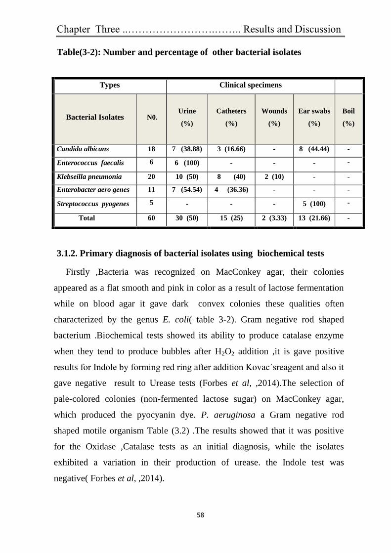
Chapter Three ..…………………….…….. Results and Discussion
58
Table(3-2): Number and percentage of other bacterial isolates
3.1.2. Primary diagnosis of bacterial isolates using biochemical tests
Firstly ,Bacteria was recognized on MacConkey agar, their colonies
appeared as a flat smooth and pink in color as a result of lactose fermentation
while on blood agar it gave dark convex colonies these qualities often
characterized by the genus E. coli( table 3-2). Gram negative rod shaped
bacterium .Biochemical tests showed its ability to produce catalase enzyme
when they tend to produce bubbles after H2O2 addition ,it is gave positive
results for Indole by forming red ring after addition Kovac΄sreagent and also it
gave negative result to Urease tests (Forbes et al, ,2014).The selection of
pale-colored colonies (non-fermented lactose sugar) on MacConkey agar,
which produced the pyocyanin dye. P. aeruginosa a Gram negative rod
shaped motile organism Table (3.2) .The results showed that it was positive
for the Oxidase ,Catalase tests as an initial diagnosis, while the isolates
exhibited a variation in their production of urease. the Indole test was
negative( Forbes et al, ,2014).
Types Clinical specimens
Bacterial Isolates N0. Urine
(%)
Catheters
(%)
Wounds
(%)
Ear swabs
(%)
Boil
(%)
Candida albicans 18 7 (38.88) 3 (16.66) - 8 (44.44) -
Enterococcus faecalis 6 6 (100) - - - -
Klebseilla pneumonia 20 10 (50) 8 (40) 2 (10) - -
Enterobacter aero genes 11 7 (54.54) 4 (36.36) - - -
Streptococcus pyogenes 5 - - - 5 (100) -
Total 60 30 (50) 15 (25) 2 (3.33) 13 (21.66) -

Chapter Three ..…………………….…….. Results and Discussion
59
Colonies of P. mirabilis bacteria were seemed to be small and colorless on
MacConkey because its non-lactose fermenter. On blood agar its ability
appeared to swarm which is the most distinguishable phenomenon for this
kind ,the bacterium can distinguish form a rod shaped vegetative cell into an
elongated (Mobley and Belase,1995).Biochemical tests for P.mirabilis
bacteria revealed that it was able to produce Catalase ,Urease enzymes, but it
was negative results for Oxidase( table 3.2).The remaining isolates grown on
the blood agar with Beta hemolysis can be identified as S.aureus they
developed yellow colonies on the mannitol salt as a result of fermentation of
mannitol.the general morphological characteristics of the colonies appeared
circular cream color to yellow. These isolates were initially identified as
Staphylococcus species Table (3.3) .All isolates were able to coagulate a
plasma and give positive result to Coagulase test (slide and tube).This gives
the impression that it may be a S. aureus (Benson, 2001).
Table (3-3) The biochemical tests of bacterial isolates
Test Bacterial species
E. coli
P. aeruginosa P. mirabilis
S.aureus
Gram stain - - - +
Catalase + + + + Oxidase - + - - Indole + - - \ Urease - V + +
Coagulase \ \ \ + Lactose fermentation on
MacConkey + - - \
(+) positive test , (-) negative test , V (variable ),\ (not tested)

Chapter Three ..…………………….…….. Results and Discussion
60
3.1.3. The identification of bacterial isolates was confirmed using API
system strips :
API 20 E for E. coli , P. mirabilis , API 20N E system for diagnosis of
Ps. aeruginosa and API Staph system for diagnosis S. aureus.
3.2. Antibiotic susceptibly testing of bacterial isolates
A total of 150 bacterial isolates of E. coli ( 65), Ps. aeruginosa ( 35) , P.
mirabilis (30) and S. aureus (20) were tested for their antibiotic resistance
against 21 antibiotics using disk diffusion method. In this study, all tested
isolates were resistant to minimum of 3 classes of antibiotics which they
were tested, so the isolates were considered multidrug resistant. MDR has
been identified as a major threat to the public health of human being by the
World Health Organization(Sun et al.,2014). These bacteria drastically
reduced the efficacy of antibiotic , consequently, increasing the frequency of
therapeutic failure and mortality. This rising antimicrobial resistance has
been accompanied by a decline in new antibiotic discovery over the last few
decades, which now poses a serious threat to public health (Kovač et
al.,2015)
The results revealed that showed different degrees of resistance to
antibiotics that used in the study. Table 3-4 showed that the percentage of
resistant isolates of E. coli and P. mirabilis to antibiotics were as to
Ampicillin, Amoxillin- clavulanic acid ,Azethronam, Cefotaxime , Cefepime
, Trimethoprim, Nitrofurantoin , Chloramphenicol Nalidaxic acid Piperacillin
and Gentamycin. while E.coli did not show any resistance to Imipenem and
Doxcycline , but it gave less resistance for both Amikacin and Ciprofloxacin
The isolates of P. aeruginosa also resistant to the same groups of
antibiotics resisted by E. coli and P. mirabilis have different degrees of
resistance to Azethronam , Cefepime, Piperacillin, Gentamycin ,
Imipenem , Ciprofloxacin and Amikacin.

Chapter Three ..…………………….…….. Results and Discussion
61
Table(3.4): Antibiotic resistant patterns of bacterial isolates against different antibio
Antibiotic
No. (%) of resistant of bacterial isolates
E. coli (n=65) P. aeruginosa
(n=35)
P. mirabilis
(n=30)
Ampicillin 50 (76.9%) / 26(86.6)
Amoxicillin-clv 50 (67.9%) / 20(66.6)
Cefoxitin / /
Cefipime 30 (46.1) 25(71.4) 15(50)
Cefotaxime 32 (49.2) - 20(66.6)
Amikacin 10 (15.3) 15(42.8) 2(6.6)
Gentamycin 40 (61.5) 15(42.8) 15(50)
Imipenem (0) 1(2.8%) 0
Azethronam 50 (76.9) 35(100) 30(100)
Nalidixic acid 36 (55.3) / 15(50)
Ciprofloxacin 5(7.9) 10(28.5) 8(26.6)
Doxcycline (0) / 10(33.3)
Chloramphenicol 26 (40 %) / 22(73.3)
Trimethoprim 35(53.8) / 20(66.6)
Nitrofurantoin 35(53.8) / 20(66.6)
Piperacillin 45(69.2) 30(85.7) 28(93.3)

Chapter Three ..…………………….…….. Results and Discussion
62
Cefoxitin were used in this test according to CLSI( 2016)
recommendations for detection of MRSA isolates. Results showed that out of
20 S. aureus isolates, 5 (25%) isolates were resistant to both of these
antibiotics Table (3-5) .The isolates of S. aureus were tested against
antibiotics under study
In this study all MRSA isolates exhibited resistance rate to antibiotics as
Cefotaxime , Cefipime,Rifampicin ,Trimethoprim , Nitrofurantoin
Chloramphenicol, Gentamycin ,Doxycycline , Ciprofloxacin , Penicillin G
and Cefoxitin .
Table(3.5): Antibiotic resistant patterns of bacterial isolates against different
antibiotic
Antibiotic
No. (%) of resistant of bacterial
isolates
S. aureus (n=20)
Penicillin G 20(100)
Cefoxitin 7(35)
Cefipime 15(75)
Cefotaxime 18(90)
Amikacin 12(60)
Gentamycin 10(50)
Rifampicin 8(40)
Ciprofloxacin 12(60)
Doxcycline 3(15)
Chloramphenicol 12(60)
Trimethoprim 13 (59.1)
Nitrofurantoin 8(40)

Chapter Three ..…………………….…….. Results and Discussion
63
In this study 30 isolates of E. coli , P. aeruginosa and P.mirabilis are
considered MDR are resistant to representative of three or more classes of
antimicrobial agents (Table 3.6).
Table(3.6): MDR and non MDR Bacterial isolates
Isolates E. coli P. aeruginosa
P. mirabilis
MRSA
Total No.
MDR
isolates
MDR 10 10 10 5 35
Non
MDR
55 25 20 15 115
Total 65 35 30 20 150
3.3. Characterization of biosynthesized silver nanoparticles (Ag-NPs)
3.3.1. Visual detection of Ag-NPs
Ag-NPs were visually detected by changing color from yellow to dark brown
of suspension. Figure3-1 containing cell free filtrate and silver nitrate .The
reduction of silver ions to Ag-NPs (Ag+ to Ag0) lead to changing color from
transparent or light yellow to brown(Sudhakar et al.,2014), which indicated
the formation of Ag-NPs. This brown color was due to the excitation of the
surface Plasmon vibrations in the metal nanoparticles, The control (soil)did
not show any change in its initial color when incubated under the same
conditions (Karwa et al.,2011) . Al- Ziadi et al ,(2015) from Iraq revealed
that the approach of dark brown synthesis seems to be cost effective, eco-
friendly and easy alternative to conventional methods of silver nanoparticles
synthesis by Trichoderma harizanum .Ag-NPs release silver ions, generating
an amplified biocidal effect, which is size- and dose-dependent (Marambio-
Jones and Hoek 2010).
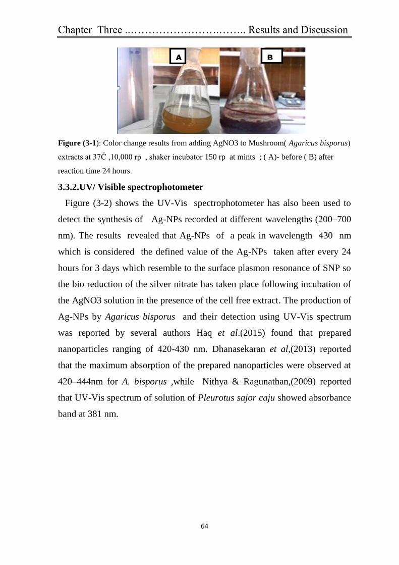
Chapter Three ..…………………….…….. Results and Discussion
64
Figure (3-1): Color change results from adding AgNO3 to Mushroom( Agaricus bisporus)
extracts at 37C̊ ,10,000 rp , shaker incubator 150 rp at mints ; ( A)- before ( B) after
reaction time 24 hours.
3.3.2.UV/ Visible spectrophotometer
Figure (3-2) shows the UV-Vis spectrophotometer has also been used to
detect the synthesis of Ag-NPs recorded at different wavelengths (200–700
nm). The results revealed that Ag-NPs of a peak in wavelength 430 nm
which is considered the defined value of the Ag-NPs taken after every 24
hours for 3 days which resemble to the surface plasmon resonance of SNP so
the bio reduction of the silver nitrate has taken place following incubation of
the AgNO3 solution in the presence of the cell free extract. The production of
Ag-NPs by Agaricus bisporus and their detection using UV-Vis spectrum
was reported by several authors Haq et al.(2015) found that prepared
nanoparticles ranging of 420-430 nm. Dhanasekaran et al,(2013) reported
that the maximum absorption of the prepared nanoparticles were observed at
420–444nm for A. bisporus ,while Nithya & Ragunathan,(2009) reported
that UV-Vis spectrum of solution of Pleurotus sajor caju showed absorbance
band at 381 nm.
A B

Chapter Three ..…………………….…….. Results and Discussion
65
Figure(3.2): A Peak(430) of silver nanoparticles synthesized by Agaricus bisporus
after (24, 48 and72 hours)by UV-visible spectroscopy .
3.3.3. Scanning and transmission electron microscopy analysis
(SEM,TEM) .
Characterization of Ag-NPs were observed under SEM revealed that
nanoparticles had varied shaped (spherical , irregular ) with size in the range
of 5-50 nm Figure(3-3A).Spherical silver nanoparticles aggregates were
reported by Nithya and Ragunathan (2009) that synthesized silver
nanoparticles by Pleurotus sajorcaju of size range 5-50nm and Narasimha et
al,(2011) who reported that their size range 5-50nm Ag-NPs by Agaricus
bisporus, while Karwa et al,(2011) who obtained Ag-NPs prepared by
Ganoderma lucidium showed the polydisperse nature of their nanoparticles
with size range (10 to 70 )nm. On the other hand, the morphology of Ag-NPs
is apparently spherical particles, sized range (4.5-35)nm-poly-dispersed by
TEM Figure( 3-3B). The that a peak of 430 nm indicates that the NPSs have
spherical form (Sudhakar et al ., 2014)
1: extract mushroom
2

Chapter Three ..…………………….…….. Results and Discussion
66
A
B
Figure (3.3 A,B): Image of TEM and SEM of Ag-NPs synthesized by Agaricus
bisporus showing shape and size at: (Isfahan university-engineering college /Iran
and Al kuffa university- college of science research center/ Iraq) respectively
3.3.4.Fourier transforms infrared spectroscopy (FTIR)
These results confirm the presence of primary and secondary amines
bonds; C=O, N=O, C=N and COOH bonds of proteins (Ramezani et al.,
2012).The amide linkages between amino acids residues in polypeptides and
proteins give rise to well-known signatures in the infrared region ,the
representative spectra in the region of 3500 to 450cm -1
revealed the presence
of different functional groups like 3290 secondary amide (N-H stretch ,H-
bonded ), 2928 (CH-stretching), 2161 (C=C stretching),1771 an hydride
(C=O stretching),1613 (C=C stretching), 1538 aromatic (C-C stretching),
1386 ,1313 and 1080 primary alcohol (C-O stretching), and 1333cm (CH3 )

Chapter Three ..…………………….…….. Results and Discussion
67
correspond to carbon hydrogen bending vibration respectively Figure (3-4).
Proteins present in the extract can bind to Ag-NPs through either free amino
or carboxyl groups in the proteins. The potential bimolecular in cell free
extract acts as stabilizing and capping agent . Singh et al (2014) reported
different functional groups absorb. Characteristic frequencies of FTIR
radiation . similar results were also represented by Haq et al (2015). the
carbonyl groups from the amino acid residues and peptides of proteins have a
stronger ability to bind metal, so that the proteins could form a coat over the
metal nanoparticle to prevent gathering of the particles(Ramezani et al.,
2012).It can be concluded that proteins or peptides have formed a coating on
silver nanoparticles, which in turn support their stability)Dhanasekaran et
al.,2013)
Figure (3-4):Image of spectrum of biosynthesized silver nanoparticles by Agaricus
bisporus .
3.4. Effect of optimum volumes and concentrations of Ag-NPs
It has been used several volumes and concentration prepared Ag-NPs to
get the optimal results for synthesis silver nanoparticles at 20, 30,40 and 50
µl was evaluated against strains bacteria by testing 17mg/ml Ag-NPs , 10
mg/ml Ag-NPs and 85 mg/ml Ag-NPs Table (3-7). Antibacterial activity of

Chapter Three ..…………………….…….. Results and Discussion
68
Ag-NPs was measured as the diameter of inhibition zone in (mm) by agar-
well diffusion methods against standard bacteria and MDR isolates were
examined according to Sudhakar et al(2014).
The results found that the Ag-NPs of volume of 50 ml of mushroom
extract with 50ml AgNo3 of 17 mg/ml was the most effective concentration
against the growth of standard strains Table(3-7) and MDR isolates Figure
(3-5). On the other hand the results concluded that the inhibition zone in
diameters were increased by using 50 µl Ag-NPs by edible mushroom
Agaricus bisporus. The exact mechanism behind the conversion of AgNo3 to
Ag-NPs by mushroom extract was not known (Raja and Singh,2012). The
results showed inhibition zone obtained in bacteria strains; E. coli
ATCC28739 (≥20) mm Ps. aeruginosa ATCC27853 (≥18) mm P. mirabilis
ATCC 16404 (≥15) mm and less inhibition zone it was MRSA ATCC 43300
(≥14 )mm. respectively Table (3-7).
Table ( 3-7) Zone of growth inhibition (mm) of standard strains tested with Ag-NPs
.
*= Significant association (p <0.05), SD = Standard Deviation
Table 3-7 showed that 17mg/ml concentration of Ag-NPs more effected
on bacterial isolates in compared with other concentration of Ag-NPs,
standard strains
Nanoparticles
Zone of growth inhibition (mm)
MRSA
ATCC 43300
E. coli
ATCC 28739
P. mirabilis
ATCC 16404
P. aeruginosa
ATCC 27853
Mean ± SD Mean ± SD Mean ± SD Mean ± SD
AgNO3 ( control) 10 ± 1 16 ± 4.36 12 ± 3.46 12 ± 3
Nanoparticles (Ag-NPs )
17.0 mg/ml 14*± 4 20*± 2.65 15 ± 4.58 18*± 6
Nanoparticles (Ag-NPs )
10mg/ml 12 ± 2 15 ± 2.01 10 ± 2.1 14 ± 4.16
Nanoparticles (Ag-NPs )
85mg /ml 12 ± 1.73 13 ± 3.3 10 ± 1.1 15 ± 3.11

Chapter Three ..…………………….…….. Results and Discussion
69
especially on E. coli (Mean of Zone of inhibition is 20mm and SD±2.65),
P. aeruginosa (Mean of Zone of inhibition is 18mm and SD±6), P. mirabilis
(Mean of Zone of inhibition is 15mm and SD±4.58). and MRSA (Mean of
Zone of inhibition is 14mm and SD±4).
The results revealed that the Ag-NPs were quite effective to inhibiting the
growth of all tested organisms and it found that by giving inhibition zone
against MDR isolates, the inhibition zone obtained in E.coli (≥16mm) , P.
aeruginosa (≥15mm) , while the lowest inhibition zone obtained with P.
mirabilis( ≥13mm ) and MRSA of (≥13 mm) respectively as in Table(3-8) ,
Figures (3-6 ). The effect of silver nanoparticles on gram negative bacteria
was more as compared to gram positive bacteria. The reason behind this
might be the cell wall composition of gram positive bacteria as described by
Birla et al. (2009). The current study disagree with Sudhakar et al.,(2014 )
who showed that inhibition zone was 19 mm by using Ag-NPs by Agaricus
bisporus mushroom. The present study agreed with Haq et al.,(2015) who
synthesized Ag-NPs using Pleurotus sajor caju (Mushroom) was tested
against Ps. aeruginosa and P.mirabilis produced zone of inhibition 12mm.
The present study approaching to Nithya et al.( 2009) who synthesized
Ag-NPs using pleurotus (mushroom) was tested against E. coli, P.
aeruginosa S. aureus produced zone of inhibition of (12mm), (14mm) and
(11mm) respectively. Whereas Rahi & Barwal (2015) revealed that Ag-NPs
synthesized by the Ganoberma applanatum (mushroom) measured as the
inhibition zone of MRSA 30mm,S. aureus 20mm , P. aeruginosa and E. coli
24mm. Devika et al ( 2012) found that the inhibition zone of in E. coli (13
mm) and for each Ps. aeruginosa and S. aureus (8 mm) antibacterial Ag-
NPs by using Pleurotus ostreatus .Haq et a l. (2015) suggested that the Ag-
NPs from Agaricus bisporus explored medicinally and nutritionally important
species of dried mushroom. pH has play an important role in NPs formation
and protein denaturized so that protein will lose its activity in low pH

Chapter Three ..…………………….…….. Results and Discussion
70
whereas at high pH, SNPs become smallest and more regular in compared of
that of low pH (Banu et al.,2011).The bactericidal effect of metal
nanoparticles has been attributed to their small size and high surface to
volume ratio, which allows them to interact closely with microbial
membranes and is not merely due to the release of metal ions in solution
(Morones et at.,2005).
There is a fact that fungi produce many proteins and enzymes involved in
synthesis of Ag-NPs and are simpler to grow both in laboratory and industrial
level and also the yield is high(Janardhanan and Nayana,2000).Although
beneficial as antimicrobial agents, silver nanoparticles have adverse effects
on cells such as the production of reactive oxygen species which are toxic to
both bacteria and eukaryotic cells (Park et al. 2009; Carlson et al. 2008).
Table(3-8): Zone of growth inhibition (mm) of different concentration(Ag-NPs )
against MDR bacterial isolates
NS= NonSignificant (p > 0.05), SD= Standard Deviation
MDR
Bacterial
isolates
Ag-NPs zone of inhibition(mm)
X2
P –
value
µl 50
No. of isolate 17 mg/ml 10 mg /ml 85 mg /ml
Mean ± SD Mean ± SD Mean ± SD
MRSA 2,3,7,10,12 13±3.3 11±1.22 10±0.90
0.478 >0.05
[ NS ]
E. coli 4,8,6,12,14,18,22,23,25 16±5.1 14±2.79 11±1.23
1,4,6 15±4.4 13±2.03 11±1.99
P. mirabilis 1,2,5,6,7,13,14,15,17,19 13±2.7 11±0.9 10±0.87
P .aeruginosa 6,9,10,11,16,18,20, 22,23 15±3.9 13±2 14±3.011
19,20,10,23 14±3.03 12±1.09 12±1.88

Chapter Three ..…………………….…….. Results and Discussion
71
Fig
ure(3-5):Zone of growth inhibition of different concentration (Ag-NPs ) against
isolated MDR , A= 17mg/ml , B= 10mg/ml and C= 85 mg/ml
Figure(3-6):Zone of growth inhibition (mm) bacterial isolates MRSA, E. coli , P
.aeruginosa and P. mirabilis with 17mg /ml Ag-NPs with 20,30,40,50µl.
B:10mg/ml
B:10mg/ml
B:10mg/ml
A:17mg/ml
C:85 mg/ml
B:10mg/ml
C:85 mg/ml
C:85 mg/ml
C:85 mg/ml
A:17mg/ml
A:17mg/ml
A:17mg/ml
MRSA

Chapter Three ..…………………….…….. Results and Discussion
72
3.5. Comparison between AgNO3 solution and (Ag-NPs) nanoparticles:
The viability of AgNo3 solution and Ag- nanoparticles using Agaricus
bisporus extract were examined in the inhibition growth of bacterial isolates
by well diffusion in agar method. Inhibition growth of bacterial isolates by
Ag-NPs was compared with AgNO3 solution and found that Ag-NPs was the
most efficient of AgNO3 solution in the inhibition of bacterial isolates (Table
3-9 and Figure 3-7) showed the inhibition zones for bacterial isolates treated
with AgNO3 solution and Ag-NPS, Sudhakar et al.( 2014) found that
mushroom extract plays role in the reduction of silver nitrate to silver
nanoparticles their by producing bactericidal properties. Rasheed (2014) from
Iraq revealed that increases the inhibition zone around the implanted
bacteria reached to 15 mm for Proteus, Remarkable increment of antibiotics
when silver nanoparticles with laser ablation added to improve the
antimicrobial effect against gram-negative and gram-positive bacteria
The most popular of Agaricus bisporus is considered safe for most
people to eat because it is grown in controlled, sterilized environments (Singh
et al., 2011), so the use of fungi especially mushrooms in synthesis of
nanoparticles is a less time and in fact, fungi produce many proteins and
enzymes involved in synthesis of Ag-NPs and are simpler to grow both in
laboratory and industrial level and also the yield is high (Janardhanan and
Nayana 2000). Rasheed (2014) found that the most sensitive bacteria for
silver nanoparticles was Proteus and the minimum inhibition concentration of
l5 mg/ml was obtained for S. aureu.

Chapter Three ..…………………….…….. Results and Discussion
73
Figure (3.7) Zone of growth inhibition (mm) of E. coli with AgNO3 1.0 mg/ml and
Ag-NPs (A:. AgNO3 alone, B: 85mg/ml Ag-NPs, C: 10 mg/ml Ag-NPs, D: 17mg/ml
Ag-NPs.
Table (3-9) Demonstrated that bacterial isolates more sensitive to Ag-NPs 17mg/ml
compared with AgNO3 (X2= 7.603, p<0.05).
SD = Standard Deviation
MDR
Bacterial isolates
Zone of growth inhibition(mm) X2
test P – value
AgNO3
50μl
Ag-NPs 17mg/ml
50μl
Mean ± SD Mean ± SD
1 MRSA 0.0± 0 13±4
7.603 <0.05
2 E. coli 10±2 16±4.12
3 P.mirabilis 9±1.7 13±3.61
4 Ps .aeruginosa 9±2.65 15±4.04
: gNO3 1.0 mg/ml
C: 10mg/ml
:85 mg/ml
:17mg/ml

Chapter Three ..…………………….…….. Results and Discussion
74
3.6. Estimation the combination effect of antibiotic with Ag-NPs
Thirty five MDR isolates out of 150 isolates were subjected for antibiotic
susceptibility test using disc diffusion methods . The combination effect
between antibiotic and silver nanoparticles by disc diffusion method gave
an increasing fold diameters of inhibition zone of bacterial isolates in
comparison with antibiotic alone. The Combinations of antibiotics and Ag-
NPs resulted in average fold-area increases in antibacterial activity. The zone
of inhibition in E.coli to antibiotic amoxicillin clavulanic acid ,ampicillin
,piperacillin and Azethronam were(11-18 mm),(11-15 mm), (12-18 mm) and
(14-18 mm) respectively with deferent average fold-area(0.3-3.5) ,(0.2-5.2)
,(0.4-5.2)and (0.2-5.2) respectively [ Table 3-10and Figure 3-8] .
This result is in agreement with Kaviya et al., (2011) who reported that
silver nanoparticles exhibited good antibacterial activity against both gram
negative and gram positive bacteria, they showed higher antibacterial activity
against E.coli and P.aeroginosa (Gram negative) than S.aureus (Gram
positive) .
The study concluded that Ag-NPs showed variable effectiveness against
bacterial isolates, the inhibition zones observed for P. aeruginosa with
antibiotics piperacillin and Azethronam were (14-18mm) and (15-19mm)
respectively and fold-area (0.5-4.4)and (0.5-5.2) respectively Table(3-11)
Figure (3-9). Most Pseudomonas spp. are naturally resistant to Penicillin and
the majority of related beta-lactam antibiotics (Cobos-Trigueros et al .,2015).
This result do not agree with Rahi and Barwal (2015) found that inhibition
zone to amoxicillin and ampicillin increase of activity against bacterial
pathogens of E. coli and Ps. aeruginosa isolates when using Ag-NPs by
mushroom Ganoderma , which was 17.7 for E. coli and 10.11 Ps. aeruginosa
against amoxicillin and 6.11 for E. coli toward Ampicillin either Ps.
aeruginosa was 12.4 mm.

Chapter Three ..…………………….…….. Results and Discussion
75
The high bactericidal activity is certainly due to the silver cations released
from Ag nanoparticles that act as reservoirs for the Ag+ bactericidal agent.
Silver nanoparticles have ability to anchor to bacterial cell wall and
subsequently penetrate it, thereby causing structural changes in cell
membrane like the permeability of the cell membrane and death of cell.
There is a formation of ‘pits’ on cell surface, and there is accumulation of
the nanoparticles on the cell surface.( Mahmood ,2012) .
In contrast, P.mirabilis isolates showed same inhibitory to Ag-NPs with
edible mushroom by inhibition zone average (12-18mm),(12-15mm),(14-
19mm) and (10-15mm) respectively ,for amoxicillin clavulanic acid ,
ampicillin, piperacillin and Azethronam. The average fold area were (0.4-
5.2),(0.4-3.5) (0.5-4.4) and (0.2-3.5) of each one respectively Table(3-12)
Figure (3-10). This result is high than Buszewski et al.,(2016) who found
that combination of Ag-NPs with ampicillin (9.3mm) zone inhibition
against P.mirabilis. Tawfeeq et al.2017 proved that results showed the
synergistic action of Ag-NPs and antibiotics against (MDR) bacteria were
also leading to enhance antibacterial activity.

76
Table (3-10)Zone of inhibition (mm) of different antibiotics against 10 isolates MDR , (in absence and in presence of Ag-NPs at
content of 50 µl per disc)A: antibiotic ,B: antibiotic with Ag-NPs, C: increase fold area=(B2-A2)/A2 .
Table (3-11)Zone of inhibition (mm) of different antibiotics against 10 isolates MDR, (in absence and in presence of Ag-NPs at
content of 50 µl per disc)A: antibiotic ,B: antibiotic with Ag-NPs, C: increase fold area=(B2-A2)/A2 .

Chapter Three ..………….………………….…………….. Results and Discussion
77
In contrast, Sondi and Salopek -Sondi (2004) reported that the antimicrobial
activity of silver nanoparticles on Gram-negative bacteria was dependent on the
concentration of silver nanoparticles, and was closely associated with the
formation of pits in the cell wall of bacteria. Ag-Nps accumulated in the bacterial
membrane caused the permeability, resulting in cell death. Singh et al .,(2012 )
suggested that electron spin resonance spectroscopy studies that silver
nanoparticles when contact with bacteria, and these free radicals have an ability
to damage cell membrane and make it porous which can ultimately lead to cell
death.
A high occurrence of resistance of S .aureus against penicillin G, were
(100%).The results showed that bacteria have become sensitive and there is an
increase in the inhibition of antibiotic tablets regions with Ag-NPs as by edible
mushroom , average inhibition zone (10-18mm) while average fold-area ( 0.5-2.2)
presented in Table (3-11).Figure (3-113). Rahi and Barwal (2015) noted that
inhibition zone of penicillin G ,amoxicillin and ampicillin increase inhibition zone
against the isolates of MRSA were (11,7,7 )with fold area(2.3,6 and13)
respectively Birla et al.,(2009) were indicated the synergistic activity observed was
better in E. coli and Ps. aeruginosa than S. aureus. Fayaz et al. (2010) showed that
Ag-NPs can act synergistically with several antibiotics, preferentially against
Gram-negative bacteria .Mahmood (2012) revealed that a synergistic effect was
observed between Ag-NPs and both amoxicillin and penicillin for Gram-positive
isolates
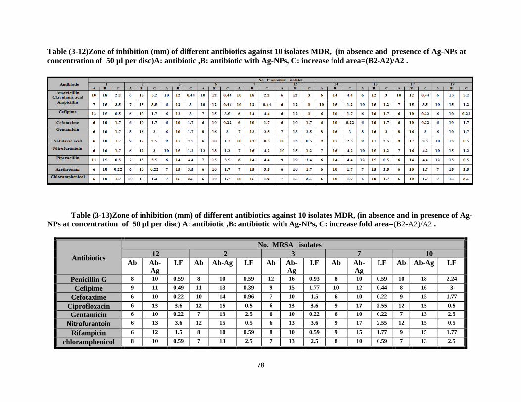
78
Table (3-12)Zone of inhibition (mm) of different antibiotics against 10 isolates MDR, (in absence and presence of Ag-NPs at
concentration of 50 µl per disc)A: antibiotic ,B: antibiotic with Ag-NPs, C: increase fold area=(B2-A2)/A2 .
Table (3-13)Zone of inhibition (mm) of different antibiotics against 10 isolates MDR, (in absence and in presence of Ag-
NPs at concentration of 50 µl per disc) A: antibiotic ,B: antibiotic with Ag-NPs, C: increase fold area=(B2-A2)/A2 .
Antibiotics
No. MRSA isolates
12 2 3 7 10
Ab Ab-
Ag
I.F Ab Ab-Ag I.F Ab Ab-
Ag
I.F Ab Ab-
Ag
I.F Ab Ab-Ag I.F
Penicillin G 8 10 0.59 8 10 0.59 12 16 0.93 8 10 0.59 10 18 2.24
Cefipime 9 11 0.49 11 13 0.39 9 15 1.77 10 12 0.44 8 16 3
Cefotaxime 6 10 0.22 10 14 0.96 7 10 1.5 6 10 0.22 9 15 1.77
Ciprofloxacin 6 13 3.6 12 15 0.5 6 13 3.6 9 17 2.55 12 15 0.5
Gentamicin 6 10 0.22 7 13 2.5 6 10 0.22 6 10 0.22 7 13 2.5
Nitrofurantoin 6 13 3.6 12 15 0.5 6 13 3.6 9 17 2.55 12 15 0.5
Rifampicin 6 12 1.5 8 10 0.59 8 10 0.59 9 15 1.77 9 15 1.77
chloramphenicol 8 10 0.59 7 13 2.5 7 13 2.5 8 10 0.59 7 13 2.5
Chapter Four ……………………………………... Results and Discussion
79
The bacterial cells which in contact with silver ions, inhibit several
functions in the cell and damage the cells, there is a generation of reactive
oxygen species, which are produced possibly through the inhibition of a
respiratory enzyme by silver ions and attack the cell itself. Silver is a soft acid,
and there is a natural tendency of acid to react with a base in this case, a soft
acid to react with a soft base (Yuan et al .,2007) . Hwang et al. (2012) proposed
that silver nanoparticles modulate the phosphor tyrosine profile of putative
bacterial peptides that can affect cellular signaling, which leads to growth
inhibition in bacteria. Samantha (2015) showed that silver nanoparticles have
reactive antibacterial activity against test pathogens including S. aureus, P.
mirabilis and E. coli .
According to Yuan et al . (2007) silver nanoparticles can be used as
effective broad spectrum antibacterial agents to Gram-negative and Gram-
positive bacteria, including antibiotic-resistant bacteria, for P. aeruginosa and
S.aureus , Ag-NPs act as efficient barriers against antimicrobial agents and the
host immune system to protect the bacterial colony. The present study clearly
indicates that Ag-NPs showed a good antibacterial action against MDR isolates
,all isolates gave a clear inhibition zone so the average of zone inhibition
obtained in E.coli (12-16mm) and (12-19mm) to cefotaxime and Cefipime, the
fold area were (0.2-3),(0.4-6.2), while P. aeruginosa zone inhibition against
Cefipime were (12-16mm) and the fold area were (0.3-3). P.mirabilis zone
inhibition (10mm),(10-15mm) with fold area (0.2-1.7) ,(0.2-4.4) cefotaxime
and Cefipime .while average inhibition zone to cefotaxime and
Cefipime against MRSA were(10-15mm), (11-16mm) with fold area (0.2-1.7),
(0.3-3) respectively. Haq et al.,(2015) reported the fold area were 8.6% by Ag-
NPs using Agaricus bisporus with Ceftriaxone, the combination of antibiotics
with Ag-NPs have better antimicrobial activities, the antibacterial activity of

Chapter Four ……………………………………... Results and Discussion
80
Cephalexin was increased by AgNPs with Cephalexin antibiotic against E.coli
and S. aureus. Singh and Raja.( 2012). Mahmood (2012) proposed that a
synergistic effect was observed between Ag-NPs and ceftriaxone for Gram-
negative isolates .
The combination between Gentamycin and Ag-NPs showed a good effect
on both E. coli, Ps. aeruginosa and P.mirabilis and the average inhibition zone
was observed when Ag-NPs were combined with antibiotic Gentamycin against
E.coli the effectiveness increased with inhibition zone (10-18mm) with fold area
(0.5-4). P. aeruginosa were found to response to combination with inhibition
zone(10-16mm) increased fold area (0.2-3). This result is less than of Birla et al
(2009) found that (0.5, 0.1) increased fold area against Gentamycin of E. coli
and Ps. aeruginosa isolates. P.mirabilis represented show combination between
Gentamycin and Ag-NPs, isolates showed diameter average (10- 16 mm) and
fold area (1.7-3).the results of Buszewski et al .,(2016) noted that inhibition
zone of Ag-NPs and Gentamycin (8.3) against P.mirabilis . The results of
combination assay for MRSA were 10-13mm and rate fold area ( 0.2- 2.5).
The result was approaching of results reported by Birla et al (2009) were
indicated the fold area (0.36) against Gentamycin synthesized Ag-NPs using
Phoma glomerata and Haq et al.,(2015) reported that fold area were 23.8% by
Ag-NPs using Agaricus bisporus. aeruginosa is a nosocomial pathogen of
particular clinical concern not only because of its extraordinary resistance
mechanisms armamentarium but also for its formidable ability to 44 Infection
control adapt very well to the hospital environment (Moradali et al.,2017).
The main reason why nanoparticles is considered an alternative to
antibiotics is that nanoparticles can effectively prevent microbial drug
resistance in some cases because the bacteria have powerful ability of
developing resistance against antibiotics,. The widespread use of antibiotics has

Chapter Four ……………………………………... Results and Discussion
81
led to emergence of numerous hazards to public health, such as superbugs that
do not respond to any existing drug and non-medicated epidemics. ( Khameneh
et al ., 2016)
Gurunathan et al. (2015) proposed that silver nanoparticles inhibit bacterial
growth by inactivating the proteins. Silver atoms bind to thiol groups (SH) in
enzymes which deactivates the enzymes. Silver alters the function of
compounds in cell membrane which is important in transmembrane energy
generation and ion transport, by forming a stable S-Ag bond with the thiol
group of the compounds. Silver acts as a catalyst in the formation of
disulfide bonds in the reaction of oxygen molecules in cell and hydrogen atoms
of thiol groups (R-S-S-R). including P.aeruginosa, Shigella flexneri, S. aureus,
Streptococcus pneumoniae, Helicobacter pylori, Helicobacter felis, E. coli, K.
pneumoniae, and Bacillus subtilis .Quinolone group include
Ciprofloxacin and Nalidixic acid .
The results indicated that the percentage of inhibition zone diameters were
increased when used Ag-NPs by mushroom with Nalidixic acid against each of
E. coli and P.mirabilis , the average inhibition zone were (15-18mm) and
(10-17mm) respectively .
On the other hand increasing the antibacterial activities in fold area were
(0.3-3.5 ) and (0.5-2.5). while inhibition zone diameters with Ciprofloxacin
when used Ag-NPs by mushroom against Ps. aeruginosa and MRSA the
average of inhibition zone were(13-19mm ) and (13-17mm) , increasing the
antibacterial activities in fold area were ( 2.1-3.5) and (0.5-3.6) respectively .
Sotiriou and Pratsinis ,(2010).hypothesized that bactericidal activity of silver
nanoparticles is attributable to Ag+ ions which enter the cell and intercalate
between the purine and pyrimidine base of DNA. It is also possible for Ag-NPs
not only interact with the surface of the membrane, but also penetrate inside the

Chapter Four ……………………………………... Results and Discussion
82
bacteria and inactivate DNA replicating ability causing the devastation of the
cell.
The results of isolates showed that Nitrofuration gave the highest average
inhibitory zone of E.coli which was (12-18 mm) with fold area (0.5-3) by
using AgNps, compared with using disc diffusion alone. Silver nanoparticles
target protein synthesis, nucleic acid synthesis, and Gram positive cell wall
synthesis, which explains why these bacteria were more susceptible to silver
nanoparticles. Indeed, silver nanoparticles attach to the surface of the cell
membrane and disturb its function, penetrate bacteria and release silver ions
(Sondi et al., 2004).Combination effect of Ag-NPs with edible mushroom
were observed against P.mirabilis isolates by increasing the antibacterial
activities of Nitrofuration with inhibition zone (10-18mm) with increases fold
arae(1.2- 4.2) .
This study appeared that the high occurrence of resistance of S .aureus
against Nitrofuration so the results showed that the bacteria have become more
sensitive and there is an increase in the inhibition of the antibiotic tablets
regions with Ag-NPs as by edible mushroom, became (13-17mm), the results
of the combination assay were (0.5-3.6) .
The most important problem caused by the chemical antimicrobial agents is
multidrug resistance. Therefore, an alternative way to overcome the drug resis-
tance of various microorganisms is needed desperately, especially in medical
devices(Rahi et al.,2013)
The high resistance here with antibiotics Rifampicin were (100%), against
MRSA, on the other hand all isolates became susceptible to this antibiotic with
rate (10-15mm) by using Ag-Nps with folded area(0.5-1.7). This resistance was
presumed to be due to mutation(s) in the gene encoding the beta-subunit of
DNA-dependent RNA polymerase, the target of the rifampin class of

Chapter Four ……………………………………... Results and Discussion
83
antibiotics. Ag-NPs can cause further damage to bacterial cells by permeating
the cell, where they interact with DNA, proteins and other phosphorus- and
sulfur-containing cell constituents (Marambio-Jones and Hoek 2010).Results
have demonstrated that MDR isolates E.coli and P.mirabilis have notably rates
of resistance to chloramphenicol .The results showed that chloramphenicol
gave inhibitory zone of E.coli which was (15-16mm) with folded area (1.7-3)
by using Ag-Nps, compared with using disc diffusion alone. However, the
results indicated that the percentage of inhibition zone (diameter) in P.
mirabilis were increased when used Ag-Nps to reach a rate of 15mm and the
folded area were (1.2-3.5) respectively.
Results approach to Mahmood (2012) revealed that a combination effect was
detected between Ag-NPs and chloramphenicol for Gram-negative isolates was
14mm. Proteus is widespread in the environment and makes up part of the
normal flora of the human gastrointestinal tract. Proteus species are the major
cause of diseases acquired outside the hospital, where many of these diseases
eventually require hospitalization are often the cause of urinary tract infections
(UTIs) (Schaffer and Pearson, 2015).
The isolates of MRSA showed resistant to chloramphenicol which was
determined (Table 3-10). all isolates were increased with AgNps in zone
inhibition (10-13mm ) so the folded area were (0.5-2.5). However, the
resistance rates in the present study were higher than those reported by others
(Hwang et al.,2012), who found that the synergistic interactions of Ag-Nps and
chloramphenicol were found only against Enterococc faecium and P.
aeruginosa while no Synergistic to E.coli and S.aureus. AgNPs are able to
physically interact with the cell surface of various bacteria. This is
particularly important in the case of Gram-negative bacteria where numerous
studies have observed the adhesion and accumulation of AgNPs to the
bacterial surface. Many studies have reported that AgNPs can damage cell

Chapter Four ……………………………………... Results and Discussion
84
membranes leading to structural changes, which render bacteria more
permeable. This effect is highly influenced by the nanoparticles’ size, shape
and concentration and a study using Escherichia coli (Periasamy et al, 2012).
they accumulation on the membrane cell creates gaps in the integrity of the
bilayer which predisposes it to a permeability increase and finally bacterial cell
death. (Rai et al.,2014)
Figure (3-8) Zone of growth inhibition (mm) of E.coli with antibiotic alone and in
combination with Ag-NPs .(Ab= Antibiotic)
Ampicillin =Amp
Amoxicillin-clv =Amc
Cefipime =Cpm
Cefotaxime =Ctx
Gentamycin =Gen
Azethronam =At
Nalidixic acid =Na ; Chloramphenicol = C ;Nitrofurantoin =Nit and Piperacillin =PI
Ab alone
Ab alone
Ab alone
Ab alone Ab + Ag-NPs Ab + Ag-NPs
Ab + Ag-NPs Ab + Ag-NPs

Chapter Four ……………………………………... Results and Discussion
85
Figure (3-9) Zone of growth inhibition (mm) of P. aeruginosa with antibiotic alone and
in combination with Ag-NPs .(Ab= Antibiotic)
Cefipime = CPM
Gentamycin = Gen
Azethronam = At
Ciprofloxacin= Cip
Piperacillin = Pi
Ab alone Ab alone Ab + Ag-NPs Ab + Ag-NPs
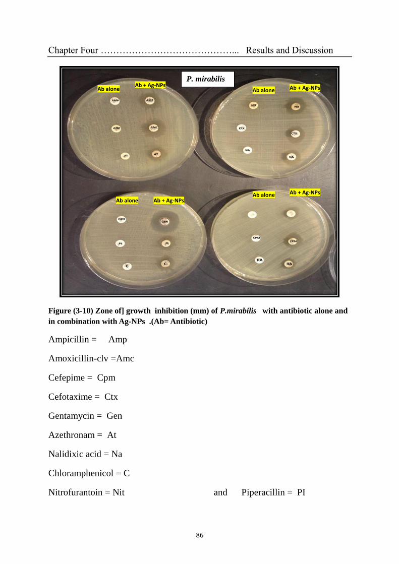
Chapter Four ……………………………………... Results and Discussion
86
Figure (3-10) Zone of] growth inhibition (mm) of P.mirabilis with antibiotic alone and
in combination with Ag-NPs .(Ab= Antibiotic)
Ampicillin = Amp
Amoxicillin-clv =Amc
Cefepime = Cpm
Cefotaxime = Ctx
Gentamycin = Gen
Azethronam = At
Nalidixic acid = Na
Chloramphenicol = C
Nitrofurantoin = Nit and Piperacillin = PI
P. mirabilis
Ab alone
Ab alone
Ab alone
Ab alone
Ab + Ag-NPs
Ab + Ag-NPs
Ab + Ag-NPs Ab + Ag-NPs

Chapter Four ……………………………………... Results and Discussion
87
Figure (3-11) Zone of growth inhibition (mm) of MRSA with antibiotic alone and in
combination with Ag-NPs . .(Ab= Antibiotic)
Cefipime =Cpm
Cefotaxime =Ctx
Gentamycin =Gen
Chloramphenicol = C
Nitrofurantoin =Nit
Piperacillin =PI
Ciprofloxacin= Cip
Rifampicin =RA
3.7. Detection of virulence factors
Investigation of some virulence factors that are important in the process of
settlement and the continuation of infection, biofilm formation and ATPase
important virulence factors in bacteria. Because there are no local studies on
how nanoparticles affect the inhibition of both properties, this study chose these
two factors and studied them for several reasons :
Ab alone Ab alone Ab + Ag-NPs Ab + Ag-NPs Ab + Ag-NPs
Ab alone

Chapter Four ……………………………………... Results and Discussion
88
Most chronic and persistent bacterial infections are associated with biofilm
growth, a strategy that has accelerated the emergence and rapid spread of MDR
bacteria. It has been known for decades that biofilm associated bacteria is much
more difficult to be eradicated by bactericidal antimicrobials than planktonic
cells (Lewis, 2001 ). Among the four general mechanisms that cause antibiotic
resistance including target alteration, drug inactivation, decreased permeability
and increased efflux, drug extrusion by the multidrug efflux pumps serves as an
important mechanism of MDR. Efflux pumps not only can expel a broad range
of antibiotics owing to their poly-substrate specificity, but also drive the
acquisition of additional resistance mechanisms by lowering intracellular
antibiotic concentration and promoting mutation accumulation (Sun et
al.,2014). Biofilm formation as a mechanism to remain in a favorable nook .
Humans have developed intricate immune systems for one critical reason:
microorganisms are continually trying to inhabit their bodies. The body, or at
least parts of it, is nutrient rich and relatively stable with respect to water
content, oxygen availability, and temperature. Consequently, there is a never-
ending race between the development of the host immune system and the
progression of bacterial strategies to evade it. In some cases, a compromise has
been made, and as such, the body is inhabited by a large number of
commensals, many of which exist as biofilms. As the body is obviously an
appealing place for bacteria to live, it may be that the primary motivation for
switching to the biofilm mode of growth is to remain fixed. (Lewis, 2001 ) . A
second possibility, which supports the biofilm as a mode of defense is that
bacteria may have evolved to interpret elevated glucose levels as a cue that it is
in the bloodstream, and that it needs to form a biofilm to remove itself from
circulation and protect itself from the immune system.

Chapter Four ……………………………………... Results and Discussion
89
Alternatively polysaccharide production may function as a mechanism of
glucose storage during times of plenty, and/or as a mechanism to augment the
accumulation phase so that when the organism finds itself in an environment
rich in nutrients it can occupy that niche. With all of the complex mechanisms
that pathogenic and commensal bacteria have evolved to survive in the human
body, it is clear that the benefits that we afford them outweigh the
hurdles synthesis (Sun et al.,2014) .
3.7.1. Detection of bacterial ability to produce biofilm by tissue culture
plate and tube methods .
Biofilm(s) are groups of microorganisms coated in an exopolymeric coat.
They have been associated with variety of persistent infections that respond
poorly to conventional antibiotics (Bose et al.,2009). This method was achieved
by Christensen et al. (1985) to detect the slime layer production by bacteria.
In the present study bacterial ability to produce biofilm were applied on 65
isolates shown in table (3-14) TM method and TCP, Optical density (OD) of
stained adherent biofilm was obtained using ELISA auto reader at wavelength
590 nm, and the interpretation of the results were conducted . A standard
method discovered 35(53.84%) as strong, 10(15.38%) as moderate and
20(30.67%) as weak/non biofilm producers by TCP table (3-15) while the
results detected By TM the number of strong biofilm producers were
30(46.15%) , moderate were 18(27.69%) and weak or non-biofilm producers
were 17(26.15%) table (3-16).
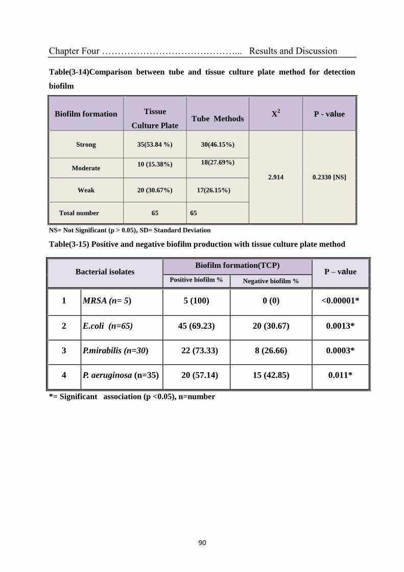
Chapter Four ……………………………………... Results and Discussion
90
Table(3-14)Comparison between tube and tissue culture plate method for detection
biofilm
Biofilm formation
Tissue
Culture Plate
Tube Methods X
2 P - value
Strong 35(53.84 %) 30(46.15%)
2.914 0.2330 [NS]
Moderate 10 (15.38%)
18(27.69%)
Weak 20 (30.67%) 17(26.15%)
Total number 65 65
NS= Not Significant (p > 0.05), SD= Standard Deviation
Table(3-15) Positive and negative biofilm production with tissue culture plate method
*= Significant association (p <0.05), n=number
Bacterial isolates Biofilm formation(TCP)
P – value Positive biofilm % Negative biofilm %
1 MRSA (n= 5) 5 (100) 0 (0) <0.00001*
2 E.coli (n=65) 45 (69.23) 20 (30.67) 0.0013*
3 P.mirabilis (n=30) 22 (73.33) 8 (26.66) 0.0003*
4 P. aeruginosa (n=35) 20 (57.14) 15 (42.85) 0.011*

Chapter Four ……………………………………... Results and Discussion
91
Table(3-16) Positive and negative biofilm production with Tube method
*= Significant association (p <0.05), NS= Non Significant (p > 0.05), n=number
The results approaching to Ahmed (2013) demonstrated that out of 118
Gram-positive bacteria the percentage of biofilm producers by TCP method was
95 (80.5%) higher than in TM were 78 (66.10%) producer as (moderate and
strong) , in another study Hassan et al., (2011) they described the incidence of
biofilm producers among 81 isolates of Gram-positive and Gram-negative
bacteria were 51 (63%) strong bacteria biofilm and 30 (37%) moderate bacteria
biofilm , in which Gram-positive bacteria generally higher than Gram-negative
bacteria in producing biofilm. Depending on comparison between the results of
biofilm production ,its appear that TCP is the more sensitive than TM methods
as in Figure 3.18 so it depends, the results showed strong and moderate biofilm
are 45 (69.23%) isolates E. coli, 20 (57.14%) P. aeruginosa ,P.mirabilis and
MRSA were 22 (73.33%) and 5 (100 %) respectively while by TM Figure
(3.19) the majority 9of the organisms associated with biofilm production were
E. coli 30 (46.15%) ; 17 (48, 53%) for P. aeruginosa and 16(53.33) P.mirabilis
and MRSA were 5(7.69%).
These isolates were tested on antibiotics and found to be MDR. Similar
results reported by Pramodhini et al., (2012) who showed that there was a
bacterial isolates Biofilm formation (TM)
P – value Positive biofilm (%) Negative biofilm (%)
1 MRSA (n=5) 5( 100 ) 0 (0) <0.00001*
2 E.coli (n=65) 35 (53.84) 30(46.15) >0.05 [NS]
3 P.mirabilis (n=30) 16(53.33) 14(46.66) >0.05[NS]
4 P. aeruginosa (n=35) 17(48.57) 18(51.42) >0.05[NS]

Chapter Four ……………………………………... Results and Discussion
92
relationship between biofilm production and MDR 80% of strains producing
biofilm were multi drug resistant phenotypic . Strong biofilm production was
caused by urinary catheter tips. Bakir & Ali (2016) in consistent finding that for
biofilm production by TCP method as (moderate and strong) by bacteria P.
aeruginosa were (81.8%) and TM (45.5%). There is an association between
biofilm production with persistent infection and antibiotic therapy failure(Bose
et al .,2009).They exhibit resistance to antibiotics by various methods like
restricted penetration of antibiotic in to biofilm, decreased growth rate and
expression of resistance genes (Sasirekha et al., 2012).
A suitable and reproducible method is necessary for screening of biofilm
producers in any healthcare setup and this TCP method can be recommended
(Bose et al .,2009).Tube method was good for strongly biofilm producing
isolates but it was difficult to discriminate between weak and biofilm negative
isolates due to the variability in observed results by different observers.
Consequently, high variability was observed and classification in biofilm
positive and negative was difficult by tube method (Mathur et al., 2006).
3.7.2. Evaluation of biofilm inhibition assay
This study examined the effects of Ag-NPs by Agaricus bisporus at 30µl in
combination with several antibiotic was evaluated against bacterial isolates
adopting biofilm inhibition spectrophotometric assay. The antibiotics used in
our study belong to several classes and have various cellular targets, modes of
action and bacterial resistance mechanisms penicillin G, Ampiciline , cefotaxim
, chloramphenicol and rifampicin.
These 35 isolates selected according to multi-drug resistance pattern TM and
TCP as the most isolates obtained the highest OD value, Combination of Ag-
NPs with antibiotics to inhibit the biofilm formation was performed according
to Hwang et al. (2012) with few modifications.

Chapter Four ……………………………………... Results and Discussion
93
The readings of OD have decreased within the range (0.120) ,whereas the
OD of strong biofilm were ≥ 0.240 and the non-biofilm were ≤ 0.120.Gram
positive cultures of bacterial isolated was S.aureus (MRSA) while of Gram
negative E.coli, Ps. aeruginosa and P.mirabilis .The isolates showed different
response to the antibiotics after they were treated, the highest response to
Cefipime and chloramphenicol were 70% by E. coli, while Ampicillin and
Gentamycin were 40%,50% respectively Figure3.12 and Table 3.17.This result
agreed with the results obtained by Hwang et al. (2012) they revealed that
combinations of Ag-NPs with conventional antibiotics (Ampicillin, Kanamycin
and chloramphenicol) also appeared to actively inhibit biofilm formation to
varying degrees. There is an association between biofilm production and
antibiotic resistance. Therefore UTI caused by biofilm producing E. coli,
(Gopinathl et al 2015).
Figure(3-12): Biofilm inhibition of E. coli determined using TCP method, the effect
of addition of Ag-NPs in combination with antibiotics(antibiofilm =sensitive).
0%
20%
40%
60%
80%
AMP+AgNPsCPM+AgNPs
C+AgNPs GN+ AgNPs
40%
70% 70%
50%30%
30% 40%20%
0%0% 10%%
Bio
film
fo
rmat
ion
antibiofilm intermediate biofilm formation strong biofilm formation

Chapter Four ……………………………………... Results and Discussion
94
Table (3-17) The optical density value of Biofilm activity in TCP obtained by using
antibiotics alone and in combination with Ag-NPs for E.coli
**Significant (p < 0.001) in compared with control, *=Significant (p < 0.05) compared
with control.
The highest response in P. aeruginosa was 60% for both Piperacillin and
Cefipime ,while Gentamycin were 50% Figure (3.13) and Table (3.18).Hwang
et al. (2012)reported differentiate results between inhibition of biofilm
formation by TCP method were effective than the various conventional
antibiotics Ampicillin, chloramphenicol , Kanamycin. This opportunistic
pathogen is able to form biofilm and is characterized by its multifactorial and
increasing antibiotic resistance (Rewatkar and Wadher, 2013). Ag-NPs are also
active against bacterial biofilms (Kalishwaralal et al., 2010), so they may prove
effective in combatting biofilm-mediated. It was shown that biofilm formation
is inhibited by the ability of Ag-NPs to prevent the initial steps in their
development, microbial adhesion to various surfaces (Monteiro et al., 2012).
Biofilm producing bacteria that have been isolated from urinary catheter tips
were also multi drug resistant. Microbial biofilm have been associated with a
variety of persistent infections which respond poorly to conventional antibiotic
treatment (Rewatkar and Wadher, 2013).

Chapter Four ……………………………………... Results and Discussion
95
Figure (3-13) Biofilm inhibition of P. aeruginosa determined using TCP method,
the effect of addition of Ag-NPs in combination antibiotics (antibiofilm =sensitive).
Table (3-18) The optical density value of biofilm activity in TCP obtained by using
antibiotics alone and in combination with Ag-NPs for P. aeruginosa
**Significant (p < 0.001) compared with control, *=Significant (p < 0.05) compared
with control
60% 60%
50%
20%
40%
0%
40%
0%
10%
20%
30%
40%
50%
60%
70%
PIP+AgNPs CPM+AgNPs GEN+ AgNPs
%B
iofi
lm f
orm
atio
n
antibiofilm intermediate biofilm formation strong biofilm formation

Chapter Four ……………………………………... Results and Discussion
96
The highest level of inhibition for p. mirabilis showed in Figure (3.14) 60%
for Ampicillin and Cefipime, 50% for chloramphenicol and Gentamycin
Additionally the result showed that the OD values acquired by Ag-NPs with
antibiotics were less than the OD values obtained by antibiotics alone
Table(3.19) Which confirms that the nanoparticles united with the antibody
molecules to be compounded to reduce the process of forming biofilm. Bacterial
biofilm has long been considered as a virulence factor contributing to infection
associated with various medical devices and causing nosocomial infection(Bose
et al .,2009).
Figure (3-14): Biofilm inhibition of P.mirabilis determined using TCP method.
addition of Ag-NPs in combination with antibiotics ( antibiofilm =sensitive).
60% 60%
50% 50%
10%
20%
30%
20%
30%
20% 20%
30%
0%
10%
20%
30%
40%
50%
60%
70%
AMP+AgNPs CPM+AgNPs C+AgNPs GEN+ AgNPs
%b
iofi
lm f
orm
ati
on
antibiofilm intermediate biofilm formation strong biofilm formation

Chapter Four ……………………………………... Results and Discussion
97
Table (3-19) The optical density value of biofilm activity in TCP obtained by using
antibiotics alone and in combination with Ag-NPs for
P.mirabilis
**Significant (p < 0.001) in compared with control, *=Significant (p < 0.05) compared
with control
S.aureus can readily form biofilm on artificial surfaces, such as stents,
prostheses and catheters in recent years, implanted medical devices have been
crucial in the advancement of patient care and the management of serious
medical conditions. however, They are embedded in a matrix of extracellular
polymeric substances (EPS) they have produced, and exhibit an altered
phenotype with respect to growth rate and gene transcription. (Donlan and
Costerton,2012).
The response of MRSA for Penicillin G 40% and 60% for each Cefipime
and chloramphenicol, while 20%for Rifampicin Figure (3.15) and Table
(3.20).Khalid et al. (2015) revealed that Ag-NPs had an inhibitory activity on
biofilm formation greater than 55%. Combinations of Ag-NPs and antibiotics,
showed a greater inhibitory activity than Ag-NPs alone, yet ampicillin did not
show any increase in activity and anti-biofilm activity was due to Ag-NPs.

Chapter Four ……………………………………... Results and Discussion
98
( Bose et al .,2009) reported anti biofilm effect of biologically synthesized
silver nanoparticles against S.aureus and its synergistic effect with
chemotherapeutics products ,Their studies reveals biogenic synthesized silver
nanoparticles inhibited biofilm of both the tested organism and the inhibition
was increased .Gurunathan et al.( 2014) they were revealed Ag-NPs and
Vancomycin Combination inhibited biofilm activity in Gram-negative and
Gram-positive bacteria by 55% and 75%, respectively. Bacterial cells were
grown to form biofilm and then treated with Ag-NPs in combination with
antibiotics There are many mechanisms used bacteria to resist antibiotics. It is
possible to have more than one mechanism in one organism. It may be a recipe
to be a biofilm. Ag-NPs are also active against bacterial biofilm(s)
(Kalishwaralal et al.,2010).
Figure (3-15): Biofilm inhibition of MRSA determined using TCP method. The
effect of addition of Ag-NPs in combination with antibiotics ( antibiofilm =sensitive).
40%
60% 60%
20%20%
40%
60%
20% 20% 20%
0%
10%
20%
30%
40%
50%
60%
70%
P.G+AgNPs Cpm+AgNPs C+AgNPs RF+ AgNPs
%B
iofi
m f
orm
atio
n
antibiofilm intermediate biofilm formation strong biofilm formation

Chapter Four ……………………………………... Results and Discussion
99
Table (3-20) The optical density value of biofilm activity in TCP obtained by using
antibiotics alone and in combination with Ag-NPs for MRSA
**Significant (p < 0.001) in compared with control, *=Significant (p < 0.05) compared with
control
The main problem is that biofilms are extremely resistant to host defense
mechanisms and antibiotic treatment, comparing to their planktonic analogues,
making biofilm-related infections a major cause of morbidity and mortality.
(Bookstaver et al. 2009). So they may prove effective in combatting biofilm-
mediated, drug-resistant and device-centred infections. It was shown that
biofilm formation is inhibited by the ability of Ag-NPs to prevent the initial
steps in their development, i.e. microbial adhesion to various surfaces (Monteiro
et al., 2012) .
The results of optical density readings of bacterial culture showed that the
presence of nanoparticles with antibiotics inhibited the growth of these
bacterial isolates and thus inhibited their ability to form biofilm with the
presence of antibiotics The selected antibiotics showed best compatibility with
the Ag-NPs on the biofilm inhibition .

Chapter Four ……………………………………... Results and Discussion
100
The results of TCP method by using Ag-NPs with antibiotics were effective
than antibiotics alone. Therefore, combining Ag-NPs with different antibiotics
at lower concentrations has the potential to become an effective anti-biofilm and
antibacterial treatment Gurunathan et al ( 2014).
3.7.3. Antibacterial activity with detergents and ATPase inhibitors.
ATPase is an assay designed to indicate the nature of the interaction
between the compound and the transporter. ATPase assays are used in two
different modes: ATPase activation and ATPase inhibition. Transported
substrates increase baseline atpase activity, while inhibitors or slowly
transported compounds inhibit baseline atpase activity (Glavinas et al.,2008).
ATPase is a membrane assay that indirectly measure the activity of efflux
transporters. ATP Binding Cassette or efflux transporters mediate the transport
of substrates cross cell membranes against a concentration gradient. ATP
cleavage is tightly linked to substrate translocation, as the energy for the
substrate translocation is derived from ATP hydrolysis. ATP hydrolysis yields
inorganic phosphate (Pi), which can be measured by a simple colorimetric
reaction. The amount of (Pi )liberated is directly proportional to the activity of
the transporter. (Glavinas et al.,2008).Combination of Ag-NPs with inhibitors
in MDR bacteria was performed according to the Hwang et al. (2012) with few
modifications. To elucidate whether the antibacterial activity of Ag-NPs was
associated with the altered membrane permeability or the action of adenosine
triphosphatase (ATPase), we examined the antibacterial activity of Ag-NPs in
the presence of detergents and ATPase-inhibiting agents.
To determine the detergent-induced permeabilization, a particular
concentration of Ag-NPs was determined using detergents, TX-100 , Tris and
NaN3 .

Chapter Four ……………………………………... Results and Discussion
101
This study examined the NaN3, tris, triton X-100 ,alone and the effects of
Ag-NPs by edible mushroom in combination compared to control (Fig 3-16 ).
The results clarified that there was only little changes in the optical density
following treatment with NaN3,Tris,Triton X-100 alone in comparison whit
combination Ag-NPs ,showed high changes in the optical density when using
Ag-NPs in comparison between using of each inhibitors alone.
Figure (3-16): Effect of addition Ag-NPs in combination with ATPase inhibitors
determined using the TCP method, various conventional antibiotics. (a)Control ,
(b)Tris, (c) Tris+ Ag-NPs,(d) NaN3 ,(e) NaN3+ Ag-NPs ,(f) Triton ,(g)Triton + Ag-NPs.
The isolates showed different response after they were treated Tables [(3-
21),(3-22 ), (3-23),(3-24)]. The result showed that OD values acquired by NaN3
,alone and the OD values in combination with Ag-NPs .The level of inhibition
of NaN3showed in Figure (3-17) 60% ,50%, 40% and40% respectively against
each of E. coli, Ps. aeruginosa , P .mirabilis and MRSA, Ps. aeruginosa gave
the highest response . NaN3 metabolic inhibitors that can decrease ATP levels
by disrupting electrochemical proton gradients in a bacterial environment were
used as an inhibitor of ATPase (Swallow et al.,1990).

Chapter Four ……………………………………... Results and Discussion
102
Renau et al.(1999).suggested that the difference in susceptibility of
antimicrobial agents against Gram-positive and Gram-negative determinants
present in all microorganisms. With few exceptions,they are chromosomally
encoded and present a conserved. bacteria has been caused by a permeability
barrier, such as an outer membrane or MDR pumps. Bacterial multidrug efflux
pumps are antibiotic resistance.Bacterial multidrug efflux pumps are antibiotic
resistance.
Figure (3.17):Effects of membrane-permeabilizing agents on the susceptibility of
bacterial isolates. The presence of NaN3 alone and in combination with Ag-NPs
Organization both at genetic and at protein levels that multidrug efflux
pumps are ancient elements encoded in bacterial genomes long before the recent
use of antibiotics for human and animal therapy Blanco- Andujar et al. (2016).
The results showed changes in the optical density when using Ag-NPs with
Triss Figure (3.18) the highest response to Tris were 80% by E. coli, while Ps.
aeruginosa, P.mirabilis and MRSA were 60%,50% and 40%respectively, while
the result acquired by Triton X-100 with Ag-NPs showed percentage 20%,30%
and 10% respectively against E. coli, Ps. aeruginosa and P.mirabilis, While
MRSA growth was not inhibited by combination Figure(3.19) .
0%
20%
40%
60%
E. coli
P.mirabilisMRSA
30%30%
40%
20%
60%
50%
40%40%
Pe
rce
nta
ge o
f co
ntr
ol
NaN3 NaN3 +Ag-NPs

Chapter Four ……………………………………... Results and Discussion
103
The increased effect of detergent-induced membrane permeability on the
activity of Ag-NPs. strains used in experiments.
The effect of the membrane-permeabilizing agent Triton X-100 (TX-100) on
the susceptibility of the four microorganisms to Ag-NPs and treatments.
compared to the OD 600 value of alone . The Binding of inhibitors and
Peptidoglycan . Hwang et al. (2012) revealed that Ag-NPs had an inhibitory
activity influenced by ATP associated with metabolism rather than by the
permeability of the outer membrane.
The permeability of outer membrane was not remarkably affected by the
activity of triton x-100 in combination Ag-NPs, and additionally the result
exhibited low response of Gram-negative bacteria against Tris in combination
with Ag-NPs, as compared to Gram-positive bacteria multidrug efflux pumps
have been increasingly found to be associated with clinically relevant drug
resistance. On the other hand, accumulating evidence has suggested that efflux
pumps also have physiological functions in bacteria and their expression is
subject tight regulation in response to various of environmental and
physiological signals (Sun et al.,2014).

Chapter Four ……………………………………... Results and Discussion
104
Figure (3-18):Effect of Tris alone and in combination with Ag-NPs on the
susceptibility of bacterial isolates
Figure (3-19):Effect of Triton X-100 alone and in combination with Ag-NPs on the
susceptibility of bacterial isolates
60%
40%
30%
20%
80%
60%
50%
40%
0%
10%
20%
30%
40%
50%
60%
70%
80%
90%
E. coli Ps. aeruginosa P.mirabilis MRSA
Pe
rce
nta
ge
of
con
tro
lTriss Triss +Ag-NPs
0%
10%
20%
30%
E. coli
P.mirabilisMRSA
20%20%
10%
0%
20%
30%
10%
0%
Pe
rce
nta
ge o
f co
ntr
ol
Tiriton X-100 Triton X-100 +Ag-NPs

Chapter Four ……………………………………... Results and Discussion
105
However, to facilitate their clinical applications, future bids to lower their
improve solubility, as well as search for the candidates that can inhibit different
classes of efflux pumps is necessary Efflux pumps are prominent in terms of
both their high efficiency of drug extrusion and broad substrate specificities,
underlying their roles in MDR ( Piddock et al .,2010). FoF1-ATPase catalyzes
ATP synthesis coupled with the proton flow across the energy-transducing
membranes such as the bacterial plasma membranes, mitochondrial inner
membranes and chloroplast thylakoid membranes (Boyer ,1997) AgNPs can
interact with bacterial membranes and increase membrane permeability, thereby
causing changes in membrane structure and inducing DNA damage and enzyme
inactivation, which eventually leads to cell death (Vardanyan et al.,2015).
Table (3-21) The optical density value of ATPase activity in TCP obtained by using
inhibitors alone and inhibitors -Ag-NPs for E. coli
**Significant (p < 0.001) in compared with control, *=Significant (p < 0.05) compared
with control.

Chapter Four ……………………………………... Results and Discussion
106
Table (3-22) The optical density value of ATPase activity in TCP obtained by using
inhibitors alone and inhibitors -Ag-NPs for P. aeruginosa
**Significant (p < 0.001) in compared with control, *=Significant (p < 0.05) compared with
control
Table (3-23) The optical density value of ATPase activity in TCP obtained by using
inhibitors alone and in combination with Ag-NPs for P.
mirabilis
**Significant (p < 0.001) in compared with control, *=Significant (p < 0.05) compared
with control

Chapter Four ……………………………………... Results and Discussion
107
Table (3-24) The optical density value of ATPase activity in TCP obtained by using
inhibitors alone and in combination with Ag-NPs for MRSA
**Significant (p < 0.001) in compared with control, *=Significant (p < 0.05) compared
with control
3.7.4. Antibacterial Activity of ATPase in TCP obtained by using
inhibitors in combination with Ag-NPs and drugs by using standard
strains
To elucidate whether the antibacterial activity of Ag-NPs was associated
with either the altered membrane permeability or the mechanism of multidrug
resistance (MDR), we examined the antibacterial activity of Ag-NPs in the
presence of detergents and ATPase-inhibiting agents, respectively. To
determine the detergent-induced permeabilization, a particular concentration of
Ag-NPs was determined using the detergent Tris and Triton X-100 (Cordwell
et al .,2002) .
NaN3, a metabolic inhibitor that can decrease ATP levels by disrupting
electrochemical proton gradients in a bacterial environment, were used as
inhibitors of ATPase(Shibata et al .,2012). The antibacterial activity of Ag-NPs
was measured in the presence of 30mM Triss, 0.01% TX-100, and 0.001%
,NaN3

Chapter Four ……………………………………... Results and Discussion
108
Generally, antibacterial drugs inhibit bacterial growth in different targets,
including inhibition of cell wall synthesis, disruption of cell membrane function,
inhibition of protein, and nucleic acid synthesis (Cordwell et al .,2002).
According to our results, Ag-NPs showed the increasing cytoplasmic membrane
permeability and inhibiting ATPase Bacterial strains viability was highly
decreased when Ag-NPs (30 𝜇g/mL), Ampicillin and inhibitors were used
together.The effect of the membrane-permeabilizing agent Tris and Triton X-
100 (TX-100) on the susceptibility of Bacterial strains to AMP treatment. The
viability of bacteria was determined spectrophotometrically optical density at
600 nm, OD600 (sun et al.,2014).The data is represented as an average of three
independent experiments. 𝑃 < 0.001 as compared to the control
alone.Comparison with the antibacterial activity of Ag-NPs in the presence of
Tris was markedly increased (Steinfels et al.,2004). Actually, it has been
reported that 0.001% NaN3 significantly increased susceptibility toward AMP
in clinical P. aeruginosa isolates (Jung and Lee,2008).
Mechanism of antibacterial activity of AMP involved the membrane
disruption and cell lyses. The results of AMP treatment in combination with
TX-100 and NaN3 showed that AMP has role in increasing cytoplasmic
membrane permeability and decreasing activity of ABC transporter. These
results show the promising effect for the use of AMP based products in the
treatment Further, in vivo experiments are needed for the clinical use of AMP
on different infectedpatients. Ag-NPs attach to the cell surface, alter the
physical and chemical properties of the cell membranes and the cell wall and
disturb important functions such as permeability, osmoregulation, electron
transport and respiration (Marambio-Jones and Hoek 2010). Another study
NaN3 is a metabolic inhibitor, which reduces ATP level by disrupting
electrochemical proton gradients in a bacterial cell . Actually, it has been

Chapter Four ……………………………………... Results and Discussion
109
reported that 0.001% NaN3 significantly increased susceptibility (Jung and G.
Lee,2008).
Table (3-25) The optical density value of ATPase activity in TCP obtained by using
inhibitors in combination with Ag-NPs and AMP for stander strains.
**Significant (p < 0.001) in compared with control, *=Significant (p < 0.05) compared
with control
Isolated Bacterial Number
Inhibitors-
Ag-NPs
E. coli
ATCC- 28739
P. mirabilis
ATCC- 16404
Control 0.270 0.268 Tris-AgNps 0.122/
0.00012 0.196/
0.00033
Tris- Ag-NPs- AMP 0.130/ 0.0015
0.105/ 0.00020**
NaN3-AgNps- 0.119/ 0.00036** 0.107/ 0.00035
NaN3- Ag-NPs-Amp 0.109/ 0.00002** 0.200/ 0.00018*
Triton X-100-AgNps 0.205/0.012 0.283/ 0.0033*
Triton X-100Ag-NPs-AMP 0.155/0.0011 0.137/ 0.00018

110
Conclusions and Recommendations
Conclusions
1. Most of bacterial isolates were Multi-drug resistant thought anti biotic
resistant biofilm and ATPase inhibition .
2. Edible mushroom (Agaricus bisporus) has positive efficiency in biosynthesis
of slivernanoparticles due to its containing compound which have play
major role in acting as a reductant and capping material in order to synthesis
of a novel Ag-NPs.
3. The biogenic of Ag-NPs showed antibacterial activity against MDR
pathogenic bacteria under study and a reduction in biofilm
production and ATPase activity.
4. Ag-NPs have potential applications in the biomedical field
and the straightforward procedure used has several advantages
for a large- scale commercial production.

111
Recommendations
1. As a result of increasing the resistance of microbes to the antibiotics
specially of those nosocomial infection ,it is necessary to invade a
new methods to stopped or militated of this problem ,nanotechnology
involved to resolution sooner.
2. Synthesized of Ag-NPs showed comprehensive bactericidal activity
against Eschershia coli, Pseudomonas aeruginosa, protues mirabilis
and Methicillin resistant Staphylococcus aureus. Ag-NPs enhanced
the antimicrobial activity of antibiotics. synthesis of nanoparticles
using Agaricus bisporus can potentially eliminate the problem of
chemical agents, which may have adverse effects in its application,
thus making nanoparticles more biocompatible
3. Further studies must be done to make in vivo by mammals against
Gram-negative and Gram-positive bacteria in combination with AgNps
in Iraq, will be helpful in monitoring and evolution antimicrobial
resistance and to guide interference to minimize its occurrence and to
amusement of the toxicity .
4. Screening of other microorganisms to select the efficient in the
production of silver nanoparticles.
5. Determination and the compound that responsible of production of
nanoparticles.

112
References

113
Absar,A. ; Priyabrata,M. ; Satyajyoti, S.; Deendayal,M. ; IslamKhan,M. ;
Rajiv,K.and Murali,S. (2003) Extracellular biosynthesis of silver
nanoparticles using the fungus Fusarium oxysporum, Colloids and
SurfacesB: Biointerfaces, 28(4): 313–318.
Afreen, R. V.and Ranganath ,E. (2011) .Synthesis of monodispersed silver
nanoparticles by Rhizopus stolonifer and its antibacterial activity against
MDR strains of Pseudomonas aeruginosa from burnt patients. International
Journal of Environmental Sciences, 1: 1583-1592.
Ahamed, M.; AlSalhi, M. S. and Siddiqui, M. K. J. (2010). Silver
nanoparticle applications and human health; Clinica Chimica Acta, 411:18-
41.
Ahmed, S .S. (2013).Microbiological Study of Multi-Drug Resistant Pathogens
Isolated From Women With Genital Tract Infection in Erbil. MSc
thesis.College of Midicine. Medical University of Hawler, Erbil (Iraq).
Al- Ziadi,S.A.; Al- Kalidy,T.M. and Maen,A. (2015). Antifungal Activity of
Silver Nanoparticles Synthesized by Ttichoderma harizanum. AL-Qadisiyha
Journal For Science, 20 (1): 1-10
Alvarez-Ortega, C.; Olivares, J. and Martínez, J.L. (2013)RND multidrug
efflux pumps: What are they good for? . Front. Microbiol ., 5:4-7.
Anju, V. R.; Anandhi, P.; Arunadevi, R.; Boovisha,A.; Sounthari, P.;
Saranya, J.; Parameswari, K .and Chitra, S. (2015). Satin leaf
(Chrysophyllum oliviforme) Extract Mediated Green Synthesis of Silver
Nanoparticles: Antioxidant and Anticancer Activities. J. Pharm. Sci. &Res.,
7(6):266-273
Ansari, M.M.; Khan, M.O.;Ansari, J.and Lee, M.H.(2014).ChoVisible light-
driven photocatalytic and photo electrochemical studies of Ag–

114
SnO2nanocomposites synthesized using an electrochemically active
biofilm.J. RSC Adv., 4(49): 26013-26021
Atlas, R.M. (1995) :Principle of Microbiology. 1st.ed. Mosby-Year book
company. USA.
Bakir, S. H.and Ali F. A. (2016). Comparison of Different Methods for
Detection of Biofilm Production in Multi-Drug Resistance Bacteria Causing
Pharyngotonsillitis.Intern. J. Res. in Pharmacy and Biosciences ,3(2): 13-22
.
Banu ,A.; Rathod,V. and Ranganath,E. (2011). Silver nanoparticle
production by Rhizopus stolonifer and its antibacterial activity against
extended spectrum β-lactamase producing (ESBL) strains of
Enterobacteriaceae. Materials Rese.Bulletin,46(9):1417-1423
Barraud, N.; Kelso, M.J.; Rice, S.A. and Kjelleberg, S.(2015). Nitric oxide:
A key mediator of biofilm dispersal with applications in infectious diseases.
Curr. Pharm., 21: 31–42.
Benson, H.J. (2002). Microbiological Application Laboratory Manual in
General Microbiology .8th
ed .The McGraw-Hill Companies.USA.
Bhosale, R.S. ; Hajare, K.Y. ; Mulay, B. ; Mujumdar, S. and Kothawade
M. (2015).Biosynthesis, characterization and study of antimicrobial effect of
silver nanoparticles by Actinomycetes spp Int. J. Curr. Microbiol. Appl. Sci.,
144–151
Bhowmik ,D; Chiranjib, Chandira; RM, Tripathi; K.K. and Kumar
KPS.(2010). Nanomedicine-an overview. Int. J. Pharm.Tech.
Res.,2(4):2143–2151.
Bilal, A. W; Bodha, R. H. and Wani , A. H. (2010).Nutritional and medicinal
importance of mushrooms. Medicinal Plants Research, 4(24) : 2598-2604.
Birla, S.S.; Tiwari, V.V. ; Gade, A.K. ; Ingle, A.P. ; Yadav, A.P. and Rai.
M.K. (2009). Fabrication of silver nanoparticles by Phoma glom- erata and

115
its combined effect agaist Escherichia coli, Pseudomonas aeruginosa and
Staphylococcus aureus. Lett. Appl. Microbiol., 48: 173–179.
Blanco-Andujar, C.; Tung ,L.D. and Thanh ,N.T.K.(2010) Synthesis of
nanoparticles for biomedical applications. Annual Reports Section “A”
(Inorganic Chemistry,106:553–568.
Bonsak,J.; Mayandi,J.; Thøgersen,A.; Marstein,E.S. and Mahalingam,U.
(2011) Chemical synthesis of silver nanoparticles for solar cell applications.
Phys. Status Solidi , 8(3): 924–927
Bookstaver ,P.B.; Williamson, .J.C.; Tucker, B.K.; Raad, I. and Sherertz
,J.(2009).Activity of novel antibiotic lock solutions in a model against
isolates of catheter-related bloodstream infections. Ann. Pharmacother.,
43:210-219
Bordi, C. and de Bentzmann, S. (2011). Hacking into bacterial biofilms: a
new therapeutic challenge. Ann. Intensive Care, 1:(19):322-331.
Bose, S.; Khodke, M.; Basak, S. and Mallik, S. K. (2009). Detection of
Biofilm Producing Staphylococci: Need of the Hour. J. Clin. and Diagnostic
Res.,3: 1915-1920.
Boyer, P. D. (1997) .The ATP synthase--a splendid molecular machine. Annu.
Rev. Biochem., 66: 717–749
Brooks,G.F.; Carroll,K.C.; Butel,J.S.and Morse,S.A. (2007). Jawets,
Melnick, Adelberg's Medical microbiology. 24th
ed. McGraw-Hill com. PP.
174.
Buszewski ,B.; Railean-Plugaru ,V.; Pomastowski ,P.; Rafi_nska
,K.;Szultka-Mlynska, M.; Golinska ,P.; Wypij ,M.; Laskowski,D.and
Dahm,H. (2016). Antimicrobial activity of biosilver nanoparticles produced
by a novel Streptacidiphilus durhamensis strain. Journal of Microbiology,
Immunology and Infection ,1-10.
Carlson, C.; Hussain ,S.M.;Schrand, A.M.; Braydich-Stolle ,L.K.; Hess
,K.L.; Jones, R.L.and Schlager, J.J..(2008).Unique cellular interaction of

116
silver nanoparticles: size-dependent generation of reactive oxygen species.J.
Phys. Chem.,112(43):13608-13619.
Cerceo, E.; Deitelzweig, S. B.; Sherman, B. M.and Amin, A. N.
(2016). Multidrug-Resistant Gram-Negative Bacterial Infections in the
Hospital Setting: Overview, Implications for Clinical Practice, and Emerging
Treatment Options. Microbial Drug Resistance (Larchmont, N.Y.), 22 (5):
412–431
Chen, J.; Ouyang, J.; Kong, J.; Zhong, W. and Xing, M.M.(2013). Photo-
cross-linked and pH-sensitive biodegradable micelles for doxorubicin
delivery. ACS Appl Mater Interfaces, 5(8):3108–3117.
Chethana, G. S. ; Hari venkatesh K. R.; Mirzaei, F.; and Gopinath, S. M.
(2013) Review on multidrug resistant bacteria and its implication in medical
sciences. Journal of Biological Scientific Opinion, 1(1): 32–37.
Christensen, G. D.; Simpson, W. A.; Younger, J. J.; Baddour, L.
M.;Barrett, F. F.; Melton, D. M. and Beachey, E. H. (1985). Adherence of
coagulase-negative staphylococci to plastic tissue culture plates: a
quantitative model for the adherence of staphylococci to medical devices. J.
Clin. Microbiol., 22:996–1006.
Chua, S.L.; Hultqvist, L.D.; Yuan, M.; Rybtke, M.; Nielsen, T.E.; Givskov,
M.; Tolker-Nielsen, T.and Yang, L. ( 2015). In vitro and in vivo
generation and characterization of Pseudomonas aeruginosa biofilm-
dispersed cells via c-di-GMP manipulation . Nature Protocols, 10 (8): 1165–
1180.
CLSI (2016). Performance Standards for Antimicrobial Susceptibility Testing;
24th Informational Supplement M100- S26. Wayne, PA: Clinical and
Laboratory Standards Institute
Clark ,D. P., and Pazdernik, N. J.(2009).Biotechnology: Applying the
genetic revolution Biochem. Mol. Biol ., 37( 4): 262–263

117
Cobos-Trigueros, N.; Solém, M.; Castro, P
.; Torres, J.L
.; Hernández, C
.; Rinaudo, M.
; Fernández ,S.; Soriano, Á.
; Nicolás, J.M.
; Mensa,
J. Vila, J
and Martínez, J.A.(2015). Acquisition of Pseudomonas
aeruginosa and its resistance phenotypes in critically ill medical patients:
role of colonization pressure and antibiotic exposure. Crit Care.,19(1):218.
Cordwell, S. J. ; Larsen, M. R. ; Cole, R. T. and Walsh, B.
J.(2002).Comparative proteomics of Stamphylococcus aureus and the
response of methicillin-resistant and methicillin-sensitive strains to Triton X-
100,” Microbiology, 148(9) 2765–2781.
Dakal,T.C.; Kumar,A.; Majumdar
,
R.S.
and Yadav ,
V.(2016).Mechanistic Basis of Antimicrobial Actions of Silver
Nanoparticles. Front. Microbiol., 7
Danquah-Amoah,A. and Morya,S. (2017). Application of nanotechnology in
bioengineering industry and its potential hazards to human health and the
environment .The Pharma Innovation Journal ,6(7): 49-53
Davies, J.(1994). Inactivation of antibiotics and the dissemination of resistance
genes. Science , 264: 375–382.
De Castro ,G.P.;Dowling, M.B.; Kilbourne, M.;Keledjian, K.; Driscoll,
I.R.; Raghavan, S.R.; Hess, J.R.; Scalea, T.M.and Bochicchio, G.V.
(2012). Determination of effi cacy of novel modifi ed chitosan sponge
dressing in a lethal arterial injury model in swine. J. Trauma Acute Care
Surg., 72: 899–907.
DeAssis, D.N.; Mosqueira, V.C.; Vilela, J.M.; Andrade, M.S.and Cardoso,
V.N. (2008). Release profi les andmorphological characterization by atomic
force microscopy and photon correlation spectroscopy of 99m Technetium—fl
uconazole nanocapsules. Int J. Pharm., 349:152–160.

118
DeLencastre ,H.; Oliveira, D. and Tomasz, A. (2007)Antibiotic resistant
Staphylococcus aureus: a paradigm of adaptive power. Curr. Opin.
Microbiol .,10:428–435.
Devika, R., Elumalai, S., Manikandan, E., and Eswaramoorthy1 D.(2012).
Biosynthesis of silver nanoparticles using the fungus Pleurotus osteatus and
their antimicrobial activity. 1(12):557
Dhanasekarana ,D.; Lathaa,S.; Sahaa,S.; Thajuddina,N. and
Panneerselvamb, A. (2013). Extracellular biosynthesis, characterisation
and in-vitro antibacterial potential of silver nanoparticles using Agaricus
bisporus Journal of Experimental Nanoscience, 8( 4): 579–588.
Donlan, R.M. (2001).Biofilms and device-associated infections. Emerg. Infect
.Dis., 7(2): 277-281.
Donlan, R.M.and Costerton, J.W. (2012).Biofilms: Survival mechanisms of
clinically relevant microorganisms.Clin. Microbiol. Rev., 15: 167–193.
Efferson, K.K.(2004). What drives bacteria to produce a biofilm? FEMS
Microbiol .Lett., 236:163-173.
Eftekhar ,F.; Rastegar, M.; Golalipoor, M.and Mansour ,S. N.(2012).
Detection of extended spectrum beta-lactamases in urinary isolates
of Klebsiella pneumonia in relation to Bla SHV, Bla TEM, Bla CTX-M gene
carriage. Iran J. Public Health., 41:127–32
Fayaz ,A.M.; Balaji, K. M.; Girilal, R. ;Yadav, P.T.; Kalaichelvan and R.
Venketesan. 2010. Biogenic synthesis of silver nanoparticles and their
synergistic effect with antibiotics: a study against gram-positive and gram-
negative bacteria. Nanomedicine ,6: 103–109.
Filipponi,D. and Sutherland.(2013).Nanotechnologies: Principles,
Applications, Implications and Hands-on Activities, Directorate-General for
Research and Innovation Industrial technol.19-137
Floyd, J. L.; Smith, K. P.; Kumar, S. H.; Floyd, J. T. and Varela, M. F.
(2010). LmrS is a multidrug efflux pump of the major facilitator

119
superfamily from Staphylococcus aureus. Antimicrob. Agents
Chemother., 54:5406–5412 .
Forbes, B.; Sahm, D. and Weissfeld, A. (2014). Bailey & Scott’s Diagnostic
Microbiology, 13th edn. St. Louis, MO: Mosby, Inc., 201–229.
Foulongne-Orio M., Murat C., Castanera R., Ramírez L., Sonnenberg
A.D.W. (2013).Genome-wide survey of repetitive DNA elements in the
button mushroom Agaricus bisporus. Fungal Genet. Biol., 55: 6–21.
Galdiero,S.;Falanga,A.;Vitiello,M.;Cantisani,M.;Marra,V. and Galdiero,
M.(2011).Silver nanoparticles as potential antiviral agents. Molecules,
16:8894–8918
Glavinas, H.; Méhn, D.;Jani, M.; Oosterhuis, B.; Herédi-Szabó ,K.and
Krajcsi, P. (2008). Utilization of membrane vesicle preparations to study
drug-ABC transporter interactions. Expert Opinion on Drug Metabolism &
Toxicology, 4 (6): 721–732.
Gopinath, P.; Gogoi, S.K.; Sanpuic, P.; Paul, A.; Chattopadhyay, A.and
Ghosh, S.S. (2010).Signaling gene cascade in silver nanoparticle induced
apoptosis. ColloidsSurf., 77:240-245. 240–
5. Gopinathl,P.M.;Narchonai,G.;Dhanasekaran,D.;Ranjani,A.andThaju
ddin,N.(2015). Mycosynthesis, characterization and antibacterial properties
of AgNPs against multidrug resistant (MDR) bacterial pathogens of female
infertility Cases. Asian J. of Pharmaceutical Sci .,10(2):138-145.
Guglielmo, D.; Lo´pez, C.; De Lapuente, D.R.; Mallafre, J.and Sua`rez,
J.M.L. (2010). Embryotoxicity of cobalt ferrite and gold nanoparticles: a first
in vitro approach reproductive toxicology. Reprod Toxicol. 30:271–276.

120
Gurunathan, S.; Kim, E.; Han, J.W.; Park, J.H.; Kim, J.H. (2015)Green
chemistry approach for synthesis of effective anticancer palladium
nanoparticles. Molecules, 20: 22476–22498.
Gurunathan,S.;Han, J.W.; Kwon,D.; and Kim,J.; (2014).Enhanced
antibacterial and anti-biofilm activities of silver nanoparticles against Gram-
negative and Gram-positive bacteria .J Nanoscale Res Lett. ,9(1) :373.
Hall, J.B.; Dobrovolskaia, M.A.; Patri, A.K.; McNeil, S.E.
(2007).Characterization of nanoparticles for therapeutics. Nanomedicine.
2(6):789-803.
Haq, M.U.; Rathod,V.; Singh,D.; Singh,A.K.; Ninganagouda,S., and
Hiremath,J. (2015).Dried Mushroom Agaricus bisporus mediated synthesis of
silver nanoparticles from Bandipora District (Jammu and Kashmir) and their
efficacy against Methicillin Resistant Staphylococcus aureus (MRSA) strains
.Gulbarga University and Kalaburagi, Karnataka, Int.J.,5(1):1-8 .
Hassan A, Usman J, Kaleem F, Omair M, Khalid A. and Iqbal M. (2011).
Detection and antibiotic susceptibility pattern of biofilm producing Gram
positive and Gram negative bacteria isolated from a tertiary care hospital of
akistan. Malaysian Journal of Microbiology, 7 (1): 57-60.
Hett, A.( 2004). Nanotechnology: Small Matter, Many Unknowns.
Zurich:Swiss Reinsurance Company.
Available: http://www.swissre.com/INTERNET/pwsfilpr.nsf/vwFilebyIDKEYL
u/ULR-68AKZF/$FILE/Publ04_nano_en.pdf
Heyneman, A.; Hoeksema, H.; Vandekerckhove, D.; Pirayesh, A. and
Monstrey, S (2016). The role of silver sulphadiazine in the conservative

121
treatment of partial thickness burn wounds: A systematic review. Burns :
journal of the International Society for Burn Injuries, 42: 1377–1386
Honary, S.; Barabadi, H.; Gharaei-Fatahabad, E. and Naghibi, F. (2013).
Green synthesis of silver nanoparticles induced by the fungus Penicillium
citrinum. Trop J. Pharma Res., 12:7–11.
Hussain, T.(2015). Pakistan at the verge of potential epidemics by multi-drug
resistant pathogenic bacteria . Adv. Life Sci., 2(2):46-47
Hwang ,I-S.; Hwang,J.H.;Choi,H.; Kim,K. and Lee,D.G.(2012) Synergistic
effects between silver nanoparticles and antibiotics and the mechanisms
involved J. .Med. Microbiol . ,61: 1719–1726 .
Hwang, I.-S.; Lee, J.; Hwang, J. H.; Kim, K.-J. and Lee, D. G. (2012).Silver
nanoparticles induce apoptotic cell death in Candida albicans through the
increase of hydroxyl radicals. FEBS. J., 279: 1327–1338
Ishijima, A.and Yanagida, T.(2001) Single molecule Nano bioscience. Trends
Biochem Sci, 26:438-444.
Jack D.L., Yang N.M., Saier M.H., Jr.(2001). The drug/metabolite
transportersuperfamily. Eur. J. Biochem. FEBS., 268:3620–3639
Janardhanan, K.K. and Nayana,J.(2000). Antioxidant and antitumor activity
of Pleurotus florida. Current Science, 79 (7): 941–943.
Jayaraman A, Wood TK (2008).Bacterial quorum sensing]signals, circuits,
and implications for biofilms and disease. Annu Rev. Biomed. Eng., 10:145–
167
Jena, P.; Mohanty S., Mallick R., Jacob B.and Sonawane A.
(2012). Toxicity and antibacterial assessment of chitosan-coated silver
nanoparticles on human pathogens and macrophage cells. Int. J.
Nanomedicine, 7: 1805–1818.

122
Jha, A. K., Prasad, K., Prasad, K., and Kulkarni, A. R. (2009). Plant system:
nature's nanofactory. Colloids and Surfaces B:Biointerfaces,73(2): 219-223.
Jores. K.; Mehnert. W.; Drecusler, M.; Bunyes, H.; Johan, C. and MAder,
K.;(2004). Investigation on the stricterof solid lipid nanopartuicles and oil-
loaded solid nanoparticles by photon correlation spectroscopy, field flow
fractionasition and transmission electron microscopy. J. Control Release.
17:217–27.
Jung, H. J. and Lee, D. G. (2008). Synergistic antibacterial effect between
silybin and N,N9-dicyclohexylcarbodiimide in clinical Pseudomonas
aeruginosa isolates. J. Microbiol., 46: 462–467.
Kalishwaralal, K.; BarathManiKanth ,S.; Pandian, S.R.; Deepak, V. and
Gurunathan, S. (2010) Silver nanoparticles impede the biofilm formation
by Pseudomonas aeruginosa and Staphylococcus epidermidis. Colloids Surf
B: Biointerfaces,9:340–344.
Karwa, A; Gaikwar, S. and Rai, M.(2011). Mycosynthesis of silver
nanoparticles using Lingzhi or Reishi medicinal mushroom, Ganoderma
lucidum (W. Curt.:Fr.) P. Karst. and their role as antimicrobials and
antibiotic activity enhancers, Int. J. Med. Mushroom,13(5): 483- 491.
Kaviya, S. ; Santhanalakshmi, J. ; Viswanathan, B.; Muthumary, J. and
Srinivasan, K. (2011).Biosynthesis of silver nanoparticles using citrus
sinensis peel extract and its antibacterial activity . Aug .Spectrochim. Acta.
A. Mol. Biomol. Spectrosc.,79(3):594-8
Khalid ,A.; Ali Abdel Rahim,A. and Mohamed,A.M.A. (2015).Bactericidal
and Antibiotic Synergistic Effect of Nanosilver Against Methicillin-
Resistant Staphylococcus aureus Nov. Jundishapur J. Microbiol., 8(11):
25867

123
Khameneh B, Diab R, Ghazvini K, Fazly Bazzaz BS.) 2016(.Breakthroughs
in bacterial resistance mechanisms and the potential ways to combat
them. Microb Pathog.,95:32–42
Kim ,H.U.;Kim ,T.Y.and Lee, S.Y.(2010).Genome-scale metabolic network
analysis and drug targeting of multi-drug resistant pathogen Acinetobacter
baumannii AYE. Mol Biosyst., 6(2):339–348.
Kim, K.-J.; Sung, W. S.; Suh, B. K.; Moon, S.-K.;, Choi, J.-S.;Kim, J. G.
and Lee, D. G. (2009). Antifungal activity and mode of action of silver
nano-particles on Candida albicans. 22(2):235-242.
Klasen, H.J.( 2010). A historical review of the use of silver in the treatment of
burns. Part I early uses. Burns, 30:1-9.
Kostakioti, M.; Hadjifrangiskou, M. and Hultgren, S.J. (2013) Bacterial
biofilms: Development, dispersal,and therapeutic strategies in the dawn of
the postantibiotic era. Cold Spring Harb. Perspect. Med., 3(4):
Kovac .J.; Simunovic ,K.Wu .Z.; Klancnik, A.; Bucar, F.; Mozina,
S.S.(2015). Antibiotic Resistance Modulation and Modes of Action of (−)-α-
Pinene in Campylobacter jejuni. P.LoS one.;10(4).
Kumar,s. T.; Abdul Rahuman, A.; Rajakumar, G.; Marimuthu, S.;
Bagavan, A.; Jayaseelan, S.; Adduz Zahir ,A.;Elango, G. and Kamaraj,
C.(2011). Synthesis of silver nanoparticles using Nelumbonucifera leaf
extract and its larvicidal activity against malaria and filariasis vectors.
Parasitol Res., 108:693-702
Kvitek ,DJ.; Will, J.L.and Gasch, A.P. 2008. Variations in stress sensitivity
and genomic expression in diverse S. cerevisiae isolates. PLoS. Genet., 4:
Lee, H.Y.; Park, H.K.; Lee, Y.M.; Kim, K. and Park, S.B.(2007) A practical
procedure for producing silver nanocoated fabric and its antibacterial
evaluation for biomedical applications. Chem. Commun.,61:28:2959

124
Lengke, M.F.; Fleet, M.E. and Southam, G.(2007).Biosynthesis of silver
nanoparticles by filamentous cyanobacteria from a silver(I) nitrate
complex. Langmuir, 23:2694–2699.
Lewis,K.(2001), Riddle of biofilm resistance, Antimicrob. Agents Chemother.,
745:999–1007
Li, G.; Qian, H. D. Y.; Guan, B.; Gao, S.; Cui ,Y.; Yokoyama ,K. and Wang
,L.(2012) Fungus-mediated green synthesis of silver nanoparticles
using Aspergillus terreus. Int. J. Mol .Sci. ,13:466–476
Linnett, P. E. and Beechey, R. B. (1979). Inhibitors of the ATP synthetase
systems. Methods Enzymol., 55: 472–518.
MacFaddin, J.F. (2000).Biochemical tests for identification of medical
bacteria, 3nd
ed. the Williamsand Wilkins. London.
Mahmood,M.A. (2012). The antibacterial effect of silver nanoparticles on some
bacterial pathogens. Iraqi J. Physics, 10 (18): 56-61.
Mahmood,M.A. (2012). The antibacterial effect of silver nanoparticles on some
bacterial pathogens. Iraqi J. Physics, 10 (18): 56-61.
Mahmood,M.A.(2012). The antibacterial effect of silver nanoparticles on some
bacterial pathogens Iraqi Journal of Physics,10 (18):56-61
Maillard, J.Y.and Hartemann , P. (2013). Silver as an antimicrobial: facts and
gaps in knowledge. Crit. Rev. Microbiol., 39 (4): 373–83.
Mallikarjuna,K.A.; Sushma,N.J.B.; Narasimha,G. C.;Manoj ,L. B.and
Deva Prasad Raju ,D.P. (2014) Phytochemical fabrication and
characterization of silver nanoparticles by using Pepper leaf broth. Arabian J.
Chem., 7: 1099–1103.
Mallmann, E.; Cunha, F.; Castro ,B.and Maciel ,A. (2015)
Antifungalactivity of silver nanoparticles obtained by Green synthesis. Rev
.Inst. Med. Trop. Sao. Paulo, .57:165–167

125
Marambio-Jones, C. and Hoek, E.M.V. (2010) .A review of the antibacterial
effects of silver nanomaterials and potential implications for human health
and the environment. J. Nanopart. Res., 12: 1531–1551.
Mathur, T.; Singhal, S.; Khan, S.; Upadhyay ,D.J.; Fatma ,T. and Rattan.
A.(2006). Detection of biofilm formation among the clinical isolates of
Staphylococci: An evaluation of three different screening methods. Indian
Journal of Medical Microbiology, 24(1): 25-29
Mobley, H.L.T.; Island, M.D.and Hausinger, R.P.(1995). Molecular Biology
of Microbial Ureases. Microbiol.Rev., 59(3): 451-480.
Monteiro, D. R.; Gorup, L. F.; Takamiya, A. S.; de Camargo, E. R.; Filho,
A. C. R.and Barbosa, D. B. (2012). Silver distribution and release from an
antimicrobial denture base resin containing silver colloidal nanoparticles. J.
Prosthodont., 21: 7–15.
Moradali, M.F.; Ghods,S. and Rehm.B.H. (2017) .Pseudomonas
aeruginosa Lifestyle: A Paradigm for Adaptation, Survival, and Persistence
Frontiers in Cellular and Infection Microbiology,7:39.
Morones, J.R., Elechiguerra, J.L., Camacho, A. and Ramirez, J.T. (2005)
The bactericidal effect of silver nanoparticles. Nanotechnology, 16:2346–
2353.
Mun, S.H.; Kim, S.B.; Kong, R.; Choi, J.G.; Kim, Y.C. and Shin, D.W.(
2014) Curcumin reverse methicillin resistance in Staphylococcus aureus.
Molecules, 19(11): 18283-18295.
Muszyńska, B.; Sułkowska-Ziaja, K.; Łojewski, M.; Opoka, W.; Zając,
M.and Rojowski, J. (2013a ).Edible mushrooms in prophylaxis and
treatment of human diseases. Med. Inter. Rev., 101:170–183.
Muszyńska,B.; Kała1,K.; Rojowski,J.; Grzywacz1,A. and imierz Opoka,A.
(2017a) Composition and Biological Properties of Agaricus bisporus
Fruiting Bodies –a Review. Pol. J. Food Nutr. Sci., 67(3) :173–181

126
Muszyńska. B.; Kała, K.; Sułkowska-Ziaja, K.; Gaweł, K.; Zając, M.;
Opoka, W. (2015a).Determination of indole compounds released from
selected edible mushrooms and their biomass to artificial stomach juice.
LWT – Food Sci. Technol., 62: 27–31.
Narasimha,G.;Praveen,B.;Mallikarjuna.K.;Raju.B.B.D.;(2011)Mushrooms(
Agaricusbisporus) mediated biosynthesis of sliver nanoparticles,
characterization and their antimicrob. Activity Int. J. Nano Dim., 2(1): 29-
36.
Narayanan, K.M. and Sakthivel, N. (2010).Biological synthesis of metal
nanoparticles by microbes. Adv. Colloid .Interface Sci. ,156:1-13.
Nithya ,R. and Ragunathan, R. (2009). Synthesis of silver nanoparticle using
Pleurotus Sajor Caju and its antimicrobial activity. Digest Journal of
Nanomaterials and Biostructures 4(4) :623-629.
Novaes, M.R.; Valadares, F.; Reis, M.C.; Gonçalves, D.R.and Menezes
M.C.(2011). The effects of dietary supplementation with Agaricales
mushrooms and other medicinal fungi on breast cancer: evidence-based
medicine. Clinics .Sao Paulo, 66: 2133–2139.
Pakutinskiene, J.; Kiuberis, P.; Bezdicka, J.; Senvaitiene and A.
Kareiva,(2007) Analytical characterization of Baltic amber by FTIR, XRD and
SEM’’ in Canadian Journal of Analytical Science Spectroscopy, (52): 287- 294.
Park, H.J.; Kim. J.Y, Kim, J.; Lee, J.H.; Hahn, J.S.; Gu, M.B. and Yoon,
J.(2009).Silver-ion-mediated reactive oxygen species generation affecting
bactericidal activity.Water Res. 43:1027–1032.
Pawlowski, A. C; Wang, W.; Koteva, K.; Barton, H.; McArthur, Andrew
G; Wright, G. D. (2016). A diverse intrinsic antibiotic resistome from a
cave bacterium. Nature Communications. 7: 1–10.
Periasamy, S.; Joo, H.S.; Duong, A.C.; Bach, T.H.; Tan, V.Y.; Chatterjee,
S.S.; Cheung, G.Y.and Otto, M.(2012).How Staphylococcus

127
aureus biofilms develop their characteristic structure. Proc. Natl. Acad.
Sci., USA 109:1281–1286.
Piddock, L.J. (2010) Multidrug-resistance efflux pumps? Not just for
resistance, Nat.Rev. Microbiol. 4 :629–636
Pramodhini, S.; Niveditha, S.; Umadevi ,S.; Kumar, S.; and Stephen, S.
(2012). Antibiotic resistance pattern of biofilm-forming uropathogens
isolated from catheterized patients in Pondicherry, India. Australasian
Mdical. J.,5 (7): 344-348.
Rahi, A.A.; Ali,M.A. and Al-Charrakh, A.H.(2013). Biosynthesis of silver
nanoparticles by Leishmania tropica African J. Biotechnology ,12(48):6718-
6722.
Rahi.D.R, and Barwal,M.(2015) Biosynthesis Of Silver Nanoparticles By
Ganoderma Applanatum, Evaluation Of Their Antibacterial and Antibiotic
Activity Enhancing Potential. World Journal of Pharmacy and
Pharmaceutical Sciences, 4(10):1234-1247
Rai, M.; Kon, K.; Ingle, A.; Duran, N.; Galdiero, S. and Galdiero, M.
(2014). Broad-spectrum bioactivities of silver nanoparticles: The emerging
trends and future prospects. Appl. Microbiol. Biotechnol., 98:1951–1961.
Raja,R.B. and Singh.P. (2012) Synergistic effect of silver nanoparticles with
the cephalexin antibiotic against the test strains Bioresearch Bulletin, 2(4):
171–179.
Ramezani, A.; Alasty, A.; Akbari, J. (2006). Influence of van der Waals force
on the pull-in parameters of cantilever type nanoscale electrostatic actuators. J.
Microsyst Technol., 12(12):1153–1161.
Ramezani, F.; Jebali ,A.and Kazemi B. (2012). A green approach for
synthesis of gold and silver nanoparticles by Leishmania sp. Appl. Biochem.
Biotechnol, 168:1549–1555.

128
Ranganathan, R.; Madanmohan, S.; Kesavan, A.; Baskar, G.;
Krishnamoorthy, Y. R.; Santosham, R.; Ponraju, D.; Rayala, S. K. and
Venkatraman, G. (2012). Nanomedicine: Towards development of patient-
friendly drug-delivery systems for oncological applications. Int.J.
Nanomedicine, 7: 1043 -1060.
Rasheed,B.C.(2014).Antibacterial Activity of Silver Nanoparticles Produced
by Laser. Tikrit Journal of Pure Science 19 (5):113-121
Rewatkar, A.R. and Wadher, B. J. (2013). Staphylococcus aureus and
Pseudomonas aeruginosa-Biofilm formation Methods. J. Pharmacy and
Biological Sciences, 8( 5): 36-40.
Sadeghi, B.; Rostami, A. and Momei, S.S. (2015) Facile Green Synthesis of
Silver Nanoparticles Using Seed Aqueous Extract of Pistacia atlantica and
Its Antibacterial Activity.Spectrochimica Acta Part A: Molecular and
Biomolecular Spectroscopy, 134: 326-332.
Sadekuzzaman ,M.; Yang, S.; Mizan, M. F. R.; Ha, S. D.; (2015). Current
and recent advanced strategies for combating biofilms. Comp. Rev. Food
Sci. Food Saf. 14: 491–509.
Sasirekha, B.; Usha, M. S.; Amruta, A. J.; Ankit, S. Brinda, N. and Divya,
R. (2012). Evalution and Comparsion of Different Phenotypic tests to Detect
Methicillin Resistance Staphylococcus aureus and their Biofilm Production.
International Journal of Pharm Tech Res., 4 (2): 532-541.
Sato, T. ; Yokota, S.-I. ; Okubo, T. ; Ishihara, K. ; Ueno, H.; Muramatsu,
Y. Fujii, N. ; Tamura, Y.(2013).Contribution of the AcrAB-TolC efflux
pump to high-level fluoroquinolone resistance in Escherichia coli isolated
from dogs and
Schaffer ,J.N.and Pearson, M.M . (2015) Proteus mirabilis and Urinary Tract
Infections. Microbiol Spectr., 3(5):122-129.

129
Sheskin, D. J., (2004). Handbook of parametric and nonparametric statistical
procedures. 3rd
ed. Boca Raton: Chapman & Hall /CRC, 1-77.
Shibata, H.; Saito, H.; Yomota, C.; Kawanishi, T. and Okuda, H. (2012).
Alterations in the detergent-induced membrane permeability and
solubilization of saturated phosphatidylcholine/cholesterol liposomes: effects
of poly(ethylene glycol)-conjugated lipid, Chemical and Pharmaceutical
Bulletin, 60(9):1105–1111.
Shin ,S.H. and Ye, M.K.(2012). The effect of nano-silver on allergic rhinitis
model in mice. Clin. Exp .Otorhinolaryngol,5(4):222–227
Singh, M., Manikandan, S. and Kumaraguru, A.K., (2011). Magnetization
reversal of Co/Pd multilayers on nanoporous templates. Res. J. Nanosci.
Nanotech.,1(1):1-11.
Singh,D.; Rathod,V.; Ninganagouda,S.; Hiremath,J.; Kumar,A.and,
Mathew,S.G. (2014). Optimization and Characterization of Silver
Nanoparticle by Endophytic Fungi Penicillium sp. Isolated from Curcuma
longa (Turmeric) and Application Studies against MDR E. coli and S. aureus
Bioinorganic Chemistry and Applications , -1 8.
Singh,P. and Raja ,B.R.(2012) .Synergistic effect of silver nanoparticles with
the cephalexin antibiotic against the test strains. Bioresearch Bulletin 4: 171-
179.
Sintubin, L.; De Windt ,W.; Dick, J.; Mast, J.; Verstraete, H.A. and
Boon, D.V. (2009).Lactic acid bacteria as reducing and capping agent for
the fast and efficient production of silver nanoparticles. Appl. Microbiol
Biotechnol., 84(4):741–749.
Sondi, I.and Salopek-Sondi, B. (2004). Silver nanoparticles as antimicrobial
agent: a case study on E. coli as a model for Gram-negative bacteria. J.
Colloid Interface Sci., 275:177 - 182.
130
Sotiriou, G.A. and Pratsinis, S.E. (2010) Antibacterial activity of
nanosilversions and particles. Environ. Sci. Technol., 44: 5649–5654
Steinfels, E.; Orelle, C. and Fantino J.-R. (2004). Characterization of YvcC
(BmrA), a multidrug ABC transporter constitutively expressed in Bacillus
subtilis. Biochemistry, 43( 23): 7491–7502.
Stephenson, C.;and Hubler, A. (2015). Stability and conductivity of self
assembled wires in a transverse electric field . Sci.Rep.,5(5): 15044.
Striebich, R.C.; Smart, C.E.; Gunasekera, T.S.; Mueller, S.S.; Strobel
,E.M.; McNichols, B.W.and Ruiz, O.N. (2014). Characterization of the F-76
diesel and Jet-A aviation fuel hydrocarbon degradation profiles of
Pseudomonas aeruginosa and Marinobacter
hydrocarbonoclasticus.. International Biodeterioration & Biodegradation, 93:
33–43.
Sudhakar,T.; Nanda,A.; George,S.; Janani,B.S.; Evans, M.D.and
Markose, T.K.(2014).Synthesis of Silver Nanoparticles from Edible
Mushroom and Its Antimicrobial Activity against Human Pathogens.
Sathyabama University, Jeppiaar nagar, Rajiv Gandhi Salai Int .J. Pharm.Tech.
Research,6(5) :1718-1723.
Sun, J.; Deng, Z.and Yan, A. (2014). Bacterial multidrug efflux pumps:
Mechanisms, physiology and pharmacological exploitations. Biochemical and
Biophysical Research Communications, 453 (2): 254–67.
Swallow, C. J.; Grinstein, S. and Rotstein, O. D. (1990). A vacuolar type H+-
ATPase regulates cytoplasmic pH in murine macrophages. J. Biol. Agents.
Chemother., 36: 695–703 .
Tawfeeq,M.S.:Maaroof, M. N.and Al-Ogaidi I. (2017).Synergistic effect of
biosynthesized silver nanoparticles with antibiotics against multi-drug
resistance bacteria isolated from children with diarrhoea under five years. Iraqi
Journal of Science, 58(1):41 -52

131
Thirumalai ,A. V.; Prabhu, D. and Soniya, M. (2010). Stable silver
nanoparticle synthesizing methods and its applications. Research Journal of
Biological Science, 1: 259-270.
Vardanyan, Z.; Gevorkyan, V.; Ananyan, M.; Vardapetyan, H.; Trchounian,
A.(2015) Effects of various heavy metal nanoparticles on Enterococcus hirae
and Escherichia coli growth and proton-coupled membrane transport. J.
Nanobiotechnol.,13:69
Viera, A. J., (2008). Odds ratios and risk ratios: what's the difference and why
does it matter? . South Med J., 101 (7): 730-734.
Wang, Z.L. (2001). Characterization of nanophase materials. Wiley-VCH
Verlag: GmbH; Transmission electron microscopy and spectroscopy of
nanoparticles. 37–80.
Williams, D.B.and Carter, C.B.(2009). Transmission electron
microscopy. Springer.The transmission electron microscope.3–22 better spatial
resolution and capabil
Wilson,D.N.(2013).Ribosome-targeting antibiotics and mechanisms of bacterial
resistance. Nat. Rev. Microbiol.,12:35–48
Wise, J.; Goodale, B.; Wise, S.; Craig, G.; Pongan, A.; Walter,
R.;Thompson, D. N.g.; Aboueissa, A.; Mitani, H.; Spalding, M. and
Mason, M. (2010). Silver nanospheres are cytotoxic and genotoxicto fish cells.
Aquatic Toxicology ,97: 34–41
Yamanaka M, Hara K and Kudo J. (2005). Bactericidal Actions of a Silver Ion
Solution on Escherichia coli, Studied by Energy-Filtering Transmission
Electron microscopy and proteomic analysis. Applied and Environmental
Microbiology,71(11):7589-93
Yuan ,Y-G.; Peng ,Q –L.; and Gurunathan,S. (2007).Effects of Silver
Nanoparticles on Multiple Drug-Resistant Strains of Staphylococcus aureus

132
and Pseudomonas aeruginosa from Mastitis-Infected Goats: An Alternative
Approach forAntimicrobial Therapy. Int. J. Mol. Sci., 18: 569.
Zhang, G.; Zhao, X. and Chen, D. (2013).Dual bonding between H2O/H2S and
AgCl/CuCl: Cu/Ag bond,sister bond to Au bond. J. Phys. Chem., 117:10944–
10950.

133
Appendix (1) Ddiagnostic tests for E.coli and P. mirabilis using API 20 E (Bio Merieux, API
20 E system)
TEST REACTION NEGATIVE POSITIVE
ONPG -galactosidase Colourless Yellow (maybe pale)
ADH Arginine dihydrolase Yellow Orange or red
LDC Lysine decarboxylase Yellow Orange or red
ODC Ornithine decarboxylase Yellow Orange or red
CIT Citrate utilisation Light green Blue-green or blue
H2S H2S production Colourless Black
URE Urea hydrolysis Yellow Pink
TDA Tryptophan deamination Yellow Dark brown
IND Indole production Colourless
reagent
Pink
VP Acetoin production Colourless Pink or red
GEL Gelatin hydrolysis Colourless Black diffuse pigment
GLU Glucose fermentation Blue Yellow
MAN Mannitol Blue Yellow
INO Inositol Blue Yellow
SOR Sorbitol Blue Yellow
RHA Rhamnose Blue Yellow
SAC Sucrose Blue Yellow
MEL Melibiose Blue Yellow
AMY Amygdalin Blue Yellow
ARA Arabinose Blue Yellow
Oxidase Cytochrome oxidase Colourless Purple

134
Appendix (2) Diagnostic tests for P. aeruginosa using API 20 NE( Bio
Merieux, API 20 NE system)
Test Substrate Reactions/Enzymes Resultats
Negative Positive
No3 Potassium nitrate Reduction of nitrate to
nitrite
NIT1 + NIT2/5min
Colour less red
TRP Tryptophane Indole production James / immediate
Colour less red
GLU Glucose Fermentation/oxidation blue to green Yellow
ADH Arginine Arginine - dehydrolase Yellow red/ orange
URE Urea Urease Yellow red/ orange
ESC esculin ferric
citrate
Hydrolysis (B-
glucosidase) (Esculin)
Yellow grey/brown
/black
GEL gelatin (bovine
origin)
Hydrolysis (Protease)
(Gelatin)
no pigment
diffusion
diffusion
of black
pigment
PNPG 4-nitrophenyl-βD-
galactopyranoside
β – galactosidase (Para-
Nitrophenyl- βD –
Galactopyr-anosidase
colourless yellow
GLU
D - glucose assimilation (Glucose) Transparent Opaque
ARA
L – arabinose assimilation (Arabinose) = =
MNE
D – mannose assimilation (Mannose) = =
MAN
D – mannitol assimilation (Mannitol) = =
NAG
N-acetyl-
glucosamine
assimilation (N-Acetyl-
Glucos amine)
= =
MAL
D – maltose assimilation (Maltose) = =
GNT
Potassium
gluconate
assimilation (Potassium
gluconate)
= =
CAP
Capric acid assimilation (Capric
acid)
= =
ADI
adipic acid assimilation (Adlic acid) = =
MLT
Malic acid assimilation (Malate) = =
CIT
Trisodium citrate assimilation (Trisodium
citrate)
= =
PAC
Phenylacetic acid assimilation (phenyl
Acetic acid)
= =

135
Appendix (3)Diagnostic test for S. aureus API staph ( Bio Merieux, API
20system)
Tests Substrate Reactions/
Enzymes
Result
Negative Positive
0 No substrate Negative Control Red -
GLU
FRU
MNE
MAL
LAC
TRE
MAN
XLT
MEL
D-Glucose
D-Fructose
D-Mannose
Maltose
Lactose
D-Trehalose
D.Mannitol
Xylitol
D.Melibiose
(Positive
Control)
Acidification
due to
Carbohydrate
Utilization
red Yellow
NIT Potassium
nitrate
Reduction of
nitrate to
nitrite
NIT1+NIT2 - 10 min
Colourless red
PAL B-
naphthylacid
phosphate
Alkaline
phosphatase
ZYMA+ZYMB - 10 min
Yellow Violet
VP Sodium
Pyruvate
Production of
acetylmethyl
carbinol
Vp1 + Vp2 - 10 min
Colourless Violet – pink
RAF
XYL
SAC
MDG
NAG
Raffinose
Xylose
Saccharose
a - methyl - D
-glucoside
N-acetyl-
glucosamine
Acidification
dueto
carbohydrate
utilization
red Yellow
ADH
URE
Arginine
Urea
Arginine
dihydrolase
Urease
Yellow
Yellow
Red Red -
Violet

136
الخالصة
الفضة النانوية التصنيع الحيوي لجسيماتتقييم لالدراسة الحالية اجريت
nanoparticles (AgNps) silver لك لال باستخدام فطر المشروم الصالحAgaricus
bisporus ، وتأثيرها لكعام مثبط لوحده او بخلطها مع المضادات الحيوية تجاه بعض
عينة سريريه من المرضى )الذلكور 250جمعت . المتعددة لألدوية البكتريا ذات المقاومة
مستشفى الديوانية العام ومستشفى االطفال التعليمين في مدينة الراقدين في واالناث( من
( لغرض عزل البكتريا ذات 2017( ولغاية شباط )2016الديوانية للفترة من تشرين االول)
، Escherichia coliلعزالت القياسية للبكتريا )المقاومة المتعددة, فضال عن استحصال ا
Pseudomonas aeruginosa Proteus mirabilis ,Staphylococcus aureus) .
%( من 40) 100 وبكتيريا ايجابيا من العزالت هي%( 60) 150ان اظهرت نتائج العزل
ة عزالت البكتيريالالنتائج أن اظهرت اعتمادا على مصدر العيناتو ولم تعطي نموا العينات
16%( الجروح 17.3) 26%( القسطرة، 32) 48%( االدرار، 40) 60وزعت الى ت
و مسحات األذن. جلدية %( دمام 10.6)
65ان البكتيريا المعزولة لكانت نتائج الزرع واالختبارات الكيموحيوية وضحتا
Pseudomonasلىتعود ا%( 23) 35و، Escherichia coliتعود الىمنها %( 34)
aeruginosa تعود الى%( 20) 30، وProteus mirabilis تعود الى %( 14) 20و
Staphylococcus aureus.
في تصنيع دقائق الفضة مختزل حيويلك Agaricus bisporusالفطر تم استخدام
من خالل تغير اللون من االصفر الى اللون البني Ag-NPs لكشف عنتم او ، النانوية
واالشعة تحت باستخدام جهاز االشعة فوق البنفسجية Ag-NPs تم توصيف دقائقالغامق .
-Agجزيئات اظهرت ان SEM والماسح TEM الحمراء والمجهر االلكتروني النافذ
NPs 5 و فذنانوميتر من خالل المجهر اإللكتروني النا 35-5 ذات شك لكروي بحجم-
ان FTIRأظهر تحلي جهر اإللكتروني الماسح ،فيما نانوميتر من خالل الم 50

137
.بينما اظهر التحلي الطيفي ان الدقائق 1-سم 4000إلى 250بين NPs-Agأطياف
نانوميتر .اظهرت 430النانوية المحضرة ذات امتصاصية
ة مع بالمقارن ضد العزالت البكتيرية المرضيةنشاطا مضاد احتوائها Ag-NPs دقائق
باالعتماد على مقاومتها بكتيرية عزلة 35لوحدها. اختبرت فعالية المضادات الحيوية
لغرض قياس الفعالية المضادة للبكتريا لجسيمات الفضه النانوية اتيةللمضادات الحيالعالية
,Methicillin- resistance, S. aureus (MRSA) المخلوطة مع المضادات الحيوية لك من
P. aeruginosa, E. coli and P. mirabilis
للنمو مابين Increase fold area )) اظهرت النتائج وجود زيادة في منطقة التثبيط
3.6) -0.2(و P. aeruginosaلبكتريا )4.4-0.2(و E. coliلبكتريا )4 -(0.2
.and P. mirabilis (MRSA ) لبكتريا
biofilm الغشاء الحيويالعزالت البكتيرية إلنتاج ط قدرة تثبيمن ناحية أخرى، تم
. وأظهرت النتائج أن نبوبالوطريقة أصفيحة الزرع النسيجي لكعام ضراوة باستخدام
صفيحة ولكانت طريقة biofilmة جميع العزالت البكتيرية التي تم اختبارها لكانت منتج
ذات للبكتيريا شف عن الغشاء الحيوي في الكالطريقة األلكثر حساسية هي الزرع النسيجي
المضادات خلوطة مع الم Ag-NPs دقائق المتعددة. باإلضافة إلى ذلك فإن المقاومة
بالمقارنة مع تأثير للغشاء الحيويإنتاج البكتريا في الحيوية أظهر انخفاضا ملحوظا
>p)معنوية لوحدها, وقد اظهر التحلي االحصائي وجود فروقات المضادات الحيوية
( .control) ةبالمقارنة مع معام السيطر 0.05.(
مثبطات الجدار باستخدام ATPase على تثبيط Ag-NPsتم التحري عن قابلية
لكان اعلى مستوى تثبيطان اوضحت النتائج . وNaN3و Tris ،Triton X100الخلوي
الدراسة ان دقائق الفضة النانوية بواسطة . استنتجت Ag-NPs معTris هو مع المثبط
قد برهنت على امكانية استخدامها لكمضادات بكتيرية للتغلب Agaricus bisporus فطر
. للمضادات الحياتية المستخدمة على مشكلة التأثيرات الجانبية

138
وزارة التعليم العالي
حث العلميوالب
جامعة القادسية
كلية الطب
فرع االحياء المجهرية
مستخلص التصنيع الحيوي لجسيمات الفضة النانوية باستخدام
ضد بكتيري كمضاد وفعاليته Agaricus bisporus الفطر
للمضادات المقاومة ا المتعددةالبكتيري
أطروحة
القادسية جامعة /الطب كلية مجلس إلى مقدمة
في فلسفة دكتوراه درجة نيل متطلبات من جزء وھي
الطبية المجهرية األحياء
قبل من
لخفاجيا عقيل عباس كريم
2005 -بكالوريوس علوم في علوم الحياة
2010 - ةبيطة مجهري أحياء ماجستير
بإشراف
. عدنان حمد الحمدانيد. أ
ھ 1438 م 2017


